β -Barrel scaffolds for the grafting of extracellular loops ... - KOBRA
β -Barrel scaffolds for the grafting of extracellular loops ... - KOBRA
β -Barrel scaffolds for the grafting of extracellular loops ... - KOBRA

Create successful ePaper yourself
Turn your PDF publications into a flip-book with our unique Google optimized e-Paper software.
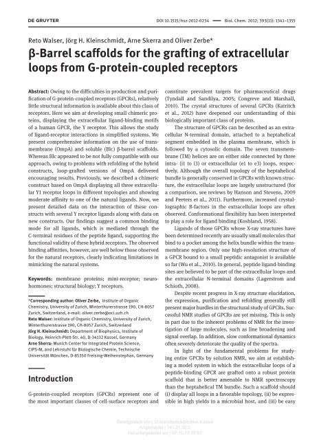
DOI 10.1515/hsz-2012-0234 Biol. Chem. 2012; 393(11): 1341–1355<br />
Reto Walser , J ö rg H. Kleinschmidt , Arne Skerra and Oliver Zerbe *<br />
<strong>β</strong>-<strong>Barrel</strong> <strong>scaffolds</strong> <strong>for</strong> <strong>the</strong> <strong>grafting</strong> <strong>of</strong> <strong>extracellular</strong><br />
<strong>loops</strong> from G-protein-coupled receptors<br />
Abstract : Owing to <strong>the</strong> difficulties in production and purification<br />
<strong>of</strong> G-protein-coupled receptors (GPCRs), relatively<br />
little structural in<strong>for</strong>mation is available about this class <strong>of</strong><br />
receptors. Here we aim at developing small chimeric proteins,<br />
displaying <strong>the</strong> <strong>extracellular</strong> ligand-binding motifs<br />
<strong>of</strong> a human GPCR, <strong>the</strong> Y receptor. This allows <strong>the</strong> study<br />
<strong>of</strong> ligand-receptor interactions in simplified systems. We<br />
present comprehensive in<strong>for</strong>mation on <strong>the</strong> use <strong>of</strong> transmembrane<br />
(OmpA) and soluble (Blc) <strong>β</strong> -barrel <strong>scaffolds</strong>.<br />
Whereas Blc appeared to be not fully compatible with our<br />
approach, owing to problems with refolding <strong>of</strong> <strong>the</strong> hybrid<br />
constructs, loop-grafted versions <strong>of</strong> OmpA delivered<br />
encouraging results. Previously, we described a chimeric<br />
construct based on OmpA displaying all three <strong>extracellular</strong><br />
Y1 receptor <strong>loops</strong> in different topologies and showing<br />
moderate affinity to one <strong>of</strong> <strong>the</strong> natural ligands. Now, we<br />
present detailed data on <strong>the</strong> interaction <strong>of</strong> <strong>the</strong>se constructs<br />
with several Y receptor ligands along with data on<br />
new constructs. Our findings suggest a common binding<br />
mode <strong>for</strong> all ligands, which is mediated through <strong>the</strong><br />
C-terminal residues <strong>of</strong> <strong>the</strong> peptide ligand, supporting <strong>the</strong><br />
functional validity <strong>of</strong> <strong>the</strong>se hybrid receptors. The observed<br />
binding affinities, however, are well below those observed<br />
<strong>for</strong> <strong>the</strong> natural receptors, clearly indicating limitations in<br />
mimicking <strong>the</strong> natural systems.<br />
Keywords: membrane proteins; mini-receptor; neurohormones;<br />
structural biology; Y receptors.<br />
*Corresponding author: Oliver Zerbe, Institute <strong>of</strong> Organic<br />
Chemistry, University <strong>of</strong> Zurich, Winterthurerstrasse 190, CH-8057<br />
Zurich , Switzerland , e-mail: oliver.zerbe@oci.uzh.ch<br />
Reto Walser: Institute <strong>of</strong> Organic Chemistry , University <strong>of</strong> Zurich,<br />
Winterthurerstrasse 190, CH-8057 Zurich , Switzerland<br />
Jörg H. Kleinschmidt: Department <strong>of</strong> Biophysics , Institute <strong>of</strong><br />
Biology, Heinrich-Plett-Str. 40, D-34132 Kassel , Germany<br />
Arne Skerra: Munich Center <strong>for</strong> Integrated Protein Science ,<br />
CIPS-M, and Lehrstuhl f ü r Biologische Chemie, Technische<br />
Universität München, D-85350 Freising-Weihenstephan , Germany<br />
Introduction<br />
G-protein-coupled receptors (GPCRs) represent one <strong>of</strong><br />
<strong>the</strong> most important classes <strong>of</strong> cell-surface receptors and<br />
constitute prevalent targets <strong>for</strong> pharmaceutical drugs<br />
(Tyndall and Sandilya , 2005 ; Congreve and Marshall ,<br />
2010 ). The crystal structures <strong>of</strong> several GPCRs (Katritch<br />
et al. , 2012 ) have deepened our understanding <strong>of</strong> this<br />
biologically important class <strong>of</strong> proteins.<br />
The structure <strong>of</strong> GPCRs can be described as an <strong>extracellular</strong><br />
N-terminal domain, attached to a heptahelical<br />
segment embedded in <strong>the</strong> plasma membrane, which is<br />
followed by a cytosolic domain. The seven transmembrane<br />
(TM) helices are on ei<strong>the</strong>r side connected by three<br />
intra- (i1 to i3) or <strong>extracellular</strong> (e1 to e3) <strong>loops</strong>, respectively.<br />
Although <strong>the</strong> overall topology <strong>of</strong> <strong>the</strong> heptahelical<br />
bundle is generally conserved in GPCRs with known structure,<br />
<strong>the</strong> <strong>extracellular</strong> <strong>loops</strong> are largely unstructured (<strong>for</strong><br />
a comparison, see reviews by Hanson and Stevens , 2009<br />
and Peeters et al. , 2011 ). Fur<strong>the</strong>rmore, increased crystallographic<br />
B-factors in <strong>the</strong> <strong>extracellular</strong> <strong>loops</strong> are <strong>of</strong>ten<br />
observed. Con<strong>for</strong>mational flexibility has been interpreted<br />
to play a role <strong>for</strong> ligand binding (Koshland , 1958 ).<br />
Ligands <strong>of</strong> those GPCRs whose X-ray structures have<br />
been determined recently are usually small molecules that<br />
bind to a pocket among <strong>the</strong> helix bundle within <strong>the</strong> transmembrane<br />
region. Only one high-resolution structure <strong>of</strong><br />
a GPCR bound to a small peptidic antagonist is available<br />
so far (Wu et al. , 2010 ). In general, peptide ligand-binding<br />
sites are believed to be part <strong>of</strong> <strong>the</strong> <strong>extracellular</strong> <strong>loops</strong> and<br />
<strong>the</strong> <strong>extracellular</strong> N-terminal domains (Lagerstrom and<br />
Schioth , 2008 ).<br />
Despite recent progress in X-ray structure elucidation,<br />
<strong>the</strong> expression, purification and refolding generally still<br />
present major hurdles in <strong>the</strong> structural study <strong>of</strong> GPCRs. Successful<br />
NMR studies <strong>of</strong> GPCRs are yet missing. This is only<br />
in part due to <strong>the</strong> inherent problems <strong>of</strong> NMR <strong>for</strong> <strong>the</strong> investigation<br />
<strong>of</strong> large molecules, such as line broadening and<br />
signal overlap. In addition, slow con<strong>for</strong>mational dynamics<br />
<strong>of</strong>ten severely deteriorate <strong>the</strong> quality <strong>of</strong> <strong>the</strong> spectra.<br />
In light <strong>of</strong> <strong>the</strong> fundamental problems <strong>for</strong> studying<br />
entire GPCRs by solution NMR, we aim at establishing<br />
a model system in which <strong>the</strong> <strong>extracellular</strong> <strong>loops</strong> <strong>of</strong> a<br />
peptide-binding GPCR are grafted onto a robust protein<br />
scaffold that is better amenable to NMR spectroscopy<br />
than <strong>the</strong> heptahelical TM bundle. Such a scaffold should<br />
(i) display all <strong>loops</strong> in a favorable topology, (ii) be expressible<br />
in high yields in a microbial host, and (iii) be easy<br />
Bereitgestellt von | Universitaetsbiblio<strong>the</strong>k Kassel<br />
Angemeldet | 141.51.38.5<br />
Heruntergeladen am | 07.10.13 13:53
1342 R. Walser et al.: Grafting <strong>of</strong> GPCR <strong>loops</strong><br />
to purify, solubilize and/or refold. Such a chimeric receptor<br />
may be useful <strong>for</strong> pharmacological studies with regard<br />
to <strong>the</strong> strength and specificity <strong>of</strong> ligand binding or to <strong>the</strong><br />
competitive binding behavior between various agonists<br />
and antagonists.<br />
Recently, we described <strong>the</strong> development <strong>of</strong> a model<br />
that mimics <strong>the</strong> <strong>extracellular</strong> domains <strong>of</strong> <strong>the</strong> human Y<br />
receptors based on a <strong>β</strong> -barrel scaffold from <strong>the</strong> Escherichia<br />
coli outer membrane protein A (OmpA) (Walser et al. ,<br />
2011 ). The model takes advantage <strong>of</strong> <strong>the</strong> membrane-integral<br />
<strong>β</strong> -barrel fold <strong>of</strong> OmpA and displays grafted <strong>loops</strong> in a<br />
favorable topology. The Y receptors are targeted by neurohormones<br />
from <strong>the</strong> neuropeptide Y family: neuropeptide<br />
Y (NPY), peptide YY (PYY) and pancreatic polypeptide<br />
(PP) (Larhammar , 1996a ). To date, four different subtypes<br />
<strong>of</strong> receptors have been characterized (Y1, Y2, Y4 and Y5;<br />
Larhammar , 1996b ; Larhammar and Salaneck , 2004 ),<br />
which are associated with different pharmacological<br />
effects. In <strong>the</strong>se, <strong>the</strong> three <strong>extracellular</strong> <strong>loops</strong>, and possibly<br />
also <strong>the</strong> N-terminal domain, are proposed to be<br />
involved in ligand binding (Zou et al. , 2008 ).<br />
We now describe <strong>the</strong> development <strong>of</strong> this receptor<br />
model in much more detail. As <strong>scaffolds</strong> we have initially<br />
employed two different, yet structurally related <strong>β</strong> -barrel<br />
proteins, <strong>the</strong> soluble bacterial lipocalin (Blc) (Bishop ,<br />
2000 ; Schiefner et al. , 2010 ) and OmpA (Tamm et al. ,<br />
2003 ) from E. coli . We demonstrate that all three <strong>extracellular</strong><br />
Y1 receptor <strong>loops</strong> and its N-terminus can be successfully<br />
transferred to <strong>the</strong> OmpA scaffold.<br />
Results<br />
Design aspects<br />
or<br />
To date, structural details at atomic resolution are available<br />
<strong>for</strong> nine major different GPCRs (<strong>for</strong> a summary see <strong>the</strong><br />
supplementary material online). We selected <strong>the</strong> N-termini<br />
and <strong>extracellular</strong> <strong>loops</strong> <strong>of</strong> <strong>the</strong> Y receptors based on<br />
<strong>the</strong> predicted topology as annotated in <strong>the</strong> GPCRDB (e.g.,<br />
http://www.gpcr.org/7tm/proteins/npy1r_human <strong>for</strong> <strong>the</strong><br />
human Y1 receptor). In <strong>the</strong> case <strong>of</strong> <strong>the</strong> human Y1 receptor,<br />
<strong>the</strong> <strong>extracellular</strong> <strong>loops</strong> comprise 13 and 14 residues <strong>for</strong> e1<br />
(Y99 to M111) and e3 (F284 to N297), respectively, and 34<br />
residues <strong>for</strong> e2 (Q176 to S209). The predicted N-terminal<br />
domains <strong>of</strong> <strong>the</strong> Y1, Y2 and Y4 receptors are <strong>the</strong> first 40, 50<br />
and 41 residues, respectively.<br />
The rationale <strong>for</strong> <strong>the</strong> design <strong>of</strong> <strong>the</strong> loop-grafted receptor<br />
models was as follows. First, we defined anchor points<br />
as <strong>the</strong> positions <strong>of</strong> <strong>the</strong> terminal C α atoms <strong>of</strong> <strong>the</strong> α -helix<br />
<strong>the</strong> <strong>β</strong> -strand that is connected to a loop. The mutual<br />
distances between <strong>the</strong>se anchor points define <strong>the</strong> overall<br />
topology <strong>of</strong> <strong>the</strong> set <strong>of</strong> <strong>extracellular</strong> <strong>loops</strong>. Figure 1 depicts a<br />
comparison <strong>of</strong> <strong>the</strong> distances between <strong>the</strong> anchor points on<br />
<strong>the</strong> <strong>extracellular</strong> side <strong>for</strong> a set <strong>of</strong> 10 different GPCR crystal<br />
structures with known structure at <strong>the</strong> onset <strong>of</strong> our study<br />
(<strong>for</strong> a list see <strong>the</strong> materials and methods section). The<br />
spacing between <strong>the</strong> anchor points <strong>for</strong> <strong>the</strong> three <strong>extracellular</strong><br />
<strong>loops</strong> is on average 13 Å <strong>for</strong> e1 and e2 and 14 Å <strong>for</strong> e3,<br />
with a narrow distribution <strong>of</strong> ± 3 Å . The distances between<br />
anchor points that are not part <strong>of</strong> <strong>the</strong> same loop are much<br />
less conserved, indicating that <strong>the</strong> relative positions <strong>of</strong><br />
two helices anchoring <strong>the</strong> same <strong>extracellular</strong> loop is more<br />
conserved than <strong>the</strong> relative positions between helices not<br />
directly connected.<br />
A similar analysis was conducted <strong>for</strong> <strong>the</strong> available<br />
high-resolution structures <strong>of</strong> Blc (Campanacci et al. , 2004 ;<br />
Schiefner et al. , 2010 ) and OmpA (Pautsch and Schulz ,<br />
1998, 2000 ). Because both published OmpA X-ray structures<br />
lack defined electron density <strong>for</strong> a substantial number<br />
<strong>of</strong> residues located in <strong>the</strong> <strong>extracellular</strong> <strong>loops</strong>, we mostly<br />
relied on <strong>the</strong> NMR structures <strong>for</strong> this protein (Arora et al. ,<br />
2001 ; Cierpicki et al. , 2006 ). Figure 1 depicts a statistical<br />
analysis <strong>of</strong> <strong>the</strong> distances observed in <strong>the</strong> crystal structure<br />
<strong>of</strong> Blc (Campanacci et al. , 2004 ) and <strong>the</strong> 10 lowest energy<br />
con<strong>for</strong>mers <strong>of</strong> two NMR structures <strong>of</strong> OmpA (Arora et al. ,<br />
2001 ; Cierpicki et al. , 2006 ), as well as a shortened loop<br />
construct <strong>of</strong> <strong>the</strong> OmpA scaffold (Johansson et al. , 2007 ).<br />
A comparison <strong>of</strong> <strong>the</strong> pairwise distances between<br />
anchor points in GPCRs and <strong>the</strong>se scaffold proteins<br />
revealed that <strong>the</strong> distance distribution observed in <strong>the</strong><br />
GPCRs falls within <strong>the</strong> distribution observed <strong>for</strong> <strong>the</strong> OmpA<br />
structures and is also close to <strong>the</strong> distances observed <strong>for</strong><br />
Blc, suggesting that <strong>the</strong> <strong>β</strong> -barrels <strong>of</strong> Blc and OmpA might<br />
indeed provide suitable frameworks <strong>for</strong> <strong>grafting</strong> <strong>the</strong> <strong>extracellular</strong><br />
<strong>loops</strong> <strong>of</strong> GPCRs. Whereas <strong>the</strong> distances between<br />
directly connected anchor points are between 10 and 17 Å<br />
in OmpA, in Blc those distances are significantly shorter:<br />
approximately 5 Å <strong>for</strong> three anchor point pairs and 10 Å<br />
<strong>for</strong> <strong>the</strong> fourth pair. GPCRs possess three, whereas OmpA<br />
carries four <strong>extracellular</strong> <strong>loops</strong>, leaving at least one ‘ acceptor<br />
’ site in <strong>the</strong> scaffold unoccupied. To rule out interference<br />
with <strong>the</strong> remaining native loop, it was replaced by a<br />
minimal turn-inducing motif <strong>of</strong> one to two residues compatible<br />
with <strong>the</strong> OmpA <strong>β</strong> -barrel structure (Koebnik , 1999a ).<br />
Blc construct design<br />
We have previously demonstrated that <strong>the</strong> <strong>extracellular</strong><br />
N-terminal domain <strong>of</strong> <strong>the</strong> Y4 receptor (NY4) interacts with<br />
Bereitgestellt von | Universitaetsbiblio<strong>the</strong>k Kassel<br />
Angemeldet | 141.51.38.5<br />
Heruntergeladen am | 07.10.13 13:53
R. Walser et al.: Grafting <strong>of</strong> GPCR <strong>loops</strong> 1343<br />
A<br />
B<br />
C<br />
D<br />
E<br />
Distance (Å) Distance (Å) Distance (Å)<br />
Distance (Å) Distance (Å) Distance (Å) Distance (Å)<br />
Figure 1 Geometries <strong>of</strong> GPCRs and <strong>β</strong>-barrel proteins.<br />
(A) Ribbon representation <strong>of</strong> bovine rhodopsin with <strong>the</strong> first, second and third <strong>extracellular</strong> <strong>loops</strong> (e1– 3) colored yellow, red and blue,<br />
respectively. On <strong>the</strong> right <strong>the</strong> arrangement <strong>of</strong> <strong>the</strong> <strong>extracellular</strong> loop anchor points is presented as viewed from <strong>the</strong> <strong>extracellular</strong> side (same<br />
color coding used as in <strong>the</strong> ribbon representation). Each anchor point is labeled by its terminal/initial residue and <strong>the</strong> TM-helix to which it<br />
belongs. (B) Ribbon representation <strong>of</strong> Blc with its four variable <strong>loops</strong> colored green and <strong>the</strong> anchor points yellow. On <strong>the</strong> right <strong>the</strong> arrangement<br />
<strong>of</strong> <strong>the</strong> loop anchor points is presented as viewed from <strong>the</strong> top (same color coding as in ribbon representation). Directly connected<br />
anchor points are indicated by black arrows. For clarity <strong>the</strong> N-terminal 3 10<br />
-helix and <strong>the</strong> C-terminal α -helix <strong>of</strong> Blc is omitted in <strong>the</strong> ribbon representation.<br />
(C) Same representation as in (B) <strong>for</strong> OmpA. (D) Histograms <strong>of</strong> <strong>the</strong> distances between <strong>the</strong> anchor points <strong>for</strong> <strong>extracellular</strong> <strong>loops</strong><br />
1, 2 and 3 as found in a set <strong>of</strong> 10 GPCR crystal structures (see <strong>the</strong> supplementary material and methods online <strong>for</strong> a full list) (top panel) and<br />
(E) <strong>for</strong> <strong>the</strong> <strong>extracellular</strong> <strong>loops</strong> in <strong>the</strong> NMR structures <strong>of</strong> two OmpA structures and one loop-shortened OmpA construct (10 con<strong>for</strong>mers each)<br />
(bottom panel). Average distances between <strong>the</strong> anchor points <strong>for</strong> <strong>the</strong> e1-, e2- and e3-loop in <strong>the</strong> GPCR structures are indicated by yellow,<br />
red and blue bars, respectively.<br />
PP (Zou et al. , 2008, 2009 ). Because <strong>of</strong> its well-behaved<br />
nature in terms <strong>of</strong> expression and stability, we have initially<br />
chosen <strong>the</strong> Y2 receptor N-terminus (NY2) <strong>for</strong> our<br />
studies aiming at determining suitable attachment points<br />
to <strong>the</strong> Blc scaffold. Accordingly, we fused <strong>the</strong> NY2 domain<br />
to different positions in <strong>the</strong> N-terminal region <strong>of</strong> Blc. In<br />
<strong>the</strong>se constructs, varying portions <strong>of</strong> <strong>the</strong> Blc N-terminus<br />
were replaced with NY2 to test how close a grafted N-<br />
terminal sequence can be brought to <strong>the</strong> first strand <strong>of</strong><br />
<strong>the</strong> Blc <strong>β</strong> -barrel, without impairing its fold. The tolerance<br />
<strong>of</strong> <strong>the</strong> scaffold towards <strong>the</strong> fused sequence was determined<br />
by [ 15 N, 1 H]-HSQCs (see Figure 2 ). Moving <strong>the</strong> fusion<br />
point between NY2 and Blc too close to <strong>the</strong> characteristic<br />
3 10<br />
-helix ( < 10 residues) that in many lipocalins precedes<br />
<strong>the</strong> <strong>β</strong> -barrel (Flower et al. , 2000 ) resulted in insoluble<br />
constructs, whereas fusion at more N-terminal positions<br />
was well tolerated. However, no interaction <strong>of</strong> <strong>the</strong> chimera<br />
with NPY family neurohormones could be detected. We<br />
also attempted to investigate whe<strong>the</strong>r <strong>the</strong> interaction<br />
detected between NY4 and PP (Zou et al. , 2008 ) could<br />
be reproduced in <strong>the</strong> context <strong>of</strong> <strong>the</strong> Blc scaffold, but no<br />
stably folded fusion protein with NY4 could be obtained<br />
(data not shown). We also set out to incorporate <strong>the</strong> <strong>extracellular</strong><br />
<strong>loops</strong> <strong>of</strong> <strong>the</strong> Y1 receptor (e1Y1 to e3Y1), but <strong>the</strong> Blc<br />
scaffold did not tolerate <strong>the</strong> necessary modifications in its<br />
<strong>loops</strong>.<br />
Bereitgestellt von | Universitaetsbiblio<strong>the</strong>k Kassel<br />
Angemeldet | 141.51.38.5<br />
Heruntergeladen am | 07.10.13 13:53
1344 R. Walser et al.: Grafting <strong>of</strong> GPCR <strong>loops</strong><br />
A<br />
B<br />
T23<br />
P25<br />
S20<br />
T14<br />
L40<br />
F34<br />
L11 N32<br />
Figure 2 Summary <strong>of</strong> Y2 receptor N-terminus <strong>grafting</strong> attempts using Blc.<br />
(A) Blc sequence with colored arrows indicating <strong>the</strong> <strong>β</strong> -strand secondary structural elements in <strong>the</strong> crystal structure. Red: additional<br />
two-stranded <strong>β</strong> -sheet which has arisen as a cloning artifact (Schiefner et al. , 2010 ); yellow: N-terminal 3 10<br />
- and C-terminal α -helix, both<br />
characteristic <strong>for</strong> <strong>the</strong> lipocalin fold; green: eight-stranded <strong>β</strong> -barrel, <strong>the</strong> central motif <strong>of</strong> <strong>the</strong> lipocalin fold. The sequence 23 – 177<br />
corresponds to <strong>the</strong> natural Blc protein, to which a His 6<br />
tag was appended at <strong>the</strong> N-terminus and a Strep -tag II at <strong>the</strong> C-terminus. Residues<br />
to which <strong>the</strong> NY2 sequence has been N-terminally fused are shaded in green or red, indicating constructs resulting in soluble or insoluble<br />
protein, respectively. (B) Ribbon representation <strong>of</strong> Blc with <strong>the</strong> residues to which <strong>the</strong> NY2 sequence was N-terminally fused colored in green<br />
or red, as in panel (A). The [ 15 N, 1 H]-HSQC spectra <strong>of</strong> Blc and <strong>the</strong> respective NY2-grafted/fused constructs are depicted next to <strong>the</strong> structure.<br />
Properly folded constructs are characterized by good signal dispersion in <strong>the</strong> spectra.<br />
OmpA construct design<br />
Our initial studies concentrated on probing <strong>the</strong> compatibility<br />
<strong>of</strong> <strong>the</strong> Y receptor <strong>extracellular</strong> <strong>loops</strong> with <strong>the</strong><br />
OmpA scaffold. Considering that most <strong>of</strong> <strong>the</strong> data from<br />
mutagenesis studies are available <strong>for</strong> <strong>the</strong> Y1 receptor,<br />
we exchanged each <strong>of</strong> <strong>the</strong> four <strong>extracellular</strong> <strong>loops</strong><br />
<strong>of</strong> OmpA with each <strong>of</strong> <strong>the</strong> three eY1 <strong>loops</strong> individually<br />
to generate 12 constructs altoge<strong>the</strong>r, dubbed ‘ one-loop<br />
exchange constructs ’ . Fur<strong>the</strong>r, we constructed a series<br />
<strong>of</strong> OmpA mutants in which a single eY1 loop was grafted<br />
into one OmpA acceptor site while <strong>the</strong> o<strong>the</strong>r three sites<br />
were filled with a minimal turn-inducing motif (Koebnik ,<br />
1999b ). These were called ‘ one-loop graft constructs ’ .<br />
All constructs could be expressed in E. coli , solubilized<br />
in urea and refolded (see Figure S1 in <strong>the</strong> supplementary<br />
material online), indicating that OmpA is suitable as a<br />
generic scaffold <strong>for</strong> <strong>grafting</strong> individual eY1 <strong>loops</strong>, both in<br />
<strong>the</strong> presence and absence <strong>of</strong> <strong>the</strong> o<strong>the</strong>r three <strong>of</strong> its natural<br />
<strong>loops</strong>.<br />
In principle, <strong>the</strong> set <strong>of</strong> three eY1 <strong>loops</strong> can be arranged<br />
in 24 different ways (4 × 3 × 2) on <strong>the</strong> OmpA scaffold according<br />
to this approach. To avoid unsuitable constructs when<br />
combining <strong>the</strong> individually grafted <strong>loops</strong>, we calculated a<br />
‘ mismatch score ’ accounting <strong>for</strong> <strong>the</strong> distance mismatches<br />
<strong>of</strong> all relevant anchor points between <strong>the</strong> model scaffold<br />
and <strong>the</strong> GPCRs (see Figure 3 ). Among <strong>the</strong> candidates with<br />
low mismatch scores, only those with a correct topological<br />
Bereitgestellt von | Universitaetsbiblio<strong>the</strong>k Kassel<br />
Angemeldet | 141.51.38.5<br />
Heruntergeladen am | 07.10.13 13:53
R. Walser et al.: Grafting <strong>of</strong> GPCR <strong>loops</strong> 1345<br />
A<br />
Mismatch score (Å)<br />
B<br />
Topology<br />
I<br />
II<br />
III<br />
IV<br />
V<br />
VI<br />
VII<br />
VIII<br />
N<br />
C<br />
Linker<br />
Figure 3 Design <strong>of</strong> receptor constructs.<br />
(A) Calculation <strong>of</strong> <strong>the</strong> mismatch score (in Å ) <strong>for</strong> all 24 possible ‘ receptor constructs ’ (<strong>for</strong> a description <strong>of</strong> <strong>the</strong> calculation procedure see<br />
materials and methods section). Constructs displaying <strong>the</strong> Y receptor <strong>extracellular</strong> <strong>loops</strong> in an appropriate topological orientation <strong>for</strong> <strong>grafting</strong><br />
are colored. The four chimeric GPCR-OmpA constructs selected <strong>for</strong> expression (Y1L1, Y1L2, Y1L3 and Y1L4) are marked in green. (B)<br />
Topography <strong>for</strong> <strong>the</strong> selected constructs <strong>of</strong> <strong>the</strong> three e1 – 3 <strong>loops</strong> <strong>of</strong> Y1 on <strong>the</strong> eight-stranded OmpA <strong>β</strong> -barrel. The remaining unused fourth<br />
loop <strong>of</strong> <strong>the</strong> OmpA scaffold was replaced by a short linker sequence. Amino acid sequences <strong>for</strong> <strong>the</strong> three Y1 <strong>loops</strong> as well as <strong>the</strong> short linker<br />
are depicted at <strong>the</strong> bottom. Serine residues highlighted in red correspond to cysteine side chains in <strong>the</strong> natural Y1 sequences, which were<br />
substituted to avoid <strong>for</strong>mation <strong>of</strong> undesired disulfide crosslinks.<br />
loop arrangement (i.e., <strong>the</strong> C-terminus <strong>of</strong> e1 to be followed<br />
by <strong>the</strong> N-terminus <strong>of</strong> e2 in a clockwise manner and so<br />
on) were considered. Four <strong>of</strong> those arrangements were<br />
selected and will be referred to in <strong>the</strong> following as ‘ receptor<br />
constructs ’ , abbreviated as Y1L1, Y1L2, Y1L3 and Y1L4<br />
(see Figure 3).<br />
The initial topological analysis <strong>of</strong> <strong>the</strong> anchor<br />
point distances revealed that although <strong>the</strong> pairwise<br />
distances <strong>for</strong> each loop in <strong>the</strong> known GPCR structures<br />
fall within <strong>the</strong> range <strong>of</strong> those observed in OmpA, <strong>the</strong><br />
overall match is not perfect. To account <strong>for</strong> <strong>the</strong>se structural<br />
differences, additional flexible linker residues at<br />
<strong>the</strong> termini <strong>of</strong> <strong>the</strong> <strong>loops</strong> were introduced by inserting<br />
glycine-serine spacers <strong>of</strong> different lengths. Based on<br />
<strong>the</strong> Y1L3 topology six constructs were designed: two in<br />
which each Y1 receptor loop was flanked on both sides<br />
by a Ser-Gly dipeptide or a Ser-Gly-Ser-Gly tetrapeptide<br />
(Y1L3-GS and Y1L3-GSGS), respectively, two where only<br />
<strong>the</strong> short e1- and e3-<strong>loops</strong> were flanked by <strong>the</strong>se spacers<br />
(Y1L3-gs and Y1L3-gsgs), and two where only <strong>the</strong> longer<br />
e2-loop was equipped with <strong>the</strong> spacers (Y1L3-e2gs and<br />
Y1L3-e2gsgs).<br />
Notably, all <strong>the</strong>se constructs lack <strong>the</strong> N-terminal<br />
receptor domain, which may also be involved in ligand<br />
binding (Robin -Jagerschmidt et al., 1998 ; Wieland et al. ,<br />
1998 ; Zou et al. , 2008 ). Un<strong>for</strong>tunately, in OmpA <strong>the</strong> N-terminus<br />
<strong>of</strong> <strong>the</strong> <strong>β</strong> -barrel is located opposite to <strong>the</strong> (<strong>extracellular</strong>)<br />
face used <strong>for</strong> <strong>grafting</strong>. We inserted <strong>the</strong> sequence <strong>of</strong><br />
<strong>the</strong> N-terminal Y1 receptor domain (NY1) into <strong>the</strong> third,<br />
so far ‘ empty ’ acceptor position <strong>of</strong> Y1L3, flanked by an<br />
N-terminal (Gly-Ser) 3<br />
and a 3C protease cleavage site,<br />
allowing <strong>the</strong> in situ generation <strong>of</strong> a free N-terminus via<br />
proteolytic cleavage after refolding (construct Y1L3-NY1<br />
in Figure 3).<br />
Bereitgestellt von | Universitaetsbiblio<strong>the</strong>k Kassel<br />
Angemeldet | 141.51.38.5<br />
Heruntergeladen am | 07.10.13 13:53
1346 R. Walser et al.: Grafting <strong>of</strong> GPCR <strong>loops</strong><br />
Biosyn<strong>the</strong>tic aspects <strong>of</strong> hybrid GPCR-OmpA<br />
constructs<br />
All <strong>the</strong>se constructs were purified, solubilized and<br />
refolded in unlabeled and 15 N-labeled <strong>for</strong>m from inclusion<br />
bodies produced in E. coli with yields <strong>of</strong> ∼ 200 and<br />
∼ 100 mg/l <strong>of</strong> LB rich medium or 15 N-labeled M9 minimal<br />
medium, respectively. The folding state <strong>of</strong> OmpA was<br />
monitored by SDS-PAGE in which <strong>the</strong> sample was mixed<br />
with SDS sample buffer but not heated prior to loading<br />
on <strong>the</strong> gel, leaving <strong>the</strong> OmpA fold intact and referred<br />
to here as ‘ non-denaturing SDS-PAGE ’ (Reithmeier<br />
and Bragg , 1974 ; Schweizer et al. , 1978 ). During refolding<br />
screens, solutions <strong>of</strong> <strong>the</strong> urea-denatured chimeric<br />
OmpA were diluted into different buffers containing<br />
various detergents at concentrations above <strong>the</strong>ir critical<br />
micellar concentrations (cmc) and at detergent/<br />
protein ratios > 500 (<strong>for</strong> a list <strong>of</strong> <strong>the</strong> relevant biophysical<br />
parameters, see Table S1 in <strong>the</strong> supplementary material<br />
online).<br />
All <strong>of</strong> <strong>the</strong> 12 ‘ one-loop exchange constructs ’ and 6 <strong>of</strong><br />
<strong>the</strong> ‘ one-loop graft constructs ’ , as well as <strong>the</strong> 4 selected<br />
‘ receptor constructs ’ were expressed, purified and <strong>the</strong>ir<br />
refolding capability was assessed by non-denaturing<br />
SDS-PAGE (see Figure 4 ). Whereas <strong>the</strong> expression level<br />
<strong>of</strong> all 22 constructs was similar to that <strong>of</strong> wild type (wt)-<br />
OmpA, <strong>the</strong> refolding efficiency was clearly lower <strong>for</strong><br />
some <strong>of</strong> <strong>the</strong>se constructs. Never<strong>the</strong>less, each <strong>of</strong> <strong>the</strong> 22<br />
constructs could be refolded at least to 50 % (data not<br />
shown). Generally, refolding efficiency increased with<br />
increasing pH (Kleinschmidt et al. , 1999 ). Whereas <strong>for</strong><br />
some constructs rapid dilution <strong>of</strong> <strong>the</strong> urea-denatured<br />
protein solution into detergent buffer at high pH 10.0<br />
resulted in nearly complete refolding, some constructs<br />
required more gentle conditions <strong>of</strong> slow dilution at a<br />
lower temperature <strong>of</strong> 4 ° C.<br />
In <strong>the</strong> Y1L3-NY1 construct complete refolding under<br />
similar conditions <strong>of</strong> pH and detergent was possible<br />
(see Figure 5 B). After refolding in DHPC micelles, Y1L3-<br />
NY1 was incubated with 3C protease and <strong>the</strong> efficiency<br />
<strong>of</strong> cleavage and integrity <strong>of</strong> <strong>the</strong> <strong>β</strong> -barrel were assessed<br />
by denaturing and non-denaturing SDS-PAGE, respectively.<br />
As can be seen in Figure 5B, bands corresponded<br />
closely to <strong>the</strong> expected sizes. Y1L3-NY1 refolded in DHPC<br />
micelles showed <strong>the</strong> same electrophoretic mobility be<strong>for</strong>e<br />
and after treatment with 3C protease, indicating integrity<br />
<strong>of</strong> its tertiary fold even after cleavage. The appearance<br />
<strong>of</strong> two bands around 14 kDa and <strong>the</strong> concomitant complete<br />
disappearance <strong>of</strong> <strong>the</strong> band at 27 kDa under denaturing<br />
SDS-PAGE conditions proved that <strong>the</strong> proteolytic<br />
cleavage was highly efficient (<strong>for</strong> results with alternative<br />
detergents see Figure S2 in <strong>the</strong> supplementary material<br />
online).<br />
Whenever folded <strong>for</strong>ms <strong>of</strong> <strong>the</strong> chimeric OmpA constructs<br />
were detected by SDS-PAGE, <strong>the</strong> presence <strong>of</strong> tertiary<br />
structure and <strong>for</strong>mation <strong>of</strong> <strong>the</strong> <strong>β</strong> -barrel was also<br />
apparent from <strong>the</strong> large signal dispersion in <strong>the</strong> [ 15 N, 1 H]-<br />
HSQC spectra <strong>of</strong> <strong>the</strong>se preparations (<strong>for</strong> [ 15 N, 1 H]-HSQC<br />
spectra <strong>of</strong> <strong>the</strong> four receptor constructs Y1L1-4, see Figure<br />
S3 in <strong>the</strong> supplementary material online).<br />
A<br />
Unfold<br />
Unfold<br />
Unfold Unfold Unfold Unfold<br />
B<br />
Unfold Unfold Unfold Unfold<br />
Unfold<br />
Unfold<br />
C<br />
Unfold<br />
Unfold<br />
Unfold<br />
Unfold<br />
Figure 4 Folding properties <strong>of</strong> grafted receptor constructs using non-denaturing SDS-PAGE.<br />
(A) pH dependence <strong>of</strong> <strong>the</strong> refolding efficiency <strong>of</strong> Y1L1 in a variety <strong>of</strong> different detergents (DDM: α -dodecyldimaltoside, <strong>β</strong> -OG:<br />
<strong>β</strong> -octylglucoside, C8E4: tetraethyleneglycol monooctyle<strong>the</strong>r, LDAO: N-lauryldimethyl amineoxide, DPC: dodecylphosphocholine, DHPC:<br />
dihexanoylphosphatidylcholine). Although refolding is not efficient in all detergents, a clear trend to increased efficiency apparent from <strong>the</strong><br />
presence <strong>of</strong> a lower band different from <strong>the</strong> heat-denatured (unfolded) species at higher pH is observed. (B) Whereas Y1L1 and Y1L2 can be<br />
refolded with fairly high efficiency at pH 10 in most detergents tested, corresponding efficiencies <strong>for</strong> Y1L3 and Y1L4 are much lower.<br />
(C) Optimization <strong>of</strong> <strong>the</strong> refolding procedure towards more gentle conditions (lower temperature, slow dilution <strong>of</strong> <strong>the</strong> denatured stock<br />
solution) results in increased refolding efficiency especially <strong>for</strong> Y1L3 and Y1L4.<br />
Bereitgestellt von | Universitaetsbiblio<strong>the</strong>k Kassel<br />
Angemeldet | 141.51.38.5<br />
Heruntergeladen am | 07.10.13 13:53
R. Walser et al.: Grafting <strong>of</strong> GPCR <strong>loops</strong> 1347<br />
A<br />
B<br />
- -<br />
- -<br />
C<br />
Chemical shift change<br />
relative to 0 eq.<br />
I<br />
Y<br />
Y<br />
Q<br />
A<br />
L<br />
L<br />
R<br />
L<br />
H<br />
I<br />
R<br />
Y<br />
T<br />
R<br />
A<br />
N<br />
S<br />
Figure 5 Interaction studies with <strong>the</strong> ‘ split ’ receptor model that additionally displays <strong>the</strong> N-terminal domain <strong>of</strong> Y1, Y1L3-NY1.<br />
(A) Schematic representation <strong>of</strong> <strong>the</strong> construct. The OmpA <strong>β</strong> -barrel is shown in cyan, <strong>the</strong> grafted eY1 <strong>loops</strong> in dark red, <strong>the</strong> (Gly-Ser) 3<br />
-linker<br />
and 3C protease cleavage site in yellow and NY1 in red. (B) SDS-PAGE <strong>of</strong> Y1L3-NY1 under different conditions: a different migration behavior<br />
under non-denaturing (lane 2) and denaturing (lane 3) conditions indicates successful refolding <strong>of</strong> <strong>the</strong> construct. After treatment with 3C<br />
protease <strong>the</strong> construct shows <strong>the</strong> same migration behavior (lane 4) as be<strong>for</strong>e (lane 2) under non-denaturing conditions. Under denaturing<br />
conditions two bands with sizes <strong>of</strong> approximately 14 kDa can be seen after cleavage with 3C protease (lane 5). (C) The chemical shift<br />
changes observed in <strong>the</strong> [ 15 N, 1 H]-HSQC spectrum <strong>of</strong> NPY upon addition <strong>of</strong> 20 Eq. <strong>of</strong> cleaved Y1L3-NY1 protein plotted <strong>for</strong> each residue and<br />
color-coded onto <strong>the</strong> α -helical structure <strong>of</strong> NPY where gray indicates no change and <strong>the</strong> intensity <strong>of</strong> red is proportional to <strong>the</strong> chemical shift<br />
change. Changes are most prominent on one side <strong>of</strong> <strong>the</strong> helix. A helical wheel representation <strong>of</strong> <strong>the</strong> C-terminal α -helix (L17-Q34) <strong>of</strong> NPY<br />
shows that this side exclusively comprises hydrophobic and aromatic residues.<br />
Interaction studies <strong>of</strong> neurohormones<br />
<strong>of</strong> <strong>the</strong> NPY family with chimeric Y1-OmpA<br />
receptor constructs<br />
Binding <strong>of</strong> NPY, PYY and PP to <strong>the</strong> receptor constructs<br />
was tested using chemical shift mapping or saturation<br />
transfer difference (STD) (Mayer and Meyer , 1999, 2001 )<br />
techniques. 15 N-labeled neurohormones (<strong>for</strong> assignments,<br />
see Table S2 in <strong>the</strong> supplementary material online) were<br />
titrated with unlabeled receptor constructs. The resulting<br />
changes <strong>for</strong> <strong>the</strong> NPY spectra upon addition <strong>of</strong> 20 Eq. <strong>of</strong><br />
<strong>the</strong> chimeric receptor constructs are depicted in Figure 6 .<br />
No interactions could be detected with Y1L1 or Y1L2,<br />
whereas Y1L3 and Y1L4 induced significant changes in<br />
<strong>the</strong> [ 15 N, 1 H]-HSQC spectra <strong>of</strong> NPY when present in excess.<br />
Interestingly, no shift in peak positions, but a decrease<br />
in peak intensities was observed. This finding is consistent<br />
only with an exchange process slow on <strong>the</strong> NMR time<br />
scale, which usually results in two sets <strong>of</strong> peaks, one corresponding<br />
to <strong>the</strong> bound and <strong>the</strong> o<strong>the</strong>r to <strong>the</strong> non-bound<br />
<strong>for</strong>m. We suspect that excessive broadening <strong>of</strong> <strong>the</strong> resonances<br />
due to small con<strong>for</strong>mational fluctuations in <strong>the</strong><br />
receptor-bound state or a large number <strong>of</strong> different states<br />
that do not interconvert fast on <strong>the</strong> NMR time scale has led<br />
to <strong>the</strong> disappearance <strong>of</strong> <strong>the</strong> bound-state signals.<br />
Figure 6 depicts <strong>the</strong> volume changes <strong>of</strong> <strong>the</strong> peaks from<br />
<strong>the</strong> neurohormones upon titration with an excess <strong>of</strong> Y1L3<br />
(<strong>for</strong> similar results obtained with <strong>the</strong> SG linker versions, see<br />
Figure S4 in <strong>the</strong> supplementary material online). The data<br />
clearly indicate that <strong>the</strong> C-terminal residues <strong>of</strong> <strong>the</strong> peptide<br />
were much more affected by interaction with <strong>the</strong> chimeric<br />
receptor than those <strong>of</strong> <strong>the</strong> N-terminus. In agreement with<br />
previous studies (Beck -Sickinger et al., 1994 ), this indicated<br />
that <strong>the</strong> C-terminal α -helix <strong>of</strong> <strong>the</strong> neurohormone is involved<br />
in receptor binding. Despite <strong>the</strong> qualitative similarities <strong>of</strong><br />
<strong>the</strong> peak volume changes between all three neurohormones<br />
tested, <strong>the</strong> attenuations were less pronounced <strong>for</strong> PYY and,<br />
in particular, <strong>for</strong> PP than <strong>for</strong> NPY. Considering that <strong>the</strong><br />
binding pr<strong>of</strong>ile <strong>of</strong> PP to <strong>the</strong> Y receptor subtypes has been<br />
shown to be different from <strong>the</strong> binding pr<strong>of</strong>iles <strong>of</strong> NPY and<br />
PYY (Larhammar and Salaneck , 2004 ), this may indicate<br />
differences in <strong>the</strong> binding mode in our model system, too.<br />
Specificity <strong>of</strong> <strong>the</strong> interaction was corroborated by a<br />
competition experiment with unlabeled NPY (Figure 6C)<br />
Bereitgestellt von | Universitaetsbiblio<strong>the</strong>k Kassel<br />
Angemeldet | 141.51.38.5<br />
Heruntergeladen am | 07.10.13 13:53
1348 R. Walser et al.: Grafting <strong>of</strong> GPCR <strong>loops</strong><br />
A<br />
B<br />
C<br />
110<br />
112<br />
114<br />
116<br />
118<br />
120<br />
122<br />
124<br />
D<br />
8.6 8.4 8.2 8.0 7.8 8.6 8.4 8.2 8.0 7.8 8.6 8.4 8.2 8.0 7.8<br />
E F G<br />
ppm<br />
110<br />
112<br />
114<br />
116<br />
118<br />
120<br />
122<br />
124<br />
8.6 8.4 8.2 8.0 7.8 8.6 8.4 8.2 8.0 7.8 8.6 8.4 8.2 8.0 7.8 8.6 8.4 8.2 8.0 7.8 ppm<br />
H<br />
Peak volume<br />
rel. to 0 eq.<br />
3 4 6 7 9 1011 12 14 15 16 17 18 19 20 21 22 23 24 25 26 27 28 29 30 31 32 33 34 35 36<br />
I<br />
Peak volume<br />
rel. to 0 eq.<br />
3 4 6 7 9 10 11 12 13 15 16 17 18 19 20 21 22 23 24 25 26 27 28 29 30 31 32 33 34 35 36<br />
J<br />
Peak volume<br />
rel. to 0 eq.<br />
3 4 6 7 9 10111213151617181920212223242526272829303132333536<br />
Figure 6 Interaction studies between peptide hormones and receptor constructs using NMR.<br />
[ 15 N, 1 H]-HSQC spectra <strong>of</strong> <strong>the</strong> 15 N-NPY in 3 % DHPC, 20 m m phosphate pH 6.5, 100 m m NaCl at 310 K (A) in <strong>the</strong> absence, (B) in <strong>the</strong> presence <strong>of</strong><br />
20 Eq. <strong>of</strong> unlabeled Y1L3 protein and (C) in presence <strong>of</strong> 20 Eq. <strong>of</strong> unlabeled Y1L3 protein and 50 Eq. <strong>of</strong> unlabeled NPY. [ 15 N, 1 H]-HSQC spectra<br />
<strong>of</strong> mutant peptide 15 N-NPY-R35L (D) in <strong>the</strong> absence, (E) in <strong>the</strong> presence <strong>of</strong> 20 Eq. <strong>of</strong> unlabeled Y1L3 protein. [ 15 N, 1 H]-HSQC spectra <strong>of</strong> mutant<br />
peptide 15 N-NPY-R33L (F) in <strong>the</strong> absence, (G) in <strong>the</strong> presence <strong>of</strong> 20 Eq. <strong>of</strong> unlabeled Y1L3. The relative residue-specific volume change <strong>of</strong><br />
(H) NPY, (I) PYY and (J) PP resonances upon addition <strong>of</strong> Y1L3 protein plotted <strong>for</strong> each residue. The relative volume change is color-coded<br />
onto <strong>the</strong> structure <strong>of</strong> <strong>the</strong> (micelle-bound) species <strong>of</strong> each neurohormone, with gray stretches indicating no change and <strong>the</strong> intensity <strong>of</strong> <strong>the</strong><br />
red color being proportional to <strong>the</strong> relative volume change.<br />
Bereitgestellt von | Universitaetsbiblio<strong>the</strong>k Kassel<br />
Angemeldet | 141.51.38.5<br />
Heruntergeladen am | 07.10.13 13:53
R. Walser et al.: Grafting <strong>of</strong> GPCR <strong>loops</strong> 1349<br />
(Walser et al. , 2011 ). Fur<strong>the</strong>rmore, binding assays with<br />
NPY-R33L and NPY-R35L, two mutants <strong>of</strong> NPY that exhibit<br />
much reduced affinity to <strong>the</strong> Y1 receptor in vivo (Beck-<br />
Sickinger et al., 1994 ), were conducted. These mutant NPY<br />
peptides displayed a markedly decreased affinity <strong>for</strong> Y1L3<br />
as visible from <strong>the</strong> reduced attenuation <strong>of</strong> peaks <strong>for</strong> <strong>the</strong><br />
C-terminal residues (see Figure 6D – G). In summary, <strong>the</strong>se<br />
findings confirmed a specific interaction <strong>of</strong> <strong>the</strong> neurohormones<br />
<strong>of</strong> <strong>the</strong> NPY family with <strong>the</strong> chimeric GPCR-OmpA<br />
receptor construct Y1L3.<br />
Interestingly, <strong>the</strong> chemical shift mapping data from<br />
titration <strong>of</strong> 15 N-NPY with cleaved or uncleaved Y1L3-NY1<br />
showed a behavior very different from Y1L3, revealing<br />
shifts in <strong>the</strong> positions <strong>of</strong> certain peaks (see Figure 5C and<br />
Figure S5 in <strong>the</strong> supplementary material online). Large<br />
chemical shift changes were exclusively observed in <strong>the</strong><br />
C-terminal helix <strong>of</strong> NPY, showing a pronounced i + 3 or i + 4<br />
periodicity, thus indicating an interaction involving residues<br />
located on <strong>the</strong> same side <strong>of</strong> <strong>the</strong> helix.<br />
STD experiments conducted with NPY in <strong>the</strong> presence<br />
<strong>of</strong> Y1L3 or cleaved Y1L3-NY1 showed <strong>the</strong> most pronounced<br />
saturation transfer effects <strong>for</strong> <strong>the</strong> aromatic resonances <strong>of</strong><br />
peptide residues, which – with <strong>the</strong> exception <strong>of</strong> <strong>the</strong> N-<br />
terminal Tyr – are all located in <strong>the</strong> C-terminal half <strong>of</strong> NPY<br />
(Walser et al. , 2011 ).<br />
To obtain fur<strong>the</strong>r in<strong>for</strong>mation on <strong>the</strong> ligand-receptor<br />
interactions, all four 15 N-labeled receptor constructs<br />
Y1L1-4 were mixed with unlabeled NPY (see Figure S6<br />
in <strong>the</strong> supplementary material online). Surprisingly, <strong>the</strong><br />
[ 15 N, 1 H]-HSQC spectra <strong>of</strong> <strong>the</strong> all chimeric receptor constructs<br />
did not display major changes upon addition <strong>of</strong><br />
<strong>the</strong> neurohormone. We speculate that most peaks from<br />
<strong>the</strong> short e1- and e3-<strong>loops</strong>, which have been proposed to<br />
mediate interaction with <strong>the</strong> peptide hormones (Walker<br />
et al. , 1994 ; Merten et al. , 2007 ), are exchange-broadened<br />
beyond detection. This view is supported by <strong>the</strong> observation<br />
that <strong>the</strong> actual number <strong>of</strong> observed sharp peaks in<br />
<strong>the</strong> receptor constructs, which most likely originate from<br />
<strong>the</strong> flexible <strong>loops</strong>, is only ∼ 40 out <strong>of</strong> an expected 60. To<br />
verify that resonances from <strong>the</strong> e1 and e3 <strong>loops</strong> are absent<br />
in <strong>the</strong> spectra <strong>of</strong> <strong>the</strong> receptor-peptide complexes, we<br />
attempted to assign <strong>the</strong> backbone <strong>of</strong> Y1L3 using perdeuterated<br />
protein (see Figure S7 in <strong>the</strong> supplementary material<br />
online).<br />
Characterization <strong>of</strong> <strong>the</strong> chimeric receptor<br />
species responsible <strong>for</strong> binding<br />
Because <strong>the</strong> refolding efficiency <strong>of</strong> <strong>the</strong> two apparently<br />
functional receptor constructs Y1L3 and Y1L4 was < 100 % ,<br />
it is a priori not clear whe<strong>the</strong>r it was <strong>the</strong> folded or <strong>the</strong><br />
unfolded component <strong>of</strong> <strong>the</strong> mixture that showed interaction<br />
with <strong>the</strong> neurohormones in <strong>the</strong> NMR titration<br />
experiments. Given <strong>the</strong> lack <strong>of</strong> a method to fully separate<br />
<strong>the</strong> folded from <strong>the</strong> unfolded protein species, we chose<br />
to produce Y1L3 under conditions that resulted in <strong>the</strong><br />
completely unfolded chimeric receptor and repeated <strong>the</strong><br />
chemical shift mapping experiments on NPY. To this end,<br />
‘ refolding ’ was per<strong>for</strong>med at <strong>the</strong> favorable pH 10, however,<br />
in <strong>the</strong> presence <strong>of</strong> <strong>the</strong> detergent DPC which is incapable<br />
<strong>of</strong> inducing refolding. Addition <strong>of</strong> 30 Eq. <strong>of</strong> this unfolded<br />
Y1L3 preparation in DPC to <strong>the</strong> NPY neurohormone had<br />
a much smaller effect than <strong>the</strong> addition <strong>of</strong> 20 Eq. <strong>of</strong> <strong>the</strong><br />
(partially) folded Y1L3 in <strong>the</strong> previous experiments using<br />
DHPC as detergent (see Figure S8 in <strong>the</strong> supplementary<br />
material online).<br />
Discussion<br />
Peptide hormone binding to GPCRs is largely mediated<br />
through association <strong>of</strong> <strong>the</strong> ligand with <strong>the</strong> <strong>extracellular</strong><br />
receptor <strong>loops</strong> (Bockaert and Pin , 1999 ). Considerable biochemical<br />
in<strong>for</strong>mation on <strong>the</strong> interactions <strong>of</strong> <strong>the</strong> Y receptors<br />
with <strong>the</strong>ir ligands exists. For example, a complete<br />
alanine scan <strong>for</strong> NPY revealed a drop in affinity towards<br />
<strong>the</strong> human Y1 receptor by approximately four orders <strong>of</strong><br />
magnitude <strong>for</strong> point mutations at ei<strong>the</strong>r Arg33 or Arg35<br />
(Beck -Sickinger et al., 1994 ). On <strong>the</strong> receptor side, acidic<br />
residues have been proposed to be involved in ionic interactions<br />
with <strong>the</strong>se Arg residues <strong>of</strong> <strong>the</strong> peptide ligand, most<br />
prominently <strong>the</strong> highly conserved Asp6.59 at <strong>the</strong> interface<br />
<strong>of</strong> TM6 and e3 (Merten et al. , 2007 ). In addition, it was<br />
recently proposed that transient contacts are <strong>for</strong>med by<br />
<strong>the</strong> peptide with <strong>the</strong> N-terminal receptor domain, facilitating<br />
transfer <strong>of</strong> <strong>the</strong> ligand from a membrane-associated<br />
state to <strong>the</strong> binding site <strong>of</strong> <strong>the</strong> receptor (Bader and Zerbe ,<br />
2005 ; Zou et al. , 2008 ).<br />
To circumvent <strong>the</strong> biochemical and spectroscopic<br />
problems when studying entire GPCRs, we tested whe<strong>the</strong>r<br />
<strong>the</strong> <strong>extracellular</strong> domains can be transferred onto a suitable<br />
more robust protein scaffold. Individual GPCR loop<br />
sequences have been investigated previously as free peptides<br />
or attached to some support. For example, Yeagle<br />
and coworkers structurally characterized peptides corresponding<br />
to <strong>the</strong> <strong>extracellular</strong> <strong>loops</strong> <strong>of</strong> rhodopsin (Yeagle<br />
et al. , 1997a,b ). Similarly, Mierke et al. syn<strong>the</strong>sized peptides<br />
comprising <strong>the</strong> cytosolic <strong>loops</strong> <strong>of</strong> <strong>the</strong> PTH receptor (Mierke<br />
et al. , 1996 ). Pham et al. described peptides that contained<br />
<strong>the</strong> sequence <strong>of</strong> <strong>the</strong> e1 loop <strong>of</strong> <strong>the</strong> sphingosine-1-phosphate<br />
Bereitgestellt von | Universitaetsbiblio<strong>the</strong>k Kassel<br />
Angemeldet | 141.51.38.5<br />
Heruntergeladen am | 07.10.13 13:53
1350 R. Walser et al.: Grafting <strong>of</strong> GPCR <strong>loops</strong><br />
receptor 4 (S1P 4<br />
) flanked by soluble self-assembling segments<br />
that mimicked <strong>the</strong> helical N-terminus <strong>of</strong> TM 2 and<br />
<strong>the</strong> C-terminus <strong>of</strong> TM 3 (Pham et al. , 2007 ). A conceptual<br />
disadvantage <strong>of</strong> <strong>the</strong>se approaches is that <strong>the</strong> loop-constraining<br />
entities are <strong>the</strong>mselves ra<strong>the</strong>r flexible. Hence, it<br />
was <strong>the</strong> purpose <strong>of</strong> <strong>the</strong> present study to provide a more<br />
rigid, three-dimensional scaffold that en<strong>for</strong>ces orientation<br />
<strong>of</strong> <strong>the</strong> entire set <strong>of</strong> GPCR <strong>loops</strong> into a defined overall<br />
geometry.<br />
For <strong>the</strong> success <strong>of</strong> such an approach <strong>the</strong> proper choice<br />
<strong>of</strong> <strong>the</strong> protein scaffold is crucial (Skerra , 2000a ). Examples<br />
<strong>of</strong> widely employed protein <strong>scaffolds</strong> include antibodies<br />
(i.e., <strong>the</strong> immunoglobulin fold represented by single<br />
Ig and Ig-like domains; Hudson and Souriau , 2003 ), protease<br />
inhibitors such as Kunitz-type domains (Dennis and<br />
Lazarus , 1994 ), lipocalins (Skerra , 2000b ), natural (Brunet<br />
et al. , 1993 ) and artificial helix bundles (Houston et al. ,<br />
1996 ), as well as smaller peptides rich in disulfide bonds,<br />
so-called knottins (Smith et al. , 1998 ). Eight-stranded<br />
<strong>β</strong> -barrel proteins, both soluble and membrane-embedded,<br />
display four <strong>extracellular</strong> <strong>loops</strong> and usually provide<br />
high-folding stability (Skerra , 2000a ; Schulz , 2002 ). Previous<br />
work has demonstrated that binding specificity <strong>for</strong><br />
small and large molecules can be engineered into lipocalins<br />
by directed evolution (Skerra , 2000a ; Kim et al. , 2009 ;<br />
Schonfeld et al. , 2009 ). However, our initial attempts to<br />
employ <strong>the</strong> bacterial lipocalin, Blc (Campanacci et al. ,<br />
2004 ; Schiefner et al. , 2010 ), as a scaffold <strong>for</strong> <strong>grafting</strong><br />
<strong>the</strong> <strong>extracellular</strong> <strong>loops</strong> <strong>of</strong> <strong>the</strong> Y receptor, un<strong>for</strong>tunately<br />
resulted in mutants that could not be efficiently refolded.<br />
A likely explanation is that Blc itself has a particularly low<br />
<strong>the</strong>rmal stability around 45 ° C. An interesting alternative<br />
candidate in this regard might be <strong>the</strong> newly discovered<br />
<strong>the</strong>rmostable ‘ slim lipocalin ’ from a Gram-positive bacterium<br />
(Wu et al. , 2012 ). The absence <strong>of</strong> detergents and<br />
<strong>the</strong> concomitant decrease in molecular weight and lower<br />
complexity <strong>of</strong> <strong>the</strong> system <strong>of</strong>fers attractive advantages <strong>for</strong><br />
development <strong>of</strong> a soluble protein scaffold.<br />
Considering that binding <strong>of</strong> <strong>the</strong> neurohormones <strong>of</strong> <strong>the</strong><br />
NPY family to <strong>the</strong> Y receptors has been postulated to occur<br />
from a membrane-bound state (Bader and Zerbe , 2005 ), a<br />
membrane-embedded <strong>β</strong> -barrel scaffold seems to be more<br />
suitable despite <strong>the</strong> above-mentioned technical problems<br />
with such systems. OmpA is structurally (Pautsch and<br />
Schulz , 2000 ; Arora et al. , 2001 ) and biochemically (Ried<br />
et al. , 1994 ; Kleinschmidt et al. , 2011 ) well characterized,<br />
can be easily solubilized with detergents and is <strong>of</strong> a size<br />
still amenable to routine NMR studies.<br />
It is likely that no single heterologous scaffold will<br />
perfectly match all <strong>the</strong> loop geometries encountered in<br />
GPCRs. Never<strong>the</strong>less, a statistical comparison <strong>of</strong> distances<br />
between loop anchor points in <strong>the</strong> solution structures<br />
<strong>of</strong> OmpA and in <strong>the</strong> available X-ray structures <strong>of</strong> GPCRs<br />
revealed that <strong>the</strong>se distances fall into similar ranges.<br />
Taking into account <strong>the</strong> presumed plasticity <strong>of</strong> anchoring<br />
points in OmpA and <strong>the</strong> known flexibility <strong>of</strong> loop residues<br />
in <strong>the</strong> GPCRs, we believe that <strong>the</strong> <strong>β</strong> -barrel <strong>of</strong> OmpA should<br />
provide a viable scaffold to present <strong>the</strong> <strong>extracellular</strong> <strong>loops</strong><br />
<strong>of</strong> many GPCRs. Remaining mismatches <strong>of</strong> distances in<br />
<strong>the</strong> model may be partially compensated by structural<br />
adaptation or by choosing appropriate linkers.<br />
The protein engineering studies described herein<br />
demonstrate that OmpA represents a biosyn<strong>the</strong>tically<br />
suitable scaffold as most <strong>of</strong> its mutants could be successfully<br />
refolded. For all constructs studied <strong>the</strong> refolding<br />
efficiency generally increased at elevated pH, as previously<br />
observed <strong>for</strong> wt-OmpA (Surrey and Jähnig , 1992 ;<br />
Kleinschmidt et al. , 1999 ). However, <strong>the</strong> chimeric receptors<br />
could not be refolded quantitatively. For all <strong>the</strong> four<br />
designed receptor constructs Y1L1 – 4, [ 15 N, 1 H]-HSQC data<br />
indicate that <strong>the</strong> <strong>β</strong> -barrel has remained intact. This observation<br />
corroborates <strong>the</strong> notion by o<strong>the</strong>rs (Johansson et al. ,<br />
2007 ) and from our previous work (Walser et al. , 2011 ) that<br />
OmpA can serve as a generic scaffold <strong>for</strong> loop-<strong>grafting</strong><br />
purposes.<br />
NMR chemical shift mapping techniques revealed<br />
that some <strong>of</strong> our chimeric receptor constructs indeed<br />
bind <strong>the</strong> cognate peptides with reasonable affinity. The<br />
fact that 15 N-labeled NPY can be displaced from <strong>the</strong> chimeric<br />
OmpA receptor constructs with unlabeled NPY in<br />
a competition assay strongly argues in favor <strong>of</strong> a specific<br />
interaction with <strong>the</strong> ligand and against a general mode<br />
<strong>of</strong> lipid association or o<strong>the</strong>r non-specific binding events<br />
(Walser et al. , 2011 ). This fact is fur<strong>the</strong>r corroborated by<br />
<strong>the</strong> observation that peptides with reduced affinities <strong>for</strong><br />
<strong>the</strong> wild type Y receptors also bound with lower affinity<br />
to our recombinant receptor model. Also, <strong>the</strong> addition <strong>of</strong><br />
wt-OmpA or <strong>of</strong> a minimal length OmpA with all four <strong>extracellular</strong><br />
<strong>loops</strong> replaced by short, turn-inducing motifs<br />
(Koebnik , 1999a ) failed to reveal an interaction. Finally,<br />
only some <strong>of</strong> <strong>the</strong> receptor constructs (Y1L3/Y1L4) bound<br />
<strong>the</strong> peptide ligands whereas o<strong>the</strong>rs (Y1L1/Y1L2) did not,<br />
indicating that <strong>the</strong> precise arrangements <strong>of</strong> <strong>the</strong> <strong>loops</strong> is<br />
indeed important.<br />
Owing to <strong>the</strong> fact that <strong>the</strong> neurohormones bind to <strong>the</strong><br />
micelles with micromolar dissociation constants (Lerch<br />
et al. , 2005 ), a precise determination <strong>of</strong> <strong>the</strong> K d<br />
<strong>for</strong> binding<br />
<strong>the</strong> receptor construct is difficult to obtain. The observation<br />
<strong>of</strong> slow exchange on <strong>the</strong> NMR time scale in <strong>the</strong><br />
chemical shift mapping experiments however allows<br />
to estimate that <strong>the</strong> affinities <strong>of</strong> <strong>the</strong> peptide ligands to<br />
<strong>the</strong> model receptor are lower by approximately three<br />
Bereitgestellt von | Universitaetsbiblio<strong>the</strong>k Kassel<br />
Angemeldet | 141.51.38.5<br />
Heruntergeladen am | 07.10.13 13:53
R. Walser et al.: Grafting <strong>of</strong> GPCR <strong>loops</strong> 1351<br />
orders <strong>of</strong> magnitude when compared to <strong>the</strong> wild type<br />
GPCRs (low micromolar vs. low nanomolar K d<br />
values).<br />
This reduced affinity is probably due to con<strong>for</strong>mational<br />
imperfections <strong>of</strong> <strong>the</strong> OmpA scaffold, although we cannot<br />
exclude that residues not from <strong>the</strong> <strong>loops</strong> are additionally<br />
involved in binding. This argues <strong>for</strong> <strong>the</strong> fact that <strong>the</strong><br />
exact loop arrangement is <strong>of</strong> utmost importance, and that<br />
already seemingly small deviations from an ideal geometry<br />
result in much reduced binding affinities. There<strong>for</strong>e,<br />
even though OmpA might serve as a convenient plat<strong>for</strong>m<br />
<strong>for</strong> displaying <strong>the</strong> <strong>extracellular</strong> <strong>loops</strong> <strong>of</strong> GPCRs, it may<br />
not be possible to modify <strong>the</strong> system to reproduce in vivo<br />
binding affinities.<br />
It was previously demonstrated that <strong>the</strong> N-terminal<br />
domain <strong>of</strong> class A GPCRs are involved in ligand binding,<br />
too (Robin -Jagerschmidt et al., 1998 ; Wieland et al. , 1998 ;<br />
Zou et al. , 2008 ). Un<strong>for</strong>tunately, <strong>the</strong> topology <strong>of</strong> OmpA<br />
is such that its N-terminus is located on <strong>the</strong> periplasmic<br />
side and hence opposite to <strong>the</strong> grafted <strong>loops</strong>. We <strong>the</strong>re<strong>for</strong>e<br />
inserted <strong>the</strong> sequence <strong>of</strong> NY1 into <strong>the</strong> third <strong>extracellular</strong><br />
loop position <strong>of</strong> <strong>the</strong> OmpA scaffold. Because <strong>the</strong> N-terminus<br />
<strong>of</strong> this receptor domain was still covalently linked to<br />
an anchor point, we re-established <strong>the</strong> free N-terminus <strong>of</strong><br />
NY1 by proteolytic cleavage with a site-specific protease<br />
after refolding/insertion <strong>of</strong> <strong>the</strong> chimeric Y1-OmpA protein<br />
into <strong>the</strong> DHPC micelle. Interestingly, in certain detergent<br />
micelles <strong>the</strong> cleaved construct was sufficiently stable even<br />
at elevated temperatures (47 ° C) to allow <strong>for</strong> extended<br />
NMR experiments.<br />
The chemical shift mapping experiments using 15 N-<br />
NPY with <strong>the</strong> cleaved Y1L3-NY1 chimeric receptor revealed<br />
a different binding mode <strong>of</strong> both versions <strong>of</strong> this protein<br />
when compared to Y1L3. Again, chemical shift changes<br />
were exclusively observed in <strong>the</strong> C-terminal α -helix <strong>of</strong><br />
NPY, but appeared clustered on <strong>the</strong> hydrophobic side <strong>of</strong><br />
<strong>the</strong> helix. This indicates that <strong>the</strong> primary binding region<br />
is <strong>the</strong> same <strong>for</strong> all constructs, whereas <strong>the</strong> exact binding<br />
mode is changed by <strong>the</strong> presence <strong>of</strong> NY1.<br />
Materials and methods<br />
Materials<br />
15<br />
N-H 4<br />
Cl was from Spectra Stable Isotopes (Andover, MA, USA). DHPC<br />
was from Avanti Polar Lipids (Alabaster, AL, USA). All o<strong>the</strong>r chemicals<br />
were from Sigma-Aldrich (Buchs, Switzerland).<br />
All primers were purchased from Microsynth (Balgach, Switzerland).<br />
Primers <strong>for</strong> deletions were purchased as desalted oligos and<br />
used without fur<strong>the</strong>r purification. Primers <strong>for</strong> <strong>the</strong> insertion constructs<br />
were self-made by PCR using two short, desalted oligos. PCR products<br />
were purified with a Sigma PCR clean-up kit (NA1020-1KT) and used<br />
in subsequent QuikChange mutagenesis reactions. The sequences <strong>of</strong><br />
all constructs were confirmed by dideoxy sequencing (Sanger et al. ,<br />
1977 ) by Synergene Biotech GmbH (Zurich, Switzerland).<br />
Cloning and purification <strong>of</strong> <strong>the</strong> Blc-derived<br />
constructs<br />
The cDNA sequence <strong>of</strong> <strong>the</strong> human Y1, Y2 and Y4 receptors as obtained<br />
from <strong>the</strong> Missouri S&T cDNA Resource Center ( www.cdna.org ) were<br />
used as templates <strong>for</strong> <strong>the</strong> receptor N-terminal domains without<br />
fur<strong>the</strong>r optimization.<br />
The plasmids coding <strong>for</strong> <strong>the</strong> constructs with <strong>the</strong> NY2 N-terminal<br />
domain inserted at positions preceding L11, T14, S20, N32, F34 and<br />
L40 were generated from pBlc3 (Schiefner et al. , 2010 ) by an overlapping<br />
PCR strategy. The NY2 segment had to be inserted between <strong>the</strong><br />
OmpA periplasmic signal sequence and <strong>the</strong> mature Blc sequence.<br />
This was achieved by generating via PCR three overlapping constructs<br />
comprising (i) <strong>the</strong> Xba I restriction site at <strong>the</strong> 5 ′ -end <strong>of</strong> <strong>the</strong><br />
expression cassette and <strong>the</strong> OmpA signal sequence (Skerra , 1994 );<br />
(ii) <strong>the</strong> Y receptor N-terminal domain; and (iii) <strong>the</strong> Blc core plus a<br />
Hin dIII restriction site at <strong>the</strong> 3 ′ -end <strong>of</strong> <strong>the</strong> expression cassette. The<br />
fragments were generated by standard PCR procedures using Vent<br />
DNA polymerase (Fermentas, Thermo Scientific, Wohlen, Switzerland).<br />
PCR products were analyzed and purified by 1.5 % agarose gel<br />
electrophoresis containing ethidium bromide <strong>for</strong> DNA staining. A<br />
QiaGen gel purification kit was used <strong>for</strong> all PCR purifications. Then,<br />
500 ng <strong>of</strong> <strong>the</strong> resulting DNA fragments were digested with Xba I<br />
(5 U) and Hin dIII (10 U) in Tango buffer (Fermentas, Thermo Scientific,<br />
Wohlen, Switzerland) at 37 ° C <strong>for</strong> 2 h, purified on a 1.5 %<br />
agarose gel and ligated with <strong>the</strong> pBlc3 vector backbone, obtained<br />
by digestion with Xba I and Hin dIII. The variants Blc-NY2(T23) and<br />
-NY2(P25) were constructed using QuikChange mutagenesis. pBlc3-<br />
NY2S20 was used as <strong>the</strong> starting construct from which three and<br />
five residues between <strong>the</strong> NY2R sequence and <strong>the</strong> Blc sequence<br />
were deleted to generate pBlc3-NY2T23 and pBlc3-NY2P25, respectively.<br />
Table S3 in <strong>the</strong> supplementary material lists all PCR primers<br />
that were used.<br />
Calculation <strong>of</strong> loop mismatch scores<br />
between GPCRs and <strong>the</strong> OmpA scaffold<br />
The overall topology <strong>of</strong> <strong>the</strong> <strong>loops</strong> is defined by 15 unique distances<br />
between <strong>the</strong> anchor points. Likewise, <strong>the</strong> topology <strong>of</strong> <strong>the</strong> 8 anchor<br />
points <strong>of</strong> <strong>the</strong> 4 <strong>extracellular</strong> <strong>loops</strong> <strong>of</strong> OmpA is defined by 28 unique<br />
distances. A total <strong>of</strong> 24 different modes are possible <strong>for</strong> arranging 3<br />
<strong>for</strong>eign <strong>loops</strong> on <strong>the</strong> 4 acceptor sites <strong>of</strong> <strong>the</strong> scaffold. To rank <strong>the</strong>m<br />
according to <strong>the</strong> similarity with a GPCR structure, a mismatch score<br />
was computed based on average distances between <strong>the</strong> anchor<br />
points <strong>of</strong> <strong>the</strong> <strong>extracellular</strong> <strong>loops</strong> in published GPCR crystal structures<br />
(i.e., <strong>the</strong> C α<br />
atoms <strong>of</strong> those residues located at <strong>the</strong> beginning and end<br />
<strong>of</strong> <strong>the</strong> flanking transmembrane helices) (<strong>for</strong> a list <strong>of</strong> <strong>the</strong> used GPCR<br />
coordinates see <strong>the</strong> supplementary material online).<br />
The C α<br />
atoms <strong>of</strong> <strong>the</strong> residues at <strong>the</strong> beginning and end <strong>of</strong> <strong>the</strong><br />
flanking <strong>β</strong> -strands in <strong>the</strong> NMR structure <strong>of</strong> a loop-shortened OmpA<br />
variant (2JMM) (Johansson et al. , 2007 ) were considered as <strong>the</strong><br />
Bereitgestellt von | Universitaetsbiblio<strong>the</strong>k Kassel<br />
Angemeldet | 141.51.38.5<br />
Heruntergeladen am | 07.10.13 13:53
1352 R. Walser et al.: Grafting <strong>of</strong> GPCR <strong>loops</strong><br />
anchor points <strong>of</strong> <strong>the</strong> <strong>extracellular</strong> <strong>loops</strong>. Then, <strong>the</strong> distances between<br />
<strong>the</strong> 6 involved anchor points <strong>of</strong> <strong>the</strong> 3 grafted GPCR <strong>loops</strong> <strong>for</strong> each <strong>of</strong><br />
<strong>the</strong> possible 24 arrangements on <strong>the</strong> 4 acceptor sites <strong>of</strong> <strong>the</strong> OmpA<br />
scaffold were calculated and compared to <strong>the</strong> distances calculated<br />
<strong>for</strong> an average GPCR. Mismatch scores were calculated according<br />
6 6<br />
to ∑∑ dij<br />
, with d i,j<br />
being <strong>the</strong> difference in separation distance<br />
i= 1 j= i+<br />
1<br />
between <strong>the</strong> anchor points i and j corresponding to one loop in an<br />
average GPCR and <strong>the</strong> corresponding distance in OmpA (see also<br />
Figure 2).<br />
Syn<strong>the</strong>sis and purification <strong>of</strong> <strong>the</strong><br />
neurohormones<br />
The sequences <strong>of</strong> porcine NPY (pNPY) (Bader et al. , 2001 ) and PYY<br />
(pPYY) (Lerch et al. , 2004 ) and <strong>of</strong> bovine PP (bPP) (Lerch et al. , 2002 )<br />
were used throughout this study.<br />
The syn<strong>the</strong>sis <strong>of</strong> unlabeled neurohormones was carried out<br />
using standard Fmoc-based solid-phase peptide syn<strong>the</strong>sis using<br />
an automated system (ABI433A, Applied Biosystems, Carlsbad, CA,<br />
USA). 15 N-labeled neurohormones were produced as described in<br />
detail elsewhere (Bader et al. , 2001 ; Lerch et al. , 2002 ).<br />
All peptide masses were confirmed by ESI-MS.<br />
Biosyn<strong>the</strong>sis and purification <strong>of</strong><br />
OmpA-based receptor constructs<br />
All genetic deletions/insertions/mutations were per<strong>for</strong>med using<br />
<strong>the</strong> QuikChange mutagenesis method. Table S4 in <strong>the</strong> supplementary<br />
material lists all <strong>the</strong> primers used. The coding region <strong>for</strong> <strong>the</strong><br />
transmembrane domain (TMD) <strong>of</strong> OmpA from E. coli (UniProt entry<br />
P0A910 positions 22 – 346 with a D77E mutation) (Ramakrishnan<br />
et al. , 2005 ) served as starting point.<br />
OmpA loop sequences to be replaced with <strong>the</strong> Y receptor loop<br />
sequences were selected based on a previously described loop-shortening<br />
study (Koebnik and Kramer , 1995 ). Residues H19-H31, P62-Y72,<br />
K107-G118 and I147-P157 correspond to <strong>the</strong> <strong>extracellular</strong> <strong>loops</strong> 1, 2, 3<br />
and 4, respectively. According to hydrophobicity plots <strong>the</strong> N-terminal<br />
domain and <strong>the</strong> <strong>extracellular</strong> <strong>loops</strong> <strong>of</strong> <strong>the</strong> human Y1 receptor (see<br />
http://www.gpcr.org/7tm/proteins/npy1r_human) were assumed to<br />
comprise <strong>the</strong> stretches M1-I40, Y100-M112, Q177-S210 and F286-N299.<br />
Any cysteines in <strong>the</strong>se sequences were replaced by serines. The<br />
cDNA sequences <strong>of</strong> <strong>the</strong>se <strong>loops</strong> were optimized by gene syn<strong>the</strong>sis<br />
(Microsynth AG, Balgach, Switzerland) to account <strong>for</strong> optimal E. coli<br />
codon usage (Kane , 1995 ; Makrides , 1996 ). Y receptor N-terminal<br />
sequences were as described above.<br />
The chimeric constructs were generated by first deleting all<br />
four original OmpA loop sequences, followed by insertion <strong>of</strong> <strong>the</strong><br />
<strong>for</strong>eign Y1 receptor loop sequences via QuikChange mutagenesis.<br />
The desired topological arrangement <strong>of</strong> <strong>the</strong> Y receptor <strong>loops</strong> on <strong>the</strong><br />
OmpA scaffold was achieved in four rounds <strong>of</strong> mutagenesis, filling<br />
three positions with Y1 receptor <strong>loops</strong> and <strong>the</strong> fourth one with a<br />
minimal turn-inducing sequence (Koebnik , 1999a ). The construct<br />
carrying <strong>the</strong> Y1 receptor N-terminus at <strong>the</strong> position <strong>of</strong> <strong>the</strong> third<br />
<strong>extracellular</strong> loop <strong>of</strong> OmpA (Y1L3-NY1) had <strong>the</strong> first 40 residues <strong>of</strong><br />
<strong>the</strong> human Y1 receptor N-terminally flanked by a (Gly-Ser) 3<br />
spacer<br />
as well as <strong>the</strong> 3C protease (Pallai et al. , 1989 ) cleavage sequence<br />
(LEVLFQGP).<br />
OmpA and its derivatives were expressed in E. coli BL21(DE3)<br />
using <strong>the</strong> vector pET22b (Novagen, Madison, WI, USA). Unlabeled<br />
and 15 N-labeled proteins, respectively, were expressed in LB rich<br />
medium and M9 minimal medium containing 15 NH 4<br />
Cl as <strong>the</strong> sole<br />
nitrogen source. Cultures were grown at 37 ° C and induced with 1 m m<br />
isopropyl- <strong>β</strong> - d -thiogalactopyranoside (IPTG) at an OD <strong>of</strong> 0.8. Cells<br />
600<br />
were harvested by centrifugation at 4 ° C and cell pellets were frozen<br />
at -20 ° C until processing.<br />
All chimeric receptor constructs were obtained as inclusion<br />
bodies and purified as described previously (Johansson et al. , 2007 ).<br />
Inclusion bodies were solubilized in 8 m urea, 10 m m Tris pH 8, 1 m m<br />
EDTA to a final protein concentration <strong>of</strong> 20 mg/ml.<br />
Refolding <strong>of</strong> chimeric GPCR-OmpA<br />
receptors<br />
Buffers <strong>for</strong> <strong>the</strong> refolding screens were 10 m m Na-acetate pH 4, 10 m m<br />
HEPES/NaOH pH 7, 10 m m Tris/HCl pH 8.8, and 10 m m Na-borate pH<br />
10, always containing 1 m m EDTA. The detergent concentration was<br />
chosen to achieve at least a 500-fold excess <strong>of</strong> detergent over protein<br />
or twice <strong>the</strong> critical micellar concentration (cmc) <strong>of</strong> <strong>the</strong> detergent.<br />
Protein was added, mixed by vortexing and incubated at 30 ° C <strong>for</strong><br />
5 h. Refolding efficiency was assessed by ‘non-denaturing ’ 18 % SDS-<br />
PAGE (Schweizer et al. , 1978 ).<br />
The solution was <strong>the</strong>n buffer-exchanged in an Amicon Ultra-4<br />
centrifugal concentrator (10 kDa MWCO; Millipore, Billerica MA,<br />
USA; cat. no. UFC801024) to NMR buffer (3 % w/v DHPC, 20 m m NaP i<br />
pH 6.5, 100 m m NaCl, 10 % v/v D 2<br />
O).<br />
3C protease cleavage <strong>of</strong> Y1L3-NY1<br />
In total, 0.7 mg <strong>of</strong> 3C protease (<strong>for</strong> expression and purification <strong>of</strong> 3C<br />
protease see supplementary material online) per mg <strong>of</strong> Y1L3-NY1<br />
protein was added and <strong>the</strong> solution was incubated at 4 ° C <strong>for</strong> 15 h.<br />
3C protease was removed by incubation with Ni-NTA resin (Sigma,<br />
Buchs, Switzerland) at 4 ° C.<br />
NMR spectroscopy<br />
All spectra were recorded on Bruker AV-600 or AV-700 spectrometers<br />
equipped with cryoprobes. Proton chemical shifts were calibrated<br />
to <strong>the</strong> water signal and nitrogen shifts were referenced indirectly to<br />
liquid NH 3<br />
(Live et al. , 1984 ).<br />
Proton-nitrogen correlation maps <strong>of</strong> <strong>the</strong> receptor constructs<br />
were measured as [ 15 N, 1 H]-TROSY experiments. Raw data were processed<br />
using <strong>the</strong> Bruker Topspin s<strong>of</strong>tware version 2.0 or 2.1 and<br />
transferred to XEASY (Bartels et al. , 1995 ) or CARA (Keller , 2004 ) <strong>for</strong><br />
fur<strong>the</strong>r analysis.<br />
The reported assignments <strong>for</strong> pNPY (Bader et al. , 2001 ), pPYY<br />
(Lerch et al. , 2004 ) and bPP (Lerch et al. , 2002 ) in DPC micelles at<br />
pH 4.5 served as <strong>the</strong> starting points <strong>for</strong> <strong>the</strong> assignments <strong>of</strong> <strong>the</strong> amide<br />
resonances <strong>of</strong> pNPY, pNPY-R33L and pNPY-R35L, pPYY and bPP in<br />
DHPC micelles at pH 6.5 using a strategy reported by our group previously<br />
(Bader et al. , 2001 ).<br />
Bereitgestellt von | Universitaetsbiblio<strong>the</strong>k Kassel<br />
Angemeldet | 141.51.38.5<br />
Heruntergeladen am | 07.10.13 13:53
R. Walser et al.: Grafting <strong>of</strong> GPCR <strong>loops</strong> 1353<br />
To detect interactions <strong>of</strong> <strong>the</strong> hormones with <strong>the</strong> receptor construct<br />
via chemical shift mapping, 15 N-labeled neurohormones were<br />
dissolved in 0.25 ml NMR buffer and increasing amounts <strong>of</strong> <strong>the</strong><br />
refolded receptor constructs (0.5 – 100 Eq.) were added. In an analogous<br />
manner, uni<strong>for</strong>mly 15 N-labeled Y1L3 at concentrations between<br />
0.25 and 1 m m was dissolved in NMR buffer and TROSY spectra were<br />
recorded at 320 K in <strong>the</strong> presence <strong>of</strong> increasing amounts <strong>of</strong> unlabeled<br />
peptide.<br />
On- and <strong>of</strong>f-resonance irradiations in <strong>the</strong> STD NMR experiment<br />
(Mayer and Meyer , 1999, 2001 ) were applied at -0.5 ppm and 40 ppm,<br />
respectively.<br />
Acknowledgments : We thank Simon Jurt <strong>for</strong> technical<br />
assistance during <strong>the</strong> setup and measurement <strong>of</strong> NMR<br />
experiments, Dr. Geetika Patel <strong>for</strong> technical assistance<br />
with <strong>the</strong> refolding <strong>of</strong> <strong>the</strong> chimeric OmpA receptors and <strong>the</strong><br />
Forschungskredit <strong>of</strong> <strong>the</strong> University <strong>of</strong> Zurich <strong>for</strong> funding<br />
(grant no. 57132601 to R.W.).<br />
Received June 17, 2012; accepted July 15, 2012<br />
References<br />
Arora, A., Abildgaard, F., Bushweller, J.H., and Tamm, L.K. (2001).<br />
Structure <strong>of</strong> outer membrane protein A transmembrane domain<br />
by NMR spectroscopy. Nat. Struct. Biol. 8 , 334 – 338.<br />
Bader, R. and Zerbe, O. (2005). Are hormones from <strong>the</strong> neuropeptide<br />
Y family recognized by <strong>the</strong>ir receptors from <strong>the</strong> membranebound<br />
state ? ChemBioChem 6 , 1520 – 1534.<br />
Bader, R., Bettio, A., Beck-Sickinger, A.G., and Zerbe, O. (2001).<br />
Structure and dynamics <strong>of</strong> micelle-bound neuropeptide Y:<br />
comparison with unligated NPY and implications <strong>for</strong> receptor<br />
selection. J. Mol. Biol. 305 , 307 – 392.<br />
Bartels, C., Xia, T.H., Billeter, M., G ü ntert, P., and W ü thrich, K. (1995).<br />
The program XEASY <strong>for</strong> computer-supported NMR spectral<br />
analysis <strong>of</strong> biological macromolecules. J. Biomol. NMR 5 , 1 –10.<br />
Beck-Sickinger, A.G., Wieland, H.A., Wittneben, H., Willim, K.D.,<br />
Rudolf, K., and Jung, G. (1994). Complete L-alanine scan <strong>of</strong><br />
neuropeptide Y reveals ligands binding to Y1 and Y2 receptors<br />
with distinguished con<strong>for</strong>mations. Eur. J. Biochem. 225 ,<br />
947 – 958.<br />
Bishop, R.E. (2000). The bacterial lipocalins. Biochim. Biophys. Acta<br />
1482 , 73 – 83.<br />
Bockaert, J. and Pin, J.P. (1999). Molecular tinkering <strong>of</strong> G proteincoupled<br />
receptors: an evolutionary success. EMBO J. 18 ,<br />
1723 – 1729.<br />
Brunet, A.P., Huang, E.S., Huffine, M.E., Loeb, J.E., Weltman, R.J.,<br />
and Hecht, M.H. (1993). The role <strong>of</strong> turns in <strong>the</strong> structure <strong>of</strong> an<br />
α -helical protein. Nature 364 , 355 – 358.<br />
Campanacci, V., Nurizzo, D., Spinelli, S., Valencia, C., Tegoni, M.,<br />
and Cambillau, C. (2004). The crystal structure <strong>of</strong> <strong>the</strong><br />
Escherichia coli lipocalin Blc suggests a possible role in<br />
phospholipid binding. FEBS Lett. 562 , 183 – 188.<br />
Cierpicki, T., Liang, B., Tamm, L.K., and Bushweller, J.H. (2006).<br />
Increasing <strong>the</strong> accuracy <strong>of</strong> solution NMR structures <strong>of</strong><br />
membrane proteins by application <strong>of</strong> residual dipolar<br />
couplings. High-resolution structure <strong>of</strong> outer membrane<br />
protein A. J. Am. Chem. Soc. 128 , 6947 – 6951.<br />
Congreve, M. and Marshall, F. (2010). The impact <strong>of</strong> GPCR structures<br />
on pharmacology and structure-based drug design. Br. J.<br />
Pharmacol. 159 , 986 – 996.<br />
Dennis, M.S. and Lazarus, R.A. (1994). Kunitz domain inhibitors<br />
<strong>of</strong> tissue factor-factor VIIa. I. Potent inhibitors selected from<br />
libraries by phage display. J. Biol. Chem. 269 , 22129 – 22136.<br />
Flower, D.R., North, A.C., and Sansom, C.E. (2000). The lipocalin<br />
protein family: structural and sequence overview. Biochim.<br />
Biophys. Acta 1482 , 9 – 24.<br />
Hanson, M.A. and Stevens, R.C. (2009). Discovery <strong>of</strong> new GPCR<br />
biology: one receptor structure at a time. Structure 17 , 8 – 14.<br />
Houston, M.E.J., Wallace, A., Bianchi, E., Pessi, A., and Hodges, R.S.<br />
(1996). Use <strong>of</strong> a con<strong>for</strong>mationally restricted secondary<br />
structural element to display peptide libraries: a two-stranded<br />
α -helical coiled-coil stabilized by lactam bridges. J. Mol. Biol.<br />
262 , 270 – 282.<br />
Hudson, P.J. and Souriau, C. (2003). Engineered antibodies. Nat.<br />
Med. 9 , 129 – 134.<br />
Johansson, M.U., Alioth, S., Hu, K., Walser, R., Koebnik, R., and<br />
Pervushin, K. (2007). A minimal transmembrane <strong>β</strong> -barrel<br />
plat<strong>for</strong>m protein studied by nuclear magnetic resonance.<br />
Biochemistry 46 , 1128 – 1140.<br />
Kane, J.F. (1995). Effects <strong>of</strong> rare codon clusters on high-level<br />
expression <strong>of</strong> heterologous proteins in Escherichia coli . Curr.<br />
Opin. Biotechnol. 6 , 494 – 500.<br />
Katritch, V., Cherezov, V., and Stevens, R.C. (2012). Diversity and<br />
modularity <strong>of</strong> G protein-coupled receptor structures. Trends<br />
Pharmacol. Sci. 33 , 17 – 27.<br />
Keller, R. (2004). The Computer Aided Resonance Assignment<br />
(Goldau, Switzerland: Cantina Verlag).<br />
Kim, H.J., Eichinger, A., and Skerra, A. (2009). High-affinity<br />
recognition <strong>of</strong> lanthanide(III) chelate complexes by a<br />
reprogrammed human lipocalin 2. J. Am. Chem. Soc. 131 ,<br />
3565 – 3576.<br />
Kleinschmidt, J.H., Wiener, M.C., and Tamm, L.K. (1999). Outer<br />
membrane protein A <strong>of</strong> E. coli folds into detergent micelles,<br />
but not in <strong>the</strong> presence <strong>of</strong> monomeric detergent. Protein Sci. 8 ,<br />
2065 – 2071.<br />
Kleinschmidt, J.H., Bulieris, P.V., Qu, J., Dogterom, M., and den<br />
Blauuwen, T. (2011). Association <strong>of</strong> neighboring <strong>β</strong> -strands <strong>of</strong><br />
outer membrane protein A in lipid bilayers revealed by site<br />
directed fluorescence quenching. J. Mol. Biol. 407 ,<br />
316 – 332.<br />
Koebnik, R. (1999a). Membrane assembly <strong>of</strong> <strong>the</strong> Escherichia<br />
coli outer membrane protein OmpA: exploring sequence<br />
constraints on transmembrane <strong>β</strong> -strands. J. Mol. Biol. 285 ,<br />
1801 – 1810.<br />
Koebnik, R. (1999b). Structural and functional roles <strong>of</strong> <strong>the</strong> surfaceexposed<br />
<strong>loops</strong> <strong>of</strong> <strong>the</strong> <strong>β</strong> -barrel membrane protein OmpA from<br />
Escherichia coli . J. Bacteriol. 181 , 3688 – 3694.<br />
Koebnik, R. and Kramer, L. (1995). Membrane assembly <strong>of</strong> circularly<br />
permuted variants <strong>of</strong> <strong>the</strong> E. coli outer membrane protein OmpA.<br />
J. Mol. Biol. 250 , 617 – 626.<br />
Bereitgestellt von | Universitaetsbiblio<strong>the</strong>k Kassel<br />
Angemeldet | 141.51.38.5<br />
Heruntergeladen am | 07.10.13 13:53
1354 R. Walser et al.: Grafting <strong>of</strong> GPCR <strong>loops</strong><br />
Koshland, D.E. (1958). Application <strong>of</strong> a <strong>the</strong>ory <strong>of</strong> enzyme specificity<br />
to protein syn<strong>the</strong>sis. Proc. Natl. Acad. Sci. USA 44 , 98 – 104.<br />
Lagerstrom, M.C. and Schioth, H.B. (2008). Structural diversity <strong>of</strong> G<br />
protein-coupled receptors and significance <strong>for</strong> drug discovery.<br />
Nat. Rev. Drug Discov. 7 , 339 – 357.<br />
Larhammar, D. (1996a). Evolution <strong>of</strong> neuropeptide Y, peptide YY and<br />
pancreatic polypeptide. Regul. Pept. 62 , 1 – 11.<br />
Larhammar, D. (1996b). Structural diversity <strong>of</strong> receptors <strong>for</strong><br />
neuropeptide Y, peptide YY and pancreatic polypeptide. Regul.<br />
Pept. 65 , 165 – 174.<br />
Larhammar, D. and Salaneck, E. (2004). Molecular evolution <strong>of</strong> NPY<br />
receptor subtypes. Neuropeptides 38 , 141 – 151.<br />
Lerch, M., Gafner, V., Bader, R., Christen, B., Folkers, G., and<br />
Zerbe, O. (2002). Bovine pancreatic polypeptide (bPP)<br />
undergoes significant changes in con<strong>for</strong>mation and dynamics<br />
upon binding to DPC micelles. J. Mol. Biol. 322 , 1117 – 1133.<br />
Lerch, M., Mayrh<strong>of</strong>er, M., and Zerbe, O. (2004). Structural<br />
similarities <strong>of</strong> micelle-bound peptide YY (PYY) and<br />
neuropeptide Y (NPY) are related to <strong>the</strong>ir affinity pr<strong>of</strong>iles at <strong>the</strong><br />
Y receptors. J. Mol. Biol. 339 , 1153 – 1168.<br />
Lerch, M., Kamimori, H., Folkers, G., Aguilar, M.-I., Beck-Sickinger,<br />
A.G., and Zerbe, O. (2005). Strongly altered receptor binding<br />
properties in PP and NPY chimera are accompanied by<br />
changes in structure and membrane binding. Biochemistry 44 ,<br />
9255 – 9264.<br />
Live, D.H., Davis, D.G., Agosta, W.C., and Cowburn, D. (1984).<br />
Observation <strong>of</strong> 1000-fold enhancement <strong>of</strong> 15 N NMR via protondetected<br />
multiple-quantum coherences: studies <strong>of</strong> large<br />
peptides. J. Am. Chem. Soc. 106 , 6104 – 6105.<br />
Makrides, S.C. (1996). Strategies <strong>for</strong> achieving high-level<br />
expression <strong>of</strong> genes in Escherichia coli . Microbiol. Rev. 60 ,<br />
512 – 538.<br />
Mayer, M. and Meyer, B. (1999). Characterization <strong>of</strong> ligand binding<br />
by saturation transfer difference NMR spectrosocpy. Angew.<br />
Chem. Int. Ed. 38 , 1784 – 1788.<br />
Mayer, M. and Meyer, B. (2001). Group epitope mapping by<br />
saturation transfer difference NMR to identify segments <strong>of</strong> a<br />
ligand in direct contact with a protein receptor. J. Am. Chem.<br />
Soc. 123 , 6108 – 6117.<br />
Merten, N., Lindner, D., Rabe, N., Rompler, H., Morl, K., Schoneberg,<br />
T., and Beck-Sickinger, A.G. (2007). Receptor subtype-specific<br />
docking <strong>of</strong> Asp6.59 with C-terminal arginine residues in Y<br />
receptor ligands. J. Biol. Chem. 282 , 7543 – 7551.<br />
Mierke, D.F., Royo, M., Pellegrini, M., Sun, H.M., and Chorev, M.<br />
(1996). Peptide mimetic <strong>of</strong> <strong>the</strong> third cytoplasmic loop <strong>of</strong> <strong>the</strong><br />
PTH/PTHrP receptor. J. Am. Chem. Soc. 118 , 8998 – 9004.<br />
Pallai, P.V., Burkhardt, F., Skoog, M., Schreiner, K., Bax, P., Cohen,<br />
K.A., Hansen, G., Palladino, D.E., Harris, K.S., Nicklin, M.J., and<br />
Wimmer, E. (1989). Cleavage <strong>of</strong> syn<strong>the</strong>tic peptides by purified<br />
poliovirus 3C proteinase. J. Biol. Chem. 264 ,<br />
9738 – 9741.<br />
Pautsch, A. and Schulz, G.E. (1998). Structure <strong>of</strong> <strong>the</strong> outer<br />
membrane protein A transmembrane domain. Nat. Struct. Biol.<br />
5 , 1013 – 1017.<br />
Pautsch, A. and Schulz, G.E. (2000). High-resolution structure <strong>of</strong> <strong>the</strong><br />
OmpA membrane domain. J. Mol. Biol. 298 , 273 – 282.<br />
Peeters, M.C., van Westen, G.J., Li, Q., and IJzerman, A.P. (2011).<br />
Importance <strong>of</strong> <strong>the</strong> <strong>extracellular</strong> <strong>loops</strong> in G protein-coupled<br />
receptors <strong>for</strong> ligand recognition and receptor activation.<br />
Trends Pharmacol. Sci. 32 , 35 – 42.<br />
Pham, T.C., Kriwacki, R.W., and Parrill, A.L. (2007). Peptide design<br />
and structural characterization <strong>of</strong> a GPCR loop mimetic.<br />
Biopolymers 86 , 298 – 310.<br />
Ramakrishnan, M., Qu, J., Pocanschi, C.L., Kleinschmidt, J.H., and<br />
Marsh, D. (2005). Orientation <strong>of</strong> <strong>β</strong> -barrel proteins OmpA and<br />
FhuA in lipid membranes. Chain length dependence from<br />
infrared dichroism. Biochemistry 44 , 3515 – 3523.<br />
Reithmeier, R.A. and Bragg, P.D. (1974). Purification and<br />
chara cterization <strong>of</strong> heat-modifiable protein from <strong>the</strong> outer<br />
membrane <strong>of</strong> Escherichia coli . FEBS Lett. 41 ,<br />
195 – 198.<br />
Ried, G., Koebnik, R., Hindennach, I., Mutschler, B., and<br />
Henning, U. (1994). Membrane topology and assembly <strong>of</strong> <strong>the</strong><br />
outer membrane protein OmpA <strong>of</strong> Escherichia coli K12. Mol.<br />
Gen. Genet. 243 , 127 – 135.<br />
Robin-Jagerschmidt, C., Sylte, I., Bihoreau, C., Hendricksen, L.,<br />
Calvet, A., Dahl, S.G., and Benicourt, C. (1998). The ligand<br />
binding site <strong>of</strong> NPY at <strong>the</strong> rat Y1 receptor investigated by<br />
site-directed mutagenesis and molecular modeling. Mol. Cell.<br />
Endocrinol. 139 , 187 – 198.<br />
Sanger, F., Nicklen, S., and Coulson, A.R. (1977). DNA sequencing<br />
with chain-terminating inhibitors. Proc. Natl. Acad. Sci. USA 74 ,<br />
5463 – 5467.<br />
Schiefner, A., Chatwell, L., Breustedt, D.A., and Skerra, A. (2010).<br />
Structural and biochemical analyses reveal a monomeric<br />
solution state <strong>of</strong> <strong>the</strong> bacterial lipocalin, Blc. Acta Cryst. Sect. D<br />
66 , 1308 – 1315.<br />
Schonfeld, D., Matschiner, G., Chatwell, L., Trentmann, S., Gille, H.,<br />
Hulsmeyer, M., Brown, N., Kaye, P.M., Schlehuber, S.,<br />
Hohlbaum, A.M., et al. (2009). An engineered lipocalin specific<br />
<strong>for</strong> CTLA-4 reveals a combining site with structural and con<strong>for</strong>mational<br />
features similar to antibodies. Proc. Natl. Acad. Sci.<br />
USA 106 , 8198 – 8203.<br />
Schulz, G.E. (2002). The structure <strong>of</strong> bacterial outer membrane<br />
proteins. Biochim. Biophys. Acta 1565 , 308 – 317.<br />
Schweizer, M., Hindennach, I., Garten, W., and Henning, U. (1978).<br />
Major proteins <strong>of</strong> <strong>the</strong> Escherichia coli outer cell envelope<br />
membrane. Interaction <strong>of</strong> protein II with lipopolysaccharide.<br />
Eur. J. Biochem. 82 , 211 – 217.<br />
Skerra, A. (1994). Use <strong>of</strong> <strong>the</strong> tetracycline promoter <strong>for</strong> <strong>the</strong> tightly<br />
regulated production <strong>of</strong> a murine antibody fragment in<br />
Escherichia coli . Gene 151 , 131 – 135.<br />
Skerra, A. (2000a). Engineered protein <strong>scaffolds</strong> <strong>for</strong> molecular<br />
recognition. J. Mol. Recogn. 13 , 167 – 187.<br />
Skerra, A. (2000b). Lipocalins as a scaffold. Biochim. Biophys. Acta<br />
1482 , 337 – 350.<br />
Smith, G.P., Patel, S.U., Windass, J.D., Thornton, J.M., Winter, G.,<br />
and Griffiths, A.D. (1998). Small binding proteins selected from<br />
a combinatorial repertoire <strong>of</strong> knottins displayed on phage.<br />
J. Mol. Biol. 277 , 317 – 332.<br />
Surrey, T. and Jähnig, F. (1992). Refolding and oriented insertion <strong>of</strong><br />
a membrane protein into a lipid bilayer. Proc. Natl. Acad. Sci.<br />
USA 89 , 7457 – 7461.<br />
Tamm, L.K., Abildgaard, F., Arora, A., Blad, H., and Bushweller, J.H.<br />
(2003). Structure, dynamics and function <strong>of</strong> <strong>the</strong> outer<br />
membrane protein A (OmpA) and influenza hemagglutinin<br />
fusion domain in detergent micelles by solution NMR. FEBS<br />
Lett. 555 , 139 – 143.<br />
Tyndall, J.D. and Sandilya, R. (2005). GPCR agonists and antagonists<br />
in <strong>the</strong> clinic. Med. Chem. 1 , 405 – 421.<br />
Bereitgestellt von | Universitaetsbiblio<strong>the</strong>k Kassel<br />
Angemeldet | 141.51.38.5<br />
Heruntergeladen am | 07.10.13 13:53
R. Walser et al.: Grafting <strong>of</strong> GPCR <strong>loops</strong> 1355<br />
Walker, P., Munoz, M., Martinez, R., and Peitsch, M.C. (1994). Acidic<br />
residues in <strong>extracellular</strong> <strong>loops</strong> <strong>of</strong> <strong>the</strong> human Y1 neuropeptide<br />
Y receptor are essential <strong>for</strong> ligand binding. J. Biol. Chem. 269 ,<br />
2863 – 2869.<br />
Walser, R., Kleinschmidt, J.H., and Zerbe, O. (2011). A chimeric<br />
GPCR model mimicking <strong>the</strong> ligand binding site <strong>of</strong> <strong>the</strong> human Y1<br />
receptor studied by NMR. ChemBioChem 12 , 1690 – 1693.<br />
Wieland, H.A., Eckard, C.P., Doods, H.N., and Beck-Sickinger, A.G.<br />
(1998). Probing <strong>of</strong> <strong>the</strong> neuropeptide Y-Y1-receptors interaction<br />
with anti-receptor antibodies. Eur. J. Biochem. 255 ,<br />
595 – 603.<br />
Wu, B., Chien, E.Y., Mol, C.D., Fenalti, G., Liu, W., Katritch, V.,<br />
Abagyan, R., Brooun, A., Wells, P., Bi, F.C., et al. (2010).<br />
Structures <strong>of</strong> <strong>the</strong> CXCR4 chemokine GPCR with small-molecule<br />
and cyclic peptide antagonists. Science 330 , 1066 – 1071.<br />
Wu, Y., Punta, M., Xiao, R., Acton, T.B., Sathyamoorthy, B., Dey, F.,<br />
Fischer, M., Skerra, A., Rost, B., Montelione, G.T., et al. (2012).<br />
NMR structure <strong>of</strong> lipoprotein YxeF from Bacillus subtilis reveals<br />
a calycin fold and distant homology with <strong>the</strong> lipocalin Blc from<br />
Escherichia coli . PLoS One 7 , e37404.<br />
Yeagle, P.L., Alderfer, J.L., and Albert, A.D. (1997a). Threedimensional<br />
structure <strong>of</strong> <strong>the</strong> cytoplasmic face <strong>of</strong> <strong>the</strong> G<br />
protein receptor rhodopsin. Biochemistry 36 ,<br />
9649 – 9654.<br />
Yeagle, P.L., Alderfer, J.L., Salloum, A.C., Ali, L., and Albert, A.D.<br />
(1997b). The first and second cytoplasmic <strong>loops</strong> <strong>of</strong> <strong>the</strong><br />
G-protein receptor, rhodopsin, independently <strong>for</strong>m <strong>β</strong> -turns.<br />
Biochemistry 36 , 3864 – 3869.<br />
Zou, C., Kumaran, S., Markovic, S., Walser, R., and Zerbe, O. (2008).<br />
Studies <strong>of</strong> <strong>the</strong> structure <strong>of</strong> <strong>the</strong> N-terminal domain from <strong>the</strong> Y4<br />
receptor, a G-protein coupled receptor, and its interaction with<br />
hormones from <strong>the</strong> NPY family. ChemBioChem 9 ,<br />
2276 – 2284.<br />
Zou, C., Kumaran, S., Walser, R., and Zerbe, O. (2009). Properties<br />
<strong>of</strong> <strong>the</strong> N-terminal domains from Y receptors probed by NMR<br />
spectroscopy. J. Pept. Sci. 15 , 184 – 191.<br />
Bereitgestellt von | Universitaetsbiblio<strong>the</strong>k Kassel<br />
Angemeldet | 141.51.38.5<br />
Heruntergeladen am | 07.10.13 13:53