PDF (Author's version) - OATAO (Open Archive Toulouse Archive ...
PDF (Author's version) - OATAO (Open Archive Toulouse Archive ...
PDF (Author's version) - OATAO (Open Archive Toulouse Archive ...

Create successful ePaper yourself
Turn your PDF publications into a flip-book with our unique Google optimized e-Paper software.
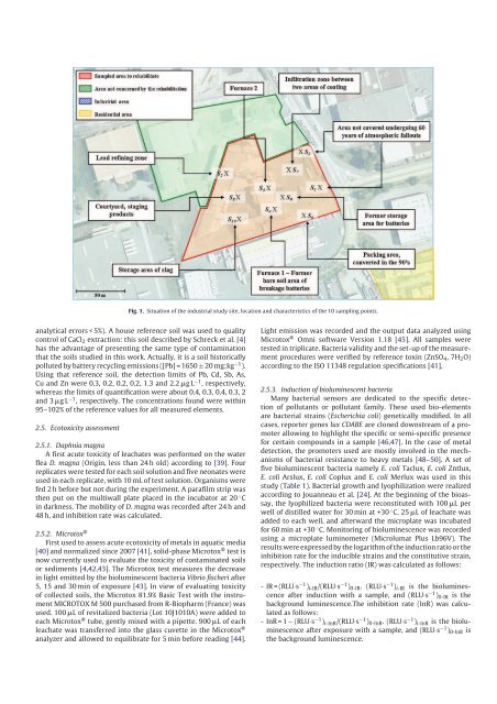
Fig. 1. Situation of the industrial study site, location and characteristics of the 10 sampling points.<br />
analytical errors < 5%). A house reference soil was used to quality<br />
control of CaCl 2 extraction: this soil described by Schreck et al. [4]<br />
has the advantage of presenting the same type of contamination<br />
that the soils studied in this work. Actually, it is a soil historically<br />
polluted by battery recycling emissions ([Pb] = 1650 ± 20 mg;kg −1 ).<br />
Using that reference soil, the detection limits of Pb, Cd, Sb, As,<br />
Cu and Zn were 0.3, 0.2, 0.2, 0.2, 1.3 and 2.2 g L −1 , respectively,<br />
whereas the limits of quantification were about 0.4, 0.3, 0.4, 0.3, 2<br />
and 3 g L −1 , respectively. The concentrations found were within<br />
95–102% of the reference values for all measured elements.<br />
2.5. Ecotoxicity assessment<br />
2.5.1. Daphnia magna<br />
A first acute toxicity of leachates was performed on the water<br />
flea D. magna (Origin, less than 24 h old) according to [39]. Four<br />
replicates were tested for each soil solution and five neonates were<br />
used in each replicate, with 10 mL of test solution. Organisms were<br />
fed 2 h before but not during the experiment. A parafilm strip was<br />
then put on the multiwall plate placed in the incubator at 20 ◦ C<br />
in darkness. The mobility of D. magna was recorded after 24 h and<br />
48 h, and inhibition rate was calculated.<br />
2.5.2. Microtox ®<br />
First used to assess acute ecotoxicity of metals in aquatic media<br />
[40] and normalized since 2007 [41], solidphase Microtox ® test is<br />
now currently used to evaluate the toxicity of contaminated soils<br />
or sediments [4,42,43]. The Microtox test measures the decrease<br />
in light emitted by the bioluminescent bacteria Vibrio fischeri after<br />
5, 15 and 30 min of exposure [43]. In view of evaluating toxicity<br />
of collected soils, the Microtox 81.9% Basic Test with the instrument<br />
MICROTOX M 500 purchased from RBiopharm (France) was<br />
used. 100 L of revitalized bacteria (Lot 10J1010A) were added to<br />
each Microtox ® tube, gently mixed with a pipette. 900 L of each<br />
leachate was transferred into the glass cuvette in the Microtox ®<br />
analyzer and allowed to equilibrate for 5 min before reading [44].<br />
Light emission was recorded and the output data analyzed using<br />
Microtox ® Omni software Version 1.18 [45]. All samples were<br />
tested in triplicate. Bacteria validity and the setup of the measurement<br />
procedures were verified by reference toxin (ZnSO 4 , 7H 2 O)<br />
according to the ISO 11348 regulation specifications [41].<br />
2.5.3. Induction of bioluminescent bacteria<br />
Many bacterial sensors are dedicated to the specific detection<br />
of pollutants or pollutant family. These used bioelements<br />
are bacterial strains (Escherichia coli) genetically modified. In all<br />
cases, reporter genes lux CDABE are cloned downstream of a promoter<br />
allowing to highlight the specific or semispecific presence<br />
for certain compounds in a sample [46,47]. In the case of metal<br />
detection, the promoters used are mostly involved in the mechanisms<br />
of bacterial resistance to heavy metals [48–50]. A set of<br />
five bioluminescent bacteria namely E. coli Taclux, E. coli Zntlux,<br />
E. coli Arslux, E. coli Coplux and E. coli Merlux was used in this<br />
study (Table 1). Bacterial growth and lyophilization were realized<br />
according to Jouanneau et al. [24]. At the beginning of the bioassay,<br />
the lyophilized bacteria were reconstituted with 100 L per<br />
well of distilled water for 30 min at +30 ◦ C. 25 L of leachate was<br />
added to each well, and afterward the microplate was incubated<br />
for 60 min at +30 ◦ C. Monitoring of bioluminescence was recorded<br />
using a microplate luminometer (Microlumat Plus Lb96V). The<br />
results were expressed by the logarithm of the induction ratio or the<br />
inhibition rate for the inducible strains and the constitutive strain,<br />
respectively. The induction ratio (IR) was calculated as follows:<br />
IR = (RLU·s −1 ) iIR /(RLU·s −1 ) 0IR . (RLU·s −1 ) iIR is the bioluminescence<br />
after induction with a sample, and (RLU·s −1 ) 0IR is the<br />
background luminescence.The inhibition rate (InR) was calculated<br />
as follows:<br />
InR = 1 − (RLU·s −1 ) iInR /(RLU·s −1 ) 0InR . (RLU·s −1 ) iInR is the bioluminescence<br />
after exposure with a sample, and (RLU·s −1 ) 0InR is<br />
the background luminescence.