Australian Cerebral Palsy Child Study: protocol of ... - BioMed Central
Australian Cerebral Palsy Child Study: protocol of ... - BioMed Central
Australian Cerebral Palsy Child Study: protocol of ... - BioMed Central

You also want an ePaper? Increase the reach of your titles
YUMPU automatically turns print PDFs into web optimized ePapers that Google loves.
Boyd et al. BMC Neurology 2013, 13:57 Page 3 <strong>of</strong> 12<br />
http://www.biomedcentral.com/1471-2377/13/57<br />
outcomes for children by GMFCS classification [25].<br />
Two potential limitations <strong>of</strong> the Ontario Motor <strong>Study</strong><br />
were that it included only minimal data on children less<br />
than 3 years <strong>of</strong> age and it was a not an entire population<br />
based sample [9].<br />
In the European <strong>Cerebral</strong> <strong>Palsy</strong> study [6], with a representative<br />
cohort <strong>of</strong> children with CP from eight European<br />
countries, children are classified according to brain injury<br />
diagnosed using MRI. This group used a classification<br />
system based on the presumed timing and nature <strong>of</strong> the<br />
insult that resulted in CP and included both genetic and<br />
non-genetic aetiologies such as genetic cortical malformations<br />
(e.g. lissencephaly) and hypoxic ischaemic injury<br />
[6,10]. Again this cohort is representative rather than<br />
entire population based and these investigators from Surveillance<br />
<strong>of</strong> <strong>Cerebral</strong> <strong>Palsy</strong> in Europe (SCPE) have guided<br />
our classifications <strong>of</strong> motor type and <strong>of</strong> the brain injury on<br />
MRI [26-28].<br />
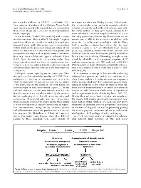
Pathogenic events impacting on the brain cause different<br />
patterns <strong>of</strong> structural abnormality in CP [29]. These<br />
pathogenic events may be environmental or genetic.<br />
Their consequences will depend not only on the nature<br />
<strong>of</strong> the event, but also the timing <strong>of</strong> the event during the<br />
different stages <strong>of</strong> brain development (Figure 1). The 1st<br />
and 2nd trimesters are the most critical times for cortical<br />
development and are characterized by the sequential<br />
yet overlapping steps <strong>of</strong> proliferation, migration and<br />
organization <strong>of</strong> neuronal cells and their connections.<br />
Brain pathology secondary to events during these stages<br />
<strong>of</strong> brain development is usually characterised by significant<br />
malformations. During the 3rd trimester, growth<br />
and differentiation events are predominant and persist<br />
into postnatal life. Disturbances <strong>of</strong> brain development<br />
during this period cause lesions, <strong>of</strong>ten <strong>of</strong> a different<br />
pattern to those resulting from earlier insults or<br />
developmental disorders. During the early 3rd trimester,<br />
the periventricular white matter is especially affected;<br />
whereas towards the end <strong>of</strong> the 3rd trimester grey matter,<br />
either cortical or deep grey matter, appears to be<br />
more vulnerable. Understanding the aetiologies <strong>of</strong> CP in<br />
the living patient has advanced significantly since the increased<br />
use <strong>of</strong> MRI in the evaluation <strong>of</strong> children with<br />
congenital or early-onset neurological deficits. Using<br />
MRI, a number <strong>of</strong> studies have shown that the most<br />
common causes <strong>of</strong> CP are structural brain lesions<br />
[27,30-33], especially prematurity-related injuries, and<br />
malformations <strong>of</strong> brain development [34-36]. Guidelines<br />
by the American Academy <strong>of</strong> Neurology strongly recommend<br />
that all children with a suspected diagnosis <strong>of</strong> CP<br />
undergo neuroimaging, with MRI preferable to CT [37].<br />
Determination <strong>of</strong> brain structural abnormality will provide<br />
a final diagnosis that is more than a label <strong>of</strong> ‘cerebral<br />
palsy’[38].<br />
It is necessary to attempt to determine the underlying<br />
aetiology/pathogenesis to confirm the suspicion <strong>of</strong> a<br />
static lesion, exclude a treatable disorder and diagnose a<br />
malformation, which may have significant genetic counselling<br />
implications for the family. In addition, these patterns<br />
<strong>of</strong> brain maldevelopments or lesions <strong>of</strong>fer excellent<br />
models to study the normal mechanisms <strong>of</strong> organisation<br />
and reorganisation in the developing brain [30,31,39].<br />
Despite these advances, limited studies exist correlating<br />
the specific MR imaging appearance and outcome measures<br />
such as motor function [27]. Such data may prove<br />
invaluable in providing accurate prognostic counselling<br />
at the time <strong>of</strong> diagnosis, as well as potentially guiding<br />
the most appropriate treatments tailored to each individual’s<br />
pattern <strong>of</strong> CP and type <strong>of</strong> lesion on imaging.<br />
A recent systematic review investigated the relationship<br />
between brain structure on MRI and motor<br />
Figure 1 Major events in human brain development. Pathogenic events (both genetic and non-genetic) affect the developing brain to cause<br />
malformations or lesions, the patterns <strong>of</strong> which will depend on the stage <strong>of</strong> brain development during which the event occurs.