Australian Cerebral Palsy Child Study: protocol of ... - BioMed Central
Australian Cerebral Palsy Child Study: protocol of ... - BioMed Central
Australian Cerebral Palsy Child Study: protocol of ... - BioMed Central

You also want an ePaper? Increase the reach of your titles
YUMPU automatically turns print PDFs into web optimized ePapers that Google loves.
Boyd et al. BMC Neurology 2013, 13:57 Page 5 <strong>of</strong> 12<br />
http://www.biomedcentral.com/1471-2377/13/57<br />
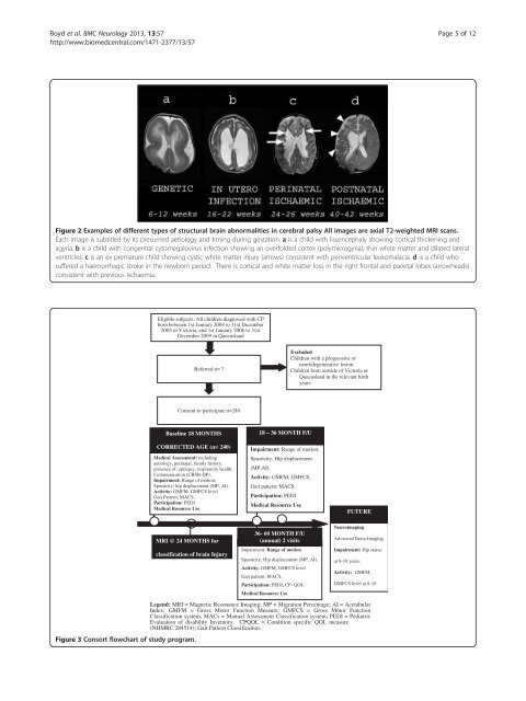
Figure 2 Examples <strong>of</strong> different types <strong>of</strong> structural brain abnormalities in cerebral palsy All images are axial T2-weighted MRI scans.<br />
Each image is subtitled by its presumed aetiology and timing during gestation. a is a child with lissencephaly showing cortical thickening and<br />
agyria. b is a child with congenital cytomegalovirus infection showing an overfolded cortex (polymicrogyria), thin white matter and dilated lateral<br />
ventricles. c is an ex premature child showing cystic white matter injury (arrows) consistent with periventricular leukomalacia. d is a child who<br />
suffered a haemorrhagic stroke in the newborn period. There is cortical and white matter loss in the right frontal and parietal lobes (arrowheads)<br />
consistent with previous ischaemia.<br />
Eligible subjects: All children diagnosed with CP<br />
born between 1st January 2004 to 31st December<br />
2005 in Victoria, and 1st January 2006 to 31st<br />
December 2009 in Queensland<br />
Referred n= ?<br />
Excluded<br />
<strong>Child</strong>ren with a progressive or<br />
neurodegenerative lesion<br />
<strong>Child</strong>ren born outside <strong>of</strong> Victoria or<br />
Queensland in the relevant birth<br />
years<br />
Consent to participate n=240<br />
Baseline 18 MONTHS<br />
CORRECTED AGE (n= 240)<br />
Medical Assessment: including<br />
aetiology, perinatal, family history,<br />
presence <strong>of</strong>: epilepsy, respiratory health.<br />
Communication (CBSB-DP).<br />
Impairment: Range <strong>of</strong> motion;<br />
Spasticity; hip displacement (MP, AI).<br />
Activity: GMFM, GMFCS level<br />
Gait Pattern; MACS,<br />
Participation: PEDI<br />
Medical Resource Use<br />
18 – 36 MONTH F/U<br />
Impairment: Range <strong>of</strong> motion;<br />
Spasticity, Hip displacement<br />
(MP,AI).<br />
Activity: GMFM, GMFCS,<br />
Gait pattern: MACS,<br />
Participation: PEDI<br />
Medical Resource Use<br />
FUTURE<br />
MRI @ 24 MONTHS for<br />
classification <strong>of</strong> brain Injury<br />
36- 60 MONTH F/U<br />
(annual) 2 visits<br />
Impairment: Range <strong>of</strong> motion<br />
Spasticity; Hip displacement (MP, AI).<br />
Activity: GMFM, GMFCS level<br />
Gait pattern; MACS,<br />
Participation: PEDI, CP- QOL<br />
Medical Resource Use<br />
Neuroimaging:<br />
Advanced Brain Imaging<br />
Impairment: Hip status<br />
at 8-10 years.<br />
Activity: GMFM,<br />
GMFCS level at 8-10<br />
Legend: MRI = Magnetic Resonance Imaging; MP = Migration Percentage; AI = Acetabular<br />
Index; GMFM = Gross Motor Function Measure; GMFCS = Gross Motor Function<br />
Classification system; MACs = Manual Assessment Classification system; PEDI = Pediatric<br />
Evaluation <strong>of</strong> disability Inventory. CPQOL = Condition specific QOL measure<br />
(NHMRC 284514); Gait Pattern Classification.<br />
Figure 3 Consort flowchart <strong>of</strong> study program.