Keratin Nanofibers as a Biomaterial - ipcbee
Keratin Nanofibers as a Biomaterial - ipcbee
Keratin Nanofibers as a Biomaterial - ipcbee

You also want an ePaper? Increase the reach of your titles
YUMPU automatically turns print PDFs into web optimized ePapers that Google loves.
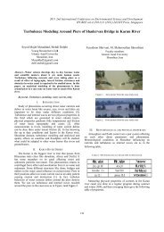
2010 International Conference on Nanotechnology and Biosensors<br />
IPCBEE vol.2 (2011) © (2011) IACSIT Press, Singapore<br />
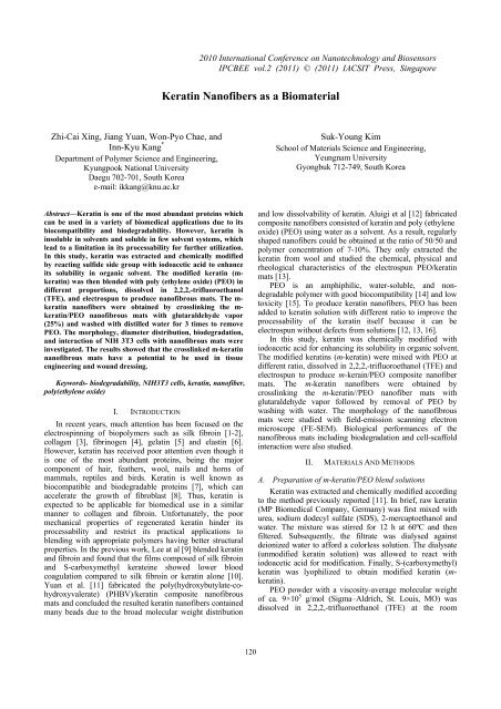
<strong>Keratin</strong> <strong>Nanofibers</strong> <strong>as</strong> a <strong>Biomaterial</strong><br />
Zhi-Cai Xing, Jiang Yuan, Won-Pyo Chae, and<br />
Inn-Kyu Kang *<br />
Department of Polymer Science and Engineering,<br />
Kyungpook National University<br />
Daegu 702-701, South Korea<br />
e-mail: ikkang@knu.ac.kr<br />
Suk-Young Kim<br />
School of Materials Science and Engineering,<br />
Yeungnam University<br />
Gyongbuk 712-749, South Korea<br />
Abstract—<strong>Keratin</strong> is one of the most abundant proteins which<br />
can be used in a variety of biomedical applications due to its<br />
biocompatibility and biodegradability. However, keratin is<br />
insoluble in solvents and soluble in few solvent systems, which<br />
lead to a limitation in its processability for further utilization.<br />
In this study, keratin w<strong>as</strong> extracted and chemically modified<br />
by reacting sulfide side group with iodoacetic acid to enhance<br />
its solubility in organic solvent. The modified keratin (mkeratin)<br />
w<strong>as</strong> then blended with poly (ethylene oxide) (PEO) in<br />
different proportions, dissolved in 2,2,2,-trifluoroethanol<br />
(TFE), and electrospun to produce nanofibrous mats. The m-<br />
keratin nanofibers were obtained by crosslinking the m-<br />
keratin/PEO nanofibrous mats with glutaraldehyde vapor<br />
(25%) and w<strong>as</strong>hed with distilled water for 3 times to remove<br />
PEO. The morphology, diameter distribution, biodegradation,<br />
and interaction of NIH 3T3 cells with nanofibrous mats were<br />
investigated. The results showed that the crosslinked m-keratin<br />
nanofibrous mats have a potential to be used in tissue<br />
engineering and wound dressing.<br />
Keywords- biodegradability, NIH3T3 cells, keratin, nanofiber,<br />
poly(ethylene oxide)<br />
I. INTRODUCTION<br />
In recent years, much attention h<strong>as</strong> been focused on the<br />
electrospinning of biopolymers such <strong>as</strong> silk fibroin [1-2],<br />
collagen [3], fibrinogen [4], gelatin [5] and el<strong>as</strong>tin [6].<br />
However, keratin h<strong>as</strong> received poor attention even though it<br />
is one of the most abundant proteins, being the major<br />
component of hair, feathers, wool, nails and horns of<br />
mammals, reptiles and birds. <strong>Keratin</strong> is well known <strong>as</strong><br />
biocompatible and biodegradable proteins [7], which can<br />
accelerate the growth of fibrobl<strong>as</strong>t [8]. Thus, keratin is<br />
expected to be applicable for biomedical use in a similar<br />
manner to collagen and fibroin. Unfortunately, the poor<br />
mechanical properties of regenerated keratin hinder its<br />
processability and restrict its practical applications to<br />
blending with appropriate polymers having better structural<br />
properties. In the previous work, Lee at al [9] blended keratin<br />
and fibroin and found that the films composed of silk fibroin<br />
and S-carboxymethyl kerateine showed lower blood<br />
coagulation compared to silk fibroin or keratin alone [10].<br />
Yuan et al. [11] fabricated the poly(hydroxybutylate-cohydroxyvalerate)<br />
(PHBV)/keratin composite nanofibrous<br />
mats and concluded the resulted keratin nanofibers contained<br />
many beads due to the broad molecular weight distribution<br />
and low dissolvability of keratin. Aluigi et al [12] fabricated<br />
composite nanofibers consisted of keratin and poly (ethylene<br />
oxide) (PEO) using water <strong>as</strong> a solvent. As a result, regularly<br />
shaped nanofibers could be obtained at the ratio of 50/50 and<br />
polymer concentration of 7-10%. They only extracted the<br />
keratin from wool and studied the chemical, physical and<br />
rheological characteristics of the electrospun PEO/keratin<br />
mats [13].<br />
PEO is an amphiphilic, water-soluble, and nondegradable<br />
polymer with good biocompatibility [14] and low<br />
toxicity [15]. To produce keratin nanofibers, PEO h<strong>as</strong> been<br />
added to keratin solution with different ratio to improve the<br />
processability of the keratin itself because it can be<br />
electrospun without defects from solutions [12, 13, 16].<br />
In this study, keratin w<strong>as</strong> chemically modified with<br />
iodoacetic acid for enhancing its solubility in organic solvent.<br />
The modified keratins (m-keratin) were mixed with PEO at<br />
different ratio, dissolved in 2,2,2,-trifluoroethanol (TFE) and<br />
electrospun to produce m-kerain/PEO composite nanofiber<br />
mats. The m-keratin nanofibers were obtained by<br />
crosslinking the m-keratin//PEO nanofiber mats with<br />
glutaraldehyde vapor followed by removal of PEO by<br />
w<strong>as</strong>hing with water. The morphology of the nanofibrous<br />
mats were studied with field-emission scanning electron<br />
microscope (FE-SEM). Biological performances of the<br />
nanofibrous mats including biodegradation and cell-scaffold<br />
interaction were also studied.<br />
II.<br />
MATERIALS AND METHODS<br />
A. Preparation of m-keratin/PEO blend solutions<br />
<strong>Keratin</strong> w<strong>as</strong> extracted and chemically modified according<br />
to the method previously reported [11]. In brief, raw keratin<br />
(MP Biomedical Company, Germany) w<strong>as</strong> first mixed with<br />
urea, sodium dodecyl sulfate (SDS), 2-mercaptoethanol and<br />
water. The mixture w<strong>as</strong> stirred for 12 h at 60ºC and then<br />
filtered. Subsequently, the filtrate w<strong>as</strong> dialysed against<br />
deionized water to afford a colorless solution. The dialysate<br />
(unmodified keratin solution) w<strong>as</strong> allowed to react with<br />
iodoacetic acid for modification. Finally, S-(carboxymethyl)<br />
keratin w<strong>as</strong> lyophilized to obtain modified keratin (mkeratin).<br />
PEO powder with a viscosity-average molecular weight<br />
of ca. 9×10 5 g/mol (Sigma–Aldrich, St. Louis, MO) w<strong>as</strong><br />
dissolved in 2,2,2,-trifluoroethanol (TFE) at the room<br />
120
temperature for about 12 h. The concentration w<strong>as</strong> adjusted<br />
at 2 wt %.<br />
The m-keratin/PEO blend solutions were prepared by<br />
adding m-kerain to the PEO solution and stirring for 12 h at<br />
room temperature. The blend solutions of the m-keratin/PEO<br />
were adjusted at the concentration of 2 wt% and the ratio of<br />
m-keratin and PEO w<strong>as</strong> changed from 50:50 to 90:10.<br />
B. Preparation of m-keratin/PEO blend nanofibers<br />
The blend solution w<strong>as</strong> delivered to a metal needle<br />
connected to a high-voltage power supply. Upon applying a<br />
high voltage, a fluid jet w<strong>as</strong> ejected from the needle. As the<br />
jet accelerated towards a grounded collector, the solvent<br />
evaporated and a charged polymer fiber w<strong>as</strong> deposited on the<br />
collector in the form of a nanofibrous mat. The typical<br />
parameters for electrospinning were <strong>as</strong> follows: 9 kV<br />
(voltage), 12 cm (distance between tip and receptor),<br />
1.0mLh −1 (feed rate), 60% (humidity) and 25ºC<br />
(temperature). For analysis of the morphology of the<br />
electrospun fibers, the samples were sputter-coated with gold,<br />
and examined using FE-SEM (Hitachi S-4300, Japan). The<br />
diameters of the electrospun nanofibres were me<strong>as</strong>ured at<br />
100 different points from SEM pictures for each sample<br />
produced.<br />
C. Preparation of m-keratin nanofibers<br />
The m-keratin/PEO nanofiber mats need to be<br />
crosslinked to reduce their solubility in water. The<br />
electrospun m-keratin/PEO nanofibrous mats were<br />
crosslinked by treating them with glutaraldehyde vapor and<br />
saturated with a 25% glutaraldehyde aqueous solution at<br />
room temperature for 4 h. This w<strong>as</strong> followed by treatment<br />
with 0.1 M glycine aqueous solution to block unreacted<br />
aldehyde groups. The crosslinked m-keratin/PEO<br />
nanofibrous mat w<strong>as</strong> then w<strong>as</strong>hed with distilled water for<br />
three times (10 minutes each) to produce m-keratin nanofiber<br />
mats. To examine the presence of PEO in the crosslinked m-<br />
keratin/PEO mat after removal by water, fluorescein-tagged<br />
PEO (F-PEO) w<strong>as</strong> synthesized by reacting hydroxyl end<br />
group of PEO with group of fluorescein isothiocyanate<br />
(FITC) [20].<br />
D. In vitro biodegradation<br />
The m-keratin/PEO and m-keratin nanofiber mats were<br />
cut into rectangles (20 × 20 × 0.05 mm) for in vitro<br />
degradation testing. Each specimen w<strong>as</strong> placed in a test tube<br />
containing 10 ml of phosphate-buffered saline (PBS, pH 7.0,<br />
Gibco) and incubated for 12 h at 37ºC. After incubation, the<br />
samples were w<strong>as</strong>hed and lyophilized for 24 h. In order to<br />
me<strong>as</strong>ure the enzymatic degradation of nanofibrous mats, the<br />
samples were incubated in a PBS containing trypsin (10<br />
mg/ml) at 37ºC. After incubation for a requisite time (2 h or<br />
12 h), the samples were w<strong>as</strong>hed with distilled water and then<br />
lyophilized for 24 h. Morphological changes were observed<br />
with a FE-SEM.<br />
E. Cell adhesion<br />
In order to examine the interaction of nanofiber mats<br />
with cells (NIH 3T3), the circular nanofibrous mats were<br />
fitted in a 24-well culture plate and subsequently immersed<br />
in a DMEM medium containing 10% fetal bovine serum<br />
(FBS, Gibco) and 1% penicillin G-streptomycin. To seed the<br />
cells, 1 ml of NIH 3T3 cell solution (3×10 4 cells) w<strong>as</strong> added<br />
and incubated in a humidified atmosphere of 5% CO 2 at 37ºC.<br />
After incubation for a 4 h, the medium solution w<strong>as</strong> removed.<br />
These samples were w<strong>as</strong>hed twice with the PBS, and fixed<br />
by 2.5% glutaraldehyde aqueous solution for 20 min. The<br />
sample mats were then dehydrated in a graded concentration<br />
of ethanol (25, 50, 75, 90, and 100) for 10 min each. Finally,<br />
the sample mats were air dried in a fume hood overnight.<br />
Dry cellular structures were sputter-coated with gold and<br />
observed with a FE-SEM.<br />
F. Determination of cell viability<br />
A standard Live/Dead <strong>as</strong>say w<strong>as</strong> used to image cell<br />
survival, adhesion, and spatial organization. After 6 days<br />
incubation, cells were collected by centrifugation and<br />
incubated in calcein-AM (1 mM in PBS) and ethidium<br />
homodimer-1 (2.5 mg/ml PBS) solution for 15 min. Cells<br />
with compromised membranes exhibit red-fluorescence from<br />
the live-cell impermanent nucleic acid stained with ethidium<br />
homodimer-1. Cells with intact membranes are able to use<br />
nonspecific cytosolic ester<strong>as</strong>es to convert nonfluorescent<br />
calcein-AM into bright green-fluorescent calcein. Cells were<br />
observed under a fluorescence microscope using a band-p<strong>as</strong>s<br />
filter (Nikon Eclipse E600-POL, Japan).<br />
Cell viability w<strong>as</strong> me<strong>as</strong>ured after 2, 4 and 6 days of<br />
culture using a commercially available MTT <strong>as</strong>say kit<br />
(Sigma). After incubation of certain time, the medium w<strong>as</strong><br />
replaced with a (3-[4,5-dimethylthiazol-2-yl]-2,5-diphenyl<br />
tetrazolium bromide (MTT) solution and incubated for<br />
further 4h. Mitochondrial dehydrogen<strong>as</strong>es of viable cells<br />
cleave the tetrazolium ring, yielding purple formazan crystals.<br />
Formazan crystals were then dissolved in PBS solution. The<br />
optical density (OD) of the solvent is proportional to the<br />
mitochondrial activity of the cells on the surface. OD w<strong>as</strong><br />
me<strong>as</strong>ured at 570 nm using a kinetic microplate reader (EL 9<br />
800, Bio, Tek, Instruments, Inc, Highland Park, USA).<br />
Background absorbance at 690 nm w<strong>as</strong> subtracted from the<br />
me<strong>as</strong>ured absorbance.<br />
G. Statistical analysis<br />
Results are displayed <strong>as</strong> mean ± standard deviation.<br />
Statistical differences were determined by a student’s twotailed<br />
t-test. Scheffe’s method w<strong>as</strong> used for multiple<br />
comparison tests at a level of 95%.<br />
III.<br />
RESULTS AND DISCUSSION<br />
A. Nanofiber morphology<br />
Figure 1 shows the SEM images of nanofibers obtained<br />
from electrospinning of the m-keratin/PEO blend solutions.<br />
The electrospinning of a 2 wt% PEO pure solution produced<br />
nanofibers without defects at the condition of a flow rate of<br />
1ml/h and a voltage of 9 kV. However the diameter<br />
distribution of PEO nanofibers w<strong>as</strong> so broad, ranging from<br />
200 nm to 2000 nm. The diameter distribution of PEO<br />
became narrow with an incre<strong>as</strong>e of m-keratin content (Figure<br />
121
1b,c,d). Therefore, it could be say that the blend composition<br />
plays an important role in determining the diameter<br />
distribution of the nanofibers. In addition, the average<br />
diameter of the blend nanofibers gradually decre<strong>as</strong>ed from<br />
950±50 to 400±30 nm <strong>as</strong> the m-keratin content incre<strong>as</strong>es<br />
(Figure 1b and Figure 1d). The viscosity of m-keratin/PEO<br />
(50/50) blend solution (224 cP) w<strong>as</strong> significantly decre<strong>as</strong>ed<br />
down to 34 cP with the incre<strong>as</strong>e of m-keratin (90/10). The<br />
conductivity of m-keratin/PEO blend solution also incre<strong>as</strong>ed<br />
with an incre<strong>as</strong>e of the m-keratin data not shown. In fact, it is<br />
known that lower viscosity promotes the formation of finer<br />
nanofibers [17] and higher charge density carried by jet<br />
forms smoother and finer nanofibers because the stronger<br />
whipping instability of the jet enhances the filament<br />
stretching [18,19].<br />
B. Crosslinking of m-keratin/PEO nanofibers<br />
The m-keratin/PEO blend nanofibers can be e<strong>as</strong>ily<br />
dissolved in water. Therefore, the nanofibers need to be<br />
crosslinked to reduce their solubility. The most popular<br />
crosslinking reagent used in proteins is glutaraldehyde vapor.<br />
Glycine solution w<strong>as</strong> used to block residual aldehyde group<br />
after treatment of m-keratin/PEO nanofibers with<br />
glutaraldehyde vapor. Figure 2 showed the morphology of<br />
m-keratin/PEO (90/10) nanofibrous mats before and after the<br />
crosslinking with glutaraldehyde vapor for 4h. Obviously,<br />
the m-keratin/PEO nanofiber mat lost its fibrous morphology<br />
slightly after the crosslinking. Figure 3 showed the<br />
fluorescence images of the m-keratin/F-PEO(90/10) mat<br />
(Figure 3a) and the m-keratin nanofibrous mat obtained by<br />
removal of F-PEO by water. As a result, the m-keratin/F-<br />
PEO showed the image of green color due to the presence of<br />
F-PEO. However, the green color w<strong>as</strong> almost disappeared<br />
after w<strong>as</strong>hing the crosslinked m-keratin/F-PEO nanofibrous<br />
mats with water (Figure 3b). It is revealed, from the data of<br />
fluorescence image, that m-keratin nanofiber mat could be<br />
obtained by crosslinking the m-keratin/PEO mat with<br />
glutaraldehyde vapor followed by w<strong>as</strong>hing with water.<br />
Figure 2. SEM images of the m-keratin/PEO (90/10) nanofibrous mats<br />
before (a) and after (b) crosslinking with glutaraldehyde vapor for 4h.<br />
C. In vitro biodegradation<br />
The image of the mat biodegraded w<strong>as</strong> examined with<br />
FE-SEM. Figure 4 illustrates the morphological changes of<br />
the electrospun mats during in vitro degradation. The<br />
crosslinked m-keratin nanofibrous mats lost their nanofibrous<br />
form after 2h degradation in trypsin aqueous solution, and<br />
the mat seriously degraded and remained <strong>as</strong> debris after 12h<br />
incubation. Yuan et al [11] reported on biodegradation of m-<br />
keratin/PHBV nanofibrous mats by trypsin solution. As a<br />
result, fibrous morphology almost not changed when the m-<br />
keratin/PHBV mat w<strong>as</strong> incubated in trypsin agueous solution<br />
for 24 h. It is concluded that m-keratin/PEO nanofibrous<br />
mats underwent a higher biological degradation than the<br />
keratin/PHBV nanofibrous mats. This result suggests that the<br />
m-keratin mats studied in this study are suitable for further<br />
biomedical and biotechnological applications [21].<br />
Figure 3. Fluorescence images of the crosslinked m-keratin/F-PEO mat<br />
before (a) and after (b) removal of F-PEO by water nanofibours mats.<br />
Figure 4. SEM images of the crosslinked m-keratin nanofibrous mats<br />
incubated in trypsin solution for 2h (a) and 12h (b).<br />
Figure 1. SEM images of electrospun nanofibers with different ratio of m-<br />
keratin and PEO (a) pure PEO, (b) 50/50, (c) 70/30 and (d) 90/10.<br />
122
D. Cell-scaffold interaction.<br />
To evaluate cellular behavior on electrospun fibers,<br />
fibrobl<strong>as</strong>ts were seeded and cultivated on the crosslinked m-<br />
keratin nanofibers and the tissue culture polystyrene<br />
(control). As shown in Figure 5, cells were more adhered to<br />
the surface of the crosslinked m-keratin nanofiber mat, and<br />
showed much more spread morphology than the tissue<br />
culture polystyrene. The viability of NIH 3T3 cells on the<br />
surface of crosslinked m-keratin nanofibrous mat and the<br />
tissue culture polystyrene were also investigated. NIH 3T3<br />
survival w<strong>as</strong> <strong>as</strong>sessed through live/dead fluorescence<br />
staining. Images taken on a fluorescence microscope indicate<br />
that the surface of crosslinked m-keratin nanofiber mat w<strong>as</strong> a<br />
favorable template for cell adhesion. NIH 3T3 cells seeded<br />
on crosslinked m-keratin nanofibers displayed a high level of<br />
viability <strong>as</strong> <strong>as</strong>sessed using a standard MTT <strong>as</strong>say (Figure 7).<br />
The viability of cells on the surface of crosslinked m-keratin<br />
nanofiber is significantly higher than that on the tissue<br />
culture polystyrene after incubation of 6 days, indicating that<br />
the NIH 3T3 cells seeded on the crosslinked m-keratin<br />
nanofiber surface are healthy and there are no cytotoxic<br />
effects (Figure 6). Further, fluorescence images of live cells<br />
seeded on surface of crosslinked m-keratin nanofiber showed<br />
higher degree of spreading compared to that on the tissue<br />
culture polystyrene. These results suggest that the<br />
crosslinked m-keratin nanofiber mat is a good scaffold for<br />
the adhesion and spread of NIH3T3 cells compared to tissue<br />
culture polystyrene.<br />
Figure 5. SEM images of NIH 3T3 cells cultured for 4 h on the tissue<br />
culture polystyrene (a) and the crosslinked m-keratin nanofibrous mats (b).<br />
Figure 7. MTT <strong>as</strong>say, Formozan absorbance expressed <strong>as</strong> a me<strong>as</strong>ure of<br />
cell viability from the NIH 3T3 cells cultured on the tissue culture<br />
polystyrene and the crosslinked m-keratin/PEO nanofibrous mats (Data are<br />
expressed <strong>as</strong> means ± SD (n=6) for the specific absorbance, * p < 0.05,<br />
values are significantly different from those of the previous group).<br />
IV. CONCLUSIONS<br />
The m-keratin/PEO blend solutions prepared in 2,2,2,-<br />
trifluoroethanol (TFE) were electrospun to produce<br />
nanofibers. All blend solutions were electrospun successfully.<br />
Morphological investigation showed that <strong>as</strong> the m-keratin<br />
amount in the blend solution incre<strong>as</strong>ed, the nanofibers<br />
became thinner and more homogeneous. In addition to being<br />
biocompatible and biodegradable, crosslinked m-keratin<br />
nanofibers induced an enhanced NIH 3T3 cells response.<br />
The results demonstrated that the crosslinked m-keratin<br />
nanofibers enhanced NIH 3T3 cells adhesion and<br />
proliferation <strong>as</strong> compared to the tissue culture polystyrene.<br />
The performance of the crosslinked m-keratin nanofiber<br />
warrants future work aimed at in vivo characterization and<br />
fabrication of 3-D mats.<br />
ACKNOWLEDGMENTS<br />
This research w<strong>as</strong> supported by the research grants of the<br />
Biotechnology development project (2009-0090907) and by<br />
the grant of 2010-0011125 from Ministry of Education,<br />
Science and Technology of Korea.<br />
Figure 6. Fluorescence images of NIH 3T3 cells cultured for 6days on the<br />
tissue culture polystyrene (a) and the crosslinked m-keratin nanofiber mat<br />
(b).<br />
REFERENCES<br />
[1] C. J. Buchko, L. C. Chen, Y. Shen, D. C. Martin “Processing and<br />
microstructural characterization of porous biocompatible protein<br />
polymer thin films,” Polymer, vol. 40, Dec. 1999, pp. 7397-7407,<br />
doi:10.1016/S0032-3861(98)00866-0.<br />
[2] K. Ohgo, C. Zhao, M. Kobay<strong>as</strong>hi, T. Asakura, “Preparation of nonwoven<br />
nanofibers of Bombyx mori silk, Samia cynthia ricini silk and<br />
recombinant hybrid silk with electrospinning method,” Polymer, vol.<br />
44, 2003, pp. 841-846, doi:10.1016/S0032-3861(02)00819-4.<br />
[3] J. A. Matthews, G. E. Wnek, D. G. Simpson, G. L. Bowlin,<br />
“Electrospinning of Collagen <strong>Nanofibers</strong>,” Biomacromolecules, vol. 3,<br />
Mar. 2002, pp.232-238, doi: 10.1021/bm015533u.<br />
123
[4] G. E. Wnek, M. E. Carr, D. G. Simpson, G. L. Bowlin, “Solution-<br />
Ph<strong>as</strong>e Synthesis of Cu2O Nanocubes,” Nano Letters, vol. 3, Feb.<br />
2003, pp. 231-216, doi: 10.1021/nl025866c.<br />
[5] Z. M. Huang, Y. Z. Zhang, S. Ramakrishma, C. T. Lim,<br />
“Electrospinning and mechanical characterization of gelatin<br />
nanofibers,” Polymer, vol. 45, Jul. 2004, pp. 5316-5368,<br />
doi:10.1016/j.polymer.2004.04.005.<br />
[6] L. Huang, R. A. McMillan, R. P. Apkarian, B. Pourdeyhimi, V. P.<br />
Conticello, E. L. Chaikof, “Generation of Synthetic El<strong>as</strong>tin-Mimetic<br />
Small Diameter Fibers and Fiber Networks,” Macromolecules, vol. 33,<br />
Apr. 2000, pp. 2989-2997, doi: 10.1021/ma991858f.<br />
[7] K. Yamauchi, A. Yamauchi, T. Kusunoki, A. Kohda, Y. J. Konishi,<br />
“Preparation of stable aqueous solution of keratins, and<br />
physiochemical and biodegradational properties of films,” Journal of<br />
Biomedical Materials Research, vol. 31, 1996, pp. 439-444.<br />
[8] K. Yamauchi, M. Maniwa, T. Mori, “ The full text electronic article is<br />
available for purch<strong>as</strong>e. You will be able to download the full text<br />
electronic article after payment,” Journal of <strong>Biomaterial</strong>s Science,<br />
Polymer Edition, vol. 9, 1998, pp. 259-270, doi:<br />
10.1163/156856298X00640.<br />
[9] K. Y. Lee, W. S. Ha, “DSC studies on bound water in silk fibroin/Scarboxymethyl<br />
kerateine blend films,” Polymer, vol. 40, Jun. 1999,<br />
pp. 4131–4134, doi:10.1016/S0032-3861(98)00611-9.<br />
[10] K. Lee, S. Kong, W. Park, W. Ha, I. Kwon, “Effect of surface<br />
properties on the antithrombogenicity of silk fibroin/S-carboxymethyl<br />
kerateine blend films,” Journal of <strong>Biomaterial</strong>s Science, Polymer<br />
Edition, vol. 9, 1998, pp. 905-914, doi: 10.1163/156856298X00235.<br />
[11] J. Yuan, Z. C. Xing, S. W. Park, J. Geng, J. Shen, W. Meng, K. J.<br />
Shim, I. S. Han, J. C. Kim, I. K. Kang, “Fabrication of PHBV/<strong>Keratin</strong><br />
Composite Nanofibrous Mats for Biomedical Applications”<br />
Macromolecular Research, vol. 17, Nov. 2009, pp. 850-855.<br />
[12] A. Aluigi, A. Varesano, A. Montarsolo, C. Vineis, F. Ferrero, G.<br />
Mazzuchetti, C. Tonin, “Electrospinning of keratin/poly(ethylene<br />
oxide)blend nanofibers,” Journal of Applied Polymer Science, vol.<br />
104, Apr. 2007, pp. 863-870, doi: 10.1002/app.25623.<br />
[13] A. Aluigi, C. Vineis, A. Varesano, G. Mazzuchetti, F. Ferrero, C.<br />
Tonin, “Structure and properties of keratin/PEO blend nanofibres,”<br />
European Polymer Journal, vol. 44, Aug. 2008, pp. 2465-2475,<br />
doi:10.1016/j.eurpolymj.2008.06.004.<br />
[14] N. P. Desai, J. A. Hubbel, “Solution technique to incorporate<br />
polyethylene oxide and other water-soluble polymers into surfaces of<br />
polymeric biomaterials,” <strong>Biomaterial</strong>s, vol. 12, Mar. 1991, pp. 144-<br />
153, doi:10.1016/0142-9612(91)90193-E.<br />
[15] J. E. Bergsma, F. R. Rozema, R. R. M. Bos, W. C. de Bruijn, G.<br />
Boering, A. J. Pennings, “In vivo degradation and biocompatibility<br />
study of in vitro pre-degraded <strong>as</strong>-polymerized polylactide particles,”<br />
<strong>Biomaterial</strong>s, vol. 16, Mar. 1995, pp. 267-274, doi:10.1016/0142-<br />
9612(95)93253-A.<br />
[16] V. Alessio, A. Annalisa, V. Claudia, T. Claudio, “Study on the shear<br />
viscosity behavior of keratin/PEO blends for nanofibre<br />
electrospinning,” Journal of Polymer Science Part B: Polymer Physics,<br />
vol. 46, Jun. 2008, pp. 1193-1201, dOI: 10.1002/polb.21452.<br />
[17] L. Larrondo, R. Manley, “Electrostatic fiber spinning from polymer<br />
melts. I. Experimental observations on fiber formation and<br />
properties,” Journal of Polymer Science Part B: Polymer Physics, vol.<br />
19, Jun. 1981, pp. 909-920, doi: 10.1002/pol.1981.180190601.<br />
[18] D. H. Reneker, A. L. Yarin, H. Fong, S. Koombhongse, “Bending<br />
instability of electrically charged liquid jets of polymer solutions in<br />
electrospinning,” Journal of Applied Physics, vol. 87, Jun. 2000, pp.<br />
4531-4547, doi:10.1063/1.373532.<br />
[19] M. M. Hohman, M. Shin, G. Rutledge, M. P. Brenner,<br />
“Electrospinning and electrically forced jets. I. Stability theory,”<br />
Physics of Fluids, vol. 13, Dec. 2001, pp. 2201-2220,<br />
doi:10.1063/1.1383791.<br />
[20] Z. Fu, M. M. Santore, “Competitive Adsorption of Poly(ethylene<br />
oxide) Chains with and without Charged End Groups,” Langmuir, vol.<br />
14, July. 1998, pp. 4300-4307, doi: 10.1021/la971358k.<br />
[21] V. Andreia, F. Giuliano, C. P. Artur, “Biodegradable Materials B<strong>as</strong>ed<br />
on Silk Fibroin and <strong>Keratin</strong>,” Biomacromolecules, vol. 9, Apr. 2008,<br />
pp. 1299-1305, doi: 10.1021/bm7012789.<br />
124