Program Book and Abstracts - ISCD
Program Book and Abstracts - ISCD
Program Book and Abstracts - ISCD

You also want an ePaper? Increase the reach of your titles
YUMPU automatically turns print PDFs into web optimized ePapers that Google loves.
Poster Number 148<br />
Epidemiology<br />
Ultrasound Densitometry Evaluation in Postmenopausal Women with<br />
Colles’ Fracture<br />
Vladyslav Povoroznyuk, Institute of Gerontology AMS Ukraine; Volodymyr Vayda,<br />
Institute of Gerontology AMS Ukraine<br />
This research was aimed at studying the bone tissue state among women with Colles’<br />
fracture by means of the ultrasound densitometry method.<br />
Materials <strong>and</strong> methods. The total of 34 healthy postmenopausal women 42–74 years<br />
old (62,1±7,5) with Colles’ fracture in their anamnesis (CF) were examined by<br />
ultrasound bone densitometer “Achilles+” (Lunar Corp., Madison, WI). The control<br />
group included postmenopausal women without any osteoporotic fractures in their<br />
anamnesis (WF), being st<strong>and</strong>ardized by age, BMI, etc. The speed of sound (SOS, m/s),<br />
broadb<strong>and</strong> ultrasound attenuation (BUA, dB/MHz) <strong>and</strong> a calculated “Stiffness” index<br />
(SI, %) were measured. The main risk factors for the osteoporotic Colles’ fracture<br />
turned out to be a menarche after 15 years, an early <strong>and</strong> late menopause. 29,3% of<br />
patients with Colles’ fractures had a bone tissue Stiffness index coinciding with the<br />
baseline of fracture risk or under it.<br />
Results. There was no revealed relation among the age <strong>and</strong> the ultrasound<br />
densitometry indices among women of posmenopausal age without fractures. Only<br />
12,5% of patients with Colles’ fractures were noticed to have a normal bone tissue.<br />
The ultrasound parameters were veritably lower among postmenopausal women<br />
with CF than among WF (SOS: CF – 1524±28,4; WF – 1543±24,3, p < 0,05; BUA:<br />
CF – 102±17,8; WF – 109±12,0, p < 0,05; SI: CF – 76±14,9; WF – 85±13,5, p <<br />
0,05; all values are the mean ± SD). It is caused by the decrease of bone tissue<br />
mineral density, accelerated aging, <strong>and</strong> the development of osteopaenia <strong>and</strong><br />
osteoporosis.<br />
Conclusion. The most tangible differences in these indices were noticed among the<br />
elderly patients. Colles’ fracture indicates osteopaenia <strong>and</strong> osteoporosis in<br />
postmenopausal period. In summary, ultrasound densitometry is an effective screening<br />
method to reveal the women of risk group with future osteoporotic Colles’ fracture<br />
in postmenopausal period.<br />
Poster Number 149<br />
Epidemiology<br />
Ultrasound Densitometry of the Calcaneus in Children <strong>and</strong> Adolescents of<br />
Ukraine<br />
Vladyslav Povoroznyuk, Institute of Gerontology AMS Ukraine<br />
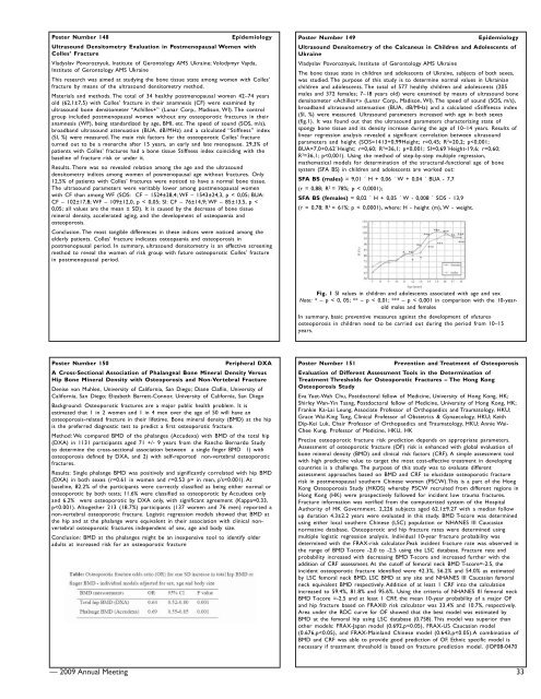
The bone tissue state in children <strong>and</strong> adolescents of Ukraine, subjects of both sexes,<br />
was studied. The purpose of this study is to determine normal values in Ukrainian<br />
children <strong>and</strong> adolescents. The total of 577 healthy children <strong>and</strong> adolescents (205<br />
males <strong>and</strong> 372 females; 7–18 years old) were examined by means of ultrasound bone<br />
densitometer «Achilles+» (Lunar Corp., Madison, WI). The speed of sound (SOS, m/s),<br />
broadb<strong>and</strong> ultrasound attenuation (BUA, dB/MHz) <strong>and</strong> a calculated «Stiffness» index<br />
(SI, %) were measured. Ultrasound parameters increased with age in both sexes<br />
(fig.1). It was found out that the ultrasound parameters characterizing state of<br />
spongy bone tissue <strong>and</strong> its density increase during the age of 10–14 years. Results of<br />
linear regression analysis revealed a significant correlation between ultrasound<br />
parameters <strong>and</strong> height (SOS=1413+0,99Height; r=0,45; R 2 =20,2; p