14th ICID - Poster Abstracts - International Society for Infectious ...
14th ICID - Poster Abstracts - International Society for Infectious ...
14th ICID - Poster Abstracts - International Society for Infectious ...

Create successful ePaper yourself
Turn your PDF publications into a flip-book with our unique Google optimized e-Paper software.
When citing these abstracts please use the following reference:<br />
Author(s) of abstract. Title of abstract [abstract]. Int J Infect Dis 2010;14S1: Abstract number.<br />
Please note that the official publication of the <strong>International</strong> Journal of <strong>Infectious</strong> Diseases 2010, Volume 14, Supplement 1<br />
is available electronically on http://www.sciencedirect.com<br />
Final Abstract Number: 23.001<br />
Session: Antibiotic Resistance Gram-Negative<br />
Date: Wednesday, March 10, 2010<br />
Time: 12:30-13:30<br />
Room: <strong>Poster</strong> & Exhibition Area/Ground Level<br />
Type: <strong>Poster</strong> Presentation<br />
Clinico epidemiologic and molecular characterization of metallo beta lactamases (MBLs)<br />
producing nosocomial Pseudomonas aeruginosa (PSA)<br />
S. Chatterjee 1 , A. Kumar 2 , K. N. prasad 3 , D. Mathai 1 , A. Manoharan 1<br />
1 Christian Medical College and Hospital, Vellore, Tamil Nadu, India, 2 Amrita Institute of Medical<br />
Science, Kochi, India, 3 Sanjay Gandhi Post Graduate Institute of Medical Sciences, Lucknow,<br />
India<br />
Background: The high prevalence of co-resistance to betalactam, aminoglycoside and quinolone<br />
against PSA has necessitated increased use of carbapenems. MBL production among PSA is<br />
one of the several mechanisms causing carbapenem resistance (CARB-R) transferable by<br />
integrons. We surveyed MBL production among PSA isolates collected via multicentric<br />
surveillance study.<br />
Methods: During March-September 2009 BMPLIII received 75 consecutively collected PSA<br />
causing infections of (skin and soft tissue-48, blood and respiratory tract 11 each, and other sites,<br />
5) from four (50-AIMS,Kochi, 21-SGPGIMS, 2-MGIMS,2-AIIMS,Delhi) Indian Medical centers.<br />
Antimicrobial susceptibility testing by Kirby Bauer method against ceftazidime (CZD),<br />
cefepime(FEP), piperacillin/tazobactam (TZP), ticarcillin /clavulanic acid (TIM), gentamicin (GEN),<br />
amikacin(AK), ciprofloxacin(CIP), imipenem(IMP), meropenem(MEM), aztreonam(ATM) and<br />
colistin(CL) was done. MBL was screened by the Combined Disk Diffusion Test (CDDT) method<br />
using 0.5M EDTA (930g) as the inhibitor with IMP, positives (inhibition 7mm) confirmed by<br />
IMP+EDTA Etest and PCR (VIM and IMP genes).<br />
Results: Of the 75 PSA isolates, 12(16%) were MBL producers. Diabetes mellitus was found to<br />
be the major risk factor in PSA infections. Overall resistance to CIP 50.7 %> CZD 37.3% >AK 36<br />
%> FEP 34.7%. Resistance pattern among MBL/NMBL was CZD(91.7%/23.8%),<br />
FEP(91.7%/20.6%), TZP(91.7%/15.9%), TIM(100%/27%), GEN(100%/23.8), AK(100%/20.6%),<br />
CIP(100%/38.1%), IMP(100%/6.3%), MEM(75%/9.5%), ATM(91.7%/38.1%). Resistance to IMP<br />
and MEM was 22.7% and 21.3% respectively. All isolates were susceptible to CL. MDR<br />
(resistant to 2 classes of antimicrobials) was 34(45.3%).Overall 14(18.7%) were CDDT positive.<br />
IMP+EDTA Etest and PCR confirmed 12 to be MBL positives. Among the MBL isolates, one<br />
was also positive <strong>for</strong> IMP gene, which on sequencing was confirmed to be IMP 7. Sensitivity and<br />
specificity of CDDT was 100% and 96.8% respectively when compared to PCR. History of prior<br />
antibiotic usage of aminoglycosides (66.7%), 3rd generation cephalosporins (58.3%), quinolones<br />
(58.3%), carbapenems (50%), penicillins (25%), betalactam+betalactamase inhibitors (8.3%)<br />
was noted <strong>for</strong> the patients with MBL infection. Clinical improvement or cure with modification of<br />
initial antibiotics was found in 63.6% (7/11) patients with MBL PSA.<br />
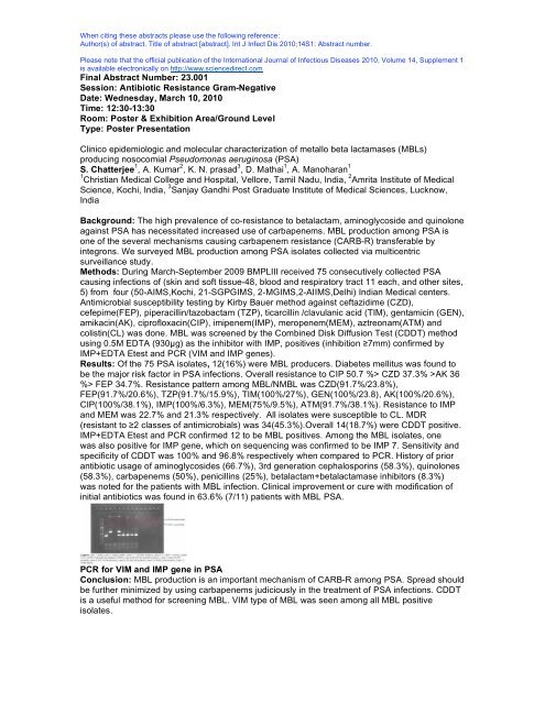
PCR <strong>for</strong> VIM and IMP gene in PSA<br />
Conclusion: MBL production is an important mechanism of CARB-R among PSA. Spread should<br />
be further minimized by using carbapenems judiciously in the treatment of PSA infections. CDDT<br />
is a useful method <strong>for</strong> screening MBL. VIM type of MBL was seen among all MBL positive<br />
isolates.
When citing these abstracts please use the following reference:<br />
Author(s) of abstract. Title of abstract [abstract]. Int J Infect Dis 2010;14S1: Abstract number.<br />
Please note that the official publication of the <strong>International</strong> Journal of <strong>Infectious</strong> Diseases 2010, Volume 14, Supplement 1<br />
is available electronically on http://www.sciencedirect.com<br />
Final Abstract Number: 23.002<br />
Session: Antibiotic Resistance Gram-Negative<br />
Date: Wednesday, March 10, 2010<br />
Time: 12:30-13:30<br />
Room: <strong>Poster</strong> & Exhibition Area/Ground Level<br />
Type: <strong>Poster</strong> Presentation<br />
The resistance patterns of Pseudomonas aeruginosa in hospitals from Greece<br />
and Romania and its importance <strong>for</strong> the therapy of nosocomial infections and infection control<br />
practices<br />
L. M. Junie 1 , S. KASTANAKIS 2 , M. Petrascu 1 , C. Bobo 1 , A. Tsouri 2 , P. Karagianni 2 , E.<br />
Papadomanolaki 2 , G. Aleuraki 2 , M. Gatzima 2 , I. Varthalitis 3<br />
1 University of Medicine and Pharmacy , Cluj Napoca, Romania, 2 St. George General Hospital ,<br />
Chania, Greece, 3 Infection Control Committee, Chania, Greece<br />
Background: One of the most difficult problems in hospitals is the appearance of an increased<br />
number of Pseudomonas antibiotic resistant strains. The objective of our study is to describe the<br />
resistance pattern of Pseudomonas aeruginosa strains.<br />
Methods: For isolating Pseudomonas aeruginosa strains, the usual nutritive media were used.<br />
Identification was made with Vitek2 system (BioMerieux). The susceptibility to the antibiotics of<br />
strains was per<strong>for</strong>med by Kirby Bauer method as recommended by CLSI and Vitek2 system.<br />
Were tested 229 strains isolated from urine and surgical wounds, in Cluj Napoca, Romania, and<br />
36 strains isolated from blood samples in Chania, Greece. The majority of the strains (86.1%)<br />
were isolated from patients in wards and a percentage of 13.8 from Intensive Care Unit.<br />
Results: The strains isolated in Romania from urine showed a high resistance to beta-lactamins,<br />
remaining susceptible to Carbenicillin, Carbapenems, Ceftazidime and Cefepime. Over 80% of<br />
the strains, were resistant to others of third generation cephalosporins. The strains isolated from<br />
blood, presented resistance to Aztreonam (30.43%) and Ceftazidime (13%). 13% were resistant<br />
to Imipenem and Meropenem. Pseudomonas aeruginosa isolated strains in Romania, showed<br />
resistance to Amikacin (71.4% isolated strains from urine and 29.7% from surgical wounds), and<br />
a low resista322nce to Gentamycin (18.6%), Tobramycin (25%). 29.6% were resistant to Colistin.<br />
The strains isolated in Chania, showed resistance to Gentamycin (21.4%). All strains isolated<br />
from urine, were resistant to Pefloxacin and a high resistance was detected to Nalidixic acid<br />
(75%), Ciprofloxacin (75%) and Ofloxacin (50%). The strains isolated from surgical wounds,<br />
presented a resistance to quinolones, oscillating between 33.1% and 51.9%. The isolated strains<br />
from blood showed resistance to Nalidixic acid (38.5%), Pefloxacin (38.5%) and Ciprofloxacin<br />
(38.5)<br />
Conclusion: Our results could reflect the implication of some hospital multi resistant<br />
Pseudomonas aeruginosa strains in nosocomial infections. In Greece, a decline in drugs<br />
resistance of Pseudomonas strains has been noted. As a fact, it is rather encouraging, and<br />
probably we can attribute it to adherence to infection control practices and prudent<br />
chemotherapeutic agents use as proposed by <strong>Infectious</strong> Diseases Control Committee. The<br />
majority of the strains was detected in ward patients and owing to reduced morbidity factors<br />
compared with patients in ICU, the reduced resistance to antibiotics can be explained.
When citing these abstracts please use the following reference:<br />
Author(s) of abstract. Title of abstract [abstract]. Int J Infect Dis 2010;14S1: Abstract number.<br />
Please note that the official publication of the <strong>International</strong> Journal of <strong>Infectious</strong> Diseases 2010, Volume 14, Supplement 1<br />
is available electronically on http://www.sciencedirect.com<br />
Final Abstract Number: 23.003<br />
Session: Antibiotic Resistance Gram-Negative<br />
Date: Wednesday, March 10, 2010<br />
Time: 12:30-13:30<br />
Room: <strong>Poster</strong> & Exhibition Area/Ground Level<br />
Type: <strong>Poster</strong> Presentation<br />
Extended spectrum -lactamase production at the Komfo Anokye Teaching Hospital, Kumasi,<br />
Ghana<br />
Y. Adu-Sarkodie<br />
School of Medical Sciences, KUMASI, Ghana<br />
Background: In recent times enterobacteriaceae isolated at the Komfo Anokye TeachingHospital<br />
(KATH) have shown significant resistance to 2nd and 3rd generation cephalosporins. In 2006, 18-<br />
32% of all enterobacteriaceae isolated from urine and blood were resistant to the cephalosporins<br />
cefuroxime, ceftriaxone , ceftazidime and cefotaxime. These antibiotics are the mainstay <strong>for</strong> the<br />
treatment of severe infections in the hospital. Microbial resistance to these antibiotics if due to the<br />
production of Extended spectrum -lactamases (ESBL) may also indicate resistance to the<br />
fluoroquinolones, aminoglycosides and other antibiotics. This limits therapeutic options <strong>for</strong> the<br />
treatment of severe infections. We studied the extent of ESBL production amongst Klebsiella sp<br />
and E. Coli at KATH.<br />
Methods: ESBL production in 300 non-selected, non-duplicate isolates of Klebesiella sp and<br />
E.coli obtained from blood, urine, wounds, and sputum of both in-patients and out-patients was<br />
determined by the combined disc method using ceftazidime, ceftriaxone and cefpodoxime discs<br />
singly and in combination with clavulanic acid.<br />
Results: 44% and 55% respectively of E.coli and Klebsiella sp ( 57.8% K.pneumo, ) were ESBL<br />
producers. ESBL production in the organisms was commoner in in-patients (75.4%) than outpatients<br />
(24.6%), though not statistically significant (OR=1.40, 95% CI 0.79-2.46, p=0.31). In<br />
general, ESBL producing organisms apart from being resistant to cephalosporins were also<br />
resistant to gentamicin and ciprofloxacin. They were however susceptible to the carbapenems.<br />
Conclusion: The high level of ESBL production found in these enterobacteriaceae with the<br />
resultant microbial resistance to available cephalosporins and other agents implies difficulties with<br />
the choice of therapeutic options <strong>for</strong> the treatment of severe infections. Carbapenems are<br />
expensive on the Ghanaian market and their use in non-severe infections (as may be suggested<br />
by these results) may be inappropriate. Both prescribers and pharmaceutical agents will need to<br />
reflect soberly on their contribution to this sordid state of affairs. We need to put an end to our<br />
practice of cracking nuts with sledge hammers!
When citing these abstracts please use the following reference:<br />
Author(s) of abstract. Title of abstract [abstract]. Int J Infect Dis 2010;14S1: Abstract number.<br />
Please note that the official publication of the <strong>International</strong> Journal of <strong>Infectious</strong> Diseases 2010, Volume 14, Supplement 1<br />
is available electronically on http://www.sciencedirect.com<br />
Final Abstract Number: 23.004<br />
Session: Antibiotic Resistance Gram-Negative<br />
Date: Wednesday, March 10, 2010<br />
Time: 12:30-13:30<br />
Room: <strong>Poster</strong> & Exhibition Area/Ground Level<br />
Type: <strong>Poster</strong> Presentation<br />
Antimicrobial susceptibility profile of Pseudomonas aeruginosa strains isolated at a tertiary-care<br />
University Hospital (S. Orsola Hospital, Bologna, Italy)<br />
R. Manfredi<br />
University of Bologna, Bologna, Italy<br />
Background: The increased rate of antimicrobial resistance among Gram-negative bacilli and<br />
Enterobacteriaceae is a general concern,especially in the hospital setting.A prospective<br />
microbiological monitoring including a continued surveillance of antimicrobial susceptibility rates<br />
of all relevant pathogens,is ongoing at our Hospital.<br />
Methods: The temporal variations of in vitro antimicrobial sensitivity rates were registered at<br />
quarterly intervals <strong>for</strong> all suitable Pseudomonas aeruginosa strains,during the year 2008.The<br />
same pathogen cultured more than once from the same patient within one month,has been<br />
considered once.<br />
Results: Among Pseudomonas aeruginosa isolates (494 strains tested on the whole),the best<br />
per<strong>for</strong>mance was obtained by the old colistin (colimycin),with a 100% susceptibility rate,followed<br />
by piperacillin-tazobactam (73.9-78.6.4% of tested strains),amikacin (71.7-84.5% of tested<br />
strains),imipenem (69.9-79.1% of tested strains),ceftazidime (from 68.0 to 82.5% of tested<br />
strains),tobramycin (from 63.0 to 76.4% of tested strains).On the other hand,significantly less<br />
effective sensitivity profiles were shown by gentamicin (57.5% to 71.3% of tested<br />
strains),ciprofloxacin (51.3-68.0% of tested strains),aztreonam (59.5-61.2% of tested<br />
strains),ticarcillin-clavulanate (54.2-66.9%),and mezlocillin (45.3-54.2% of tested strains).When<br />
examining temporal trends of antibiotic sensitivity figures in the examinewd period (January-<br />
December 2008), significantly favorable changes were observed only <strong>for</strong> ceftazidime and<br />
ciprofloxacin (p
When citing these abstracts please use the following reference:<br />
Author(s) of abstract. Title of abstract [abstract]. Int J Infect Dis 2010;14S1: Abstract number.<br />
Please note that the official publication of the <strong>International</strong> Journal of <strong>Infectious</strong> Diseases 2010, Volume 14, Supplement 1<br />
is available electronically on http://www.sciencedirect.com<br />
Final Abstract Number: 23.005<br />
Session: Antibiotic Resistance Gram-Negative<br />
Date: Wednesday, March 10, 2010<br />
Time: 12:30-13:30<br />
Room: <strong>Poster</strong> & Exhibition Area/Ground Level<br />
Type: <strong>Poster</strong> Presentation<br />
Characterization of ertapenem resistance in Klebsiella pneumoniae from Croatia<br />
B. Bedenic 1 , J. Vranes 2 , Z. Bosnjak 3 , A. Budimir 4 , S. Kalenic 3<br />
1 School of Medicine, University of Zagreb, Clinical Hospital Center Zagreb, Zagreb, Croatia,<br />
2 Zagreb Institue of Public Health "Andrija Stampar", Zagreb, Croatia, 3 Clinical Hospital Center<br />
Zagreb, Zagreb, Croatia, 4 School of Medicine University of Zagreb, Clinical Hospital Center<br />
Zagreb, Zagreb, Croatia<br />
Background: Klebsiella pneumoniae isolates with reduced susceptibility to carbapenems were<br />
recently reported in USA, UK, Turkey and some other countries of the world. Recently eight<br />
Klebsiella pneumoniae strains with reduced susceptibility to carbapenems were isolates in four<br />
different hospitals in Croatia. The aim of the study was to determine the mechanisms of<br />
ertapenem resistance in these strains.<br />
Methods: Antibiotic susceptibilities were determined by broth microdilution method according to<br />
CLSI. Transferability of ertapenem resistance was determined by conjugation (broth mating<br />
method) using E. coli A15 R- as recipient. Production of metallo -lactamases was detected by<br />
double-disk synergy test and E test. -lactamases were characterized by PCR with primers<br />
specific <strong>for</strong> extended-spectrum -lactamases, plasmid-mediated ampC -lactamases, metallo -<br />
lactamases of VIM and IMP series, KPC and OXA-48 -lactamases. Genotyping of the strains<br />
was per<strong>for</strong>med by PFGE.<br />
Results: All strains were resistant to ceftazidime, cefotaxime, ceftriaxone, piperacillin alone and<br />
combined with tazobactam, amoxycillin/clavulanate, gentamicin and ciprofloxacin. All except one<br />
strains showed resistance to ertapenem, intermediate susceptibility or resistance to meropenem<br />
and intermediate susceptibility or full susceptiblity to imipenem. One strain was resistant to all<br />
three carbapenems. Ertapenem resistance was not transferable by conjugation to E. coli recipient<br />
in neither of our strains. PCR revealed the presence of blaSHV and blaCTX-M genes. Multiplex<br />
PCR was positive <strong>for</strong> group 1 CTX-M -lactamases. Sequencing of representative blaCTX-M<br />
genes revealed the presence of CTX-M-15 beta-lactamase. The strain resistant to all three<br />
carbapenem was positive by E test <strong>for</strong> MBLs. However, PCR was negative <strong>for</strong> VIM and IMP -<br />
lactamases. No KPC or plasmid-mediated ampC -lactamases were found. The strains were not<br />
clonally related as shown by PFGE and displayed distinct PFGE fingerprints.<br />
Conclusion: This is the first report of carbapenem resistant Klebsiellae in Croatia. Ertapenem<br />
resistance in Klebsiella was previously reported in UK, Turkey and Israel mainly due to the<br />
production of CTX-M -lactamases of group 1 combined with porin loss (OmpK36 or OmpK35).<br />
The characterization of outer membrane porins needs to be done to clarify the mechanisms of<br />
ertapenem resistance in our strains. Further testing is necessary to determine the mechanism of<br />
carbapenem resistance in the strain with positive E test–MBL.
When citing these abstracts please use the following reference:<br />
Author(s) of abstract. Title of abstract [abstract]. Int J Infect Dis 2010;14S1: Abstract number.<br />
Please note that the official publication of the <strong>International</strong> Journal of <strong>Infectious</strong> Diseases 2010, Volume 14, Supplement 1<br />
is available electronically on http://www.sciencedirect.com<br />
Final Abstract Number: 23.006<br />
Session: Antibiotic Resistance Gram-Negative<br />
Date: Wednesday, March 10, 2010<br />
Time: 12:30-13:30<br />
Room: <strong>Poster</strong> & Exhibition Area/Ground Level<br />
Type: <strong>Poster</strong> Presentation<br />
Changing trends in antimicrobial resistance among salmonella serotypes in Southern India<br />
S. rao 1 , B. Kabir 2<br />
1 kasturba Medical College & Hospital ,Manipal, Karnataka, India, 2 Kasturba Medical College and<br />
Hospital, Madhav Nagar, Manipal, India<br />
Background: Enteric fever caused by drug resistant Salmonella enterica serotype Typhi and<br />
Salmonella enterica serotype Paratyphi A has been the major public health concern in the Indian<br />
Subcontinent.<br />
Methods: A Retrospective analysis of antibiogram and resistance pattern to Ciprofloxacin,<br />
Nalidixic acid, Ceftriaxone Azithromicin and other routine antibiotics to Salmonella isolates from<br />
PUO cases from blood cultures during 2005-2006 combined with a follow up during 2007-2008<br />
represents the data presented in this study.<br />
Results: Of the 2247 from PUO cases 198 salmonella species (65 were Salmonella typhi, 132<br />
Salmonella paratyphi A and 1 Salmonella enteritidis. Salmonella typhi and Salmonella paratyphi<br />
A serotypes were sensitive to Chloramphenicol, Ampicillin, Cotrimoxazole, and Cefriaxone and<br />
sensitive /intermediate to Ciprofloxacin and resistant to Nalidixic acid (except one). MIC <strong>for</strong><br />
Ciprofloxacin and Nalidixic acid resistance strains was 0.5/1 g/ml except <strong>for</strong> 3 MDR salmonella<br />
strains which had MIC value of 16 g/ml. This was reflected on disc diffusion test as intermediate<br />
zone of inhibition. Retrospective blood culture analysis of 2005-2006 has shown that MDR<br />
Salmonella typhi was common isolate then and the strains were sensitive <strong>for</strong> Ciprofloxacin (MIC<br />
being 0.125g/ml). No antibiotic resistant Salmonella paratyphi A was isolated during this period.<br />
Conclusion: Salmonella strains with Nalidixic acid resistance and reduced susceptibility and MIC<br />
to Ciprofloxacin have emerged as major cause of enteric fever in Indian Subcontinent. Nalidixic<br />
acid susceptibility (30g disc) can be reliably used to monitor Ciprofloxacin resistance.
When citing these abstracts please use the following reference:<br />
Author(s) of abstract. Title of abstract [abstract]. Int J Infect Dis 2010;14S1: Abstract number.<br />
Please note that the official publication of the <strong>International</strong> Journal of <strong>Infectious</strong> Diseases 2010, Volume 14, Supplement 1<br />
is available electronically on http://www.sciencedirect.com<br />
Final Abstract Number: 23.007<br />
Session: Antibiotic Resistance Gram-Negative<br />
Date: Wednesday, March 10, 2010<br />
Time: 12:30-13:30<br />
Room: <strong>Poster</strong> & Exhibition Area/Ground Level<br />
Type: <strong>Poster</strong> Presentation<br />
Molecular epidemiology of aminoglycosides resistance in Acinetobacter spp. with emergence of<br />
multidrug-resistant strains in hospitalized patients in Iran<br />
R. Moniri 1 , R. Kheltabadi Farahani 2<br />
1 Kashan University of Medical Sciences, Kashan, Iran, Islamic Republic of, 2 Kashan University Of<br />
Medical Sciences , Kashan, Iran, Islamic Republic of<br />
Background: Acinetobacter spp. is emerging as an important nosocomial pathogen and is<br />
characterized by increasing antimicrobial resistance. Our aim was to evaluate antimicrobial<br />
susceptibility and aminoglycosides resistance genes of Acinetobacter spp. isolated from<br />
hospitalized patients.<br />
Methods: Sixty isolates were identified as Acinetobacter species. The isolates were tested <strong>for</strong><br />
antibiotic resistance by disc diffusion method <strong>for</strong> 12 antimicrobials. The presence of aphA6,<br />
aacC1 aadA1, and aadB genes were detected using PCR.<br />
Results: From the isolated Acinetobacter spp. the highest resistance rate showed against<br />
amikacin, tobramycin, and ceftazidim, respectively; while isolated bacteria were more sensitive to<br />
ampicillic/subactam. More than 66% of the isolates were resistant to at least three classes of<br />
antibiotics, and 27.5% of MDR strains were resistant to all seven tested classes of antimicrobials.<br />
The higher MDR rate presented in bacteria isolated from the ICU and blood samples. More than<br />
60% of the MDR bacteria were resistance to amikacin, ceftazidim, ciprofloxacin,<br />
piperacillin/tazobactam, doxycycline, tobramycin and levofloxacin. Also, more than 60% of the<br />
isolates contained phosphotransferase aphA6, and acetyltransferase genes aacC1, but<br />
adenylyltransferase genes aadA1 (41.7%), and aadB (3.3%) were less prominent. In this study<br />
21.7% of the strains contain three aminoglycoside resistance genes (aphA6, aacC1 and aadA1).<br />
Conclusion: The rising trend of resistance to aminoglycosides poses an alarming threat to<br />
treatment of such infections. The findings showed that clinical isolates of Acinetobacter spp. in<br />
our hospital carrying various kinds of aminoglycoside resistance genes.
When citing these abstracts please use the following reference:<br />
Author(s) of abstract. Title of abstract [abstract]. Int J Infect Dis 2010;14S1: Abstract number.<br />
Please note that the official publication of the <strong>International</strong> Journal of <strong>Infectious</strong> Diseases 2010, Volume 14, Supplement 1<br />
is available electronically on http://www.sciencedirect.com<br />
Final Abstract Number: 23.008<br />
Session: Antibiotic Resistance Gram-Negative<br />
Date: Wednesday, March 10, 2010<br />
Time: 12:30-13:30<br />
Room: <strong>Poster</strong> & Exhibition Area/Ground Level<br />
Type: <strong>Poster</strong> Presentation<br />
Incidence of Carbapanemase Resistance Gene (KPC) among Klebsiella pneumoniae isolates<br />
and its Clinical Implications<br />
B. Yegneswaran, W. Numsuwan, D. Alcid<br />
Drexel University College of Medicine / St Peter's University Hospital, New Brunswick, NJ, USA<br />
Background: Carbapenem antibiotics (Imipenem, Ertapenem, and Meropenem) idicated <strong>for</strong><br />
infections caused by extended-spectrum {beta}-lactamase (ESBL) carrying pathogens.<br />
Carbapenem resistance has been unusual in isolates of Klebsiella pneumoniae. The aim of this<br />
study is to identify the prevalence of KPC positive Klebsiella pneumoniae, and it's clinical<br />
significance.<br />
Methods: All isolates of Klebsiella pneumoniae species from October 1, 2007 to September 30,<br />
2009 were tested <strong>for</strong> the presence of KPC gene using the modified Hodge test. Medical records<br />
of patients with KPC were studied.<br />
Results: Over the period of two-years 40,309 samples were submitted <strong>for</strong> culture and sensitivity,<br />
out of which 7,836 were positive. Of the positives, there were 106 isolates of K pneumoniae and<br />
11 were ESBL positive. Of the ESBL producing isolates, 8 carried the Carbapenem-hydrolyzing<br />
-lactamase. Of the eight, three isolates were reported as being susceptible to Imipenem.<br />
Although all the eight isolates were resistant using the Hodge test. Piperacillin/Tazobactam (PT)<br />
and Vancomycin were the antibiotic used 7 of the 8 patients prior to isolation of Klebsiella<br />
pneumoniae resistant to Carbapenems. One patient was excluded in outcome as one patient’s<br />
sample was clinically thought to be a contaminant was not treated. 3 patients in whom resistance<br />
to carbapenem was reported had their antibiotic was changed to Tigecycline and Polymyxin B<br />
resulting in clinical improvement. Of the remaining 4 patients who were reported as sensitive to<br />
carbapenem three patients had to undergo a repeat surgery due to clinical deterioration and one<br />
patient clinically died.<br />
Conclusion: The incidence of KPC gene at our hospital was 7.5%. KPC positive isolatesare<br />
rapidly emerging pathogens. It is very important to keep this organism in mind as if not treated<br />
there is a 100% probability of having a poor outcome. There is a complete cross resistance to all<br />
Carbapenems containing KPC, there<strong>for</strong>e if KPC is present, the K pneumoniae will be resistant to<br />
all Carbapenem regardless of the routine susceptibility testing as shown in three isolates that are<br />
KPC positive but susceptible to Imipenem. Current automated systems used <strong>for</strong> susceptibility<br />
testing may not accurately identify all these isolates. We must also control the use of antibiotics<br />
specially PT to prevent emergence of KPC positive organisms.
When citing these abstracts please use the following reference:<br />
Author(s) of abstract. Title of abstract [abstract]. Int J Infect Dis 2010;14S1: Abstract number.<br />
Please note that the official publication of the <strong>International</strong> Journal of <strong>Infectious</strong> Diseases 2010, Volume 14, Supplement 1<br />
is available electronically on http://www.sciencedirect.com<br />
Final Abstract Number: 23.009<br />
Session: Antibiotic Resistance Gram-Negative<br />
Date: Wednesday, March 10, 2010<br />
Time: 12:30-13:30<br />
Room: <strong>Poster</strong> & Exhibition Area/Ground Level<br />
Type: <strong>Poster</strong> Presentation<br />
In vitro activity of Tigecycline against molecularly defined Carbapenemase producing<br />
Acinetobacter baumannii<br />
M. Hackel 1 , P. Higgins 2 , H. Seifert 2 , S. Bouchillon 1 , B. Johnson 1 , R. Badal 1 , J. johnson 1 , D.<br />
Hoban 1 , S. Hawser 3 , M. Dowzicky 4<br />
1 <strong>International</strong> Health Management Associates, Inc., Schaumburg, IL, USA, 2 Institute <strong>for</strong> Medical<br />
Microbiology, Cologne, Germany, 3 IHMA Europe Sàrl, Epalinges, Switzerland, 4 Pfizer Inc,<br />
Collegeville, PA, USA<br />
Background: Acinetobacter baumannii are important opportunistic pathogens with increasing<br />
rates of multi-antibiotic resistance due to both intrinsic and acquired mechanisms. Carbapenems<br />
are often used to treat these infections, however carbapenem resistance is increasingly reported,<br />
leaving few therapeutic options. This resistance is most often associated with acquired or<br />
intrinsic OXA-group carbapenemase production. While A. baumannii carry the intrinsic OXA-51-<br />
like carbapenemase gene, carbapenem resistance has only been associated with these genes<br />
when the insertion sequence ISAba1 is upstream. In this study, we evaluated the in vitro activity<br />
of tigecycline against genetically defined A. baumannii from the Tigecycline Evaluation<br />
Surveillance Trial.<br />
Methods: A total 352 imipenem resistant Acinetobacter baumanii from 35 countries (2004 to<br />
2006) were evaluated. MICs were determined by broth microdilution and interpreted according to<br />
CLSI guidelines. Carbapenemase genes were detected by multiplex PCR.<br />
Results: All isolates tested in this study contained an OXA-51-like gene. Additional genotypes<br />
are listed below.<br />
Conclusion: Tigecycline demonstrated excellent in vitro activity against carbapenem resistant A.<br />
baumannii regardless of carbapenemase type. Tigecycline’s in vitro activity against isolates with<br />
either intrinsic ISAba1-OXA-51 or the acquired oxacillinases (OXA-23, OXA-40 and OXA-58)<br />
suggests that it may be effective against resistant isolates of this clinically important pathogen.
When citing these abstracts please use the following reference:<br />
Author(s) of abstract. Title of abstract [abstract]. Int J Infect Dis 2010;14S1: Abstract number.<br />
Please note that the official publication of the <strong>International</strong> Journal of <strong>Infectious</strong> Diseases 2010, Volume 14, Supplement 1<br />
is available electronically on http://www.sciencedirect.com<br />
Final Abstract Number: 23.010<br />
Session: Antibiotic Resistance Gram-Negative<br />
Date: Wednesday, March 10, 2010<br />
Time: 12:30-13:30<br />
Room: <strong>Poster</strong> & Exhibition Area/Ground Level<br />
Type: <strong>Poster</strong> Presentation<br />
Molecular typing of multi-drug resistant Acinetobacter baumannii from London hospitals<br />
D. Wareham, D. Bean<br />
Queen Mary University London, London, United Kingdom<br />
Background: Acinetobacter baumannnii has emerged as a major nosocomial pathogen causing<br />
outbreaks of infection in the immunosuppressed and critically ill worldwide. A number of<br />
molecular typing techniques have been developed <strong>for</strong> use in both local surveillance and global<br />
epidemiological studies. We applied a recently described multi-locus sequence typing (MLST)<br />
scheme to multi-drug resistant A. baumannii (MDRAB) recovered from London Hospitals in<br />
comparison with other molecular methods.<br />
Methods: Sixteen MDRAB were identified using multiplex PCR <strong>for</strong> OXA-carbapenemases and<br />
included representatives previously assigned to the UK ‘South East’, OXA-23 (1) and OXA-23 (2)<br />
clones. Stains were typed using pulsed field gel electrophoresis (PFGE), random-amplified<br />
polymorphic DNA-PCR (RAPD), a multiplex PCR typing (MPT) scheme (ompA, csuE and<br />
blaOXA-51 like genes) and an A. baumannii MLST scheme (gltA, gyrB, gdhB, recA, cpn60, gpi,<br />
rpoD).<br />
Results: Five clearly distinguishable profiles were observed using PFGE and six with RAPD.<br />
MPT typing assigned all but one of the strains (OXA-23 (2) representative) to group 1. Four allelic<br />
profiles (sequence types ST) were obtained using MLST, two of which appeared to be novel.<br />
Comparison of results revealed isolates designated ‘South East’ clone by PFGE / RAPD<br />
belonged to the previously described ST22 and those designated OXA-23 (1) to ST53. The allelic<br />
profile of the OXA-23 clone 2 strain was novel and contained a new gyrB allele.<br />
Conclusion: Molecular techniques were comparable <strong>for</strong> typing UK MDRAB. Isolates belonging to<br />
ST22, (found previously in Italy, Portugal, Czech Republic, China, Korea and Australia) and<br />
isolates belonging to ST53 (found previously in Italy) were the most frequently types, highlighting<br />
the widespread dissemination of these clones. Two novel sequence types were identified and are<br />
undergoing further investigation.
When citing these abstracts please use the following reference:<br />
Author(s) of abstract. Title of abstract [abstract]. Int J Infect Dis 2010;14S1: Abstract number.<br />
Please note that the official publication of the <strong>International</strong> Journal of <strong>Infectious</strong> Diseases 2010, Volume 14, Supplement 1<br />
is available electronically on http://www.sciencedirect.com<br />
Final Abstract Number: 23.011<br />
Session: Antibiotic Resistance Gram-Negative<br />
Date: Wednesday, March 10, 2010<br />
Time: 12:30-13:30<br />
Room: <strong>Poster</strong> & Exhibition Area/Ground Level<br />
Type: <strong>Poster</strong> Presentation<br />
Emerging multi drug-resistant Acinetobacter in Iran: Study of 800 cases<br />
F. Abbasi 1 , D. Yadegarynia 2 , M. Mardani 2 , B. Frasinejad 2 , T. Yaghubi 2 , S. Gholamin 2<br />
1 Shaheed Beheshti Medical University, Tehran, Iran, Islamic Republic of, 2 Shaheed Beheshti<br />
Medical University, tehran, Iran, Islamic Republic of<br />
Background: Acinetobacter baumannii is a multi drug resistant organism associated with high<br />
morbidity and mortality. It is an emerging nosocomial pathogen in many parts of the world. The<br />
aim of the present study was to review the incidence of Acinetobacter in several hospitals in<br />
Tehran, Iran and also to find out pattern of antibiotic resistance of Acinetobacter. We published<br />
primary results of this study when it was consisted of 100 samples. It was accepted in 48th<br />
Annual ICAAC/ 46th Annual IDSA congress, Oct. 2008.<br />
Methods: In a retrospective study we detected 800 positive cultures of Acinetobacter from 800<br />
patients in different wards of several tertiary care hospitals in Tehran, Iran. Disk diffusion method<br />
was used to determine the resistance of isolated Acinetobacter baumannii. Antimicrobial<br />
sensitivity to 14 available antibiotics was analyzed. The following antibiotics were tested:<br />
cefriaxone, cefotaxime, ceftazidime, cefepime, trimethoprim-sulfamethoxazole, gentamicin,<br />
tobramycin, amikacin, ciprofloxacine, ofloxacine, imipenem, meropenem, piperacillin-tazobactam<br />
and tigecycline.<br />
Results: The most frequently sites of infection were wound, respiratry tract, blood stream, and<br />
urinary tract. The most increminated wards were burn and intensive care unit with a high<br />
prevalence of wound infection, pneumonia and septicemia due to Acinetobacter. Acinetobacter<br />
was isolated from wound in 58.9%, tracheal discharge 9.2%, sputum in 7.1%, urine in 6.6%,<br />
blood in 5.8%, CSF in 3.4%, pleural fluid in 2%, tip of catheter (CV line) in 0.6% and<br />
brochoalveolar lavage (BAL) in 0.2%.<br />
Acinetobacter baumannii showed resistance to cefriaxone, cefotaxime, ceftazidime and cefepime<br />
more than 90%. Resistance to gentamicin and trimethoprim-sulfamethoxazole were more than<br />
80%. Resistance to amikacin, imipenem and ciprofloxacine were 72.7%, 70.5% and 45.7%<br />
respectively. Tigecycline, piperacillin-tazobactam, tobramycin and ofloxacine were evaluated in<br />
100 samples. Sensitivity to tobramycin and ofloxacine were 28.9% and 25.3% respectively.<br />
Tigecycline and piperacillin-tazobactam were active against 100% and 74% of the strains<br />
respectively.<br />
Conclusion: Acinetobacter baumanii is an increasingly isolated pathogen in Iran. Prevalence of<br />
carbapenem resistant acinetobacter is high in our study. Newer antimicrobial compound is<br />
needed <strong>for</strong> treatment of this infection.
When citing these abstracts please use the following reference:<br />
Author(s) of abstract. Title of abstract [abstract]. Int J Infect Dis 2010;14S1: Abstract number.<br />
Please note that the official publication of the <strong>International</strong> Journal of <strong>Infectious</strong> Diseases 2010, Volume 14, Supplement 1<br />
is available electronically on http://www.sciencedirect.com<br />
Final Abstract Number: 23.012<br />
Session: Antibiotic Resistance Gram-Negative<br />
Date: Wednesday, March 10, 2010<br />
Time: 12:30-13:30<br />
Room: <strong>Poster</strong> & Exhibition Area/Ground Level<br />
Type: <strong>Poster</strong> Presentation<br />
The prevalence of extended spectrum betalactamases at a tertiary hospital in South Africa<br />
N. Mbelle<br />
National health laboratory Services, Johannesburg, South Africa<br />
Background:<br />
The extent of extended spectrum beta lactamase (ESBL) producing clinical isolates is now a<br />
global concern. The prevalence of ESBLs varies between species, geographically and with time.<br />
Delays in commencing appropriate therapy have been shown to have an impact on morbidity,<br />
mortality and hospital associated costs. The availability of current South African data will provide<br />
the evidence required to implement appropriate health measures.<br />
Methods: A retrospective descriptive study was per<strong>for</strong>med <strong>for</strong> the period from May 2008 to April<br />
2009. Consecutive clinical specimens were obtained from admitted patients and patients referred<br />
from primary health care clinics to Dr. George Mukhari tertiary hospital in Pretoria, South Africa.<br />
Isolates from blood, cerebrospinal fluid, urine, sputum, pus and stools were processed using<br />
conventional laboratory methods. Isolates were identified and susceptibility testing done on the<br />
Microscan® using the CLSI guidelines.Demographic, clinical and laboratory data were evaluated.<br />
Results: ESBL production was identified in 454 (18%) of the enterobacteriaceae isolated this<br />
period. The clinical source was predominantly non-sterile sites, 386 (83%) compared to 78 (17%)<br />
from sterile sites. The majority of isolates were from pus swabs (170) 44%, while 134 (35%) were<br />
from urine. Of the isolates identified, 271 (29%) were Klebsiella pneumoniae, 89 (9%) Escherichia<br />
coli, 25 (3%) Proteus mirabilis, 25 (5%) Enterobacter cloacae, 12 (1%) Klebsiella oxytoca and the<br />
rest Enterobacter aerogenes, Morganella morganii, Proteus vulgaris, Citrobacter freundii and<br />
Serratia marcescens. The prevalence of ESBLs in the Klebsiella pneumoniae isolates was found<br />
to be 271 (46%) and that <strong>for</strong> Escherichia coli 89 (12%).<br />
Conclusion: At our institution, the overall prevalence of ESBL producing enterobacteriaceae of<br />
18%, reaffirms the global spread of ESBL producing bacteria. The presence of ESBLs in most of<br />
the common enterobacteriacea is consistent with the horizontal spread of ESBL genetic material.<br />
Due to the significant epidemiological implications; detection and surveillance would benefit<br />
health interventions at our institution and the country.
When citing these abstracts please use the following reference:<br />
Author(s) of abstract. Title of abstract [abstract]. Int J Infect Dis 2010;14S1: Abstract number.<br />
Please note that the official publication of the <strong>International</strong> Journal of <strong>Infectious</strong> Diseases 2010, Volume 14, Supplement 1<br />
is available electronically on http://www.sciencedirect.com<br />
Final Abstract Number: 23.013<br />
Session: Antibiotic Resistance Gram-Negative<br />
Date: Wednesday, March 10, 2010<br />
Time: 12:30-13:30<br />
Room: <strong>Poster</strong> & Exhibition Area/Ground Level<br />
Type: <strong>Poster</strong> Presentation<br />
First characterization of blaIMP and blaVIM cassette-containing Novel Integron in metallo-ßlactamase<br />
producing Pseudomonas aeruginosa in Malaysia<br />
Y. Khosravi, S. T. Tay, V. Jamuna<br />
University Of Malaya, Kuala Lumpur, Malaysia<br />
Background: Carbapenem play an important role in the treatment of infections caused by<br />
Pseudomonas aeruginosa. The increasing frequency of carbapenem-resistant P. aeruginosa<br />
isolates is of concern world wide. The carbapenemases can be class A, B (metallo-ß-lactamases)<br />
and D. The types of MBLs that have been described so far are, IMP, VIM, SPM, GIM, SIM, with<br />
three recently reported AIM, KHM and NDM. They are typically present as integrons, often in<br />
association with other resistance gene cassettes. The objective of this study is characterization of<br />
MBl cassette-containing integron from P. aeruginosa in Malaysian isolates.<br />
Methods: A total of 90 IMP-resistant clinical isolates were randomly isolated from the patients<br />
admitted at University Malaya Medical Center (UMMC), during 2005-2008. MBL producing<br />
isolates were phenotypically identified using MBL Etest and double disk synergy tests. Duplex<br />
PCR and PCR-RFLP, were carried out to determine the presence of MBL and class of integron.<br />
The gene cassette regions <strong>for</strong> the class1 integrons were amplified using primer specific <strong>for</strong> the<br />
5´CS and 3´CS combining by VIM-R/IMP-R and VIM-F/IMP-F respectively.<br />
Results: Among 90 IMP-resistant isolates tested by the MBL Etest and double disk synergic test,<br />
34 (37%) isolates were found to be MBL producers. Of these 34 MBL-positive strains, PCR<br />
analysis confirmed blaIMP and blaVIM genes in 32 isolates (94%). In the present study, DNA<br />
sequencing shows that the MBL-positive genes were present as class1 integrons. The gene<br />
cassette array of class 1 integrons containing MBL genes are listed as following: blaIMP-7,<br />
aacC1; single gene cassette of blaVIM-11; aacA7 and blaVIM-2. The blaIMP-4 gene was absent<br />
in the integrons of two strains (attI; aacA7). Amplicons with lengths of 1.1 kb to 3.3 kb were<br />
amplified using 5´-CS and 3´-CS primers <strong>for</strong> gene cassette regions and 4 types of PCR<br />
amplicons of gene cassette were identified in 32 isolates with class 1 integron-associated MBL<br />
genes.<br />
Conclusion: We confirmed that the integrons were prevalent and may play an important role in<br />
multidrug resistance of P. aeruginosa. In addition, the class I was the most abundant type of<br />
integron in Malaysian isolates.
When citing these abstracts please use the following reference:<br />
Author(s) of abstract. Title of abstract [abstract]. Int J Infect Dis 2010;14S1: Abstract number.<br />
Please note that the official publication of the <strong>International</strong> Journal of <strong>Infectious</strong> Diseases 2010, Volume 14, Supplement 1<br />
is available electronically on http://www.sciencedirect.com<br />
Final Abstract Number: 23.014<br />
Session: Antibiotic Resistance Gram-Negative<br />
Date: Wednesday, March 10, 2010<br />
Time: 12:30-13:30<br />
Room: <strong>Poster</strong> & Exhibition Area/Ground Level<br />
Type: <strong>Poster</strong> Presentation<br />
Carbapenem hydrolyzing multidrug resistant Acinetobacter baumannii<br />
J. Koirala 1 , I. Tyagi 1 , V. Sundareshan 1 , S. Bergman 2 , J. Lawhorn 3 , C. Drake 3 , C. Speil 1<br />
1 Southern Illinois University School of Medicine, Springfield, IL, USA, 2 Southern Illinois University<br />
School of Pharmacy, Edwardsville, IL, USA, 3 Memorial Medical Center, Department of<br />
Microbiology, Springfield, IL, USA<br />
Background: Multidrug resistant (MDR) Acinetobacter baumannii has emerged as a major<br />
pathogen causing healthcare-associated infections. Carbapenem susceptibility in our local<br />
hospitals has declined from over 90% to less than 50% in past five years.<br />
Methods: We analyzed A. baumannii isolated from various patient specimens which were<br />
identified using standard biochemical tests. Antibiotic susceptibility testing was done using<br />
standard disk diffusion method. Multidrug resistance was defined as resistance to 3 or more<br />
classes of antibiotics including aminoglycosides, antipseudomonal penicillins, carbapenems,<br />
cephalosporins, and quinolones. MDR isolates were tested <strong>for</strong> carbapenemase production using<br />
modified Hodge test. Phenotypic determination <strong>for</strong> metallo-beta-lactamase (MBL) production was<br />
done using combined disk (imipenem+EDTA) and two different types of double-disk synergy<br />
methods (imipenem+EDTA and ceftazidime+EDTA).<br />
Results: We studied 60 non-duplicate isolates of A. baumannii that were grown from patient<br />
specimens including blood, bone, wound, sputum, and other sources, obtained over a 12-month<br />
period in 2008-2009. Among these isolates, 65% were resistant to 3 or more classes of antibiotics<br />
meeting criteria <strong>for</strong> MDR organisms, and 50% showed reduced susceptibility to carbapenems.<br />
Modified Hodge test was positive in 19 (83%) of carbapenem-resistant isolates showing<br />
carbapenemase production. All MDR isolates were negative <strong>for</strong> metallo-beta-lactamase (MBL)<br />
production when phenotypically tested with double-disk synergy method, using imipenem+EDTA<br />
and ceftazidime+EDTA. These findings were similar when we tested with combined-disk synergy<br />
test using imipenem+EDTA.<br />
Conclusion: This study confirms that majority of the currently prevalent A. baumannii are<br />
multidrug-resistant organisms, and half of them are carbapenem resistant. Carbapenemase<br />
production appears to be the most common mechanism of carbapenem resistance by phenotype<br />
screening method. Since these isolates were negative <strong>for</strong> MBL phenotype, production of<br />
carbapenem hydrolyzing oxacillinase is the most likely mechanism of resistance.
When citing these abstracts please use the following reference:<br />
Author(s) of abstract. Title of abstract [abstract]. Int J Infect Dis 2010;14S1: Abstract number.<br />
Please note that the official publication of the <strong>International</strong> Journal of <strong>Infectious</strong> Diseases 2010, Volume 14, Supplement 1<br />
is available electronically on http://www.sciencedirect.com<br />
Final Abstract Number: 23.015<br />
Session: Antibiotic Resistance Gram-Negative<br />
Date: Wednesday, March 10, 2010<br />
Time: 12:30-13:30<br />
Room: <strong>Poster</strong> & Exhibition Area/Ground Level<br />
Type: <strong>Poster</strong> Presentation<br />
Antimicrobial resistance in non-typhoidal Salmonellae<br />
B. Harish 1 , M. Khan 2 , J. Hays 3 , G. Menezes 4<br />
1 Jawaharlal Institute of Postgradute Medical Education & Research (JIPMER), Pondicherry, India,<br />
2 Erasmus MC, ROTTERDAM, Netherlands, 3 Erasmus MC, Rotterdam, Netherlands, 4 Jawaharlal<br />
Institute of Postgraduate Medical education and Research (JIPMER), Pondicherry, India<br />
Background: Non-typhoidal salmonellae are among the primary causes of food-borne<br />
gastroenteritis worldwide. Treatment failures due to the in-vivo acquisition ESBL gene or<br />
fluoroquinolone resistance in these bacteria are well documented, particularly in India, though<br />
actual molecular in<strong>for</strong>mation is currently lacking. In this study we investigated ESBL genes, and<br />
fluoroquinolone resistance in non-typhoidal salmonellae isolated.<br />
Methods: 23 isolates of non-typhoidal salmonellae were collected. Isolates were identified<br />
biochemically and agglutination, with antibiotic profiles being obtained. PCR and sequencing<br />
were used to determine the genetic determinants of antibiotic resistance. Isolates were screened<br />
<strong>for</strong> qnr gene and integrons by PCR, and plasmid analysis was per<strong>for</strong>med using PCR-based<br />
replicon typing. Crude extracts of -lactamase were subjected to isolelectric focussing and the<br />
role of efflux pumps in fluoroquinolone resitance was also studied. Isolates were genotyped<br />
using PFGE and ERIC-PCR.<br />
Results: Six different Salmonella serovars were identified, the majority being S. Agona (48%),<br />
with one of the S. Agona isolates being unusually positive <strong>for</strong> a combination of TEM-1, SHV-12,<br />
OXA-1-like and DHA-1 genes. Twelve isolates (49%) were phenotypically ESBL producers, with<br />
12, 11, 3, 2 and 1 of these isolates being TEM-1, SHV-12, DHA-1, OXA-1-like and CTX-M-15<br />
gene postive, respectively. Four out of these 12 ESBL positive isolates were also resistant to<br />
fluorquinolones with 3 out of 4 possessing double mutations in the gyrA gene. No mutations were<br />
detected in the gyrB and parE genes of these 12 isolates. Inc/rep typing detected the presence of<br />
FIIS replicons in the majority of isolates. Phenotypic evidence <strong>for</strong> efflux pumps was detected in 4<br />
isolates. Eight of the isolates were positive <strong>for</strong> integrons. Genotypic diversity was observed<br />
among the isolates, even within isolates from the same hospital.<br />
Conclusion: Twelve of 23 (52.2%), a very high proportion of non-typhoidal salmonellae from<br />
India produced ESBLs. The SHV-12 ESBL is one of the most common non-CTX-M ESBLs and is<br />
identified in many Gram-negative species, including Salmonella species. Our study confirms the<br />
role of double mutation in gyrA combined with 1 or 2 mutations in the parC and efflux<br />
mechanisms in conferring high resistance to ciprofloxacin. In India, non-typhoidal salmonellae<br />
constitute approximately 20% of the Salmonella serovars and continued surveillance <strong>for</strong> the<br />
presence of new ESBLs and fluoroquinolone resistances is required in India.
When citing these abstracts please use the following reference:<br />
Author(s) of abstract. Title of abstract [abstract]. Int J Infect Dis 2010;14S1: Abstract number.<br />
Please note that the official publication of the <strong>International</strong> Journal of <strong>Infectious</strong> Diseases 2010, Volume 14, Supplement 1<br />
is available electronically on http://www.sciencedirect.com<br />
Final Abstract Number: 23.016<br />
Session: Antibiotic Resistance Gram-Negative<br />
Date: Wednesday, March 10, 2010<br />
Time: 12:30-13:30<br />
Room: <strong>Poster</strong> & Exhibition Area/Ground Level<br />
Type: <strong>Poster</strong> Presentation<br />
First description of CTX-M extended-spectrum -lactamase-producing clinical Escherichia coli<br />
strains from Macao, China<br />
Q. H. Ye, Y. Lau<br />
Macao Polytechnic Institute, China, China<br />
Background: Extended-spetrum ß-lactamases (ESBLs) producing Escherichia coli (E.coli) is an<br />
emerging major pathogen worldwide. It has the ability to hydrolyze and cause resistance to<br />
various types of newer ß-lactamases antibiotics, including third generation cephalosporins and<br />
monobactams. Organisms that produce ESBLs remain an important reason <strong>for</strong> therapy failure<br />
with cephalosporin and have serious consequences <strong>for</strong> infection control. Those clinical<br />
microbiology laboratories detect and report ESBL-producing organism is there<strong>for</strong>e important.The<br />
objectives of this study were to determine the prevalence, genotype and clonal relationship of<br />
extended-spectrum -lactamases (ESBLs) in 209 clinical Escherichia coli strains from Macao,<br />
China.<br />
Methods: Phenotypic detection was used by the standard disk diffusion method, double-disk<br />
synergy test and E-test. The genotypic characterization was detected by isoelectric focusing<br />
analysis (IEF). The clonal relationship between the different isolates was studies by pulsed-field<br />
gel electrophoresis (PFGE)<br />
Results: The prevalence rate of ESBLs was 30.1% according to the Clinical and Laboratory<br />
Standards Institute. By isoelectric focusing analysis, polymerase chain reaction and sequencing,<br />
we detected the major genotypic characterization of ESBLs was CTX-M-14 (76.2%). Two strains<br />
showed indistinguishable patterns by pulsed-field gel electrophoresis.<br />
Conclusion: This study documented the CTX-M family as the predominant ESBL type among<br />
Macao population. The spread of CTX-M enzymes is concerning and deserves close monitoring<br />
in further investigation.
When citing these abstracts please use the following reference:<br />
Author(s) of abstract. Title of abstract [abstract]. Int J Infect Dis 2010;14S1: Abstract number.<br />
Please note that the official publication of the <strong>International</strong> Journal of <strong>Infectious</strong> Diseases 2010, Volume 14, Supplement 1<br />
is available electronically on http://www.sciencedirect.com<br />
Final Abstract Number: 23.017<br />
Session: Antibiotic Resistance Gram-Negative<br />
Date: Wednesday, March 10, 2010<br />
Time: 12:30-13:30<br />
Room: <strong>Poster</strong> & Exhibition Area/Ground Level<br />
Type: <strong>Poster</strong> Presentation<br />
Utilizing hospital generated antibiograms to examine state trends in antibiotic resistance<br />
S. Onofrey, M. Morrison, M.-Y. Lin, B. Bolstorff, A. DeMaria<br />
Massachusetts Department of Public Health, Jamaica Plain, MA, USA<br />
Background: Antibiograms are aggregated, hospital-generated reports on susceptibility of<br />
bacteria of interest to specific antibiotics. They are utilized within hospitals to assist in effective<br />
use of antibiotics. The Massachusetts Department of Public Health (MDPH) has been requesting<br />
voluntary submission of antibiograms from hospitals annually since 1999.<br />
Methods: Susceptible proportions reported in antibiograms were analyzed to evaluate changes<br />
in levels of susceptibility over five years, while accounting <strong>for</strong> the effect of hospital characteristics.<br />
Trends were examined <strong>for</strong> specific antibiotic and bacteria combinations as well as antibiotic class<br />
susceptibility patterns. Data were analyzed using SAS software version 9.1 (SAS Institute Inc.,<br />
USA).<br />
Results: Significant trends in antibiotic resistance were seen with a strong decreasing trend in<br />
E.coli fluoroquinolone-susceptibility and a moderate decrease in Klebsiella pneumoniae and<br />
Enterobacter cloacae. Specifically, E.coli susceptibility to ciprofloxacin decreased substantially<br />
over five years, and this trend was more pronounced in specific regions of the state. Other<br />
hospital characteristics such as bed count and hospital type did not appear to have a significant<br />
association with antibiotic resistance trends.<br />
Conclusion: Antibiograms may serve as useful tools in examining regional antibiotic resistance<br />
trends. Trends identified may be used to in<strong>for</strong>m further studies and pinpoint areas of concern <strong>for</strong><br />
hospitals.
When citing these abstracts please use the following reference:<br />
Author(s) of abstract. Title of abstract [abstract]. Int J Infect Dis 2010;14S1: Abstract number.<br />
Please note that the official publication of the <strong>International</strong> Journal of <strong>Infectious</strong> Diseases 2010, Volume 14, Supplement 1<br />
is available electronically on http://www.sciencedirect.com<br />
Final Abstract Number: 23.018<br />
Session: Antibiotic Resistance Gram-Negative<br />
Date: Wednesday, March 10, 2010<br />
Time: 12:30-13:30<br />
Room: <strong>Poster</strong> & Exhibition Area/Ground Level<br />
Type: <strong>Poster</strong> Presentation<br />
A Comparative study on gram-negative bacterial infections in Mansoura University Hospitals,<br />
Egypt<br />
W. El-Naggar, R. Ibrahim, E. Habib, S. Gerorge, E. Abd-Elmagid<br />
Faculty of Pharmacy, Mansoura, Egypt<br />
Background: Gram negative bacteria are responsible <strong>for</strong> numerous infectious diseases. These<br />
diseases can occur in and harm any part of the body, the skin, eyes and the nervous,<br />
cardiovascular, respiratory, gastrointestinal and urogenital systems.<br />
Methods: In the present study, some phenotyic and molecular typing techniques were applied on<br />
108 strains of E. coli, 88 strains of Ps. aeruginosa and 8 strains of Serratia isolated from different<br />
clinical lesions in Mansoura University Hospitals, Egypt.<br />
Results: The distribution of antibiotic resistance among the isolated strains showed high<br />
incidence of resistance and imipenem was the most active antibiotic. Using the active pyocin<br />
typing, 72 strains of Ps. aeruginosa could be typed into 35 pyotypes. SDS-PAGE of total cell<br />
protein extracts showed that the presence of fifteen patterns among E. coli strains and eleven<br />
patterns among Ps. aeruginosa strains. Using PCR technique it was found that 84% of the 50<br />
tested strains were found to have at least one of the tested ESBLs. Also TolC and AcrA genes<br />
were present in all tested E. coli except 4 strains and did not present in Ps. aeruginosa except 4<br />
strains. Plasmid profiles of 23 tested E. coli appear to be diverse. Also the prevalence of plasmids<br />
in 22 tested Ps. aeruginosa strains was lower than in tested E. coli there<strong>for</strong>e 59.1% of tested Ps.<br />
aeruginosa strains harbored plasmids. Using Pyrosequencing technique, the sequenced region of<br />
gyrA, gyrB and ParC were able to differentiate between the tested strains and neighbor-joining<br />
tree was constracted to determine relatedness between the isolated strains. Morover, Molecular<br />
cloning of the whole sequence of bla-TEM, bla-SHV and bla-CTX-M was carried out<br />
experimentally to study the expression of these genes and determine which genes of them<br />
responsible <strong>for</strong> the resistance<br />
Conclusion: Molecular-based methods of typing are more advantageous compared with<br />
phenotypic methods of typing in terms of better discrimination and reproducibility. Significant<br />
genetic variation was observed among different strains represented by the diversity of their<br />
plasmid profiles. All molecular genetic methods <strong>for</strong> distinguishing organism subtypes are based<br />
on differences in the DNA sequence.
When citing these abstracts please use the following reference:<br />
Author(s) of abstract. Title of abstract [abstract]. Int J Infect Dis 2010;14S1: Abstract number.<br />
Please note that the official publication of the <strong>International</strong> Journal of <strong>Infectious</strong> Diseases 2010, Volume 14, Supplement 1<br />
is available electronically on http://www.sciencedirect.com<br />
Final Abstract Number: 23.019<br />
Session: Antibiotic Resistance Gram-Negative<br />
Date: Wednesday, March 10, 2010<br />
Time: 12:30-13:30<br />
Room: <strong>Poster</strong> & Exhibition Area/Ground Level<br />
Type: <strong>Poster</strong> Presentation<br />
Extended spectrum beta-lactamases in Escherichia coli and Klebsiella spp. from Eastern<br />
Romania<br />
E. Miftode 1 , O. Dorneanu 2 , D. Leca 3 , A. Teodor 4 , A. Badescu 5 , G. Juganariu 5 , A. Vita 5 , C.<br />
Dorobat 5<br />
1 Univ. of Medicine and Pharmacy - Hospital of <strong>Infectious</strong> Diseases, Iasi, Romania, 2 Univ Medicine<br />
and Pharmacy Iasi, Iasi, Romania, 3 Univ. of Medicine and Pharmacy, Iasi, Romania, 4 Univ of<br />
Medicine and Pharmacy, Iasi, Romania, 5 Univ Medicine and Pharmacy, Iasi, Romania<br />
Background: The emergence and dissemination of ESBL are problems of major importance <strong>for</strong><br />
the population health; ESBLs represent a first example of factor that contribute to the global crisis<br />
concerning the treatment of Klebsiella pneumoniae and Escherichia coli against which the third<br />
generation cephalosporins are not efective anymore.<br />
Methods: Clinically isolates of E. coli (n= 642) and Klebsiella (n=92 ) were collected from patients<br />
with different types of infections (sepsis, urinary tract infections, etc), hospitalyzed between<br />
september 08 and september 09 in a Hospital of <strong>Infectious</strong> Diseses from Eastern Romania.<br />
Double disc synergy test using cefotaxime and amoxicillin/clavulanic acid discs was used to<br />
screen ESBL producers and these strains were subsequently subjected to confirmatory Etest.<br />
Results: E. coli resistance to cefotaxime, ceftazidime, cefoxitin, ciprofloxacin and imipenem was<br />
found to be 40, 29, 6, 30 and 1% respectively. Klebsiella resistance to cefotaxime, ceftazidime,<br />
cefoxitin, ciprofloxacin and imipenem was 70, 57, 22, 41 and 4% respectively. % of ESBL<br />
producers E. coli was 15% (97 strains) and Klebsiella was 38% (35). All the ESBL producing<br />
strains were susceptible in 100% to imipenem and meropenem.<br />
Conclusion: Carbapenems remain the most active agents against Gram-negative isolates,<br />
including ESBL producers strain of E. coli and Klebsiella spp. isolated from community-acquired<br />
and nosocomial infections from Eastern Romania.
When citing these abstracts please use the following reference:<br />
Author(s) of abstract. Title of abstract [abstract]. Int J Infect Dis 2010;14S1: Abstract number.<br />
Please note that the official publication of the <strong>International</strong> Journal of <strong>Infectious</strong> Diseases 2010, Volume 14, Supplement 1<br />
is available electronically on http://www.sciencedirect.com<br />
Final Abstract Number: 23.020<br />
Session: Antibiotic Resistance Gram-Negative<br />
Date: Wednesday, March 10, 2010<br />
Time: 12:30-13:30<br />
Room: <strong>Poster</strong> & Exhibition Area/Ground Level<br />
Type: <strong>Poster</strong> Presentation<br />
Can we rely on automated VITEK2 system the detection of KPC and other class A<br />
carbapenemase producers enterobacteriaceae?<br />
F. Pasteran, C. lucero, R. soloaga, A. Corso<br />
Instituto Nacional de Enfermedades Infecciosas "Dr. C. Malbrán" , Buenos Aires, Argentina<br />
Background: Class A carbapenemases have become more prevalent within Enterobacteriaceae.<br />
Proficient methods are needed <strong>for</strong> their early detection in clinical microbiology laboratories in any<br />
attempt aimed <strong>for</strong> targeting optimal antimicrobial therapy and controlling their spread. Automated<br />
systems, as VITEK2, are increasingly used <strong>for</strong> routine susceptibility testing to decrease the inlaboratory<br />
turnaround time. However, the per<strong>for</strong>mance of VITEK2 <strong>for</strong> the whole class A<br />
carbapenemase family detection has never been assessed be<strong>for</strong>e. Objective: to determine the<br />
per<strong>for</strong>mance of VITEK2 <strong>for</strong> carbapenemase detection compared with both, CLSI agar dilution<br />
MIC and the genotype obtained by molecular methods.<br />
Methods: Methods: we designed a panel composed by diverse bacterial genera with distinct<br />
carbapenem susceptibility patterns composed by 37 carbapenemase producers and 34<br />
nonproducers (n): KPCs (17) Sme (10), NMC-A/IMI (2), GES (4), VIM/IMP (4) and CTX-M (12),<br />
AmpCs (12), combined mechanisms and others (10), respectively. The resistance mechanisms of<br />
the strains were assessed by PCR/DNA sequencing. Each isolate was tested with the VITEK2<br />
using the AST-N082 cards specifically designed <strong>for</strong> South American countries (which included<br />
only imipenem -IPM- and meropenem -MEM-), according to the manufacturer's instructions and<br />
by CLSI agar dilution MICs <strong>for</strong> both carbapenems. Discrepant results were resolved by retesting<br />
the isolates.<br />
Results: Overall categorical interpretations with VITEK2 showed a 72% and 79% of agreement<br />
with reference MICs <strong>for</strong> IPM and MEM, respectively. Very major (VM), major (MA) and minor (MI)<br />
errors were: IPM, VM 3%, MA 11% and MI 13%; MEM, VM 4%, MA 6% and MI 11%. Most of the<br />
errors (>80%) occurred among carbapenemase producers. The expert system showed sensitivity<br />
(SN) of 76% and specificity (SP) of 87% <strong>for</strong> carbapenemase detection, when confronted to the<br />
genotype, with the greatest SN <strong>for</strong> KPCs (82%) and the lower <strong>for</strong> MBLs (25%). The recognition of<br />
suspected carbapenemase producers could be increased with the combined used of IPM and<br />
MEM with modified cut-off points of >=2.0mg/L and >=1.0mg/L, respectively (SN 97%, SP 90%).<br />
Conclusion: VITEK2 may be suitable in clinical laboratories <strong>for</strong> Class A carbapenemase<br />
detection, but should be accompanied with modifications in the cut-off used <strong>for</strong> screening of<br />
suspected carbapenemase producers to ensure their proper detection.
When citing these abstracts please use the following reference:<br />
Author(s) of abstract. Title of abstract [abstract]. Int J Infect Dis 2010;14S1: Abstract number.<br />
Please note that the official publication of the <strong>International</strong> Journal of <strong>Infectious</strong> Diseases 2010, Volume 14, Supplement 1<br />
is available electronically on http://www.sciencedirect.com<br />
Final Abstract Number: 23.021<br />
Session: Antibiotic Resistance Gram-Negative<br />
Date: Wednesday, March 10, 2010<br />
Time: 12:30-13:30<br />
Room: <strong>Poster</strong> & Exhibition Area/Ground Level<br />
Type: <strong>Poster</strong> Presentation<br />
Emergence of multidrugresistant gram negative bacilli and enterococci from rectal swabs of<br />
newborn and their mothers from Central India<br />
S. Chitnis 1 , V. Chitnis 2 , D. Chitnis 1<br />
1 Choithram Hospital and research center, Indore, India, 2 Medical University of Americas, St<br />
James Parish, Saint Kitts and Nevis<br />
Background: Newborn babies acquire gut flora mainly from mother and surrounding. We had<br />
observed in the faecal samples of newborns prevalence, colonization of multidrug resistant ,<br />
ESBL pandrug resistant gram negative bacteria, vancomycin resistant enterococci as a new<br />
threat in the newborn admitted in hospital.The influx of these bacteria into hospitals has major<br />
implications <strong>for</strong> infection–control and empirical treatment strategies<br />
Methods: A total of 140 samples of faeces from neonates and mothers admitted in general<br />
maternity ward and ICU of two hospital in central India were examined within 24-48 hours <strong>for</strong><br />
presence of ESBL, pandrug resistant gram negative bacilli, and vancomycin resistant<br />
enterococci. Antibiotic susceptibility test were per<strong>for</strong>med using Kirby-Bauer disc diffusion method<br />
and results were interpreted according to CLSI. Van A and ESBL gene was confirmed using E<br />
test, PCR and RT PCR.<br />
Results: 1. A total of 48 E.coli, 49 Klebsiella, 21 Pseudomonas and 52 Entercocci isolates were<br />
obtained. The percentage of multidrug resistant E.coli, Klebsiella, Pseudomonas and enterococci<br />
was 78.79, 66.67, 58.8 and 91.18% respectively. For gram negative bacilli % resistance <strong>for</strong><br />
chloramphenicol (47%) , cabapenem 58.9% and ampicillin (74.3%) aminoglycosides (70.9%),<br />
quinolones (65.8%) and cefoparazone+ sulbactum (58.1%). piperacillin+tazobactum (69.2%)<br />
cotrimoxazole (47%) cephalosporin (71.1%).The prevalence of ESBL gene (TEM and SHV)<br />
among E.coli and Klebsiella was 100% and 75% respectively. The pandrug resistant was<br />
18.15% among E.coli and 20.4% among Klebsiella and 1.5% among Pseudomonas<br />
Of 52 enterococci, 47.06%. of them were vancomycin resistant strain and harboured van A gene.<br />
Enterococci were showing a high level resistance to aminoglycosides (82.35%), ampicillin<br />
(82.35%), chloramphenicol (38.24%), teicoplanin (44.12%) and linezolid (8.82%).<br />
Conclusion: We report high rates of colonization with ESBL and pandrug resistant gram<br />
negative organism and vancomycin resistant enterococci among faecal samples from newborn<br />
admitted in ICU, maternity ward with in 24-48 hours from two hospitals in central India. The<br />
colonization is mainly due to antibiotic usage by the mother as well as newborn in ICU. Antibiotics<br />
used were cefotaxime, gentamicin amikacin and ciprofloxacin. It poses a great threat to infection<br />
control measures and complicates the selection of empirical treatment.
When citing these abstracts please use the following reference:<br />
Author(s) of abstract. Title of abstract [abstract]. Int J Infect Dis 2010;14S1: Abstract number.<br />
Please note that the official publication of the <strong>International</strong> Journal of <strong>Infectious</strong> Diseases 2010, Volume 14, Supplement 1<br />
is available electronically on http://www.sciencedirect.com<br />
Final Abstract Number: 23.022<br />
Session: Antibiotic Resistance Gram-Negative<br />
Date: Wednesday, March 10, 2010<br />
Time: 12:30-13:30<br />
Room: <strong>Poster</strong> & Exhibition Area/Ground Level<br />
Type: <strong>Poster</strong> Presentation<br />
Patients with hematological malignancies who are prescribed quinolone prophylaxis postchemotherapy<br />
in a hospital with high gram-negative quinolone resistance are at a higher risk <strong>for</strong><br />
febrile neutropenia<br />
E. Ng 1 , Y. X. Liew 2 , Y. Ding 1 , L. P. Koh 1 , J. Jin 1 , L. Y. Hsu 1<br />
1 National University Health System, Singapore, Singapore, 2 Singapore General Hospital,<br />
Singapore, Singapore<br />
Background: Febrile neutropenia (FN) remains a major cause of morbidity and mortality in<br />
patients receiving chemotherapy. Major prophylactic strategies include granulocyte colony<br />
stimulating factor and antibiotics, the most widely used of which are quinolones. While quinolone<br />
prophylaxis has been shown to be effective in areas where fluoroquinolone resistance is low, this<br />
same efficacy has not been proven in areas where resistance is high. We reviewed the efficacy of<br />
quinolone prophylaxis in preventing FN in patients with leukemia and lymphoma undergoing<br />
chemotherapy at our institute - a tertiary academic medical center where quinolone resistance<br />
among aerobic gram-negative bacilli (GNB) exceed 40%.<br />
Methods: A retrospective chart review was per<strong>for</strong>med on all patients with acute leukemia and<br />
lymphoma who underwent chemotherapy and were at high risk (>5%) of FN at our institute<br />
between 1 October 2008 and 30 September 2009. Demographic and clinical characteristics <strong>for</strong><br />
each episode of chemotherapy were collected, with the main outcome being presence/absence of<br />
FN. Generalized estimating equation (GEE) models were used to analyze the data to adjust <strong>for</strong><br />
serial autocorrelation due to repeat measurements of both outcome and covariates <strong>for</strong> each<br />
patient.<br />
Results: There were a total of 510 chemotherapy episodes from 119 patients within the study<br />
period, with median number of chemotherapy episodes being 3. Demographic and clinical<br />
characteristics - including disease stage - were similar between patients on and off quinolone<br />
prophylaxis. Among all patients, those on quinolone prophylaxis were at significantly higher risk<br />
<strong>for</strong> both FN (OR:5.4; 95%CI:3.1-9.4; p
When citing these abstracts please use the following reference:<br />
Author(s) of abstract. Title of abstract [abstract]. Int J Infect Dis 2010;14S1: Abstract number.<br />
Please note that the official publication of the <strong>International</strong> Journal of <strong>Infectious</strong> Diseases 2010, Volume 14, Supplement 1<br />
is available electronically on http://www.sciencedirect.com<br />
Final Abstract Number: 23.023<br />
Session: Antibiotic Resistance Gram-Negative<br />
Date: Wednesday, March 10, 2010<br />
Time: 12:30-13:30<br />
Room: <strong>Poster</strong> & Exhibition Area/Ground Level<br />
Type: <strong>Poster</strong> Presentation<br />
Multidrug resistant and sensitive strains of Pseudomonas aeruginosa: Establishing clonal<br />
relationship by Pulsed Field Gel Electrophoresis and in vitro antibiotic synergy testing by E test<br />
S. F. A. Mohd Nawi 1 , R. Karunakaran 2 , K.-L. Thong 3 , M. Y. Yusof 2 , J. Vadivelu 2<br />
1 Universiti Teknologi MARA, Shah Alam, Selangor, Malaysia, 2 Department of Medical<br />
Microbiology, Faculty of Medicine, University of Malaya, Kuala Lumpur, Malaysia, 3 Institute of<br />
Biological Sciences, Laboratory of Biomedical Science and Molecular Microbiology, Faculty of<br />
Science, University of Malaya, Kuala Lumpur, Malaysia<br />
Background: The emergence of multidrug resistant Pseudomonas aeruginosa (MDRPA) as one<br />
of the leading nosocomial pathogens imposes a serious health threat due to limited antibiotic<br />
options. In this study, a sample of clonally distinct MDRPA and sensitive strains of Pseudomonas<br />
aeruginosa (SSPA) were tested against various antibiotics combinations to establish synergistic<br />
activity against these strains.<br />
Methods: A total of 39 retrospectively collected MDRPA strains isolated over three years with a<br />
particular antibiogram (sensitive to colistin but resistant to ceftazidime, cefepime, cefoperazone,<br />
cefoperazone/sulbactam , ciprofloxacin, imipenem, meropenem, piperacillin, piperacillintazobactam,<br />
gentamicin, netilmicin, with varying susceptibility to amikacin) and 19 SSPA<br />
collected prospectively were genotyped using Spe1 DNA macrorestriction analysis and pulsedfield<br />
gel electrophoresis (PFGE). The strains were from different patients from various sites<br />
(blood, tissue, fluid, catheter tip, bronchiol alveolar lavage (BAL), urine, sputum, swab, tracheal<br />
secretion and nasal swab). Clonal relatedness was determined at 85% level of similarity and<br />
grouped into different clusters. A representative strain from each cluster was tested against 10<br />
different paired combinations of various antimicrobials (-lactams, quinolone, aminoglycoside,<br />
tigecycline, rifampicin and colistin) to establish synergistic activity against these strains using the<br />
investigational E test fixed ratio method.<br />
Results: There were 46 distinct clones out of a total of 58 MDRPA and SSPA resolved by PFGE<br />
from which 10 clonally distinct strains (8 MDRPA and 2 SSPA strains) were selected <strong>for</strong> synergy<br />
testing. The antibiotic combination which most frequently demonstrated synergy was cefepime<br />
with amikacin (4/10 strains). Other synergistic combinations were aztreonam with either<br />
ceftazidime or cefepime, and meropenem with ciprofloxacin (3/10 strains respectively),<br />
ceftazidime with either ciprofloxacin or amikacin (2/10 strains respectively) and imipenem with<br />
ciprofloxacin (1/10 strains). The combination of ceftazidime with amikacin was antagonistic in<br />
1/10 strains. The other antibiotics combinations were either additive or indifferent.<br />
Conclusion: PFGE was a useful and discriminative molecular subtyping tool to establish clonal<br />
relatedness among strains. Results of antimicrobial synergy testing indicate that although certain<br />
combinations may act synergistically, it was still strain dependent. These preliminary findings<br />
require further confirmation with testing of a larger number of isolates.<br />
This study was funded by the University of Malaya Research Grant F vote: FS242/2008B
When citing these abstracts please use the following reference:<br />
Author(s) of abstract. Title of abstract [abstract]. Int J Infect Dis 2010;14S1: Abstract number.<br />
Please note that the official publication of the <strong>International</strong> Journal of <strong>Infectious</strong> Diseases 2010, Volume 14, Supplement 1<br />
is available electronically on http://www.sciencedirect.com<br />
Final Abstract Number: 23.024<br />
Session: Antibiotic Resistance Gram-Negative<br />
Date: Wednesday, March 10, 2010<br />
Time: 12:30-13:30<br />
Room: <strong>Poster</strong> & Exhibition Area/Ground Level<br />
Type: <strong>Poster</strong> Presentation<br />
KPC Antibiogram in a large teaching Brazilian Hospital<br />
J. Almeida 1 , A. P. Cury 2 , M. Maffucci 2 , H. Caiaffa 2 , J. A. Duarte 2 , F. ROSSI 3<br />
1 University of São Paulo, São Paulo, São Paulo, Brazil, 2 University of São Paulo, São Paulo,<br />
Brazil, 3 HOSPITAL DAS CLINICAS, SAO PAULO, NA, Brazil<br />
Background: Describe susceptibility profile of KPC Klebsiella pneumoniae producers recovered<br />
in a teaching hospital from São Paulo.<br />
Methods: We analyzed 27 isolates of KPC, one per patient, from July 2008 to April 2009.<br />
Identification was per<strong>for</strong>med by GNI650 Cards Vitek (Biomerieux, Marcy l'Etoile, France);<br />
susceptibility test was per<strong>for</strong>med by disk diffusion and the Hodge Test was done according to<br />
CLSI-M100S19 recommendations. blaKPC detection was done by PCR with previously described<br />
primers and Pulsed Field Gel Electrophoresis (PFGE) was also per<strong>for</strong>med. Minimal Inhibitory<br />
Concentration (MIC) <strong>for</strong> polymyxin B, tigecycline and carbapenens was per<strong>for</strong>med by Etest<br />
(Biomerieux, Marcy l'Etoile, France).<br />
Results: KPC antibiogram showed multiple resistance as expected: 100% to ertapenem, 81,4%<br />
to meropenem, 77,7% to imipenem, 93,6% to amikacin; 82,5% to gentamicin, 89% to cefepime<br />
and ceftazidime and 100% to ciprofloxacin, sulphametoxazol/trimetropim, piperacillin/tazobactam<br />
and to aztreonam. MIC 50/90 were 2mg/l and 3mg/l to tigecyclin, and 2mg/l and 48mg/l to<br />
polymyxin B. PFGE showed that 89% belonged to the same molecular profile.<br />
Conclusion: KPC isolates showed very few therapeutically options being tygecyclin the most<br />
active drug among those isolates. In our institution treatment with polymyxin B alone has to be<br />
carefully monitored until MIC is available. The same molecular profile emphasizes the horizontal<br />
spreading of these multiresistant bacteria.
When citing these abstracts please use the following reference:<br />
Author(s) of abstract. Title of abstract [abstract]. Int J Infect Dis 2010;14S1: Abstract number.<br />
Please note that the official publication of the <strong>International</strong> Journal of <strong>Infectious</strong> Diseases 2010, Volume 14, Supplement 1<br />
is available electronically on http://www.sciencedirect.com<br />
Final Abstract Number: 23.025<br />
Session: Antibiotic Resistance Gram-Negative<br />
Date: Wednesday, March 10, 2010<br />
Time: 12:30-13:30<br />
Room: <strong>Poster</strong> & Exhibition Area/Ground Level<br />
Type: <strong>Poster</strong> Presentation<br />
Decreased susceptibility to polymyxins emerging during treatment <strong>for</strong> carbapenem-resistant<br />
Enterobacter aerogenes infection<br />
J. Almeida 1 , H. Caiaffa 1 , A. P. Cury 1 , G. D. Almeida 1 , D. O. Garcia 2 , M. N. Burattini 1 , F. ROSSI 1<br />
1 University of São Paulo, São Paulo, Brazil, 2 Instituto Adolfo Lutz, São Paulo, Brazil<br />
Background: Emergence of carbapenem resistant enterobacteriaceae (CRE) is worrisome and<br />
polymyxins are possible therapeutic options. The objective of this article is to describe emergency<br />
of polymyxins resistance during therapy.<br />
Methods: A 30 years old male patient with bilioma after liver transplantation had eighteen<br />
successive cultures isolates of E. aerogenes (Ea)from blood and peritoneum fluid recovered<br />
during thirty days hospitalization. Identification was done by Vitek (Biomerieux) and<br />
antimicrobial susceptibility test were done by disk-diffusion (DD) and Etest (AB Biodisk)<br />
according to CLSI. Polymixins agar dilution was also per<strong>for</strong>med. All isolates were submitted to<br />
pulsed field gel electrophoresis analysis.<br />
Results: In the third day of hospitalization the first blood culture was positive <strong>for</strong> Ea susceptible to<br />
carbapenens. MIC <strong>for</strong> polymyxin B was 1 mg/L and <strong>for</strong> colistin 0,5 mg/L. DD showed 14mm of<br />
zone inhibition <strong>for</strong> colistin. After six days of meropenem therapy Eaisolates became resistant to<br />
carbapenens with MIC higher than 32mg/L. Colistin therapy was initiated until patient’s death.<br />
The eight initials isolates recovered be<strong>for</strong>e colistin treatment had MIC less than 2 g/ml <strong>for</strong><br />
polymyxins and eight Ea isolates recovered after this period had MIC above 12g/ml <strong>for</strong><br />
polymyxins. The fourteenth isolate, one day be<strong>for</strong>e the patient’s death, had MIC of 64 mg/L <strong>for</strong><br />
polymyxin B and colistin, DD inhibition zone <strong>for</strong> colistin was 7mm. Correlation between DD and<br />
MIC above 8g/ml <strong>for</strong> polymyxins was seen with disk inhibition zones inferior than 13mm. All Ea<br />
belonged to the same clone.<br />
Conclusion: CRE is a therapeutically and epidemiological challenge in every hospital. Colistin is<br />
one of the therapeutically options but resistance may emerge during treatment and its in vitro<br />
activity is not routinely recommended by CLSI.
When citing these abstracts please use the following reference:<br />
Author(s) of abstract. Title of abstract [abstract]. Int J Infect Dis 2010;14S1: Abstract number.<br />
Please note that the official publication of the <strong>International</strong> Journal of <strong>Infectious</strong> Diseases 2010, Volume 14, Supplement 1<br />
is available electronically on http://www.sciencedirect.com<br />
Final Abstract Number: 23.026<br />
Session: Antibiotic Resistance Gram-Negative<br />
Date: Wednesday, March 10, 2010<br />
Time: 12:30-13:30<br />
Room: <strong>Poster</strong> & Exhibition Area/Ground Level<br />
Type: <strong>Poster</strong> Presentation<br />
Outbreak of (OXA-66 carbapenemase) multidrug-resistant Acinetobacter baumannii in a Spanish<br />
tertiary-care hospital: Epidemiology and study of patient movements<br />
F. Gonzalez, E. Culebras, J. Head, M. Gomez, G. Morales, J. Picazo<br />
Hospital Clinico San Carlos, Madrid, Spain<br />
Background: Acinetobacter baumannii is an increasingly common nosocomial pathogen.<br />
Carbapenems have been the agents of choice <strong>for</strong> severe Acinetobacter infections. We describe<br />
an outbreak of multidrug-resistant (MDR) A. baumannii that produced OXA-66 carbapenemase<br />
and was resistant to imipenem. We also analyze the relationship between the spread of this strain<br />
and patients’ movements within the hospital.<br />
Methods: Thirty-one isolates of A. baumannii with very similar susceptibility patterns from 15<br />
patients, recovered in a 2 months period, were studied. We analyzed 8 more isolates recovered<br />
during the following year. ERIC-PCR and RAPD genotyping methods were used to define<br />
clusters of clonally related isolates. PFGE was used to confirm the results and to check the<br />
maintenance of the epidemic strain over the following year. Patterns of possible transmission<br />
were analyzed by recording patient movements within the hospitals. Antibiotic susceptibility<br />
testing to 28 agents was per<strong>for</strong>med by microdilution and by E-test. The isolates were screened by<br />
PCR analysis with primers specific <strong>for</strong> 6 carbapenemase genes. Amplification products were<br />
sequenced to determine the gene present.<br />
Results: Twelve of the 15 patients studied were hospitalizated at the ICU. The most frequent<br />
sites of isolation were the respiratory tract (16) and the blood (11). With the exception of colistin<br />
(0% resistance) there were no antibiotics with good activity against these isolates. Of the other<br />
antibiotics tested, tigecycline showed the best activity with an MIC90 value of 2 mg/L. The<br />
genotypic study revealed that the same strain was responsible <strong>for</strong> all the infections. All the<br />
isolates harboured the bla-OXA-51–like gene and the 6 of them chosen to sequence the gene<br />
were identical and 100% homologous with the bla-OXA-66 gene. PCR showed that the insertion<br />
sequence ISAba1 was present upstream of the oxacillinase gene in all the isolates.<br />
Conclusion: a) Molecular typing revealed that the outbreak detected in our hospital was due to a<br />
single A. baumannii clonal group. b) The epidemic strain of A. baumannii produces an OXA-66-<br />
ISAba1 carbapenem-hydrolyzing oxacillinase. c) Even though the outbreak was controlled, and<br />
the number of isolates decreased significantly, the clone responsible of the outbreak persisted at<br />
the hospital during the following year.
When citing these abstracts please use the following reference:<br />
Author(s) of abstract. Title of abstract [abstract]. Int J Infect Dis 2010;14S1: Abstract number.<br />
Please note that the official publication of the <strong>International</strong> Journal of <strong>Infectious</strong> Diseases 2010, Volume 14, Supplement 1<br />
is available electronically on http://www.sciencedirect.com<br />
Final Abstract Number: 23.027<br />
Session: Antibiotic Resistance Gram-Negative<br />
Date: Wednesday, March 10, 2010<br />
Time: 12:30-13:30<br />
Room: <strong>Poster</strong> & Exhibition Area/Ground Level<br />
Type: <strong>Poster</strong> Presentation<br />
Online games teaching children hygiene and antibiotic resistance: Evaluation of the e-Bug games<br />
D. Farrell 1 , P. Kostkova 1 , J. Weinberg 1 , D. Lecky 2 , C. McNulty 2<br />
1 City University, London, United Kingdom, 2 Health Protection Agency, Gloucester, United<br />
Kingdom<br />
Background: e-Bug is a EC-funded antibiotic, hygiene teaching resource aiming to rein<strong>for</strong>ces an<br />
awareness of microbes, respiratory hygiene and prudent antibiotics use among junior and senior<br />
school children across Europe. e-Bug junior web games were developed <strong>for</strong> children age 9-12<br />
years.<br />
The e-Bug junior game has a number of “levels” teaching the given set of learning outcomes<br />
(LOs). Player, shrunken inside human body, interacts with good and bad cartoon microbes (Fig 1)<br />
as well as antibiotics and viruses. Teaching the LOs is implemented through interaction with<br />
microbes (making yogurt (Fig 3), finishing course of antibiotics (Fig 2). Knowledge is tested<br />
seamlessly be<strong>for</strong>e and after each level in a Game Show style similar to the game “Do you want to<br />
be a Millionaire?”<br />
Methods: Evaluation was primarily conducted in the UK and online demonstrating statistically<br />
significant knowledge gain of the learning outcomes. This was complemented by focus groups<br />
and observational studies with 29 pupils taking part (and fully completing the pre and post<br />
questionnaire) from three schools. Be<strong>for</strong>e playing the game, only 4 pupils “agreed” that fungi<br />
were microbes while after playing 18. Smaller improvements were seen in other questions<br />
including: “We use microbes to make things like bread and yogurt” (11 correct be<strong>for</strong>e, 23 correct<br />
after) and “Soap can be used to wash away bad bugs” (20 be<strong>for</strong>e vs 24 after).<br />
Results: The main evaluation took place in May – August 2009. Each of the completed levels<br />
was evaluated <strong>for</strong> statistical significance of knowledge change of the LOs. As many questions<br />
were correct be<strong>for</strong>e and after the game, the statistically significant improved responses were<br />
measured (using McNemar’s test), <strong>for</strong> the following questions: “if you cannot see a microbe it is<br />
not there”, “most coughs and colds get better without medicine” and in particular “we use good<br />
microbes to make things like bread and yogurt”. There was a trend towards improved knowledge<br />
however in other questions did not reach statistic significance.<br />
Conclusion: The study demonstrated that computer games can teach children about hygiene<br />
and antibiotics in an enjoyable way. Further study is needed to evaluate an impact on behaviour<br />
change.
When citing these abstracts please use the following reference:<br />
Author(s) of abstract. Title of abstract [abstract]. Int J Infect Dis 2010;14S1: Abstract number.<br />
Please note that the official publication of the <strong>International</strong> Journal of <strong>Infectious</strong> Diseases 2010, Volume 14, Supplement 1<br />
is available electronically on http://www.sciencedirect.com<br />
Final Abstract Number: 23.028<br />
Session: Antibiotic Resistance Gram-Negative<br />
Date: Wednesday, March 10, 2010<br />
Time: 12:30-13:30<br />
Room: <strong>Poster</strong> & Exhibition Area/Ground Level<br />
Type: <strong>Poster</strong> Presentation<br />
Antimicrobial utilization and susceptibility patterns of a sentinel group of bacterial isolates prior<br />
and subsequent to the introduction of Ertapenem to the hospital <strong>for</strong>mulary<br />
J. ARAUJO 1 , C. RODRGUEZ-OSORIO 1 , E. CRIOLLO-MORA 1 , A. RAMOS-HINOJOSA 1 , A.<br />
Macías Hernandez 2 , A. Ponce-de-Leon 1 , J. SIFUENTES-OSORNIO 1<br />
1 INSTITUTO NACIONAL DE CIENCIAS MEDICAS Y NUTRICION SALVADOR ZUBIRAN,<br />
TLALPAN, Mexico, 2 National Institute of Medical Sciences, Mexico City, Mexico<br />
Background: It has been suggested that after the introduction of ertapenem into hospital use,<br />
the amount of other carbapenems and the rate of resistance to other antimicrobials decreases.<br />
We conducted a retrospective study to evaluate these outcomes.<br />
Methods: We studied 48 months (two 24 month-periods) be<strong>for</strong>e (PRE) and after (POST) the<br />
introduction of ertapenem in August 2004. Antibiotic use was determined using the standard<br />
defined daily dose (DDD) per 1000 patient/days. Antimicrobial susceptibility testing to Escherichia<br />
coli, Pseudomonas aeruginosa, Klebsiella pneumoniae and Acinetobater baumanii was<br />
per<strong>for</strong>med with the Vitek automated system. We used chi-square <strong>for</strong> trend to compare rates of<br />
resistance and changes in antimicrobial use.<br />
Results: In the PRE-period, we analyzed 4072 E. coli, 677 P. aeruginosa, 571 K. pneumoniae,<br />
and 67 A. baumanii isolates; in the POST 4648, 884, 559, and 109, respectively; antibiotic<br />
consumption was as follows: PRE-period: meropenem 89.9 DDD/1000/patients/days,<br />
antipseudomonal cephalosporins 90.9, and ceftriaxone 195.65; POST period: 75.3, 103.58, and<br />
184.77, respectively. The rate of antimicrobial susceptibility was: E. coli, to meropenem in PREperiod<br />
99.6% and in POST-period 98.05%, to ceftazidime 88.67% and 84.75% (p
When citing these abstracts please use the following reference:<br />
Author(s) of abstract. Title of abstract [abstract]. Int J Infect Dis 2010;14S1: Abstract number.<br />
Please note that the official publication of the <strong>International</strong> Journal of <strong>Infectious</strong> Diseases 2010, Volume 14, Supplement 1<br />
is available electronically on http://www.sciencedirect.com<br />
Final Abstract Number: 23.029<br />
Session: Antibiotic Resistance Gram-Negative<br />
Date: Wednesday, March 10, 2010<br />
Time: 12:30-13:30<br />
Room: <strong>Poster</strong> & Exhibition Area/Ground Level<br />
Type: <strong>Poster</strong> Presentation<br />
Clinical characterization of patients with carbapenem-resistant versus carbapenem-susceptible<br />
Acinetobacter baumannii infections<br />
I. Tyagi, J. Koirala<br />
Southern Illinois University School of Medicine, Springfield, IL, USA<br />
Background:<br />
Steady rise in carbapenem resistance in Acinetobacter baumannii has become a major challenge<br />
<strong>for</strong> clinicians. The objective of this study is to characterize clinical features of infections secondary<br />
to carbapenem-resistant organisms compared to carbapenem-susceptible organisms.<br />
Methods: We conducted a retrospective analysis of data <strong>for</strong> 39 patients from two tertiary care<br />
medical centers who had positive cultures <strong>for</strong> A. baumannii. Collected data included clinical<br />
presentations, underlying illnesses, treatment course, clinical outcome, microbiological data and<br />
other laboratory data. Statistical comparisons were done between patients infected with<br />
carbapenem-resistant (CRAB) and carbapenem-susceptible (CSAB) isolates using Fisher’s exact<br />
test and Student’s T-test. Multidrug resistance (MDR) was defined as resistance to more than two<br />
classes of antibiotics generally active against A. baumannii.<br />
Results:<br />
Total 17 (44%) patients had carbapenem-resistant (CRAB) and 24 (62%) had multidrug resistant<br />
(MDR) A. baumannii. Mean age (53±5.7 years) and male predominance (70%) were comparable<br />
in both groups. Predisposing factors included diabetes mellitus (15), pressure ulcers (12),<br />
malignancy (8), paraplegia (7), burn (5), peripheral vascular disease (5), and chronic renal failure<br />
(5). Majority of the infections involved wound (13) and bone (10) followed by sputum (8), urine (5)<br />
and bloodstream (3). Mean duration <strong>for</strong> first positive A. baumannii cultures after admission to the<br />
hospital was 6.6 (±3.4) days. Patients infected with CRAB had significantly higher rates of<br />
respiratory or other organ failures (47% vs. 14%, p
When citing these abstracts please use the following reference:<br />
Author(s) of abstract. Title of abstract [abstract]. Int J Infect Dis 2010;14S1: Abstract number.<br />
Please note that the official publication of the <strong>International</strong> Journal of <strong>Infectious</strong> Diseases 2010, Volume 14, Supplement 1<br />
is available electronically on http://www.sciencedirect.com<br />
Final Abstract Number: 24.001<br />
Session: Arboviruses<br />
Date: Wednesday, March 10, 2010<br />
Time: 12:30-13:30<br />
Room: <strong>Poster</strong> & Exhibition Area/Ground Level<br />
Type: <strong>Poster</strong> Presentation<br />
Doxycycline modify the cytokine storm in patients with dengue and dengue hemorrhagic fever<br />
J. Zavala Castro 1 , T. Fredeking 2<br />
1 Universidad Autonoma de Yucatan, Merida, Yucatán, Mexico, 2 Antibody Systems Inc., Hurst,<br />
TX, USA<br />
Background: Dengue virus infection is an acute febrile disease caused by a virus of the genus<br />
Flavivirus, family Flaviridae. Usually is a mild self-limiting acute illness, but may evolve to a<br />
hemorrhagic <strong>for</strong>m characterized by plasma leakage and hemorrhagic manifestations. The<br />
treatment of both <strong>for</strong>ms has been limited to the management of the signs and symptoms, and<br />
sometimes a retroviral treatment has been used. The cytokine cascade has a crucial role in the<br />
pathogenesis of dengue (DF) and dengue hemorrhagic fever (DHF), and the presence of the Th1<br />
and Th2 cytokines are responsible to cross-regulate the disease from a mild illness (DF; Th1-<br />
type response), to a severe illness (DHF; Th2-type response). Recently, doxycycline was show to<br />
inhibit the cytokine storm caused by Staphylococcal exotoxins, specifically the cytokines involved<br />
in the Th2-type response. In this work, we use the doxycycline and tetracycline treatment in<br />
patients with dengue and dengue hemorrhagic fever, in order to regulate the Th1 and Th2<br />
cytokines.<br />
Methods: Thirty patients were included in the work. The patients were divided in groups of 5<br />
each: DF, DF+ doxycycline, DF+ tetracycline, DHF, DHF+ doxycycline, DHF+ tetracycline. The<br />
doxycycline groups were treated with oral doxycycline 200 mg/day/7 days, the tetracycline groups<br />
were treated with oral tetracycline 1.5 g/day/7 days. The patients were bleeding in days 0, 3 and<br />
7. Serum concentrations of IL-6, IL-1, IL-1ra, TNF-, and sTNF-R1 were determined by ELISA.<br />
Clinical laboratory were per<strong>for</strong>med with each bleeding, and a clinical control of signs and<br />
symptoms were carry out every day.<br />
Results: Doxycycline groups shows the higher immune-regulation of cytokines IL-6, IL-1, and<br />
TNF-, with an increase in the levels of IL-1ra and sTNF-R1, followed by the tetracycline groups<br />
which presents an slightly higher regulation than the control group without treatment. The<br />
improvement of the patients was better in the doxycycline groups, with a faster remission of the<br />
symptoms than the other groups.<br />
Conclusion: The use of doxycycline in the treatment of dengue and dengue hemorrhagic fever<br />
patients could regulate the cytokine cascade and improve the recovery of the patients with<br />
dengue and dengue hemorrhagic fever.
When citing these abstracts please use the following reference:<br />
Author(s) of abstract. Title of abstract [abstract]. Int J Infect Dis 2010;14S1: Abstract number.<br />
Please note that the official publication of the <strong>International</strong> Journal of <strong>Infectious</strong> Diseases 2010, Volume 14, Supplement 1<br />
is available electronically on http://www.sciencedirect.com<br />
Final Abstract Number: 24.002<br />
Session: Arboviruses<br />
Date: Wednesday, March 10, 2010<br />
Time: 12:30-13:30<br />
Room: <strong>Poster</strong> & Exhibition Area/Ground Level<br />
Type: <strong>Poster</strong> Presentation<br />
Field hospital <strong>for</strong> fluid intake: The solution <strong>for</strong> the decrease mortality in dengue fever<br />
D. Borghi, M. D. Canetti, W. Braz, L. Cortes, R. C. Vasconcellos<br />
Military Fire Corps, Rio de Janeiro, Brazil<br />
Background: December 2007, some cases of dengue hemorrhagic fever were diagnosed in<br />
clildren in Rio de Janeiro, Brazil. Along the following months, the has ravaged the state, infecting<br />
more than 150,000 people with 232 suspected deaths. 42% of fatal cases were in children. The<br />
deaths showed us that the plasma leakage and shock are more common than hemorrhagic<br />
phenomena. In many cases, the presence of pleural, pericardial and peritoneal effusions were<br />
associated with a severe disease. The emergency rooms in the state were not capableto absorb<br />
the extra demand and causing the collapse of the healthcare system. The last outbreak in Rio de<br />
Janeiro was happen in 2002 with the serotype 3 and now the serotype 2 e 3 has been reported.<br />
Methods: On February 2008, a hundred of new dengue cases were being reported/ hour, so the<br />
health department of Rio the Janeiro State, the Mylitary Fire Corps and the Armed Forces<br />
assembled 7 field hospitals to support the emergency rooms, working with more 1000 health care<br />
providers. The average field hospital was equipped with an electronic blood cell counting<br />
machine, 30 beds <strong>for</strong> hydratation and 1 advanced ambulance. This intervention was based in the<br />
cocept of disaster medicine. The patients were triaged in the hospitals, had their blood taken <strong>for</strong><br />
diagnosis, kept in observation and hydratated. The caes with deterioration were admitted to the<br />
hospital and if the patient got better, he was sent home.<br />
Results: The intravenous fluid administration during 12 hour observation period was associated<br />
with a decreased risk <strong>for</strong> the death and complications. On April, 29,000 cases were treated in the<br />
field hospitals and less than 2% of the patients were admitted to the emergency hospitals.<br />
Conclusion: The Field hospitals were a practical solution to reduce the mortality and morbility in<br />
this outbreak.
When citing these abstracts please use the following reference:<br />
Author(s) of abstract. Title of abstract [abstract]. Int J Infect Dis 2010;14S1: Abstract number.<br />
Please note that the official publication of the <strong>International</strong> Journal of <strong>Infectious</strong> Diseases 2010, Volume 14, Supplement 1<br />
is available electronically on http://www.sciencedirect.com<br />
Final Abstract Number: 24.003<br />
Session: Arboviruses<br />
Date: Wednesday, March 10, 2010<br />
Time: 12:30-13:30<br />
Room: <strong>Poster</strong> & Exhibition Area/Ground Level<br />
Type: <strong>Poster</strong> Presentation<br />
Utility of dengue antigen-capture ELISA in the diagnosis of dengue Fever in the real world<br />
S. Kalimuddin, H. N. Leong, X. L. Bai, S. H. Lim, K. P. Chan<br />
Singapore General Hospital, Singapore, Singapore<br />
Background: Dengue fever is an endemic and potentially fatal viral infection threatening more<br />
than 2.5 million people in over 100 regions around the world - hence the need <strong>for</strong> timely and<br />
accurate diagnosis using an easy and af<strong>for</strong>dable assay.<br />
Methods: In our study, the use of a commercial dengue antigen-capture ELISA (PLATELIA<br />
DENGUE NS1 AG by Bio-Rad) was evaluated to demonstrate its usefulness in diagnosing acute<br />
dengue viral infection in an acute tertiary centre in Singapore. Our country is endemic <strong>for</strong> dengue<br />
fever with more than 7000 cases reported in 2008. Retrospective analysis was per<strong>for</strong>med on 197<br />
patients in whom serum dengue antigen-capture ELISA was carried out. The diagnosis of dengue<br />
was made based on dengue IgM positivity and the presence of classic clinical features of dengue<br />
fever.<br />
Results: A total of 75 patients were identified to have dengue fever. The overall sensitivity of<br />
dengue antigen-capture ELISA was 64% (48/75). Sensitivity was 81.5% when testing was carried<br />
out on serum samples taken during Day 1 to 4 of fever, 59.4% during Day 5-6 and 42.9% during<br />
Day 7-8. In patients who did not have dengue fever, the specificity was 100%.<br />
Conclusion: The results suggest that the dengue antigen-capture ELISA is most useful in the<br />
diagnosis of dengue fever within the first 4 days of fever.
When citing these abstracts please use the following reference:<br />
Author(s) of abstract. Title of abstract [abstract]. Int J Infect Dis 2010;14S1: Abstract number.<br />
Please note that the official publication of the <strong>International</strong> Journal of <strong>Infectious</strong> Diseases 2010, Volume 14, Supplement 1<br />
is available electronically on http://www.sciencedirect.com<br />
Final Abstract Number: 24.004<br />
Session: Arboviruses<br />
Date: Wednesday, March 10, 2010<br />
Time: 12:30-13:30<br />
Room: <strong>Poster</strong> & Exhibition Area/Ground Level<br />
Type: <strong>Poster</strong> Presentation<br />
Specific point mutations in the envelope protein of Tick-borne encephalitis virus enhance non-viraemic<br />
transmission efficiency in a tick vector.<br />
M. Khasnatinov 1 , K. Ustanikova 2 , T. V. Frolova 3 , V. V. Pogodina 3 , N. G. Bochkova 3 , L. S. Levina 3 , M.<br />
Slovak 4 , M. Kazimirova 4 , M. Labuda 4 , B. Klempa 2 , E. Eleckova 2 , E. A. Gould 5 , T. S. Gritsun 6<br />
1 SC FHHR SD RAMS, Irkutsk, Russian Federation, 2 Institute of Virology, Slovak Academy of Sciences,<br />
Bratislava, Slovakia, 3 Chumakov Institute of Poliomyelitis and Viral Encephalitides RAMS, Moscow, Russian<br />
Federation, 4 Institute of Zoology, Slovak Academy of Sciences, Bratislava, Slovakia, 5 Centre <strong>for</strong> Ecology<br />
and Hydrology Ox<strong>for</strong>d, NERC , Ox<strong>for</strong>d, United Kingdom, 6 The University of Reading, Reading, United<br />
Kingdom<br />
Background: Tick-borne encephalitis virus (TBEV) is transmitted to humans by Ixodid ticks causing<br />
>10,000 cases of disease annually. The risk of human infection relates to the efficiency of virus transmission<br />
between infected and uninfected ticks. Here we identify specific mutations in the viral envelope protein that<br />
affect transmission efficiency of TBEV between ticks.<br />
Methods: The genomes of 4 field isolates of TBEV deficient in haemagglutination, were sequenced and<br />
recreated by site-directed mutagenesis, in a TBEV infectious clone. They were then compared with the wildtype<br />
infectious clone in mice, porcine kidney PS cells and adult and nymphal I. ricinus ticks.<br />
Results: Sequence analysis revealed unique amino acid substitutions D67G, E122G or D277A in the<br />
envelope glycoprotein. Each mutation resulted in an increase of net charge and hydrophobicity on the virion<br />
surface. When introduced individually into the TBEV infectious clone (IC), each substitution inhibited<br />
haemagglutination and reduced mouse neuroinvasiveness from 65% to 15-30%. Antibody production in<br />
infected mice was 1.5-3 times lower <strong>for</strong> IC-E122G and IC-D277A suggesting lower levels of viraemia and/or<br />
deficient immune stimulation induced by these viruses. All mutants demonstrated delayed growth in PS cells<br />
during the first 24hpi; however, mutant IC-D67G exhibited significantly better growth characteristics than IC-<br />
E122G and IC-D277A. The reproduction of IC-E122G and IC-D277A in fasting ticks was similar to that of<br />
control HA positive virus whereas the titres of IC-D67G were significantly lower (2.5-3 vs. 1-2 log10PFU/ml,<br />
respectively). In feeding ticks, the titre of IC-E122G increased approximately 1000-fold and IC-D277A and<br />
IC-D67G - approximately 300-fold, whereas <strong>for</strong> control virus the increase was about 10-fold. Non-viraemic<br />
transmission efficiency from infected to uninfected ticks was increased by each individual substitution in<br />
nymphal I. ricinus (figure 1).<br />
Figure 1. Tick-to-tick transmission rate (clear bars) is expressed as the proportion of infected<br />
I.ricinus nymphs. Black triangles show the average virus titres in individually infected recipient<br />
nymphs as determined by plaque assay.<br />
Conclusion: We hypothesize that the mechanism of adaptation of TBEV to its host utilizes the shift of<br />
charge/hydrophobicity at several critical aminoacid residues exposed on the virion surface. This shift results<br />
in different biological consequences depending on the localisation of certain aminoacid residue. The results<br />
provide valuable in<strong>for</strong>mation concerning the maintenance in nature and the emergence of pathogenic<br />
variants of TBEV.
When citing these abstracts please use the following reference:<br />
Author(s) of abstract. Title of abstract [abstract]. Int J Infect Dis 2010;14S1: Abstract number.<br />
Please note that the official publication of the <strong>International</strong> Journal of <strong>Infectious</strong> Diseases 2010, Volume 14, Supplement 1<br />
is available electronically on http://www.sciencedirect.com<br />
Final Abstract Number: 24.005<br />
Session: Arboviruses<br />
Date: Wednesday, March 10, 2010<br />
Time: 12:30-13:30<br />
Room: <strong>Poster</strong> & Exhibition Area/Ground Level<br />
Type: <strong>Poster</strong> Presentation<br />
A pre-exposure prophylactic <strong>for</strong> arenaviral hemorrhagic fever in the pirital virus-Syrian golden<br />
hamster model<br />
E. Vela, R. Stammen, J. Garver, S. Sarrazine<br />
Battelle , Columbus, OH, USA<br />
Background: Arenaviral infections in humans have the capacity to lead to hemorrhagic fever and<br />
may be fatal. Supportive care and ribavirin remain the only options <strong>for</strong> arenaviral infections in<br />
humans; however, treatment is often ineffective because of the time frame be<strong>for</strong>e the disease is<br />
recognized. Pirital virus (PIRV) is a New World arenavirus that was isolated from the cotton rat<br />
(Sigmodon alstoni) in the Municipality of Guanarito, Portuguesa State, Venezuela in 1994. This<br />
virus is not associated with any <strong>for</strong>m of disease in humans and can be studied in a biosafety level<br />
3 (BSL-3) laboratory environment; there<strong>for</strong>e, the development of a small animal model system is<br />
ideal <strong>for</strong> testing and/or screening of vaccines and therapeutics against arenavirus hemorrhagic<br />
fever.<br />
Methods: Treated and mock-treated female Syrian golden implanted with telemetry units<br />
measuring temperature and activity, were utilized to test the antiviral effects of the compound<br />
named BAT-V1. The animals were followed <strong>for</strong> 14 days post-challenge. Clinical signs of disease<br />
were monitored. Temperatures and acitivty levels were recorded by telemetry. Clinical<br />
chemistry, hematology, and coagulation parameters were measured and viral titers in the tissues<br />
and blood were analyzed.<br />
Results: We demonstrate that PIRV infection in Syrian golden hamsters leads to morbidity, fever,<br />
lethargy, hemorrhagic fever manifestations, viremia, and replication in select tissues and results<br />
in 100% mortality within 8 days after challenge. Treating hamsters with BAT-V1 prior to<br />
challenge significantly protected the animals from death, which is important because survival of<br />
hamsters infected with PIRV has not previously been reported. Abnormal temperatures,<br />
hematology, clinical chemistry, and coagulation parameters associated with PIRV infection in<br />
hamsters all rebounded to normal levels in the treated animals. Lower viremia and inhibition of<br />
viral replication in select tissues were also observed in treated animals when compared to mocktreated<br />
animals.<br />
Conclusion: Results from this study demonstrate a BSL-3 arenaviral hemorrhagic fever animal<br />
model that can be utilized to test and screen multiple antivirals in a time efficient and cost<br />
effective manner. Additionally, the data demonstrate pre-exposure protective efficacy of an<br />
antiviral against PIRV-induced hemorrhagic fever in the Syrian golden hamster model.
When citing these abstracts please use the following reference:<br />
Author(s) of abstract. Title of abstract [abstract]. Int J Infect Dis 2010;14S1: Abstract number.<br />
Please note that the official publication of the <strong>International</strong> Journal of <strong>Infectious</strong> Diseases 2010, Volume 14, Supplement 1<br />
is available electronically on http://www.sciencedirect.com<br />
Final Abstract Number: 24.006<br />
Session: Arboviruses<br />
Date: Wednesday, March 10, 2010<br />
Time: 12:30-13:30<br />
Room: <strong>Poster</strong> & Exhibition Area/Ground Level<br />
Type: <strong>Poster</strong> Presentation<br />
Characterization of a novel neutralizing monoclonal antibody that recognizes the fusion loop of<br />
Flavivirus envelope protein<br />
Y. Deng 1 , G. Ji 2 , Y. Kang 2 , T. Jiang 1 , J. Dai 2 , E. Qin 1 , Y. Guo 2 , C. Qin 1<br />
1 Beijing Institute of Microbiology and Epidemiology, Beijing, China, 2 <strong>International</strong> Joint Cancer<br />
Institute, Second Military Medical University, Shanghai, China<br />
Background: Dengue, West Nile and yellow fever viruses are major human pathogens that<br />
belong to the Flavivirus genus, and cause large epidemics and deaths worldwide. Given the lack<br />
of approved antiviral treatment, recombinant monoclonal antibodies (MAbs) have been verified as<br />
candidate <strong>for</strong> the treatment of flavivirus infections.<br />
Methods: A panel of MAbs against dengue 2 virus was produced according to the standard<br />
procedure. Indirect immunofluorescence assay and ELISA were per<strong>for</strong>med to identify the crossreactivity<br />
against flaviviruses. In vitro andin vivo experiments were per<strong>for</strong>med to analyze the<br />
neutralizing and protection profiles of a selected MAb against dengue and other flavivirus.<br />
Epitope mapping and in vitro binding inhibition assays were further carried out to characterize this<br />
specific MAb.<br />
Results: Plaque reduction neutralization test demonstrated that MAb 2A10G6 was active to<br />
neutralize dengue 1-4, yellow fever and West Nile viruses. In vivo protection experiments showed<br />
that mAb 2A10G6 protected sucking mice from lethal dengue 1-4 viruses challenge in a dosedependent<br />
manner. Thus, we have established a novel flavivirus cross-reactive neutralizing mAb<br />
2A10G6. Phage-displayed random peptide library mapped the epitope of mAb 2A10G6 to a<br />
common antigenic site within the highly conserved N-terminal fusion loop peptide of flavivirus<br />
envelope protein. Functional assays confirmed that mAb 2A10G6 bind with the fusion peptide and<br />
blocks infection primarily at a step after viral attachment.<br />
Conclusion: Together, these experiments define the characteristics of a novel flavivirus crossreactive<br />
neutralizing MAb 2A10G6 and make it a suitable candidate <strong>for</strong> humanization into a<br />
therapeutic antibody to treat severe flavivirus infections in human.
When citing these abstracts please use the following reference:<br />
Author(s) of abstract. Title of abstract [abstract]. Int J Infect Dis 2010;14S1: Abstract number.<br />
Please note that the official publication of the <strong>International</strong> Journal of <strong>Infectious</strong> Diseases 2010, Volume 14, Supplement 1<br />
is available electronically on http://www.sciencedirect.com<br />
Final Abstract Number: 24.007<br />
Session: Arboviruses<br />
Date: Wednesday, March 10, 2010<br />
Time: 12:30-13:30<br />
Room: <strong>Poster</strong> & Exhibition Area/Ground Level<br />
Type: <strong>Poster</strong> Presentation<br />
Gene optimization <strong>for</strong> expression of Crimean-Congo haemorrhagic fever viral nucleoprotein<br />
R. Samudzi 1 , F. J. BURT 2<br />
1 University of the Free State, Bloemfontein, Free State, South Africa, 2 UNIVERSITY OF THE<br />
FREE STATE, 9324, ZA, South Africa<br />
Background: Crimean-Congo haemorrhagic fever (CCHF) virus causes a severe and often fatal<br />
infection. The virus has the propensity to cause nosocomial infections and hence a rapid and<br />
sensitive diagnosis is important <strong>for</strong> isolation of the patient <strong>for</strong> protection of health care workers<br />
and implementation of supportive therapy. Current serological diagnostic assays are based on<br />
enzyme linked immunosorbent assays (ELISA) or immunofluorescence (IF) tests using<br />
inactivated virus which necessitates biosafety level (BSL) 4 facilities <strong>for</strong> preparation of reagents.<br />
The aim of the study was to prepare a safe recombinant nucleoprotein (NP) and determine its<br />
suitability <strong>for</strong> detection of antibody responses in survivors of CCHF infection. To facilitate protein<br />
expression the gene encoding the nucleoprotein was optimized <strong>for</strong> E.coli usage.<br />
Methods: The CCHF nucleoprotein gene coding sequence was submitted to GenScript (USA).<br />
OptimumGene software was used to optimize codon usage, GC content, eliminate<br />
polyadenylation sites and modify cis-acting sites. The optimized gene was synthesized and the<br />
nucleoprotein expressed in an E.coli bacterial expression system with a His tag <strong>for</strong> purification.<br />
CCHF antibody positive human sera were tested in an ELISA using the recombinant NP antigen.<br />
Results: A direct ELISA was developed in which plates were coated with the recombinant NP<br />
antigen and reacted with convalescent human sera from confirmed CCHF patients. The antigen<br />
expressed from the optimized NP gene was found to detect IgG antibodies against CCHF virus.<br />
Conclusion: Recombinant proteins have been shown to be safe, cost effective reagents that can<br />
be prepared <strong>for</strong> biohazardous pathogens without the requirements of BSL 4 facilities.<br />
Optimization of the CCHF N gene was essential <strong>for</strong> high expression of soluble NP. The protein<br />
expressed was shown to react against antibodies in convalescent CCHF patients. A panel of<br />
serum samples from confirmed patients will be used to determine the specificity and sensitivity of<br />
the ELISA <strong>for</strong> diagnostic purposes.
When citing these abstracts please use the following reference:<br />
Author(s) of abstract. Title of abstract [abstract]. Int J Infect Dis 2010;14S1: Abstract number.<br />
Please note that the official publication of the <strong>International</strong> Journal of <strong>Infectious</strong> Diseases 2010, Volume 14, Supplement 1<br />
is available electronically on http://www.sciencedirect.com<br />
Final Abstract Number: 24.008<br />
Session: Arboviruses<br />
Date: Wednesday, March 10, 2010<br />
Time: 12:30-13:30<br />
Room: <strong>Poster</strong> & Exhibition Area/Ground Level<br />
Type: <strong>Poster</strong> Presentation<br />
Preparation of antigenically active recombinant yellow fever viral envelope domain III protein<br />
S. Smouse 1 , F. J. BURT 2<br />
1 University of the Free State, Bloemfontein, Free State, South Africa, 2 UNIVERSITY OF THE<br />
FREE STATE, 9324, ZA, South Africa<br />
Background: Yellow fever virus belongs to the genus Flavivirus, of the family Flaviviridae. It is a<br />
mosquito-borne virus endemic in tropical regions of Africa and South America. Although an<br />
effective vaccine is available, the virus remains a major public health threat, particularly in Africa<br />
where vaccination is limited by poverty, civil wars and the inaccessibility of rural areas prone to<br />
outbreaks. It is a re-emerging pathogen with case-fatality rates that can exceed 50%. The<br />
diagnosis of infection and testing of the immune status of vaccinees require reagents that are<br />
prepared in biosafety level 3 and 4 facilities. The viral envelope protein plays an important role in<br />
eliciting antibodies and hence serves as an ideal diagnostic and research tool <strong>for</strong> the detection of<br />
antibodies. The aim of the study was to compare various bacterial expression systems <strong>for</strong> the<br />
preparation of a recombinant yellow fever viral envelope protein <strong>for</strong> the detection of antibodies<br />
against yellow fever in vaccinated individuals.<br />
Methods: The domain III region of the envelope (EDIII) gene was amplified with primers<br />
identified using sequence data retrieved from GenBank. The EDIII gene was cloned into three<br />
different E.coli bacterial expression systems <strong>for</strong> comparison of protein yield, solubility and<br />
suitablity in an ELISA. Proteins were all expressed with an N+ terminal His tag <strong>for</strong> purification. A<br />
direct ELISA was developed in which the plates were coated with antigen and reacted with serum<br />
samples from vaccinees.<br />
Results: Each antigen was evaluated using serum samples collected from vaccinees and<br />
serological cross reactivity of the antigen against heterologous flaviviral antibody was determined<br />
using convalescent serum samples from patients with known flaviviral infections, such as West<br />
Nile. Protein yields varied significantly between the different expression systems with higher<br />
yields and increased solubility obtained from expression at lower temperatures. Higher reactivity<br />
was associated with homologous antibody.<br />
Conclusion: Preliminary results suggest that bacterially expressed recombinant EDIII protein is<br />
antigenically active and potentially useful <strong>for</strong> detecting antibodies against yellow fever virus.
When citing these abstracts please use the following reference:<br />
Author(s) of abstract. Title of abstract [abstract]. Int J Infect Dis 2010;14S1: Abstract number.<br />
Please note that the official publication of the <strong>International</strong> Journal of <strong>Infectious</strong> Diseases 2010, Volume 14, Supplement 1<br />
is available electronically on http://www.sciencedirect.com<br />
Final Abstract Number: 24.009<br />
Session: Arboviruses<br />
Date: Wednesday, March 10, 2010<br />
Time: 12:30-13:30<br />
Room: <strong>Poster</strong> & Exhibition Area/Ground Level<br />
Type: <strong>Poster</strong> Presentation<br />
Development of a recombinant antigen and multiplex PCR <strong>for</strong> differentiation of tick-borne and<br />
mosquito-borne flaviviruses.<br />
L. MATHENGTHENG, F. J. BURT<br />
UNIVERSITY OF THE FREE STATE, 9324, ZA, South Africa<br />
Background: Crimean Congo haemorrhagic fever (CCHF) virus is widely distributed in Africa.<br />
Though cases of suspected CCHF infections are routinely investigated, less than 10 % are<br />
confirmed. A proportion are usually Rickettsia spp. infections but a large number remain<br />
undiagnosed. This warrants investigation of other tick-borne pathogens such as flaviviruses.<br />
Although the presence of mosquito-borne flaviviruses in southern Africa is known, that of tickborne<br />
flaviviruses was suggested by serological evidence in cattle but not confirmed. Ixodes ticks<br />
are the known principal vectors of tick-borne flaviviruses and are endemic in southern Africa. The<br />
domain III of the flavivirus envelope protein was reported to differentiate between the highly<br />
cross-reactive tick- and mosquito-borne flaviviruses. Our aim was to prepare a non-cross reactive<br />
representative tick-borne recombinant antigen that will differentiate between antibodies against<br />
the tick- and mosquito-borne members, and to develop a nested multiplex PCR that can be used<br />
<strong>for</strong> differentiation. Langat virus, a tick-borne flavivirus, was selected as representative <strong>for</strong><br />
preparation of a recombinant antigen because it can be handled in a BSL 2 laboratory.<br />
Methods: The domain III region of the envelope protein (EDIII) of Langat virus was amplified by<br />
PCR and cloned into a pQE-80L expression vector. The recombinant protein was expressed in a<br />
bacterial expression system, purified in Nickel-charged columns and characterized using Western<br />
blot. The protein was evaluated in an ELISA <strong>for</strong> cross-reactivity with mosquito-borne flaviviruses.<br />
For the nested multiplex PCR, primers that amplify conserved regions of flaviviruses and CCHF<br />
were identified.<br />
Results: The assay was nested to increase sensitivity and detected the viral nucleic acid of 10<br />
known flaviviruses and CCHF virus. A 13kDa EDIII recombinant protein was expressed and<br />
purified. The antigen was able to differentiate between antibodies directed against tick- and<br />
mosquito-borne flaviviruses and also showed clear cut-offs when tested against known positive<br />
antibodies directed against louping ill, tick-borne encephalitis and West Nile viruses.<br />
Conclusion: The recombinant antigen is an important tool <strong>for</strong> the differentiation of flaviviral<br />
groups and will be used to screen human and cattle sera <strong>for</strong> antibody against tick-borne flavivirus.<br />
Similarly the multiplex PCR will be a useful tool <strong>for</strong> screening acute serum samples and ticks.
When citing these abstracts please use the following reference:<br />
Author(s) of abstract. Title of abstract [abstract]. Int J Infect Dis 2010;14S1: Abstract number.<br />
Please note that the official publication of the <strong>International</strong> Journal of <strong>Infectious</strong> Diseases 2010, Volume 14, Supplement 1<br />
is available electronically on http://www.sciencedirect.com<br />
Final Abstract Number: 24.010<br />
Session: Arboviruses<br />
Date: Wednesday, March 10, 2010<br />
Time: 12:30-13:30<br />
Room: <strong>Poster</strong> & Exhibition Area/Ground Level<br />
Type: <strong>Poster</strong> Presentation<br />
A novel cell encapsulation mode <strong>for</strong> delivery of therapeutic antibodies against West Nile Virus<br />
infections that maintains steady plasma antibody levels throughout therapy<br />
A. J. S. Chua 1 , E. M. Brandtner 2 , J. A. Dangerfield 2 , B. Salmons 2 , W. H. Gunzburg 2 , M. L. Ng 1<br />
1 National University of Singapore, Singapore, Singapore, 2 SGaustria, Singapore, Singapore<br />
Background: West Nile virus (WNV) is currently endemic in various parts of all five continents in<br />
the world. Being a member of the Japanese encephalitis virus subgenus, WNV can cause<br />
potentially fatal neuro-invasive diseases such as encephalitis and meningitis. Un<strong>for</strong>tunately, to<br />
date, no vaccine or antiviral therapy has yet been approved. One antiviral strategy in<br />
development involves the passive administration of neutralizing antibodies. As with most immunotherapies,<br />
plasma antibody levels diminish between treatments. This is especially detrimental<br />
in flavivirus immuno-therapies as sub-neutralizing concentrations of antibodies can instead<br />
enhance infection. Here, we report a proof-of-concept <strong>for</strong> a novel mode of delivery using<br />
encapsulation of hybridoma cells producing therapeutic antibodies which enables the<br />
maintenance of a steady level of antibody in the plasma, thus preventing any possible antibodydependent<br />
enhancement (ADE).<br />
Methods: Recombinant domain III of the envelope glycoprotein (rE-DIII) of WNV was cloned,<br />
expressed in bacteria, and purified. It was then inoculated into balb/c female mice and<br />
splenocytes harvested to generate hybridomas. Limiting dilution was subsequently per<strong>for</strong>med and<br />
producers of antibody specific to WNV were selected. Following which, the selected hybridomas<br />
were encapsulated in polymers of sodium cellulose sulfate. Viability of the encapsulated<br />
hybridoma clones, their ability to continuously secrete antibodies, and most importantly, whether<br />
antibodies can be released from the capsules were then characterized.<br />
Results: Pilot batches of hybridomas remained viable and divided beyond 100 days postencapsulation.<br />
Moreover, the encapsulated hybridoma cells and their progeny were able to<br />
continuously secrete WNV-specific antibodies, and the secreted antibodies could be released<br />
from the capsules.<br />
Conclusion: Our preliminary results showed that encapsulation of cells producing therapeutic<br />
antibodies can indeed be a potential solution to improving treatment outcomes <strong>for</strong> immunotherapies.<br />
This is especially essential <strong>for</strong> immunotherapies against flavivirus infections so as to<br />
minimize any potential ADE side-effect. However, the application of this technology <strong>for</strong> the<br />
treatment of infectious diseases is still at its infancy and we are currently in the midst of further<br />
optimizing and characterizing this technology.
When citing these abstracts please use the following reference:<br />
Author(s) of abstract. Title of abstract [abstract]. Int J Infect Dis 2010;14S1: Abstract number.<br />
Please note that the official publication of the <strong>International</strong> Journal of <strong>Infectious</strong> Diseases 2010, Volume 14, Supplement 1<br />
is available electronically on http://www.sciencedirect.com<br />
Final Abstract Number: 24.011<br />
Session: Arboviruses<br />
Date: Wednesday, March 10, 2010<br />
Time: 12:30-13:30<br />
Room: <strong>Poster</strong> & Exhibition Area/Ground Level<br />
Type: <strong>Poster</strong> Presentation<br />
Association of tumor necrosis factor-alpha gene promoter regions polymorphism in Japanese<br />
encephalitis patients<br />
S. K. Pujhari 1 , R. K. Ratho 2 , S. Prabhakar 1 , B. Mishra 1 , M. sharma 1 , M. Modi 1<br />
1 Postgraduate Institute of Medical Education and Research, 160012, UT, India, 2 Postgraduate<br />
Institute of medical Education and Research, 160012, India<br />
Background: More than three billion populations are living under the threat of Japanese<br />
encephalitis in the south East Asian countries including India. The pathogenesis of this disease is<br />
not clearly understood, and is possibly attributed to the genomic variation in virus as well the host<br />
genetic make up. The present study focused to determine the role of -238G/A, -857C/T and -<br />
863C/A polymorphism of tumor necrosis factor-alpha (TNF-a), gene promoter polymorphism in<br />
Japanese encephalitis patients as a part of host role in disease severity and pathogenesis.<br />
Methods: Twenty encephalitis cases (PCR/IgM positive) 16 JE IgM positive fever cases with out<br />
encephalitis were considered as subject group I and II respectively, and from A 46 healthy<br />
individuals were taken as controls. The genomic DNA was extracted from whole blood/clotted<br />
blood. TNF-a promoter polymorphism was per<strong>for</strong>med by PCR restriction fragment length<br />
polymorphism. The statistical analysis was per<strong>for</strong>med by X2 using GraphPad software.<br />
Results: The genotype frequencies of C/C at -863 are 90 % and 93.75% in subject group I and II<br />
respectively and 47.83% in controls. The distribution AA in subject group I and II are 10% and<br />
6.25% respectively and 32.61% in control, in fever and in encephalitis patients. Where as<br />
distribution of C/A was found only in controls (19.56%). Significantly higher frequency of<br />
distribution was observed in JE encephalitis (Subject group I) and fever cases (Subject group II)<br />
as compared to that of healthy controls (X2 10.522, p=0.005, X2 10.77, p= =< 0.005).<br />
The frequency of C/C at-857 are in the range of 65.22 to 75%, of C/T are 15% to 32.6%, where<br />
as the frequency distributions of G/G at –238 position are in the range of 87.5% to 100% and of<br />
G/A 6.25% to 12.5% There was no difference observed in the subjects and control groups.<br />
Conclusion: With the available TNF-a promoter variations data analysis reveals that position -<br />
863promoter likely to play a substantial role in severity of Japanese encephalitis viral infection.
When citing these abstracts please use the following reference:<br />
Author(s) of abstract. Title of abstract [abstract]. Int J Infect Dis 2010;14S1: Abstract number.<br />
Please note that the official publication of the <strong>International</strong> Journal of <strong>Infectious</strong> Diseases 2010, Volume 14, Supplement 1<br />
is available electronically on http://www.sciencedirect.com<br />
Final Abstract Number: 24.012<br />
Session: Arboviruses<br />
Date: Wednesday, March 10, 2010<br />
Time: 12:30-13:30<br />
Room: <strong>Poster</strong> & Exhibition Area/Ground Level<br />
Type: <strong>Poster</strong> Presentation<br />
A novel antagonistic relationship between human Sec3 exocyst and flavivirus capsid protein<br />
B. Raghavan, K. L. Yeo, M. L. Ng<br />
National University of Singapore, Singapore, Singapore<br />
Background: The Flaviviridae family comprises several medically important pathogens such as<br />
West Nile virus (WNV) and Dengue virus (DENV). Flavivirus capsid (C) protein is a key structural<br />
component of virus particles. However, the role of C protein in the pathogenesis of arthropodborne<br />
flaviviruses is poorly understood. Examination of whether flavivirus C protein can associate<br />
with cellular proteins and contribute to viral pathogenesis would define the plat<strong>for</strong>m towards the<br />
development of new anti-virals against flavivirus infection.<br />
Methods: Yeast-two-hybrid screening of brain library was employed to identify the host<br />
interacting partners of WNV/DENV C protein. Co-immunoprecipitation and mutagenesis studies<br />
were used to delineate the interacting domains of host and C proteins. We used a combination of<br />
lentivirus-mediated gene knock-down/over-expression analyses, real-time RT-PCR, coimmunoprecipitation,<br />
Western blotting, densitometry, pull-down, proteasome activity and<br />
competition assays to unveil the biological significance of identified host-C protein association.<br />
Results: This study identified human Sec3 exocyst protein (hSec3p) as a novel interacting<br />
partner of WNV/ DENV C protein. Mutagenesis studies showed that SH2 domain-binding motif of<br />
hSec3p binds to the first 15 amino acids of C protein. We reported that hSec3p can modulate<br />
virus production by affecting viral RNA transcription and translation through the sequestration of<br />
elongation factor 1 alpha (EF1). We also demonstrated that flavivirus C protein degrades<br />
hSec3p through proteasome-mediated pathway.<br />
Conclusion: This study highlighted <strong>for</strong> the first time that hSec3p functions as a new<br />
transcriptional and translational repressor of flavivirus replication. Our study also demonstrated<br />
that flavivirus C protein plays an important role in nullifying the antiviral checkpoints imposed by<br />
hSec3p and allowing the virus to establish a microenvironment that facilitates successful<br />
replication and infection. Understanding the molecular mechanism of flavivirus C protein and<br />
hSec3p interaction, besides revealing the new cellular functions of hSec3p, could possibly pave<br />
the way towards the development of potential hSec3p-derived antiviral agent against flaviviruses.
When citing these abstracts please use the following reference:<br />
Author(s) of abstract. Title of abstract [abstract]. Int J Infect Dis 2010;14S1: Abstract number.<br />
Please note that the official publication of the <strong>International</strong> Journal of <strong>Infectious</strong> Diseases 2010, Volume 14, Supplement 1<br />
is available electronically on http://www.sciencedirect.com<br />
Final Abstract Number: 24.013<br />
Session: Arboviruses<br />
Date: Wednesday, March 10, 2010<br />
Time: 12:30-13:30<br />
Room: <strong>Poster</strong> & Exhibition Area/Ground Level<br />
Type: <strong>Poster</strong> Presentation<br />
Effect of dengue virus infection on the host signal transduction pathways<br />
M. K. Chong, A. J. S. Chua, M. L. M. Ng<br />
National University of Singapore, Singapore, Singapore<br />
Background: Dengue virus (DENV) is a single, positive-stranded RNA virus from the family<br />
Flaviviridae. Dengue infection is a medically important infectious disease affecting many tropical<br />
and subtropical countries. To date, vaccine and anti-viral drugs <strong>for</strong> DENV infection is still not<br />
available. This is mainly due to the incomplete understanding of the pathogenesis and the<br />
underlying molecular mechanism involved during infection. This project aims to study the virushost<br />
interplay by investigating the signal transduction pathways that are activated during DENV<br />
infection.<br />
Methods: RT2 Profiler PCR array from SABiosciences was employed to screen <strong>for</strong> the pathways<br />
that were affected during DENV infection on Human Embryonic Kidney (HEK)-293 cells. A panel<br />
of 84 genes from 18 biological pathways were analyzed and quantified simultaneously. The<br />
HEK293 cells were infected with DENV at multiplicity of infection of 1 and the total RNA were<br />
extracted after 12hr, 24hr, and 48hr post infection (p.i.). The RNA of infected cells and mock<br />
infected cells were reverse-transcribed into cDNA which were then served as templates <strong>for</strong> realtime<br />
polymerase chain reaction in a 96-well plate containing pre-dispensed gene specific primer<br />
sets. The gene expression profiling of DENV-infected cells were compared to that of mockinfected<br />
cells.<br />
Results: The PCR array data showed that there was no significant change in the gene<br />
expression profile of infected cells at 12hr p.i. as compared to mock infected cells. Nevertheless,<br />
the number of activated genes increased from 24hr p.i. to 48hr p.i. Among 18 signal transduction<br />
pathways, the most affected pathways are NFB, CREB, Jak-Stat, phospholipase C, insulin and<br />
low density lipoprotein pathways. Besides, there are also novel genes identified to be activated in<br />
this study. All the activated genes were analyzed and an interactome map was generated. It<br />
provides a better understanding of the virus-host interplay during different stages of DENV<br />
infection.<br />
Conclusion: PCR array allows fast and convenient analysis of many gene expression profiles<br />
simultaneously <strong>for</strong> different samples. There<strong>for</strong>e, the comparison of the gene expression profiles<br />
of all four different DENV serotypes can be carried out to study the similarities and differences in<br />
virus-host interaction.
When citing these abstracts please use the following reference:<br />
Author(s) of abstract. Title of abstract [abstract]. Int J Infect Dis 2010;14S1: Abstract number.<br />
Please note that the official publication of the <strong>International</strong> Journal of <strong>Infectious</strong> Diseases 2010, Volume 14, Supplement 1<br />
is available electronically on http://www.sciencedirect.com<br />
Final Abstract Number: 24.014<br />
Session: Arboviruses<br />
Date: Wednesday, March 10, 2010<br />
Time: 12:30-13:30<br />
Room: <strong>Poster</strong> & Exhibition Area/Ground Level<br />
Type: <strong>Poster</strong> Presentation<br />
Jumping over the sharp edge of dengue shock syndrome<br />
V. Pinto<br />
Faculty of Medicine, University of Peradeniya., Kandy, Sri Lanka<br />
Background: Dengue is a mosquito-borne viral infection which sometimes can lead to a<br />
potentially lethal complication called Dengue Hemorrhagic Fever (DHF) and dengue shock<br />
syndrome (DSS). In severe cases, the patient may rapidly go into a critical state of shock and<br />
die within 12 to 24 hours. Major pathophysiological hallmark of DHF is an increasing vascular<br />
permeability leading to leakage of plasma and hypovolaemic shock. Also the haematological<br />
abnormalities, leucopenia, thrombocytopenia, immune complex <strong>for</strong>mation, vasculopathy,<br />
thrombopathy myocarditis and Disseminated Intravascular Coagulopathy (DIC) with massive<br />
bleeding contribute to shock and fatal outcome. They also can complicate with pericardial and<br />
pleural effusions, ascitis and Liver failure.<br />
Methods: With the available data of case studies with 11 mortalities and 3 successfully managed<br />
cases two phases of fluid derangement were identified in severe DHF. Namely leaking phase and<br />
auto transfusion phase which can lead to more complex situations like pulmonary oedema.<br />
Both phases were very difficult to diagnose as well as to manage since the situations can<br />
complicate with internal bleeding and myocarditis.<br />
Serveral protocols had been <strong>for</strong>waded and the aggressive management according to the<br />
protocols is proved to be effective. Though these protocols were not addressing the different<br />
phases, a clear identification of each phase was mandatory since each phase has complete<br />
opposite management strategies.<br />
Results: Evidences were <strong>for</strong>waded from retrospective analysis and management of 9 fatalities of<br />
DHF, and successfully managed patient with DHF grade IV who was complicated with<br />
Hypotension ascitis, bilateral pleural effusion mild pericardial effusion and acute liver failure .<br />
Conclusion: These comparison of dilemmas of identifying the phases and complications and<br />
maintenance of critical fluid balance will be discussed in this paper . .<br />
This will also prove the importance of early involvement of the intensive care <strong>for</strong> the management<br />
of patients with DHF with severe leakage.
When citing these abstracts please use the following reference:<br />
Author(s) of abstract. Title of abstract [abstract]. Int J Infect Dis 2010;14S1: Abstract number.<br />
Please note that the official publication of the <strong>International</strong> Journal of <strong>Infectious</strong> Diseases 2010, Volume 14, Supplement 1<br />
is available electronically on http://www.sciencedirect.com<br />
Final Abstract Number: 24.015<br />
Session: Arboviruses<br />
Date: Wednesday, March 10, 2010<br />
Time: 12:30-13:30<br />
Room: <strong>Poster</strong> & Exhibition Area/Ground Level<br />
Type: <strong>Poster</strong> Presentation<br />
Japanese encephalitis virus and neuronal cell interaction : a study on cellular receptor and gene<br />
expression profile<br />
S. Das, R. Vasanthapuram<br />
National Institute of Mental Health and Neurosciences (NIMHANS), 560029, India<br />
Background: Japanese encephalitis virus (JEV) is a mosquito borne flavivirus responsible <strong>for</strong><br />
acute encephalitis in humans. Very little in<strong>for</strong>mation is available on the cellular receptor <strong>for</strong><br />
JEV as well as changes in host gene expression following JEV infection in the CNS.<br />
Consequently, the present study was undertaken to (i) identify the cellular proteins involved<br />
in JEV entry and (ii) to study JEV mediated alteration in cellular gene expression.<br />
Methods: A ‘Virus Overlay Protein Blot Assay (VOPBA) was used to identify cell membrane<br />
protein on mouse neuroblastoma cells (Neuro2a) interacting with JEV. The identity of the<br />
interacting protein was established using MALDI TOF. A series of experiments including`infection<br />
inhibition assay’, and flowcytometric analysis further confirmed the identity of the protein.<br />
Additionally, using the bioin<strong>for</strong>matic tool - FTDOCK, protein-protein interaction was studied.<br />
Total RNA extracted from JEV infected Neuro2a cells was subjected to microarray analysis to<br />
study alteration in gene expression profiles. Standard assays <strong>for</strong> apoptosis (TUNEL and Realtime<br />
PCR) were used to validate the microarray results.<br />
Results: Heat shock protein 70 (Hsp70) was identified as the receptor <strong>for</strong> JEV on Neuro2a cells<br />
based on the following observations (i) surface expression of Hsp70 on Neuro2a cells<br />
(ii) reduction in virus infectivity using anti-Hsp70 antibodies, co-immunoprecipitation results<br />
demonstrating JEV -Hsp70 interaction and delineating the residues in the interacting pockets<br />
using bioin<strong>for</strong>matic tools.<br />
Microarray analysis revealed upregulation of 660 genes and downregulation of 949 genes in<br />
JEV infected Neuro2a cells. A large number of differentially expressed genes were found to be<br />
involved in apoptosis, oncogenesis, cellular metabolism, neurodegeneration and immunological<br />
functions. Upregulation of pro-apoptotic genes (p53, VEGF, Gadd45) and downregulation of antiapoptotic<br />
gene (bcl-2) were observed. TUNEL assay and DNA fragmentation followed by<br />
conventional and real time PCR further confirmed apoptosis in JEV infected Neuro2a cells.<br />
Conclusion: In conclusion, this study <strong>for</strong> the first time identified Hsp70 as the receptor <strong>for</strong> JEV<br />
on Neuro2a cells. Further, this study illustrated that apoptosis is one of the mechanisms of JEV<br />
induced damage of infected neuronal cells.
When citing these abstracts please use the following reference:<br />
Author(s) of abstract. Title of abstract [abstract]. Int J Infect Dis 2010;14S1: Abstract number.<br />
Please note that the official publication of the <strong>International</strong> Journal of <strong>Infectious</strong> Diseases 2010, Volume 14, Supplement 1<br />
is available electronically on http://www.sciencedirect.com<br />
Final Abstract Number: 24.016<br />
Session: Arboviruses<br />
Date: Wednesday, March 10, 2010<br />
Time: 12:30-13:30<br />
Room: <strong>Poster</strong> & Exhibition Area/Ground Level<br />
Type: <strong>Poster</strong> Presentation<br />
Chikungunya virus (CHIKV) infection: Analytical per<strong>for</strong>mance of real-time NASBA assay<br />
G. Rossini 1 , F. CAVRINI 2 , P. GAIBANI 3 , A. Pierro 4 , M. P. Landini 5 , V. Sambri 6<br />
1 Centro Riferimento Regionale Emergenze Microbiologiche (CRREM), Bologna, Italy, 2 S. Orsola-<br />
Malpighi Hospital, BOLOGNA, Italy, 3 S.Orsola-Malpighi Hospital, Section of Microbiology,<br />
BOLOGNA, Italy, 4 S.Orsola-Malpighi, Bologna, Italy, 5 S.Orsola-Malpighi Hospital, section of<br />
Microbiology, BOLOGNA, Italy, 6 University of Bologna, Bologna, Italy<br />
Background: Chikungunya virus (CHIKV), an alphavirus belonging to the Togaviridae family, is<br />
transmitted to human by several species of mosquitoes, with Aedes Aegypti and A. Albopictus<br />
being the two main vectors. The virus is endemic in Africa, India, South-East Asia and recently in<br />
southern-Europe and is responsible <strong>for</strong> an acute infection of abrupt onset characterized by high<br />
fever, asthenia, headache, rash, myalgia and a painful polyarthralgia. Occurrence of CHIKV<br />
asymptomatic infections, whose epidemiological consistency is still to be assessed, leaves<br />
hypothesize spread of infection by blood transfusion or by tissue or organ transplantation and<br />
highlights the need <strong>for</strong> highly sensitive CHIKV-specific tests. Objective of this study is to analyze<br />
the analytical per<strong>for</strong>mance of real-time nucleic acid sequence-based amplification (RT-NASBA).<br />
Methods: The analytical sensitivity of the assay was validated with a panel of blood donor<br />
plasma samples spiked with 10-fold serial dilutions of CHIKV, previously quantified by TCID50<br />
assay, with viral titers ranging from 105 to 1 TCID50/mL. 10 replicates <strong>for</strong> each viral concentration<br />
were analyzed. Following RNA extraction, all samples were amplified by RT-NASBA and <strong>for</strong> each<br />
virus concentration, the detection rate (n.positives/n.total) was evaluated. Finally the analytical<br />
sensitivity of the NASBA assay has been compared with real- time PCR based methods (RT-<br />
PCR).<br />
Results: RT-NASBA assay has an amplification rate of 100% <strong>for</strong> plasma samples spiked with<br />
CHIKV titers >1 TCID50/mL. RT-NASBA has higher analytical sensibility than RT-PCR, which in<br />
turn has an amplification rate of 100% <strong>for</strong> viral concentrations > 20 TCID50/mL.<br />
Conclusion: The results of this study document a good analytical sensitivity of the RT-NASBA<br />
assay, even higher if compared with RT-PCR methods. Thus, this assay can be used as routine<br />
laboratory test <strong>for</strong> diagnosis of CHIKV infection in plasma samples. Furthermore, the high<br />
sensibility of the test and shortness of turnaround time to obtain the results, make this assay<br />
suitable <strong>for</strong> routine screening of blood donations and organ transplantations.
When citing these abstracts please use the following reference:<br />
Author(s) of abstract. Title of abstract [abstract]. Int J Infect Dis 2010;14S1: Abstract number.<br />
Please note that the official publication of the <strong>International</strong> Journal of <strong>Infectious</strong> Diseases 2010, Volume 14, Supplement 1<br />
is available electronically on http://www.sciencedirect.com<br />
Final Abstract Number: 24.017<br />
Session: Arboviruses<br />
Date: Wednesday, March 10, 2010<br />
Time: 12:30-13:30<br />
Room: <strong>Poster</strong> & Exhibition Area/Ground Level<br />
Type: <strong>Poster</strong> Presentation<br />
Wild yellow fever cases in Sao Paulo state, Brazil, 2009<br />
M. Mascheretti 1 , A. Ribeiro 1 , C. Tengan 1 , H. K. Sato 1 , P. Opromolla 1 , A. Suzuki 2 , R. Brasil 2 , C.<br />
Fortaleza 3 , F. Chudk 4 , M. S. Carli 4 , R. Albernaz 1 , R. Souza 2<br />
1 Centro de Vigilância Epidemiológica CVE/CCD/Secretaria de Estado de Saúde de São Paulo,<br />
Sao Paulo, Brazil, 2 Instituto Adolfo Lutz CCD/ Secretaria de Estado da Saúde de São Paulo, Sao<br />
Paulo, Brazil, 3 Faculdade de Medicina de Botucatu, Universidade Estadual Paulista, Botucatu,<br />
Brazil, 4 Grupo de Vigilância Epidemiológica de Botucatu and Itapeva CVE/CCD/Secretaria de<br />
Estado da Saúde de São Paulo, Sao Paulo, Brazil<br />
Background: Yellow fever (YF) is an arboviral disease caused by a virus from Flaviviridae family<br />
and genus Flavivirus endemic in tropical regions of America and Africa. Transmission occurs after<br />
mosquito bite, Aedes and Haemogogus. Urban YF was eradicated in Brazil in 1942, sporadic wild<br />
transmission has been maintained in Amazon area. Two laboratory-confirmed cases were<br />
reported in 2000 and two cases in 2008 in Sao Paulo state suggesting a re-emergence after 50<br />
years.<br />
Methods: Descriptive study of YF cases in Sao Paulo state, Brazil in 2009.<br />
Results: From February to April 28 confirmed cases of YF were reported including 11 deaths,<br />
case fatality rate 39,2%. 18 were male (64,3%), the mean age was 29 years old (range 8 days to<br />
52 years old). Four cases occurred in children under 16 years old (8 and 12 days; 14 and 15<br />
years old). Newborns mother’s YF onset symptoms started two to five days be<strong>for</strong>e delivery<br />
suggesting perinatal transmission. Symptoms varied from mild to severe disease, 75% (21/28) of<br />
the cases were hospitalized. Most common symptoms were fever (25/26), headache (15/19),<br />
jaundice (5/20), abdominal pain (16/25), vomit (9/18) and hemorrhage (12/27). Aspartate<br />
aminotransferase mean was 4,772UL (range 32–28,900UL; reference value 40,00UL), missing<br />
data in 6 cases. Direct bilirrubine mean was 2,82mg/dL (range 0,20–21,50mg/dL; reference value<br />
1,5mg/dL), missing data in 7 cases. Four cases had renal failure. All cases were laboratoryconfirmed:<br />
YF IgM antigen-capture ELISA (24/26), blood or tissue virus isolation (5/15), blood or<br />
tissue RT-PCR (14/16), immunohistochemistry (5/13). Human transmission was associated with<br />
leisure and work activities in rural areas of Sarutaia, Piraju, Buri, Avare and Tejupa municipalities.<br />
All cases occurred among unvaccinated person. Mass vaccination campaigns were implemented<br />
in 50 cities with more than 1 million doses (vaccination coverage was 87%). Five fatal cases of<br />
YF vaccine-associated viscerotropic disease were reported during February to October 2009.<br />
Other epidemiological control measures were adopted including entomologic assessments and<br />
monkey deaths surveillance investigation.<br />
Conclusion: The early identification of YF transmission re-emergence in Sao Paulo state was<br />
important to establish effective public health intervention to prevent and control future outbreaks.
When citing these abstracts please use the following reference:<br />
Author(s) of abstract. Title of abstract [abstract]. Int J Infect Dis 2010;14S1: Abstract number.<br />
Please note that the official publication of the <strong>International</strong> Journal of <strong>Infectious</strong> Diseases 2010, Volume 14, Supplement 1<br />
is available electronically on http://www.sciencedirect.com<br />
Final Abstract Number: 25.001<br />
Session: Clinical Bacterial Infections<br />
Date: Wednesday, March 10, 2010<br />
Time: 12:30-13:30<br />
Room: <strong>Poster</strong> & Exhibition Area/Ground Level<br />
Type: <strong>Poster</strong> Presentation<br />
Clinical significance of polymicrobial bacteremial as determined by the pattern of gram stain<br />
M. NORIZUKI, S. YAMAMOTO, N. HOSOKAWA<br />
Kameda Medical Center, Kamogawa, Japan<br />
Background: Polymicrobial bacteremia has been associated in higher mortality in past studies.<br />
The objective of this study was to investigate etiology, source of infection and mortality.<br />
Methods: We retrospectively reviewed microbiological result of blood cultures in 865-bed tertiary<br />
hospital in Japan during the past 22-months ends 31 September 2009. In-hospital mortality, 28-<br />
day mortality, follow up period, site of infection, past medical history, gram stain result of blood<br />
culture (gram positive cluster, gram positive chain, enterobacteriaceae like gram negative rod,<br />
non-fermenting gram negative rod (e.g. P.aeruginosa), gram positive rod and fungi) and<br />
organisms were collected.<br />
Results: Ten-thousand eight hundred thirty-eight blood cultures were taken and 16.3 %<br />
(n=1,771) were positive. Seventeen cases of polymicrobial bacteremia were excluded because of<br />
contamination. There were fifty-one (2.9 % of positive blood culture) episodes of polymicrobial<br />
bacteremia. Four patients were excluded because of admission at the end of investigation. Sixty<br />
percent of patients were male and the average age was 72.7 years. Although over all hospital<br />
mortality was 42.3%(n=20), 28-day mortality was 21%(n=10). The three leading sources of<br />
infection were billiary tract infection (49%), urinary tract infection (15%) and neutropenic fever<br />
(11%). There were 24 cases of hospital acquired infection and 23 cases of community acquired<br />
infection. Hospital acquired polymicrobial bacteremia had higher mortality than community<br />
acquired (63% vs.22%). Enterococcus species (18%), E.Coli (17%) and Klebsiella (15%) were<br />
common pathogens cultured. The hospital mortality was different in combination of organisms.<br />
Three or four organisms were higher mortality than two organisms (57% vs. 36%). The<br />
combination of gram positive coccus and non-fermenting gram negative rod were higher mortality<br />
than gram positive coccus and enterobacteriaceae like gram negative rod (57% vs.27%).<br />
Conclusion: We conclude that polymicrobial bacteremia is an important entity especially in<br />
hospital acquired setting. We were able to predict mortality, prognosis or determination of<br />
contamination from the gram stain result of blood culture.
When citing these abstracts please use the following reference:<br />
Author(s) of abstract. Title of abstract [abstract]. Int J Infect Dis 2010;14S1: Abstract number.<br />
Please note that the official publication of the <strong>International</strong> Journal of <strong>Infectious</strong> Diseases 2010, Volume 14, Supplement 1<br />
is available electronically on http://www.sciencedirect.com<br />
Final Abstract Number: 25.002<br />
Session: Clinical Bacterial Infections<br />
Date: Wednesday, March 10, 2010<br />
Time: 12:30-13:30<br />
Room: <strong>Poster</strong> & Exhibition Area/Ground Level<br />
Type: <strong>Poster</strong> Presentation<br />
Infective endocarditis after dental surgery<br />
V. Krcmery 1 , A. Demitrovicova 1 , E. Kalavsky 1 , P. Kisac 1 , M. Karvaj 2 , F. Bauer 2 , R. Kovac 2 , P.<br />
Mlkvy 2<br />
1 St. Elizabeth University College of Health and Social Sciences, Bratislava, Slovakia, 2 Slovak<br />
Medical University, Bratislava, Slovakia<br />
Background: Dental surgery is one of the major risk factor <strong>for</strong> occurrence of infective<br />
endocarditis, mainly in patients with pre-existing cardiac disorders (prior cardiac surgery,<br />
rheumatic heart diseases, prosthetic valve, congenital vitium cordis, previous endocarditis etc.).<br />
Methods: Within 606 cases in our national database, 52 (8.6%) patients with infective<br />
endocarditis occurred within 7 days after dental surgery (teeth extraction 40 – 76.9%, periodontal<br />
abscess 3 – 5.8%, caries debridement 3 - 5.8% and other dental procedures 6 – 11.5%). Ten<br />
(19.2%) of them were breakthrough – occurred despite of antibiotic prophylaxis with amoxycillin<br />
(3 – 5.8%) roxithromycin (6 – 11.5%) or clindamycin (1 – 1.9%).<br />
Results: Comparing risk factors of 52 patients with infective endocarditis after dental surgery and<br />
entire group of 606 patients, neoplasia (9.2% vs. 0%; p
When citing these abstracts please use the following reference:<br />
Author(s) of abstract. Title of abstract [abstract]. Int J Infect Dis 2010;14S1: Abstract number.<br />
Please note that the official publication of the <strong>International</strong> Journal of <strong>Infectious</strong> Diseases 2010, Volume 14, Supplement 1<br />
is available electronically on http://www.sciencedirect.com<br />
Final Abstract Number: 25.003<br />
Session: Clinical Bacterial Infections<br />
Date: Wednesday, March 10, 2010<br />
Time: 12:30-13:30<br />
Room: <strong>Poster</strong> & Exhibition Area/Ground Level<br />
Type: <strong>Poster</strong> Presentation<br />
Efficacy of serum semi-quantitative procalcitonin measurement kit PCT-Q <strong>for</strong> bacteremia<br />
T. Kodama, H. Wakatake, M. Yanai, S. Fujitani<br />
St. Marianna University School of Medicine, Kawasaki-City, Japan<br />
Background: Serum procalcitonin (PCT) concentration has been used as a specific biomarker<br />
<strong>for</strong> diagnosis and severity of the bacterial infections. Although data of quantitative PCT<br />
concentration <strong>for</strong> bacterial infections have been accumulated, clinical implications <strong>for</strong> semiquantitative<br />
PCT concentration have not been well defined. Thus, we report the clinical utility of<br />
PCT-Q especially <strong>for</strong> cases with bacteremia.<br />
Methods: PCT-Q concentration was measured in those who were suspected bacterial<br />
infections among all patients who were evaluated in our Emergency Department from September<br />
2007 to March 2008. PCT-Q concentration was divided into four classes (=0.5, >=2.0,<br />
>=10.0ng/mL) and above 0.5ng/mL that was the cut-off value were defined as bacterial<br />
infections. We compared the results of PCT-Q with quantitative PCT concentration, white blood<br />
cells (WBC) and C-reactive protein (CRP) . The results of blood culture of all recruited patients<br />
were collected and the results of PCT-Q positive <strong>for</strong> bacteremia were also analyzed.<br />
Furthermore we compared the rates of detection of Methicillin-resistant Staphylococcus aureus<br />
(MRSA) in this period with that of previous one year.<br />
Results: A total of 291 patients among all 16,700 patients were evaluated. The concentration<br />
between PCT-Q and quantitative PCT were almost concordant. Discordance between PCT-Q<br />
concentration and WBC was observed, while significant correlation between PCT-Q<br />
concentration and CRP concentration was obtained. The sensitivity and specificity of PCT-Q<br />
among patients with bacteremia was 72.1% and 64.4%, respectively. The rate of detection of<br />
MRSA fell from 6.5% (50/766) to 3.8% (29/773).<br />
Conclusion: PCT-Q is useful <strong>for</strong> diagnosing severe bacterial infections including bacteremia.<br />
PCT-Q could be useful to restrain from the emergence of bacterial resistant strains by<br />
decreasing unnecessary antimicrobial usage.
When citing these abstracts please use the following reference:<br />
Author(s) of abstract. Title of abstract [abstract]. Int J Infect Dis 2010;14S1: Abstract number.<br />
Please note that the official publication of the <strong>International</strong> Journal of <strong>Infectious</strong> Diseases 2010, Volume 14, Supplement 1<br />
is available electronically on http://www.sciencedirect.com<br />
Final Abstract Number: 25.004<br />
Session: Clinical Bacterial Infections<br />
Date: Wednesday, March 10, 2010<br />
Time: 12:30-13:30<br />
Room: <strong>Poster</strong> & Exhibition Area/Ground Level<br />
Type: <strong>Poster</strong> Presentation<br />
Salivary IgA responses in newborn against pathogen of oral cavity. Influence of prematurity in this<br />
response<br />
R. D. Nogueira, M. L. T. Sesso, M. C. L. Borges, L. R. Roberti, V. P. L. Ferriani<br />
University of Sao Paulo - Faculty of Medicine of Ribeirao Preto, Ribeirao Preto, SP, Brazil<br />
Background: Analysis of the mucosal immune system represents an interesting way to<br />
understand the microbial colonization in early life, particularly the response of SIgA (Secretory<br />
IgA) present in saliva because it represents the first line of defense. Previous study showed in<br />
children with 6 months of age, high complexity of SIgA response to antigens of Streptococcus<br />
mutans (MS), main pathogen of the dental caries, but little is known about the ontogeny of the<br />
mucosal immune system in the first day of life especially in preterm newborn (below 37 weeks of<br />
gestation). Thus, we compared the levels and specificity of SIgA to MS and others species<br />
enrolled with initial infection in fullterm (FT) and preterm (PT) early in life.<br />
Methods: Stimulate saliva from 160 children, with 0 day of life, were enrolled in this study.<br />
Salivary IgA and IgM levels were determined by ELISA. Subsets of 24 fullterm (FT) and 24<br />
Preterm (PT) children showing similar salivary IgA levels were paired and matched <strong>for</strong> gender,<br />
racial background, breastfeeding. SIgA antibody reactivity to MS, Streptococcus sanguinis (SSA),<br />
Streptococcus mitis (SMI) and Streptococcus gordonii (SGO) Ags was determined in Western<br />
blot assays.<br />
Results: Levels of SIgA were statistically different (Whitney test, p
When citing these abstracts please use the following reference:<br />
Author(s) of abstract. Title of abstract [abstract]. Int J Infect Dis 2010;14S1: Abstract number.<br />
Please note that the official publication of the <strong>International</strong> Journal of <strong>Infectious</strong> Diseases 2010, Volume 14, Supplement 1<br />
is available electronically on http://www.sciencedirect.com<br />
Final Abstract Number: 25.005<br />
Session: Clinical Bacterial Infections<br />
Date: Wednesday, March 10, 2010<br />
Time: 12:30-13:30<br />
Room: <strong>Poster</strong> & Exhibition Area/Ground Level<br />
Type: <strong>Poster</strong> Presentation<br />
<strong>Infectious</strong> complications of venomous snakebite: 2 cases from Eastern Nepal<br />
S. K. Sharma, S. Shrestha, B. Badhu, C. S. Agrawal, B. Khanal<br />
BP Koirala Institute of Health Sciences, Dharan, Nepal<br />
Background: Venomous snakebite is a common and deadly disease throughout the tropics, but<br />
there are few reports of infections associated with snake envenomation in Asia.We describe two<br />
patients with distinct infectious syndromes following venomous snakebites in Eastern Nepal.<br />
Methods: Two case reports.<br />
Results: Case 1: Endopthalmitis and necrotizing fasciitis. A 21 year old female victim of<br />
snake bite (common cobra) to the foot presented within 30 minutes and received 24 ampoules of<br />
antivenom (Bharat Serum, India) over 2 days. After 18 hrs she developed dusky, ascending<br />
wound discoloration associated with (pain, blisters, fevers, and leukocytosis). Necrotizing fasciitis<br />
was diagnosed and treated with antibiotics and surgical debridement. On hospital day 2nd, the<br />
patient developed right eye pain and blindness. Analysis of vitreous humor aspirate revealed<br />
gram negative bacilli, and intravenous and ocular antibiotics were administered; aspirate cultures<br />
revealed growth of Serratia marcescens. The patient was discharged after 21 days, with<br />
moderate foot dysfunction and total eye blindness.<br />
Case 2: Local soft tissue infection with Enterococcus. A 62 years old alcoholic male<br />
presented within 1 hour after snakebite to the right hand from an unknown species. He had<br />
applied two tourniquets (to right <strong>for</strong>earm and arm) that were loosened in the emergency ward. He<br />
developed mild local swelling and neurological symptoms (ptosis, opthalmoplegia) within two<br />
hours of the bite, and received 200 ml of antivenom (Bharat serum, India) over 2 hours. (Although<br />
the neurologic symptoms resolved) the patient subsequently developed fevers and the hand<br />
swelling increased over 2 days. The wound developed gangrene and purulence over requiring<br />
surgical debridement. Wound cultures grew Enterococcus species, and he received gentamicin,<br />
metronidazole, and vanomycin. He was discharged on day 18th with moderate loss of hand<br />
functions.<br />
Conclusion: Snakebite-associated infection in the tropics can manifest variously, with local,<br />
regional and remote effects on injured tissues. More studies are warranted <strong>for</strong> this important<br />
complication of envenomation.
When citing these abstracts please use the following reference:<br />
Author(s) of abstract. Title of abstract [abstract]. Int J Infect Dis 2010;14S1: Abstract number.<br />
Please note that the official publication of the <strong>International</strong> Journal of <strong>Infectious</strong> Diseases 2010, Volume 14, Supplement 1<br />
is available electronically on http://www.sciencedirect.com<br />
Final Abstract Number: 25.006<br />
Session: Clinical Bacterial Infections<br />
Date: Wednesday, March 10, 2010<br />
Time: 12:30-13:30<br />
Room: <strong>Poster</strong> & Exhibition Area/Ground Level<br />
Type: <strong>Poster</strong> Presentation<br />
Clinicomicrobiological profile of infective endocarditis in a tertiary care centre of Nepal<br />
M. Ghimire<br />
BPKIHS, Dharan, Nepal<br />
Background: Infective endocarditis is a common problem and data regarding its clinical and<br />
microbiological pattern from developing countries are sparse. We studied clinical features and the<br />
microbiological pathogens in patients with Infective Endocarditis.<br />
Methods: A hospital based, cross sectional descriptive study was carried out over a period of 1<br />
year.<br />
Results: A total of 54 patients with history of fever and underlying heart disease were evaluated<br />
<strong>for</strong> IE. Out of them 11 patients (20.4%) had Dukes definite IE. Fever was present in 100% cases<br />
(n=11) as it was the inclusion criteria of the study, followed by SOB 81.8% (n=9), anorexia 81.8%<br />
(n=9), malaise 63.6% (n=7), cough 54.4% (n=6), palpitation 45.5% (n=5), swelling of body 45.5%<br />
(n=5), myalgia 36.4% (n=4). Past history of RHD was present in 18.2% (n=2) and only 18.2%<br />
(n=2) of patients were in penicillin prophylaxis. No patients had undergone any dental procedure<br />
in last 2 weeks prior to the presentation. One patient (9.1%) was intravenous drug abuser and<br />
36.7 % (n=4) of patients were smoker. History of antibiotic therapy prior to the presentation was<br />
present in 36.7 % (n=4) patients. None of the patients had a history of prior IE. Pallor was the<br />
most common sign 63.6% (n=7), followed by edema 54.5% (n=6), icterus 36.4% (n=4). Embolic<br />
events seen in 18.2% (n=2). Dental caries and focal neurological deficit were seen in 9.1 % (n=1)<br />
each. Peripheral signs in IE were not seen in any of the cases. Petechial hemorrhage in 18.2%<br />
(n=2) patients. Anemia (Hb
When citing these abstracts please use the following reference:<br />
Author(s) of abstract. Title of abstract [abstract]. Int J Infect Dis 2010;14S1: Abstract number.<br />
Please note that the official publication of the <strong>International</strong> Journal of <strong>Infectious</strong> Diseases 2010, Volume 14, Supplement 1<br />
is available electronically on http://www.sciencedirect.com<br />
Final Abstract Number: 25.007<br />
Session: Clinical Bacterial Infections<br />
Date: Wednesday, March 10, 2010<br />
Time: 12:30-13:30<br />
Room: <strong>Poster</strong> & Exhibition Area/Ground Level<br />
Type: <strong>Poster</strong> Presentation<br />
The relation of the contraction period and the bacteria of the otorrhea of chronic otitis media<br />
M. Shinogami, H. Iwamura, W. Nakanishi, C. Yamada<br />
Tokyo Metropolitan Hospital, Tokyo, Japan<br />
Background: Chronic otitis media [ COM ] is an infectious disease to need treatment of the<br />
antibiotics many times <strong>for</strong> a long time. It is often that COM suffers from treatment because the<br />
appearance frequency of resistant bacteria or fungus increases. Aim of our study is to clarify<br />
cause bacteria of the otorrhea of COM and what kind of patient resistant bacteria increase with.<br />
Methods: 103 patients aged 2 years to 79 years [median age 53.2 years ] who complained<br />
otorrhea with COM were enrolled in a study at the Tokyo Metropolitan Police Hospital during<br />
September from 2007 to 2009. Diagnosis was made by otolaryngologists, the otorrhea was<br />
collected. The per<strong>for</strong>ation of ear drum to last <strong>for</strong> more than three months was defined as COM.<br />
Antibiotic treatment within one month was excluded. The relation of the contraction period and<br />
numbers , kind, sensitivity of bacteria was examined.<br />
Results: The patients who had illness under 1 year were 47 people, <strong>for</strong> 1 -20 years were 29, <strong>for</strong><br />
more than 20 years were 27. In total , 85 of 103 patients had positive culture. Staphylococcus<br />
aureus was found in 37[ 43 %]. Pseudomonas aeruginosa was found in 23 [ 27 %]. Candida or<br />
Aspergillus was found in 11 [13%]. Streptococcus pneumonia was found in 2. In the 27 patients<br />
who had illness <strong>for</strong> more than 20 years, Pseudomonas aeruginosa was found in 10, Candida or<br />
Aspergillus was found in 9, MRSA was found in 2, and Serratia marcescens was found in 2.<br />
Conclusion: In this study, the cause bacteria of otorrhea of the COM found that obviously it was<br />
different from that of the acute otitis media. Specially, it found that the bacteria in such case as<br />
the Pseudomonas aeruginosa and MRSA that it had a difficulty in the treatment increased as<br />
much as the year which COM took increased, and fungus which treatment by the antibiotic was<br />
invalid to had identification given, too.<br />
We have to do treatment by the antibiotic carefully until cause bacteria have identification given<br />
by a bacteriological examination about the otorrhea of the COM.
When citing these abstracts please use the following reference:<br />
Author(s) of abstract. Title of abstract [abstract]. Int J Infect Dis 2010;14S1: Abstract number.<br />
Please note that the official publication of the <strong>International</strong> Journal of <strong>Infectious</strong> Diseases 2010, Volume 14, Supplement 1<br />
is available electronically on http://www.sciencedirect.com<br />
Final Abstract Number: 25.008<br />
Session: Clinical Bacterial Infections<br />
Date: Wednesday, March 10, 2010<br />
Time: 12:30-13:30<br />
Room: <strong>Poster</strong> & Exhibition Area/Ground Level<br />
Type: <strong>Poster</strong> Presentation<br />
Bone and joint infections in elderly and young adults patients: Comparison of clinical features<br />
and outcomes (1991-2007)<br />
F. Nacinovich 1 , C. A. Pensotti 2 , C. Vizzotti 3 , P. Fernandez Oses 3 , A. Ferraris 4 , P. Luchetti 3 , M.<br />
Marin 3 , A. Sucari 3 , J. Thierer 5 , C. Di Stéfano 6 , D. Stamboulian 7<br />
1 FUNCEI and ICBA, Ciudad Autonoma de Buenos Aires, Argentina, 2 FUNCEI and Clinica Privada<br />
Monte Grande, Ciudad Autonoma de Buenos Aires, Argentina, 3 FUNCEI, Ciudad Autonoma de<br />
Buenos Aires, Argentina, 4 FUNCEI, Ciudad Autónoma de Buenos Aires, Argentina, 5 ICBA,<br />
Ciudad Autonoma de Buenos Aires, Argentina, 6 FUNCEI , Ciudad Autonoma de Buenos Aires,<br />
Argentina, 7 Clinical Director of FUNCEI, Ciudad Autonoma de Buenos Aires, Argentina<br />
Background: The effect of age on the presentation and outcome of BJI is unclear. Available data<br />
is mostly based on the analyses of mixed populations.<br />
The objective was to evaluate the clinical and microbiological features and outcomes of BJI in<br />
young adults vs. older patients.<br />
Methods: We carried out a retrospective cohort study comparing 341 episodes (ep) of BJI in<br />
elderly pts (>65 yo) vs. 336 ep in a younger (>16 yo and 65 yo (47.2% male; mean<br />
age 73.5, r:65-103) and 336 (37%)
When citing these abstracts please use the following reference:<br />
Author(s) of abstract. Title of abstract [abstract]. Int J Infect Dis 2010;14S1: Abstract number.<br />
Please note that the official publication of the <strong>International</strong> Journal of <strong>Infectious</strong> Diseases 2010, Volume 14, Supplement 1<br />
is available electronically on http://www.sciencedirect.com<br />
Final Abstract Number: 25.009<br />
Session: Clinical Bacterial Infections<br />
Date: Wednesday, March 10, 2010<br />
Time: 12:30-13:30<br />
Room: <strong>Poster</strong> & Exhibition Area/Ground Level<br />
Type: <strong>Poster</strong> Presentation<br />
Prosthetic joint infections: A multidisciplinary approach (1992-2008)<br />
C. A. Pensotti 1 , F. Nacinovich 2 , P. Fernandez Oses 3 , J. Thierer 4 , A. Ferraris 5 , C. Vizzotti 3 , C. Di<br />
Stéfano 6 , D. Stamboulian 7<br />
1 FUNCEI and Clinica Privada Monte Grande, Ciudad Autonoma de Buenos Aires, Argentina,<br />
2 FUNCEI and ICBA, Ciudad Autonoma de Buenos Aires, Argentina, 3 FUNCEI, Ciudad Autonoma<br />
de Buenos Aires, Argentina, 4 ICBA, Ciudad Autonoma de Buenos Aires, Argentina, 5 FUNCEI,<br />
Ciudad Autónoma de Buenos Aires, Argentina, 6 FUNCEI , Ciudad Autonoma de Buenos Aires,<br />
Argentina, 7 Clinical Director of FUNCEI, Ciudad Autonoma de Buenos Aires, Argentina<br />
Background: The management of prosthetic joint infections (PJI) requires combined medical<br />
treatment and surgery. The one or two-stage exchange arthroplasty (EA) or resection arthroplasty<br />
(RA) are the conventional surgical approach suggested.<br />
The objectiv was to analyze the clinical and microbiological features and the outcome of PJI<br />
treated with the conventional surgery approach.<br />
Methods: We per<strong>for</strong>med a retrospective cohort study of PJI episodes (ep.) treated with EA or<br />
RA. The medical and surgical approach were not previously standardized, but individualized<br />
according to the patient and the treating team. Statistical analysis: we used the Stata 10, applying<br />
the Chi 2 test. Difference was considered statistically significantatp50 mm/h: 66.3%.<br />
Microbiology: 57.6% grampositive cocci (S. aureus 35.2%, CoNS 13.6%, other 9.2%), 10.4%<br />
gramnegative bacilli (P. aeruginosa 8% Enterobacteriaceae 4%); polimicrobial 24%, fungi 1.6%,<br />
anaerobic bacteria 0.8%, culture negative 5.6%.<br />
Antibiotic therapy: TI: 20 weeks (median; IR 12-26); oral 50%, switch to oral therapy 37.5%,<br />
parenteral only 12.5%.TS in 28,9% ep; median 24 weeks (IR 11-37).<br />
Surgical treatment: two-stages EA 50.4%, one-stage EA 13.9%, AR 35.7%.<br />
Follow up: 27 months (median; IR 16-50).<br />
Outcome: cure/ improvement: 109 (87.2%), relapse 9 (7.2%), failure 3 (2.4%), not evaluable<br />
(3.2%).<br />
In the univariate analysis, only PJI treated with two-stages EA had a better outcome (p = 0.04).<br />
Conclusion: The management of PJI should be individualized according to the patient<br />
characteristics and evaluated with a team (orthopedic surgeons and infectologist). This is the key<br />
in determining the functional status of the prosthesis, in order to define the surgery strategy. Twostages<br />
EA is the method that achieves better control of infection. There were no significant<br />
differences in the outcome with the others variables studied.
When citing these abstracts please use the following reference:<br />
Author(s) of abstract. Title of abstract [abstract]. Int J Infect Dis 2010;14S1: Abstract number.<br />
Please note that the official publication of the <strong>International</strong> Journal of <strong>Infectious</strong> Diseases 2010, Volume 14, Supplement 1<br />
is available electronically on http://www.sciencedirect.com<br />
Final Abstract Number: 25.010<br />
Session: Clinical Bacterial Infections<br />
Date: Wednesday, March 10, 2010<br />
Time: 12:30-13:30<br />
Room: <strong>Poster</strong> & Exhibition Area/Ground Level<br />
Type: <strong>Poster</strong> Presentation<br />
Cerebrospinal meningitis outbreak in Kano state, Nigeria<br />
O. Biya 1 , O. Alabi 2 , G. Tolough 1<br />
1 Nigerian Field Epidemiology and Laboratory Training Program, Abuja, Nigeria, 2 Nigeria Federal<br />
Ministry of Agriculture, Abuja, Nigeria<br />
Background: Neisseria meningitides (Nm) is the leading cause of epidemic Cerebrospinal<br />
meningitis (CSM). Kano State in northern Nigeria is located within the ‘African meningitic belt’ and<br />
has reported frequent CSM outbreaks in the past. In March 2009, a suspected outbreak of CSM<br />
was reported from Kano. We conducted an investigation to describe the magnitude of the<br />
outbreak and institute public health control measures.<br />
Methods: We reviewed patient hospital records, interviewed healthcare workers and hospitalized<br />
cases. A total of 60 hospitalized cases chosen systematically in one of the reporting Local<br />
Government Areas (LGAs) were interviewed with a structured questionnaire. We collected<br />
Cerebrospinal fluid (CSF) specimens from suspected cases from the first 15-20 suspected cases<br />
from each LGA. A suspected case of CSM was defined as any person aged two years and above<br />
with sudden onset of fever (>38oC axillary) and any one of these signs: neck stiffness, altered<br />
consciousness, and other meningeal signs. A confirmed case was defined as a suspected case<br />
with laboratory confirmation. We analyzed data with Epi info version 3.3.2 and Microsoft excel<br />
office 2003.<br />
Results: A total of 1036 suspected cases were reported with 30 deaths, a case fatality rate of<br />
2.9%. The overall attack rate was 10.0 per 100,000 persons but varied over the weeks and by<br />
LGA. Majority of the cases were aged 2-15 years {812 (78.4%)}. The age-specific attack rate <strong>for</strong><br />
age group 2-15 years was 19.6 per 100,000 persons. Of 60 cases interviewed, only 3 (5.0%) had<br />
received CSM vaccination within previous 3 years. Out of 124 CSF samples analyzed, 69<br />
(55.6%) tested positive <strong>for</strong> Nm serogroup ‘A’ (Nm A). The recommended drugs <strong>for</strong> case<br />
management in meningitis outbreaks – oily chloramphenicol and ceftriaxone were not available.<br />
Conclusion: Nm A was the cause of the CSM outbreak that affected mostly age group 2-15<br />
years in Kano. The authorities should conduct a reactive vaccination campaign targeting this age<br />
group and procure adequate drugs <strong>for</strong> effective case management.
When citing these abstracts please use the following reference:<br />
Author(s) of abstract. Title of abstract [abstract]. Int J Infect Dis 2010;14S1: Abstract number.<br />
Please note that the official publication of the <strong>International</strong> Journal of <strong>Infectious</strong> Diseases 2010, Volume 14, Supplement 1<br />
is available electronically on http://www.sciencedirect.com<br />
Final Abstract Number: 25.011<br />
Session: Clinical Bacterial Infections<br />
Date: Wednesday, March 10, 2010<br />
Time: 12:30-13:30<br />
Room: <strong>Poster</strong> & Exhibition Area/Ground Level<br />
Type: <strong>Poster</strong> Presentation<br />
A comparison of infective endocarditis related Sequelae incidence among population- based,<br />
multicenter, and hospital-based cohorts: Data trom a systematic review <strong>for</strong> the global burden of<br />
disease project<br />
A. Bin Abdulhak 1 , M. George 2 , L. M. BADDOUR 3 , E. Patricia 4 , V. Chu 5 , M. Ezzati 6 , B. Hoen 7 , I.<br />
M. TLEYJEH 8<br />
1 King Fahd Medical City , Riyadh, Saudi Arabia, 2 pepsico, gerogia, GA, USA, 3 MAYO CLINIC<br />
COLLEGE OF MEDICINE, ROCHESTER, MN, USA, 4 Mayo clinic, Rochester, MN, USA, 5 Duke<br />
university, North carolina, NC, USA, 6 harvard, Boston, MA, USA, 7 University, Paris, Paris, France,<br />
8 KING FAHD MEDICAL CITY, RIYADH, Saudi Arabia<br />
Background: Referral bias has been shown to influence infective endocarditis (IE) epidemiology<br />
in a previous study comparing IE patients from Olmsted county, Minnesota and those referred to<br />
Myao clinic from other hospitals. Due to scarce data from population based studies in<br />
many countries, less representative IE patient populations may be needed to estimate the burden<br />
of this disease. We aimed to compare the incidence of IE related sequelae among published<br />
pouplation- based, multicenter, and hospital-based cohorts.<br />
Methods: We conducted a systematic review of puplished studies,surveys,and other data<br />
sources in order to assess the global epidemiology of IE and related disabling sequelae. A<br />
simplified model was used and it included cure, valve surgery,stroke, and death. Electronic<br />
databases searched included MIDLINE,EMBASE,LILACS,koreaMED,AMED,EXTRAMED,scopus<br />
and web of scinence.To estimate IE sequela and mortality,data from population based studies<br />
were used preferentially whenever availabe <strong>for</strong> a given country followed by multicenter cohorts<br />
and then hospital series. We copmare the incidence of IE related sequelae among published<br />
population- based, multicenter, and hospital based cohorts.<br />
Results: Data were from 40 countries and 2 international collaborations. we identified 121<br />
cohorts, 21 were population-based, 21 were muticenter, and 79 were hospital based.The<br />
incidence on IE related sequelae did not differ significantly among the different cohorts ( table).<br />
Population based Studies<br />
Valve replacement Stroke Mortality<br />
(%)<br />
(%) (%)<br />
Mean(Standard Deviation) 25(13) 7(13) 25(10)<br />
Median (Range) 21(44) 15(32) 21(35)<br />
Multicenter Cohorts<br />
Mean(Standard Deviation) 38(16) 14(19) 20(8.8)<br />
Median(Range) 43(47) 15(29) 18(37)<br />
Hospital Cohorts<br />
Mean(Standard Deviation) 28(16) 16(21) 23(25)<br />
Medican(Range) 26(63) 12(42) 23(57)<br />
Conclusion: In this systematic review of IE studies, the incidence of IE related sequelae did not<br />
differ significantly among different types of sampled populations. Due to the paucity of<br />
population-based data from many countries, hospital based series could be used as an<br />
alternative source to estimate the global burden of IE.
When citing these abstracts please use the following reference:<br />
Author(s) of abstract. Title of abstract [abstract]. Int J Infect Dis 2010;14S1: Abstract number.<br />
Please note that the official publication of the <strong>International</strong> Journal of <strong>Infectious</strong> Diseases 2010, Volume 14, Supplement 1<br />
is available electronically on http://www.sciencedirect.com<br />
Final Abstract Number: 25.012<br />
Session: Clinical Bacterial Infections<br />
Date: Wednesday, March 10, 2010<br />
Time: 12:30-13:30<br />
Room: <strong>Poster</strong> & Exhibition Area/Ground Level<br />
Type: <strong>Poster</strong> Presentation<br />
The impact of a microbial sealant to reduce surgical site infection reduction in cardiac surgery<br />
P. Dohmen 1 , T. Christ 1 , J. linneweber 2 , W. Konertz 1<br />
1 Charite Hospital, Medical University Berlin, Berlin, Germany, 2 Charite Hospital, Medical<br />
University Berlin , Berlin, Germany<br />
Background: Surgical site infection (SSI) do have a serious impact on patients undergoing<br />
cardiac surgery. This study was per<strong>for</strong>med to prove if additional preoperative care by using a<br />
microbial sealant can reduce surgical site infection.<br />
Methods: To improve preoperative surgical care two groups of patients were identified during<br />
thes same period of time. Between January and August 2007, a control group (n=718), receiving<br />
standard institutional preoperative preparation and between January and August 2008,the<br />
InteguSeal group (n=780) who received additionally a microbial sealant prior to sternotomy. Both<br />
groups were evaluated by patients characteristics and a pre-operative risk scores. End-point of<br />
this study was freedom from SSI.<br />
Results: Follow up was 100% completed. The values of the pre-operative risk score of the<br />
control group and the InteguSeal group was significantly different in both groups, respectively<br />
9.7.±1.5 and 10.1±1.8 (p=0.001).<br />
The clinical end-point, however showed a highly significant decrease of SSI in the InteguSeal<br />
group 1.4% (n=11) compared with the control group 4.3% (n=31), (p
When citing these abstracts please use the following reference:<br />
Author(s) of abstract. Title of abstract [abstract]. Int J Infect Dis 2010;14S1: Abstract number.<br />
Please note that the official publication of the <strong>International</strong> Journal of <strong>Infectious</strong> Diseases 2010, Volume 14, Supplement 1<br />
is available electronically on http://www.sciencedirect.com<br />
Final Abstract Number: 25.013<br />
Session: Clinical Bacterial Infections<br />
Date: Wednesday, March 10, 2010<br />
Time: 12:30-13:30<br />
Room: <strong>Poster</strong> & Exhibition Area/Ground Level<br />
Type: <strong>Poster</strong> Presentation<br />
The global burden of infective endocarditis: Methodology <strong>for</strong> a systematic review to assess<br />
disease burden and trends in 21 world regions <strong>for</strong> 1990-2005<br />
I. M. TLEYJEH 1 , M. George 2 , A. Bin Abdulhak 3 , E. Patricia 4 , V. Chu 5 , M. Ezzati 6 , B. Hoen 7 , L. M.<br />
BADDOUR 8<br />
1 KING FAHD MEDICAL CITY, RIYADH, Saudi Arabia, 2 pepsico, gerogia, GA, USA, 3 King Fahd<br />
Medical City , Riyadh, Saudi Arabia, 4 Mayo clinic, Rochester, MN, USA, 5 Duke university, North<br />
carolina, NC, USA, 6 harvard, Boston, MA, USA, 7 University, Paris, Paris, France, 8 MAYO CLINIC<br />
COLLEGE OF MEDICINE, ROCHESTER, MN, USA<br />
Background: Infective endocarditis(IE) is an important contributor to mortality and morbidity<br />
worldwide. However, previous work in the global burden of diseases, injuries,and risk<br />
factors(GBD) has not included specific assessment of the burden of IE and its sequelea. We<br />
aimed to describe the methdology <strong>for</strong> a comprehensive assessment of IE burden trends in 21<br />
world regions <strong>for</strong> 1990-2005 as part of the current GBD 2005 ef<strong>for</strong>t.<br />
Methods: A systematic review of published studies, surveys,and other data sources is being<br />
conducted in order to assess the global epidemiology of IE and related disabling sequelae. Cases<br />
of IE were defined according to the Duke, modified Duke, Steckelberg,Von Reyn and modified<br />
Von Reyn. A simplified model was used <strong>for</strong> the systematic review and it included cure,valve<br />
surgery,stroke, and death.Electronic databases included<br />
MEDLINE,EMBASE,LILACK,KoreaMED,AMED,EXTRAMED,scopus and web of science.Only<br />
population based studies were used to estimte the incidence.<br />
Results: We identified 121 studies: 21 population-based,21 multicenter studies , and 79 hospital<br />
cohorts. Data originated from 40 countries and 2 international collaborations. Only 18 population<br />
based studies reported on the incidence of IE/100,000 in 9 countries.Australia(3), France (2.2,3),<br />
Denmark(2.4,2.7),Italy (3.6), Netherlands(1.5,9.6),<br />
Sweden(0.39,6.2),Tunisia(5.5),UK(1.6,2.3),USA(1.7,3.8,4.2,4.95,11.6). Valve replacement was<br />
per<strong>for</strong>med on 30%(mean) and 29%(median) of IE cases.Stroke occured in 15%(mean)and<br />
14%(median).Mortality occured in 23%(mean) and 21%(median) of cases.<br />
Bias secondary to incomplete data, non representative populations, and missing data <strong>for</strong> national<br />
or regional populations remain important challenge. Specific strategies to address this limitation<br />
are ongoing and include (1) translating non-English studies (2) searching the gray literature; and<br />
(3) contacting IE experts in world regions with limited or no data.<br />
Conclusion: A comprehensive and systematic assessment of the global burden and trends in IE<br />
mortality and disability using a rigorous methodology is being conducted. IE is a relatively<br />
uncommon disease but is associated with significant morbidity and mortality. Completion of this<br />
ef<strong>for</strong>t will add substantively to the summary estimates of cardiovascular mortality and disability.
When citing these abstracts please use the following reference:<br />
Author(s) of abstract. Title of abstract [abstract]. Int J Infect Dis 2010;14S1: Abstract number.<br />
Please note that the official publication of the <strong>International</strong> Journal of <strong>Infectious</strong> Diseases 2010, Volume 14, Supplement 1<br />
is available electronically on http://www.sciencedirect.com<br />
Final Abstract Number: 25.014<br />
Session: Clinical Bacterial Infections<br />
Date: Wednesday, March 10, 2010<br />
Time: 12:30-13:30<br />
Room: <strong>Poster</strong> & Exhibition Area/Ground Level<br />
Type: <strong>Poster</strong> Presentation<br />
Psoas abscess in Korea: Etiology, clinical features, treatment and outcome<br />
M. W. Kang, Y. J. Kim, K.-W. Hong, S. I. Kim, Y. R. Kim<br />
The catholic University of Korea, Seoul, Korea, Republic of<br />
Background: Abscess of the psoas muscle is a rare disease. Because of nonspecific<br />
presentation, it is difficult to diagnosis. There is a wide spectrum in etiology, time to diagnosis,<br />
and therapeutic options. This study is to describe the etiology, clinical features, treatment, and<br />
outcome of psoas abscess from multicenter in Korea.<br />
Methods: We retrospectively reviewed the medical records of patients with psoas muscle<br />
abscess who were admitted to three university hospitals in Korea over 10 years.<br />
Results: A total of 87 patients (46 male, 41 female) were included in this study. The mean age<br />
was 56.3 ± 18.5 years old. The mean duration of hospitalization was 31.9 ± 24.37 days, time from<br />
admission to diagnosis was 13.6 ± 82.9 days. Fourteen cases as “secondary” abscess, and<br />
pyelonephritis (7/14, 50%) were most frequent etiology. Positive blood culture were obtained in<br />
25% (27/81) and the causative pathogens were S. aureus (17/27, 62.8%), E.coli (4/27, 14.8%),<br />
Coagulase negative staphylococci (3/27, 11.1%), K. pneumoniae (2/27, 7.4%) and Candida<br />
species (1/27, 3.7%). Among 49 patients who were cultivated from abscess material, 41<br />
microorganisms were grown in 34 patients (35/49, 71.4 %) and the most frequent organisms were<br />
S. aureus (17/35, 41.5 %) E. coli (6/35, 114.6%), K. pneumoniae (5/35, 12.2%). Forty<br />
patients(45.9%) underwent surgical debridement, 11 percutaneous drainage, 4 aspiration, and 31<br />
patients were received only antibiotic treatment. The overall mortality was 10% (n=9).<br />
Comparison of treatment options showed mortality was not significant different between patients<br />
<strong>for</strong> only antibiotics and <strong>for</strong> antibiotics plus invasive procedure (12.9% (4/31) vs 8.9% (5/56),<br />
p=0.55).<br />
Conclusion: The etiology of psoas abscess can vary with each countries. In Korea, “primary”<br />
psoas abscess was more prevalent than “secondary” psoas abscess, and S.aureus was the most<br />
prevalent organism. The causative organisms can be identified by abscess culture in most cases.<br />
High index of suspicion is important to early diagnosis.
When citing these abstracts please use the following reference:<br />
Author(s) of abstract. Title of abstract [abstract]. Int J Infect Dis 2010;14S1: Abstract number.<br />
Please note that the official publication of the <strong>International</strong> Journal of <strong>Infectious</strong> Diseases 2010, Volume 14, Supplement 1<br />
is available electronically on http://www.sciencedirect.com<br />
Final Abstract Number: 25.015<br />
Session: Clinical Bacterial Infections<br />
Date: Wednesday, March 10, 2010<br />
Time: 12:30-13:30<br />
Room: <strong>Poster</strong> & Exhibition Area/Ground Level<br />
Type: <strong>Poster</strong> Presentation<br />
Mortality and ICU-admission in community-acquired pneumonia: CURB-65 score validation in<br />
Uruguay<br />
H. Albornoz 1 , D. Goleniuk 2<br />
1 Hospital Departamental de Rivera and Cooperativa de Asistencia Medica de Rivera-FEMI ,<br />
Montevideo, Uruguay, 2 Hospital Departamental de Rivera, Cooperativa de Asistencia Medica de<br />
Rivera-FEMI and Cooperativa Medica de Rivera, Rivera, Uruguay<br />
Background: Community-acquired Pneumonia (CAP) is still an important health problem with<br />
high mortality. Early identification of patients with severe CAP should improve the results. CURB-<br />
65 score of the British Throracic <strong>Society</strong> is one of the simplest predictor's score of mortality and<br />
intensive care unit admission (ICU).<br />
Objetive: to validate the CURB-65 score in CAP patients admitted to three community hospitals in<br />
Rivera, Uruguay.<br />
Methods: A prospective cohort of CAP patients admitted between 1st May 2005 and 30th April<br />
2007 was included and followed up until one year of hospital discharge. Area under the ROC and<br />
specificity and sensibility were estimated <strong>for</strong> CURB-65 score <strong>for</strong> 28 days-mortality and ICUadmission.<br />
Results: 495 patients (63 ± 19 years, male 53.9%) were included. 28-days mortality and ICUadmission<br />
were 20.8% (102/491) and 19.9% (98/492), respectively. 28-days mortality in patients<br />
with CURB-65 0, 1, 2, 3 and 4-5 was 4 (6/150), 14.2 (30/211), 43.6 (41/94), 67.7 (21/31) and 80%<br />
(4/5), respectively (p
When citing these abstracts please use the following reference:<br />
Author(s) of abstract. Title of abstract [abstract]. Int J Infect Dis 2010;14S1: Abstract number.<br />
Please note that the official publication of the <strong>International</strong> Journal of <strong>Infectious</strong> Diseases 2010, Volume 14, Supplement 1<br />
is available electronically on http://www.sciencedirect.com<br />
Final Abstract Number: 25.016<br />
Session: Clinical Bacterial Infections<br />
Date: Wednesday, March 10, 2010<br />
Time: 12:30-13:30<br />
Room: <strong>Poster</strong> & Exhibition Area/Ground Level<br />
Type: <strong>Poster</strong> Presentation<br />
Encrusted Pyelitis: A kidney stone disease of infectious origin<br />
M. Lodigiani, C. Ottone, V. Bellesi, D. Cera, S. Rojas, A. Celentano, S. Ripoll<br />
Hospital de Emergencias Dr Clemente Alvarez, Rosario, Argentina<br />
Background: Encrusted Pyelitis is a rare stone disease related to group D2 Corynebacterium.<br />
It was first described in 1993 by Aguado- Morales et-al in transplanted patients and later in<br />
patients with native kidney having predisposing factors which were underlying urologic disease<br />
and/or urologic manipulation, debilitating disease, hospitalization, and prolonged antibiotic<br />
therapies.<br />
Methods: A 31- year-old female with a history of repeated urinary tract infection who required a<br />
left nephrectomy was studied. She was admitted in our service because of persistent symptoms<br />
of pyelonephritis with no growth of bacteria on urine culture, and the presence of obstructive<br />
acute renal failure.<br />
Ultrasound of the kidney detected hyperechogenic material in the pelvis. Mild dilation of right<br />
ureter with a 4 mm diameter stone in distal urether.<br />
Abdominal CT: right kidney with abnormal morphology and loss of cortico-medular differentiation.<br />
Hyperdense lesions with corali<strong>for</strong>m lithiasic aspect.<br />
A percutaneous nephrostomy was per<strong>for</strong>med.<br />
A corali<strong>for</strong>m stone was seen and the presence of Corynebacterium Urealyticum in the urine<br />
culture and culture of the stone was detected. Treatment with vancomicine was completed <strong>for</strong> 21<br />
days.<br />
Results: Encrusted Pyelitis is characterized by accumulation of struvite crystals in ulceronecrotic<br />
lesions of an inflamed and infected chorion of the urinary tract.<br />
It’s related to the colonization with C. Urealyticum, aerobic gram positive bacilli, non sporulated,<br />
with frequently groups with a V shape, positive catalasa, negative nitrate, positive ureasa.<br />
C. Urealyticum is a common hast of skin and mucosa, particularly in genital area which, in<br />
presence of predisposing conditions, colonizes the urinary tract causing infection.<br />
The clinical manifestations are a triad of pyuria-hematuria, alkaline urine and presence of struvite<br />
crystals.<br />
The growth of C. Urealyticum in urine samples increases when a selective culture is used and the<br />
period of incubation is of 48 hours.<br />
C. Urealyticum is generally resistant to B-lactamic and amuinoglucosides and sensible to<br />
vancomicine and teicoplamine; with variable sensibility to quinolones, erithromicin, rifampicin and<br />
tetracycline.<br />
Conclusion: It is the purpose of this work to study this case because of its low incidence in not<br />
transplanted patients.
When citing these abstracts please use the following reference:<br />
Author(s) of abstract. Title of abstract [abstract]. Int J Infect Dis 2010;14S1: Abstract number.<br />
Please note that the official publication of the <strong>International</strong> Journal of <strong>Infectious</strong> Diseases 2010, Volume 14, Supplement 1<br />
is available electronically on http://www.sciencedirect.com<br />
Final Abstract Number: 25.017<br />
Session: Clinical Bacterial Infections<br />
Date: Wednesday, March 10, 2010<br />
Time: 12:30-13:30<br />
Room: <strong>Poster</strong> & Exhibition Area/Ground Level<br />
Type: <strong>Poster</strong> Presentation<br />
Distant infection and the risk of Prosthetic Joint Infection (PJI): A case control study<br />
G. Tsaras, D. R. Osmon, A. D. Hanssen, L. M. BADDOUR, D. G. Greene, W. S. Harmsen, J. N.<br />
Mandrekar, J. M. Steckelberg, W. R. Wilson, E. F. Berbari<br />
MAYO CLINIC COLLEGE OF MEDICINE, ROCHESTER, MN, USA<br />
Background: The risk of prosthetic joint infection (PJI) associated with prior distant organ<br />
infection has not been assessed.<br />
Methods: We per<strong>for</strong>med a single-center, case-control study between December 2001 and May<br />
2006 in a 2000 bed tertiary care hospital in Rochester, Minnesota. Cases were patients<br />
hospitalized with total hip or knee arthroplasty infection. Controls were patients with a total hip<br />
arthroplasty (THA) or total knee arthroplasty (TKA) hospitalized within the same time<br />
period without a prosthetic joint infection. Controls were frequency matched to cases<br />
by prosthesis location. Data regarding demographic features and potential risk factors related to<br />
the development of a PJI were collected. Logistic regression was used to assess the association<br />
of variables with the odds of infection.<br />
Results: There were 339 cases and 339 controls. 164/339(48.4%) cases had a THA and<br />
175/339(51.6%) had a TKA. The two most common organisms responsible <strong>for</strong> PJI were<br />
Staphylococcus coagulase negative and S. aureus, encountered in 101/339(30%) and<br />
95/339(28%) of cases respectively. 111/339(32.7%) of cases and 62/339(18.3%) of controls<br />
had a distant organ infection in the preceding two years prior to the development of PJI (adjusted<br />
odds ratio, 2.18 [95 % CI 1.46 - 3.26] ; p-value < 0.0001). The two most common distant<br />
infections were Urinary Tract Infection (33/339 cases and 22/339 controls) and Respiratory<br />
Infection(31/339 cases and 23/339 controls). Blood stream infection within two years preceding<br />
the development of PJI occured in 14/339 cases and none in controls.<br />
Conclusion: Prior distant organ infection is independently associated with an increased risk of<br />
PJI. Patients with an arthroplasty and a distant organ infection should be evaluated and treated<br />
promptly.
When citing these abstracts please use the following reference:<br />
Author(s) of abstract. Title of abstract [abstract]. Int J Infect Dis 2010;14S1: Abstract number.<br />
Please note that the official publication of the <strong>International</strong> Journal of <strong>Infectious</strong> Diseases 2010, Volume 14, Supplement 1<br />
is available electronically on http://www.sciencedirect.com<br />
Final Abstract Number: 25.018<br />
Session: Clinical Bacterial Infections<br />
Date: Wednesday, March 10, 2010<br />
Time: 12:30-13:30<br />
Room: <strong>Poster</strong> & Exhibition Area/Ground Level<br />
Type: <strong>Poster</strong> Presentation<br />
Infective endocarditis in children: Analysis of cases admitted to a cardiac surgery referral hospital<br />
from January 2006 to July 2009<br />
L. A. Pereira 1 , G. Q. Lopes 2 , M. R. G. Vasques 1 , P. R. Travancas 3 , W. Paiva 3 , L. C. Simões 3 , G. I.<br />
D. Ferraiuoli 1 , M. D. S. Santos 1 , C. Lamas 1<br />
1 Instituto Nacional de Cardiologia, Rio de Janeiro, Brazil, 2 Associação Fluminense de Ensino,<br />
Duque de Caxias, Brazil, 3 Insituto Nacional de Cardiologia, Rio de Janeiro, Brazil<br />
Background: Paediatric infective endocarditis(PIE) is overall not a common disease, and there<br />
are few recent series describing this condition in developing countries. We describe our local<br />
experience and identify risk factors <strong>for</strong> acquisition of PIE.<br />
Methods: This is a prospective study, using the ICE case report <strong>for</strong>m (CRF). Cases were defined<br />
by the modified Duke criteria. Data extracted from these CRFs were analysed on Microsoft<br />
ExcelR. Means were expressed ±standard deviation. Chi-square values were calculated in<br />
Statcalc (EpiInfo)<br />
Results: Twenty six children were included; they were divided into 2 groups: A (age < 1 year)<br />
and B (age 1 year). Group A (gpA) had 5 children (4 male, M, 1 female, F), mean age 41 ± 46<br />
days . Group B (gpB) had 21 children (14M, 7F), with mean age 12,1± 4,4 years. Congenital<br />
heart disease was present in 15/26 (58 %), 5 in gA, 10 in gB. Rheumatic heart disease was<br />
present in 7/21(33%) of gp B. Diagnosis of PIE was definite in 21/26 ( 81%; 2 gpA,19 gpB).<br />
Mitral, tricuspid, aortic and mitroaortic valves were affected in similar proportions. Acute clinical<br />
presentation was present in 20/26 cases (81 %; 5 gpA,15 gpB); and subacute in 6 (6 gB).<br />
Hospital acquisition of infection was present in 11/26 (42%; 5/5 gpA e 6/21 do gpB). Causative<br />
microorganisms are shown in figure 1 and clinical features in figure 2. Intravenous (IV) access<br />
was a predisposing procedure in 12/26 (46%) patients (5 gA e 7 gB). Recent (same admission)<br />
cardiac surgery was present in 11/26 (42%; 5 gpA e 6 gpB). Transthoracic echocardiograms<br />
showed major criteria in 25/26(96%). Complications were emboli (2 gA, 9 gB), persistently<br />
positive blood cultures (1 gA,4 gB), central nervous system events (8 gB) and cardiac failure (1<br />
gpA ,8 gpB). Mortality was 4/5 (80%) in gpA and 9/21(43 %) in gpB.<br />
Conclusion: Children less than 1 year old had acute presentation, hospital acquired infection<br />
and high mortality more often, when compared to older children. Previous IV access and cardiac<br />
surgery were frequently seen in this group as predisposition to infection<br />
Supported by FAPERJ, Brazil
When citing these abstracts please use the following reference:<br />
Author(s) of abstract. Title of abstract [abstract]. Int J Infect Dis 2010;14S1: Abstract number.<br />
Please note that the official publication of the <strong>International</strong> Journal of <strong>Infectious</strong> Diseases 2010, Volume 14, Supplement 1<br />
is available electronically on http://www.sciencedirect.com<br />
Final Abstract Number: 25.019<br />
Session: Clinical Bacterial Infections<br />
Date: Wednesday, March 10, 2010<br />
Time: 12:30-13:30<br />
Room: <strong>Poster</strong> & Exhibition Area/Ground Level<br />
Type: <strong>Poster</strong> Presentation<br />
Anaerobic bacteria as etiological agents of intraabdominal infections from a Costa Rican hospital<br />
C. Quesada-Gómez, E. Rodríguez-Cavallini, M. D. M. Gamboa-Coronado<br />
University of Costa Rica, San José, Costa Rica<br />
Background: Intraabdominal infections (IAI) are serious life-threatening conditions that endanger<br />
patient’s life and are caused usually by the indigenous intestinal flora. The study of the anaerobic<br />
bacteria involved in the IAI contributes <strong>for</strong> a better treatment; nevertheless, it is per<strong>for</strong>med by few<br />
laboratories in Costa Rica as well as other Latin American countries.<br />
Methods: Two hundred thirty one intraabdominal samples were studied by anaerobic bacteria.<br />
Samples were taken by medical staff and sent, under anaerobic conditions, to the Anaerobic<br />
Bacteriology Research Laboratory of the University of Costa Rica. From the San Juan de Dios<br />
Hospital, Costa Rica (<strong>for</strong> nine years: 1999 to 2008).<br />
Once there, each one was inoculated into prereduced media and incubated <strong>for</strong> 48 h at 35ºC.<br />
After this, each sample was streaked in blood agar supplemented with vitamin K and hemin in<br />
order to isolate characteristic colonies. These were identified using Gram staining, physical<br />
colonial characteristics, and hemolysis. For definitive identification the RapID 32A®<br />
(bioMérieux) system was used.<br />
Results: A total of 265 anaerobic isolates were obtained from 129 positive samples (56%): 71%<br />
of the strains were Gram positive and 29% Gram negative; 2 isolates per sample. Bacteroides<br />
was the genus most frequently isolated (22%), followed by Eggerthella and Clostridium (16 and<br />
15%), The species of B. fragilis group were the most frequent (more details in the<br />
table)Anaerobic bacteria as etiological agents of intraabdominal infections from a Costa Rican<br />
hospital (1999 – 2008)<br />
Table<br />
Conclusion: Knowledge of the etiological agents involved contributes to the therapeutic<br />
success in IAI; so the study of anaerobic bacteria is helpful, not just in Costa Rican hospitals, but<br />
in those of the Latin American region.
When citing these abstracts please use the following reference:<br />
Author(s) of abstract. Title of abstract [abstract]. Int J Infect Dis 2010;14S1: Abstract number.<br />
Please note that the official publication of the <strong>International</strong> Journal of <strong>Infectious</strong> Diseases 2010, Volume 14, Supplement 1<br />
is available electronically on http://www.sciencedirect.com<br />
Final Abstract Number: 25.020<br />
Session: Clinical Bacterial Infections<br />
Date: Wednesday, March 10, 2010<br />
Time: 12:30-13:30<br />
Room: <strong>Poster</strong> & Exhibition Area/Ground Level<br />
Type: <strong>Poster</strong> Presentation<br />
Blood culture negative infective endocarditis (IE). Clinical features, long- term outcome and<br />
comparison with positive blood culture IE<br />
F. Nacinovich 1 , E. Perez Carrega 1 , M. Trivi 1 , J. Thierer 2 , D. Navia 1 , F. Piccinini 1 , M. Marin 3 , A.<br />
Sucari 3 , P. Fernandez Oses 3 , D. Stamboulian 4<br />
1 ICBA, Buenos Aires, Argentina, 2 Blanco Encalada 1543, Ciudad Autonoma de Buenos Aires,<br />
Argentina, 3 FUNCEI, Ciudad Autonoma de Buenos Aires, Argentina, 4 FUNCEI; Clinical Director,<br />
Ciudad Autonoma de Buenos Aires, Argentina<br />
Background: Blood-culture negative infective endocarditis (BCNIE) is a challenge <strong>for</strong> clinicians,<br />
due in part to the difficult diagnosis and the appropriate choice of antibiotic treatment.<br />
Methods: To evaluate clinical findings and long-term outcome of patients (pts) with BCNIE vs pts<br />
with positive blood cultures IE (BCPIE) we analize a prospective observational registry of<br />
consecutive definitive episodes (ep) of IE according to modified Duke criteria admitted at the<br />
institution. Patients follow up was made by patient visits and/or phone calls. The outcome<br />
(relapse and reinfection) was evaluated by Kaplan Mayer method.<br />
Results: From August 1998 through December 2006, 177 ep. of IE in 169 patients were<br />
included. One hundred and thirty four (75,7%) were definitive IE; 32 (23,8%) were BCNIE.<br />
Median age 67 y/o (range=16-87); male 25 (78,2%). Previous antibiotic therapy was used in<br />
14/32 ep (43,75%). NVIE 12/32, PVIE 11/32 (5 early) and 9 intracardiac devices. Aortic valve 15,<br />
Mitral valve 6, Tricuspid valve 1 and both aortic and mitral valve 1. Clinical picture: fever 25<br />
(78%), sepsis 5 (15,6%), new regurgitation heart murmur 9 (28%). Complications were found in<br />
18/32 patients (56,2%): heart failure 11 (61%), embolic events 6 (75%; 4 were in CNS). Surgery:<br />
24/32 (75%); 13/24 had a positive valve culture or valve PCR. In-hospital mortality: 4/32 (12.5%;<br />
2/24 with surgery). Long-term follow up lasted 1034 days (range 67-3492) and there were 4<br />
relapses (16,5%). The overall mortality was 46,4% (13/28); 8/13 died patients had previous<br />
surgery (61,5%). Compared with pts with BCPIE, it was noted that pts with BCNIE more<br />
frequently have intracardiac devices (prosthetic valves and pacemakers), surgical treatment and<br />
a double rate of mortality in the long-term follow up group. The other variables did not show a<br />
statistically significant difference.<br />
Conclusion: BCNIE represents a challenge in the clinical practice. It is noteworthy that half of<br />
cases had previous antimicrobial therapy; this observation must conscious us about antibiotic<br />
use, especially in those patients with intracardiac devices. The mortality and complications makes<br />
necessary a lasting and rigorous follow up of pts with BCNIE.
When citing these abstracts please use the following reference:<br />
Author(s) of abstract. Title of abstract [abstract]. Int J Infect Dis 2010;14S1: Abstract number.<br />
Please note that the official publication of the <strong>International</strong> Journal of <strong>Infectious</strong> Diseases 2010, Volume 14, Supplement 1<br />
is available electronically on http://www.sciencedirect.com<br />
Final Abstract Number: 25.021<br />
Session: Clinical Bacterial Infections<br />
Date: Wednesday, March 10, 2010<br />
Time: 12:30-13:30<br />
Room: <strong>Poster</strong> & Exhibition Area/Ground Level<br />
Type: <strong>Poster</strong> Presentation<br />
Treatment of Chronic Osteomyelitis (COM): Rwanda experience of in situ sterile Plaster Of Paris<br />
(POP) pellets containing antibiotics<br />
I. NYARUHIRIRA 1 , E. NSENGIYUMVA 2<br />
1 National University of Rwanda, Kigali, Rwanda, 2 Centre Hospitalier Universitaire de Kigali, Kigali,<br />
Rwanda<br />
Background: COM constitutes a serious public health burden in developing countries. Diagnosis<br />
of acute OM is often missed in primary care and patients are referred at a late stage of COM in<br />
referral hospitals, thus requiring heavy, long and costly treatment.<br />
Treatment in many developing countries is not standardized and patients’ outcomes are often<br />
poor.<br />
Methods: From June 2000 to March 2001, 67 patients diagnosed with COM at the University<br />
Teaching Hospital of Kigali, Rwanda were placed into two treatment groups:<br />
Group 1 (34 patients) received “classic treatment,” sequestrectomy, surgical toilet, and closing of<br />
the wound over sucking drains. Patients also received I.V. antibiotics until the wound healed and<br />
then received wide spectrum oral antibiotics until normalization of the ESR.<br />
Group 2 (33 patients) were also treated by sequestrectomy, surgical toilet, and closing of the<br />
wound over sucking drains. The latest was given 24 hours I.V. antibiotics as prophylaxis,<br />
received in situ sterile POP pellets containing fusidic acid plus oxacillin or amoxicillin.<br />
All patients were discharged when the wound was closed and fistula dried. Patients were<br />
followed post-operatively after 20 months.<br />
Results: Of all patients, 37% were referred by a health professional, 21% had self-medicated,<br />
and 42% were first treated by a traditional practitioner.<br />
The majority of cases were misdiagnosed as acute osteomyelitis (Group 1, n=22; Group 2, n=27).<br />
Aureus Staphylococcus was the most frequent germ(Group 1, n=22; Group 2, n=22): 39% were<br />
oxacillin resistant; 5% were fusidic acid resistant; none were vancomycin resistant. Hospital stay<br />
was significantly (p=.0008) longer <strong>for</strong> Group 1 (32 days) compared to Group 2 (18 days) patients.<br />
Wound healing was significantly (p=.0017) longer <strong>for</strong> Group 1 (48 days) compared to Group 2 (36<br />
days) patients.<br />
At 20-month follow-up, treatment failures and infection recurrence was significantly (p=.0017)<br />
higher <strong>for</strong> Group 1 (n=15) compared to Group 2 (n=6).<br />
Excellent results (wound healing and fistula dried) were obtained in 58% of Group 1 patients<br />
compared to 87% of Group 2 patients.<br />
Conclusion: In situ sterile POP pellets containing antibiotics is an effective and cost-efficient<br />
alternative treatment <strong>for</strong> COM and is suitable <strong>for</strong> resource poor healthcare settings.
When citing these abstracts please use the following reference:<br />
Author(s) of abstract. Title of abstract [abstract]. Int J Infect Dis 2010;14S1: Abstract number.<br />
Please note that the official publication of the <strong>International</strong> Journal of <strong>Infectious</strong> Diseases 2010, Volume 14, Supplement 1<br />
is available electronically on http://www.sciencedirect.com<br />
Final Abstract Number: 26.001<br />
Session: Foodborne Diseases and Outbreaks<br />
Date: Wednesday, March 10, 2010<br />
Time: 12:30-13:30<br />
Room: <strong>Poster</strong> & Exhibition Area/Ground Level<br />
Type: <strong>Poster</strong> Presentation<br />
Shiga-toxin producing Escherichia coli in fresh milk from small-holder dairy farms in Kaduna,<br />
Nigeria<br />
N. E. Waziri 1 , J. U. Umoh 2 , H. M. Kazeem 2 , I. Ajogi 2<br />
1 National Veterinary Research, Institute, Plateau State, Plateau, Nigeria, 2 Ahmadu Bello<br />
University, Zaria, Zaria, NG, Nigeria<br />
Background: Untreated milk is a good source of transmission of human and animal disease<br />
causing agents. In Nigeria, milking of cows is generally done using unhygienic procedures and<br />
pastuerization of milk is rarely done. Shiga toxin-producing Escheriachia coli are food-borne<br />
pathogens associated with hemorrhagic colitis and hymolytic-uremic syndrome. We conducted a<br />
study to determine hygienic practices during milking, assess the microbial quality and presence of<br />
E .coli in fresh milk from samllholder dairy farms in Kaduna.<br />
Methods: We used a multistage cluster sampling to select <strong>for</strong>ty farms from three senetorial<br />
districts in Kaduna State. Fifteen milk samples collected from each farm were analyzed <strong>for</strong> total<br />
plate (TPC), and coli<strong>for</strong>m counts (CC), and presence of E. coli. The E. coli isolates were analyzed<br />
<strong>for</strong> the presence of virulent genes by polymerase chain reaction. Data on variables that describe<br />
the milking facilities and hygienic practices during milking were obtained. The mean TPC and CC<br />
were normalized by a logarithm base ten trans<strong>for</strong>mation and used as dependent variables.<br />
Independent sample t-test and one way analysis of variance were used to check associations.<br />
Fig.1 PCR amplifications from the E. coli isolates.<br />
Results: Thirty eight (98%) farms milked cows by hand and had TPC and CC of 5.5±0.17 and<br />
3.46±11. In the 23 (57.5%) farms that washed the cows' udders be<strong>for</strong>e milking, TPC and CC<br />
were 5.39±0.09 and 3.38±0.15, while in farms that did not wash, TPC and CC were 5.65±0.15<br />
and 3.51±0.08 (p
When citing these abstracts please use the following reference:<br />
Author(s) of abstract. Title of abstract [abstract]. Int J Infect Dis 2010;14S1: Abstract number.<br />
Please note that the official publication of the <strong>International</strong> Journal of <strong>Infectious</strong> Diseases 2010, Volume 14, Supplement 1<br />
is available electronically on http://www.sciencedirect.com<br />
Final Abstract Number: 26.002<br />
Session: Foodborne Diseases and Outbreaks<br />
Date: Wednesday, March 10, 2010<br />
Time: 12:30-13:30<br />
Room: <strong>Poster</strong> & Exhibition Area/Ground Level<br />
Type: <strong>Poster</strong> Presentation<br />
Investigation and control of a meningitis outbreak in Birbhum-Murshidabad border of West<br />
Bengal, India, March 2009<br />
G. Roy<br />
NIE, ICMR, Chennai, India, 700060, West Bengal, India<br />
Background: Sudden onset of high fever, severe headache, nausea and convulsion, followed by<br />
unconsciousness, struck in nine villages of both banks of river Brahmani in Birbhum-Murshidabad<br />
border of West Bengal, India, from 19.03.2009 to 25.03.2009 causing panic after eight deaths.<br />
We along with Rapid Response Team investigated the outbreak to determine the cause and<br />
worked <strong>for</strong> containment.<br />
Methods: We defined the case, confirmed the outbreak and line listed by house to house visit;<br />
arranged hospitalization, laboratory investigation and treatment of case- patients; examined,<br />
treated and monitored contacts; undertook hygienic, disinfective and environmental measures<br />
involving community; strengthened immunization, health- education and advocacy.<br />
Results: Attack Rate (AR) of two villages of Nalhati II block of district Birbhum and western part<br />
of Brahmani was 2.21 with higher incidence among male and 16-49 years age group with<br />
hospitalization of 27 cases. Case fatality Rate (CFR) was 6.45. AR of seven villages of Nabagram<br />
and Khargram blocks of Murshidabad district and eastern part of Brahmani was 3.60 with higher<br />
incidence among male and 16-49 years age group with hospitalization of 116 cases. CFR was<br />
1.90. These areas are riverine low lands with marshy paddy fields full of culex and other<br />
mosquitoes; several water bodies where migratory birds used to come; scattered bushes and<br />
trees with bat population; congested unhygienic hutments with backyard poultry and pig rearing<br />
by a section. Poverty, lack of awareness and poor nutritional status were common. Frequent<br />
migration <strong>for</strong> better livelihood through masonry and other labouring works is rampant. Blood<br />
slides, culture, IgM, virology; urine and CSF examinations were per<strong>for</strong>med. CSF suggested<br />
Pneumococcal meningitidis.<br />
Conclusion: It was a fatal bacterial meningitis outbreak which was controlled by prompt<br />
treatment and public health intervention. Improvement of personal hygiene, nutritional status,<br />
housing and environment and persistent advocacy and surveillance are recommended to prevent<br />
future outbreak.
When citing these abstracts please use the following reference:<br />
Author(s) of abstract. Title of abstract [abstract]. Int J Infect Dis 2010;14S1: Abstract number.<br />
Please note that the official publication of the <strong>International</strong> Journal of <strong>Infectious</strong> Diseases 2010, Volume 14, Supplement 1<br />
is available electronically on http://www.sciencedirect.com<br />
Final Abstract Number: 26.003<br />
Session: Foodborne Diseases and Outbreaks<br />
Date: Wednesday, March 10, 2010<br />
Time: 12:30-13:30<br />
Room: <strong>Poster</strong> & Exhibition Area/Ground Level<br />
Type: <strong>Poster</strong> Presentation<br />
Burden of illness <strong>for</strong> food-borne Salmonella: a cohort study in an agricultural community in<br />
Yucatan, Mexico<br />
M. B. Zaidi 1 , F. D. Campos 1 , F. Martinez 1 , F. Gutierrez 1 , M. Leon 1 , A. Polanco 1 , T. Estrada-<br />
Garcia 2 , J. J. Calva 3<br />
1 Hospital General O'Horan, Merida, Yucatan, Mexico, 2 Centro de Investigacion y Estudios<br />
Avanzados del IPN, Mexico City, Mexico, 3 Instituto Nacional de Ciencias Medicas y Nutricion<br />
"Salvador Zubiran", Mexico City, Mexico<br />
Background: There is a great need <strong>for</strong> conducting community-based integrated food-chain<br />
surveillance in highly endemic settings to assess the impact of Salmonella infections on human<br />
health.<br />
Methods: A 15-month cohort study was conducted in Buctzotz, a small, well nourished,<br />
agricultural community in Yucatan, Mexico. Twice-weekly household visits <strong>for</strong> surveillance of<br />
diarrheal disease, collection of stool samples and health education were per<strong>for</strong>med in 126<br />
infants less than 3 years of age and 120 adults over 74 years. Ten samples each of food-animal<br />
intestines, raw retail meat, and ready-to-eat food and beverages were tested weekly. Salmonella<br />
isolates were serotyped, and tested <strong>for</strong> antimicrobial susceptibility and PFGE according to<br />
standard methods.<br />
Results: Salmonella contamination rates in chicken, swine and cattle intestine and raw retail<br />
meat were 20% and 27%; 82% and 69%; and 74% and 75%, respectively. Salmonella was also<br />
recovered from 5% of cooked pork, 4% of cooked chicken and 4% of fresh fruit beverages. The<br />
annual incidence of diarrhea of any etiology was 2.1 episodes/infant and 0.7 episodes/elder.<br />
Incidence of Salmonella-associated diarrhea was 21 episodes/100 infants and 13<br />
episodes/100 elders. Asymptomatic carriage was detected in 18% of household members. None<br />
of the cohort subjects suffered dehydration or required hospitalization. The distribution of<br />
Salmonella serotypes in humans closely correlated with those isolated from animals and foods.<br />
PFGE analysis of the two top serotypes, Agona and Meleagridis, and two virulent serotypes,<br />
Enteritidis and Typhimurium, showed that many human isolates were indistinguishable or closely<br />
related to animal isolates. In the few household clusters detected, the infant was symptomatic<br />
and older family members were usually diarrhea-free. Overall, 3.5% of isolates were resistant to<br />
ceftriaxone, 17.7% to nalidixic acid, and 0% to ciprofloxacin; 5.6% were resistant to ampicillin,<br />
chloramphenicol and trimethoprim-sulphamethoxazole.<br />
Conclusion: In highly endemic settings, multiple clones of Salmonella circulate in the food chain<br />
and are continuously transmitted to humans of all ages. Constant exposure results in sporadic<br />
symptomatic infections at the extremes of age and asymptomatic infections at other ages. Future<br />
preventive interventions should focus on health education aimed at reducing household<br />
transmission, and on the rational use of antimicrobials in food animal production.
When citing these abstracts please use the following reference:<br />
Author(s) of abstract. Title of abstract [abstract]. Int J Infect Dis 2010;14S1: Abstract number.<br />
Please note that the official publication of the <strong>International</strong> Journal of <strong>Infectious</strong> Diseases 2010, Volume 14, Supplement 1<br />
is available electronically on http://www.sciencedirect.com<br />
Final Abstract Number: 26.004<br />
Session: Foodborne Diseases and Outbreaks<br />
Date: Wednesday, March 10, 2010<br />
Time: 12:30-13:30<br />
Room: <strong>Poster</strong> & Exhibition Area/Ground Level<br />
Type: <strong>Poster</strong> Presentation<br />
Laboratory investigation <strong>for</strong> clonality of a foodborne outbreak due to Vibrio parahaemolyticus in<br />
Singapore, 2009<br />
M.-V. La 1 , S. Zulaina 1 , R. Jureen 2 , R. Lin 2<br />
1 National Public Health Laboratory, Singapore, Singapore, 2 National University Hospital,<br />
Singapore, Singapore<br />
Background: We report a laboratory investigation of a gastroenteritis outbreak caused by Vibrio<br />
parahaemolyticus following the consumption of the local salad dish “Indian rojak” from a popular<br />
hawker stall in Singapore in April 2009. The total number of involved cases was 154, with 48<br />
cases hospitalized and 2 dead. The National Public Health Laboratory collaborated with the<br />
investigation of collected isolates of V. parahaemolyticus to determine genetic relatedness of<br />
these isolates.<br />
Methods: Repetitive extragenic palindromic PCR (REP-PCR), PCR <strong>for</strong> the thermostable direct<br />
hemolysin gene (tdh) and the tdh-related hemolysin gene (trh) as well as serotyping were<br />
per<strong>for</strong>med on all isolates from suspected outbreak cases and some unrelated control strains. The<br />
REP-PCR fingerprint was generated with the Agilent® Bioanalyzer using DNA 1000 LabChip®<br />
kit, and then analyzed with Bionumerics software.<br />
Results: REP-PCR profiles obtained from 15 of 16 investigated isolates were identical. REP-<br />
PCR typing appeared to be as discriminatory as pulse-field gel electrophoresis in this outbreak<br />
investigation. All above outbreak isolates were positive <strong>for</strong> tdh, negative <strong>for</strong> trh and had serotype<br />
O4:K55.<br />
Conclusion: REP-PCR in this setting was a rapid and useful molecular typing method <strong>for</strong> the<br />
laboratory evaluation of genetic and epidemiological relationships among V. parahaemolyticus<br />
strains.
When citing these abstracts please use the following reference:<br />
Author(s) of abstract. Title of abstract [abstract]. Int J Infect Dis 2010;14S1: Abstract number.<br />
Please note that the official publication of the <strong>International</strong> Journal of <strong>Infectious</strong> Diseases 2010, Volume 14, Supplement 1<br />
is available electronically on http://www.sciencedirect.com<br />
Final Abstract Number: 26.005<br />
Session: Foodborne Diseases and Outbreaks<br />
Date: Wednesday, March 10, 2010<br />
Time: 12:30-13:30<br />
Room: <strong>Poster</strong> & Exhibition Area/Ground Level<br />
Type: <strong>Poster</strong> Presentation<br />
Detection waterborne diseases associated viruses in the river water Metro Manila and Bulacan,<br />
the Philippines<br />
T. Imagawa 1 , A. Suzuki 1 , M. Saito 2 , Y. Masago 3 , C. Okumura 3 , S. Lupisan 4 , R. Olveda 4 , T.<br />
Omura 3 , H. Oshitani 1<br />
1 Tohoku University Graduate School of Medicine, Sendai, Japan, 2 Research Center <strong>for</strong> Emerging<br />
and Re-emerging Infections, Manila, Philippines, 3 Tohoku University , Sendai, Japan, 4 Research<br />
Institute <strong>for</strong> Tropical Medicine, Manila, Philippines<br />
Background: Untreated groundwater is responsible <strong>for</strong> about half of the waterborne disease.<br />
Inadequate water distribution system in that may not be able to provide clean water, and use<br />
river water <strong>for</strong> the living are two main cause of water borne disease in developing countries. In<br />
addition, viruses causing diarrhea are stable in environmental water and serve as threat to<br />
humans In the Philippines, about 10,000 children died of severe diarrhea annually and data <strong>for</strong><br />
viruses in the environment is not enough. So, detection of those viruses from river water is<br />
important as it indicates what viruses are circulating among human living in the area. We<br />
conducted environmental sampling in Metro Manila, the capital of the Philippines, and Bulacan,<br />
the area with a fifth of population of Metro Manila, to detect enteric viruses.<br />
Methods: From March, April and August 2009 water samples were collected from 14 sites of<br />
river running in Metro Manila and Bulacan region. Water was concentrated by Poly ethylene<br />
glycol precipitation method. We per<strong>for</strong>med real time PCR and conventional PCR to detect virus<br />
that can cause water borne disease. All positive samples by conventional PCR underwent<br />
sequence analysis and phylogenic tree were constructed.<br />
Results: By both real time PCR and conventional PCR, almost all water samples were positive<br />
<strong>for</strong> viruses, including enteroviruses(100%), adenovirus(64.3%), rotavirus(85.7%), hepatitis A virus<br />
(HAV) (100%), astrovirus(42.9%) and noroviruses(87.5%) in these areas. Detected rotavirus<br />
belonging to G serotype 1 and this is identical to the virus detected in China Detected hepatitis A<br />
virus is belonging to genotype IA. The viral titers of samples in April, enterovirus, hepatitis A<br />
virus, norovirus G1, and rotavirus were higher than that of August.<br />
Conclusion: The result of our study substantially showed that seasonality of the rotavirus in the<br />
environment agreed with that of human rotavirus infection in the Philippines. In addition, our study<br />
suggested that other enteric viruses would spread in dry season in the Philippines. We believe<br />
focusing on the aspect may improve water quality as well as prevention of enteric viral diseases<br />
in the Philippines.
When citing these abstracts please use the following reference:<br />
Author(s) of abstract. Title of abstract [abstract]. Int J Infect Dis 2010;14S1: Abstract number.<br />
Please note that the official publication of the <strong>International</strong> Journal of <strong>Infectious</strong> Diseases 2010, Volume 14, Supplement 1<br />
is available electronically on http://www.sciencedirect.com<br />
Final Abstract Number: 26.006<br />
Session: Foodborne Diseases and Outbreaks<br />
Date: Wednesday, March 10, 2010<br />
Time: 12:30-13:30<br />
Room: <strong>Poster</strong> & Exhibition Area/Ground Level<br />
Type: <strong>Poster</strong> Presentation<br />
Detection of human enteric viruses in shellfish, vegetables, waters and environmental samples: a<br />
preliminary study<br />
V. Cannella 1 , G. Purpari 1 , A. Ferrari 2 , A. Migliazzo 1 , P. Di Marco 1 , A. Guercio 1<br />
1 Istituto Zooprofilattico Sperimentale della Sicilia, Palermo, Italy, Italy, 2 Istituto Zooprofilattico<br />
Sperimentale del Piemonte,Liguria e Valle D'Aosta, Genoa, IT, Italy<br />
Background: Human enteric viruses contaminations of foods destined <strong>for</strong> human use, as<br />
shellfish, vegetables and waters, are considered a Public Health problem. Many epidemiological<br />
studies show that Adenoviruses, HAV (Hepatitis A Virus) and Norovirus gastrointestinal infections<br />
are increasing in industrialized countries. These viruses are largely excreted in feces and show a<br />
high resistance in the environment. Environment pollution can occur in many manners. However<br />
one of the major source is represented by the personal hygiene of food-handlers and consumers.<br />
Moreover, filter-feeder organisms such as mussels are bio-accumulators of viruses in waters.<br />
Thus, undercooked shellfish consumption involves sanitary risk. Irrigation and fertilization of fields<br />
with sewage may externally contaminate vegetables and fruits. In order to warrant an high level of<br />
food safety, European Commission, introduced the concept of ”HACCP” (Hazard Analysis and<br />
Critical Control Points). This rule (EC 178/2002) provides <strong>for</strong> bacteria quantitative limits and<br />
analytical methods only. Viral detection methods and limits are being studied. Recently, the<br />
Environmental Protection Agency has included Adenovirus into the “Contamination Candidate<br />
List” among the microorganisms to monitor to warrant foods and environment health. Aim of this<br />
study is to improve knowledge about Adenovirus, Norovirus and HAV diffusion in shellfishes,<br />
vegetables, waters and environmental swabs samples.<br />
Methods: Specimens were collected from food-production centers, hospitals, military and school<br />
canteens from January 2008 to October 2009. They were tested through biomolecular methods<br />
(Nested RT-PCR, Nested PCR and sequencing) and virus isolation in cell lines (A549 and FrhK-<br />
4).<br />
Results: 19 samples of a total of 270 tested, resulted positives by Nested PCR <strong>for</strong> Adenovirus<br />
and 4 of them were positive also <strong>for</strong> virus isolation in A549 cell lines. Only one mussel sample<br />
was positive through Nested RT-PCR <strong>for</strong> HAV and negative by virus isolation into FrhK4.<br />
Sequence analysis confirmed the results and showed the presence of Adenovirus serotype 2 and<br />
41.<br />
Conclusion: These results support the hypothesis that Adenovirus serotypes 2 and 41 are the<br />
most frequent contaminants and their role as indicators of viral environmental contaminations.
When citing these abstracts please use the following reference:<br />
Author(s) of abstract. Title of abstract [abstract]. Int J Infect Dis 2010;14S1: Abstract number.<br />
Please note that the official publication of the <strong>International</strong> Journal of <strong>Infectious</strong> Diseases 2010, Volume 14, Supplement 1<br />
is available electronically on http://www.sciencedirect.com<br />
Final Abstract Number: 26.007<br />
Session: Foodborne Diseases and Outbreaks<br />
Date: Wednesday, March 10, 2010<br />
Time: 12:30-13:30<br />
Room: <strong>Poster</strong> & Exhibition Area/Ground Level<br />
Type: <strong>Poster</strong> Presentation<br />
Gastroenteritis outbreak investigation in Adamawa State Nigeria, 2009<br />
M. Adamu Kida 1 , S. A. Idris 2<br />
1 Nigerian Field Eppidemiology and Laboratory Training program,ABU Zaria, Abuja, Nigeria,<br />
2 Ahmadu Bello University, Zaria, Nigeria<br />
Background: In developing countries Vibrio cholerae is the leading cause of rapidly, progressive,<br />
large-scale outbreak of gastroenteritis. The transmission is mainly faeco-oral associated with poor<br />
sanitation. Adamawa State is among Nigerian States that have reported frequent gastroenteritis<br />
in the past. In August, 2009 a suspected outbreak was reported in the State following floods in<br />
the northern parts. Over 2,000 people displaced and many of them with no access to clean<br />
drinking water. We conducted an outbreak investigations to confirm, the outbreak, described the<br />
magnitude in view to instituted public health control measures.<br />
Methods: The nine reporting Local Gogernment Areas (LGAs) were indentified. we reviewed<br />
patient's hospital records, interviewed patients and health care workers, we conducted active<br />
case search in the communities and collected stools specimens from the suspected cases from<br />
reporting LGAs. Suspected case is defined as person of any age with profuse, ef<strong>for</strong>tless watery<br />
diarrhea with three or more stolls in 24 hours residing in the affected areas of Adamawa State. A<br />
confirmed case is any suspected case with laboratory confirmation (presence of Vibrio cholerae<br />
in the stools). Data analysis was per<strong>for</strong>med and cases were mapped.<br />
Results: From 29th July to 10th October, 2009 a total of 2,373 cases with 204 deaths were<br />
reported, case fatality rate {204[8.6%] 2,373} was recorded in nine LGAs. The overall attack rate<br />
was {2,373 [163.8] 1,447,725} per 100,000 persons but varies over week and by LGAs. Majority<br />
of the cases were aged 2-30 years {1,681 [71%] 2,373}. The attack rate among 2-30 years was<br />
{1,,681[187.3] 897,580} per 100,000 populations. Of the 22 stools samples analyzed {15 [68.2%]<br />
22} were positive <strong>for</strong> vibrio cholerae sero-group 01, Ogawa.<br />
Conclusion: Vibrio Cholerae Serogroup 01, Ogawa was the cause of the outbreak that affected<br />
mostly age 2-30 years with high attack rate and Case Fatality Rate. Active case management,<br />
health education, environmental sanitation and decontimation of wells were mounted.
When citing these abstracts please use the following reference:<br />
Author(s) of abstract. Title of abstract [abstract]. Int J Infect Dis 2010;14S1: Abstract number.<br />
Please note that the official publication of the <strong>International</strong> Journal of <strong>Infectious</strong> Diseases 2010, Volume 14, Supplement 1<br />
is available electronically on http://www.sciencedirect.com<br />
Final Abstract Number: 26.008<br />
Session: Foodborne Diseases and Outbreaks<br />
Date: Wednesday, March 10, 2010<br />
Time: 12:30-13:30<br />
Room: <strong>Poster</strong> & Exhibition Area/Ground Level<br />
Type: <strong>Poster</strong> Presentation<br />
Phenotypical profile against antibiotics of Lactobacillus sp isolated from artisanal cheeses<br />
G. Rozos 1 , A. Vatopoulos 2 , X. Voidarou 3 , D. Vassos 3 , A. Alexopoulos 4 , S. Plessas 4 , E.<br />
BEZIRTZOGLOU 4<br />
1 Cyclades Prefecture, Syros, Greece, 2 National School of Public Health, Laboratory of<br />
Microbiology, Athens, Greece, 3 Arta Prefecture, Arta, Greece, 4 Democritus University of Thrace,<br />
Greece, Orestiada, Greece<br />
Background: “Kopanisti” and “Tyrovolia” are traditional artisanal cheeses produced in Mykonos<br />
island, Greece, from raw unpasteurized milk of cows and ewes. Their organoleptic properties are<br />
exquisite and they are considered to be of high nutritional value. Our aim was to identify the<br />
lactobacilli composing the fermenting microflora of these cheeses and to evaluate their<br />
susceptibility against commonly used antibiotics.<br />
Methods: Isolation and identification of the strains was per<strong>for</strong>med by selective culture in MRS<br />
agar and by utilization of the API 50 test respectively. Phenotypical resistance was assayed by<br />
the broth microdilution method, curves were plotted against M.I.C s and deviation from the<br />
Gaussian distribution (“wild type” and “not wild type” strains) was assessed. Bimodal curves<br />
indicating separate subpopulation to the right end of the distributions, “hills”, “valleys” and “tails”<br />
were the criteria of assessment. The antimicrobial agents in test represented all possible modes<br />
of pharmacological action and were the following: penicillin G, streptomycin, sulbactam/ampicillin,<br />
ampicillin, vancomycin, teicoplanin, erythromycin, clindamycin, oxytetracycline, chloramphenicol,<br />
gentamicin, metronidazole, trimethoprim, fusidic acid and quinopristin/dalfopristin.<br />
Results: Lactobacillus dominating the microflora of these cheeses belongs to the following<br />
fifteen species: L acidophilus (9.77%), L brevis (4.89%), L curvatus (3.16%), L helveticus<br />
(7.47%), L plantarum (13, 5%), L parapalantarum (10.34%), L paracasei (8.33%), L reuteri<br />
(3.74%), L johnsonii (2.01%), L delbrueckii subsp lactis (7.18%), L delbrueckii subsp bulgaricus<br />
(6.32%), L gasseri (2.58%), L fermentum (6.03%), L rhamnosus (4.6%), and L pentosus (10.5%).<br />
Tentative ECOFF values are also proposed, often different than those proposed by SCAN and<br />
NCCLS. All species had resistant strains to at least one antimicrobial. MIC 50 and MIC 90 as well<br />
as microbiological breakpoints clearly are species dependent. A multiresistance antibiotic profile<br />
was effective <strong>for</strong> most bacterial strains, and pronounced resistance profiles were observed <strong>for</strong> the<br />
commonly used antibiotics. Our results strongly suggest resistant patterns to the Lactobacillus<br />
genus which could be either intrinsic (against vancomycin, trimethoprim and metronidazole) or<br />
acquired (against penicillin, oxytetracycline and erythromycin). However, some strains with MIC<br />
close to the breakpoint values can be attributed to natural variation within the species.<br />
Conclusion: As antibiotic resistance is a growing problem, an holistic approach strategy based<br />
on animal, plant and food control should be capable to stop its spread.
When citing these abstracts please use the following reference:<br />
Author(s) of abstract. Title of abstract [abstract]. Int J Infect Dis 2010;14S1: Abstract number.<br />
Please note that the official publication of the <strong>International</strong> Journal of <strong>Infectious</strong> Diseases 2010, Volume 14, Supplement 1<br />
is available electronically on http://www.sciencedirect.com<br />
Final Abstract Number: 26.009<br />
Session: Foodborne Diseases and Outbreaks<br />
Date: Wednesday, March 10, 2010<br />
Time: 12:30-13:30<br />
Room: <strong>Poster</strong> & Exhibition Area/Ground Level<br />
Type: <strong>Poster</strong> Presentation<br />
An outbreak of Cerebrospinal meningitis in Jigawa state Nigeria 2009.<br />
M. O. Akhimien, H. Akpan H.<br />
Nigerian Field Epidemiology and Laboratory Training Program, Abuja, Ab, Nigeria<br />
Background: Cerebrospinal Meningitis (CSM) is a fatal infection with high morbidity and<br />
mortality. Jigawa State with a population of 5 million people lies within the nation’s meningitis belt.<br />
We conducted a descriptive study of the outbreak.<br />
Methods: Health workers as well as cases were interviewed. Hospital records, laboratory and<br />
CSM surveillance data in the state were reviewed.<br />
Results: There were community mobilization/advocacies and health staff sensitization<br />
/motivations as part of pre-epidemic preparation. The index case a 13 years old male presented<br />
in the clinic on the 23/12/08. The cases increased from 12 and one death in epidemiologic week<br />
one to 1,238 cases and 43 deaths in week 13. The most affected age group was 5 – 15 years<br />
(>65%). A reactive vaccination was carried out at 11 of the 26 affected LGAs targeting age group<br />
2-30 years at week 15. Of the 113 cerebrospinal fluid samples taken 84 was positive <strong>for</strong> Neisseria<br />
meningitides type A, two <strong>for</strong> Haemophilus influenza and others were negative. Case fatality rate<br />
(CFR) was dependent on the area. The epidemic ended in the state with 8,616 cases, 306<br />
deaths, CFR of 3.6 and an attack rate of 181.9. The CSM surveillance was 100% timely and 87%<br />
complete.<br />
Conclusion: The State ran out of prepositioned drugs/vaccine as the epidemic was propagating<br />
faster than the control measures. Supplies were delayed from the Federal level due to delay in<br />
resource mobilization. Pockets of vaccination were done at those LGAs that had crossed the alert<br />
threshold and schools that were experiencing outbreaks through the assistance of medicine san<br />
frontiers. Most cases had lived in overcrowded rooms and had not been vaccinated in the last five<br />
years. The laboratories ran out of supplies.<br />
Unequal distribution of qualified personnel led to the varied CFR observed in various part of the<br />
state and this was further complicated by erratic drugs supplies to the hospitals. The<br />
recommendations from this investigation led to the establishment of an active Federal Emergency<br />
Preparedness and Response (EPR) committee and the decision by all States to preposition<br />
vaccines and drugs independent of the Federal Government.
When citing these abstracts please use the following reference:<br />
Author(s) of abstract. Title of abstract [abstract]. Int J Infect Dis 2010;14S1: Abstract number.<br />
Please note that the official publication of the <strong>International</strong> Journal of <strong>Infectious</strong> Diseases 2010, Volume 14, Supplement 1<br />
is available electronically on http://www.sciencedirect.com<br />
Final Abstract Number: 26.010<br />
Session: Foodborne Diseases and Outbreaks<br />
Date: Wednesday, March 10, 2010<br />
Time: 12:30-13:30<br />
Room: <strong>Poster</strong> & Exhibition Area/Ground Level<br />
Type: <strong>Poster</strong> Presentation<br />
Risk factors <strong>for</strong> repeated cholera outbreak in Arua municipal council, north-western Uganda<br />
M. Andrawa 1 , P. Anguzu 2 , A. Anguaku 2 , C. Nalwadda 1 , O. Namusisi 3 , R. Tweheyo 4<br />
1 Makerere University School of Public Health, Kampala, Uganda, 2 Arua District Local<br />
Government, Arua, Uganda, 3 African Field Epidemiology Network, kampala, Uganda, 4 Makerere<br />
University School of Public Health, Kampala, Kampala, Uganda<br />
Background: Cholera outbreaks have occurred annually in Arua district especially in the<br />
Municipal Council (AMC) since 2005 with an average case-fatality rate (CFR) of 2.1%. The study<br />
objective was to establish the risk factors <strong>for</strong> repeated cholera outbreak in AMC with a view of<br />
designing appropriated strategies <strong>for</strong> preventing future cholera outbreaks.<br />
Methods: Unmatched case control study was conducted in July 2009. Cases were defined as<br />
individuals who had lived in the Municipality <strong>for</strong> at least two years and had symptoms of cholera,<br />
while controls were persons that lived in the neighbourhood of a case, but did not have symptoms<br />
of cholera. Case definitions of the Epidemiology and Surveillance division, Ministry of Health<br />
Uganda were used as eligibility criterion <strong>for</strong> cases. A total of 23 cases and 46 controls were<br />
interviewed using a semi structured questionnaire and analysis done using EPI INFO 2008<br />
version 3.5.1. Univariate and bivariate analysis was done. Odds ratios and 95% confidence<br />
intervals were used to determine association and statistical significance at p
When citing these abstracts please use the following reference:<br />
Author(s) of abstract. Title of abstract [abstract]. Int J Infect Dis 2010;14S1: Abstract number.<br />
Please note that the official publication of the <strong>International</strong> Journal of <strong>Infectious</strong> Diseases 2010, Volume 14, Supplement 1<br />
is available electronically on http://www.sciencedirect.com<br />
Final Abstract Number: 26.011<br />
Session: Foodborne Diseases and Outbreaks<br />
Date: Wednesday, March 10, 2010<br />
Time: 12:30-13:30<br />
Room: <strong>Poster</strong> & Exhibition Area/Ground Level<br />
Type: <strong>Poster</strong> Presentation<br />
Top health concerns in rural Honduras following the introduction of clay water filters<br />
R. Hemrajani 1 , B. Morehouse 2 , K. Elam 1 , D. Markley 1 , L. F. Stevens 1 , G. Bearman 3 , M. P.<br />
Stevens 1<br />
1 Virginia Commonwealth University, Richmond, Va, USA, 2 Virginia Commonwealth University,<br />
Richmond, VA, USA, 3 Richmond, VA, USA<br />
Background: Access to clean water is a major health issue <strong>for</strong> many living in rural Honduras. In<br />
June 2008, clay water filters were distributed to individual homes in La Hicaca, a rural mountain<br />
community in the Yoro area of Honduras. In June 2009, a needs assessment survey was<br />
administered to residents in this area.<br />
Methods: In June 2009, a needs assessment survey was administered in La Hicaca and other<br />
communities in the Yoro area that collected data on demographics, water source and principle<br />
health concerns. One question asked, “What are the three biggest health care problems that you<br />
worry about? (Check all that apply)” There were 15 options in the areas of water sanitation,<br />
nutrition, education, maternal/child health, access to doctors and medicine and specific disease<br />
concerns. Survey respondents in La Hicaca who used clay water filters and all other respondents<br />
were compared regarding their principle health concerns. Respondents were excluded if they<br />
filtered their water but did not live in La Hicaca or if they did not respond to the question regarding<br />
health concerns.<br />
Results: Of 101 completed surveys, 12 survey respondents from La Hicaca used clay water<br />
filters and 70 respondents from the Yoro area did not. For the respondents who did not use<br />
filtered water, water sanitation (48.6%) and nutrition (42.9%) were the principle health concerns.<br />
For the La Hicaca residents who utilized water filters, access to healthcare in terms of distance<br />
(41.7%) and overall cost of healthcare (50%) were the principle concerns.<br />
Conclusion: When comparing the primary health concerns of rural Hondurans in La Hicaca who<br />
utilize clay water filters to those who use a different water purification method, the principle health<br />
concerns differ. Water sanitation and nutrition were the principle concerns <strong>for</strong> those who do not<br />
use filtered water, and access to healthcare in terms of distance and cost were the concerns <strong>for</strong><br />
those respondents who utilize water filters. As far as perceived health concerns are an indicator,<br />
the distribution of clay water filters in one rural Honduran community may have had a positive<br />
health impact.
When citing these abstracts please use the following reference:<br />
Author(s) of abstract. Title of abstract [abstract]. Int J Infect Dis 2010;14S1: Abstract number.<br />
Please note that the official publication of the <strong>International</strong> Journal of <strong>Infectious</strong> Diseases 2010, Volume 14, Supplement 1<br />
is available electronically on http://www.sciencedirect.com<br />
Final Abstract Number: 26.012<br />
Session: Foodborne Diseases and Outbreaks<br />
Date: Wednesday, March 10, 2010<br />
Time: 12:30-13:30<br />
Room: <strong>Poster</strong> & Exhibition Area/Ground Level<br />
Type: <strong>Poster</strong> Presentation<br />
E.coli infectious complicated with HUS in Georgia<br />
E. vashakidze 1 , T. megrelishvili 1 , E. Pachkoria 1 , P. Imnadze 2 , S. canava 3 , L. Tevzadze 3 , M.<br />
Lashkarashvili 4<br />
1 tbilisi state medicine university, tbilisi, Georgia, 2 National Center <strong>for</strong> Disease Control and Public<br />
Health, Tbilisi, Georgia, 3 National Center <strong>for</strong> Disease Control and Public Health, tbilisi, Georgia, 4<br />
National Center <strong>for</strong> Disease Control and Public Health, tbilisi, Georgia<br />
Background: <strong>Infectious</strong> diarrhea with its morbidity is the main issue of Georgian population and<br />
at the same time it is the multifactor problem ( climato-geographic, social-economic, etiological).<br />
Noticeable increasing rate of E.coli infectious, (including EHEC- Enterohemorrhagic E.coli, 0124,<br />
0111, and et al.) has been revealed in etiological structure of infectious diarrheas in recent<br />
years. During this period some sporadic cases of hemorrhagic colitis have been registered,<br />
and among the patients of different ages they were complicated with HUS syndrome that<br />
indicates to possible circulation of E.coli. 0157.<br />
Methods: Since 2009. 07- up to2009. 09 18 cases of E.coli complicated with HUS syndrome<br />
have been registered. Most patients were rural residents ( 14 - 78%), only 4 ( 22%) were urban.<br />
8(44% ) patients were children, 10 (55%) were adults. Among male and female patients HUS<br />
complication rate was equal. All the patients associated the onset of the disease with the<br />
ingesting of unwashed and raw fruits or vegetables. Hemorrhagic colitis dynamics in clinical<br />
estimation of cases revealed development of hemolytic anemia, thrombocytopenia, and renal<br />
failure which were confirmed by clinical-laboratory findings.<br />
Results: The onset of disease was acute: low grade fever ( 37,5-37,8 C), severe diffuse pain in<br />
abdomen accompanied with cramps, frequent bowel movement with loose, watery stool 5-6<br />
times a day. On the 2-3rd days of disease condition was worsened by increasing of intoxication<br />
and frequent bowel movement with bloody stool about 10-15-20 times a day. Acute renal failure<br />
developed in 11( 61%) patients , in spite of adequate treatment with infusion of crioplasma and<br />
polyionic solutions 9(50%) patients required haemodialysis in specialized department.<br />
Severe Thrombohemorrhagic syndrome - 2(11%) patients, poliserositis - 6( 33% ), ileus -1( 5%),<br />
encephalopathy - 2 (11%), psychosis - 1(5%) were revealed in various rates.<br />
Conclusion: Outcome was lethal in 5(28%) cases due to delayed hospitalization and inadequate<br />
therapy.
When citing these abstracts please use the following reference:<br />
Author(s) of abstract. Title of abstract [abstract]. Int J Infect Dis 2010;14S1: Abstract number.<br />
Please note that the official publication of the <strong>International</strong> Journal of <strong>Infectious</strong> Diseases 2010, Volume 14, Supplement 1<br />
is available electronically on http://www.sciencedirect.com<br />
Final Abstract Number: 26.013<br />
Session: Foodborne Diseases and Outbreaks<br />
Date: Wednesday, March 10, 2010<br />
Time: 12:30-13:30<br />
Room: <strong>Poster</strong> & Exhibition Area/Ground Level<br />
Type: <strong>Poster</strong> Presentation<br />
Epidemiology of alimentary toxiinfection in western Romania<br />
S.-M. Draghici, C. Laslau, A. Jarca<br />
University of Oradea, Oradea, Romania<br />
Background: Knowledge of the epidemiology of alimentary toxiinfections (AT) is one of the main<br />
tools <strong>for</strong> public health care control in Romania.<br />
Methods: We made a retrospective study of the cases of alimentary toxiinfections in Bihor county<br />
between the years 2005 and 2009, emphasizing the epidemiological process.<br />
Results: In the past 5 years in Bihor county (610,000 inhabitants), there were reported 601 cases<br />
(9.85%000 inhabitants) of AT, 442 sporadic cases, and 29 foci with 159 cases. The 17 familial<br />
foci were prevalent (74%), versus the 6 collective foci (27%). We presume that 40-50% of food<br />
borne illnesses go unreported to health departments due to mild symptomatology and quick<br />
recovery. Etiology was found in 220 cases (38.26%), Salmonella (29.13%), Staphylococcus<br />
(20.45%), E. coli (15.45%), Clostridium botulinum (14%), Campylobacter, Pseudomonas, Bacillus<br />
cereus, associated germs. As sources of infection there were identified eggs, poultry, dairy<br />
products, cream-filled cakes and pies, home-packed cans, sausages, ham, and sometimes water<br />
contaminated by animal feces.<br />
Improper food handling from the farm to the table creates conditions <strong>for</strong> the growth of bacteria<br />
that make people sick. Vegetables that are eaten raw may be contaminated by bacteria in the<br />
soil, water, and dust during washing and packing. Home canned and commercially canned food<br />
may be improperly processed at too low a temperature or <strong>for</strong> too short a time to kill the bacteria.<br />
Cooked food can also be contaminated after cooking by bacteria carried by food handlers or from<br />
bacteria in the environment. The preparation of vegetable cans and keeping of home-produced<br />
smoked meat in unhygienic conditions in the presence of vectors (flies, cockroaches) are also<br />
involved in producing AT in the western region of Romania, mainly botulism.<br />
Conclusion: Contaminated, adulterated, and mishandled food and beverages are the key<br />
element or source <strong>for</strong> contracting alimentary toxiinfections.<br />
The main etiology was Salmonella, followed by Staphylococcus, E.coli and C. botulinum. The<br />
public health authorities must take further measures to stop the illegal commerce with alimentary<br />
products and to keep the population in<strong>for</strong>med on the food associated risks.
When citing these abstracts please use the following reference:<br />
Author(s) of abstract. Title of abstract [abstract]. Int J Infect Dis 2010;14S1: Abstract number.<br />
Please note that the official publication of the <strong>International</strong> Journal of <strong>Infectious</strong> Diseases 2010, Volume 14, Supplement 1<br />
is available electronically on http://www.sciencedirect.com<br />
Final Abstract Number: 27.001<br />
Session: HIV: Epidemiology and Prevention<br />
Date: Wednesday, March 10, 2010<br />
Time: 12:30-13:30<br />
Room: <strong>Poster</strong> & Exhibition Area/Ground Level<br />
Type: <strong>Poster</strong> Presentation<br />
The vertical transmission of the HIV/AIDS. 22 years of experience in Cuba<br />
I. González Nuñez<br />
Instituto de Medicina Tropical Pedro Kouri, Ciudad De La Habana, Cuba<br />
Background: Since 1986 a controlled program was established in the primary health care<br />
system in order to reduce vertical transmission in Cuba.<br />
Methods: The usual approach followed with each HIV+ pregnant woman who decides to keep<br />
her pregnancy, is to administer HAART independently her immunologic status, from week 14 to<br />
the time when a caesarean operation is carried out (week 38). Maternal nursing is strongly<br />
discouraged. The newborn child receives ZDV (2mg/Kg/dose) every 6 hours <strong>for</strong> the first 6 weeks.<br />
The children are followed-up in IPK outpatient office, where their HIV infection status is<br />
determined. Infected children are treated with antiretrovirals according to the presence of<br />
opportunistic diseases, CD4 cell count and viral load. Quantification of CD4 is determined by flow<br />
cytometry, using a FACScan cytometer. Quantification of viral RNA levels was made with the<br />
Nuclisens system from Biomerieux.<br />
Results: A total of 2074 seropositive women have been reported between January 1st, 1986 and<br />
December 2008 of all the seropositive cases (10655) reported <strong>for</strong> the country (19.4%). Threehundred<br />
thirty and seven seropositive women (16,2%) have given birth to a total of 364 children<br />
(22 women have delivered twice and 5 women have delivered twins); of the 364 children 35<br />
(9,6%) are HIV+ , 33 (33/35=94,2%) have been classified as AIDS patients, 21(21/35=60.0%) are<br />
under treatment with HAART ; 3 (3/35=8,5%) are asymptomatic and 11(11/35=31,4 %) have died.<br />
No infection has been shown in 255 children (255/364=70,0%) by PCR and Western Blot, and 74<br />
(74/364=20,3%) are still under study.<br />
Conclusion: The Program of Prevention and Control of Vertical transmission is effective since<br />
the number of infected children is low, similar to the figures reported <strong>for</strong> developed countries.
When citing these abstracts please use the following reference:<br />
Author(s) of abstract. Title of abstract [abstract]. Int J Infect Dis 2010;14S1: Abstract number.<br />
Please note that the official publication of the <strong>International</strong> Journal of <strong>Infectious</strong> Diseases 2010, Volume 14, Supplement 1<br />
is available electronically on http://www.sciencedirect.com<br />
Final Abstract Number: 27.002<br />
Session: HIV: Epidemiology and Prevention<br />
Date: Wednesday, March 10, 2010<br />
Time: 12:30-13:30<br />
Room: <strong>Poster</strong> & Exhibition Area/Ground Level<br />
Type: <strong>Poster</strong> Presentation<br />
Community involvement in HIV/AIDS prevention<br />
C. OKONKWOH<br />
Youth Repositioning Foundation, 234, Nigeria<br />
Background: Due to high prevalence rates of HIV in limited resource locations in Nigeria,<br />
communities have been trained to be more involved in designing and implementing HIV/AIDS<br />
prevention programmes .This project after months of research Identified community members as<br />
“significant others” in the prevention of HIV/AIDS. Mother to child transmission of HIV/AIDS have<br />
been used as a case study to butress this fact.<br />
Methods: The data was generated through a descriptive cross sectional study among one<br />
hundred positive nursing mothers in Ijesha community in Surulere Local Government Area, Lagos<br />
State, Nigeria. Household interviews were carried out and these interviews were used to assess<br />
community knowledge, beliefs, opinions, perception and attitude on Mother to child transmission<br />
of HIV/AIDS and the need to reduce the gap and misin<strong>for</strong>mation on the prevention of mother to<br />
child transmission of HIV/AIDS.<br />
Results: The data generated revealed that seventy percent of the research population responded<br />
to medical treatments because they received support from their families and other community<br />
members. Furthermore mother-to-child transmission of HIV/AIDS was greatly reduced. Contrarily,<br />
thirty percent of the research population had a high rate of mother to child transmission of<br />
HIV/AIDS due to cultural barriers from the community and fear of stigma and discrimination from<br />
the community.<br />
Conclusion: To be most effective, biomedical breakthroughs on mother to child transmission of<br />
HIV/AIDS must be combined with educational ef<strong>for</strong>ts that in<strong>for</strong>m communities about HIV/AIDS<br />
prevention. Interventions to reduce the risk of mother to child transmission of HIV/AIDS will<br />
succeed to a large extent when adequate in<strong>for</strong>mation, education and services are properly<br />
packaged <strong>for</strong> community members who represent significant others in women’s lives.<br />
Involving community members in mother to child transmission of HIV/AIDS is an excellent<br />
approach to breaking down barriers that influence HIV transmission from mother to child.
When citing these abstracts please use the following reference:<br />
Author(s) of abstract. Title of abstract [abstract]. Int J Infect Dis 2010;14S1: Abstract number.<br />
Please note that the official publication of the <strong>International</strong> Journal of <strong>Infectious</strong> Diseases 2010, Volume 14, Supplement 1<br />
is available electronically on http://www.sciencedirect.com<br />
Final Abstract Number: 27.003<br />
Session: HIV: Epidemiology and Prevention<br />
Date: Wednesday, March 10, 2010<br />
Time: 12:30-13:30<br />
Room: <strong>Poster</strong> & Exhibition Area/Ground Level<br />
Type: <strong>Poster</strong> Presentation<br />
Community awareness including male involvement in prevention of mother to child transmission<br />
of HIV infection is crucial<br />
K. A. Myint<br />
UNFPA-Myanmar, Yangon, Myanmar<br />
Background: Myanmar is one of the developing countries in South East Asia and HIV<br />
prevalence among pregnant women is 1.4%. UNFPA has established the Prevention of Mother to<br />
Child Transmission of HIV infection (PMCT) programme collaborated with National AIDS<br />
Programme since 2005 . Low education status, social, traditional, cultural, and financial barriers<br />
limitted many women not access to sexual and reproductive health in<strong>for</strong>mation and services<br />
including PMCT services. There<strong>for</strong>e, number of pregnant women access to PMCT services and<br />
HIV testing is still low and should promote community awareness including male involvement in<br />
HIV/AIDS activities.<br />
Methods: Sharing knowledge and in<strong>for</strong>mation via TV spots, video clips, role plays, stage shows,<br />
and distribution of pamphlets , posters, and wall sheets are effective and it reaches to grass root<br />
level where 70% of total Myanmar population live and most of them are illiterate. It also improves<br />
awareness on HIV/AIDS and male participation. Peer knowledge and in<strong>for</strong>mation sharing among<br />
men and women, health talk to targeted population with two ways discussion improve knowledge<br />
and utilization of services. Delivery of in<strong>for</strong>mation by influential people is also effective in male<br />
involvement of PMCT activities. Partner counseling is better than individual counseling. Basic<br />
health staff are main actors in PMCT activities. PMCT activities should be integrated with other<br />
health activities.<br />
Results: Address and stress on community awareness and male involvement by different ways<br />
made improvement of male participation in health care of pregnant women and it enhanced the<br />
utilization of PMCT services. Partner involvement has been increased as there was no partner<br />
has taken HIV testing in the project started time and now gradually increased in number of male<br />
HIV testing. Husbands become aware on safe deliveries of their wives and taking regular<br />
antenatal care. In some areas, husbands got peer in<strong>for</strong>mation sharing from their friends and they<br />
lead and bring their wives to health sectors.<br />
Conclusion: Community awareness and male involvement in sexual and reproductive health<br />
care activities including HIV/AIDS is important and crucial <strong>for</strong> utilization of PMCT services. If<br />
male involvement is improved, PMCT programe would be improved and successful. Integration of<br />
other activities is also needed to improve the PMCT programme.
When citing these abstracts please use the following reference:<br />
Author(s) of abstract. Title of abstract [abstract]. Int J Infect Dis 2010;14S1: Abstract number.<br />
Please note that the official publication of the <strong>International</strong> Journal of <strong>Infectious</strong> Diseases 2010, Volume 14, Supplement 1<br />
is available electronically on http://www.sciencedirect.com<br />
Final Abstract Number: 27.004<br />
Session: HIV: Epidemiology and Prevention<br />
Date: Wednesday, March 10, 2010<br />
Time: 12:30-13:30<br />
Room: <strong>Poster</strong> & Exhibition Area/Ground Level<br />
Type: <strong>Poster</strong> Presentation<br />
Frequency of isolated antibody to hepatitis B core antigen in HIV- HCV coinfected individuals<br />
A. Ramezani 1 , A. Aghakhani 1 , A. Eslamifar 1 , M. Mohraz 2 , M. Banifazl 3<br />
1 Pasteur Institute of Iran, Tehran, Iran, Islamic Republic of, 2 Iranian Research Center <strong>for</strong><br />
HIV/AIDS, Tehran, Iran, Islamic Republic of, 3 Iranian society <strong>for</strong> support patients with infectious<br />
diseases , Tehran, Iran, Islamic Republic of<br />
Background: An association between isolated anti-HBc and HCV infection has been noted in<br />
human immunodeficiency virus (HIV)-infected persons. Isolated anti-HBc was more frequently<br />
seen in HIV-HCV coinfected patients than subjects with HIV infection alone. Occult hepatitis B<br />
may be encountered in HIV-HCV co-infected patients with isolated anti-HBc. This study describes<br />
the frequency of isolated anti-HBc and its possible value <strong>for</strong> detection of HBV-DNA in HIV<br />
infected patients with or without HCV co-infection.<br />
Methods: 92 HIV infected patients were enrolled in this study. HBsAg, anti-HBs, anti-HBc, anti-<br />
HCV, ALT, HIV viral load and CD4 count were tested in all subjects. We compared 63 subjects<br />
with HIV-HCV co-infection with 29 subjects with HIV infection alone regarding isolated anti-HBc<br />
(HBsAg negative, anti-HBs negative and anti-HBc positive). Presence of HBV-DNA was<br />
determined quantitatively by real-time PCR in serum samples of patients with isolated anti-HBc.<br />
Results: Of the 63 anti-HCV positive patients, 18 subjects (28.6%, 95% CI, 22.6%-34.6%) and of<br />
29 anti-HCV negative patients 5 subjects (17.2%, 95% CI, 11.5%-22.9%) had isolated anti-HBc.<br />
HBV-DNA was detectable in 3 out of 18 anti-HCV positive patients with isolated anti-HBc (16.7%,<br />
95% CI, 9.7%-23.7%) and none of anti-HCV negative patients with isolated anti-HBc. There was<br />
no significant difference between the HIV patients with and without HCV coinfection regarding<br />
isolated anti-HBc and occult HBV infection.<br />
Conclusion: Our study showed that HIV-HCV coinfected individuals were more likely to have<br />
isolated anti-HBc than subjects with HIV alone. It demonstrated that the presence of isolated anti-<br />
HBc in HIV-HCV co-infected individuals may reflect occult HBV infection in these patients.
When citing these abstracts please use the following reference:<br />
Author(s) of abstract. Title of abstract [abstract]. Int J Infect Dis 2010;14S1: Abstract number.<br />
Please note that the official publication of the <strong>International</strong> Journal of <strong>Infectious</strong> Diseases 2010, Volume 14, Supplement 1<br />
is available electronically on http://www.sciencedirect.com<br />
Final Abstract Number: 27.005<br />
Session: HIV: Epidemiology and Prevention<br />
Date: Wednesday, March 10, 2010<br />
Time: 12:30-13:30<br />
Room: <strong>Poster</strong> & Exhibition Area/Ground Level<br />
Type: <strong>Poster</strong> Presentation<br />
HIV voluntary counseling and testing (VCT) in three sites in Nigeria<br />
M. L. Alkan 1 , O. A. Akinwande 2 , U. I. Gebi 2 , R. Owolabi 2 , P. S. Dakum 2<br />
1 Ben Gurion University of the Negev Beer Sheva Israel, 84101, Israel, 2 Institute of Human<br />
Virology Nigeria, Abuja, FCT, Nigeria<br />
Background: VCT is an entry point to many HIV care and treatment programs. This operation is<br />
mostly a walk-in clinic, and clients come to be tested by self referral or following guidance from<br />
health care personnel. The question of the efficacy of this method is open, and the clients mix<br />
does not necessarily reflect the typical clients who need to be tested. In order to learn more about<br />
the characteristics of this population, data were collected from three centers of a single project in<br />
Nigeria, one in the Muslim north, one in the Christian south, and one near the central region of<br />
the capital during the years 2006-2007.<br />
Methods: VCT rosters and client <strong>for</strong>ms of consecutive clients were reviewed <strong>for</strong> demographic<br />
data and personal details, compared with HIV test results, from three VCT centers: Kano (1130<br />
clients), Nnewi (4312 clients) and Gwagwalada (1073 clients). Data were logged on EXCEL<br />
sheets and analyzed with EPI-INFO and ANOVA software.<br />
Results: In Kano 56.6%v were HIV+, in Nnewi 30.7% and 53.2% in Gwagwalada. In all three<br />
sites female gender and age were risk factors. Muslims had significantly lower rates than others,<br />
and marital status was a marker <strong>for</strong> HIV: Married clients were more frequently positive (66.6%)<br />
than single ones (41.7%) OR=2.97), separated or widowed clients had the highest prevalence of<br />
HIV (89 and 90%) (p=0.00024). Clients' professions influenced the relative risk <strong>for</strong> HIV: Among<br />
drivers (OR=2.7), musicians (OR=9.3) and restaurant workers (OR=3.9) the prevalence was high,<br />
while clergymen (OR=0.5), teachers (OR=0.4) and health care professionals (OR=0.4) were less<br />
frequently afflicted. Higher levels of education were found to be protective (71.4% versus 36.7%).<br />
Conclusion: The question of who should come to be tested is addressed, but not answered. We<br />
identified criteria which identify high and low prevalence groups, these could be useful when<br />
populations are targeted <strong>for</strong> promotion of counseling and testing.
When citing these abstracts please use the following reference:<br />
Author(s) of abstract. Title of abstract [abstract]. Int J Infect Dis 2010;14S1: Abstract number.<br />
Please note that the official publication of the <strong>International</strong> Journal of <strong>Infectious</strong> Diseases 2010, Volume 14, Supplement 1<br />
is available electronically on http://www.sciencedirect.com<br />
Final Abstract Number: 27.006<br />
Session: HIV: Epidemiology and Prevention<br />
Date: Wednesday, March 10, 2010<br />
Time: 12:30-13:30<br />
Room: <strong>Poster</strong> & Exhibition Area/Ground Level<br />
Type: <strong>Poster</strong> Presentation<br />
Does knowing someone who has died of AIDS affect condom use? An analysis from Ivory Coast<br />
A. Ngui Ngamini<br />
University of Montreal, Montreal, QC, Canada<br />
Background: Prior research indicates that personally, knowing someone who has died of AIDS<br />
is associated with greater perceived risk behaviour risk of contracting HIV and changes in sexual<br />
risk behaviours.<br />
Methods: The current study with a sample of 9,686 persons examined whether personally<br />
knowing someone who died of AIDS influence condom use using the 2005 Ivory Coast<br />
Demographic and Health Survey (ICDHS).<br />
Results: Unadjusted model suggests that those who reported personally knowing someone with<br />
AIDS were1.72 times more likely to have used condom at last sexual intercourse (p
When citing these abstracts please use the following reference:<br />
Author(s) of abstract. Title of abstract [abstract]. Int J Infect Dis 2010;14S1: Abstract number.<br />
Please note that the official publication of the <strong>International</strong> Journal of <strong>Infectious</strong> Diseases 2010, Volume 14, Supplement 1<br />
is available electronically on http://www.sciencedirect.com<br />
Final Abstract Number: 27.007<br />
Session: HIV: Epidemiology and Prevention<br />
Date: Wednesday, March 10, 2010<br />
Time: 12:30-13:30<br />
Room: <strong>Poster</strong> & Exhibition Area/Ground Level<br />
Type: <strong>Poster</strong> Presentation<br />
The features of HIV-infected patients at A. Wahab Sjahranie General Hospital Samarinda,<br />
Indonesia<br />
C. gunawan<br />
Mulawarman University School of Medicine/ A. Wahab Sjahranie General Hospital, Samarinda,<br />
Indonesia<br />
Background: Indonesia is a country with rapid growth of HIV cases. With population of more<br />
than 230 millions, it is estimated there will be one to five million people infected with HIV in 2010.<br />
In East Kalimantan Province, until August 2009, more than 1100 cases of HIV are reported,<br />
although the exact number of cases must me much higher.<br />
Objectives. To know the features of HIV-infected patients who come to Voluntary Counseling<br />
and Testing (VCT) Clinic and treated at the <strong>Infectious</strong> Diseases Ward of A. Wahab Sjahranie<br />
General Hospital Samarinda, East Kalimantan, Indonesia.<br />
Methods:<br />
An observational study was per<strong>for</strong>med at VCT Clinic and <strong>Infectious</strong> Diseases Ward of A. Wahab<br />
Sjahranie General Hospital Samarinda from December 2005 to August 2009. Data collected were<br />
included number of visits to VCT Clinic, number of positive HIV test, sex, age, specific groups,<br />
route of transmission, stages of HIV, CD4 counts on admission, opportunistic infections, ARV<br />
treatment, mortality.<br />
Results: During the period of study there were 2591 visits to VCT Clinic, 2176 people were<br />
tested <strong>for</strong> HIV, and positive results were 143 (6.6 %). Most patients with positive HIV were 25-34<br />
years old (55.2 %). Male patients were 87 (60.8 %) and females 36 (39.2 %). Specific groups of<br />
infected patients were customers of sex workers (32.2 %), injecting drug users (IDUs) (23.8 %),<br />
female sex workers (19.6 %), housewives (18.2 %), children born from HIV positive mothers (4.9<br />
%), male sex workers (1.4 %). Routes of transmission were vaginal sex (69.9 %), shared needles<br />
use (22.4 %), perinatal (4.9 %), tattoo (2.1 %), anal sex (0.7 %). Stages of infection while<br />
diagnosed were stage IV (60.1 %), stage I-III (39.9 %). Most patients came to hospital with<br />
opportunistic infections (72 %), including pulmonary tuberculosis (50.5 %), chronic diarrhea due<br />
to parasitic infections (40.8 %), oral candidiasis (16.5 %). Patients on ARV treatment were 67<br />
(46.9 % of total patients). Initial CD4 counts were 1 - 458. Most patients had initial CD4 counts <<br />
50 (62.5 %), while others with CD4 50 – 100 (15.6 %), 101 – 200 (9.4 %), 201 – 350 (9.4 %), ><br />
350 (3.1 %). Total mortality was 44 (10 on ARV treatment, 34 had not got ARV).<br />
Conclusion:<br />
The majority of HIV patients are young people 25-34 years old (55.2 %). Vaginal sex is the main<br />
route of transmission (69.9 %), while shared needles use is 22.4 %. Most patients come to<br />
hospital in late stage (stage IV) (60.1 %) that correlates with high mortality rate. Opportunistic<br />
infections are found in 72 % of patients when first diagnosed and the most common opportunistic<br />
infection is pulmonary tuberculosis (50.5 % of cases).
When citing these abstracts please use the following reference:<br />
Author(s) of abstract. Title of abstract [abstract]. Int J Infect Dis 2010;14S1: Abstract number.<br />
Please note that the official publication of the <strong>International</strong> Journal of <strong>Infectious</strong> Diseases 2010, Volume 14, Supplement 1<br />
is available electronically on http://www.sciencedirect.com<br />
Final Abstract Number: 27.008<br />
Session: HIV: Epidemiology and Prevention<br />
Date: Wednesday, March 10, 2010<br />
Time: 12:30-13:30<br />
Room: <strong>Poster</strong> & Exhibition Area/Ground Level<br />
Type: <strong>Poster</strong> Presentation<br />
Bile salt stimulated lipase genotype distribution in Ghanaian couples discordant <strong>for</strong> HIV-1<br />
infection<br />
Y. Affram<br />
University of Ghana Medical School, Accra, NA, Ghana<br />
Background: Some individuals remain HIV-1 seronegative despite multiple sexual exposures to<br />
HIV-1 virus. This study analyzed the possible role of bile salt stimulated lipase (BSSL) genotypes<br />
in the lack of HIV-1 transmission in Ghanaian HIV-1 serologically discordant couples (SDCs).<br />
BSSL is a Lewis X-carrying glycoprotein secreted by the pancreas and present in human milk, the<br />
testes, adrenals and blood plasma of humans. BSSL has been postulated to have variant<br />
capacity to bind Dendritic cell-specific ICAM-3 grabbing non-integrin (DC-SIGN) and potentially<br />
block viral transmission across a mucosal surface.<br />
Methods: A total of 32 couples were enrolled in the study. These comprised of 12 SDCs and 20<br />
serologically concordant couples (SCC). Five milliliters of blood was taken from couples. HIV-1<br />
antibody testing was done using Abbott HIV-1/2 Determine assay and confirmed with Innolia HIV-<br />
1/HIV/2 assay. HIV-1 negative serostatus of discordant negative partners was confirmed by<br />
polymerase chain reaction (PCR) and BSSL genotypes of all couples were also identified by<br />
PCR.<br />
Results: HIV antibody testing with PCR confirmation revealed 8 SDC and 24 SCC.<br />
BSSL genotypes were grouped into high high (HH), high low (HL) and low low (LL) genotypes<br />
based on the number of repeats (ranged from 6 to 19 repeats; 16 repeats or more was denoted<br />
as high (H) and less than 16 repeats was low (L)). Each patient had 2 types of the repeats. Fifty<br />
five percent of SDCs had HL genotype found to be associated with strong binding of BSSL to DC-<br />
SIGN, 20% had HH genotype and 25% had LL genotype both of which are associated with weak<br />
binding of BSSL to DC-SIGN. On the other hand, 40% of SCCs had HL genotype, 45% had LL<br />
genotype and 15% had HH genotype. SDC positives were also compared to SCCs in terms of<br />
BSSL genotype distribution. The HH genotypes in both groups were the same (15%). The HL<br />
genotype was higher in SDC positives (60%) than in SCCs (40%) and the LL genotypes was<br />
higher in SCCs (55%) than in SDC positive partners (25%).<br />
Conclusion: SDCs could be more protected against HIV-1 transmission from DC-SIGN to CD4<br />
cells than SCCs.
When citing these abstracts please use the following reference:<br />
Author(s) of abstract. Title of abstract [abstract]. Int J Infect Dis 2010;14S1: Abstract number.<br />
Please note that the official publication of the <strong>International</strong> Journal of <strong>Infectious</strong> Diseases 2010, Volume 14, Supplement 1<br />
is available electronically on http://www.sciencedirect.com<br />
Final Abstract Number: 27.009<br />
Session: HIV: Epidemiology and Prevention<br />
Date: Wednesday, March 10, 2010<br />
Time: 12:30-13:30<br />
Room: <strong>Poster</strong> & Exhibition Area/Ground Level<br />
Type: <strong>Poster</strong> Presentation<br />
Safety and immunogenicity of measles vaccine in HIV-infected children: Systematic review and<br />
meta-analysis<br />
P. Scott 1 , W. J. Moss 2 , Z. Gilani 2 , N. Low 1<br />
1 Institute of Social and Preventive Medicine, University of Bern, Berne, Switzerland, 2 Bloomberg<br />
School of Public Health, Johns Hopkins University, Baltimore, MD, USA<br />
Background: Measles vaccines could be less immunogenic in immunosuppressed people. We<br />
conducted a systematic review to identify and synthesize evidence about the immunogenicity and<br />
safety of measles vaccination in HIV-infected children.<br />
Methods: We searched eight electronic databases <strong>for</strong> studies published through February 12th<br />
2009. Identified studies were independently screened by two reviewers <strong>for</strong> eligibility based on<br />
predefined criteria. In<strong>for</strong>mation was extracted independently by two reviewers. Meta-analysis was<br />
conducted where appropriate, and heterogeneity in results between studies was investigated<br />
through stratification of results.<br />
Results: Seven-hundred and twenty-three articles were identified. Twenty-five studies with<br />
comparison groups were included. Thirteen studies without comparison groups and one case<br />
report were also examined <strong>for</strong> adverse event data.<br />
After vaccination at 6 months, measles antibody levels were similar in HIV-infected and HIVunexposed<br />
(combined relative risk (RR) 1.05, 95% confidence interval (CI) 0.83-1.34) or HIVexposed<br />
but uninfected children (RR 0.91, 95%CI 0.80-1.04). Among HIV-uninfected children,<br />
slightly more HIV-exposed but uninfected children responded when vaccinated at 6 months than<br />
HIV-unexposed children (RR 1.11, 95%CI 0.99-1.25).<br />
By nine months of age, fewer HIV-infected children responded to measles vaccine than HIVunexposed<br />
(RR 0.79, 95% CI 0.61-1.02,) or HIV-exposed but uninfected children (RR 0.70, 95%<br />
CI 0.56-0.88). HIV-uninfected children had similar levels of response after vaccination at nine<br />
months regardless their mother’s HIV-infection status (RR 1.01, 95%CI 0.98-1.04).<br />
Vaccination at twelve months of age resulted in poorer responses in HIV-infected children relative<br />
to HIV-unexposed children (RR 0.52, 95% CI 0.21-1.33) and HIV-exposed but uninfected children<br />
(RR 0.61, 95% CI 0.50-0.73).<br />
No reference was made to adverse events in half the studies. In studies describing adverse<br />
events, most reported no serious adverse events. There were limited data comparing vaccinated<br />
to unvaccinated HIV-infected children.<br />
Conclusion: Our findings suggest that children of HIV-infected women, regardless of the child’s<br />
HIV infection status, might benefit from initial vaccination at 6 months of age in regions where<br />
children are at risk <strong>for</strong> measles. Confirmation of the child’s HIV-infection status prior to<br />
vaccination would not be needed. Measles vaccines appear to be safe in HIV-infected children<br />
but there is an absence of studies reporting adverse events.
When citing these abstracts please use the following reference:<br />
Author(s) of abstract. Title of abstract [abstract]. Int J Infect Dis 2010;14S1: Abstract number.<br />
Please note that the official publication of the <strong>International</strong> Journal of <strong>Infectious</strong> Diseases 2010, Volume 14, Supplement 1<br />
is available electronically on http://www.sciencedirect.com<br />
Final Abstract Number: 27.010<br />
Session: HIV: Epidemiology and Prevention<br />
Date: Wednesday, March 10, 2010<br />
Time: 12:30-13:30<br />
Room: <strong>Poster</strong> & Exhibition Area/Ground Level<br />
Type: <strong>Poster</strong> Presentation<br />
Metabolic changes in HIV-infected children and adolescents from São Paulo city, Brazil<br />
C. Zanin Palchetti, R. Vega Patin, A. D. F. Thomé Barbosa Gouvea, F. Bononi do Carmo, A. M.<br />
Rufino, V. L. Szejnfeld, R. C. de Menezes Succi, F. L. Ceragioli Oliveira<br />
Universidade Federal de São Paulo - UNIFESP/EPM, São Paulo, Brazil<br />
Background: HIV infection and the use of antiretroviral therapy can lead to clinical and metabolic<br />
changes first described in adults and later in children and adolescents. In this study, we evaluated<br />
the presence of lipodystrophy, dyslipidemia, hyperglycemia and insulin resistance determined by<br />
biochemical parameters and clinical assessment<br />
Methods: Prospective transversal study including 40 prepubertal children and adolescents of<br />
both gender, aged 7 to 12 years, attended at the Pediatric <strong>Infectious</strong> Disease Clinic -<br />
Universidade Federal de São Paulo, São Paulo city, Brazil, from august to december, 2008.<br />
Epidemiological parameters (age,sex, HIV transmission), clinical and immune status (CDC, 1994)<br />
and HAART were recorded. Presence of clinical signs of lipodystrophy was assessed by a trained<br />
clinician. Lipid panel, glucose and insulin levels were evaluated after an overnight fast<br />
(Kwiterovich, 2008; ADA, 2008). HOMA-IR was calculated to assess insulin resistance. Statistical<br />
tests such as t-distribution and chi-squared test were per<strong>for</strong>med. Statistical significance was<br />
considered as P < 0.05, data were analysed using STATA 8.0 software.<br />
Results: The mean age was 9,8±1,2 years, 50% were boys, 82,5% children from B e C<br />
categories and 97,5% were infected by vertical transmission. Lipodystrophy was present in 11<br />
patients (27,5%) , while hypertriglyceridaemia, hypercholesterolaemia, low HDL cholesterol levels<br />
and high LDL cholesterol levels were present in 40%, 32,5%, 32,5% and 17,5% respectively. The<br />
prevalence of hyperglycemia (5%) and insulin resistance (2,5%) was lower in these patients.<br />
There was a significant association between lipodystrophy and insulin levels (P = 0,043). In our<br />
study, 52,5% patients received protease inhibitors, which showed significant association with<br />
higher levels of triglycerides ( P= 0,003) and total cholesterol (P=0,005).<br />
Conclusion: There was a higher prevalence of lipodystrophy and dyslipidemia in comparison<br />
with hyperglycemia and insulin resistance. The protease inhibitors were associated with impaired<br />
lipid metabolism, increasing the risk of early cardiovascular disease.
When citing these abstracts please use the following reference:<br />
Author(s) of abstract. Title of abstract [abstract]. Int J Infect Dis 2010;14S1: Abstract number.<br />
Please note that the official publication of the <strong>International</strong> Journal of <strong>Infectious</strong> Diseases 2010, Volume 14, Supplement 1<br />
is available electronically on http://www.sciencedirect.com<br />
Final Abstract Number: 27.011<br />
Session: HIV: Epidemiology and Prevention<br />
Date: Wednesday, March 10, 2010<br />
Time: 12:30-13:30<br />
Room: <strong>Poster</strong> & Exhibition Area/Ground Level<br />
Type: <strong>Poster</strong> Presentation<br />
Body composition measurements and fat redistribution in HIV-infected children and adolescents<br />
from São Paulo city, Brazil<br />
C. Zanin Palchetti, R. Vega Patin, D. M. Machado, S. Vasconcelos Beltrão, E. R. Paulino, V. L.<br />
Szejnfeld, R. C. de Menezes Succi, F. L. Ceragioli Oliveira<br />
Universidade Federal de São Paulo - UNIFESP/EPM, São Paulo, Brazil<br />
Background: Body composition changes are frequently present in HIV –infected individuals. We<br />
studied the relation between body composition measurements and lipodystrophy in children and<br />
adolescents infected with human immunodeficiency virus.<br />
Methods: Prospective transversal study including 40 prepubertal children and adolescents of<br />
both gender, aged 7 to 12 years, attended at the Pediatric <strong>Infectious</strong> Disease Clinic -<br />
Universidade Federal de São Paulo, São Paulo city, Brazil, from august to december, 2008. Age,<br />
gender, clinical and immune status (CDC, 1994), weight and height were recorded. Body Mass<br />
Index (BMI) z-score and height-<strong>for</strong>-age z-score was calculated according to WHO, 2007.<br />
Circumferences and skinfolds measurements were assessed. Body fat mass (%) was determined<br />
by DXA. Presence of clinical signs of lipodystrophy was assessed by a trained clinician. Statistical<br />
tests such as t-distribution and chi-squared test were per<strong>for</strong>med. Statistical significance was<br />
considered as P < 0.05, data were analysed using STATA 8.0 software.<br />
Results: The mean age was 9,8±1,2 years, 50% were girls and 82,5% children from B e C<br />
categories. The mean values of BMI z-score and height-<strong>for</strong>-age z-score were -0,10±1,58 and -<br />
0,80±1,26 respectively. Lipodystrophy was present in 14 (27,5%) patients ; four had lipoatrophy<br />
(10%), three lipohypertrophy (7,5%) and four a mixed pattern (10%). Considering the body<br />
composition measurements, the waist circumference (61±7,47cm; P=0,044) and trunk-arm ratio<br />
(0,95±0,28; P=0,001) were related to lipodystrophy presence. No significant association was<br />
found between lipodystrophy and arm and calf circumferences (18,76±2,58cm; 25,93±2,93cm),<br />
body fat mass (17,61± 8,26%),tricipital and bicipital skinfolds (8,37±2,91mm;5,67±2,13mm).<br />
Conclusion: Waist circumference and trunk-arm ratio were sensitive measurements to show<br />
body composition changes in patients with lipodystrophy.
When citing these abstracts please use the following reference:<br />
Author(s) of abstract. Title of abstract [abstract]. Int J Infect Dis 2010;14S1: Abstract number.<br />
Please note that the official publication of the <strong>International</strong> Journal of <strong>Infectious</strong> Diseases 2010, Volume 14, Supplement 1<br />
is available electronically on http://www.sciencedirect.com<br />
Final Abstract Number: 27.012<br />
Session: HIV: Epidemiology and Prevention<br />
Date: Wednesday, March 10, 2010<br />
Time: 12:30-13:30<br />
Room: <strong>Poster</strong> & Exhibition Area/Ground Level<br />
Type: <strong>Poster</strong> Presentation<br />
Antioxidant nutritional status and superoxide dismutase (SOD) levels in school–aged children<br />
infected with human immunodeficiency virus<br />
R. Vega Patin, F. L. Ceragioli Oliveira, R. C. de Menezes Succi, O. M. Silvério Amâncio, A. D. F.<br />
Thomé Barbosa Gouvea, C. Zanin Palchetti, D. M. Machado, F. Ancona Lopez<br />
Universidade Federal de São Paulo - UNIFESP/EPM, São Paulo, Brazil<br />
Background:<br />
Antioxidant micronutrients play an important role in HIV infection. Deficiencies of micronutrients<br />
are associated with immune deficiency, rapid disease progression and mortality. The aim of the<br />
study was evaluate antioxidant nutritional status in prepubertal school-aged children in two<br />
situations: HIV exposure and the control group.<br />
Methods: Prospective transversal study including 51 HIV-infected children attended at the<br />
Pediatric <strong>Infectious</strong> Disease Clinic - Universidade Federal de São Paulo, São Paulo city, Brazil<br />
and their respective exposed siblings not infected by HIV (n=31) and the ones not exposed to HIV<br />
(n=32). Antioxidant substances related to the clinical, dietary and biochemical variables in the<br />
groups were evaluated.<br />
Results: Vitamin A, C, E, beta-carotene, licopene, zinc and copper intake and plasma levels of<br />
vitamins A, C, E, beta-carotene, serum copper and superoxide dismutase (SOD) and protein C<br />
reactive, did not significantly differ in the groups. Major probability of vitamin A inadequacy was<br />
identified in the HIV-infected group (43.1%) as well as in the exposed but non-infected by HIV<br />
(48.4%), when compared to the non-exposed to HIV (37.5%) (P>0.05). Lower rates of dietary<br />
vitamin E in HIV-infected children with inadequate trans fatty acid intake (P
When citing these abstracts please use the following reference:<br />
Author(s) of abstract. Title of abstract [abstract]. Int J Infect Dis 2010;14S1: Abstract number.<br />
Please note that the official publication of the <strong>International</strong> Journal of <strong>Infectious</strong> Diseases 2010, Volume 14, Supplement 1<br />
is available electronically on http://www.sciencedirect.com<br />
Final Abstract Number: 27.013<br />
Session: HIV: Epidemiology and Prevention<br />
Date: Wednesday, March 10, 2010<br />
Time: 12:30-13:30<br />
Room: <strong>Poster</strong> & Exhibition Area/Ground Level<br />
Type: <strong>Poster</strong> Presentation<br />
Lipodystrophy and antioxidants in school-aged children infected with human immunodeficiency<br />
virus<br />
R. Vega Patin, F. L. Ceragioli Oliveira, R. C. de Menezes Succi, D. M. Machado, C. Zanin<br />
Palchetti, S. Pessoa, O. M. Silvério Amâncio, F. Ancona Lopez<br />
Universidade Federal de São Paulo - UNIFESP/EPM, São Paulo, Brazil<br />
Background: To identify association between clinical-metabolic repercussions of the<br />
lipodystrophy with micronutrient nutritional status and the enzyme superoxide dismutase in HIVinfected<br />
children.<br />
Methods: Prospective and transversal study was assessed in 51 HIV-infected children attended<br />
at the Pediatric <strong>Infectious</strong> Disease Clinic - Universidade Federal de São Paulo, São Paulo city,<br />
Brazil. The clinical, nutritional and biochemical variables were related to the presence or absence<br />
of clinical lipodystrophy.<br />
Results: Lipodystrophy was present in 25.5% of the children. This group was identified <strong>for</strong><br />
subjects of younger age at the moment of diagnosis and antiretroviral therapy (ART) was more<br />
damaging concerning current clinical and immune status (P
When citing these abstracts please use the following reference:<br />
Author(s) of abstract. Title of abstract [abstract]. Int J Infect Dis 2010;14S1: Abstract number.<br />
Please note that the official publication of the <strong>International</strong> Journal of <strong>Infectious</strong> Diseases 2010, Volume 14, Supplement 1<br />
is available electronically on http://www.sciencedirect.com<br />
Final Abstract Number: 27.014<br />
Session: HIV: Epidemiology and Prevention<br />
Date: Wednesday, March 10, 2010<br />
Time: 12:30-13:30<br />
Room: <strong>Poster</strong> & Exhibition Area/Ground Level<br />
Type: <strong>Poster</strong> Presentation<br />
Factors influencing pregnancy among HIV positive women receiving anti-retroviral therapy in<br />
Tororo district, Uganda<br />
M. Busuulwa<br />
Medical Epidemiologist, Kampala, US, Uganda<br />
Background: In Uganda HIV prevalence among women is estimated at 7.5% relative to 5.0%<br />
among men. The women are disproportionately affected at the younger age compared with men.<br />
This there<strong>for</strong>e has an implication on their fertility in the era of anti-retroviral therapy (ART). The<br />
improvement in health in the advent of ART has resulted in high proportions of unplanned<br />
pregnancies in HIV positive women on treatment estimated at 97%. Purpose of study: establish<br />
factors <strong>for</strong> unplanned pregnancies among HIV positive women on ART care to help health district<br />
department implement and strengthen family planning services in HIV care in Tororo District and<br />
prevent unplanned pregnancies in women with HIV/AIDS on ART to improve their quality of life.<br />
Methods: Facility based unmatched case control study. Cases were women who had an<br />
unplanned pregnancy or had ever had one whilst on ART; controls were women on ART who had<br />
never had an unplanned pregnancy. Sample size was 414 with 207 cases and 207controls.<br />
Results: Age 31years (OR= 1.70, 95% CI =1.140-2.53), seeking FP in<strong>for</strong>mation (p=0.004),<br />
barriers to FP use (p= 0.00001) and provision of male condoms (p=0.00004) by the ART clinics<br />
were significantly associated with unplanned pregnancies in HIV positive women on ART. On the<br />
other hand, using injectable hormonal FP (p= 0.00002) and dual contraceptives (OR = 0.22 95%<br />
CI=0.12- 0.41) was associated with less likelihood of occurrence of unplanned pregnancies<br />
Conclusion: Age31years; seeking FP in<strong>for</strong>mation from health workers and presence barriers to<br />
FP use were risk factors to unplanned pregnancies. On the other hand, using male condoms<br />
alone;using dual FP methods of contraception was protective against unplanned pregnancies in<br />
HIV positive women on ART. Providing comprehensive family planning in HIV/AIDS care settings<br />
is necessary to reduce/prevent unplanned pregnancies in HIV+ women.
When citing these abstracts please use the following reference:<br />
Author(s) of abstract. Title of abstract [abstract]. Int J Infect Dis 2010;14S1: Abstract number.<br />
Please note that the official publication of the <strong>International</strong> Journal of <strong>Infectious</strong> Diseases 2010, Volume 14, Supplement 1<br />
is available electronically on http://www.sciencedirect.com<br />
Final Abstract Number: 27.015<br />
Session: HIV: Epidemiology and Prevention<br />
Date: Wednesday, March 10, 2010<br />
Time: 12:30-13:30<br />
Room: <strong>Poster</strong> & Exhibition Area/Ground Level<br />
Type: <strong>Poster</strong> Presentation<br />
Epidemiologic characteristics and transmitted drug resistance mutation patterns among newly<br />
diagnosed HIV infected individuals in a large tertiary care hospital in Detroit<br />
M. Huaman, J. Aguilar, N. Markowitz, D. Baxa, A. Golembieski, I. Brar<br />
Henry Ford Hospital, Detroit, MI, USA<br />
Background: Although evidence is mounting <strong>for</strong> earlier initiation of antiretroviral therapy, late<br />
presentation and transmitted drug resistance are major obstacles <strong>for</strong> current treatment and<br />
prevention strategies. We describe the epidemiology and transmitted drug resistance mutation<br />
patterns of newly diagnosed HIV individuals in a large tertiary care hospital in Detroit.<br />
Methods: A retrospective analysis among adults newly diagnosed with HIV infection was<br />
per<strong>for</strong>med using a computer-based medical record system. Individuals who received care at<br />
Henry Ford Hospital at the time of diagnosis and underwent genotypic resistance testing were<br />
included. Demographic, clinical and laboratory data were collected. Drug resistance mutations<br />
were detected using the Trugene HIV-1 genotype assay. Mutations evaluated were selected from<br />
the IAS-USA mutation list (12/2008).<br />
Results: From 01/2006-12/2008, 137 individuals were newly diagnosed with HIV and underwent<br />
genotypic resistance testing. Mean age was 37 years; 75% were male; 77% African-Americans.<br />
Transmission risk in males: 60% MSM, 35% high-risk heterosexual, 2% injection drug use (IDU).<br />
Transmission risk in females: 79% high-risk heterosexual, 9% IDU. Overall, 53% were diagnosed<br />
in outpatient clinics, 29% during a hospital admission, and 10% in the Emergency Department.<br />
Reasons <strong>for</strong> HIV testing included: symptoms suspicious <strong>for</strong> immunosupression in 45%; routine<br />
checkup <strong>for</strong> 29%; suspected sexually transmitted disease in 14%, partner notification/recent highrisk<br />
sexual contact in 12%. At the time of diagnosis, 30% had an opportunistic disease: 14%<br />
Pneumocystis pneumonia; 10% esophageal candidiasis. Mean CD4 count was 272 cells/mm3;<br />
47% of patients had 200 CD4 cells/mm3; 35% had a viral load >100,000 copies/ml. Prevalence<br />
of transmitted drug resistance mutations was 18.9%. Prevalence of one, two and three class<br />
mutations were 13.1%, 5.1% and 0.7% respectively. NNRTI resistance mutations occurred in<br />
11% (K103N in 8%); 7% had PI resistance mutations (L90M in 5%); and 7% had NRTI resistance<br />
mutations. Bivariate and multivariate analyses showed no significant associations between<br />
resistance mutations and demographic variables.<br />
Conclusion: Late presentation and transmitted drug resistance mutations are common among<br />
newly diagnosed HIV infected individuals in Detroit. There is a high burden of disease among<br />
young and heterosexual populations. Scaling up of programs to diagnose HIV infection in a<br />
timely manner is essential <strong>for</strong> appropriate treatment intervention and enhanced preventive ef<strong>for</strong>ts.
When citing these abstracts please use the following reference:<br />
Author(s) of abstract. Title of abstract [abstract]. Int J Infect Dis 2010;14S1: Abstract number.<br />
Please note that the official publication of the <strong>International</strong> Journal of <strong>Infectious</strong> Diseases 2010, Volume 14, Supplement 1<br />
is available electronically on http://www.sciencedirect.com<br />
Final Abstract Number: 27.016<br />
Session: HIV: Epidemiology and Prevention<br />
Date: Wednesday, March 10, 2010<br />
Time: 12:30-13:30<br />
Room: <strong>Poster</strong> & Exhibition Area/Ground Level<br />
Type: <strong>Poster</strong> Presentation<br />
Occupational risk of HIV infection among Nigerian dentists<br />
C. Azodo<br />
University of Benin Teaching Hospital, Benin City, Edo State, Nigeria<br />
Background: Dentists are often victims of occupational injuries by contaminated sharps of<br />
projection of contaminated fluids to mucous membranes and are there<strong>for</strong>e a occupational<br />
transmission HIV acquisition.<br />
Objective: To analyze the occupational risk of HIV infection among Nigerian dentists.<br />
Methods: A multi-staged sampling technique was used to select 300 practicing dentists from all<br />
parts of Nigeria. Data collection tool was a self-administered questionnaire. Measures were:<br />
frequency of percutaneous injury, and action taken and precautions to mucocutaneous exposure.<br />
Results: Percutaneous injury was recorded among 69.3% of respondents and only 1.2% postexposure<br />
prophylaxis. Those with abraded skin that will treat patient with additional barrier was<br />
8.6%. Percutaneous injury was positively related to gender, position, additional qualifications,<br />
location of practice and experience (p
When citing these abstracts please use the following reference:<br />
Author(s) of abstract. Title of abstract [abstract]. Int J Infect Dis 2010;14S1: Abstract number.<br />
Please note that the official publication of the <strong>International</strong> Journal of <strong>Infectious</strong> Diseases 2010, Volume 14, Supplement 1<br />
is available electronically on http://www.sciencedirect.com<br />
Final Abstract Number: 27.017<br />
Session: HIV: Epidemiology and Prevention<br />
Date: Wednesday, March 10, 2010<br />
Time: 12:30-13:30<br />
Room: <strong>Poster</strong> & Exhibition Area/Ground Level<br />
Type: <strong>Poster</strong> Presentation<br />
Outbreak investigation of HIV/AIDS in Jalalpur Jattan (JPJ), Pakistan- 2008<br />
J. Ansari 1 , M. Salman 1 , R. M. Safdar 2 , N. Ikram 3 , T. Mahmood 4 , H. A. Zaheer 3 , B. M. Kazi 5 , H.<br />
Walke 6 , R. J. Asghar 7<br />
1 Public Health Division, National Institue of Health, Islamabad, Pakistan, 2 Public Health<br />
Laboratories Division, Islamabad, Pakistan, 3 National AIDS Control Program (NACP), Islamabad,<br />
Pakistan, Islamabad, Pakistan, 4 Department of Health, Punjab, Islamabad, Pakistan, 5 National<br />
Institue of Health, Islamabad, Pakistan, 6 Centers <strong>for</strong> Disease Control and Prevention, Iatlanta,<br />
GA, USA, 7 Centers <strong>for</strong> Disease Control and Prevention, CDC, USA, Islamabad, Pakistan<br />
Background: In Summer, 2008 an NGO in Jalalpur Jattan (JPJ) arranged two voluntary HIV<br />
screening camps after observing many HIV infected persons in their treatment center. 88<br />
(35.8%) of 246 persons screened were positive by rapid test. Intense media coverage made<br />
village residents hostile to further inquires. The Pakistan Field Epidemiology Training and<br />
Laboratory Training Program(FELTP) was requested by the Provincial AIDS Control Program to<br />
carry out an epidemiological investigation.<br />
Methods: HIV-positive persons or family members of patients who died of AIDS and consented<br />
<strong>for</strong> an interview during 15 December 2008 to 2nd January 2009 were investigated. Enhanced<br />
contact tracing was done to identify additional cases. A structured questionnaire was used to<br />
collect data regarding clinical history, HIV knowledge & practices. The national HIV/STI Referral<br />
Laboratory collected blood samples <strong>for</strong> HIV serology and molecular studies independently<br />
following pre and post counseling.<br />
Results: Of 53 HIV-infected persons investigated; 47 (88.7%) were alive, 27 (50.9%) female.<br />
Median age was 35 years (mean 34.7, range 3-70), including six children of 10 years or less.<br />
Clinical symptoms included; unexplained fever (79.2%), diarrhea (64.15%) and skin infections<br />
(50.9%). 24.5% had co-infection with tuberculosis and 18.9% with hepatitis (B or C). Unsafe<br />
injections (96.2%), dental procedures (40%) and barber shop visits among males (72%) were<br />
common risk factors. Extramarital sex was reported by 9.4% including 3.8% who admitted paid<br />
sex. Only 19 (35.8%) were aware that HIV can be sexually transmitted or transmitted by blood, 18<br />
(34%). Phylogenetic analysis revealed HIV infection in this group was HIV-1 Subtype A,<br />
transmitted over a decade, likely endemic, and not an outbreak.<br />
Conclusion: Investigation indicates high rates of HIV infection in JPJ. Unlike other studies from<br />
Pakistan, increased rates were observed in females and under 12 children. Socio-cultural norms<br />
and stigmatization limited in-depth investigation of sexual practices and history of drug abuse.<br />
Shift of HIV from high-risk to general population was evident and requires vigilant surveillance<br />
besides targeted health education, VCT and clinical management services to limit disease<br />
spread.
When citing these abstracts please use the following reference:<br />
Author(s) of abstract. Title of abstract [abstract]. Int J Infect Dis 2010;14S1: Abstract number.<br />
Please note that the official publication of the <strong>International</strong> Journal of <strong>Infectious</strong> Diseases 2010, Volume 14, Supplement 1<br />
is available electronically on http://www.sciencedirect.com<br />
Final Abstract Number: 27.018<br />
Session: HIV: Epidemiology and Prevention<br />
Date: Wednesday, March 10, 2010<br />
Time: 12:30-13:30<br />
Room: <strong>Poster</strong> & Exhibition Area/Ground Level<br />
Type: <strong>Poster</strong> Presentation<br />
A randomized controlled field trial of HIV-STI risk reduction program among undergraduate<br />
students at a University in Northern Nigeria<br />
A. Saad 1 , L. Rampal 1 , K. Sabitu 2 , H. AbdulRahman 1 , B. AbuSamah 1 , A. Y. Ibrahim 3 , A. Awaisu 4<br />
1 Universiti Putra Malaysia, Serdang, Selangor DE, Malaysia, 2 Ahmadu Bello University , Zaria,<br />
Kaduna, Nigeria, 3 Infectitious Disease Hospital, Kano, Kano, Nigeria, 4 Universiti Sains Malaysia,<br />
Penang, Malaysia<br />
Background: Nigeria with over 2.9 million people living with HIV is at risk of increased burden<br />
and transmission HIV. It has the second largest burden of HIV infection in the world with young<br />
people at higher risk. The aim of this study was to evaluate the effectiveness of a peer-led HIV-<br />
STI intervention program framed on the In<strong>for</strong>mation-Motivation-Behavioral skills model among<br />
undergraduate students at Ahmadu Bello University, Zaria- Nigeria.<br />
Methods: Randomized controlled field trial design was used to investigate the effectiveness of an<br />
intervention program developed <strong>for</strong> HIV-STI risk reduction. Participants were randomized to either<br />
the intervention group or control group. An 8-hour integrated HIV-STI prevention program<br />
comprising of four structured modules was developed and delivered to the intervention group<br />
while the control group received another 8-hour program on career development. Both programs<br />
were delivered by trained peers. Two-way repeated measure ANOVA was applied to assess the<br />
effectiveness of the intervention. The outcome measures including HIV-related and STI<br />
knowledge, sexual risk behaviors, attitude and stigma were assessed at baseline, immediately<br />
post-intervention, at 3 months and 6 months post-intervention.<br />
Results: Respondents in the intervention arm showed significant improvements in HIV-related<br />
and STI knowledge, sexual risk behaviors and attitudes towards HIV-STI prevention. Conversely,<br />
there was no difference in tolerance towards PLHIV, which was assessed using the stigma scale.<br />
There were significant main effects <strong>for</strong> group [F = 155.94, p= < 0.001, 2= 0.401]; time [F =<br />
248.35, p= < 0.001, 2= 0.516] and group x time interaction [F = 162.96, p= < 0.001, 2= 0.412]<br />
<strong>for</strong> HIV-related knowledge. Similarly, the main effects <strong>for</strong> group, time, and group x time interaction<br />
<strong>for</strong> STI knowledge, sexual risk behaviors, and attitudes were also significant.<br />
Conclusion: The peer-led HIV-STI intervention program developed was effective in improving<br />
knowledge and attitudes towards HIV prevention and reducing sexual risk behaviors among<br />
Nigerian university students.
When citing these abstracts please use the following reference:<br />
Author(s) of abstract. Title of abstract [abstract]. Int J Infect Dis 2010;14S1: Abstract number.<br />
Please note that the official publication of the <strong>International</strong> Journal of <strong>Infectious</strong> Diseases 2010, Volume 14, Supplement 1<br />
is available electronically on http://www.sciencedirect.com<br />
Final Abstract Number: 27.019<br />
Session: HIV: Epidemiology and Prevention<br />
Date: Wednesday, March 10, 2010<br />
Time: 12:30-13:30<br />
Room: <strong>Poster</strong> & Exhibition Area/Ground Level<br />
Type: <strong>Poster</strong> Presentation<br />
Qualitative analysis of a new web-based system <strong>for</strong> monitoring and evaluation of HIV/AIDS, El<br />
Salvador 2009<br />
M. Mengel 1 , J. Armero 2 , F. Job 1<br />
1 Instituto Carlos III, Madrid, Spain, 2 Minstry of Public Health and Social Assistance, San Salvador,<br />
El Salvador<br />
Background: In February 2009, the Ministry of Health (MoH) of El Salvador launched SUMEVE,<br />
a unitary web-based system <strong>for</strong> monitoring and evaluation of HIV/AIDS. SUMEVE collects data<br />
on every person seeking testing or treatment <strong>for</strong> HIV/AIDS.<br />
In May 2009 we per<strong>for</strong>med an early, qualitative analysis to verify whether the system was<br />
operating as designed.<br />
Methods: We selected a convenience sample of 5/30 collection centres and 6/30 laboratories at<br />
regional and central level and per<strong>for</strong>med a qualitative analysis assessing the system’s resources<br />
and functioning, following CDC (Atlanta)’s “Updated Guidelines <strong>for</strong> Evaluating Public Health<br />
Surveillance Systems”.<br />
Standardized interviews with SUMEVE-professionals were conducted to explore the system’s<br />
strengths, weaknesses, opportunities and threats (SWOT-<strong>for</strong>mat).<br />
Results: Patient data are entered on paper <strong>for</strong>ms when HIV rapid testing is requested at primary<br />
health-care facilities and <strong>for</strong>warded with the HIV test results to the collection-centres <strong>for</strong> entry into<br />
the online database.<br />
97% of primary health care facilities and 100% of HIV laboratories are reporting to SUMEVE,<br />
producing exhaustive data and showing wide system acceptability.<br />
Timeliness varies 1-7 days between generating and digitizing notification <strong>for</strong>ms <strong>for</strong> instant<br />
analysis.<br />
SWOT analysis shows as strengths a comprehensive legal framework <strong>for</strong> SUMEVE which is part<br />
of the National HIV/AIDS plan. Registration by name avoids double notification. Results are<br />
regularly published on MoH website.<br />
Data confidentiality is guaranteed by password-restricted access.<br />
The system has a flexible modular design allowing to amend and remove indicators.<br />
Identified weaknesses were: lacking reliable internet connection at two of the centres and that<br />
notification <strong>for</strong>ms there could not be stored confidentially.<br />
As opportunities, we identified advocating renewed political commitment of the current<br />
government to guarantee the stability of SUMEVE and take actions in benefit of vulnerable<br />
groups identified by SUMEVE.<br />
No current threats to the system per<strong>for</strong>mance could be identified.<br />
Conclusion: SUMEVE is operating as planned on all levels, collecting exhaustive data of at least<br />
97% of primary health care facilities. Identified weaknesses are being improved. SUMEVE is the<br />
only surveillance system to direct public health interventions <strong>for</strong> HIV/AIDS. We recommend<br />
per<strong>for</strong>ming a re-evaluation after one year to assess the value of the SUMEVE <strong>for</strong> planning and<br />
executing intervention measures.
When citing these abstracts please use the following reference:<br />
Author(s) of abstract. Title of abstract [abstract]. Int J Infect Dis 2010;14S1: Abstract number.<br />
Please note that the official publication of the <strong>International</strong> Journal of <strong>Infectious</strong> Diseases 2010, Volume 14, Supplement 1<br />
is available electronically on http://www.sciencedirect.com<br />
Final Abstract Number: 27.020<br />
Session: HIV: Epidemiology and Prevention<br />
Date: Wednesday, March 10, 2010<br />
Time: 12:30-13:30<br />
Room: <strong>Poster</strong> & Exhibition Area/Ground Level<br />
Type: <strong>Poster</strong> Presentation<br />
HIV infection in elderly (patients over 65 years)<br />
F. Almasi Nokiani<br />
University Paris 6, Courbevoie, France<br />
Background: After 1996 with highly active antiretroviral therapy, not only HIV infected people live<br />
longer but also new HIV infection occurs in older people.<br />
Methods: This is a Retrospective observational study in Registered HIV infected patients older<br />
than 65 years old in order to review special epidemiologic, clinical and biologic aspects of HIV<br />
infection in elderly.<br />
Results: Among 1680 registered HIV infected patients in this center, there are 61 HIV infected<br />
patients older than 65 years old including 13 women (21%) and 48 men (79%).The oldest patient<br />
is an 84 years old man and male-female ratio is 4/1. There are 29 homosexual-bisexual<br />
transmission (47%), 24 heterosexual transmission (40%), only one patient blood transmission<br />
and 7 patients who denied any risk factors (11%).<br />
23 patients (38%) are coming with AIDS defining condition (stage C), 13 patients (21%) are<br />
symptomatic (stage B) and 25 patients (41%) are asymptomatic (stage A).<br />
Be<strong>for</strong>e antiretroviral treatment, CD4 count was between 9 and 493 as a mean 231cell/ml. There<br />
were 24 patients (40%) with CD4 count less than 200 and 7 patients (11%) with CD4 count less<br />
than 100 cells/ml. Recent CD4 count, was between10 and 1080 and as a mean 478. There is an<br />
important increase in CD4 count (247 cells/ml) after antiretroviral treatment.<br />
Viral load be<strong>for</strong>e treatment was ranged between 6410 to 1740000 copies/ml. Mean viral load was<br />
230184. There are only 5 untreated patients (8%). After antiretroviral treatment 8 patients<br />
present viral load more than 500 copies/ml including 2 untreated patients. It means 6 patients<br />
(10%) suffered from confirmed virologic failure and 7 patients (11%) present detectable viral load<br />
but less than 500 copies.<br />
Conclusion: 80% of HIV infected people older than 65 are men. Homosexual contact is the<br />
major risk factor in this group. In spite of 40% asymptomatic patients there are near 40% stage C.<br />
40% of patients were coming with low CD4 count (less than 200). Mean CD4 count be<strong>for</strong>e<br />
treatment was 231 which is less than younger HIV infected patients, may be because of low CD4<br />
count in older patients and/or late diagnosis of HIV infection in elderly. Virologic response is as<br />
well as younger patients even better.
When citing these abstracts please use the following reference:<br />
Author(s) of abstract. Title of abstract [abstract]. Int J Infect Dis 2010;14S1: Abstract number.<br />
Please note that the official publication of the <strong>International</strong> Journal of <strong>Infectious</strong> Diseases 2010, Volume 14, Supplement 1<br />
is available electronically on http://www.sciencedirect.com<br />
Final Abstract Number: 27.021<br />
Session: HIV: Epidemiology and Prevention<br />
Date: Wednesday, March 10, 2010<br />
Time: 12:30-13:30<br />
Room: <strong>Poster</strong> & Exhibition Area/Ground Level<br />
Type: <strong>Poster</strong> Presentation<br />
Primary human immunodeficiency virus-1 infection: Clinical, virological and immunological<br />
characteristics of a braziliam cohort<br />
D. Pellegrino, E. Boccardo, M. Eira, R. Silva, I. Moreira, M. Bocoli Rossi<br />
Institute of <strong>Infectious</strong> Diseases Emilio Ribas, Sao Paulo, Brazil<br />
Background: Primary HIV-1 infection (PHI) consists in the period of time between viral<br />
acquisition and seroconversion, its hallmark is high viremia and consequently increased<br />
infectiousness. The occurrence, severity and duration of symptoms are predictive factors of<br />
clinical deterioration. We report here the epidemiological, clinical, virological and immunological<br />
characteristics of a cohort of patients with PHI.<br />
Methods: Prospective observational study of patients with PHI at the Emilio Ribas Institute of<br />
<strong>Infectious</strong> Diseases, a tertiary hospital in Sao Paulo, Brazil. Inclusion criteria included negative or<br />
undetermined HIV-1 serology associated with viral detection, or clinical and serological evidence<br />
of seroconversion during the last 6 months. Epidemiological history, clinical data, HIV-1 plasma<br />
viral load, CD4 cell count, genotypic resistance testing, serology <strong>for</strong> hepatitis B, C, A,<br />
toxoplasmosis, cytomegalovirus, herpes and syphilis were recorded as well as the use of highly<br />
active antiretroviral treatment (HAART).<br />
Results: Between 2007 and 2009, 10 patients met the inclusion criteria (8 males and 2 females,<br />
median age was 34). Two patients were asymptomatic and eight were symptomatic. The main<br />
symptoms were fever (80%), myalgia (60%), rash (30%), hepatitis (20%) aseptic meningitis<br />
(20%) and renal failure (10%). Only 4 patients had a mononucleosis-like illness. Homosexual<br />
transmission route was more frequent (60%). Five patients had plasma viral load above the upper<br />
limit of detection and the median CD4 cell count was 395cel/mm (range: 47-835cel/mm). Five<br />
patients received HAART and among 5 patients who did not receive HAART, 2 patients had<br />
clinical and immunological criteria <strong>for</strong> initiating HAART after 12 months of follow-up. Genotypic<br />
resistance testing was available <strong>for</strong> 4 patients. Overall patients had triple class susceptible HIV-1<br />
sub-type B strain. One patient had primary resistance to non-nucleoside reverse transcriptase<br />
inhibitors and several protease inhibitors mutations and this finding was correlated with clinical<br />
severity.<br />
Conclusion: Clinical, virological and immunological parameters in PHI may be heterogenous,<br />
atypical clinical presentation is frequent. Determinating resistance profile is useful <strong>for</strong> early<br />
therapeutic intervention, which is associated with better outcome.
When citing these abstracts please use the following reference:<br />
Author(s) of abstract. Title of abstract [abstract]. Int J Infect Dis 2010;14S1: Abstract number.<br />
Please note that the official publication of the <strong>International</strong> Journal of <strong>Infectious</strong> Diseases 2010, Volume 14, Supplement 1<br />
is available electronically on http://www.sciencedirect.com<br />
Final Abstract Number: 27.022<br />
Session: HIV: Epidemiology and Prevention<br />
Date: Wednesday, March 10, 2010<br />
Time: 12:30-13:30<br />
Room: <strong>Poster</strong> & Exhibition Area/Ground Level<br />
Type: <strong>Poster</strong> Presentation<br />
Prevalence of metabolic syndrome and estimated Framingham risk score among Brazilian HIVinfected<br />
patients<br />
E. Margareth 1 , E. L. Dorea 2 , I. M. Bensenor 3 , I. R. S. Oliveira 2 , G. A. Pinto 2 , A. L. Sassaki 2 , P. A.<br />
Lotufo 3<br />
1 Instituto de Infectologia Emílio Ribas, Sao Paulo, Brazil, 2 Hospital Universitário, University of<br />
Sao Paulo, Sao Paulo, Brazil, 3 Faculdade de Medicina, University of Sao Paulo, Sao Paulo,<br />
Brazil<br />
Background: Recent studies suggest that HIV infection itself or combination ART (cART) were<br />
both associated with increased risk <strong>for</strong> cardiovascular disease (CVD). The 10-year Framingham<br />
risk score (FRS) is used to predict cardiovascular events in the non-HIV-infected patients, and its<br />
application in the HIV-infected subjects is under discussion. We evaluated the traditional CVD risk<br />
factors and metabolic syndrome (MS) components among HIV-infected patients ART-treated and<br />
ART-naïve.<br />
Methods: This was a cross-sectional study of HIV-infected subjects ART-treated (n=29), HIVinfected<br />
patients ART-naive (n=28) and controls without previous CVD events (n=32). Subjects<br />
were selected <strong>for</strong> common age range (20 to 69 years) from the Instituto de Infectologia Emilio<br />
Ribas, São Paulo. We assessed cardiovascular risk factors, HIV viral load, nadir CD4 count,<br />
high-sensivity C-reactive protein (hs-CRP) and plasma lipid concentrations. MS components<br />
included low LDL cholesterol, high triglycerides, high BMI, hypertension and diabetes. The<br />
statistical analysis were done using a SPSS 16.0.<br />
Results: Groups were matched <strong>for</strong> age (mean 43.6 years <strong>for</strong> ART-treated vs 42.0 years <strong>for</strong> ARTnaïve<br />
vs 42.8 <strong>for</strong> controls); 31%, 35.7% o and 46.8% are women, respectively. The mean<br />
duration of HIV infection was 10 years <strong>for</strong> ART-treated and 6 years <strong>for</strong> ART-naïve subjects. The<br />
mean nadir CD4 count (cells/μL) was 208 <strong>for</strong> ART-treated and 449 <strong>for</strong> ART-naïve subjects (p<<br />
0.0001); current HIV-RNA levels were undetectable on ART-treated and 13.683 copies/ml on<br />
ART-naïve subjects (p= 0.005). There were no significant differences between the groups in<br />
levels of hs-CRP, HDL and LDL-cholesterol. Total cholesterol was higher in ART-treated than in<br />
ART-naïve (mean 209 vs 182 mg/dl, respectively; p= 0.02); triglycerides was higher in ARTtreated<br />
than in ART-naïve subjects (mean 234 vs 137 mg/dl, respectively; p= 0.02). Hypertension<br />
was more frequent in ART-treated compared to the others groups (p= 0.01). 41.4% of ARTtreated<br />
patients had MS, compared to 25% of ART-naïve and 28.1% of controls (p= 0.0001);<br />
27.6% of ART-treated had a high (> 20%) 10-year FRS compared to 0% in the others groups (p<<br />
0.0001).<br />
Conclusion: Our results shows a high prevalence of MS and high FRS in HIV-patients under<br />
treatment, which can be used to predict cardiovascular risk stratification in this population.
When citing these abstracts please use the following reference:<br />
Author(s) of abstract. Title of abstract [abstract]. Int J Infect Dis 2010;14S1: Abstract number.<br />
Please note that the official publication of the <strong>International</strong> Journal of <strong>Infectious</strong> Diseases 2010, Volume 14, Supplement 1<br />
is available electronically on http://www.sciencedirect.com<br />
Final Abstract Number: 27.023<br />
Session: HIV: Epidemiology and Prevention<br />
Date: Wednesday, March 10, 2010<br />
Time: 12:30-13:30<br />
Room: <strong>Poster</strong> & Exhibition Area/Ground Level<br />
Type: <strong>Poster</strong> Presentation<br />
Factors affecting acceptance of HIV testing among antenatal care attendees in Ethiopia: With<br />
emphasis on role of male partners<br />
T. Zewde<br />
EngenderHealth , Hawassa, Ethiopia<br />
Background: Counselling and testing is an entry point <strong>for</strong> PMTCT of HIV infection. To increase<br />
uptake of PMTCT interventions and to benefit more generally from HIV testing, the greater<br />
involvement of men is important.<br />
This study was designed to assess factors affecting acceptance of HIV testing among antenatal<br />
care (ANC) attendees with emphasis on role of male partners, in Wolaita zone, southern Ethiopia.<br />
Methods: Cross-sectional study was conducted on 412 pregnant women using structured<br />
questionnaire from March to April 2008 in three public health centers of Wolaita zone, southern<br />
Ethiopia. The study was complimented and triangulated by focus group discussions (FGDs). In<br />
the absence of similar study, the sample size was determined based on the assumption that 50%<br />
of women would make joint (with their partner) decisions about HCT. The margin of error was set<br />
at 5% and degree of confidence 95%.<br />
Results: Acceptance of HIV testing among the interviewed pregnant women was 74.5%. Stigma<br />
and discrimination by the community, husband reaction and fear of positive test result were<br />
reasons that impede higher acceptance of HIV testing. Acceptance of HIV testing was<br />
significantly associated with pregnant women who had attended <strong>for</strong>mal education, reside in urban<br />
area, living with their partners and those mentioned ART as PMTCT. Only 27% of pregnant<br />
women could decide independently on accepting HIV testing. Male partners, who came to health<br />
centers along with their partners <strong>for</strong> ANC and HIV testing, were only 5.1%. Two third of the study<br />
participants (65.5%) have no habit of open discussion on HIV/STI with their male partners. Most<br />
women with positive test result do not disclose their test result to their partners. Pregnant women<br />
who could get partner support with positive test result were more likely to accept HIV testing than<br />
their counterparts.<br />
Conclusion: HIV testing acceptance is encouraging but men’s involvement was found to be low<br />
and they appear to be the secrete ingredient of PMTCT intervention. Generally, pregnant women<br />
need their male partners’ positive attitude and support to accept HIV testing. Hence PMTCT<br />
programs should give emphasis on involvement of male partners.
When citing these abstracts please use the following reference:<br />
Author(s) of abstract. Title of abstract [abstract]. Int J Infect Dis 2010;14S1: Abstract number.<br />
Please note that the official publication of the <strong>International</strong> Journal of <strong>Infectious</strong> Diseases 2010, Volume 14, Supplement 1<br />
is available electronically on http://www.sciencedirect.com<br />
Final Abstract Number: 27.024<br />
Session: HIV: Epidemiology and Prevention<br />
Date: Wednesday, March 10, 2010<br />
Time: 12:30-13:30<br />
Room: <strong>Poster</strong> & Exhibition Area/Ground Level<br />
Type: <strong>Poster</strong> Presentation<br />
Attitudes towards neonatal male circumcision among Hispanic men and women in Miami, Florida<br />
J. G. Castro 1 , D. Jones 2 , I. Barradas 2 , M. Lopez 1 , S. Weiss 2<br />
1 University of Miami, Miami Shores, FL, USA, 2 University of Miami, Miami, FL, USA<br />
Background: Hispanics in the U.S. with the lowest rates of circumcision (MC) and relatively<br />
higher rates of heterosexual HIV transmission may benefit with higher rates of MC. Be<strong>for</strong>e<br />
interventions to promote MC can be introduced in the Hispanic community, additional in<strong>for</strong>mation<br />
is necessary to determine the factors that are related to its acceptability in this population.<br />
Methods: We conducted a qualitative study to assess the attitudes towards neonatal MC in<br />
Hispanic pregnant females and males and intensive interviews with 12 providers of the Hispanic<br />
community. Gender concordant focus groups were held to address the relative acceptability of<br />
MC. Each focus group (2 male, 4 female) was lead by 2 trained facilitators (medical,<br />
psychosocial). Key in<strong>for</strong>mants of the Hispanic community had an individualized in-depth<br />
structured interviews.<br />
Results: Qualitative data was analyzed <strong>for</strong> dominant themes and collapsed into overarching<br />
themes. Thirteen themes emerged, including acceptability, appearance, circumcision and<br />
children, circumcision and HIV, cost, cultural differences, health benefits, knowledge and<br />
personal experiences, pain and injury to the penis, perceived HIV risk, religion, sexual<br />
per<strong>for</strong>mance and sexual pleasure. Men associated acceptability with the attitudes of health care<br />
professionals, knowledge about the procedure and hygiene. Women focused on cost, cultural<br />
differences, circumcised family members and decision making <strong>for</strong> circumcising children. Attitudes<br />
regarding MC differed between national/ cultural group and gender; excepting the Mohel,<br />
Hispanic male providers related MC acceptability to American Pediatric Association guidelines,<br />
personal circumcision status and were skeptical regarding its health benefits <strong>for</strong> STD/HIV risk<br />
reduction. Female providers focused on the financial burden to parents in its provision to<br />
neonates, lack of in<strong>for</strong>mation and the need to increase acceptability among Hispanic men.<br />
Conclusion: Both women and men appeared accepting of neonatal circumcision, and the<br />
women were assertive regarding their role as decision makers regarding the procedure.
When citing these abstracts please use the following reference:<br />
Author(s) of abstract. Title of abstract [abstract]. Int J Infect Dis 2010;14S1: Abstract number.<br />
Please note that the official publication of the <strong>International</strong> Journal of <strong>Infectious</strong> Diseases 2010, Volume 14, Supplement 1<br />
is available electronically on http://www.sciencedirect.com<br />
Final Abstract Number: 27.025<br />
Session: HIV: Epidemiology and Prevention<br />
Date: Wednesday, March 10, 2010<br />
Time: 12:30-13:30<br />
Room: <strong>Poster</strong> & Exhibition Area/Ground Level<br />
Type: <strong>Poster</strong> Presentation<br />
Patients with newly-diagnosed HIV in 2004 versus 2008: No apparent difference in progression<br />
A. Brown 1 , C. Ní Bhuachalla 1 , C. de Gascun 2 , R. Hagan 3 , C. Bergin 1<br />
1 St. James's Hospital, Dublin, Ireland, 2 National Virus Reference Laboratory, Dublin, Ireland,<br />
3 National Blood Transfusion & Histocompatibility Service, Dublin, Ireland<br />
Background: Trends have recently been observed suggesting more rapid progression in newlydiagnosed<br />
HIV-positive patients. This data is limited by an inability to specifically identify the time<br />
of infection. Our HIV-positive cohort displays heterogenous acquisition risk and significant<br />
immigration from countries of high prevalence, and is ideal to study changing epidemiology.<br />
Methods: A retrospective cohort study was undertaken comparing newly diagnosed HIV patients<br />
attending in the first half of 2004 with those in 2008. Baseline demographics and virological<br />
parameters were gathered. Progression was followed <strong>for</strong> the first year after diagnosis. Patients<br />
with known seroconversion were of particular interest – including those certain of time of<br />
infection, who recalled significant seroconversion illness and/or a negative test within the<br />
preceding 6 months. Others with previous negative tests outside this window were not designated<br />
as known seroconverters. Rapid progressors (RPs) were defined as those with documented<br />
seroconversion window
When citing these abstracts please use the following reference:<br />
Author(s) of abstract. Title of abstract [abstract]. Int J Infect Dis 2010;14S1: Abstract number.<br />
Please note that the official publication of the <strong>International</strong> Journal of <strong>Infectious</strong> Diseases 2010, Volume 14, Supplement 1<br />
is available electronically on http://www.sciencedirect.com<br />
Final Abstract Number: 27.026<br />
Session: HIV: Epidemiology and Prevention<br />
Date: Wednesday, March 10, 2010<br />
Time: 12:30-13:30<br />
Room: <strong>Poster</strong> & Exhibition Area/Ground Level<br />
Type: <strong>Poster</strong> Presentation<br />
Effectiveness of “positive prevention”among people living with HIV/AIDS in Yunnan Province,<br />
China<br />
L. Li, Y. Guo<br />
Peking University Health Science Center, Beijing, China<br />
Background: HIV/AIDS prevention needs not only a participation of HIV-negative people, but<br />
also the HIV-positive. "Positive prevention", which mainly targets on people live with HIV/AIDS<br />
(PLWH), aims to improve PLWH's health knowledge, self-esteem and life quality, protect their<br />
families and communities, and helps reducing HIV related infections as well as HIV<br />
transmission.This project evaluated the effectiveness of “Positive Prevention” among multiethnic<br />
people living with HIV/AIDS in Yunnan province, China.<br />
Methods: Using a quasi-experiment design and doing surveys be<strong>for</strong>e and after intervention.<br />
Results: On baseline, total 620 qualified questionnaires were finished, 312 from R county and<br />
308 from L. 227 participators of R county enrolled in training, 6 months later 89.43% were<br />
followed, as well as 88.96% of L county. There were no significant differences between initial and<br />
tracing population of both counties. Through interventions, R county got higher knowledge rates<br />
than L in most questions; its average scores on 4 belief factors - perceived susceptibility and<br />
severity of diseases, perceived benefits of engaging in health behaviors, self norms- remarkably<br />
increased; in past 3 months, the condom using rate raised slightly (from 89.91% to 92.17%)<br />
whenever having sex with his/her married partner, and unprotected intercourses with the out-ofmarriage<br />
decreased from 23.08% to 5.88%, and <strong>for</strong> the latest sex, the condom using rate<br />
increased from 77.97% to 97.69%. However, there were insufficient evidences on reducing drug<br />
using and syringe sharing.<br />
Conclusion: “Positive Prevention” effectively improved R county participators’ pertinent<br />
knowledge and belief, and also reduced high risk sexual behaviors to some extent. Moreover,<br />
with participators’ praise and welcome, the training was proved to be feasible and proper.
When citing these abstracts please use the following reference:<br />
Author(s) of abstract. Title of abstract [abstract]. Int J Infect Dis 2010;14S1: Abstract number.<br />
Please note that the official publication of the <strong>International</strong> Journal of <strong>Infectious</strong> Diseases 2010, Volume 14, Supplement 1<br />
is available electronically on http://www.sciencedirect.com<br />
Final Abstract Number: 27.027<br />
Session: HIV: Epidemiology and Prevention<br />
Date: Wednesday, March 10, 2010<br />
Time: 12:30-13:30<br />
Room: <strong>Poster</strong> & Exhibition Area/Ground Level<br />
Type: <strong>Poster</strong> Presentation<br />
Demographic and psychosocial factors associated with appointment attendance among HIVpositive<br />
outpatients<br />
L. Bofill<br />
University of Miami, Miami, FL, USA<br />
Background: Appointment attendance is an important strategy <strong>for</strong> ensuring adherence to<br />
antiretroviral treatment (ARV), and also provides valuable opportunities <strong>for</strong> HIV care professionals<br />
to assess the patient’s need <strong>for</strong> medical advice, individualized treatment, and implement behavior<br />
interventions when needed. The purpose of the present study was to identify individual and<br />
psychosocial characteristics associated with HIV-related medical appointment non-attendance<br />
among adult participants attending the Outpatient Adult HIV/AIDS Immunology Clinic at Jackson<br />
Memorial Hospital (JMH) in Miami, Florida over a 12-month period.<br />
Methods: Patient selection: Treatment naïve or no ARV <strong>for</strong> the previous 6 months at study entry.<br />
Baseline questionnaire included demographic, social and behavioral determinants, depression<br />
level, CD4 cell count and viral load. Data on medical appointment attendance was obtained from<br />
the Jackson Computer Data System retrospectively <strong>for</strong> 12 months from baseline. Mean percent of<br />
missed appointments was obtained. Pearson and Spearman correlations were used to analyze<br />
the relationship between potential predictors. A hierarchical linear multiple regression (HLMR)<br />
analysis testing the relation of the factors above to appointment non-attendance was conducted.<br />
One-way ANOVA was per<strong>for</strong>med to examine whether missed appointments would vary based on<br />
CD4 count at baseline.<br />
Results: A total of 178 patients, 110 males (61.7%) and 68 females (38.2%) were included.<br />
African Americans represented 70.2% (p
When citing these abstracts please use the following reference:<br />
Author(s) of abstract. Title of abstract [abstract]. Int J Infect Dis 2010;14S1: Abstract number.<br />
Please note that the official publication of the <strong>International</strong> Journal of <strong>Infectious</strong> Diseases 2010, Volume 14, Supplement 1<br />
is available electronically on http://www.sciencedirect.com<br />
Final Abstract Number: 27.028<br />
Session: HIV: Epidemiology and Prevention<br />
Date: Wednesday, March 10, 2010<br />
Time: 12:30-13:30<br />
Room: <strong>Poster</strong> & Exhibition Area/Ground Level<br />
Type: <strong>Poster</strong> Presentation<br />
The role of HIV Viral Load and CD4+ Cell Count in the prolongation of the QT interval in patients<br />
from an HIV outpatient clinic<br />
H. Shaaban 1 , A. Qaqa 1 , J. Slim 2 , G. Perez 2<br />
1 St Michael's Medical Center, Newark, NJ, USA, 2 St. Michael's Medical Center, Newark, NJ, USA<br />
Background: QTc Interval prolongation is associated with life-threatening arrhythmias and it<br />
has been reported to occur more frequently in the HIV-infected population.<br />
Methods: The data was collected from the charts of a cohort of 135 consecutive HIV-infected<br />
patients from our HIV outpatient clinic. QTc interval was corrected to the heart rate using Bazzets<br />
<strong>for</strong>mula .The cohort was divided into two groups(prolonged QTc and normal QTc interval). QTc<br />
interval was considered prolonged if it was >440 ms in males and >460 ms in females. Multiple<br />
variables and potential risk factors were collected including the CD4+ cell count and Viral Load<br />
(VL) measurements which were done at the same day or within few days from the time the ECG<br />
was done.<br />
Results: 23 patients were found to have prolonged QTc (17%).No significant difference between<br />
the groups was observed <strong>for</strong> any baseline characterestics ;however stastistically significant<br />
differences were observed with regard to the CD4+ cell count and VL. The ROC curves <strong>for</strong> both<br />
CD4 count and VL were obtained to establish cut-off points .The cutoff points <strong>for</strong> CD4 count and<br />
VL were 144 and 17.9 x 10³ respectively. Sixteen of 23 subjects (70%) with prolonged QTc had<br />
CD4+ cell counts < 144 cells/mm3 as opposed to only 27 of 112 (24%) of subjects with normal<br />
QTc interval (OR: 7.20; 95% CI: 2.88 to 19.33; p < 0.0001). For VL, 18 of 23 subjects (78%) with<br />
prolonged QTc had levels 17.9 x 10³ copies/ml, whereas 43 of 105 (41%) of patients with<br />
normal QTc had VL greater than the cut-off value (OR: 5.19; 95% CI: 1.79 to 15.05; p = 0.002).<br />
The simultaneous presence of both risk factors increased the OR to 14.74 (95% CI: 3.84 to<br />
56.55; p
When citing these abstracts please use the following reference:<br />
Author(s) of abstract. Title of abstract [abstract]. Int J Infect Dis 2010;14S1: Abstract number.<br />
Please note that the official publication of the <strong>International</strong> Journal of <strong>Infectious</strong> Diseases 2010, Volume 14, Supplement 1<br />
is available electronically on http://www.sciencedirect.com<br />
Final Abstract Number: 27.029<br />
Session: HIV: Epidemiology and Prevention<br />
Date: Wednesday, March 10, 2010<br />
Time: 12:30-13:30<br />
Room: <strong>Poster</strong> & Exhibition Area/Ground Level<br />
Type: <strong>Poster</strong> Presentation<br />
Modelling the between-host evolution of set-point viral load in HIV infection<br />
G. Shirreff 1 , T. D. Hollingsworth 2 , W. P. Hanage 2 , C. Fraser 2<br />
1 Imperial College, London, W2 1PG, United Kingdom, 2 Imperial College, London, London, United<br />
Kingdom<br />
Background: The Human Immunodeficiency Virus (HIV) is capable of evolving rapidly and<br />
responding to diverse selection pressures. Previous research has generally focused on the<br />
responses of HIV to selection within-host, comparatively little has been done on the response<br />
between-host selection. Previous work has proposed that the set-point viral load (SPVL) may<br />
evolve due to between-host selection as intermediate values maximise transmission potential.<br />
There is also evidence <strong>for</strong> heritability of SPVL from one infection to the next.<br />
Methods: We developed three models to examine the evolution of the SPVL distribution. One<br />
modelled change in strain prevalence in discrete generations of infection. Another incorporated<br />
continuous time into this framework. The third was extended to include explicit modelling of host<br />
dynamics and variable population size. Comparison of the simulated distribution with observed<br />
data allowed estimation of parameter values.<br />
Results: All three models demonstrated that SPVL distribution would converge on the optimum<br />
relatively rapidly regardless of the initial distribution of genotypes. The discrete generation model<br />
provided a robust measure of the amount of variation attributable to non-viral effects and mutation<br />
from one individual to the next. The dynamic population model showed the response of the SPVL<br />
distribution to host dynamics.<br />
Conclusion: The models described can be used to simulate the response of SPVL to<br />
widespread interventions such as circumcision or treatment, or the response to changing<br />
demography.
When citing these abstracts please use the following reference:<br />
Author(s) of abstract. Title of abstract [abstract]. Int J Infect Dis 2010;14S1: Abstract number.<br />
Please note that the official publication of the <strong>International</strong> Journal of <strong>Infectious</strong> Diseases 2010, Volume 14, Supplement 1<br />
is available electronically on http://www.sciencedirect.com<br />
Final Abstract Number: 27.030<br />
Session: HIV: Epidemiology and Prevention<br />
Date: Wednesday, March 10, 2010<br />
Time: 12:30-13:30<br />
Room: <strong>Poster</strong> & Exhibition Area/Ground Level<br />
Type: <strong>Poster</strong> Presentation<br />
Large scale HIV survey in Cameroon by mass HIV testing mobile units: Evidence of HIV epidemic<br />
hot spot areas and high HIV vulnerability of women over time<br />
F.-X. Mbopi-Keou 1 , G. C. M. Kalla 2 , R. Guiadem 3 , H. Tchouamani 3 , R. Mbele 3 , C. Nkada 3 , E.<br />
Voundi 3 , G. Ondobo Andze 1 , I. Takoungang 4 , L. Bélec 5 , P. Ndumbe 6 , F. Angwafo III 1<br />
1 Ministry of Health & University of Yaounde I, Yaounde, Cameroon, 2 University Teaching<br />
Hospital, Yaounde, Cameroon, 3 Ministry of Health, Yaounde, Cameroon, 4 University of Yaounde<br />
I, Yaounde, Cameroon, 5 University Rene DEscartes & Georges Pompidou University Hospital,<br />
Paris, France, 6 University of Buea, Buea, Cameroon<br />
Background: In Africa where the majority of HIV-infected adults do not know their HIV status, the<br />
use of HIV testing mobile units offers relevant public health prospects. In order to increase the<br />
HIV testing capabilities of voluntary counselling and testing centres, we developed a<br />
decentralized, large scale strategy based on bringing the needed services closer to the people,<br />
through the use of mobile units.<br />
Methods: The National Public Health Laboratory “Hygiene Mobile” acquired a van (in order to<br />
propose voluntary HIV screening) comprising separate compartments <strong>for</strong> the driver, the medical<br />
team, and laboratory facilities. The screening of HIV-specific antibodies in serum samples was<br />
carried out using SD Bioline HIV (Standard Diagnostics) & Determine (Inverness Medical<br />
Innovation). Indeterminate or positive samples were immediately retested by the<br />
ImmunoComb®II HIV 1 & 2 Bispot /Hexagon HIV. People diagnosed as HIV-infected were<br />
referred to health care centres.<br />
Results: From April 2005 to November 2008, 114 campaigns were organised in Cameroon<br />
(average of 277 volunteers tested per day). Out of 31,610 volunteers who received a pre-test<br />
counselling, 31,512 (20,330 males; 11,182 females) tested <strong>for</strong> HIV (acceptance rate of 99.7%).<br />
Their average age was 31 years. Amongst those, 31,210 (99.04%) received post-test counselling.<br />
The overall HIV prevalence was 6.06% (HIV infection in women significantly increased from 6.7<br />
% in 2005 to 9.73% in 2008). The North West region of Cameroon was the most affected with a<br />
prevalence of 15.76% in females and 7.27% in males.<br />
Conclusion: HIV testing through mobile units likely constitutes a useful tool to promote large<br />
scale up HIV testing in sub-Saharan Africa and should be integrated into national HIV control<br />
programmes. Successive campaigns in a given area allow to evidence hot spots of the HIV<br />
epidemic, and to follow the variations of HIV prevalence over time.
When citing these abstracts please use the following reference:<br />
Author(s) of abstract. Title of abstract [abstract]. Int J Infect Dis 2010;14S1: Abstract number.<br />
Please note that the official publication of the <strong>International</strong> Journal of <strong>Infectious</strong> Diseases 2010, Volume 14, Supplement 1<br />
is available electronically on http://www.sciencedirect.com<br />
Final Abstract Number: 27.031<br />
Session: HIV: Epidemiology and Prevention<br />
Date: Wednesday, March 10, 2010<br />
Time: 12:30-13:30<br />
Room: <strong>Poster</strong> & Exhibition Area/Ground Level<br />
Type: <strong>Poster</strong> Presentation<br />
Potential impact of a customized peer-facilitators training program on sexual health intervention<br />
A. Saad 1 , L. Rampal 1 , K. Sabitu 2 , H. AbdulRahman 1 , B. AbuSamah 1 , A. Awaisu 3 , A. Y. Ibrahim 4<br />
1 Universiti Putra Malaysia, Serdang, Selangor DE, Malaysia, 2 Ahmadu Bello University , Zaria,<br />
Kaduna, Nigeria, 3 Universiti Sains Malaysia, Penang, Malaysia, 4 Infectitious Disease Hospital,<br />
Kano, Kano, Nigeria<br />
Background: The human immunodeficiency virus/acquired immune deficiency syndrome<br />
(HIV/AIDS) epidemic remains of global significance. Targeting the youth in which most new<br />
infections occur especially in Nigeria where the second greatest burden of the epidemic lies is<br />
pivotal in HIV prevention. The use of peer facilitators to deliver an integrated sexual health<br />
intervention on HIV/AIDS and sexually transmitted infections (STI) in this age group is an<br />
important prevention strategy.This study aimed to determine the feasibility of developing and<br />
implementing a customized training program related to the delivery of an integrated HIV/AIDS<br />
and STI risk reduction intervention <strong>for</strong> peer facilitators and to evaluate its potential impact on their<br />
knowledge, attitudes, and perceived competence.<br />
Methods: We developed and delivered a structured training program and materials about<br />
HIV/AIDs and STI prevention in a university setting. The training was offered to facilitators who<br />
were planned to be involved in a larger project, known as Integrated HIV-STI Risk Reduction<br />
Program. A pretest-posttest survey of single group was conducted. Changes in scores be<strong>for</strong>e and<br />
after the training program in knowledge, attitude, stigma scores and perceived confidence and<br />
competence were measured.<br />
Results: Ten potential facilitators participated in the program and completed both the pretest and<br />
posttest survey questionnaire. The trainees’ median scores in HIV-related and STI knowledge,<br />
attitude and stigma scores had significantly increased compared to the baseline (HIV knowledge<br />
from 22 to 30.5; STI knowledge from 8 to 22.5; stigma scores from 5 to 9; attitude from 39 to 57,<br />
p
When citing these abstracts please use the following reference:<br />
Author(s) of abstract. Title of abstract [abstract]. Int J Infect Dis 2010;14S1: Abstract number.<br />
Please note that the official publication of the <strong>International</strong> Journal of <strong>Infectious</strong> Diseases 2010, Volume 14, Supplement 1<br />
is available electronically on http://www.sciencedirect.com<br />
Final Abstract Number: 27.032<br />
Session: HIV: Epidemiology and Prevention<br />
Date: Wednesday, March 10, 2010<br />
Time: 12:30-13:30<br />
Room: <strong>Poster</strong> & Exhibition Area/Ground Level<br />
Type: <strong>Poster</strong> Presentation<br />
Molecular epidemiology of HIV-1 in newly-diagnosed treatment-naïve patients in Singapore<br />
Y.-J. Sun 1 , P. Kaur 1 , Y. S. Leo 2<br />
1 Tan Tock Seng Hospital, Singapore, Singapore, 2 Communicable Disease Centre, Tan Tock<br />
Seng Hospital, Singapore, Singapore<br />
Background: The molecular epidemiology of HIV-1 has not been well studied in Singapore.<br />
In<strong>for</strong>mation on HIV-1 molecular epidemiology obtained from previous studies was either based on<br />
a small sample size or on a specific HIV-1 population (seroconverters). In this study we aimed to<br />
have a better understanding to HIV-1 molecular epidemiology in newly-diagnosed treatmentnaïve<br />
patients on the basis of a larger sample size.<br />
Methods: Newly-diagnosed treatment-naïve HIV-1 patients were prospectively, consecutively<br />
recruited from the Communicable Disease Centre, Tan Tock Seng Hospital, Singapore. PCR<br />
products of pol gene that encompass the entire protease gene and two-third of the reverse<br />
transcriptase (RT) gene were amplified based on PBMC-associated viruses using an in-house<br />
method. Direct sequencing was used to sequence the pol gene PCR products. The pol<br />
sequences were used <strong>for</strong> phylogenetic analysis and <strong>for</strong> genotypic analysis of drug resistance.<br />
Results: One hundred fifty-one patients were analyzed in this study. Of whom 92% (n=139) were<br />
men, 49.7% (n=75) were men who have sex with men (MSM), and 49% (n=74) were<br />
heterosexuals. Five HIV-1 subtype/CRF were identified, including CRF01_AE (n=94, 62.3%),<br />
subtype B (n=53, 35.1%), CRF33_01B (n=3, 2%), and one each of CRF34_01B and CRF07_BC.<br />
MSM accounted <strong>for</strong> 35.1% (n=33) of the CRF01_AE infections and 77.4% (n=41) of the subtype<br />
B infections. Phylogenetic analysis showed that 55.3% (n=52) of 01_AE sequences were<br />
clustered in each of 8 clusters and 73.6% (n=39) of subtype B sequences were clustered in each<br />
of 5 clusters; in addition, MSM was more common in one of the clusters compared to<br />
heterosexuals in both 01_AE clusters (55.7% vs. 37.7%, OR 11.9, 95% CI 3.9-36.7) and subtype<br />
B clusters (87.8% vs. 25%, OR 21.6, 95% CI 4.6-101.5). There was no any known drug-resistant<br />
mutation was detected by direct sequencing.<br />
Conclusion: CRF01_AE and subtype B remain the primary etiology <strong>for</strong> HIV-1 infections in<br />
Singapore. The high extent of clustering of MSM suggests active transmission of HIV-1 in this<br />
specific population, and there<strong>for</strong>e, more effective prevention intervention measures <strong>for</strong> HIV-1<br />
transmission are needed <strong>for</strong> this group of individuals.
When citing these abstracts please use the following reference:<br />
Author(s) of abstract. Title of abstract [abstract]. Int J Infect Dis 2010;14S1: Abstract number.<br />
Please note that the official publication of the <strong>International</strong> Journal of <strong>Infectious</strong> Diseases 2010, Volume 14, Supplement 1<br />
is available electronically on http://www.sciencedirect.com<br />
Final Abstract Number: 27.033<br />
Session: HIV: Epidemiology and Prevention<br />
Date: Wednesday, March 10, 2010<br />
Time: 12:30-13:30<br />
Room: <strong>Poster</strong> & Exhibition Area/Ground Level<br />
Type: <strong>Poster</strong> Presentation<br />
Factors influencing access to HIV care services by clients referred from a district home based<br />
HIV counseling and testing program in eastern Uganda, 2009<br />
D. Lubogo 1 , H. Wamani 1 , J. Ddamulira 2 , R. Tweheyo 3<br />
1 Makerere University School of Public Health, Kampala, Uganda, 2 Makerere University School of<br />
Public Health, UGANDA, Kampala, Uganda, 3 Makerere University School of Public Health,<br />
Kampala, Kampala, Uganda<br />
Background: Between 2006 and 2008 only 64.6% of all HIV positive referrals from the Home<br />
Based HIV Counselling and Testing (HBCT) program in Kumi district of Eastern Uganda<br />
accessed HIV care services at health facilities. This study to determined the factors influencing<br />
access to HIV care services <strong>for</strong> clients referred in the district<br />
Methods: We conducted a descriptive, cross-sectional study in February 2009. The study<br />
population comprised of HIV positive adults aged 18 years identified by the district HBCT<br />
program and referred to public and private health facilities <strong>for</strong> HIV care services. We administered<br />
face-to-face interviews to 352 respondents using a semi-structured questionnaire. Respondents<br />
were selected by systematic random sampling using a sampling interval of 15 out of 5044 HIV<br />
positive clients identified in the HBCT program. The main outcome measure was access to HIV<br />
care defined as access to Cotrimoxazole prophylaxis in the facility HIV program. Data was<br />
entered in Epi-Info version 3.5.1 and analyzed using Stata/SE 10.0. Logistic regression analysis<br />
was done and the level of significance used was p
When citing these abstracts please use the following reference:<br />
Author(s) of abstract. Title of abstract [abstract]. Int J Infect Dis 2010;14S1: Abstract number.<br />
Please note that the official publication of the <strong>International</strong> Journal of <strong>Infectious</strong> Diseases 2010, Volume 14, Supplement 1<br />
is available electronically on http://www.sciencedirect.com<br />
Final Abstract Number: 27.034<br />
Session: HIV: Epidemiology and Prevention<br />
Date: Wednesday, March 10, 2010<br />
Time: 12:30-13:30<br />
Room: <strong>Poster</strong> & Exhibition Area/Ground Level<br />
Type: <strong>Poster</strong> Presentation<br />
Adherence to infant feeding practices among HIV positive women with infants aged less than six<br />
months in Rakai district, South-western Uganda<br />
V. Kawooya 1 , B. T. Kirunda 1 , W. G. Maina 1 , R. Tweheyo 2<br />
1 Makerere University School of Public Health, Kampala, Uganda, 2 Makerere University School of<br />
Public Health, Kampala, Kampala, Uganda<br />
Background: In 2001, the Ministry of Health, Uganda, developed and adopted policy guidelines<br />
on feeding of infants and young children in the context of HIV/AIDS, however, little is known<br />
about the adherence to infant feeding practices. The study aimed at identifying factors affecting<br />
adherence to infant feeding practices among HIV positive mothers with infants aged below six<br />
months in Rakai district, South – western Uganda.<br />
Methods: We conducted a cross-sectional study in Rakai district, Uganda between February to<br />
May 2008. HIV positive mothers, who had infants, aged less than 6 months and had attended<br />
ANC were enrolled consecutively on every clinic day, using a semi-structured interviewer<br />
administered questionnaire. Purposive sampling of five government and three private health<br />
facilities offering PMTCT in the district was done. The outcome measure was adherence to<br />
recommended infant feeding practices categorized as a binary variable (yes, no). Logistic<br />
regression analysis was per<strong>for</strong>med in S.P.S.S version 12.0 with the level of significance<br />
considered at p
When citing these abstracts please use the following reference:<br />
Author(s) of abstract. Title of abstract [abstract]. Int J Infect Dis 2010;14S1: Abstract number.<br />
Please note that the official publication of the <strong>International</strong> Journal of <strong>Infectious</strong> Diseases 2010, Volume 14, Supplement 1<br />
is available electronically on http://www.sciencedirect.com<br />
Final Abstract Number: 28.001<br />
Session: Influenza<br />
Date: Wednesday, March 10, 2010<br />
Time: 12:30-13:30<br />
Room: <strong>Poster</strong> & Exhibition Area/Ground Level<br />
Type: <strong>Poster</strong> Presentation<br />
The new North American strain of influenza A virus (H1N1) may not be a novel variant of H1<br />
subtype<br />
Y.-H. Lu, L.-W. Ju, C.-Y. Jiang, Y.-J. Zheng, Q.-W. Jiang<br />
School of Public Health, Fudan University, Shanghai, China<br />
Background: The current influenza A virus (H1N1) epidemic raises a serious concern about the<br />
nature of the virus. The transmissibility was substantially higher <strong>for</strong> this virus compared with<br />
seasonal flu viruses and close to those from previous influenza pandemics. It’s reported that the<br />
virus had been already evolving <strong>for</strong> some time.<br />
Methods: The present study was based on HA (hemagglutinin) fragment. A total of 558 H1 fulllength<br />
sequences collected since 1930 were selected from the GenBank, including 76 H1N1<br />
isolates from 2009 outbreak, 439 H1N1 and 48 H1N2 sequences collected previously. If there<br />
were many sequences isolated simultaneously in the same area, three were randomly included.<br />
A Bayesian approach (http://beast.bio.ed.ac.uk/) was used to construct genetic phylogeny and to<br />
explore evolutionary history with incorporation of a time-scale.<br />
Results: All the H1 sequences were classified into 5 main clusters. There was a little diversity<br />
within Swine 1 (swine H1 in the US, Canada, Japan, Korea, Thailand, Philippines, China<br />
Mainland and Taiwan) and Swine 2 (swine H1 in Europe and China Mainland). Swine 2 was more<br />
close to clusters Avian 1 (avian H1 in the US and Canada) and Avian 2 (avian H1 in other areas)<br />
phylogenetically. Cluster Human consisted of human H1 worldwide with a similar identity. Further<br />
research revealed that the time to most recent common ancestor (tMRCA) of animal H1<br />
sequences was 93.6 years ago and that of human H1 sequences was 91.6 years ago.<br />
The H1 isolates in 2009 outbreak remarkably clustered within Swine 1 with tMRCA being 9.1<br />
years ago. It was noted that there were other 7 human H1 sequences clustered within Swine 1.<br />
Among them, six were isolated in the US with the earliest isolation time of 1976.<br />
Phylogenetic tree of HA fragment produced with posterior probability<br />
Conclusion: The present study suggested that H1 virus was originally circulating in human and<br />
animals simultaneously and separately. In addition there was marked spatial clustering in the<br />
evolution of H1 in animals.<br />
It’s likely that there was a long-term cross-species circulation of swine-origin H1 virus in the US<br />
and that the current outbreak was attributable to an accumulated genetic mutation induced by<br />
zoonosis rather than a novel variation.
When citing these abstracts please use the following reference:<br />
Author(s) of abstract. Title of abstract [abstract]. Int J Infect Dis 2010;14S1: Abstract number.<br />
Please note that the official publication of the <strong>International</strong> Journal of <strong>Infectious</strong> Diseases 2010, Volume 14, Supplement 1<br />
is available electronically on http://www.sciencedirect.com<br />
Final Abstract Number: 28.002<br />
Session: Influenza<br />
Date: Wednesday, March 10, 2010<br />
Time: 12:30-13:30<br />
Room: <strong>Poster</strong> & Exhibition Area/Ground Level<br />
Type: <strong>Poster</strong> Presentation<br />
NS1 protein of avian influenza A virus prevents activation of NF-B through binding to IKK and<br />
IKK<br />
S. Gao 1 , H. Peng 2 , W. Jiang 2 , L. Song 2<br />
1 Institute of microbiology, Chinese academy of Sciences, Beijing, China, 2 Beijing, China<br />
Background: Highly pathogenic strains of avian influenza A virus H5N1 have caused the deaths<br />
of more than 262 people since 2003, corresponding to a death rate of about 60% <strong>for</strong> known<br />
infections (in<strong>for</strong>mation from WHO website). If these viruses acquire the ability <strong>for</strong> efficient<br />
transmission between humans, the next pandemic will come. There<strong>for</strong>e, it is important to study<br />
the factors that limit the transmission of avian viruses within humans.<br />
Methods: The non-structure protein NS1 of H5N1 influenza virus contributes to virulence during<br />
virus infection by allowing the virus to disarm the IFN-based defence system of the host cell. To<br />
identify new potential antiviral genes regulated by NS1, we measured the expression pattern of<br />
cellular gene using DNA microarray technology. The down-regulation of several NF-B-mediated<br />
downstream genes, IL-6, IL-8, COX-2, CCL-20, etc, was observed in the presence of NS1. Realtime<br />
PCR and ELISA were used to further confirm the microarray results. Luciferase activity<br />
assay indicated that the NF-B binding sites were essential <strong>for</strong> the regulation of IL-6 and IL-8 by<br />
NS1 protein. Further studies demonstrated that NS1 protein can suppress NF-B activity in a<br />
dose-dependent manner. Western blot assay suggested that NS1 did not alter the expression<br />
level of NF-B, but prevented the translocation of NF-B from cytosol to nucleus. This inhibitory<br />
property of the NS1 protein was dependent on its ability to bind IKK and IKK, which confirmed<br />
by the GST pull down, co-immunoprecipitation and confocal assay.<br />
Results: We <strong>for</strong> the first time demonstrated that NS1 can prevent activation of NF-B through<br />
binding to IKK and IKK.<br />
Conclusion: NF-B, an important transcription factor, plays an essential role in the regulation of<br />
immune and inflammatory responses. There<strong>for</strong>e, NS1-mediated inhibition of the NF-B pathway<br />
may thus play a key role in regulating the host innate and adaptive immune responses during<br />
virus infection.
When citing these abstracts please use the following reference:<br />
Author(s) of abstract. Title of abstract [abstract]. Int J Infect Dis 2010;14S1: Abstract number.<br />
Please note that the official publication of the <strong>International</strong> Journal of <strong>Infectious</strong> Diseases 2010, Volume 14, Supplement 1<br />
is available electronically on http://www.sciencedirect.com<br />
Final Abstract Number: 28.003<br />
Session: Influenza<br />
Date: Wednesday, March 10, 2010<br />
Time: 12:30-13:30<br />
Room: <strong>Poster</strong> & Exhibition Area/Ground Level<br />
Type: <strong>Poster</strong> Presentation<br />
H5N1 NS1 change the cell cytoskeleton and interferes with host cell motility through the GTPase<br />
W. Jiang 1 , Q. Wang 1 , S. Gao 1 , L. song 2 , W. Huang 2<br />
1 Institute of Microbiology, Chinese Academy of Sciences, Beijing, PR, China, 2 Beijing, China<br />
Background: NS1 protein of highly pathogenic avian influenza virus H5N1 contributes<br />
significantly to disease pathogenesis by modulating virus replication. It can inhibit innate immunity<br />
by preventing type I IFN release and inhibit adaptive immunity by attenuating human DC<br />
maturation. The ability of the protein NS1 to induce cytoskeleton changes and alter the cell<br />
motility in infected host cells is a key event in these processes. And all these may associate with<br />
the Rho subfamily of small GTP-binding proteins which mediates many fundamental cellular<br />
functions. The commonly studied members (Rho, Rac, and Cdc42) regulate actin reorganization,<br />
affecting diverse cellular responses, including adhesion, cytokinesis, and motility.<br />
Methods: In our experiment, we use the three-dimensional cell culture system and the scanning<br />
electron microscope to detect the cell surface change after transfection of NS1 in A549 cell.<br />
Results: We found <strong>for</strong>ced expression the NS1 in A549 cell could curve the stress fibers,<br />
decrease lamellipodia and inhibit cell migration. And we found a new interaction about the NS1<br />
and Rap1, a member of the Ras family of small G proteins, which has been recognized as an<br />
important regulator of cell proliferation, differentiation, and adhesion, may impact the Rac1 activity<br />
and interfere the cell morphology and motility.<br />
Conclusion: Taken together, our results suggest that the avain influenza A virus NS1 protein is a<br />
multifunctional virulence factor which can also inhibit the cell motility and change the cell<br />
morphology through interfere the GTPase’ activity. Demonstrate the importance of the NS1<br />
protein in regulating the host cell response triggered by virus infection.
When citing these abstracts please use the following reference:<br />
Author(s) of abstract. Title of abstract [abstract]. Int J Infect Dis 2010;14S1: Abstract number.<br />
Please note that the official publication of the <strong>International</strong> Journal of <strong>Infectious</strong> Diseases 2010, Volume 14, Supplement 1<br />
is available electronically on http://www.sciencedirect.com<br />
Final Abstract Number: 28.004<br />
Session: Influenza<br />
Date: Wednesday, March 10, 2010<br />
Time: 12:30-13:30<br />
Room: <strong>Poster</strong> & Exhibition Area/Ground Level<br />
Type: <strong>Poster</strong> Presentation<br />
Novel influenza A H1N1 infection among healthcare workers<br />
M. QUERCI, F. HERRERA, E. TEMPORITI, W. ALCALA, L. FIGUERAS, F. BARBERIS, G.<br />
CARBALLAL, M. ECHAVARRIA, M. STRYJEWSKI, P. BONVEHI<br />
CEMIC, BUENOS AIRES, Argentina<br />
Background: Knowledge of epidemiological, clinical characteristics and outcomes in healthcare<br />
workers (HCW) exposed to novel Flu A H1N1 could be useful in order to improve protection<br />
measures.<br />
Methods: A longitudinal study was carried out at CEMIC among 1,465 HCW from June-July,<br />
2009. Those with influenza like illness (ILI) were swabbed <strong>for</strong> detection of novel Flu A H1N1 virus<br />
by RT-PCR. Fisher’s exact test or Wilcoxon were employed. Logistic regression (LR) model to<br />
identify variables associated with H1N1 virus were analyzed.<br />
Results: HCW with ILI n = 85<br />
H1N1 positive<br />
n=43 (%)<br />
H1N1 negative<br />
n=42 (%)<br />
Female 26 (60) 24 (57) 0.83<br />
Age, mean (±SD) 33.4 (9 ) 34.1 ( 9) 0.72<br />
Nurse or physician 33 (77) 31 (74) 0.81<br />
Comorbidities 6 (14) 7 (17) 0.77<br />
Contact with suspected/ confirmed case 30 (70) 35 (83) 0.20<br />
Use of adequate protection measures 28 (65) 26 (62) 0.82<br />
Flu vaccination † 32 (76 ) 32 (78) 1.0<br />
Fever 37 (86) 34 (81) 0.57<br />
Asthenia 31 (72) 02 (48) 0.03<br />
Cough 34 (79) 18 (43) 0.008<br />
Diarrhea 4 (9) 12 (29) 0.03<br />
Complications 5 (12) 4 (9.5) 1.0<br />
Pneumonia 2 (5) 1 (2) --<br />
Oseltamivir<br />
Prophylactic 2 (5) 13 (31) 0.002<br />
Therapy 41 (95) 36 (86) 0.16<br />
Days of oseltamivir, median (IQR) 5 (5-5) 5 (3-5) 0.002<br />
Adverse events with oseltamivir 12 (28) 8 (19) 0.44<br />
Days off, median (IQR) † 7 (5-7) 4 (2-6.5) 0.0004<br />
Hospital admission 1 (2.33) 0 1.0<br />
Death 0 0 --<br />
† In 94/96 HCW.<br />
LR with H1N1 as dependent variable showed: cough (OR 6.93; 95%CI 2.24, 21.4); prophylactic<br />
oseltamivir (OR 0.08; 95% CI 0.01, 0.43); and diarrhea (OR 0.17; 95% IC 0.04, 0.74).<br />
Conclusion: Incidence and complications of ILI was low among HCW. Seasonal flu vaccination<br />
seems to give low protection against pandemic strain. Prophylactic oseltamivir was associated<br />
with decreased risk of H1N1 infection.<br />
p
When citing these abstracts please use the following reference:<br />
Author(s) of abstract. Title of abstract [abstract]. Int J Infect Dis 2010;14S1: Abstract number.<br />
Please note that the official publication of the <strong>International</strong> Journal of <strong>Infectious</strong> Diseases 2010, Volume 14, Supplement 1<br />
is available electronically on http://www.sciencedirect.com<br />
Final Abstract Number: 28.005<br />
Session: Influenza<br />
Date: Wednesday, March 10, 2010<br />
Time: 12:30-13:30<br />
Room: <strong>Poster</strong> & Exhibition Area/Ground Level<br />
Type: <strong>Poster</strong> Presentation<br />
Demographic and epidemiological characteristics of influenza in HIMA, San Pablo Caguas<br />
Hospital, Puerto Rico<br />
M. Collado, L. Collado, C. Cortes<br />
HIMA San Pablo Caguas Hospital, 00725, PR, USA<br />
Background: On 24 April 2009, the World Health Organization (WHO) in<strong>for</strong>med of an epidemic<br />
caused by a novel influenza A/H1N1 originating from Mexico. On 25 May 2009, Puerto Rico<br />
confirms its first case. About a month later, 11 July 2009, WHO declares a worldwide pandemic<br />
(phase 6), but not be<strong>for</strong>e Puerto Rico reports its first death on 15 June 2009. Nevertheless, after<br />
results of submitted specimens were reported from CDC, HIMA•San Pablo Caguas Hospital<br />
received its first positive confirmation <strong>for</strong> influenza H1N1 from a specimen collected on May 23,<br />
2009. The purpose of this presentation is to assess the emergence and characteristics of<br />
influenza A/H1N1; specifically focused in HIMA•San Pablo Caguas Hospital. The correlation<br />
between severity of illness and clinical outcome will be analyzed in cases admitted to ICU. We<br />
will also determine the distribution among demographical characteristics, such as age, gender,<br />
and locality.<br />
Epidemic curve of collected specimens (n=379) of influenza A/H1N1 by date of symptom<br />
onset, 26 April 2009 to 14 September 2009.<br />
Methods: Clinical specimens, oral pharyngeal and/or blood, were collected and tested using rRT-<br />
PCR, rapid antigen testing, or influenza titers from April 26 2009 to September 14, 2009.<br />
Results: Influenza A, B, and both A and B viruses were identified in 47, 6 and 3 of the<br />
Oseltamivir-treated population (n=375), correspondingly. Less often associated with laboratoryconfirmed<br />
cases of influenza A/H1N1, were the incidence of mortality in obstetrical cases. Of the<br />
379 confirmed and probable cases of influenza A/H1N1, 2.6% and 8.7% were reported as<br />
deceased and pregnant, respectively. Among the 379 clinically suspected cases <strong>for</strong> influenza<br />
H1N1, only 3 were confirmed by RT-PCR sent to the Centers <strong>for</strong> Disease Control and Prevention<br />
in Atlanta. The median age <strong>for</strong> the 79 confirmed cases <strong>for</strong> influenza A and/or B was 18 years<br />
(range, 6 month to 65 years) with a female prevalence of 63%. Complicated cases of probable<br />
and/or confirmed influenza A/H1N1 were admitted to the intensive care unit of which 5 patients<br />
(6%) died.<br />
Reported cases of Influenza-like illness tested with RT-PCR (n=119)<br />
Conclusion: This was an observational study demonstrating demographic and epidemiological<br />
in<strong>for</strong>mation of influenza in HIMA•San Pablo Caguas Hospital.
When citing these abstracts please use the following reference:<br />
Author(s) of abstract. Title of abstract [abstract]. Int J Infect Dis 2010;14S1: Abstract number.<br />
Please note that the official publication of the <strong>International</strong> Journal of <strong>Infectious</strong> Diseases 2010, Volume 14, Supplement 1<br />
is available electronically on http://www.sciencedirect.com<br />
Final Abstract Number: 28.006<br />
Session: Influenza<br />
Date: Wednesday, March 10, 2010<br />
Time: 12:30-13:30<br />
Room: <strong>Poster</strong> & Exhibition Area/Ground Level<br />
Type: <strong>Poster</strong> Presentation<br />
Influenza disease burden study on 2 sentinel sites of Mongolia, 2008/09 season<br />
N. Nukiwa 1 , A. Burmaa 2 , T. Naranzul 2 , T. Kamigaki 1 , B. Darmaa 2 , B. Gantsooj 2 , H. Oshitani 1 , P.<br />
Nymadawa 2<br />
1 Tohoku University Graduate School of Medicine, Sendai, Japan, 2 National Influenza Center,<br />
National Center of Communicable Diseases, Ulaanbaatar, Mongolia<br />
Background: Influenza may pose a large public health issue besides of ongoing pandemic<br />
(H1N1) 2009 in Mongolia. However detailed burden of seasonal influenza remains unknown.<br />
Here we conducted an active surveillance during 2008/09 influenza season in Mongolia.<br />
Methods: Active data and sample collection was per<strong>for</strong>med in both Baganuur District,<br />
Ulaanbaatar City and Selenge Province during October 2008 to April 2009. Cases of influenzalike<br />
illness (ILI) who visited Family Group Practitioners as well as territorial hospital outpatient<br />
departments were enrolled in this study. In addition, contact person to cases of ILI and<br />
hospitalized cases of severe acute respiratory infection (sARI) were also enrolled. Laboratory<br />
screening <strong>for</strong> influenza viruses using rt-RT-PCR has been per<strong>for</strong>med in NIC/NCCD and statistical<br />
analysis was per<strong>for</strong>med in NIC/NCCD and Tohoku University, Japan.<br />
Results: 1,102 and 686 cases of ILI were registered from Baganuur and Selenge with 82 (7.4%)<br />
and 55 (8.0%) laboratory confirmed influenza cases respectively. Almost half of the cases of ILI<br />
were in the age group of 0-4 while only 2% were in 60 year old age group. Total of 133cases<br />
were registered as contact cases, but none of them had positive <strong>for</strong> influenza by rt-RT-PCR.<br />
Majority of them were either parents or siblings. There were 286 sARI cases from both sentinel<br />
sites, but no death was reported. Duration time of onset to hospitalization was approximately 5.2<br />
days and average length of hospitalization was 9.1 days. Temporal distribution was analyzed<br />
together with laboratory confirmation done.<br />
The weekly ILI morbidity and influenza positive samples in selected sites<br />
Conclusion: This active surveillance showed a certain existence of ILI morbidity and<br />
hospitalization especially among 0-4 age children, but there was no death observed during this<br />
period. In addition there were some cases of ILI seen while no influenza was detected. A multiple<br />
year study is necessary to figure out the disease burden of influenza and at the same time further<br />
study including etiological study is also necessary. At the moment, this community based<br />
in<strong>for</strong>mation is useful to respond current pandemic (H1N1) 2009.
When citing these abstracts please use the following reference:<br />
Author(s) of abstract. Title of abstract [abstract]. Int J Infect Dis 2010;14S1: Abstract number.<br />
Please note that the official publication of the <strong>International</strong> Journal of <strong>Infectious</strong> Diseases 2010, Volume 14, Supplement 1<br />
is available electronically on http://www.sciencedirect.com<br />
Final Abstract Number: 28.007<br />
Session: Influenza<br />
Date: Wednesday, March 10, 2010<br />
Time: 12:30-13:30<br />
Room: <strong>Poster</strong> & Exhibition Area/Ground Level<br />
Type: <strong>Poster</strong> Presentation<br />
Clinical profile and outcome in 100 patients admitted with pandemic influenza in four intensive<br />
care units in Uruguay during the winter of 2009<br />
H. Bagnulo 1 , A. Soca 2 , M. Buroni 3 , G. limongi 4 , E. echavarria 5 , S. noveri 6 , M. godino 7<br />
1 Hospital Maciel, Montevideo, Uruguay, 2 Hospital Pasteur, Montevideo, Uruguay, 3 Hospital<br />
Español, Montevideo, Uruguay, 4 asociacion española, montevideo, Uruguay, 5 hospital maciel,<br />
montevideo, Uruguay, 6 Hospital Pasteur, montevideo, Uruguay, 7 hospital Maciel, Montevideo,<br />
Uruguay<br />
Background: Pandemic Influenza Virus (AH1N1) has been identified as the cause of respiratory<br />
infection worldwide, has been linked to severe respiratory failure requiring frequently ICU<br />
admissions<br />
Methods: We describe the clinical and epidemiologic characteristics of 100 patients(p)<br />
hospitalized at these intensive care units with laboratory confirmed (RT-PCR) or acute febrile<br />
respiratory illness epidemiologically linked.<br />
Results: 60p were confirmed and 40 were epidemiogically suspected cases. Mean Age: 45 yrs (±<br />
16,8); 84%< 60yrs<br />
Clinical profile: cough ( 96%), dyspnea (93%), fever(90%), bronchospasm (51%), headache<br />
(41%), myalgias(42%), obtundness (35%)<br />
Risk Factors: 31p had a body mass index >30; in 13% obesity was the only risk factor. COPD<br />
(33%), cardiovascular disease(19%), diabetes(16%), asthma(14%), pregnancy(10%). White<br />
count cell in 70p was less than 10.000. 76p had less than 1000 lynphocites, CPK wes elevated in<br />
60p All tested patients had elevated LDH; in 37/80 p>1000 IU, Most p (82) showed bilateral<br />
interstitial alveolar images.<br />
Acute Respiratoty Distress Sindrome was present in 60 p. Invasive Mechanical Ventilation was<br />
instituted in 54 p. Alveolar recruitment maneuvers were necessary in 36 p; in 20 of which oxemia<br />
improved. When recruitment failed prone position was instituted: 7/12p also improved<br />
St pneumoniae was identified in 22 p (18 in tracheal aspirates and 4 in blood cultures). In 84 p<br />
Oseltamivir was indicated; average dose 150 mg per day <strong>for</strong> 7 days . Pts who did not respond to<br />
treatment or in obese the dose was increased to 450 mg. Corticosteroid were indicated most due<br />
to bronchospasm(61p)<br />
The most important complications were trombosis (7p) and Septic shock (35p).The most common<br />
cause of death (24/28) was respiratory failure<br />
Conclusion: The vast majority of our patients were younger than 60. High frequency of<br />
bronchospasm, myalgia and obtundness. Obesity and pregnancy, are special risk factors.<br />
Lymphopenia, elevated LDH and CPK represent laboratory findings. Both lungs are usually<br />
compromised.<br />
These patients have high mortality linked to respiratory refractory failure.
When citing these abstracts please use the following reference:<br />
Author(s) of abstract. Title of abstract [abstract]. Int J Infect Dis 2010;14S1: Abstract number.<br />
Please note that the official publication of the <strong>International</strong> Journal of <strong>Infectious</strong> Diseases 2010, Volume 14, Supplement 1<br />
is available electronically on http://www.sciencedirect.com<br />
Final Abstract Number: 28.008<br />
Session: Influenza<br />
Date: Wednesday, March 10, 2010<br />
Time: 12:30-13:30<br />
Room: <strong>Poster</strong> & Exhibition Area/Ground Level<br />
Type: <strong>Poster</strong> Presentation<br />
Intravenous peramivir <strong>for</strong> treatment of influenza A and B infection in high-risk patients<br />
S. Kohno 1 , H. Kida 2 , M. Mizuguchi 3 , N. Hirotsu 4 , T. Ishida 5 , J. Kadota 6 , J. Shimada 7<br />
1 Nagasaki University, Nagasaki, Japan, 2 Hokkaido University, Sapporo, Japan, 3 Tokyo University,<br />
Tokyo, Japan, 4 Hirotsu Clinic, Kawasaki, Japan, 5 Kurashiki Central Hospital, Kurashiki, Japan,<br />
6 Oita University, Yufu, Japan, 7 St. Marianna University School of Medicine, Kawasaki, Japan<br />
Background: The ongoing pandemic of 2009/H1N1 influenza has increased the awareness of<br />
the impact and unpredictable nature of influenza. This in turn has raised the interest in the<br />
development of additional drugs to treat the disease when vaccinations fall short. The patient<br />
population of most concern are those classified as "high-risk" where influenza causes substantial<br />
morbidity and mortality. This high-risk group comprises individuals with underlying chronic<br />
disease. In this study, efficacy and safety of intravenous administration of peramivir were<br />
evaluated in influenza patients with one or more high-risk co morbidities.<br />
Methods: The study was a double-blind, multi-center study of intravenous peramivir with dosing<br />
over multiple days. The study enrolled 42 influenza patients with complications due to one or<br />
more qualifying conditions: diagnosis with poorly controlled diabetes mellitus, a chronic<br />
respiratory disease requiring pharmacotherapy, or current treatment with any immunosuppressive<br />
drug. Peramivir was administered at 300 mg or 600 mg per day, and the duration was adjusted<br />
(up to five days) on a case-by-case basis, depending on the patient’s temperature and clinical<br />
condition as determined by the treating physician.<br />
Results: In this study, the median time to alleviation of symptoms in all 37 evaluable patients<br />
treated with either 300 mg or 600 mg peramivir daily was 68.6 hrs (90% CI, 41.5 hrs - 113.4 hrs).<br />
The median time <strong>for</strong> each group (300 mg and 600 mg) was 114.4 hrs and 42.3 hrs respectively,<br />
while the time <strong>for</strong> each group categorized as single-dosed (n=10) and multiple-dosed (n=27) was<br />
92.0 hrs and 64.1 hrs, respectively. The incidence of pneumonia, which was the main<br />
complication resulting from influenza was 8.1% (3/37). There were no drug-related SAEs<br />
throughout the study.<br />
Conclusion: The results suggest that intravenous peramivir was effective <strong>for</strong> treatment of<br />
influenza patients with a high-risk <strong>for</strong> complications co morbidities and increased risk <strong>for</strong><br />
complications due to influenza infection.
When citing these abstracts please use the following reference:<br />
Author(s) of abstract. Title of abstract [abstract]. Int J Infect Dis 2010;14S1: Abstract number.<br />
Please note that the official publication of the <strong>International</strong> Journal of <strong>Infectious</strong> Diseases 2010, Volume 14, Supplement 1<br />
is available electronically on http://www.sciencedirect.com<br />
Final Abstract Number: 28.009<br />
Session: Influenza<br />
Date: Wednesday, March 10, 2010<br />
Time: 12:30-13:30<br />
Room: <strong>Poster</strong> & Exhibition Area/Ground Level<br />
Type: <strong>Poster</strong> Presentation<br />
Neuraminidase sub typing and drug resistance among influenza A viruses circulating in western<br />
India<br />
V. Potdar, M. Dakhave, M. Chadha, A. Mishra<br />
National Institute of Virology, 421001, India<br />
Background: Influenza viruses are important respiratory pathogens in India. Clinical, virological<br />
and molecular surveillance is being carried out under a Multi- site Influenza Surveillance project<br />
(Indian Council of Medical Research-Centers <strong>for</strong> Diseases Control). National Institute of Virology,<br />
Pune, India monitors genetic variations and drug susceptibility in circulating influenza viruses.<br />
Most of the influenza A viruses are amantadine resistance worldwide and hence neuraminidase<br />
inhibitors (NAI) oseltamivir or zanamivir are recommended <strong>for</strong> the treatment of infection with<br />
influenza viruses.<br />
Methods: Neuraminidase sub typing of 42 H1 and 48 H3 viruses from the year 2003 to April<br />
2009 were carried out by one step diagnostic RT PCR using published primers. Universal M13<br />
primers <strong>for</strong>ward and reverse were used <strong>for</strong> Sequencing and subtypes of isolates were confirmed<br />
by blast analysis. 253 bp product of N1 isolates contains established mutation site H274Y leading<br />
to oseltamivir resistance .To check the mutation sites E119V, I222V, R292K, N294S and 244-<br />
247 <strong>for</strong> N2 isolates; further 1103bp N2 gene was amplified using specific primers.<br />
Results: NA Subtyping: 90 influenza A isolates from 2003- April 2009 circulating in Pune were<br />
amplified. 90 influenza A isolates were confirmed as 42 N1 and 48 N2 respectively by<br />
sequencing. No unusual combinations were observed<br />
Detection of drug resistant sites <strong>for</strong> N1 and N2 isolates:<br />
N1 isolates:253 bp amplified product of N1 isolates which included the established mutation site<br />
H274Y leading to oseltamivir resistance was analyzed by sequencing. All N1 isolates were<br />
sensitive to oseltamivir.<br />
N2 isolates: To check the mutation sites <strong>for</strong> N2 (E119V, I222V, R292K, N294S and 244-247);<br />
further 1103 bp products were amplified. All 44 N2 isolates were sensitive to both the drugs<br />
oseltamivir and zanamivir.<br />
Conclusion: All seasonal influenza A viruses A/H1N1 and A/H3N2 were sensitive to<br />
neuraminidase inhibitors. Antiviral drugs against influenza are rarely used in India as a<br />
therapeutic agent. However due to globalization, resistant viruses could be seeded in Pune from<br />
elsewhere.Though no resistant viruses were detected, continuous surveillance of influenza<br />
viruses is needed to monitor circulating strains, and their drug susceptibility in view of pandemic<br />
potential of influenza virus.
When citing these abstracts please use the following reference:<br />
Author(s) of abstract. Title of abstract [abstract]. Int J Infect Dis 2010;14S1: Abstract number.<br />
Please note that the official publication of the <strong>International</strong> Journal of <strong>Infectious</strong> Diseases 2010, Volume 14, Supplement 1<br />
is available electronically on http://www.sciencedirect.com<br />
Final Abstract Number: 28.010<br />
Session: Influenza<br />
Date: Wednesday, March 10, 2010<br />
Time: 12:30-13:30<br />
Room: <strong>Poster</strong> & Exhibition Area/Ground Level<br />
Type: <strong>Poster</strong> Presentation<br />
Influenza in the tropics - Epidemiology and burden<br />
L. Da Silva, M. H. Kyaw<br />
Novartis Vaccines and Diagnostics, Cambridge, MA, USA<br />
Background: Although influenza presents a significant burden in the tropics, the belief that this<br />
disease primarily affects people in temperate climates and developed countries persists because<br />
of peculiar circumstances of disease epidemiology, other local causes of morbidity and mortality,<br />
and better surveillance systems in countries in temperate regions. The public health burden of<br />
influenza is high in the tropics, particularly in children, yet it has been suggested that vaccination<br />
programs would not be cost-effective in this region because little is known about the true impact<br />
of the disease.<br />
Methods: Systematic literature review of Medline using search terms of influenza, tropical, and<br />
epidemiology.<br />
Results: In Medline, 155 manuscripts were published between 1969 and 2009. Influenza in<br />
tropical regions has gained more attention in the medical literature in the twenty-first century; 118<br />
of 155 papers (76%) appeared after 1999. Twenty-three studies presented data on laboratoryconfirmed<br />
cases of influenza in tropical regions. Outbreaks and epidemics present a significant<br />
public health burden in tropical regions. For example, in Madagascar in 2002, more than 27,000<br />
cases of influenza and 800 deaths occurred within a period of three months despite rapid public<br />
health intervention. In tropical regions, viral transmission is usually year-round and commonly has<br />
two peaks annually. Co-circulation of what are considered “northern” or “southern” strains occurs<br />
year-round in tropical regions. Tropical regions, thus, may be the source of new strains that are<br />
then disseminated to other latitudes. However, the clinical characteristics, impact on healthcare<br />
services, and seasonal variation of influenza in tropical regions are still not clearly understood,<br />
based on the current literature. A third category of strain recommendation <strong>for</strong> influenza vaccines<br />
may be needed <strong>for</strong> tropical regions.<br />
Conclusion: The burden of influenza in tropical countries may be higher than in North America or<br />
Europe. More studies in the tropics are necessary to define the impact and seasonality of<br />
influenza in these regions. These data could help identify the optimal timing of vaccination<br />
programs and other measures <strong>for</strong> the prevention and control of seasonal and pandemic influenza<br />
in the tropics, including protection <strong>for</strong> travelers and control of emerging strains.
When citing these abstracts please use the following reference:<br />
Author(s) of abstract. Title of abstract [abstract]. Int J Infect Dis 2010;14S1: Abstract number.<br />
Please note that the official publication of the <strong>International</strong> Journal of <strong>Infectious</strong> Diseases 2010, Volume 14, Supplement 1<br />
is available electronically on http://www.sciencedirect.com<br />
Final Abstract Number: 28.011<br />
Session: Influenza<br />
Date: Wednesday, March 10, 2010<br />
Time: 12:30-13:30<br />
Room: <strong>Poster</strong> & Exhibition Area/Ground Level<br />
Type: <strong>Poster</strong> Presentation<br />
Novel influenza A H1N1 (NIA) infection in Argentinean Children – Experience at Hospital de<br />
Niños “Ricardo Gutiérrez”<br />
E. L. Lopez, A. FALLO, M. M. Contrini, A. Mistchenko , C. Molise, S. Neyro, N. Gonzalez, D.<br />
Lazzarini, G. Manonelles, J. Grichener, M. Dastugue, A. Teper, N. Study Group<br />
Hospital de Niños "Ricardo Gutiérrez", Buenos Aires, Argentina<br />
Background: NIA pandemia (P) began in Argentina during respiratory virus season. Hospital<br />
Gutiérrez (HG) set up special outfitted examination room trailler; 5000 suspected cases were<br />
attended in 2 mo. Health care <strong>for</strong> P included 2 different stages: May24-June12, 695 outpatients<br />
mainly school-age were attended [Containment Phase(CP)]. After 17 ds with a widespread<br />
disease: June12-July11, 110 children were admitted to HG due to respiratory failure [Mitigation<br />
Phase(MP)]<br />
Objective to characterize clinical, lab and treatment of confirmed NIA patients (pts)<br />
Methods: IFA was used to rule out other RV; confirmation by RT-PCR<br />
Results: CP: 220/695(61.3%) cases were confirmed; 191students, 15 close contacts (CC), 8<br />
international travelers, 3 teachers; 2 health-workers. The 191 confirmed student cases are<br />
reported: mean age: 9±3(4-17) yr; 16(8%) had comorbidity(C). Most frequent symptoms: fever(F)<br />
178(93%) and cough(Co)160(64%). Oseltamivir(Os) was prescribed to 150(78.5%); none of them<br />
was hospitalized. Duration of F and Co was lower in treated than untreated 2,1 vs 3.6 ds, and 4.2<br />
vs 7 ds respectively(p24 mo: 38/61(62.3%) than younger 46/49(93.9%)(p
When citing these abstracts please use the following reference:<br />
Author(s) of abstract. Title of abstract [abstract]. Int J Infect Dis 2010;14S1: Abstract number.<br />
Please note that the official publication of the <strong>International</strong> Journal of <strong>Infectious</strong> Diseases 2010, Volume 14, Supplement 1<br />
is available electronically on http://www.sciencedirect.com<br />
Final Abstract Number: 28.012<br />
Session: Influenza<br />
Date: Wednesday, March 10, 2010<br />
Time: 12:30-13:30<br />
Room: <strong>Poster</strong> & Exhibition Area/Ground Level<br />
Type: <strong>Poster</strong> Presentation<br />
Assessing the risk of Influenza virus strains isolated from birds and mammals to humans<br />
G. Vdovichenko 1 , A. Sergeev 1 , O. K. Demina 1 , A. Kabanov 1 , A. S. Kudriavcev 2 , A. V. Pal'cev 2 ,<br />
U. E. Narov 2 , L. N. Shishkina 1 , I. G. Drozdov 1<br />
1 FRSI State research center of virology and biotechnology "Vector", Novosibirsk, Russian<br />
Federation, 2 Novosibirsk Regional Oncological Clinic, Novosibirsk, Russian Federation<br />
Background: Influenza A virus is the subject of research of many scientists in the world. So now<br />
the attention of scientists focused on the problem of influenza, first because of the increased<br />
outbreaks of avian influenza in the world and the incidence of transmission of the disease to<br />
people who were in contact with sick birds. Secondly the threat today is the influenza virus of<br />
H1N1 subtype, which caused many diseases among people around the world. In this connection<br />
assessment of risk to human influenza virus strains of various origins is essential <strong>for</strong> timely<br />
implementation of effective anti-epidemic measures in order to prevent the development of an<br />
epidemic or pandemic influenza. We have evaluated parameters of infectivity of influenza virus<br />
strains (H5N1, H1N1 and H3N2 subtypes) <strong>for</strong> primary cell culture obtained from human lung<br />
tissues.<br />
Methods: Three influenza /Chicken/Suzdalka/2/05 (H5N1), /Novosibirsk/1/09 (H1N1) and<br />
/Aichi/2/68 (H3N2) virus strains from the collection of research institute of FRSI SRC VB<br />
"Vector" and primary cell cultures obtained from pieces of human lung tissue kindly provided by<br />
colleagues from the Regional Oncology Center of the Novosibirsk, Russia have been used.<br />
Results: A comparison of the values 50% cell infecting dose (CID50) of strains found that the<br />
susceptibility of human lung cells to influenza /Chicken/Suzdalka/2/05 virus strain in the 10 and<br />
100 times more than values both /Novosibirsk/1/09 and /Aichi/2/68 virus strains respectively.<br />
However influenza /Chicken/Suzdalka/2/05 strain had a 10 times lower progeny yield during 20<br />
hours of replication in human lung cells compared with other strains.<br />
Conclusion: Thus, our studies revealed influenza /Chicken/Suzdalka/2/05 virus strain is able<br />
to more easily infect cells of the respiratory tract of man, but has a lower replicate activity than<br />
both /Novosibirsk/1/09 and /Aichi/2/68 virus strains. Influenza /Chicken/Suzdalka/2/05 virus<br />
strain was isolated during an outbreak of avian flu among wild birds and poultry in the Novosibirsk<br />
region. During this period of the outbreak have not been established cases of avian influenza<br />
transmission from birds to local residents. Perhaps this is due to the low replicate activity of<br />
/Chicken/Suzdalka/2/05 virus strain, which we evaluated in our studies.
When citing these abstracts please use the following reference:<br />
Author(s) of abstract. Title of abstract [abstract]. Int J Infect Dis 2010;14S1: Abstract number.<br />
Please note that the official publication of the <strong>International</strong> Journal of <strong>Infectious</strong> Diseases 2010, Volume 14, Supplement 1<br />
is available electronically on http://www.sciencedirect.com<br />
Final Abstract Number: 28.013<br />
Session: Influenza<br />
Date: Wednesday, March 10, 2010<br />
Time: 12:30-13:30<br />
Room: <strong>Poster</strong> & Exhibition Area/Ground Level<br />
Type: <strong>Poster</strong> Presentation<br />
In vitro inactivation of avian influenza virus by Capryilic acids and its derivatives<br />
N. I. Hariastuti, S. Babapoor, Y. Huang, M. I. Khan<br />
University of Connecticut, Storrs, CT, USA<br />
Background: Avian influenza is an important disease as it causes significant economic losses to<br />
the poultry industry. Further, transmission of the virus to humans enhanced the major public<br />
health significance. Since the virus is highly contagious, strict hygienic measures employed at the<br />
poultry farms can greatly help prevent the virus from spreading. Contaminated food, water, and<br />
poultry manure are most important vehicles of the virus to infect animals and humans. Effective<br />
and safe antimicrobials that kill the virus in the a<strong>for</strong>ementioned vehicles could greatly reduce<br />
spread of the virus. Caprylic acid is a natural fatty acid and its other chemical <strong>for</strong>ms, namely<br />
sodium caprylate and monocaprylin are highly effective in killing a variety of disease causing<br />
bacteria and viruses.<br />
Methods: Low pathogenic avian influenza viruses H5N1 and H5N2 were subjected to caprylic<br />
acid, sodium caprylate and monocaprylin. Concentrations of viruses were determined using<br />
TCID50 in the cell culture. Plaque assays were optimized and 100 PFU viruses were used to<br />
infect chicken embryo kidney cells. Serial dilutions of fatty acid compounds were made from<br />
0.005% to 0.5% <strong>for</strong> each virus to observe their virucidal effects in plaque assays. The effect of the<br />
caprylic acid to virus particles was observed using the transmission electron microscope.<br />
Results: Caprylic acid and its derivatives were able to inhibit the plaque <strong>for</strong>ming in chicken<br />
embryo kidney cells. The minimum inhibitory concentrations were 0.5% <strong>for</strong> caprylic acid, 0.1% <strong>for</strong><br />
sodium caprylate and 0.05% <strong>for</strong> monocaprylin. Using transmission electron microscope, negative<br />
staining of avian influenza virus particles treated with 0.5% caprylic acid indicated disruption of<br />
cell membrane and envelope of the viral particle.<br />
Conclusion: Low concentration of caprylic acid and its derivatives in-vitro can reduce or inhibit<br />
avian influenza virus. There<strong>for</strong>e, caprylic acid and its derivates are potential to be effective and<br />
safe antimicrobials that can be used in poultry feed in order to reduce transmission to humans.
When citing these abstracts please use the following reference:<br />
Author(s) of abstract. Title of abstract [abstract]. Int J Infect Dis 2010;14S1: Abstract number.<br />
Please note that the official publication of the <strong>International</strong> Journal of <strong>Infectious</strong> Diseases 2010, Volume 14, Supplement 1<br />
is available electronically on http://www.sciencedirect.com<br />
Final Abstract Number: 28.014<br />
Session: Influenza<br />
Date: Wednesday, March 10, 2010<br />
Time: 12:30-13:30<br />
Room: <strong>Poster</strong> & Exhibition Area/Ground Level<br />
Type: <strong>Poster</strong> Presentation<br />
Development of new effective antiinfluenza drugs based on extracts of basidiomycetes<br />
A. Kabanov, A. O. Sementsova, M. O. Skarnovich, T. V. Teplyakova, L. N. Shishkina , A. N.<br />
Sergeev<br />
FSRI State research center of virology and biotechnology "Vector" , Novosibirsk, Russian<br />
Federation<br />
Background: Due to the high incidence of respiratory diseases in humans caused by influenza<br />
viruses of different subtypes are now particularly relevant search <strong>for</strong> new curative and preventive<br />
drugs against influenza. Many species and strains of basidiomycetes are producers of natural<br />
compounds exhibiting anti-tumor and antiblastic activity, cytostatic effect and antiviral effect.<br />
Specific components of different bio-genetic origin, with antibacterial, anti-fungal, nematodocidal,<br />
cytotoxic, antiviral, and other pharmacological properties of the basidiomycetes were identified in<br />
different investigations. Study of antiviral activity of extracts isolated from basidiomycetes against<br />
influenza virus, including highly pathogenic avian influenza and creation on their basis of new<br />
non-toxic antiviral drugs is essential <strong>for</strong> health. In the present investigation was carried out<br />
screening and studying of antiviral activity of basidiomycete extracts in experiments on MDCK<br />
cells <strong>for</strong> avian influenza A/H5N1 virus.<br />
Methods: In present study more than 70 water extracts of fungi of the genera Rleurotus,<br />
Ganoderma, Laetiporus, Inonotus, Lentipus obtained from the laboratory collection of fungal<br />
cultures and the simplest of SRC VB "Vector" were used. The antiviral effect of extracts of fungi<br />
was assessed by highly pathogenic avian influenza A/chicken/Kurgan/05/2005 (H5N1) virus<br />
strain and MDCK cells.<br />
Results: The results showed that many of the studied extracts of basidiomycetes have not<br />
toxicity to MDCK cells and thus exhibit a high antiviral efficacy. This fact was noted in the<br />
evaluation of A/chicken/Kurgan/05/2005 infectivity in model of MDCK cells, when cultured with<br />
influenza virus strain in the presence and absence of tested drugs. Some aqueous extracts of<br />
fungi Ganoderma, Laetiporus, Inonotus and Lentipus has been shown in vitro to have high up to<br />
1000- 10000 times neutralization activity <strong>for</strong> influenza A/chicken/Kurgan/05/2005 virus strain.<br />
Conclusion: One possible reason <strong>for</strong> the effectiveness of the audited extracts isolated from the<br />
basidiomycetes is the presence of polysaccharides. The spectrum of polysaccharides, flavonoids,<br />
melanins, mycelium and fruit of body and other biologically active substances of basidiomycetes<br />
is very wide. The results of the studies of fungi extracts suggest about the great prospects<br />
associated with the study of substances from basidiomycetes to create new highly effective<br />
antiviral therapeutic preparations on their basis.
When citing these abstracts please use the following reference:<br />
Author(s) of abstract. Title of abstract [abstract]. Int J Infect Dis 2010;14S1: Abstract number.<br />
Please note that the official publication of the <strong>International</strong> Journal of <strong>Infectious</strong> Diseases 2010, Volume 14, Supplement 1<br />
is available electronically on http://www.sciencedirect.com<br />
Final Abstract Number: 28.015<br />
Session: Influenza<br />
Date: Wednesday, March 10, 2010<br />
Time: 12:30-13:30<br />
Room: <strong>Poster</strong> & Exhibition Area/Ground Level<br />
Type: <strong>Poster</strong> Presentation<br />
Multifocal cerebral hemorrhages in H1N1 SOIV influenza patients: Report of 2 cases<br />
E. Carvalho Siqueira, L. M. Carvalho valle, A. D. F. Borges, P. S. Gonçalves da Costa<br />
Hospital Monte Sinai & Suprema Medical School, Juiz de Fora MG, MG, Brazil<br />
Background: Neurological complications of influenza have been scarcely reported possible<br />
under-recognized. The full description of clinical <strong>for</strong>ms of the current pandemic influenza is<br />
currently underway. Several different neurological syndromes have been associated with<br />
influenza but no cases of multi-focal cerebral hemorrhages have been reported so far. The aim of<br />
this report is to present 2 cases of multiple focal cerebral hemorrhages in patients with influenza<br />
H1N1 pandemic.<br />
Methods: Clinical, laboratorial and imaging data of 2 cases of influenza and serious neurological<br />
symptoms are presented. Cranial CT scan was per<strong>for</strong>med in both cases. Influenza virus<br />
identification in upper respiratory secretion (UPS) was per<strong>for</strong>med by real-time-reversetranscriptase-polymerase-chain-reaction<br />
assay. Cerebrospinal fluid (CSF) was analized. No<br />
necropsy or brain biopsy were per<strong>for</strong>med<br />
Results: Case nº 1: 32 years old pregnant woman with flu like symptoms, evolved to severe<br />
pneumonia and ARDS requiring mechanical ventilation. She developed comma and cranial CT<br />
scan has shown multiple hemorrhagic foci scattered throughout the brain and sub-aracnoid<br />
hemorrhage (SAH) (Figure 1). CSF was hemorrhagic. No hemorrhagic disorder was found and<br />
eclampsia was ruled-out. Despite oseltamivir therapy she eventually died. H1N1 SOIV was<br />
isolated from UPS.<br />
Case nº 2: 51 years old male with flu like symptoms evolved to ARDS and respiratory<br />
insufficiency demanding ICU and mechanical ventilation. He developed status epilepticus. Cranial<br />
CT scan showed multifocal hemorrhages. Lumbar CSF was hemorrhagic. H1N1 SOIV was<br />
isolated from UPS. Despite therapy with oseltamivir he eventually died.<br />
Figure 1: Cranial CT scan showing multifocal hemorrhage in frontal, parietal, temporal and<br />
occipital lobes, as well as SAH and intraventricular hemorrhage<br />
Conclusion: Influenza has imposed an under-recognized burden of central nervous system<br />
disease so far. Well known neurological syndromes associated with influenza virus in humans<br />
have been febrile convulsions, acute encephalitis, acute encephalopathy, Reye’s syndrome,<br />
Guillain-Barré syndrome, Kleine-Levin syndrome and post encephalitic Parkinson’s disease.<br />
Some encephalitis cases with petechial lesions in the brain have also been reported. The highly<br />
pathogenic H5N1 influenza virus has shown neurological damage in both birds and humans.<br />
Current medical literature, including some up-to-date publications of H1N1 pandemic haven’t<br />
shown the occurrence of multifocal hemorrhagic neurological syndromes. The cases here<br />
presented should aware practitioners <strong>for</strong> such possibility especially because many seriously ill<br />
influenza patients in ICU have been kept under sedation so hiding several neurological<br />
symptoms.
When citing these abstracts please use the following reference:<br />
Author(s) of abstract. Title of abstract [abstract]. Int J Infect Dis 2010;14S1: Abstract number.<br />
Please note that the official publication of the <strong>International</strong> Journal of <strong>Infectious</strong> Diseases 2010, Volume 14, Supplement 1<br />
is available electronically on http://www.sciencedirect.com<br />
Final Abstract Number: 28.016<br />
Session: Influenza<br />
Date: Wednesday, March 10, 2010<br />
Time: 12:30-13:30<br />
Room: <strong>Poster</strong> & Exhibition Area/Ground Level<br />
Type: <strong>Poster</strong> Presentation<br />
Development of a consensus microarray method <strong>for</strong> identification of different influenza A viruses<br />
D. Barlic - Maganja 1 , A. Fratnik Steyer 2 , N. Toplak 3 , A. Zrimec 4 , O. Zorman Rojs 2<br />
1 University of Primorska, Izola, Slovenia, 2 University of Ljubljana, Ljubljana, Slovenia, 3 OMEGA<br />
Ltd., Ljubljana, Slovenia, 4 Institute of Physical Biology Ltd., Ljubljana, Slovenia<br />
Background: Rapid and reliable identification methods are fundamental <strong>for</strong> the comprehensive<br />
characterization of emerging and evolving influenza viruses. A consensus microarray approach<br />
<strong>for</strong> detection and a panel of specific probes covering 16 hemagglutinin (HA) subtypes of influenza<br />
type A viruses has been designed.<br />
Methods: Nucleotide sequences of all available influenza A virus subtype combinations, isolated<br />
from various species on different geographic regions were selected from GenBank and from<br />
influenza virus specific databases (FLU Database, Influenza Research Database). Among them<br />
192 sequences of matrix (M) protein gene and 206 sequences of hemagglutinin (HA) gene were<br />
aligned and arranged, respectively, using ClustalX and BioEdit programmes.<br />
Results: For the influenza A virus detection oligonucleotide probes were selected on the virus M<br />
gene. 16 different segments of this gene were used <strong>for</strong> the construction of 37 probes. The probes<br />
<strong>for</strong> simultaneous identification of different subtypes were selected on 6 different parts of the virus<br />
HA gene. For each HA subtype at least 10 probes were designed on the 1770 nts long segment<br />
4.<br />
Conclusion: Oligonucleotide microarray method was designed <strong>for</strong> simultaneous detection and<br />
subtyping of influenza A viruses. The evaluation of developed approach including all 16 HA<br />
subtypes is in progress.
When citing these abstracts please use the following reference:<br />
Author(s) of abstract. Title of abstract [abstract]. Int J Infect Dis 2010;14S1: Abstract number.<br />
Please note that the official publication of the <strong>International</strong> Journal of <strong>Infectious</strong> Diseases 2010, Volume 14, Supplement 1<br />
is available electronically on http://www.sciencedirect.com<br />
Final Abstract Number: 28.017<br />
Session: Influenza<br />
Date: Wednesday, March 10, 2010<br />
Time: 12:30-13:30<br />
Room: <strong>Poster</strong> & Exhibition Area/Ground Level<br />
Type: <strong>Poster</strong> Presentation<br />
Clinic-epidemiologic identification of the first wave of AH1N1 in adults in Albania<br />
N. Como 1 , D. Kraja 2 , P. Pipero 3 , S. Bino 4 , E. Meta 3 , N. Gjermeni 3 , E. Muco 3 , A. Kica 3 , E. Erindi 4 , V. Ostreni 3 , R. Petrela 1 ,<br />
S. Cibuku 3<br />
1<br />
University Hospital Centre "Mother Theresa", Tirana, AL, Albania, 2 Faculty of Medicine, Tirane, Albania, 3 HUC, Tirane,<br />
Albania, 4 Institute of Public Health, Tirane, Albania<br />
Background: Our goal is the recognition of demonstration clinical-epidemiologic of AH1N1 first wave.This material includes 32 cases of<br />
ages 15 – 66 years old,during June-September 2009.The influenza identification is based on criteria of WHO.<br />
Methods: The cases were classified based on chronology demonstration, origin, group-age and gender.There were analyzed symptoms<br />
with what influenza was represented;it was conceived its clinical syndrome based on the subject.There were evidenced and complications.<br />
Results: Epidemiological aspects:<br />
Chronological:June 9,July 5,August 17,September 1 case.<br />
Imported cases 27(England 12,Greece 7,Italy 6,Macedonia 1,Malt 1);autochton 5.<br />
Based on group-age<br />
Age in years Number of cases<br />
15-24 10<br />
25-44 13<br />
45-59 6<br />
60-66 3<br />
Where 20 were male and 12 female.<br />
Clinical aspects:<br />
General syndrome appeared with subfebrile temperature<br />
Temperature Number of Cases<br />
37.60C - 37.90C 2<br />
38.20C- 39.40C 30<br />
with hyperpyrexia 0<br />
The fever extended:<br />
Number of days Number of cases<br />
1 18<br />
2 9<br />
3 3<br />
4 1<br />
5 1<br />
Symptomps<br />
Number of cases<br />
sweating 32<br />
general myalgia 13<br />
lower extremitetes myalgia 9<br />
headache 29<br />
diarrhea 6<br />
vomiting 7<br />
loss of appetite 25<br />
weakness 27<br />
Local syndrome was presented as<br />
1)Rhinitis (nose congestion 14, exudative rhinorrhea 15, sneezing 17)<br />
2)Pharingitis 18 cases (dryness and sore throat 18, pain of pharynx 13)<br />
3)Laryngitis 3 cases with hoarseness and 1 with dyspnea;<br />
4)Catarrhal angina 19 cases;<br />
5)Tracheitis 17 cases (cough 17, sputum 7, chest pain 10)<br />
6)Bronchitis 13 cases(unproductive cough 13,productive cough 9;chest pain 7,dyspnea 2,bronchial rale 13)<br />
7)Tracheo-bronchitis 9 cases<br />
8)Bronchiolitis 5 cases<br />
14 cases were healthy subjects;7 cases had chronic diseases (diabetes mellitus 4,cirrhosis 1, cholagenosis 1,lymphoma 1);1 was 4 months<br />
pregnant.<br />
Complications:<br />
Diagnosis Number of cases<br />
Otitis 1<br />
Myositis 1<br />
Encephalopathy 2<br />
Pneumonia 8<br />
ARDS<br />
4 cases(2 with multiorganory failure)<br />
Conclusion: 1.AH1N1 first wave extended June- September; the maximum reached on August with 53.12% of cases.<br />
2. Cases were imported from 5 countries; majority 37.5% from England.<br />
3.Affected age fluctuated from 15-66 years old; predominated group-age 25-44 years old, 40.62%.<br />
4.The temperature fluctuated from 37.6- 39.4 0C; 6.25% were subfebrile.<br />
5.There were highlighted 7 clinical syndromes and 6 cases of complications.
When citing these abstracts please use the following reference:<br />
Author(s) of abstract. Title of abstract [abstract]. Int J Infect Dis 2010;14S1: Abstract number.<br />
Please note that the official publication of the <strong>International</strong> Journal of <strong>Infectious</strong> Diseases 2010, Volume 14, Supplement 1<br />
is available electronically on http://www.sciencedirect.com<br />
Final Abstract Number: 28.018<br />
Session: Influenza<br />
Date: Wednesday, March 10, 2010<br />
Time: 12:30-13:30<br />
Room: <strong>Poster</strong> & Exhibition Area/Ground Level<br />
Type: <strong>Poster</strong> Presentation<br />
Analysis of first cluster outbreak of the pandemic influenza (H1N1) in Georgia<br />
K. Zakhashvili, P. Imnadze, O. Tarkhan-Mouravi<br />
National Center <strong>for</strong> Disease Control and Public Health, Tbilisi, Georgia<br />
Background: On 30 July, 2009 a member of a Georgian dance group was confirmed to have<br />
influenza A(H1N1). For that time 7 sporadic imported cases of influenza A(H1N1) have been<br />
confirmed in Georgia. Group had just returned from touring Western Europe. We investigated<br />
transmission of influenza within the group and their close contacts upon return to Georgia.<br />
Methods: WHO case definitions of pandemic influenza A(H1N1) virus based on real-time RT-<br />
PCR confirmation were used. We collected in<strong>for</strong>mation regarding age, symptoms, and health<br />
practices using a standardized questionnaire. Received data have been analyzed in EpiInfo<br />
Results: As of 17 August 2009, we detected 5 confirmed and 9 probable case-patients with<br />
influenza A(H1N1) virus (13 of 35 group members, Attack Rate=37%; 1 of 37 in-country contacts,<br />
AR=3%). First probable case developed symptoms on 20 July, 2009 during the tour. All the<br />
subsequent a case have developed symptoms within the possible period of a transmission of<br />
infection from previous cases. Age distribution of cases was 10 to 18 years old; none of 30 adults<br />
(4 group members and 26 in-country contacts) was affected. Common symptoms of case-patients<br />
were: fever over 380C (70%), cough (35%), sore throat (35%), runny nose (35%) and, headache<br />
(29%). 93% of all cases revealed limited hand-washing during the tour and all of them had been<br />
in close contact with each other <strong>for</strong> 5 days while traveling by ferry and bus. No group members or<br />
contacts were ever vaccinated against seasonal influenza. No one received antiviral treatment.<br />
No complications or deaths have occurred.<br />
Epidemiological curve of date of symptoms onset in cluster outbreak of pandemic<br />
influenza (H1N1 ), July-August 2009, Georgia<br />
Conclusion: First cluster outbreak in Georgia coincided with summer pick of pandemic influenza<br />
A(H1N1) virus in the Western Europe. Investigations showed that more susceptible were under<br />
18 years’ old group members and no transmission was identified in adults. Poor hygiene and<br />
close contact were the most likely risk factors of transmission among the group. Discontinuation<br />
of these conditions after arrival and moderate severity of the disease may explain limited incountry<br />
transmission, as evidenced by the differences between the attack rates among group<br />
members and in-country contacts. These findings suggest that social distancing and appropriate<br />
hand hygiene could have an important role in reducing transmission of A(H1N1) virus.
When citing these abstracts please use the following reference:<br />
Author(s) of abstract. Title of abstract [abstract]. Int J Infect Dis 2010;14S1: Abstract number.<br />
Please note that the official publication of the <strong>International</strong> Journal of <strong>Infectious</strong> Diseases 2010, Volume 14, Supplement 1<br />
is available electronically on http://www.sciencedirect.com<br />
Final Abstract Number: 28.019<br />
Session: Influenza<br />
Date: Wednesday, March 10, 2010<br />
Time: 12:30-13:30<br />
Room: <strong>Poster</strong> & Exhibition Area/Ground Level<br />
Type: <strong>Poster</strong> Presentation<br />
2009 Influenza A (H1N1) first outbreak in a Spanish military training center<br />
E. BALLESTER 1 , E. Mayo-Montero 1 , P. Santa Olalla 2 , M. J. Sierra-Moros 2 , A. Fe Marques 3<br />
1 Defense Institute <strong>for</strong> Preventive Medicine “Capitán Médico Ramón y Cajal”, Madrid, Spain,<br />
2 Coordination Centre of Sanitarian Alerts and Emergencies National Health Ministry, Madrid,<br />
Spain, 3 <strong>Infectious</strong> Diseases Department; Defense Central Hospital “Gómez Ulla”., Madrid, Spain<br />
Background: The Defense Institute <strong>for</strong> Preventive Medicine and the Coordination Centre of<br />
Sanitarian Alerts and Emergencies (CCAES) were notified the 19th May 2009 of an outbreak of<br />
influenza like symptoms (ILS) in a military training center. The Spanish Army Academy in Madrid<br />
(Spain) received cadets during the whole year and approximation of 200 recruits monthly <strong>for</strong><br />
military service distributed in instruction cycles.<br />
The objective of this study is to describe the first outbreak of 2009 influenza A (H1N1) in Spain in<br />
a military training center and to analyze the adopted measures to control the virus transmission.<br />
Methods: Epidemiological and clinical data were collected with an epidemiological interview to<br />
the patients with ILS. During the interview samples of sera and nasopharyngeal swabs were<br />
obtained and later processed in the Microbiology National Center by real-time reverse<br />
transcriptase-polymerase chain reaction (RT-PCR).<br />
Results: Among the 1464 staff and students, 106 suspected cases with ILS were studied.<br />
Influenza virus A/Cali<strong>for</strong>nia/7/2009 was isolated in 31 of the 52 analyzed samples.<br />
Clinical and epidemiological characteristics compatible with 2009 influenza A (H1N1) were<br />
present in 76 % (81) of the subjects. Mean age of cases was 22.7 years (range 18-31 years).<br />
Cough (81.5 %), fever (72.8%) and rhinorrea (76.5%) were the most frequent reported symptoms.<br />
Total attack rate (AR) was 12.5%. The students of the third cycle suffered 52% of the cases (AR<br />
of 21%).<br />
All symptomatic patients were isolated in a separated building (7 days) and oseltamivir<br />
prescribed. The persons in contact with people of the cycle affected remained under quarantine<br />
without receiving antiviral chemoprophylaxis. The illness was mild in all cases and the<br />
symptomatic evolution minor .The outbreak was declared over the 1st June 2009. Neither history<br />
of trip out of Spain nor contact with cases out of the military center was found in our cases.<br />
Conclusion: The Spanish Epidemiological Surveillance was realized of the probable virus<br />
circulation without of history of trips or closed contacts with influenza A confirmed ills.<br />
Early detection of cases beneficts the early intervention and adoption of preventive measures in<br />
order to contain outbreaks of diseases like the 2009 influenza A (H1N1).
When citing these abstracts please use the following reference:<br />
Author(s) of abstract. Title of abstract [abstract]. Int J Infect Dis 2010;14S1: Abstract number.<br />
Please note that the official publication of the <strong>International</strong> Journal of <strong>Infectious</strong> Diseases 2010, Volume 14, Supplement 1<br />
is available electronically on http://www.sciencedirect.com<br />
Final Abstract Number: 28.020<br />
Session: Influenza<br />
Date: Wednesday, March 10, 2010<br />
Time: 12:30-13:30<br />
Room: <strong>Poster</strong> & Exhibition Area/Ground Level<br />
Type: <strong>Poster</strong> Presentation<br />
Investigation of pandemic influenza A (H1N1) 2009 virus: Isolation and genetic analysis of<br />
brazilian strains<br />
T. Paiva<br />
Instituto Adolfo Lutz, São Paulo, Brazil<br />
Background: Since April 2009, human cases of respiratory infections originated by novel swineorigin<br />
influenza A virus, designated pandemic A(H1N1)v, have been detected worldwide, causing<br />
immediate international concern. In Brazil, as of 06 October 2009, there have<br />
been 14.229 confirmed cases of infection and 1.167 deaths. Mortality rates of 0.61/100.000<br />
inhabitantes were observed.<br />
Methods: The virus samples were obtained from clinical specimens from patients who came from<br />
the states of São Paulo, Mato Grosso and Distrito Federal. Virus isolation was per<strong>for</strong>med in<br />
MDCK cell cultures. Viral RNA was extracted from the infected cells and submitted to reverse<br />
transcription-amplification reactions with primers set designed to cover the complete segments of<br />
the HA, NA and MP genes. The amplified products were directed sequenced. The complete<br />
genes of hemagglutin (HA), neuraminidase (NA) and matrix (MP) were sequenced.<br />
Results: Comparative sequence analysis indicated the presence of point mutations in the HA<br />
gene of the Brazilian strains when compared to the reference strain A/Cali<strong>for</strong>nia/04/H1N1 (2009).<br />
These alterations do not change the all five of the known antigenic sites of the HA protein. The<br />
canonical sites <strong>for</strong> N-linked glycosylation at NxS/T motifs were preserved among strains. Data of<br />
the MP sequence analysis revealed that the strains of this study carried the S31N mutation that<br />
confers cross-resistence to the adamantine class of anti-influenza drugs. Sequencing of the NA<br />
gene showed that the neuraminidase relative drug binding pocket represented by H275 was not<br />
altered, yielding the strains sensitive to oseltamivir.<br />
Conclusion: These results emphasize the contribution of molecular surveillance, in addition to<br />
antigenic characterization to monitor the evolutionary pattern of the pandemic A(H1N1)v, in<br />
order to vaccine development, and evaluation of antiviral drugs susceptibility.
When citing these abstracts please use the following reference:<br />
Author(s) of abstract. Title of abstract [abstract]. Int J Infect Dis 2010;14S1: Abstract number.<br />
Please note that the official publication of the <strong>International</strong> Journal of <strong>Infectious</strong> Diseases 2010, Volume 14, Supplement 1<br />
is available electronically on http://www.sciencedirect.com<br />
Final Abstract Number: 28.021<br />
Session: Influenza<br />
Date: Wednesday, March 10, 2010<br />
Time: 12:30-13:30<br />
Room: <strong>Poster</strong> & Exhibition Area/Ground Level<br />
Type: <strong>Poster</strong> Presentation<br />
An analysis of critically ill patients with the novel influenza A (H1N1) virus in Japan<br />
K. Wada 1 , H. Nishiura 2 , A. Kawana 3<br />
1 Kitasato University School of Medicine, Sagamihara, Japan, 2 PRESTO, Japan Science and<br />
Technology Agency, Saitama, Japan, 3 National Defense Medical College, Saitama, Japan<br />
Background: Japan identified its first case of novel influenza A (H1N1) on 9 May 2009, and its<br />
incidence has increased steadily. As of 26 October, 34 deaths have been reported. The aim of<br />
this study was to summarize the clinical findings of critically ill patients in Japan.<br />
Methods: Critically ill patients are defined as confirmed or probable cases who meet one of the<br />
following conditions: a case who (1) was diagnosed with influenza-associated encephalopathy,<br />
(2) was admitted to an intensive care unit (ICU), (3) was intubated or (4) died. We analyzed a<br />
total of 120 cases, including 23 deaths, who developed the disease by 6 October 2009.<br />
Results: Of the 120 cases, those aged < 20 years accounted <strong>for</strong> 71.7%. All 11 cases aged 65<br />
and older resulted in death, while the proportion of deaths among cases aged < 20 years and<br />
adults aged < 65 years was smaller (5.2% and 34.8%, respectively). The mean (SD) and median<br />
times from onset to hospitalization were 62.8 (40.2) and 48.0 hours, respectively. The mean (SD)<br />
and median times from onset to death <strong>for</strong> the 23 fatal cases were 160.4 (116.5) and 120.0 hours,<br />
respectively. The timing of antiviral administration did not appear to significantly affect the clinical<br />
course among critically ill patients.<br />
Of the 120 cases, 57 (47.5%) had at least one underlying medical condition. Of the 86 cases<br />
aged < 20 years, 27 (31.4%) had one or more comorbidity, with asthma being the most common<br />
(22.9%). Similarly, of the 34 adult cases, 30 (88.2%) had comorbidities and chronic respiratory<br />
disease (23.5%) was the most common.<br />
Conclusion: First, age below 20 were more frequently reported as critically ill than adults. As the<br />
epidemic progresses further, the age-composition of severe cases may change. Second, the<br />
clinical courses appeared to have been rapid after onset. The time from onset to death in Japan<br />
was shorter than those reported elsewhere. Third, approximately half of the cases were<br />
accompanied by at least one underlying condition, and this proportion was particularly high<br />
among adults.
When citing these abstracts please use the following reference:<br />
Author(s) of abstract. Title of abstract [abstract]. Int J Infect Dis 2010;14S1: Abstract number.<br />
Please note that the official publication of the <strong>International</strong> Journal of <strong>Infectious</strong> Diseases 2010, Volume 14, Supplement 1<br />
is available electronically on http://www.sciencedirect.com<br />
Final Abstract Number: 28.022<br />
Session: Influenza<br />
Date: Wednesday, March 10, 2010<br />
Time: 12:30-13:30<br />
Room: <strong>Poster</strong> & Exhibition Area/Ground Level<br />
Type: <strong>Poster</strong> Presentation<br />
Clinical, radiological features and outcome in children with confirmed infection by 2009 influenza<br />
A H1N1, admitted to a reference hospital of Curitiba, Brazillian South region<br />
H. Giamberardino 1 , L. F. Bricks 2 , V. H. Costa Jr. 1 , P. F. A. Boze 1 , F. A. Motta 1 , A. P. P. Oliveira 3 ,<br />
M. Guedes 1<br />
1 Hospital Pequeno Principe, Curitiba, Brazil, 2 Sanofi Pasteur, São Paulo, Brazil, 3 Hospital<br />
Pequeno Principe, Curitiba, Brazil<br />
Background: Since 16 July, when pandemic 2009 A H1N1 virus was considered widespread in<br />
Brazil, a substantial number of children with acute respiratory symptoms (ARS) demanded<br />
medical services. The impact of this outbreak was significant; especially in Parana, São Paulo<br />
and Rio Grande do Sul states (South region).We describe clinical and radiological characteristics<br />
of 32 children attended at Hospital Pequeno Principe,a pediatric reference center.<br />
Methods: After review medical charts from 133 children attended because of ARS, from 24 July<br />
till 16 Oct, we collected data from 32 patients hospitalized <strong>for</strong> at least 24 hours because of<br />
influenza-like illness, and who had rRT-PCR positive test <strong>for</strong> 2009 A H1N1 influenza virus.<br />
Results: The median age was 4.05y (range, 3m-14y1m): 51.61% were < 5 y, and 54.83% males.<br />
All patients received oseltamivir, on average 2.25 days (range 1– 22d) after the onset of illness<br />
and 87.5% had received antibiotics at the time of hospitalization. The median time of<br />
hospitalization was 5.75 days (range 3- 22) and 12.9% need intensive care. Rx exam was<br />
per<strong>for</strong>med in 22 children and was suggestive of bacterial infection in 7 (31.81%). Samples <strong>for</strong><br />
blood culture were taken from 11 children (only 1 positive <strong>for</strong> CNS). At least one underlying<br />
medical condition was found in 35% of children and the most common was congenital heart<br />
disease.Two children dead (CFR = 6.25%):both were > 2 y, had underlying conditions (1 cardiac<br />
and other pulmonary chronic disease), demanded medical help 5 days after the onset of disease<br />
and have not been immunized against pneumococcal, as recommended by National Program of<br />
Immunization <strong>for</strong> at-risk children.<br />
Conclusion: These data suggest that early treatment with oseltamivir and antibiotics can benefit<br />
children with ARD and should start early in serious cases and immunocompromised children.<br />
About 1/3 of children had radiologic condensation suggestive of co-infection by<br />
S.pneumoniae.The low positivity rate of blood culture do not exclude infection by this bacteria,<br />
because of antibiotic use and low sensitivity of this test. It is necessary to divulgate<br />
recommendations <strong>for</strong> immunization against S. pneumoniae, that is offered free of charge <strong>for</strong> atrisk<br />
children in Reference Centers <strong>for</strong> Immunobiologics, but is underused.
When citing these abstracts please use the following reference:<br />
Author(s) of abstract. Title of abstract [abstract]. Int J Infect Dis 2010;14S1: Abstract number.<br />
Please note that the official publication of the <strong>International</strong> Journal of <strong>Infectious</strong> Diseases 2010, Volume 14, Supplement 1<br />
is available electronically on http://www.sciencedirect.com<br />
Final Abstract Number: 28.023<br />
Session: Influenza<br />
Date: Wednesday, March 10, 2010<br />
Time: 12:30-13:30<br />
Room: <strong>Poster</strong> & Exhibition Area/Ground Level<br />
Type: <strong>Poster</strong> Presentation<br />
Novel influenza A 2009 - A comparison of intensive care unit vs non-intensive care unit patients<br />
C. Boehringer<br />
UCSF Fresno, fresno, CA, USA<br />
Background: Novel 2009 influenza A cases were first noted in Cali<strong>for</strong>nia in mid April. To our<br />
knowledge there are no studies to date, which have reviewed duration of illness, fever, treatments<br />
and outcomes. We reviewed patient demographics and clinical characteristics in ICU vs non-ICU<br />
patients.<br />
Methods: From June 2009 to October 2009, 41 patients were admitted to our 620 bed teaching<br />
hospital and 80 bed community hospital with confirmed H1N1 by rRT- PCR. 16 patients admitted<br />
to ICU- and 25 to non-ICU beds. Data was analyzed using multi-logistic regression analysis.<br />
Results: 51.2% Hispanic, 19.5% Caucasian, 17.1% Asian and 12.2% African-American.<br />
Seventeen males; 24 females, 8 were pregnant. The mean BMI 33.4 ICU vs 32.3 non-ICU, p<br />
=0.76. Mean age <strong>for</strong> ICU was 43.7 vs 39.6 non-ICU, p = 0.46. There were 4 deaths.<br />
Fever (>37.8 C) was noted in 75% ICU and 72% non-ICU patients (mean 38.5 ± 0.62 vs 38.9 ±<br />
0.75 respectively). Duration of fever was 4.5 days (±4.8) vs 2.2 (± 2.5) p = 0.028. Of ICU patients<br />
43.8% had leukocytosis (mean 19.6). 3 of 25 (12%) floor patients had leukocytosis, mean 8.9 (p =<br />
0.009,[95% CI, 1.062, 1.541])). Mean WBC in pregnancy was 9.11 ± 2.3.<br />
Duration of illness in ICU patients, from onset of symptoms to day of discharge was 19.93 days<br />
(SD ± 10.5) vs of 9.4 days (SD±4.99) non-ICU (p = 0.00013).<br />
Mean days on mechanical ventilation were 14.81 (SD ± 12.71). Days to ER presentation <strong>for</strong> ICU<br />
vs non-ICU patients 3.4 ± 3.0 vs 2.72 ± 1.9 (p=0.236).<br />
Antivirals were received by 15 patients (93.7%); 62% received coverage with 2 antivirals and 62%<br />
received steroids.<br />
Treatment <strong>for</strong> non-ICU patients was 63%. Three pregnant women did not receive treatment<br />
ICU treatment with Oseltamivir was begun a mean of 2.8 days into hospitalization and in non-ICU<br />
patients, 3.5 days after hospital admit (p= 0.52).<br />
Duration of treatment was 9.8 ± 4.3 days and 5.5 days ± 0.92 ICU and non-ICU respectively (p=<br />
0.0005)<br />
Secondary bacterial infection was found in 2 ICU patients by mini- BAL. One patient with<br />
Streptococcus pneumonia, 1 patient with MRSA and Haemophilus Influenzae (12.5%)<br />
Conclusion: Overall mortality was 9.75%, and 18.75% <strong>for</strong> the ICU. No pregnancy related deaths.<br />
All deaths were in ICU patients. A significant increase in length of illness, duration of fever and<br />
leukocytosis was seen in ICU patients. No difference was noted in the onset of treatment<br />
between the 2 groups but longer treatments in ICU patients were seen.Time of onset of<br />
symptoms to ER presentation did not impact outcome. There was no difference in the BMI or age<br />
of those in the ICU vs non-ICU.
When citing these abstracts please use the following reference:<br />
Author(s) of abstract. Title of abstract [abstract]. Int J Infect Dis 2010;14S1: Abstract number.<br />
Please note that the official publication of the <strong>International</strong> Journal of <strong>Infectious</strong> Diseases 2010, Volume 14, Supplement 1<br />
is available electronically on http://www.sciencedirect.com<br />
Final Abstract Number: 28.024<br />
Session: Influenza<br />
Date: Wednesday, March 10, 2010<br />
Time: 12:30-13:30<br />
Room: <strong>Poster</strong> & Exhibition Area/Ground Level<br />
Type: <strong>Poster</strong> Presentation<br />
Impact of educational measures about influenza A (H1N1) directed to healthcare workers and<br />
patients<br />
C. Freuler, C. Ezcurra, M. Mayer-Wolf, V. Rodríguez, R. Durlach<br />
Hospital Alemán, Buenos Aires, Argentina<br />
Background: The aim of this report is to describe the impact of educational measures during the<br />
influenza A(H1N1) pandemic in an acute-care hospital in Buenos Aires.<br />
Methods: This is a retrospective descriptive study based on own data and data from the National<br />
Health System during epidemiological weeks 20 to 30 (17th May-1st August).<br />
Results: Because of the influenza epidemic a crisis committee was organized under the<br />
leadership of the <strong>Infectious</strong> Diseases Department and constituted by the heads of the Internal<br />
Medicine and Pediatric Departments, the Emergency Room Coordinator, and the head of nurses.<br />
They published a daily report and outlined common guidelines about diagnostic and treatment<br />
proceedings, prevention measures, and overall hospital per<strong>for</strong>mance. A call center with specially<br />
trained physicians answering the phone decreased patients and community anxiety, advising<br />
asymptomatic patients or patients without risk to stay at home. Visits to hospital patients were<br />
restricted to 2 hours per day and suppressed <strong>for</strong> pregnant women. Alcohol <strong>for</strong> hand hygiene was<br />
offered at hospital entrances. Respiratory masks were given to symptomatic patients until<br />
physician’s evaluation.<br />
Of 1520 employees, 1083 in contact with patients, only two servants became infected, thus<br />
reflecting overall community risk. The hospital cares <strong>for</strong> 47000 members, 439 (0,93%) were<br />
studied because of influenza A(H1N1) suspicion, media age 15 years, 48% men. Forty-one<br />
required admission and 3 died.<br />
Data<br />
Suspected<br />
influenza<br />
Confirmed<br />
Influenza<br />
Deaths<br />
Deaths/suspected<br />
influenza<br />
Deaths/confirmed<br />
influenza<br />
Buenos<br />
Aires City<br />
22502 930 (4.13%) 27 0.12% p
When citing these abstracts please use the following reference:<br />
Author(s) of abstract. Title of abstract [abstract]. Int J Infect Dis 2010;14S1: Abstract number.<br />
Please note that the official publication of the <strong>International</strong> Journal of <strong>Infectious</strong> Diseases 2010, Volume 14, Supplement 1<br />
is available electronically on http://www.sciencedirect.com<br />
Final Abstract Number: 28.025<br />
Session: Influenza<br />
Date: Wednesday, March 10, 2010<br />
Time: 12:30-13:30<br />
Room: <strong>Poster</strong> & Exhibition Area/Ground Level<br />
Type: <strong>Poster</strong> Presentation<br />
How was the telephone consultation service perceived by the officers who provided it in Japan<br />
during swine-origin influenza A (H1N1) outbreak? A qualitative study<br />
K. Iwata 1 , C. Shirai 2<br />
1 Kobe University, Kobe, Hyogo, Japan, 2 Kobe City government, Kobe, Hyoto, Japan<br />
Background: For the preparation of pandemic influenza, Japan's government requested to set<br />
up telephone consultation service at each prerfectual and city government. During the initial<br />
period of swine origin influenza A (H1N1) outbreak in Japan, the telephone consultation service<br />
received a great number of calls. However, the effectiveness of this system had not been<br />
evaluated. This study reports the result of a qualitative study, which explored officers who<br />
provided the telephone consultation system during influenza outbreak.<br />
Methods: A qualitative study, using semi-structured interviews (n=3), was conducted to the<br />
officers of a major city government in Japan, who provided the service. Theory construction was<br />
conducted with a use of M-GTA and SCQRM (structure construction qualitative research<br />
method).<br />
Results: Many difficulties were expressed by the officers during the outbreak. The uncertainty of<br />
the disease characteristics, rapidly changing situation, fear of bad outcome of patients due to<br />
wrong triage, and miscommunications among health care workers were among the difficulties<br />
extracted. While inefficiency was one of the problems extracted, lessening health care burden by<br />
triaging patients was one of the achievement they felt the telephone service provided.<br />
Development of a practical manual made their provision of service easier.<br />
Conclusion: Various aspects of the telephone consultation service during swine origin influenza<br />
A outbreak were extracted through the interview. Perception of the officers helped to understand<br />
the effectiveness and shortcomings of the telephone consultation service, which may lead to<br />
further improvement of it.
When citing these abstracts please use the following reference:<br />
Author(s) of abstract. Title of abstract [abstract]. Int J Infect Dis 2010;14S1: Abstract number.<br />
Please note that the official publication of the <strong>International</strong> Journal of <strong>Infectious</strong> Diseases 2010, Volume 14, Supplement 1<br />
is available electronically on http://www.sciencedirect.com<br />
Final Abstract Number: 28.026<br />
Session: Influenza<br />
Date: Wednesday, March 10, 2010<br />
Time: 12:30-13:30<br />
Room: <strong>Poster</strong> & Exhibition Area/Ground Level<br />
Type: <strong>Poster</strong> Presentation<br />
Epidemiological profile of the A (H1N1) pandemic influenza in the state of Sao Paulo, Brazil, <strong>for</strong><br />
the April-September 2009 timeframe<br />
T. R. M. P. Carvalhanas, A. L. F. Yu, G. D. Freitas, A. C. G. Pellini, A. F. Ribeiro, B. L. Liphaus<br />
State Secretary of Health of Sao Paulo, Sao Paulo, SP, Brazil<br />
Background: On April 2009, the emergence of a new influenza virus strain set the stage <strong>for</strong> the<br />
first pandemic in the 21st century. The speed of global dissemination of the new virus has no<br />
precedents in recorded history. We have analyzed the epidemiological profile of the A/H1N1<br />
pandemic influenza virus – the reported number of cases and deaths, confirmed by lab tests.<br />
The geographical scope of the analysis comprised the state of Sao Paulo, Brazil, during the<br />
Autumn-Winter season (April to September).<br />
Methods: Epidemiological data was obtained from the National System of Acute Notifications<br />
(Sinan Influenza on line), which aggregates reported cases in a dedicated system, and are<br />
analyzed with the Epi Info/Win and Excel software. Initial definition of suspected cases was<br />
similar to that of the flu syndrome (fever, cough – associated or not with headaches, myalgia, and<br />
arthralgia). After confirmed evidence of transmission, the definition <strong>for</strong> acute respiratory syndrome<br />
(ARS) – fever, cough and respiratory distress was used. Laboratory confirmation tests were<br />
processed at Adolfo Lutz Institute in São Paulo, using real-time reverse transcriptase-polimerase<br />
chain reaction (rRT-PCR) in nasal or oropharyngeal specimens.<br />
Results: There were 25,082 reported cases till September 2009, from these, 53% had ARS<br />
criteria, 29% influenza like illness. Among the 13,651 cases with ARS, 28% were positive <strong>for</strong><br />
Influenza A(H1N1) and 4% <strong>for</strong> seasonal flu. The majority of reported and confirmed cases were<br />
notified in July and August, declining in September. Infants 24 months and young adults 20-29<br />
years of age were the groups more affected. Case fatality rates were greater in children 2 years<br />
and adults (20-59 y).<br />
Cases tested positive <strong>for</strong> A(H1N1) varied from 20-35% within this period. High risk groups<br />
included children 24 months and young adults, patients with chronic pulmonary disease,<br />
smokers, patients with metabolic disease and pregnant women.<br />
Conclusion: The characteristics and evolution of the cases were similar to those occurring in the<br />
northern hemisphere, with predominant circulation of the pandemic influenza virus. These<br />
findings can provide elements to define strategies <strong>for</strong> prevention and control of A (H1N1)<br />
outbreak.
When citing these abstracts please use the following reference:<br />
Author(s) of abstract. Title of abstract [abstract]. Int J Infect Dis 2010;14S1: Abstract number.<br />
Please note that the official publication of the <strong>International</strong> Journal of <strong>Infectious</strong> Diseases 2010, Volume 14, Supplement 1<br />
is available electronically on http://www.sciencedirect.com<br />
Final Abstract Number: 28.027<br />
Session: Influenza<br />
Date: Wednesday, March 10, 2010<br />
Time: 12:30-13:30<br />
Room: <strong>Poster</strong> & Exhibition Area/Ground Level<br />
Type: <strong>Poster</strong> Presentation<br />
H1N1 Pandemic: From imported cases in July 2009 to sustained transmission in school clusters<br />
in September, Uganda, 2009<br />
L. Lukwago 1 , J. Wamala 2 , B. Erima 3 , C. Okot 4 , B. Bakamutumaho 5 , I. Nabukenya 2 , J. J.<br />
Lutwama 6<br />
1 Ministry of Health, Uganda, Kampala, Uganda, 2 Ministry of Health, Kampala, Uganda, 3 Makerere<br />
University Walter Reed Project, Kampala, Uganda, 4 World Health Organization, Kampala,<br />
Uganda, 5 Uganda Virus Research Institute, Kampala, Uganda, 6 Uganda Virus Research<br />
Institute, Entebbe, Uganda<br />
Background: In Uganda, since the confirmation of the first case of Influenza A (H1N1) on July 1,<br />
2009, Influenza-like illnesses (ILI), were reported in school clusters in September. The clusters<br />
were investigated to confirm ILI and determine the causative strains.<br />
Methods: Clinical records were reviewed and specimens collected <strong>for</strong> laboratory testing.<br />
Epidemiological analysis of data was done <strong>for</strong> descriptive and analytical reporting. The WHO ILI<br />
case definition was adapted as any person with fever, cough, headache, runny nose, and sore<br />
throat in the last 3 to 5 days. A total of 543 specimens of throat and nasal swabs from different<br />
clusters and individuals were collected <strong>for</strong> laboratory testing and epidemiological analysis.<br />
Case based surveillance was administered to all students in the first school cluster (Kitabi<br />
Seminary), with ILI from beginning of September 2009 to assess the clinical and possible<br />
exposure history. Line lists were used to collect demographic and clinical in<strong>for</strong>mation from the<br />
other clusters.<br />
Results: Pandemic influenza A (H1N1) 2009 in was confirmed in 13/18 school clusters. The<br />
index case in the first cluster with a sustained H1N1 transmission was a student who came with<br />
the infection from the holiday. In the school clusters, the number with ILI symptoms ranged from<br />
50- 430. The major signs and symptoms from the first cluster were cough 341/403(84.6%),<br />
headache 330/403(81.9%), sneezing 288/403(71.5%), general body weakness 283/403(70.5%),<br />
fever 269/403(66.7%), runny nose 208/403(51.6%), sore throat 104/403(25.8%), and others<br />
126/403(31.3%). The attack rate as determined from first cluster by dormitory of residence<br />
ranged from 47.1 - 89.1 per 100 population, with a mean of 76.2 per 100 population.<br />
The nested case control analysis from the laboratory database revealed a significant association<br />
with confirmed cases of H1N1 and having fever OR 1.68(CI: 1.16-2.45), and cough OR 1.86(CI:<br />
1.2-2.86).<br />
Conclusion: The H1N1 pandemic in Uganda has since the confirmation of the first case, reached<br />
high levels of person to person transmission resulting in clustered epidemics. Observations from<br />
the case control analysis reveal a more likelihood of finding an H1N1 positive laboratory result<br />
from a patient with fever and cough compared from the other Influenza-like illnesses.
When citing these abstracts please use the following reference:<br />
Author(s) of abstract. Title of abstract [abstract]. Int J Infect Dis 2010;14S1: Abstract number.<br />
Please note that the official publication of the <strong>International</strong> Journal of <strong>Infectious</strong> Diseases 2010, Volume 14, Supplement 1<br />
is available electronically on http://www.sciencedirect.com<br />
Final Abstract Number: 28.028<br />
Session: Influenza<br />
Date: Wednesday, March 10, 2010<br />
Time: 12:30-13:30<br />
Room: <strong>Poster</strong> & Exhibition Area/Ground Level<br />
Type: <strong>Poster</strong> Presentation<br />
Factors associated to death in patients with confirmed diagnosis <strong>for</strong> influenza A/H1N1. State of<br />
Sao Paulo, Brazil, April to October, 2009<br />
A. Ribeiro, A. C. G. Pellini, T. R. M. P. Carvalhanas, B. D. L. Liphaus, A. L. F. L. F. Yu, G. D.<br />
Freitas, M. D. C. S. T. Timenetsky<br />
State Secretary of Health of Sao Paulo, Sao Paulo, SP, Brazil<br />
Background: In April, 2009, a new subtype of influenza A/H1N1 virus emerged from the<br />
American continent, spreading worldwide. Until the end of October, 2009, WHO reported more<br />
than 440.000 influenza A/H1N1 laboratory-confirmed cases, registering more than 5.700 deaths.<br />
Studies point out that population groups who develop higher severity and mortality are: children<br />
less than two years of age, young adults, patients with chronic diseases and pregnant women.<br />
Methods: An exploratory analysis was per<strong>for</strong>med to identify factors associated with death among<br />
confirmed cases of influenza A/H1N1, considering the residents and other people treated in<br />
healthcare facilities in SP, from EW 16 to 41. Data were collected from the In<strong>for</strong>mation System of<br />
Notifiable Hazards – Influenza Online. Association between remarkable expositions and death<br />
caused by influenza A/H1N1 was investigated by unadjusted odds ratio estimation, with 95%<br />
confidence intervals, employing the EpiInfo software.<br />
Results: In SP, since EW 16, 31.589 influenza A/H1N1 suspected cases were reported, from<br />
which 5.242 (16,6%) were confirmed and fulfilled the criteria <strong>for</strong> Severe Acute Respiratory<br />
Disease. Among these, 372 cases resulted in death, 3.619 were discharged from hospital and<br />
1.251 are still under investigation. The following variables showed association with death caused<br />
by influenza A/H1N1: chronic cardiopathy OR=3,00 (IC95%=2,05-4,40); chronic renal failure<br />
OR=3,85 (IC95%=2,02-7,33); immunosuppression OR=4,93 (IC95%=3,35–7,25); tobacco abuse<br />
OR=1,52 (IC95%=1,03-2,26); chronic metabolic disease OR=6,36 (IC95%=4,27–9,48); diarrhea<br />
OR=1,56 (IC95%=1,16-2,10); ); residence in the metropolitan area of Greater Sao Paulo in<br />
relation to the interior of state OR=1,37 (IC95%=1,11-1,70). Variables which did not present<br />
association with death were: lack of previous vaccination against influenza OR=1,65<br />
(IC95%=0,93-2,93) or pneumococcus OR=1,22 (IC95%=0,44-3,39); chronic pneumopathy<br />
OR=1,14 (IC95%=0,80-1,61); haemoglobinopathy OR=1,74 (IC95%=0,60-5,04).<br />
Conclusion: Widespread knowledge of influenza A/H1N1 epidemiology, its complications and<br />
risk factors <strong>for</strong> death is extremely important to support the implementation of strategies to prevent<br />
and control this disease in high-risk groups. Comparison of data found in the assessed population<br />
with other similar studies is recommended.
When citing these abstracts please use the following reference:<br />
Author(s) of abstract. Title of abstract [abstract]. Int J Infect Dis 2010;14S1: Abstract number.<br />
Please note that the official publication of the <strong>International</strong> Journal of <strong>Infectious</strong> Diseases 2010, Volume 14, Supplement 1<br />
is available electronically on http://www.sciencedirect.com<br />
Final Abstract Number: 28.029<br />
Session: Influenza<br />
Date: Wednesday, March 10, 2010<br />
Time: 12:30-13:30<br />
Room: <strong>Poster</strong> & Exhibition Area/Ground Level<br />
Type: <strong>Poster</strong> Presentation<br />
H1N1 Epidemic: Our experience at PGIMER Delhi, India<br />
A. yadav, T. samra, M. pawar<br />
Dr.RML Hospital,PGIMER , New Delhi, India<br />
Background: Patients infected with the H1N1 strain of the influenza A virus frequently develop<br />
rapidly progressive lower respiratory tract disease resulting in respiratory failure. We describe the<br />
clinical characteristics of patients who expired in ICU at our hospital in Delhi, India. We did the<br />
study to see cause and pattern of death in H1N1.<br />
Methods: Using guidelines developed by the Indian health ministry, all individuals with flu-like<br />
symptoms were screened by a physician into three management categories- Category A (outpatient<br />
treatment and home isolation), Category B (hospital isolation ward) and Category C<br />
(intensive care units) based on clinical presentation. Of the 236 patients with confirmed H1N1<br />
evaluated between 8th August to 27th October 2009 at our facility, 26 patients expired. Data was<br />
collected from the hospital records <strong>for</strong> patients with H1N1, who expired in the ICU. Influenza A<br />
(H1N1) virus infection was confirmed in specimens using TaqMan real-time reverse transcriptasepolymerase-chain-reaction<br />
(CDC Protocol).<br />
Results: Median age was 29 years (Range: 12 - 74); >80% of patients were < 40 years. Three<br />
(11.5%) were diabetic, 2 (7. 7%) pregnant, 5(19.2%) hypertensive, 2(7. 7%) had pre-existing<br />
respiratory disease and 14 (53.9%) had no pre-existing medical conditions. 24 (92.3%) had<br />
primary viral pneumonitis and 2 (7.7%) had exacerbation of structural respiratory disease. 14<br />
(53.9%) developed acute respiratory distress syndrome (ARDS), 4(15.4%) developed multi organ<br />
dysfunction syndrome (MODS), 3 (11.5%) developed acute renal failure (ARF), 2(7.7%)<br />
developed myocarditis/Congestive heart failure (CHF) and 3(11.5%) developed disseminated<br />
intravascular coagulopathy (DIC).All 26 required mechanical ventilation. Oseltamivir<br />
administration delay ranged from 3 to 10 days after illness onset, 52.78% received high-dose<br />
(300mg/day) treatment, and treatment duration ranged from 1 to 10 days (mean 3.0 ± 2.5days).<br />
Conclusion: Over a 12 week period, H1N1 virus infection caused ICU deaths in 26 adult patients<br />
with a high case fatality rate of 26/236 (11.0%). Deaths resulted from organ failures including<br />
ARDS, MODS, ARF, DIC and CHF. Clinicians should be aware of the severe organ complications<br />
of H1N1, particularly in pregnant and previously healthy young adults
When citing these abstracts please use the following reference:<br />
Author(s) of abstract. Title of abstract [abstract]. Int J Infect Dis 2010;14S1: Abstract number.<br />
Please note that the official publication of the <strong>International</strong> Journal of <strong>Infectious</strong> Diseases 2010, Volume 14, Supplement 1<br />
is available electronically on http://www.sciencedirect.com<br />
Final Abstract Number: 28.030<br />
Session: Influenza<br />
Date: Wednesday, March 10, 2010<br />
Time: 12:30-13:30<br />
Room: <strong>Poster</strong> & Exhibition Area/Ground Level<br />
Type: <strong>Poster</strong> Presentation<br />
Deaths in women during fertile years and pregnant women as confirmed <strong>for</strong> pandemic influenza A<br />
(H1N1) in the state of São Paulo<br />
A. Ribeiro, G. D. Freitas, A. C. G. Pellini, T. R. M. P. Carvalhanas, A. L. F. Yu, B. D. L. Liphaus,<br />
M. D. C. S. T. Timenetsky<br />
State Secretary of Health of Sao Paulo, Sao Paulo, SP, Brazil<br />
Background: Since 16 July, the new influenza virus A (H1N1) was considered widespread in<br />
Brazil. Some groups had more serious evolution, with higher hospitalization and mortality rates.<br />
The risks <strong>for</strong> complications and deaths were higher in children under two years of age, young<br />
adults, patients of chronic diseases and pregnant women.<br />
Methods: Analysis was per<strong>for</strong>med to identify factors associated with death among confirmed<br />
cases of influenza A/H1N1 in women during their fertile years (WFY:10-49 years of age) and<br />
included pregnant women, both with rRT-PCR positive <strong>for</strong> influenza A (H1N1). Data were<br />
collected from the national bank <strong>for</strong> influenza A (H1N1) reporting system, during the<br />
epidemiologic weeks (EW) 16 and 41, 2009. Software employed: Epinfo.<br />
Results: In this period (EW16- 41), we identified 31,589 suspicious cases of influenza A (H1N1);<br />
from these, 9,350 (29.6%) were confirmed, 27% (N=2,548) in WFY and 26% (n=668) in pregnant<br />
women. The case fatality rate in pregnant women was 7.5% (N=50). The proportion of gestation<br />
in this group is 26%,higher proportion wnen compared with population. The first pregnant who<br />
died was registered in the EW 26 and the last one, in EW 39, most of cases were registered<br />
during WE 28 and 29. Risk factors associated were: chronic diseases: cardiopathies OR 2,5<br />
IC95%1,2-5,3; immunosuppression OR 2,7 IC95%=1,5-5,0; metabolic diseases OR 4,9 OC95%<br />
2,5-9,7 and renal diseases OR 3,2 IC95%<br />
1,2-8,5. Among the women during their fertile years, pregnancy was not associated to the deaths<br />
OR 0,9 IC95% 0,7-1,3. In pregnant women there was an association to death with the second<br />
and third trimester of pregnancy OR 4,3 IC95%=1,3-14,1.<br />
Conclusion: These results confirmed that pregnant women should be included in at-risk dor<br />
more severe disease. For death, the risk is more important after second trimester. It is essential<br />
to alert people and doctors that pregnant women should have better control of healthy conditions,<br />
and start immediately antiviral if they develop symptoms of influenza like illness. This group<br />
should also be prioritized <strong>for</strong> influenza vaccination
When citing these abstracts please use the following reference:<br />
Author(s) of abstract. Title of abstract [abstract]. Int J Infect Dis 2010;14S1: Abstract number.<br />
Please note that the official publication of the <strong>International</strong> Journal of <strong>Infectious</strong> Diseases 2010, Volume 14, Supplement 1<br />
is available electronically on http://www.sciencedirect.com<br />
Final Abstract Number: 28.031<br />
Session: Influenza<br />
Date: Wednesday, March 10, 2010<br />
Time: 12:30-13:30<br />
Room: <strong>Poster</strong> & Exhibition Area/Ground Level<br />
Type: <strong>Poster</strong> Presentation<br />
The impact of the “novel” influenza A (N1H1) pandemic in Norway, as monitored by recorded fatal<br />
cases - end of October 2009<br />
V. Hasseltvedt<br />
Sykehuset Innlandet Trust, Lillehammer, Norway<br />
Background: This paper deals the with impact of the pandemic of “novel” influenza A (N1H1), in<br />
Norway, as documented by experiences from the end of April 2009 to the end of October 2009,<br />
as monitored by recorded fatal cases, during the six months up to October 25, 2009.<br />
From the 1980s the excess mortality from “seasonal flu” has been estimated ranging from 1000 to<br />
1500 cases during the time span of the outbreak. The duration of “seasonal flu”, caused by<br />
influenza virus A has been estimated to be approximately 10 weeks. The Norwegian projections<br />
given by The Directorate of Health, on April 27, 2009 (source- URL: dagsavisen.no) gave a<br />
scenario of: up to a possible 13 000 deaths - and up to 1.2 million infections – caused by “novel”<br />
influenza virus A (H1N1).<br />
Methods: Data on Norway’s preparedness and surveillance - available from bodies and sites<br />
(URLs) comprise some of the following, The Norwegian Public Health Institute (fhi.no), The<br />
Directorate of Health (helsedirektoratet.no) – including the general in<strong>for</strong>mation given at The<br />
Norwegian Governmental Official site - (pandemi.no).<br />
The projections and scenarios given by these governmental bodies have been compared with<br />
data from Norwegian sources – as well as from - promedmail.org and WHO data, as of October<br />
25, 2009 - from who.int/csr.<br />
Results: From the end of April 2009 to the end October 25, 2009 13 deaths were recorded. Many<br />
of these individuals had predisposing illnesses/ conditions, which may have contributed to a fatal<br />
outcome. The known impact has been 13 recorded deaths, so far, - not 13 000 deaths - i.e. one<br />
thousandth – in contrast to the worst-case scenario.<br />
Conclusion: The documentation above highlights uncertainties concerning projections made in<br />
an early epidemic/pandemic phase. It also might give reasons <strong>for</strong> caution – when extrapolating<br />
small data sets – in order to give short/intermediate term projections, to health professionals, as<br />
well as to the public in general.
When citing these abstracts please use the following reference:<br />
Author(s) of abstract. Title of abstract [abstract]. Int J Infect Dis 2010;14S1: Abstract number.<br />
Please note that the official publication of the <strong>International</strong> Journal of <strong>Infectious</strong> Diseases 2010, Volume 14, Supplement 1<br />
is available electronically on http://www.sciencedirect.com<br />
Final Abstract Number: 28.032<br />
Session: Influenza<br />
Date: Wednesday, March 10, 2010<br />
Time: 12:30-13:30<br />
Room: <strong>Poster</strong> & Exhibition Area/Ground Level<br />
Type: <strong>Poster</strong> Presentation<br />
A survey in Taiwan 2009 <strong>for</strong> the public perception and the willingness to be vaccinated of<br />
pandemic influenza A (H1N1)<br />
W.-S. Jhao, T.-H. Wang, L.-L. Ho, H.-P. Huang, C.-M. Chen, Y.-P. Chou, M.-C. Peng<br />
Taiwan CDC, Taipei, Taiwan, R.O.C.<br />
Background: Taiwan established H1N1 Central Epidemic Command Center on April 27, 2009<br />
while swine flu outbreak developed in Mexico. The first wave communication was focusing on<br />
border quarantine and travel alert. The second wave communication drifted to self prevention and<br />
the treatment after the first death on July 30. Until October 26, there are 423 H1N1 hospitalized<br />
cases and 26 deaths with fatality rate round 0.11‰.<br />
Methods: Telephone survey targeting people aged 18 and older knowing H1N1 flu sampling by<br />
the Computer Assisted Telephone Interview (CATI) was conducted during October 1-3 2009,<br />
Questionnaires were developed under the theoretical framework of the KAP model. After<br />
excluding 50 cases of respondents who haven’t heard of H1N1 flu, 1,123 valid interviews have<br />
been successfully collected at 95% confidence level with sampling error is ±2.99%.<br />
Results: Our survey explored that the perceptions of the respondents’ to H1N1 flu such as the<br />
transmission routes, treatment and vaccine <strong>for</strong> prevention have all reached to 70%. The<br />
perception of the preventive measures such as hands-washing, seeing doctor immediately after<br />
the symptoms develop reached to above 90%. The main channel of H1N1 in<strong>for</strong>mation was TV<br />
(95%), followed by newspaper (34%) and internet (18%). 95% of the respondents have prepared<br />
some materials <strong>for</strong> H1N1evention such as face mask(85%), thermometer (78%), or handkerchief<br />
or tissue paper (77%). Almost 60% worried that the H1N1 outbreak will be more serious during<br />
fall and winter in Taiwan. More than 50% worried about contracting H1N1 flu. 58% would like to<br />
receive H1N1 shot and 25% won’t mostly due to safety concern of vaccination (45%). 17%<br />
haven’t made their decision yet.<br />
Table1 Where people get their in<strong>for</strong>mation?<br />
Conclusion: Most people in Taiwan have good awareness and right perception to this novel<br />
influenza, the media played a critical role <strong>for</strong> the public awareness. The worrisome about the<br />
epidemic on autumn and winter commonly existed and drove the residents doing something such<br />
as stockpiling the face masks. 37% complained of the exaggerations of the media reports of<br />
H1N1. Striking the balance of awareness and the panic of the public is the challenge <strong>for</strong> the<br />
public communication of the H1N1.<br />
Table2 Important attitude analysis of demographic variables
When citing these abstracts please use the following reference:<br />
Author(s) of abstract. Title of abstract [abstract]. Int J Infect Dis 2010;14S1: Abstract number.<br />
Please note that the official publication of the <strong>International</strong> Journal of <strong>Infectious</strong> Diseases 2010, Volume 14, Supplement 1<br />
is available electronically on http://www.sciencedirect.com<br />
Final Abstract Number: 28.033<br />
Session: Influenza<br />
Date: Wednesday, March 10, 2010<br />
Time: 12:30-13:30<br />
Room: <strong>Poster</strong> & Exhibition Area/Ground Level<br />
Type: <strong>Poster</strong> Presentation<br />
The outcomes of H1N1 09 screening at the 17th World Transplant Games<br />
G. Thompson 1 , M. Schmittmann 2 , C. Hahne 2<br />
1 Campbelltown Hospital, Campbelltown, NSW, Australia, 2 Aspen Medical, Deakin, NSW,<br />
Australia<br />
Background: The 17th World Transplant Games occurred on the Gold Coast, Australia August<br />
22 to 30, 2009, at the end of the winter season in the southern hemisphere. Due to concerns<br />
related to the virus and after 4 countries withdrew altogether, a mandatory screening process and<br />
influenza clinic was instituted to assist in the health and safety of the participants. This review<br />
outlines the procedure and outcomes of this process.<br />
Methods: Screening consisted of a medical questionnaire and tympanic temperature scan,<br />
conducted by 4 nurses and 2 doctors. Positive symptoms and/or a temperature >37.5 C (99.5.F)<br />
were referred <strong>for</strong> physician assessment. The BinaxNOW test kit (Inverness Medical, Maine USA)<br />
was used <strong>for</strong> the rapid diagnosis of influenza A. Screening attendance was cross referenced with<br />
the registration database, and individual team managers helped rein<strong>for</strong>ce screening compliance<br />
and isolation processes.<br />
Results: A total of 2125 individuals presented <strong>for</strong> screening over a 2.5 day period, arriving from<br />
over 45 countries. Only 8 registered athletes and supporters failed to attend <strong>for</strong> screening<br />
however 29.4% of the volunteer group failed to attend screening and 7.5% of volunteers did not<br />
register <strong>for</strong> the Games altogether.<br />
Only 0.5% of attendees required further medical evaluation. Of these, 68 people were deemed to<br />
have had a mild viral illness of either respiratory or gastrointestinal origin. 31 people were<br />
swabbed nasally and all returned a negative result with the BinaxNOW Rapid Influenza test, 7<br />
were clinically deemed non infectious, 14 were placed in home isolation <strong>for</strong> 48 hours, treated<br />
conservatively and medically reassessed, 9 were prescribed influenza prophylaxis and 7<br />
convention centre staff were instructed to take a medical leave of absence until symptoms<br />
resolved.<br />
Separate to this process, just fewer than 10% of participants were prescribed influenza<br />
prophylaxis by their regular physicians. This practice varied by country and by registration status.<br />
Conclusion: The screening process was limited by time constraints and volunteer compliance.<br />
No documented cases of H1N1 09 were identified during the Games; however 25 conservatively<br />
treated upper respiratory viral illnesses were managed in the clinic.
When citing these abstracts please use the following reference:<br />
Author(s) of abstract. Title of abstract [abstract]. Int J Infect Dis 2010;14S1: Abstract number.<br />
Please note that the official publication of the <strong>International</strong> Journal of <strong>Infectious</strong> Diseases 2010, Volume 14, Supplement 1<br />
is available electronically on http://www.sciencedirect.com<br />
Final Abstract Number: 28.034<br />
Session: Influenza<br />
Date: Wednesday, March 10, 2010<br />
Time: 12:30-13:30<br />
Room: <strong>Poster</strong> & Exhibition Area/Ground Level<br />
Type: <strong>Poster</strong> Presentation<br />
Epidemiology of severe pediatric patients with novel influenza A (H1N1) in Korea<br />
S. Soo Youn 1 , J. H. Kim 1 , H. S. Kim 1 , Y. A. Kang 1 , H. G. Lee 1 , J. S. Kim 1 , W. K. Kim 2<br />
1 Korea Centers <strong>for</strong> Disease Control and Prevention, seoul, Korea, Republic of, 2 Inje University<br />
College of Medicine, Seoul, Korea, Republic of<br />
Background: Since the first outbreak of novel influenza A (H1N1) in May 2009, the virus has<br />
been spread throughout local communities. More than 4,000 diagnosed cases are being reported<br />
daily as of November 2009. One of the major infection routes is the educational institutions, so<br />
children and teenagers have high risk of viral exposure. Korea Centers <strong>for</strong> Disease Control and<br />
Prevention (KCDC) is operating nationwide monitoring system <strong>for</strong> severe hospitalization cases.<br />
The objective of this study is to highlight demographics, infection risk factors and clinical courses.<br />
Methods: Influenza A (H1N1) patients who were hospitalized in ICU or had pneumonia in needs<br />
of intubation were categorized as severe pediatric patients. Between June and October, total of<br />
22 cases under the age of 18 were identified as severe patients. After the medical chart review,<br />
we had an interview with the doctor in charge. All the patients were laboratory-confirmed<br />
influenza A (H1N1) virus infection by means of real-time PCR. Based on the Advisory Committee<br />
on Immunization Practices, the patients with high-risk medical conditions were defined as having<br />
higher risk <strong>for</strong> influenza complications.<br />
Results: Among the reported 22 severe cases, 15 were male and 7 were female. Ages ranged<br />
from 2 months to 18 years old (median 7, standard deviation 5.4). Fourteen patients (63%) had<br />
high-risk medical conditions such as 1) age less than 59 months (6 cases), 2) chronic respiratory<br />
disease (3 asthma cases), 3) neuro-developmental disorder (3 cases), 4) congenital heart<br />
disease (1 case) and 5) leukemia (1 case). Total of 7 patients have expired. Patients took antiviral<br />
agent (Tamiflu®) average 2 days after onset. Thirteen patients received ventilator care, 7 did<br />
not and 2 were unsure. Viral pneumonia was the most common complication (17 cases, 77%)<br />
and 3 patients exacerbated into acute respiratory distress syndrome. Initial symptoms were fever<br />
and cough (18 cases, 81% each). There were 11 leukocytosis, 3 leucopenia and 3<br />
thrombocytopenia cases on complete blood count.<br />
Conclusion: Half of patients with high-risk medical conditions have expired. Considering current<br />
situations, we need to maintain high-risk medical conditions category and to have continuous<br />
tracking <strong>for</strong> severe pediatric patients with influenza A (H1N1).
When citing these abstracts please use the following reference:<br />
Author(s) of abstract. Title of abstract [abstract]. Int J Infect Dis 2010;14S1: Abstract number.<br />
Please note that the official publication of the <strong>International</strong> Journal of <strong>Infectious</strong> Diseases 2010, Volume 14, Supplement 1<br />
is available electronically on http://www.sciencedirect.com<br />
Final Abstract Number: 28.035<br />
Session: Influenza<br />
Date: Wednesday, March 10, 2010<br />
Time: 12:30-13:30<br />
Room: <strong>Poster</strong> & Exhibition Area/Ground Level<br />
Type: <strong>Poster</strong> Presentation<br />
Epidemiologic characteristics of deaths associated with 2009 pandemic influenza A (H1N1) virus<br />
infection in Korea<br />
H. S. Kim 1 , J. H. Kim 2 , S. Y. Shin 2 , Y. A. Kang 1 , H. G. Lee 1 , J. S. Kim 1 , B. Cho 3<br />
1 Korea Centers <strong>for</strong> Disease Control and Prevention, seoul, Korea, Republic of, 2 Korea Centers <strong>for</strong><br />
Disease Control and Prevention, Seoul, Korea, Republic of, 3 Seoul National University Hospital,<br />
Seoul, Korea, Republic of<br />
Background: The 2009 pandemic influenza A(H1N1) virus has emerged to cause the first<br />
pandemic of the 21st century. Clinical and epidemiologic features of fatal cases associated with<br />
2009 influenza A(H1N1) infection is a important in<strong>for</strong>mation to manage the disease<br />
Methods: Nationwide enhanced public health surveillance of Korea residents who were died with<br />
laboratory evidence of pandemic 2009 influenza A(H1N1) infection reported to the Korea Centers<br />
<strong>for</strong> Disease Control and Prevention between August 15 and November 05, 2009<br />
Results: During the study period there were 51 cases of death due to pandemic 2009 influenza<br />
A(H1N1) infection reported in Korea. Among the 51 cases, the median age was 55 years (range,<br />
2 months-83 years) ; 27(52.9%) were male and 24(47.1%) were female. The median time <strong>for</strong>m<br />
symptom onset to diagnosis was 3days(range : 1-16days) and from symptom onset to death was<br />
6days. Antiviral drugs were administerd to 42patients(82.4%). 24patients(57.1%) of them start<br />
antiviral medication be<strong>for</strong>e diagnosis and others start antiviral medications as soon as the<br />
influenza confirmed. 42 patients(82.4%) had risk factors <strong>for</strong> seasonal influenza complications<br />
such as 1) age less than 59months were 4 patients, 2) age more than 65years were 22<br />
patients(19 cases of these had chronic medical conditions), 3) chronic medical conditions were<br />
16 patients(malignant tumor, DM, chronic lung disease and chronic renal disease).<br />
46 patients(90.2%) of those had pulmonary complication like pneumonia or acute respiratory<br />
distress syndrome. Evidence of coinfection was found in specimens from 19.6%(10/51) patients,<br />
including 6 caused by bacteria and 2 caused by funfus<br />
Conclusion: Fatal cases from 2009 influenza A(H1N1) in Korea occurred in high-risk patients<br />
and was associated with viral pneumonia and severe acute respiratory distress syndrome.<br />
Considering current situation, we need to manage high-risk patients actively.
When citing these abstracts please use the following reference:<br />
Author(s) of abstract. Title of abstract [abstract]. Int J Infect Dis 2010;14S1: Abstract number.<br />
Please note that the official publication of the <strong>International</strong> Journal of <strong>Infectious</strong> Diseases 2010, Volume 14, Supplement 1<br />
is available electronically on http://www.sciencedirect.com<br />
Final Abstract Number: 28.036<br />
Session: Influenza<br />
Date: Wednesday, March 10, 2010<br />
Time: 12:30-13:30<br />
Room: <strong>Poster</strong> & Exhibition Area/Ground Level<br />
Type: <strong>Poster</strong> Presentation<br />
The effect of influenza-like illness symptoms and laboratory confirmed influenza A on hand<br />
hygiene and other health habits among US University students<br />
V. Perez, S. Galea, J. Kalbfleisch, A. S. Monto, A. E. Aiello<br />
University of Michigan School of Public Health, Ann Arbor, MI, USA<br />
Background: Although hand hygiene and other non-pharmaceutical interventions have been<br />
used <strong>for</strong> mitigating influenza, the effect of influenza illness on adherence with these measures is<br />
unknown.<br />
Methods: Data analyzed came from a cluster randomized intervention trial held at the University<br />
during the 2007-2008 flu season. Immediate and sustained effects on hand hygiene and<br />
reduction in social contacts from clinically verified ILI and seasonal influenza A were examined<br />
using lagged regression to estimate beta () coefficients and 95% confidence intervals. Separate<br />
analyses were conducted <strong>for</strong> comparing (1) clinically verified ILI cases, non-clinically verified ILI<br />
cases, and healthy ILI-free controls (no reported ILI either clinically or on surveys); and (2)<br />
influenza A cases, clinically verified or survey reported ILI cases not positive or tested <strong>for</strong><br />
influenza, and healthy ILI-free controls. Clinical ILI and influenza cases were further dichotomized<br />
by ILI symptom severity (ss) based on their mean value.<br />
Results: During the week of illness verification, clinically verified ILI cases reported washing<br />
hands less than participants with non-clinically verified ILI and healthy controls (i.e. non-clinical ILI<br />
cases) (=-1.04, p=0.03; high ss cases vs. low ss/non-clinical ILI cases: =-1.55, p=0.02; low ss<br />
cases vs. high ss/non-clinical ILI cases: =-0.52, p=0.56). Clinical ILI cases also reported using<br />
hand sanitizer more, spending less time handwashing, and spending fewer hours in their own<br />
residence hall rooms during the week of illness verification (all p>0.05). Comparing influenza A<br />
cases to ILI only cases and controls (i.e. non-flu cases) during the week of viral confirmation, flu<br />
cases reported spending fewer hours in their own residence hall rooms (=-2.56, p=0.01; high ss<br />
cases vs. low ss/non-flu cases: =-3.52, p=0.03; low ss cases vs. high ss/non-flu cases: =-1.60,<br />
p=0.56). However, flu cases reportedly had better hand hygiene than non-flu cases (all p>0.05).<br />
No sustained effects were observed.<br />
Conclusion: Although adherence to non-pharmaceutical interventions varied by clinical ILI and<br />
seasonal influenza A occurrence, the temporal changes were not statistically significant.<br />
Emphasis on the importance of complying with non-pharmaceutical recommendations such as<br />
hand hygiene and voluntary reduction in social contacts is needed to potentially mitigate disease<br />
spread among university students on campus.
When citing these abstracts please use the following reference:<br />
Author(s) of abstract. Title of abstract [abstract]. Int J Infect Dis 2010;14S1: Abstract number.<br />
Please note that the official publication of the <strong>International</strong> Journal of <strong>Infectious</strong> Diseases 2010, Volume 14, Supplement 1<br />
is available electronically on http://www.sciencedirect.com<br />
Final Abstract Number: 28.037<br />
Session: Influenza<br />
Date: Wednesday, March 10, 2010<br />
Time: 12:30-13:30<br />
Room: <strong>Poster</strong> & Exhibition Area/Ground Level<br />
Type: <strong>Poster</strong> Presentation<br />
The progressive expansion of the Novel A (H1N1) v epidemic in the EpiSouth region<br />
(Mediterranean and Balkans)<br />
F. Ait Belghiti 1 , N. El Omeiri 1 , J. Gueguen 1 , A. Rachas 1 , M. Gastellu-Etchegorry 1 , S. Declich 2 ,<br />
M.-G. Dente 2 , P. Barboza 1<br />
1 Institut de Veille Sanitaire (InVS), Saint Maurice, France, 2 Istituto Superiore di Sanita, Rome,<br />
Italy<br />
Background: EpiSouth is a network covering 26 Mediterranean and Balkan countries. Since<br />
April 2009, all continents have been progressively affected by the A(H1N1)v influenza pandemic.<br />
Methods: In the scope of the project, EpiSouth countries shared, on a voluntary basis,<br />
in<strong>for</strong>mation regarding their confirmed cases, case definitions and cases management strategies.<br />
Data concerning confirmed cases were analysed on a weekly basis.<br />
Results: The first confirmed case was reported on the 27th of April in Spain. As of 06 July 2009,<br />
2,577 confirmed cases were reported by 24/26 countries. The most affected country was Spain<br />
(776 cases) followed by Israel (681) and France (330). The pandemic spread within the four<br />
EpiSouth sub-regions was slightly different: the number of cases started to increase markedly first<br />
among EpiSouth EU countries (week 19), followed by Middle-East (week 23), and finally North<br />
Africa and Balkans (Week 27).<br />
These different dynamics can be partly explained by the historical or socio-economical links<br />
existing between countries. The more rapidly affected EpiSouth countries (e.g. Spain, Israel or<br />
France) are those with close links (e.g. numerous direct daily flights with the Americas) while<br />
countries with less direct or frequent connections could delay longer the implementation of a local<br />
cycle of transmission. Later, population movements within EpiSouth countries also contributed to<br />
a further pandemic spread (e.g. cases exported from France to Algeria, Slovenia and Tunisia and<br />
from Spain to Lebanon and Serbia). The third phase was linked to relations with neighbouring<br />
areas e.g. Saudi Arabia exported cases to several EpiSouth countries.<br />
Conclusion: While all countries were faced with the same difficulties regarding implementation of<br />
control measures, the ongoing in<strong>for</strong>mation exchange between countries has proven its<br />
importance.
When citing these abstracts please use the following reference:<br />
Author(s) of abstract. Title of abstract [abstract]. Int J Infect Dis 2010;14S1: Abstract number.<br />
Please note that the official publication of the <strong>International</strong> Journal of <strong>Infectious</strong> Diseases 2010, Volume 14, Supplement 1<br />
is available electronically on http://www.sciencedirect.com<br />
Final Abstract Number: 28.038<br />
Session: Influenza<br />
Date: Wednesday, March 10, 2010<br />
Time: 12:30-13:30<br />
Room: <strong>Poster</strong> & Exhibition Area/Ground Level<br />
Type: <strong>Poster</strong> Presentation<br />
An outbreak of influenza A pandemic (H1N1) 2009 in a residential home <strong>for</strong> the disabled in Hong<br />
Kong and detection of the first local case of oseltamivir-resistant infection<br />
W. H. A. leung, L. T. T. LOH<br />
Centre <strong>for</strong> Health Protection, Hong Kong, China<br />
Background: In Hong Kong, outbreaks of pandemic influenza A (H1N1) virus infection occurred<br />
in institutions since June 2009. The Centre <strong>for</strong> Health Protection (CHP) of the Department of<br />
Health carried out epidemiological investigation and provided oseltamivir chemoprophylaxis <strong>for</strong><br />
residents of residential home <strong>for</strong> the physically and mentally disabled <strong>for</strong> confirmed influenza<br />
outbreaks. We described the epidemiological findings of an outbreak with a case of oseltamivir<br />
resistant infection detected.<br />
Methods: We calculated the effectiveness of seasonal influenza vaccination 2008-2009 which<br />
comprised A/Brisbane/59/2007 (H1N1)-like virus, A/Brisbane/10/2007 (H3N2)-like virus and<br />
B/Florida/4/2006-like virus in protection against pandemic influenza A (H1N1) 2009, and the<br />
effectiveness of oseltamivir chemoprophylaxis. Thirty-four respiratory specimens (15<br />
nasopharyngeal aspirates, 15 nasopharyngeal swabs and 4 throat swabs) were taken from 34<br />
residents <strong>for</strong> realtime RT-PCR testing <strong>for</strong> pandemic influenza A (H1N1) 2009. All of the 21<br />
positive samples were further tested <strong>for</strong> antiviral resistance.<br />
Results: Seasonal influenza vaccination did not confer protection against pandemic influenza A<br />
(H1N1) virus (OR 2.23, 0.70 to 7.00; p>0.05), but oseltamivir prophylaxis was found to be<br />
effective in preventing disease (OR 0.31, 0.10 to 0.98; p
When citing these abstracts please use the following reference:<br />
Author(s) of abstract. Title of abstract [abstract]. Int J Infect Dis 2010;14S1: Abstract number.<br />
Please note that the official publication of the <strong>International</strong> Journal of <strong>Infectious</strong> Diseases 2010, Volume 14, Supplement 1<br />
is available electronically on http://www.sciencedirect.com<br />
Final Abstract Number: 28.039<br />
Session: Influenza<br />
Date: Wednesday, March 10, 2010<br />
Time: 12:30-13:30<br />
Room: <strong>Poster</strong> & Exhibition Area/Ground Level<br />
Type: <strong>Poster</strong> Presentation<br />
Evaluation of direct immunofluorescent assay (DFA) and rapid antigen test (RAT) <strong>for</strong> diagnosis of<br />
new pandemic influenza A H1N1 2009 (FLU AH1N1) during first wave in Santiago, Chile<br />
C. Vizcaya 1 , M. Ferrés 2 , C. Perret 3 , C. Martinez 4 , P. Godoy 4 , A. M. Contreras 4 , P. Ferrer 4 , T.<br />
Azocar 4<br />
1 Clinical Hospital of Catholic University, Santiago, Chile, 2 Catholic University, Santiago, Chile,<br />
3 Pontificia Universidad Catolica de Chile, Santiago, Chile, 4 <strong>Infectious</strong> diseases and Molecular<br />
Laboratory, Catholic University, Santiago, Chile<br />
Background: Since May 17th 2009 (epidemiological week 20th), the new strain of influenza A<br />
H1N1 was detected in respiratory samples of symptomatic patients in Santiago, Chile. The<br />
circulation of the virus lasted 11 weeks, with a peak between weeks 25-27th. The objective of our<br />
study was to evaluate the per<strong>for</strong>mance of influenza tests <strong>for</strong> diagnosis of FLU AH1N1.<br />
Methods: Nasopharyngeal swabs were taken from in and outpatients with influenza like illness<br />
(ILI), between June 1st and July 19th of 2009 (weeks 23-29th) and the results of DFA and RAT<br />
were compared using RT-PCR FLU AH1N1 (Light mix Kit Influenza A virus M2 and Light Mix<br />
Kit FLU A swine H1Ò of TIB MOLBIOL) as gold standard. We analyzed sensitivity, specificity,<br />
positive predictive value (PPV) and negative predictive value (NPV) of DFA (D3 Ultra 8 DFA<br />
Respiratory & Identification Kit ä de Diagnostic Hybrid) and RAT (QuickVeuÒ of Biomerieux).<br />
Results were compared by age group and over three different periods of the outbreak: increasing,<br />
peak and decreasing.<br />
Results: 510 patients had RT-PCR <strong>for</strong> FLU AH1N1 with simultaneous DFA, 385 with RAT and 48<br />
with both tests. Average age with DFA was 25,8 years (1 month-108 years, 53% females)<br />
and with RAT 32,9 years (2 months-108 years, 51% females), (p
When citing these abstracts please use the following reference:<br />
Author(s) of abstract. Title of abstract [abstract]. Int J Infect Dis 2010;14S1: Abstract number.<br />
Please note that the official publication of the <strong>International</strong> Journal of <strong>Infectious</strong> Diseases 2010, Volume 14, Supplement 1<br />
is available electronically on http://www.sciencedirect.com<br />
Final Abstract Number: 28.040<br />
Session: Influenza<br />
Date: Wednesday, March 10, 2010<br />
Time: 12:30-13:30<br />
Room: <strong>Poster</strong> & Exhibition Area/Ground Level<br />
Type: <strong>Poster</strong> Presentation<br />
Influenza A (H1N1) pandemic in Argentina. Experience in two private general hospitals during the<br />
outbreak (June 2009)<br />
G. Vidiella 1 , P. Titanti 2 , V. Cruzat 3 , E. Parino 4 , H. Quinteros 4 , G. Diaz Colodrero 4 , M. Curone 4 , I.<br />
Moine 5 , G. Kohan 1 , J. C. Gallo 4 , A. Moreno 4 , P. Gallego 2<br />
1 Maternidad Suizo Argentina y Sanatorio Agote, Buenos Aires, Argentina, 2 FUNCEI, Buenos<br />
Aires, Argentina, 3 Sanatorio Fleni, Buenos Aires, Argentina, 4 Maternidad Suizo Argentina,<br />
Buenos Aires, Argentina, 5 Sanatorio Agote, Buenos Aires, Argentina<br />
Background: On June <strong>14th</strong> 2009 the first confirmed case of Influenza AH1N1 was hospitalized in<br />
our institution, four days after the WHO declared the pandemic.<br />
Methods: We retrospectively compared the clinical and radiological findings between the 2<br />
groups of adult patients tested positive or negative <strong>for</strong> H1N1 RT-- PCR assay (nasal swab).<br />
Results: A total of 73 adults were hospitalized with presumptive diagnosis of Influenza AH1N1,<br />
from June <strong>14th</strong> to October 18th 2009. Tests were positive <strong>for</strong> 21 patients and negative <strong>for</strong> 52.<br />
The mean age was 43 years (range 16-85), male/female ratio: 13/8 in the positive results group<br />
and 56 years (range 17- 92) male/female ratio: 29/23 in the negative. There were no significant<br />
differences in age (p: 0.14) and sex (p: 0.29). Among the 21 positive cases 15 (71%) had<br />
preexisting medical conditions, obesity being the most frequent 6 (28%). Among the 52 negative<br />
adults 38 (73%) had preexisting medical conditions, HTA being the most frequent 9 (17%). The<br />
following symptoms were present at admission in both groups (positive/negative): fever 95%/<br />
83%, myalgia 76 %/ 35%, coughing 67%/ 65% and shortness of breath 38%/ 31%.<br />
Among the 21 positive cases, both CT scan and chest X ray were obtained in 15, 5 patients only<br />
had chest X rays. There was only 1 patient showing normal images. Of the 52 negative cases, 42<br />
had both CT scan and chest X-ray, 4 had only chest X-ray and 6 had no diagnostic images. Only<br />
3 patients showed normal images.<br />
Table 1. Radiological findings<br />
Radiological Pattern n positive A H1N1(%) n negative A H1N1(%) P-value<br />
Interstitial 7/20 (35) 11/43 (25) (p: 0.21)<br />
Bilateral 16/20 (80) 26/43 (61) (p: 0.27)<br />
Pleural Effusion 5/20 (25) 11/43 (24) (p: 0.61)<br />
Lymphadenopathy 11/14 (79) 34/40 (85) (p: 0.42)<br />
Peripheral localization 4/20 (20) 12/43 (28) (p: 0.36)<br />
Conclusion: No significant differences were found in age and clinical presentation at admission,<br />
although mean age was inferior and myalgia was more frequent in the confirmed A H1N1 group.<br />
There were no significant differences in radiological findings, which could be attributed to the<br />
small size of our study population.
When citing these abstracts please use the following reference:<br />
Author(s) of abstract. Title of abstract [abstract]. Int J Infect Dis 2010;14S1: Abstract number.<br />
Please note that the official publication of the <strong>International</strong> Journal of <strong>Infectious</strong> Diseases 2010, Volume 14, Supplement 1<br />
is available electronically on http://www.sciencedirect.com<br />
Final Abstract Number: 28.041<br />
Session: Influenza<br />
Date: Wednesday, March 10, 2010<br />
Time: 12:30-13:30<br />
Room: <strong>Poster</strong> & Exhibition Area/Ground Level<br />
Type: <strong>Poster</strong> Presentation<br />
Epidemiologic and clinical finding of A H1N1 2009 pandemic influenza in the Dominican<br />
Republic<br />
M. Thormann 1 , R. Pimentel 2 , C. Then 2 , C. Perez 3<br />
1 Salvador B. Gautier Hospital, Santo Domingo, D.N., Dominican Republic, 2 Direccion General de<br />
Epidemiologia, Santo Domingo, Dominican Republic, 3 Salvador B. Gautier Hospítal, Santo<br />
Domingo, Dominican Republic<br />
Background: The influenza virus belongs to Orthomyxoviridae family and is classified into three<br />
types of virus A, B and C. These viruses have two major surface glycoprotein: hemaglutinin (H)<br />
and neuraminidase (A) which helps the ranking of multiple subtypes.<br />
One of the most important characteristics is their ability to mutation and antigenic variation.<br />
Influenza A has been linked to catastrophic pandemic. The last of these is called Swine flue<br />
(influenza A H1N1), which since its inception has caused great alarm, high morbidity and<br />
mortality especially in pregnant women. This is a sample of the features of the disease in<br />
Dominican Republic.<br />
Dominican Republic is a tropical country located in the Caribbean that receive substantial annual<br />
number of tourists, which makes influenza outbreaks throughout the year: both seasonal peaks in<br />
the northern and southern hemisphere.<br />
Methods: The Epidemiology National Program starts a surveillance since the epidemic arise at<br />
level 5, at epidemiological week 16.<br />
Diagnosis confirmation was made with rt- PCR <strong>for</strong> A H1N1 to all symptomatic respiratory cases.<br />
Results: Until the 40th epidemiology week, we have 2,664 suspicious cases, 997 (37%)<br />
hospitalized, 441 confirmed cases, from this 191 (45%) hospitalized, and 22 (5%) death, 76<br />
(17%) cases were pregnant women with 16% mortality.<br />
Clinical manifestations were: fever (94%), cough (89%), nasal congestion (76%), headache<br />
(71%), muscle pain (67%), sore throat (60%), dyspnoea (45%), nausea (25%), rales (21%),<br />
vomiting (18%) and diarrhoea (11%).<br />
Pregnancy was one of the more important risk factor associated with mortality.<br />
Necropsy findings where: edema, hemorrhage and necrosis in lung tissues, frequently associate<br />
with intravascular thrombosis.<br />
High mortality was cause due a delay in search of medical attention and no suspected diagnosis<br />
from health cares personel.<br />
Conclusion: It must be increased the Primary Care Services to obtain an early diagnosis of the<br />
disease, and began the antiviral treatment in high risk populations, when this pathology is<br />
suspected, mostly in pregnant women.
When citing these abstracts please use the following reference:<br />
Author(s) of abstract. Title of abstract [abstract]. Int J Infect Dis 2010;14S1: Abstract number.<br />
Please note that the official publication of the <strong>International</strong> Journal of <strong>Infectious</strong> Diseases 2010, Volume 14, Supplement 1<br />
is available electronically on http://www.sciencedirect.com<br />
Final Abstract Number: 28.042<br />
Session: Influenza<br />
Date: Wednesday, March 10, 2010<br />
Time: 12:30-13:30<br />
Room: <strong>Poster</strong> & Exhibition Area/Ground Level<br />
Type: <strong>Poster</strong> Presentation<br />
Prolonged prophylaxis <strong>for</strong> A H1N1 Influenza pandemic in Oncohematologic patients - Adherence,<br />
security and efficiency.<br />
S. J. Ramirez Borga 1 , A. S. Firpo 2 , L. Lopez 3 , J. J. Napal 3 , M. V. Prates 3 , S. Yantorno 3 , A.<br />
Enrico 3 , J. Milone 3<br />
1 Hospital Italiano de La Plata, La Plata, Buenos Aires, Argentina, 2 Hospital Italiano La Plata, La<br />
Plata, Buenos Aires, Argentina, 3 Hospital Italiano La Plata, La Plata, Argentina<br />
Background: Since 1 May to 31 October 2009, 1,241,612 cases of a kind of Influenza disease<br />
were notified in Argentine. 24,504 virologic samples were processed, confirming that 10,248 were<br />
cases of AH1N1 Influenza, there were 12,471 cases that required hospitalization and 600<br />
confirmed deaths.<br />
Oncohematologic patients were in the high risk group to this infection. As a preventive strategy a<br />
prolonged prophylaxis with oseltamivir was administered during 30 days.<br />
Our objetive was value the adherence, security and efficiency of the prolonged prophylaxis<br />
against AH1N1 Influenza with oseltamivir in an oncohematologic patients cohort.<br />
Methods: From July to September 2009, a prospective study was per<strong>for</strong>med from a cohort of 45<br />
oncohematologic patients above 19 years old, that received a 75 mg daily dose of oseltamivir<br />
during 30 days. Then, by an anonymous survey, age was consulted, as well as sex,<br />
oncohematologic pathology, treatment per<strong>for</strong>mance, adverse effects that patients presented,<br />
doses that were used, whether and why medication was interrupted, concomitant medication and<br />
whether the patient had to consult the doctor during that period of time.<br />
Results: Nor Influenza Syndromes were registered in the cohort, neither Influenza cases during<br />
the administration of such prophylaxis. Medium age was 54 years (range 19-87), 22 females and<br />
23 males. Basal diseases were Lymphomas in 42, 2 % of the cases; Acute Leukemias in 24 %;<br />
Multiple Myeloma in 17, 8 %; Hodgkin disease in 11,1 % and Myelodysplasia in 6,7 %.<br />
From 45 patients, 24 (53, 3 %) presented adverse effects, 60 % with digestive symptoms, 18 %<br />
neurologic and 15 % fatigue.<br />
In 34 patients (75, 5 %) a 100 % adherence was achieved. From those patients that did not finish<br />
the treatment, 5 said that it was because they had a digestive intolerance, 3 did not give their<br />
reasons and another one because he/she began late.<br />
No serious adverse effects requiring hospitalization were objectived.<br />
There were 32 patients that were already receiving other medication during antiviral prophylaxis,<br />
and only 6 patients had to consult their doctor during such period of time.<br />
Conclusion: Daily prophylaxis efficiency with oseltamivir was successful in 100 % of the cases.<br />
The adherence to oseltamivir was partial (75,5%) mainly because of digestive intolerance. No<br />
serious adverse effects were detected.
When citing these abstracts please use the following reference:<br />
Author(s) of abstract. Title of abstract [abstract]. Int J Infect Dis 2010;14S1: Abstract number.<br />
Please note that the official publication of the <strong>International</strong> Journal of <strong>Infectious</strong> Diseases 2010, Volume 14, Supplement 1<br />
is available electronically on http://www.sciencedirect.com<br />
Final Abstract Number: 28.043<br />
Session: Influenza<br />
Date: Wednesday, March 10, 2010<br />
Time: 12:30-13:30<br />
Room: <strong>Poster</strong> & Exhibition Area/Ground Level<br />
Type: <strong>Poster</strong> Presentation<br />
Homologous and heterologous immune responses to naturally-acquired influenza virus infection<br />
L. Lau, V. J. Fang, K. H. Chan, E. Ma, G. M. Leung, M. Peiris, B. J. Cowling<br />
The University of Hong Kong, Hong Kong, China<br />
Background: Humoral antibody responses, typically measured by hemagglutination inhibition<br />
(HAI) and viral neutralization, are common measures of immunity to influenza virus infection. An<br />
antibody titer of greater than 1:40 is thought to correlate to around 50% protection to infection,<br />
while a 4-fold rise in antibody titers between two times is evidence of infection during that interval.<br />
Infection from a specific strain of influenza can elicit an antibody response against other strains of<br />
influenza, which may correlate with protection.<br />
Methods: Subjects presenting with influenza like illness (ILI) were recruited from general<br />
outpatient clinics throughout Hong Kong. Those subjects who tested positive <strong>for</strong> influenza by a<br />
rapid diagnostic test, and their household members, were followed up with home visits to<br />
determine secondary infections in the household. Serum samples taken at baseline and<br />
convalescence were tested <strong>for</strong> HAI specific to pandemic H1N1, seasonal H1N1 and seasonal<br />
H3N2. Nasal and throat swabs (NTS) were collected from each individual and tested by RT-PCR<br />
to confirm active infections.<br />
Results: There were 18 pandemic H1N1, 5 seasonal H1N1, and 24 seasonal H3N2 virus positive<br />
cases confirmed by RT-PCR. Among those positive <strong>for</strong> influenza virus, 39 were adults (16<br />
years) and 8 were children. The proportion of individuals with a homologous antibody titer of<br />
1:40 rose from 17%, 20% and 4% to 78%, 100% and 38% after infection with the pandemic<br />
H1N1, seasonal H1N1 and seasonal H3N2 viruses respectively. Heterologous responses were<br />
strongest between pandemic H1N1 and seasonal H1N1. In individuals with RT-PCR confirmed<br />
seasonal H1N1 infection, the proportion with an antibody titer 1:40 <strong>for</strong> pandemic H1N1<br />
increased from 20% to 40%. In the opposite direction, the proportion of those confirmed with<br />
pandemic H1N1 with an antibody titer 1:40 <strong>for</strong> seasonal H1N1 increased from 33% to 44%. Low<br />
levels of cross-strain HAI titer rise to 1:40 were detected in other variations.<br />
Conclusion: We identified low baseline immunity prior to naturally-acquired infection with<br />
seasonal or pandemic strains of influenza in the 2009 summer season in Hong Kong. This<br />
suggests that a proportion of those infected with either seasonal H1N1 or H3N2 viruses could<br />
gain protection against the novel pandemic H1N1 strain and vice-versa.
When citing these abstracts please use the following reference:<br />
Author(s) of abstract. Title of abstract [abstract]. Int J Infect Dis 2010;14S1: Abstract number.<br />
Please note that the official publication of the <strong>International</strong> Journal of <strong>Infectious</strong> Diseases 2010, Volume 14, Supplement 1<br />
is available electronically on http://www.sciencedirect.com<br />
Final Abstract Number: 28.044<br />
Session: Influenza<br />
Date: Wednesday, March 10, 2010<br />
Time: 12:30-13:30<br />
Room: <strong>Poster</strong> & Exhibition Area/Ground Level<br />
Type: <strong>Poster</strong> Presentation<br />
The role of schools as social networks in transmission of Influenza A (H1N1 2009): The Mbulu<br />
District, Northern Tanzania, November 2009 experience<br />
E. Mwakapeje 1 , J. Mghamba 1 , E. Nyale 2 , M. A. mohamed 1 , V. Makundi 3 , O. Oleribe 1 , P. Mmbuji 4<br />
1 Tanzanian Field Epidemiology and Laboratory Training Program (TFELTP), Dar Es Salaam,<br />
Tanzania, United Republic of, 2 School of Public Health Muhimbili University of Health and Allied<br />
Sciences, DAR ES SALAAM, CA, Tanzania, United Republic of, 3 Ministry of health and social<br />
welfare, 22, Tanzania, United Republic of, 4 Tanzanian Ministry of Health and Social Welfare, Dar<br />
Es Salaam, Tanzania, United Republic of<br />
Background: Novel influenza A H1N1 virus is the latest emerging viral disease with a pandemic<br />
phase 6 status. Since July 4th when the first case was confirmed in Tanzania, there have been<br />
several sporadic cases across several regions. A cluster of cases were identified at a school in<br />
Mbulu district on September 16th 2009, Northern Tanzania. Within a week, 94 cases were<br />
confirmed (72 in the school and 22 within the community). This prompted immediate outbreak<br />
investigation, risk factor analysis and establishment of control measures. We describe the steps<br />
taken to curtail this health event in Tanzania.<br />
Methods: An unmatched case control study was done. A case was defined as any person with<br />
acute respiratory illness from Mbulu district with laboratory confirmed influenza A (H1N1) virus or<br />
who is epidemiologically linked to a confirmed case. Cases were actively searched and identified;<br />
specimens were collected and sent to the National Influenza Centre in Dar es Salaam <strong>for</strong> rt-PCR<br />
confirmatory testing <strong>for</strong> Influenza A (H1N1). Data analysis was done using Epi info 3.5.1<br />
Results: The index case was a student of the school and from September 16, through October 5,<br />
a total of 99 were confirmed with Influenza A H1N1. The mean age of cases was 16 years<br />
(SD=8.4); 78% of patients were less than 20 years. Twenty two percent were identified from the<br />
community following a graduation activity at the school on September 26, 2009. The most<br />
common presenting symptoms were fever (90%), cough (46%), headache (40%), and Joint pains<br />
(30%). Significant transmission occurred in the class and dormitory where the first case was<br />
identified (attack rate = 23.8%)..Being present in a graduation gathering was found to be a risk<br />
factor <strong>for</strong> developing the disease at the community level (OR=1.4, 95%CI= 1.2–6.0)<br />
Conclusion: Following the outbreaks and the subsequent community spread, Tanzanian FELTP<br />
residents and MoHSW were co-opted to investigate, control and prevent future spread. The<br />
epidemic was subsequently controlled and surveillance rein<strong>for</strong>ced. Early identification and<br />
institutionalization of control measures are vital tools in shifting epidemic curves to the left.
When citing these abstracts please use the following reference:<br />
Author(s) of abstract. Title of abstract [abstract]. Int J Infect Dis 2010;14S1: Abstract number.<br />
Please note that the official publication of the <strong>International</strong> Journal of <strong>Infectious</strong> Diseases 2010, Volume 14, Supplement 1<br />
is available electronically on http://www.sciencedirect.com<br />
Final Abstract Number: 28.045<br />
Session: Influenza<br />
Date: Wednesday, March 10, 2010<br />
Time: 12:30-13:30<br />
Room: <strong>Poster</strong> & Exhibition Area/Ground Level<br />
Type: <strong>Poster</strong> Presentation<br />
Co-infection between the pandemic influenza virus A H1N1 and seasonal influenza A virus in a<br />
patient presenting severe acute respiratory disease<br />
A. Feltrin 1 , K. Augusto 1 , V. Isper 1 , J. Delamain 1 , B. Kemp 2 , A. R. R. Freitas 2 , M. Pacola 3 , R.<br />
Angerami 4<br />
1 Hospital Madre Theodora, Campinas, SP, Brazil, 2 Epidemiological Surveillance Coordination of<br />
Campinas, Campinas, SP, Brazil, 3 Epidemiological Surveillance Group, Regional Division of<br />
Health VII,State Department of Health of São Paulo, Campinas, SP, Brazil, 4 State University of<br />
Campinas, Campinas, SP, Brazil<br />
Background: With the pandemic circulation of the new influenza A/H1N1 virus in 2009, became<br />
mandatory to implement the national preparedness plan to improve both health care and<br />
laboratory settings activities. However, it is also important a continuous surveillance is essential<br />
to promote a better comprehension of mechanisms of transmission, viral virulence, and treatment<br />
efficacy. The aim of this study is to describe the clinical, laboratorial, and epidemiological aspects<br />
of a fatal case of severe acute respiratory disease (SARD) with simultaneous identification of both<br />
pandemic and seasonal influenza A virus.<br />
Methods: Retrospective and descriptive study based on in<strong>for</strong>mation collected in medical records<br />
and notification file about a patient hospitalized in Campinas, State of São Paulo, during the first<br />
influenza A/H1N1 epidemic period in Brazil.<br />
Results: A white female patient, 25 years old, with antecedent of obesity and diabetes, was<br />
admitted in July/2009 with 2 days history of fever, cough, sore throat, myalgia, and malaise. On<br />
admission, chest X-ray showed bilateral, diffuse, and intersticial-alveolar opacities. Despite the<br />
started treatment with oseltamivir, ceftriaxone, and azithromycin, she presented with tachypnoea,<br />
tachycardia, cyanosis, and hypoxemia in the day-3 of disease. In day-4, her respiratory pattern<br />
progressed to severe respiratory distress, lung hemorrhage, and shock; invasive ventilatory<br />
support and vasoactive drugs were introduced. Her outcome was death in the 8-day of disease.<br />
The specific RT-PCR per<strong>for</strong>med by Instituto Adolfo Lutz – a public health laboratory reference <strong>for</strong><br />
laboratorial diagnostic of pandemic influenza virus in Brazil – identified seasonal influenza virus in<br />
respiratory secretion and pandemic A/H1N1 pandemic virus in lung specimen collected post<br />
morten.<br />
Conclusion: In the present case, it is not possible to postulate the real impact of simultaneous<br />
infection predicting the poor clinical evolution. However, it rein<strong>for</strong>ces the importance of a<br />
continuous and sensitive epidemiological and laboratorial surveillance of respiratory syndromes,<br />
during epidemic and interepidemic period, as a strategy to detect early changes in the<br />
epidemiological pattern and to know what respiratory virus are circulating, including possible<br />
emerging new influenza virus strains eventually more virulent or antiviral resistant.
When citing these abstracts please use the following reference:<br />
Author(s) of abstract. Title of abstract [abstract]. Int J Infect Dis 2010;14S1: Abstract number.<br />
Please note that the official publication of the <strong>International</strong> Journal of <strong>Infectious</strong> Diseases 2010, Volume 14, Supplement 1<br />
is available electronically on http://www.sciencedirect.com<br />
Final Abstract Number: 28.046<br />
Session: Influenza<br />
Date: Wednesday, March 10, 2010<br />
Time: 12:30-13:30<br />
Room: <strong>Poster</strong> & Exhibition Area/Ground Level<br />
Type: <strong>Poster</strong> Presentation<br />
Response to pandemic influenza in the English and Dutch Caribbean<br />
E. Boisson, B. Irons<br />
Caribbean Epidemiology Centre (CAREC), Pan American Health Organization (PAHO/WHO),<br />
Port of Spain, Trinidad and Tobago<br />
Background: In June 2009, the first cases of pandemic influenza occurred in the English<br />
Caribbean, just prior to the WHO declaration of pandemic level 6. Since then the virus has<br />
spread rapidly throughout the sub-region.<br />
Methods: In response to the pandemic, at the sub-regional level: alerts, updates and guidelines<br />
were developed and distributed; a Situation Room was activated with two 24/7 emergency phone<br />
lines; a multidisciplinary Flu Team was <strong>for</strong>med; the laboratory testing algorithm <strong>for</strong> influenza was<br />
revised; there was a rapid increase in testing capacity; specimen collection kits, laboratory<br />
supplies and reagents were sent to countries; and prepaid courier accounts were set-up<br />
<strong>for</strong> shipment of specimens. Countries revised and implemented their pandemic influenza plans.<br />
Results: As of November 18, 2009, there were 1,334 laboratory cases of pandemic (H1N1),<br />
including 298 hospitalized cases and 18 deaths identified in 20 of 21 English and Dutch<br />
Caribbean countries [Table 1]. Of the 18 deaths; 10 had underlying medical conditions, 9 were<br />
obese and 3 were pregnant. Pandemic (H1N1) is the predominant circulating influenza virus<br />
type, with seasonal influenza viruses (H1N1, H3N2 and influenza B) also circulating, but at much<br />
lower levels. Most (> 85%) influenza cases had symptoms of upper respiratory tract infections;<br />
approximately 20% of cases had gastrointestinal symptoms; and approximately 40% had other<br />
symptoms such as body pain, myalgia and/or headache. The age groups most affected by<br />
pandemic influenza were those aged 0-14 years and 20-49 years. The least affected age group<br />
was those aged >60 years.<br />
The second wave of pandemic influenza appears to have peaked in the English and Dutch<br />
Caribbean.<br />
Conclusion: The Caribbean Community responded well to a rapidly changing pandemic<br />
influenza situation. The major lessons learnt during this pandemic were the importance of:<br />
continuous communication at all levels; efficient and timely surveillance with collaborative clinical,<br />
epidemiological and laboratory input; and adequate intersectoral planning, with evaluation and<br />
adaptation to meet the changing needs of the disease situation.<br />
With another wave expected early in 2010, countries will need to continue to be adequately<br />
prepared to deal with the expected increase in influenza cases.
When citing these abstracts please use the following reference:<br />
Author(s) of abstract. Title of abstract [abstract]. Int J Infect Dis 2010;14S1: Abstract number.<br />
Please note that the official publication of the <strong>International</strong> Journal of <strong>Infectious</strong> Diseases 2010, Volume 14, Supplement 1<br />
is available electronically on http://www.sciencedirect.com<br />
Final Abstract Number: 28.047<br />
Session: Influenza<br />
Date: Wednesday, March 10, 2010<br />
Time: 12:30-13:30<br />
Room: <strong>Poster</strong> & Exhibition Area/Ground Level<br />
Type: <strong>Poster</strong> Presentation<br />
Enhanced influenza survey: Khartoum state survey in selected paediatric hospitals<br />
S. Mustafa 1 , M. S. KARSANI 2 , A. A. BADRI 3 , M. Mangory 3 , R. ELAGIB 3 , A. S. ABD ELFADEEL 4<br />
1 national public health laboratory, Khartoum, Khartoum, Sudan, 2 NATIONAL PUBLIC HEALTH<br />
LABORATOY, KHARTOUM, KHARTOUM, Sudan, 3 NATIONAL PUBLIC HEALTH<br />
LABORATORY, KHARTOUM, Sudan, 4 national public health laboratory, KHARTOUM, Sudan<br />
Background: On July 2009, the first influenza A (H1N1) virus laboratory confirmed cases were<br />
reported in Sudan. It was brought by a family coming from the UK. The situation continued static,<br />
except <strong>for</strong> another three cases coming from abroad. Two months later specifically after the return<br />
of pilgrims from omra (visit to holy Muslim places in Saudi Arabia); we started to detect clusters of<br />
H1N1 within the community with no history of travelling abroad or contact with abroad comer. An<br />
increase acute respiratory diseases incidence in Khartoum state hospitals above the base line<br />
was also reported. The National Public Health Laboratory conducted a survey aiming to<br />
determine the burden of H1N1 virus incidence among Sudanese children with acute respiratory<br />
illness attending emergency clinics in four pediatrics hospitals in Khartoum state.<br />
Methods: Pre-survey orientation about influenza pandemic and expected bacterial pneumonia<br />
was held. Training of personnel in specimen collection and preservation. From 5th to 21st of<br />
November 2009, a total of 115 nasopharyngeal swabs were collected from children selected<br />
according to the WHO criteria of case definition of influenza-like illness. Specimens were sent to<br />
the National Public Health Laboratory <strong>for</strong> real-time reverse-transcriptase–polymerase-chainreaction<br />
confirmatory testing <strong>for</strong> H1N1.<br />
Results: H1N1 was confirmed in 51 patients (44.4%), another 5 patients were flu A (untypable)<br />
positive. Male to female ratio 26:25. 4 patients were under one year of age, 10 patients were<br />
within the range from 1-5 years and 24 patients above 5 years of age. The signs and symptoms<br />
of the disease were mild and rarely required more than 24 hours hospitalization. One case of<br />
fatality was reported.<br />
Conclusion: H1N1 outbreak (>40%) among Sudanese children have been confirmed with<br />
predominance of incidence in school aged children (above 5 years).
When citing these abstracts please use the following reference:<br />
Author(s) of abstract. Title of abstract [abstract]. Int J Infect Dis 2010;14S1: Abstract number.<br />
Please note that the official publication of the <strong>International</strong> Journal of <strong>Infectious</strong> Diseases 2010, Volume 14, Supplement 1<br />
is available electronically on http://www.sciencedirect.com<br />
Final Abstract Number: 28.048<br />
Session: Influenza<br />
Date: Wednesday, March 10, 2010<br />
Time: 12:30-13:30<br />
Room: <strong>Poster</strong> & Exhibition Area/Ground Level<br />
Type: <strong>Poster</strong> Presentation<br />
Assessing H1N1 (2009) mitigation strategies under epidemiologic and programmatic uncertainty<br />
M. Zivkovic Gojovic 1 , B. Sander 2 , D. Fisman 3 , M. Krahn 3 , C. Bauch 4<br />
1 York University, Toronto, ON, Canada, 2 Toronto Health Economics and Technology Assessment<br />
Collaborative, Toronto, ON, Canada, 3 University of Toronto, Toronto, ON, Canada, 4 University of<br />
Guelph, Guelph, ON, Canada<br />
Background: Decision-makers faced substantial uncertainties during the H1N1 (2009)<br />
pandemic. Uncertainties were both epidemiologic (e.g. unknowns about likely attack rate and<br />
severity) as well as programmatic (e.g., unknowns about when vaccines would be available).<br />
Simulation models can be used to assess the effectiveness of mitigation strategies, but model<br />
projections may change according to assumptions about epidemiologic and programmatic<br />
variables.<br />
Methods: We developed a simulation model of a pandemic (H1N1) 2009 outbreak in a mediumsized<br />
Canadian city using demographic and epidemiologic influenza pandemic data. Simulated<br />
individuals were allocated into homes, schools, workplaces and communities, and the contact<br />
patterns and resulting spread of influenza were modeled. We projected the attack rate under<br />
different combinations of vaccination, school closure, antiviral drug strategies, and “trigger”<br />
thresholds and under various levels of pre-existing immunity. To assess the impact of<br />
epidemiologic and program uncertainty, we used combinatorial uncertainty analysis in which all<br />
possible scenarios combinations are simulated, and the results stratified according to questions<br />
of interest. This permitted us to identify the general features of public health response programs<br />
that resulted in the lowest attack rates.<br />
Results: Delays in vaccination of 30 days or more reduced the effectiveness of vaccination in<br />
lowering the attack rate. However, pre-existing immunity in 15% or more of the population kept<br />
the attack rates low, even if the whole population was not vaccinated or vaccination was delayed.<br />
School closure was effective in reducing the attack rate, especially if applied early in the<br />
outbreak, but this is not necessary if vaccine is available early or if pre-existing immunity is<br />
strong. These results are robust under the combinatorial uncertainty analysis. For a baseline<br />
scenario of 5% pre-existing immunity and no school closure, the attack rates under scenarios of<br />
(i) no vaccination, vaccination of 30% beginning (ii) 30 days or (iii) 60 days after the outbreak<br />
were (i) 21.7%, (ii) 7.6%, and (iii) 12.5% respectively.<br />
Conclusion: Early action, especially rapid vaccine deployment, is disproportionately effective in<br />
reducing the attack rate. Combinatorial uncertainty analysis can be useful <strong>for</strong> assessing the<br />
impacts of policies when decisions must be made in an environment of uncertainty.
When citing these abstracts please use the following reference:<br />
Author(s) of abstract. Title of abstract [abstract]. Int J Infect Dis 2010;14S1: Abstract number.<br />
Please note that the official publication of the <strong>International</strong> Journal of <strong>Infectious</strong> Diseases 2010, Volume 14, Supplement 1<br />
is available electronically on http://www.sciencedirect.com<br />
Final Abstract Number: 28.049<br />
Session: Influenza<br />
Date: Wednesday, March 10, 2010<br />
Time: 12:30-13:30<br />
Room: <strong>Poster</strong> & Exhibition Area/Ground Level<br />
Type: <strong>Poster</strong> Presentation<br />
Providing guidance during the swine flu outbreak in 2009: An evaluation study of the National<br />
Resource <strong>for</strong> Infection Control (NRIC)<br />
S. Wiseman 1 , P. Kostkova 2 , E. de Quincey 2 , G. Jawaheer 2<br />
1 Department of Health UK and City University, London, United Kingdom, 2 City University, London,<br />
United Kingdom<br />
Background: Over 40 000 professionals monthly access the evidence provided by the National<br />
Resource <strong>for</strong> Infection Control (NRIC, www.nric.org.uk) - a digital library <strong>for</strong> infection prevention<br />
and control, was launched in May 2005. The project funded by the Department of Health (UK) is<br />
endorsed by NeLI (www.neli.org.uk) and its success has been in its unique ability to provide the<br />
best available evidence published within the last 5 years (where possible) on investigation,<br />
management, prevention, control and treatment of, healthcare associated infection, and infectious<br />
diseases. The user base is coming from the UK, US and many other non-English speaking<br />
countries. The key added value is the quality appraisal of posted documents conducted in<br />
collaboration with major professional societies.<br />
Methods: During the swine flu outbreak from April until August 2009, we conducted a wide<br />
evaluation of user searches and needs and access of key resources to better understand user<br />
concerns.<br />
We conducted a number of evaluation searches that will be presented on at this conference. Most<br />
importantly, page views <strong>for</strong> resources related to swine flu peaked in late August, as illustrated on<br />
Figure 1.<br />
Results: Further, important revelation was investigation of the swine flu searches along the<br />
timeline of the outbreak clearly demonstrating the raise and increase of the keyword “pandemic”<br />
(graph in red) after 11th June 2009 and the keyword “influenza” (graph in green) after 23rd July<br />
2009 while the keyword “pandemic flu” (graph in blue) initially popular decreased. Figure 2<br />
illustrates the popularity of the three most frequent keywords.<br />
Direct access to influenza resources on NRIC was also encouraged by the placement of a<br />
dedicated link on the Home Page (green line on Figure 2). Furhter timelines, traffic and<br />
in<strong>for</strong>mation needs analysis revealed an increase in interest in evidence around pandemic<br />
influenza.<br />
Conclusion: Having learning these lessons we have updated the NRIC Home page in October<br />
2009 and are in the process of collecting more results <strong>for</strong> the autumn second wave. A<br />
comparative study of this unique evaluation and user navigation behaviour, user demographics<br />
as well as searches from popular search engines will be presented at the conference.
When citing these abstracts please use the following reference:<br />
Author(s) of abstract. Title of abstract [abstract]. Int J Infect Dis 2010;14S1: Abstract number.<br />
Please note that the official publication of the <strong>International</strong> Journal of <strong>Infectious</strong> Diseases 2010, Volume 14, Supplement 1<br />
is available electronically on http://www.sciencedirect.com<br />
Final Abstract Number: 28.050<br />
Session: Influenza<br />
Date: Wednesday, March 10, 2010<br />
Time: 12:30-13:30<br />
Room: <strong>Poster</strong> & Exhibition Area/Ground Level<br />
Type: <strong>Poster</strong> Presentation<br />
Severe acute respiratory disease caused by pandemic influenza A H1N1 virus. A case series of<br />
hospitalized patients in Southeastern Brazil during the 2009 epidemic<br />
A. Feltrin 1 , K. Augusto 1 , J. Carvalho 2 , J. Vitone 3 , A. Silva 4 , J. Morelli 5 , F. Toniatti 2 , M. Reis 6 , C.<br />
Silva 7 , B. Kemp 8 , A. R. R. Freitas 8 , M. Pacola 9 , R. Angerami 10<br />
1 Hospital Madre Theodora, Campinas, SP, Brazil, 2 Santa Casa de Valinhos, Valinhos, SP, Brazil,<br />
3 Hospital Galileo, Valinhos, SP, Brazil, 4 Hospital Samaritano, Campinas, SP, Brazil, 5 Santa Casa<br />
de Vinhedo, Vinhedo, SP, Brazil, 6 Epidemiological Surveillance Coordination of Vinhedo,<br />
Vinhedo, SP, Brazil, 7 Epidemiological Surveillance Coordination of Valinhos, Valinhos, SP, Brazil,<br />
8 Epidemiological Surveillance Coordination of Campinas, Campinas, SP, Brazil, 9 Epidemiological<br />
Surveillance Group, Regional Division of Health VII,State Department of Health of São Paulo,<br />
Campinas, SP, Brazil, 10 State University of Campinas, Campinas, SP, Brazil<br />
Background: During the pandemic influenza A H1N1 (FluAH1N1) period in 2009, it was<br />
observed a significant number of suspected cases presenting severe acute respiratory disease<br />
(SARD). In this context, all health care settings have adapted their structure – including infection<br />
control strategies, emergency and intensive care unities organization, treatment protocols – to<br />
increase their capacity of pandemic response. The main objective of this study is to describe the<br />
main clinical and epidemiological features of a case serie of severe acute respiratory disease<br />
caused by pandemic FluAH1n1 in a metropolitan region.<br />
Methods: Retrospective and descriptive study based on in<strong>for</strong>mation of medical records and<br />
epidemiological files of SARD hospitalized cases caused FluAH1N1 in 5 hospitals in the<br />
metropolitan region of Campinas, São Paulo State, during the first epidemic period in Brazil.<br />
Results: Between July/6 and October/30 there were evaluated 254 cases of SARD; of them, 49<br />
have been confirmed as FluAH1N1 infection (19.3%) and 14 (5,5%) as seasonal influenza strain.<br />
Of the 49 confirmed FluAH1N1 cases, 28 were female (57.1%) and the median age was 26 years<br />
(range: 0-75 years). The median time from onset of illness and hospital admission was 2 days<br />
(range, 1 to 5). Nine cases (18.3%) were admitted in intensive care unity and 5 (10.2%) required<br />
mechanical ventilation. The most important clinical features were fever and cough(98%),<br />
dyspnoea (77.5%), and malaise (83.6%). X-ray abnormalities were present in 75.5% of patients;<br />
leucocytes count was elevated in 5 (10.2%) and reduced in 6 (12.2%) patients. Pre-existing<br />
conditions were observed in 28 patients (55.1%); chronic respiratory disease (51.8%) and obesity<br />
(22.2%) were the most frequent underlying medical conditions. There were 4 deaths associated<br />
with FluAH1N1 (lethality 8.1%), one of them with simultaneous seasonal influenza strain virus<br />
infection.<br />
Conclusion: As observed in other case series, it has been observed a higher frequency of SARD<br />
in female gender, younger adults, and in patients with underlying medical conditions. The<br />
identification of seasonal influenza virus strains in SARD patients rein<strong>for</strong>ces the potential impact<br />
of seasonal influenza on morbidity and mortality annually and the importance of a continuous<br />
epidemiological and laboratorial surveillance of respiratory syndromes.
When citing these abstracts please use the following reference:<br />
Author(s) of abstract. Title of abstract [abstract]. Int J Infect Dis 2010;14S1: Abstract number.<br />
Please note that the official publication of the <strong>International</strong> Journal of <strong>Infectious</strong> Diseases 2010, Volume 14, Supplement 1<br />
is available electronically on http://www.sciencedirect.com<br />
Final Abstract Number: 28.051<br />
Session: Influenza<br />
Date: Wednesday, March 10, 2010<br />
Time: 12:30-13:30<br />
Room: <strong>Poster</strong> & Exhibition Area/Ground Level<br />
Type: <strong>Poster</strong> Presentation<br />
Is the 2009 influenza A (H1N1) virus uncovering health disparities in Miami?<br />
F. Leguen 1 , A. LLau 2 , G. Zhang 1 , E. O'Connell 2<br />
1 Miami-Dade County Health Department, 33126, FL, USA, 2 Miami-Dade County Health<br />
Department, Miami, FL, USA<br />
Background: Since last April 2009 when the first cases of 2009 H1N1 influenza were diagnosed<br />
in the United States, the Miami-Dade County has been the hardest hit by this disease among<br />
Florida counties. This outbreak has also uncovered large disparities in the outcome of this<br />
disease among ethnic and racial groups in our community. Sixty-two percent of the 2,400,000<br />
Miami-Dade County residents are of Hispanic origin, eighteen percent are Black Non-Hispanic,<br />
and White Non-Hispanic respectively. Among County residents, twenty-eight percent have no<br />
health insurance, and seventeen percent live below the poverty level.<br />
Methods: In<strong>for</strong>mation was retrieved from the enhanced public health surveillance database of<br />
Miami-Dade County residents who were hospitalized or died due to laboratory-confirmed 2009<br />
H1N1 influenza infection reported to the Miami-Dade County Health Department between April<br />
26th and November 30th, 2009.<br />
Results: During this period, a total of 32 patients died, and 423 were admitted at Miami-Dade<br />
hospitals due to this disease. The hospitalization rate among Black Non-Hispanic residents (26.7<br />
per 100,000) was more than three times higher than the one observed among White Non-<br />
Hispanics (7.3 per 100,000). Black Non-Hispanic residents (1.9 per 100,000) were more than<br />
twice likely to die due to this disease than Non-Hispanic Whites (0.7 per 100,000). Miami-Dade<br />
had more pronounced disparities among racial/ethnic groups than the ones observed in other<br />
areas of the State of Florida.<br />
Conclusion: The current 2009 H1N1 influenza outbreak has hit more severely the minority<br />
population in Miami-Dade, underscoring the need to address the social and environmental factors<br />
leading to increasing health disparities observed on this community.
When citing these abstracts please use the following reference:<br />
Author(s) of abstract. Title of abstract [abstract]. Int J Infect Dis 2010;14S1: Abstract number.<br />
Please note that the official publication of the <strong>International</strong> Journal of <strong>Infectious</strong> Diseases 2010, Volume 14, Supplement 1<br />
is available electronically on http://www.sciencedirect.com<br />
Final Abstract Number: 28.052<br />
Session: Influenza<br />
Date: Wednesday, March 10, 2010<br />
Time: 12:30-13:30<br />
Room: <strong>Poster</strong> & Exhibition Area/Ground Level<br />
Type: <strong>Poster</strong> Presentation<br />
Comparative evaluation of ARDS patients with and without H1N1 infection at a tertiary care<br />
referral center<br />
T. samra, M. pawar, A. yadav<br />
Dr.RML Hospital,PGIMER , New Delhi, India<br />
Background: The recent emergence of H1N1 as pandemic has raised concerns in its critical<br />
care management. The management in our ICU is primarily based on guidelines of sepsis and<br />
ARDS. So we compare our experience of Acute lung Injury patients referred to our centre with<br />
and without H1N1 infection.<br />
Methods: All patients admitted to our centre over a period of 3 months were studied. Patients<br />
with Acute lung Injury and ARDS and clinical suspicion of H1N1 were shifted to the ICU<br />
specifically assigned <strong>for</strong> management of such patients. The demographic profile, presenting<br />
features, outcome and parameters of oxygenation and ventilation were recorded and compared.<br />
Results: The clinical features were comparable in the two groups – H1N1 and nonH1N1 group,<br />
at the time of presentation in our centre. A total of 40 patients were shifted to our ICU with the<br />
clinical suspicion of H1N1 infection and having respiratory compromise requiring ventilatory<br />
support. Out of these 26 patients were tested positive <strong>for</strong> H1N1 and rest were negative. The<br />
mean age was 32.8 and 31.8 years and male: female ratio of 16: 10 and 6:8 in patients with and<br />
without H1N1 infection respectively. 12 patients in H1N1 group and 3 patients in nonH1N1 group<br />
had associated comorbidities. The mean duration of symptom prior to need of ventilator support<br />
was 6.4 and 5.9 days respectively in the two groups. The mean PaO2 at the time of ICU<br />
admission was 48.6 and 52.7 mm Hg in the two groups respectively. The The H1N1 group had<br />
100% mortality while it was 71.4% in nonH1N1 group. The mean duration of stay in ICU was 3.4<br />
and 5.5 days respectively in two groups. The acute renal failure was seen in 8 patients in H1N1<br />
group as compared to none in nonH1N1 group. 42% and 36% of the patients required inotropic<br />
support in the two groups respectively.<br />
Conclusion: The H1N1 positive patients has more florid and severe course with high mortality as<br />
compared to nonH1N1 patients with similar demographic profile, initial clinical symptoms and<br />
respiratory compromise requiring ventilatory support.
When citing these abstracts please use the following reference:<br />
Author(s) of abstract. Title of abstract [abstract]. Int J Infect Dis 2010;14S1: Abstract number.<br />
Please note that the official publication of the <strong>International</strong> Journal of <strong>Infectious</strong> Diseases 2010, Volume 14, Supplement 1<br />
is available electronically on http://www.sciencedirect.com<br />
Final Abstract Number: 28.053<br />
Session: Influenza<br />
Date: Wednesday, March 10, 2010<br />
Time: 12:30-13:30<br />
Room: <strong>Poster</strong> & Exhibition Area/Ground Level<br />
Type: <strong>Poster</strong> Presentation<br />
Comparison between three not pharmacological strategies aimed to prevent the dissemination of<br />
the A/H1N1 influenza virus in Colombia<br />
C. Alvarez 1 , J. A. CORTES 2 , M. Sossa 3 , G. Aristizabal 4 , C. Rodriguez 5<br />
1 Pontificia Universidad Javeriana, Bogota, Colombia, 2 Universidad Nacional de Colombia,<br />
BOGOTA, Colombia, 3 Unisanitas, Bogota, Colombia, 4 SecretariaDistrita de Salud, Bogota,<br />
Colombia, 5 Clinica Colsanitas, Bogota, Colombia<br />
Background: Influenza by the new A H1N1/09 virus is an emerging disease characterized by<br />
high transmissibility, rapid capacity to spread, high pandemic potential and seriousness of its<br />
complications, particularly in population with risk factors already established. The countries have<br />
been preparing <strong>for</strong> the impact of mitigation in case of appearance of a new pandemic through the<br />
adoption of different strategies of which the social distancing is one of the most used strategies.<br />
Social distancing strategies have lasted in an arbitrary manner between 7 and 21 days, and have<br />
a potential impact on the spread of the epidemic virus and at the same time an economic impact<br />
on individuals, the productive sector or insurers, depending on the time of license and various<br />
<strong>for</strong>ms of recruitment. In the literature there are different models that show the impact generated<br />
by this measure in different populations but not in our population.<br />
Methods: Our study was a simulation of discret events with the Arena Professional Edition<br />
Software Version 11, starting from the existence of a first case infected with the virus, taking as<br />
susceptible population the total population of Bogota, to compare 3 strategies of social distancing<br />
in workers with wages: 1. Only hygiene measures in the work site (washing hands and mask); 2.<br />
Unable to work <strong>for</strong> 3 days; and 3. Unable to work <strong>for</strong> 7 days. We calculated the costs of<br />
productivity lost with each of the strategies through the approach of human capital.<br />
Results: The most cost-effective strategy <strong>for</strong> our population was to give incapacity during three<br />
days. For this strategy we calculated a total of 1,862,331 of infected patients and a mortality rate<br />
of 1.0%, the lost of productivity calculated was 6 days, and the cost was 155 dollars patient<br />
Conclusion: The strategy of social distancing more cost-effective in preventing the spread of the<br />
virus influenza A H1N1 in the Colombian population is unable to work <strong>for</strong> 3 days.
When citing these abstracts please use the following reference:<br />
Author(s) of abstract. Title of abstract [abstract]. Int J Infect Dis 2010;14S1: Abstract number.<br />
Please note that the official publication of the <strong>International</strong> Journal of <strong>Infectious</strong> Diseases 2010, Volume 14, Supplement 1<br />
is available electronically on http://www.sciencedirect.com<br />
Final Abstract Number: 29.001<br />
Session: Malaria & Blood-borne Parasites<br />
Date: Wednesday, March 10, 2010<br />
Time: 12:30-13:30<br />
Room: <strong>Poster</strong> & Exhibition Area/Ground Level<br />
Type: <strong>Poster</strong> Presentation<br />
A study of acute myocardial infarction in a hospital cohort of malaria: 4 years retrospective<br />
analysis<br />
K. Jain 1 , D. C. M. 2<br />
1 JSS Medical college and hospital, Mysore, Karnataka, India, 2 Kasturba Medical college,<br />
mangalore, mangalore, Karnataka, India<br />
Background: Malaria, a protozoal disease, caused by genus plasmodium, is prevalent in about<br />
100 countries worldwide & is a major cause of morbidity & mortality especially in sub Saharan<br />
Africa, Southeast Asia & Latin-America. Myocardial infarction has not been recognized as a<br />
complication of malaria though there have been reports of myocardial involvement in<br />
experimental studies and in postmortem findings. The objective of the present study was to<br />
analyze the association of acute myocardial infarction (AMI) in malaria patients<br />
Methods: Retrospective observational study of 38,919 in-patients of Dr. TMA Pai Rotary Hospital<br />
was done from the year 1995 to 1998. Analysis had been started from 1995 as malaria resurged<br />
in Mangalore city from 1995 onwards. A year wise categorizations of patients were done &<br />
occurrence of AMI among patients with malaria was compared with the occurrence of AMI among<br />
all other in-patients <strong>for</strong> each year & cumulatively. Diagnosis of malaria cases had been<br />
established by QBC test. Diagnosis of myocardial infarction had been established by following<br />
standard ECG changes and cardiac biomarkers profile. After tabulation, p value has determined<br />
by applying standard Gaussian test. p value
When citing these abstracts please use the following reference:<br />
Author(s) of abstract. Title of abstract [abstract]. Int J Infect Dis 2010;14S1: Abstract number.<br />
Please note that the official publication of the <strong>International</strong> Journal of <strong>Infectious</strong> Diseases 2010, Volume 14, Supplement 1<br />
is available electronically on http://www.sciencedirect.com<br />
Final Abstract Number: 29.002<br />
Session: Malaria & Blood-borne Parasites<br />
Date: Wednesday, March 10, 2010<br />
Time: 12:30-13:30<br />
Room: <strong>Poster</strong> & Exhibition Area/Ground Level<br />
Type: <strong>Poster</strong> Presentation<br />
Severe malaria in East Kalimantan, Indonesia<br />
C. gunawan 1 , P. D. Nasution 2 , A. R. Magdaleni 2<br />
1 Indonesian Malaria Expert Committee/ Mulawarman University School of Medicine/ A. Wahab<br />
Sjahranie General Hospital, Samarinda, Indonesia, 2 Mulawarman University School of Medicine,<br />
samarinda, Indonesia<br />
Background:<br />
Malaria is endemic in many provinces in Indonesia, especially in Eastern Region, where 35 % of<br />
population live in areas with risk of getting infected by malaria. Mortality of severe malaria<br />
reported in Indonesia is 10 – 30 %. Abdul Wahab Sjahranie General Hospital, Samarinda is the<br />
top referral hospital in East Kalimantan Province that manage many severe malaria patients sent<br />
from some districts around Samarinda.<br />
Objectives. To know the features of severe malaria patients treated at Abdul Wahab Sjahranie<br />
General Hospital Samarinda.<br />
Methods: An observational study was per<strong>for</strong>med on severe malaria patients treated at the<br />
Department of Internal Medicine of Abdul Wahab Sjahranie General Hospital, Samarinda during 2<br />
years (January 2007 – December 2008). Severe malaria was diagnosed based on WHO criteria<br />
(positive microscopic examination of Plasmodium falciparum with one or more complications).<br />
Anti malarial drug given was quinine infusion 10 mg/ kg BW/ 8 hours <strong>for</strong> at least 48 hours, and<br />
then continued with sulfate quinine tablet if patients could take oral medicines until 7th day.<br />
Results:<br />
There were 47 severe malaria patients treated in this hospital during 2 years, consisting of 43<br />
males (91.5 %) and 4 females (8.5 %). All patients worked or lived in the <strong>for</strong>est areas out of<br />
Samarinda. Patients ages` were 13 - 63 years, most of them were 20 – 40 years (57.5 %). The<br />
most common complications found were jaundice (72.3 %), cerebral malaria (40.4 %), acute renal<br />
failure (31.9 %), while severe anemia was only 6.4 %. Most patients had 1 complication (63.8 %),<br />
while patients with 2 complications were 21.3 %, and patients with 3 or more complications were<br />
14.9 %. Mortality of patients with 1 complication, 2 complications, 3 or more complications were<br />
6.7 %, 10.0 %, 71.4 %, respectively. Overall mortality rate was 17.0 %.<br />
Conclusion: Almost all severe malaria patients treated in Samarinda are males (91.5 %) in<br />
productive ages (20 – 40 years). Jaundice, cerebral malaria and acute renal failure are the most<br />
common complications found. Mortality is related to the number of complications found, whereas<br />
patients with 3 or more complications have very high mortality rate (above 70 %). Overall<br />
mortality rate of severe malaria cases is 17.0 %.
When citing these abstracts please use the following reference:<br />
Author(s) of abstract. Title of abstract [abstract]. Int J Infect Dis 2010;14S1: Abstract number.<br />
Please note that the official publication of the <strong>International</strong> Journal of <strong>Infectious</strong> Diseases 2010, Volume 14, Supplement 1<br />
is available electronically on http://www.sciencedirect.com<br />
Final Abstract Number: 29.003<br />
Session: Malaria & Blood-borne Parasites<br />
Date: Wednesday, March 10, 2010<br />
Time: 12:30-13:30<br />
Room: <strong>Poster</strong> & Exhibition Area/Ground Level<br />
Type: <strong>Poster</strong> Presentation<br />
Per<strong>for</strong>mance of four rapid diagnostic tests <strong>for</strong> diagnosis of falciparum and non-falciparum malaria<br />
in endemic areas of Gondar region, Northern Ethiopia<br />
A. K. Gelaglie<br />
Addis Ababa University, Medical Faculty, Addis Ababa, Ethiopia<br />
Background: Malaria remains a major public health problem in Ethiopia, despite decades of a<br />
sustained national control program. One of the major obstacles to this control program is the lack<br />
of accurate and rapid diagnostic service in most resource poor settings where malaria is endemic.<br />
Very recently, ef<strong>for</strong>ts have been made to develop and implement various <strong>for</strong>mats of malaria<br />
RDTs.<br />
Methods: In view of this, the per<strong>for</strong>mance of the OptiMAL-IT, Paracheck-Pf, CareStartTM malaria<br />
pLDH 4 line test and CareStartTM malaria pLDH/HRP II combo test were investigated in<br />
comparison with microscopic examination of thick and thin blood film in malaria endemic areas of<br />
Gondar region. In order to evaluate these assays, the sensitivity, specificity, PPV and NPV values<br />
of each RDT were calculated taking microscopy results as the gold standard in a total of 588<br />
febrile patients.<br />
Results: Paracheck-Pf was the most sensitive (100%) assay <strong>for</strong> the diagnosis of P. falciparum in<br />
comparison with OptiMAL-IT (98.1%), CareStartTM malaria pLDH 4 line test (98.1%) and<br />
CareStartTM malaria pLDH/HRP II combo test (96.2%). However, OptiMAL-IT was the most<br />
specific (99.1%) as compared to Paracheck-Pf (97.9%), CareStartTM malaria pLDH/HRP II<br />
combo test (96.4%) and CareStartTM malaria pLDH 4 line test (93.8%) <strong>for</strong> falciparum malaria<br />
diagnosis. For the diagnosis of P. vivax, both CareStartTM assays had better sensitivity (94.4%<br />
<strong>for</strong> CareStartTM malaria pLDH 4 line test and 94.2% <strong>for</strong> CareStartTMTMTM malaria pLDH/HRP II<br />
combo test (97.9%). malaria pLDH/HRP II combo test) as compared to OptiMAL-IT 88.2%. But<br />
OptiMAL-IT gave the higher specificity (99.8%) than CareStart malaria pLDH 4 line test (98.1%)<br />
and CareStart<br />
Conclusion: Although microscopy remains the gold standard <strong>for</strong> malaria diagnosis, OptiMAL-IT,<br />
Paracheck-Pf, CareStartTM malaria pLDH/HRP II combo test and CareStartTM malaria pLDH 4<br />
line test may prove a useful screening <strong>for</strong> malaria control in Ethiopia where microscopic<br />
examination is not in place. Finally, further studies on RDT per<strong>for</strong>mance is recommended to be<br />
undertaken in multisite study fields, in monitoring drug therapy and with respect to molecular<br />
analysis.
When citing these abstracts please use the following reference:<br />
Author(s) of abstract. Title of abstract [abstract]. Int J Infect Dis 2010;14S1: Abstract number.<br />
Please note that the official publication of the <strong>International</strong> Journal of <strong>Infectious</strong> Diseases 2010, Volume 14, Supplement 1<br />
is available electronically on http://www.sciencedirect.com<br />
Final Abstract Number: 29.004<br />
Session: Malaria & Blood-borne Parasites<br />
Date: Wednesday, March 10, 2010<br />
Time: 12:30-13:30<br />
Room: <strong>Poster</strong> & Exhibition Area/Ground Level<br />
Type: <strong>Poster</strong> Presentation<br />
Clinical presentation and outcome of severe falciparum malaria in Eastern Nepal<br />
S. K. Sharma 1 , B. Khanal 1 , D. Manandhar 2 , S. Rijal 1<br />
1 BP Koirala Institute of Health Sciences, Dharan, Nepal, 2 Nepal Medical college, Kathmandu,<br />
Nepal<br />
Background: Malaria is endemic in 65 out of 75 districts of Nepal and >70% of the total<br />
population are at risk of the disease. The clinical presentations of severe and complicated malaria<br />
vary.<br />
Methods: Clinical profile, biochemical parameters and outcome in 138 adult patients of malaria<br />
requiring hospital admission in BP Koirala Institute of Health Sciences hospital, a tertiary care<br />
hospital in eastern Nepal during April 2002 to April 2005 were studied.<br />
Results: Mean age of the patients was 33 ±16 yrs with majority (n=88) being in age group of 15<br />
to 34 years. 67% of the patients were from terai belt (southern plain area). Mean duration of<br />
febrile illness was 13±9 days at the time of presentation and 54% patients had recent history of<br />
travel to India. Hepatic dysfunction (39%), anemia (30%), hypotension (18%), metabolic acidosis<br />
(16%), convulsion (11%), hypoglycemia (8%) and 22% (n=30) had acute renal failure according<br />
to WHO criteria. Three or more complication was present in 32%. Apart from antimalarial<br />
therapy, dialysis support and mechanical ventilation was provided to 22 and 25 patients<br />
respectively. All the patients who died (23%) had 3 complications.<br />
Conclusion: Death from complicated malaria is high. Delayed in diagnosis leading to multiple<br />
complications might have contributed to high mortality.
When citing these abstracts please use the following reference:<br />
Author(s) of abstract. Title of abstract [abstract]. Int J Infect Dis 2010;14S1: Abstract number.<br />
Please note that the official publication of the <strong>International</strong> Journal of <strong>Infectious</strong> Diseases 2010, Volume 14, Supplement 1<br />
is available electronically on http://www.sciencedirect.com<br />
Final Abstract Number: 29.005<br />
Session: Malaria & Blood-borne Parasites<br />
Date: Wednesday, March 10, 2010<br />
Time: 12:30-13:30<br />
Room: <strong>Poster</strong> & Exhibition Area/Ground Level<br />
Type: <strong>Poster</strong> Presentation<br />
APACHE III score as a prognostic marker in severe malaria in a tertiary care hospital from south<br />
India<br />
K. Nataraj 1 , M. Prabhu 2 , M. Sangar 2 , B. Ramachandran 2<br />
1 NRS Medical College, Kolkata, West Bengal, India, 2 KMC Manipal, Manipal, Karnataka, India<br />
Background: Malaria is a most common parasitic infection with transmission in 103 countries<br />
affecting > 1 billion people and causing between 1 and 3 million deaths each year. Falciparum<br />
malaria causing severe malaria is one of the commonest infections with high mortality in India in<br />
spite of potent chemotherapy. APACHE III is the most recent version of the scoring system<br />
developed by Knaus and colleagues. So far no studies are available using APACHE III in severe<br />
malaria in Indian setting<br />
Aims: Prospective clinical study of APACHE III score as a predictor of mortality in patients with<br />
severe Malaria.<br />
Methods: 41 patients with diagnosis of severe Malaria admitted in Kasturba Hospital Manipal,<br />
tertiary care hospital during the period of May 2006 to June 2008<br />
Statstics:SPSS 15.3 was used to per<strong>for</strong>m statistics, depending on the normalcy of distribution<br />
curve and skew deviation mean or median was compared using Independent T test or Non<br />
parametric t test such as Mann Whitney’s were used respectively.<br />
Results: During 2 year study period (May 2006- June 2008) 41 patients with Severe Malaria<br />
were admitted in different ICUs of Kasturba Hospital, of whom6 were females and 35 were<br />
males.11 patients succumbed to their illness and 30 survived. There was no significant<br />
difference in age and parasite index in patients who survived and succumbed to illness. Clinical<br />
parameters like tachycardia, hypotension and hypoxia were significantly seen in patients who<br />
succumbed to illness. Neurological obtundation was seen in all 11 dead patients at presentation<br />
is another independent variable predictive of severe malaria Among lab parameters<br />
thrombocytopenia, raised BUN, Creatinine, low blood sugars were significant predictive factors<br />
<strong>for</strong> mortality. Most patients 10 out of 11 had lung involvement with ARDS required ventilation.<br />
Mean APACHE III scoring among alive patients was 50.94 ± 17.25 and among dead patients was<br />
100.18 ± 26.86.The scores were compared by using Independent T test and the scores were<br />
highly statistically significant (p
When citing these abstracts please use the following reference:<br />
Author(s) of abstract. Title of abstract [abstract]. Int J Infect Dis 2010;14S1: Abstract number.<br />
Please note that the official publication of the <strong>International</strong> Journal of <strong>Infectious</strong> Diseases 2010, Volume 14, Supplement 1<br />
is available electronically on http://www.sciencedirect.com<br />
Final Abstract Number: 29.006<br />
Session: Malaria & Blood-borne Parasites<br />
Date: Wednesday, March 10, 2010<br />
Time: 12:30-13:30<br />
Room: <strong>Poster</strong> & Exhibition Area/Ground Level<br />
Type: <strong>Poster</strong> Presentation<br />
Poor impact of the primary health care (PHC) on malaria control in rural communities of<br />
Southeast Nigeria<br />
O. Odikamnoro<br />
Ebonyi State University, Abakaliki, Ebonyi, Nigeria<br />
Background: Malaria is a disease of poverty and low socio-economic status and these<br />
conditions abound in developing countries. This means that the successful implementation of<br />
malaria control programmes requires a certain level of basic health services. In countries where<br />
malaria is a serious impediment to socio-economic progress, emphasis should be on malaria<br />
control rather tan eradication. This will reduce the burden of the disease and pave the way <strong>for</strong> the<br />
speeding up of socio-economic development which may in the long run contribute to a future<br />
eradication of malaria. The resurgence of malaria is a global phenomenon. Most studies on<br />
malaria have dealt with clinical, laboratorial and entomological aspects. Few studies have dealt<br />
with human factors.<br />
Methods: 2400 volunteers from all age- groups from selected endemic rural communities of<br />
southeast Nigeria were screened <strong>for</strong> malaria parasitaemia from June 2008 to August 2009, using<br />
the thin and thick smear microscopy. Following this, 16 close-ended questionnaire items were<br />
generated and administered on 600 adults, mainly heads of households to assess their<br />
knowledge, attitudes,and practices on the various aspects of malaria transmission, management<br />
and control, thus evaluate the impact of the Primary Health Care on malaria control in the study<br />
areas.<br />
Results: The results showed the following percentage infection in the varous age-groups: 0-11<br />
months (24%); 1year-2years (43%); 2-4 years (80%); 5-9 years (74%); 10-14 years (71%); 15<br />
years and above (68%). A total of 1440 persons were positive <strong>for</strong> Plasmodium falciparum<br />
parasitaemia, representing an average infection of 60%. In the questionnaires, 93% of the<br />
respondents did not associate malaria with mosquito bite; 60% were not carrying out any <strong>for</strong>m of<br />
vector control; only 7% had adequate knowledge of the management of fever, and 15% had any<br />
knowledge of government action towards the control of malaria.<br />
Conclusion: The study revealed poor implementation of the Primary Health Care as it concerns<br />
malaria control at the peripheral level. The remoteness of some rural communities and lack of<br />
easy accessibility makes it difficult <strong>for</strong> health progrmmes to get to the grassroot. There is the<br />
need to train a large number of Village-Based Health Volunteers , who should step up community<br />
mobilizaion and health education campaigns . Parents should know how to prevent and manage<br />
malaria fever in their children.
When citing these abstracts please use the following reference:<br />
Author(s) of abstract. Title of abstract [abstract]. Int J Infect Dis 2010;14S1: Abstract number.<br />
Please note that the official publication of the <strong>International</strong> Journal of <strong>Infectious</strong> Diseases 2010, Volume 14, Supplement 1<br />
is available electronically on http://www.sciencedirect.com<br />
Final Abstract Number: 29.007<br />
Session: Malaria & Blood-borne Parasites<br />
Date: Wednesday, March 10, 2010<br />
Time: 12:30-13:30<br />
Room: <strong>Poster</strong> & Exhibition Area/Ground Level<br />
Type: <strong>Poster</strong> Presentation<br />
Malaria parasitemia in surgical patients in University of Calabar Teaching Hospital, Nigeria<br />
N. Usoro, E. E. Anwan, I. O. Uchejuru, O. J. Bam<br />
University of Calabar Teaching Hospital, Calabar, Cross River State, Nigeria<br />
Background: Malaria is endemic in Nigeria. Surgical patients in our environment are<br />
predisposed to perioperative anemia and postoperative pyrexia due to malaria. We carried out a<br />
prospective study of malaria parasitemia in surgical patients to evaluate the need <strong>for</strong> routine<br />
malaria parasite (MP) test <strong>for</strong> surgical patients in an endemic area.<br />
Methods: Fifty adult elective surgical patients were tested preoperatively <strong>for</strong> MP by Giemsa<br />
staining of thick blood smear. Pre-operative hematocrit was also recorded and patients were<br />
followed up <strong>for</strong> postoperative pyrexia.<br />
Results: Forty-four patients (88%) were positive <strong>for</strong> MP. Twenty-one of these (47.7%) were<br />
anemic with mean hematocrit of 33.3%. None of the 6 patients who were MP negative was<br />
anemic (mean hematocrit 42%). Thirty-six patients had surgery and 20 of these had<br />
postoperative pyrexia <strong>for</strong> which 15 required antimalarial medication <strong>for</strong> resolution. All 15 had<br />
tested positive <strong>for</strong> MP.<br />
Conclusion: There is significant malaria parasitemia among surgical patients in our<br />
environment. This contributes to the perioperative anemia and postoperative pyrexia and<br />
there<strong>for</strong>e the morbidity of our surgical patients. Routine preoperative MP test would facilitate<br />
prompt diagnosis and treatment of malaria, anemia, and postoperative pyrexia in this group of<br />
patients, thus reducing morbidity.
When citing these abstracts please use the following reference:<br />
Author(s) of abstract. Title of abstract [abstract]. Int J Infect Dis 2010;14S1: Abstract number.<br />
Please note that the official publication of the <strong>International</strong> Journal of <strong>Infectious</strong> Diseases 2010, Volume 14, Supplement 1<br />
is available electronically on http://www.sciencedirect.com<br />
Final Abstract Number: 29.008<br />
Session: Malaria & Blood-borne Parasites<br />
Date: Wednesday, March 10, 2010<br />
Time: 12:30-13:30<br />
Room: <strong>Poster</strong> & Exhibition Area/Ground Level<br />
Type: <strong>Poster</strong> Presentation<br />
Improving estimates of malaria intervention coverage from household surveys using GPS data<br />
C. Burgert 1 , S. E. Bradley 2 , F. Arnold 2 , E. Eckert 2<br />
1 Blue Raster, LLC, Arlington, VA, USA, 2 ICF Macro, Calverton, MD, USA<br />
Background: Standard indicators of malaria prevention interventions, such as ownership and<br />
use of insecticide-treated mosquito nets (ITNs), are restricted to the population at risk of getting<br />
malaria. Household survey measurements of those indicators, however, are typically reported at<br />
the national level, including areas with little or no malaria transmission. Moreover, many surveys<br />
are not conducted during the high malaria transmission season. For these reasons, estimates of<br />
coverage by malaria interventions are often biased downward.<br />
This paper quantifies the effect of these biases by examining two coverage measures <strong>for</strong><br />
universal coverage: 1) household possession of at least one ITN (HP1+) and 2) at least one ITN<br />
<strong>for</strong> every two usual residents (HP1/2), by<br />
A) Using GPS data to correlate these measures with levels of malaria endemicity and<br />
seasonality, and<br />
B) Recalculating coverage estimates based on the population at risk, taking into account the<br />
timing of the survey and endemicity in the area.<br />
Methods: Data come from 15 recent sub-Saharan Demographic and Health Surveys (DHS) or<br />
Malaria Indicator Surveys (MIS). GPS location of household cluster is linked with Malaria Atlas<br />
Project maps to evaluate the malaria transmission zone and season.<br />
Results: The percentage of HP1+ households varies from 3.4% in Ethiopia to 53.3% in Zambia,<br />
and the percentage of HP1/2 households varies from 0.4% in Ethiopia to 18% in Zambia. If the<br />
estimates are restricted to households located in endemic areas, coverage is considerably higher.<br />
For example, HP1/2 universal coverage is 6% nationwide in Namibia, but ranges from 2% in<br />
areas with no malaria to 11% in highly endemic areas (with parasitemia >40%). Rates of ITN use<br />
also differ substantially: in Uganda, 11% of the population in highly endemic areas interviewed<br />
during the malaria season slept under ITNs, compared to only 3% in areas with no malaria.<br />
Conclusion: Estimates of ITN ownership and use based on household surveys are strongly<br />
affected by the location of survey clusters and the month of the interview. Taking these factors<br />
into account is recommended to improve the monitoring of progress toward achieving national<br />
goals and to in<strong>for</strong>m in-country programmatic decision-making.
When citing these abstracts please use the following reference:<br />
Author(s) of abstract. Title of abstract [abstract]. Int J Infect Dis 2010;14S1: Abstract number.<br />
Please note that the official publication of the <strong>International</strong> Journal of <strong>Infectious</strong> Diseases 2010, Volume 14, Supplement 1<br />
is available electronically on http://www.sciencedirect.com<br />
Final Abstract Number: 29.009<br />
Session: Malaria & Blood-borne Parasites<br />
Date: Wednesday, March 10, 2010<br />
Time: 12:30-13:30<br />
Room: <strong>Poster</strong> & Exhibition Area/Ground Level<br />
Type: <strong>Poster</strong> Presentation<br />
Assessment of chloroquine resistance of Plasmodium in patients attending malaria clinic in a<br />
government general hospital, Kurnool; Strategies to prevent chloroquine resistance<br />
N. Mullaguri<br />
Kurnool Medical College, 518002, India<br />
Background: Prevalence of chloroquine resistant malaria is on a rise and our area is one of the<br />
declared endemic zones <strong>for</strong> malaria. Recent mortality trends of the disease have increased<br />
considerably seeking immediate modification in the treatment guidelines to decrease the<br />
complications and thus the mortality of the disease. We have attributed the present condition to<br />
the chloroquine resistance, the drug which is used to treat the disease in this area <strong>for</strong> so long.<br />
Even the effective surveillance system fails in decreasing the mortality figures by following the<br />
prescribed treatment guidelines. Hence, we have undertaken this project to assess the drug<br />
resistance and to state new treatment guidelines in the areas where chloroquine resistant malaria<br />
is rampant.<br />
Methods: 250 patients are taken as sample in this project. After diagnosing them as Malaria by<br />
peripheral smear and IgM antibody detection tests, the patients are prescribed chloroquine<br />
tablets as per the treatment guidelines in this region <strong>for</strong> 3 days closely watching them <strong>for</strong><br />
complications. The number of patients cured of the disease are noted and the number of uncured<br />
cases are assessed <strong>for</strong> the continuation of symptoms. The percentage of cured to uncured is<br />
calculated and this serves as an evaluation tool <strong>for</strong> chloroquine resistance. The Uncured subjects<br />
are prescribes Tablet Artesunate <strong>for</strong> 3 days.<br />
Results: 106 patients are not cured after Standard chloroquine treatment and prescribed<br />
Artesunate treatment.<br />
144 patients are cured after the chloroquine treatment.<br />
% of cured patients=57.6%<br />
% of uncured patients=42.4%<br />
The ratio of Uncured to Cured=0.736<br />
the ratio >0.5<br />
Full details will be submitted in the conference.<br />
Conclusion: As the Ratio of Cured to Uncured is greater than 0.5 in this area, We want to<br />
intervene in the modifications of the standard treatment guidelines by introducing Artesunate<br />
instead of Chloroquine <strong>for</strong> the Patients suffering from Malaria in Our region.<br />
Any Endemic region with the ratio of Uncured to cured >O.5 should modify the treatment<br />
guidelines to decrease the complication rates and thus the mortality caused by this disease.<br />
For the regions with the ratio less than 0.5, Co-prescription of Artesunate is advised instead of<br />
relying only on Chloroquine.
When citing these abstracts please use the following reference:<br />
Author(s) of abstract. Title of abstract [abstract]. Int J Infect Dis 2010;14S1: Abstract number.<br />
Please note that the official publication of the <strong>International</strong> Journal of <strong>Infectious</strong> Diseases 2010, Volume 14, Supplement 1<br />
is available electronically on http://www.sciencedirect.com<br />
Final Abstract Number: 29.010<br />
Session: Malaria & Blood-borne Parasites<br />
Date: Wednesday, March 10, 2010<br />
Time: 12:30-13:30<br />
Room: <strong>Poster</strong> & Exhibition Area/Ground Level<br />
Type: <strong>Poster</strong> Presentation<br />
Blood microfilarial-stage specific gene expression profile of Brugia malayi<br />
S. Nuchprayoon 1 , S. Sungpradit 2 , A. E. Jedlicka 3 , J. Bailey 3 , A. L. Scott 3<br />
1 Chulalongkorn University, 10330, Thailand, 2 Mahidol University, 10300, Thailand, 3 Johns<br />
Hopkins University, Baltimore, MD, USA<br />
Background: Lymphatic filariasis, a mosquito-borne disease, is mainly caused by the nematodes<br />
Wuchereria bancrofti and Brugia malayi. The adult worms reside in the lymphatic vessels where<br />
they cause damage and the female release an abundance of offsprings (microfilariae; Mf) into the<br />
host’s circulatory system. The Mf stage is associated with disease transmission, complex disease<br />
pathology, and host immunomodulation. Potential targets from the Mf stage <strong>for</strong> drug and vaccine<br />
development were investigated in order to reduce Mf density, improve disease morbidity, and<br />
prevent disease transmission.<br />
Methods: The Filarial Nematode Oligonucleotide Array-Version 2 (BmV2array) slides comprising<br />
18,153 oligonucleotide elements in duplicate that represent expressed genes and predicted<br />
ORFs from B. malayi, Onchocerca volvulus, Wuchereria bancrofti, and Wolbachia, were used to<br />
investigate gene expression changes in triplicate. 300,000 B. malayi Mf cultured in vitro to identify<br />
potential therapeutic targets.<br />
Results: By 48 hours, significant increase of Mf gene expression was found in succinate<br />
dehydrogenase, malate dehydrogenase, NADH dehydrogenase, and cytochrome, which are<br />
important in glycolysis/gluconeogenesis, citrate cycle, ubiquinone biosynthesis, and oxidative<br />
phosphorylation, respectively. Furthermore, expression of immunomodulatory genes (e.g.,<br />
macrophage inhibitory factor, trans<strong>for</strong>ming growth factor beta, serpin, and glutathione<br />
peroxidase), cathepsin-like cysteine protease, microfilarial sheath protein, motility genes (e.g.,<br />
actin, myosin, tropomyosin, tubulin, and calmodulin), and ribosomal RNA were also found upregulated.<br />
Conclusion: Microarray analysis is a valuable screening tool <strong>for</strong> identifying stage specific B.<br />
malayi Mf genes and related metabolic pathways. The roles of these genes as a target <strong>for</strong><br />
developing novel antifilarial drugs or vaccines should be verified.
When citing these abstracts please use the following reference:<br />
Author(s) of abstract. Title of abstract [abstract]. Int J Infect Dis 2010;14S1: Abstract number.<br />
Please note that the official publication of the <strong>International</strong> Journal of <strong>Infectious</strong> Diseases 2010, Volume 14, Supplement 1<br />
is available electronically on http://www.sciencedirect.com<br />
Final Abstract Number: 29.011<br />
Session: Malaria & Blood-borne Parasites<br />
Date: Wednesday, March 10, 2010<br />
Time: 12:30-13:30<br />
Room: <strong>Poster</strong> & Exhibition Area/Ground Level<br />
Type: <strong>Poster</strong> Presentation<br />
Nitric oxide synthase immunity in the malaria non-vector Anopheles culicifacies species B: a<br />
putative transmission blocking Plasmodium vivax immune responsive mechanism <strong>for</strong><br />
refractoriness<br />
A. SHARMA, S. vijay, M. Rawat, K. Raghavendra<br />
National Institute of Malaria Research, New Delhi, India<br />
Background: Innate immune-related anti-parasite defenses mounted by Anopheles may<br />
suppress the growth of Plasmodium in mosquitoes. Nitric oxide (NO) produced by the action of an<br />
inducible NO synthase (NOS) and its gene elements kill malaria parasite in vitro and may be<br />
central to the anti-parasitic arsenal of this mosquito.<br />
Methods: In the present study, we have identified and characterized the expression of Anopheles<br />
culicifacies nitric oxide synthase (AcNOS) in non refractory (species A) and refractory (species B)<br />
in order to elucidate a plausible mechanism of refractoriness in terms of NOS physiologies.<br />
Understanding the difference between vector and non-vector mosquitoes can facilitate<br />
development of novel malaria control strategies<br />
Results: The specific activity of AcNOS and circulating levels of nitrite/nitrate in mid-guts and<br />
haemolymph, end products of NO synthesis, were found to be significantly higher in the refractory<br />
species B as compared to non-refractory species A soon after invasion of the mid-gut by<br />
Plasmodium vivax at the beginning and during the course of blood feeding. Dietary feeding of a<br />
NOS inhibitor, L-NAME significantly decreased AcNOS enzyme activity and increased the<br />
parasite numbers (oocysts) in infected mosquitoes, confirming that An. culicifaces species B<br />
limits Plasmodium development via a NO mechanism. Amplification of the AcNOS gene fragment<br />
(200bp) and sequence analysis show the highest level of homology to other characterized<br />
inducible NOS genes. Increased levels of mRNAs (encoding iNOS) were observed at 7 days and<br />
9-14 days after ingestion of an infected blood meal using semiquantitative RT-PCR analyses in<br />
the refractory species. Nitric oxide synthase (NOS) gene elements inhibitory to growth of malaria<br />
parasite in vitro and An. culicifaces NOS gene (AcNOS) is transcriptionally activated by the<br />
malaria parasite Plasmodium vivax in refractory mosquitoes<br />
Conclusion: Our studies have revealed that AcNOS may be used as an additional effector gene<br />
to block the development of the malaria parasite in An. culicifaces mosquitoes. Our studies are<br />
important <strong>for</strong> understanding of innate immune-related anti-parasite defenses of the mosquito,<br />
parasite-vector interactions and may relate to/elucidate the mechanism of refractoriness and fight<br />
against the disease.
When citing these abstracts please use the following reference:<br />
Author(s) of abstract. Title of abstract [abstract]. Int J Infect Dis 2010;14S1: Abstract number.<br />
Please note that the official publication of the <strong>International</strong> Journal of <strong>Infectious</strong> Diseases 2010, Volume 14, Supplement 1<br />
is available electronically on http://www.sciencedirect.com<br />
Final Abstract Number: 29.012<br />
Session: Malaria & Blood-borne Parasites<br />
Date: Wednesday, March 10, 2010<br />
Time: 12:30-13:30<br />
Room: <strong>Poster</strong> & Exhibition Area/Ground Level<br />
Type: <strong>Poster</strong> Presentation<br />
Malaria vector studies in the Republic of Korea: Vector parasite rates and habitat distribution<br />
T. Klein 1 , H.-C. Kim 2 , L. M. Rueda 3 , D. H. Foley 3 , C. Li 3 , R. C. Wilkerson 3<br />
1 65th Medical Brigade, Seoul, Korea, Republic of, 2 168th Multifunctional Medical Battalion, Seoul,<br />
Korea, Republic of, 3 Walter Reed Army Institute of Research, Suitland, MD, USA<br />
Background: In 1993, vivax malaria reemerged along the demilitarized zone (DMZ) of the<br />
Republic of Korea (ROK) and rapidly increased to more than 4,000 cases by 2000. Although it<br />
was presumed that malaria would rapidly spread throughout Korea, malaria transmission<br />
remained concentrated near the DMZ. In 2005, two new species of Anopheles mosquitoes were<br />
identified, with studies indicating that Anopheles pullus and An. kleini were likely the primary<br />
vectors, while An. sinensis s.s., was a secondary vector. New evidence also suggests that An.<br />
belenrae is a potential vector. Preliminary studies indicate that, although An. pullus, An. kleini,<br />
and An. belenrae are found throughout Korea, population densities are highest near the DMZ and<br />
possibly accounting <strong>for</strong> the high rates of transmission in this area.<br />
Methods: More than 5,000 larvae were collected from selected habitats near Warrior Base<br />
(approximately 3 Km south of the DMZ), labeled, placed in 100% ethanol, and shipped to the<br />
Walter Reed Biosystematics Unit where they were identified by PCR to species. Additionally,<br />
>7,000 adult anopheline mosquitoes were collected by light traps, Mosquito Magnets, and resting<br />
collections at selected sites in northern Gyeonggi and Gangwon Provinces (1-30 Km south of the<br />
DMZ). The head and thorax of individual specimens were identified to species by PCR and<br />
sporozoites, and malaria infected mosquitoes identified by single step and semi-nested multiplex-<br />
PCR.<br />
Results: Larvae were identified to species from selected habitats and include Anopheles sinensis<br />
s.s., An. pullus, An. kleini, An. belenrae, An. lesteri, and An. sineroides. Rice paddies were the<br />
predominant habitat sampled. From adult collections, Plasmodium vivax was identified in An.<br />
belenrae, An. kleini, An. pullus, and An. sinensis s.s. We discuss the potential role of these vector<br />
species in maintaining malaria in the ROK.<br />
Conclusion: The identification of potential malaria vectors, their role in malaria transmission, and<br />
their distributions, including population density, are important in understanding the dynamics of<br />
transmission and epidemiology of human cases in the ROK. Studies to determine the<br />
distributions of Anopheles spp. and their relative population densities over their range are<br />
needed.
When citing these abstracts please use the following reference:<br />
Author(s) of abstract. Title of abstract [abstract]. Int J Infect Dis 2010;14S1: Abstract number.<br />
Please note that the official publication of the <strong>International</strong> Journal of <strong>Infectious</strong> Diseases 2010, Volume 14, Supplement 1<br />
is available electronically on http://www.sciencedirect.com<br />
Final Abstract Number: 29.013<br />
Session: Malaria & Blood-borne Parasites<br />
Date: Wednesday, March 10, 2010<br />
Time: 12:30-13:30<br />
Room: <strong>Poster</strong> & Exhibition Area/Ground Level<br />
Type: <strong>Poster</strong> Presentation<br />
Cardiac function and haemodynamics in African children with severe malaria<br />
S. Yacoub 1 , H.-J. Lang 2 , M. shebbe 3 , M. Twimba 3 , E. Ohuma 3 , R. Tulloh 4 , K. Maitland 3<br />
1 Imperial College, W12 0NN, United Kingdom, 2 Imperial college, London, United Kingdom,<br />
3 Kenya Medical Research Institute- Wellcome Trust Programme, Kilifi, Kenya, 4 Bristol Royal<br />
Hospital <strong>for</strong> Children, University Hospitals Bristol NHS Foundation Trust, Bristol, United Kingdom<br />
Background: Mortality from severe malaria remains unacceptably high in sub-Saharan Africa.<br />
Several markers of cardiovascular compromise and metabolic acidosis correlate with mortality.<br />
The role of cardiac dysfunction in the pathogenesis of severe childhood malaria remains<br />
unknown.<br />
In this study we aimed to investigate cardiac function and haemodynamic status of children<br />
admitted with severe malaria, assessing changes over time and response to fluid resuscitation.<br />
Methods: Setting:High Dependency Unit, Kilifi District Hospital, Kenya.<br />
We examined thirty children admitted with severe malaria using portable echocardiography to<br />
assess their cardiac function and haemodynamic status on admission (day 0), day 1, and<br />
discharge. We compared haemodynamic parameters in two study groups: children presenting<br />
with metabolic acidosis (base deficit > 8) and children without acidosis. Acidotic patients received<br />
fluid resuscitation with either Dextran 70 or Hetastarch at admission.<br />
Results:<br />
Several markers of haemodynamic compromise were noted on admission including severe<br />
tachycardia, low stroke volume index (SVI) and high inferior vena cava collapsibility index (IVCCI)<br />
that improved with subsequent readings (fig 1). Overall, cardiac function assessed by ejection<br />
fraction (63.1% ± 5.2 vs. 71.9% ± 2.8 P=
When citing these abstracts please use the following reference:<br />
Author(s) of abstract. Title of abstract [abstract]. Int J Infect Dis 2010;14S1: Abstract number.<br />
Please note that the official publication of the <strong>International</strong> Journal of <strong>Infectious</strong> Diseases 2010, Volume 14, Supplement 1<br />
is available electronically on http://www.sciencedirect.com<br />
Final Abstract Number: 29.014<br />
Session: Malaria & Blood-borne Parasites<br />
Date: Wednesday, March 10, 2010<br />
Time: 12:30-13:30<br />
Room: <strong>Poster</strong> & Exhibition Area/Ground Level<br />
Type: <strong>Poster</strong> Presentation<br />
Transfusion-associated Babesia infections: Reports received by the FDA 1997 to 2008<br />
D. Gubernot, K. C. Lee, G. B. Conley, L. G. Holness, S. O'Callaghan, S. Cannon, E. Cowan, H.<br />
Nakhasi, R. P. Wise<br />
Food and Drug Administration, Rockville, MD, USA<br />
Background: Babesiosis is a known transfusion-transmitted disease risk and there is no FDAlicensed<br />
test <strong>for</strong> donor screening. Approximately 80 transfusion-transmitted cases have been<br />
reported 1979 through 2008. This research evaluated the Babesia-related transfusion events<br />
reported to the Food and Drug Administration (FDA) with particular focus on numbers and<br />
characteristics of transfusion-related babesiosis fatality reports.<br />
Methods: Data were collected from FDA's Blood Collection and Transfusion Fatality Reporting<br />
and Biological Product Deviation Reports (BPDRs) surveillance systems.<br />
Results: From 2005 through 2008, the FDA received 10 transfusion-related babesiosis fatality<br />
reports after only one prior report in 1998. Recipients presented with symptoms 2.5 to 9 weeks<br />
after transfusion of implicated red blood cell units. In recent years, FDA has also seen an<br />
increase in Babesia-related BPDRs involving donors with post donation illness and reports of<br />
potential transfusion-transmitted disease. Most reports submitted to the FDA implicate B. microti.<br />
Conclusion: The reports received by the FDA indicate Babesia infection may be a rare but<br />
increasing risk to the blood supply. Without a licensed screening test to prevent these<br />
transmissions, enhanced clinician awareness of the possibility of babesiosis in febrile, recent<br />
transfusion recipients may facilitate prompt diagnosis, improved prognosis, and more timely<br />
investigations to interdict infected units. Prompt reporting of babesiosis donor and transfusionrelated<br />
events assists the FDA in assessing the scope of this risk and developing appropriate<br />
public health control measures.<br />
Disclaimer: The findings and conclusions in this abstract have not been <strong>for</strong>mally disseminated by<br />
the Food and Drug Administration and should not be construed to represent any Agency<br />
determination or policy.
When citing these abstracts please use the following reference:<br />
Author(s) of abstract. Title of abstract [abstract]. Int J Infect Dis 2010;14S1: Abstract number.<br />
Please note that the official publication of the <strong>International</strong> Journal of <strong>Infectious</strong> Diseases 2010, Volume 14, Supplement 1<br />
is available electronically on http://www.sciencedirect.com<br />
Final Abstract Number: 29.015<br />
Session: Malaria & Blood-borne Parasites<br />
Date: Wednesday, March 10, 2010<br />
Time: 12:30-13:30<br />
Room: <strong>Poster</strong> & Exhibition Area/Ground Level<br />
Type: <strong>Poster</strong> Presentation<br />
Clinical and epidemiological study of patients with malaria treated at the hospital central de Las<br />
Fuerzas Armadas in Dominican Republic<br />
C. Rodriguez-Taveras, L. C. Socias, C. Blanco, J. R. Reyes, J. M. Puello<br />
Hospital Central de Las Fuerzas Armadas, Santo Domingo, Dominican Republic<br />
Background: As a tropical country, Dominican Republic is a malaria endemic area and although<br />
it has reduced its incidence, continues to be in the differential diagnosis of febrile illnesses.<br />
Methods: This is a retrospective study to assess the epidemiological and clinical characteristics<br />
of patients diagnosed with Malaria admitted from January 2008 to November 2009, at the<br />
hospital, a tertiary-care center, and regional reference, that serves to the military population,<br />
relatives of these and the civilian population as social action. Inclusion criteria were suspected<br />
diagnosis of Malaria at the time of admission and confirmed by the presence of asexual <strong>for</strong>ms in<br />
thin blood smears or thick made at the time of initial evaluation.<br />
Results: Of 93 febrile patients admitted with suspected malaria, this diagnosis was confirmed in<br />
13 (13.9%), 11 (84.6%) were men. The mean age found was 32 Years (range 15-62). While 46.1<br />
% were found within Santo Domingo and Distrito Nacional, the distribution by region was: South<br />
(7.7 %), North (7.7%), the border region (38.5%). All were symptomatic at diagnosis. The typical<br />
clinical presentation observed in most cases: fever (98.4%), headache (85.6%), chills (46.6%),<br />
myalgia (34.5%) and jaundice (10.9 %). All were managed within the hospital, required standard<br />
of this military institution. All cases were autochthonous and P. falciparum were the only parasite<br />
found in all the patients, responding satisfactory to chloroquine and primaquine treatment. Death<br />
occurred in 1 (7.7%). The average time between the start of clinical setting and the diagnosis was<br />
6.5 days <strong>for</strong> patients who were transfer from other parts of the country and 2.8 days (p
When citing these abstracts please use the following reference:<br />
Author(s) of abstract. Title of abstract [abstract]. Int J Infect Dis 2010;14S1: Abstract number.<br />
Please note that the official publication of the <strong>International</strong> Journal of <strong>Infectious</strong> Diseases 2010, Volume 14, Supplement 1<br />
is available electronically on http://www.sciencedirect.com<br />
Final Abstract Number: 29.016<br />
Session: Malaria & Blood-borne Parasites<br />
Date: Wednesday, March 10, 2010<br />
Time: 12:30-13:30<br />
Room: <strong>Poster</strong> & Exhibition Area/Ground Level<br />
Type: <strong>Poster</strong> Presentation<br />
Artemisinin combination therapies: Public and private market and policy surveys in Burundi and<br />
Sierra Leone<br />
J. AMUASI 1 , Y. KARIKARI 1 , I. BOAKYE 1 , S. BLAY 1 , L. NSABIYUMVA 2 , A. JAMBAI 3 , G. DIAP 4<br />
1 Komfo Anokye Teaching Hospital, KUMASI, Ghana, 2 National Malaria Control Program,<br />
BUJUMBURA, Burundi, 3 Ministry of Health, FREETOWN, Sierra leone, 4 Drugs <strong>for</strong> Neglected<br />
Diseases Initiative, Geneva, Switzerland<br />
Background: Access to Artemisinin-based Combination Therapies (ACTs) is still limited in Africa,<br />
even though ACTs are recommended by the WHO since 2002. In order to better understand the<br />
situation where access has traditionally been limited, policy analyses and market surveys on<br />
antimalarial availability and accessibility have been conducted in selected countries where the<br />
ACT, artesunate and amodiaquine (AS+AQ), has been adopted as first line.<br />
Methods: The analyses described here were conducted in the public and private sectors of<br />
Burundi and Sierra Leone using internationally standardized methodologies. Data were obtained<br />
by reviewing publications; interviewing relevant health stakeholders; and visiting hospitals, health<br />
centres, pharmacies, and drug sale outlets.<br />
Results: Patients are not accessing ACTs due to systemic barriers varying between countries. In<br />
Burundi, quinine was the most common over-the-counter antimalarial found in all sectors despite<br />
not being first line and being more expensive than ACTs. Chloroquine (CQ), which has been<br />
banned from importation, was not found. Public sector cost of AS+AQ (0.16 USD) was found to<br />
be 1/5 the cost in the private sector (p=0.00021). In Sierra Leone, no antimalarial importation<br />
policy exists, the average cost of AS+AQ was approximately three times the cost of CQ. Although<br />
antimalarials like AS+AQ are not expected to be sold in the public sector, AS+AQ was found in<br />
three public sector outlets at a higher cost (1.88 USD) than either the mission/NGO cost or the<br />
private-sector cost (p
When citing these abstracts please use the following reference:<br />
Author(s) of abstract. Title of abstract [abstract]. Int J Infect Dis 2010;14S1: Abstract number.<br />
Please note that the official publication of the <strong>International</strong> Journal of <strong>Infectious</strong> Diseases 2010, Volume 14, Supplement 1<br />
is available electronically on http://www.sciencedirect.com<br />
Final Abstract Number: 29.017<br />
Session: Malaria & Blood-borne Parasites<br />
Date: Wednesday, March 10, 2010<br />
Time: 12:30-13:30<br />
Room: <strong>Poster</strong> & Exhibition Area/Ground Level<br />
Type: <strong>Poster</strong> Presentation<br />
Role of ultrasonography in malaria pregnancy<br />
M. Brock 1 , F. E. M. Espinosa 2 , J. R. D. T. Leao 3<br />
1 University of Amazon State (UEA), Manaus, AM, Brazil, 2 Fundação de Medicina Tropical do<br />
Amazonas, Manaus, Brazil, 3 Universidade do Estado do Amazonas - Fundação de Medicina<br />
Tropical do Amazonas, Manaus, AM, Brazil<br />
Background: Malaria acquired during pregnancy is one of the major causes of poor maternal<br />
and birth outcomes in tropical areas endemic <strong>for</strong> this disease. Malaria during pregnancy induces<br />
deterioration of placental function, resulting in transient fetal hypoxia and can induces grouth<br />
restriction. Ultrassound assessment of intrauterine growth, fetal and Placental abnormalities is<br />
often used as a important clinical tool to identify these patological findings.<br />
Methods: Fetal biometric mesurements, placental and amniotic fluid evaluation were obtained<br />
and thereafter at 4 week intervals from pregnant women enrolled in a longitudinal study.<br />
The study population comprised 162 singleton gestations with ultrasound derived gestational age<br />
in the first trimester. Estimated fetal biometric parameters and weight were calculated at each<br />
ultrasound examination using the Hadlock algorithm. Placental thickness were estimated using<br />
Lubschenco data and amniotic fluid was avaliated based in Phelan study.<br />
Were included on these study pregnant womens with any Malaria infection in treatment.<br />
Were excluded of thete study pregnant womens without treatment, with any patology that can<br />
afect fetal growth, and multiple gestations.<br />
Results: Fetal growth restriction occured in 7 patients (4%) . Placental thickness was increased<br />
in 4 patients (2,4%) and decreased in 1(1,2%), 3 pacients had oligodramnious. 1 fetus (1,2%) had<br />
bilateral cleft lip detected by ultrasound scan.<br />
Conclusion: These datas are against the normal populations datas were the incidence of fetal<br />
growth restriction is 3% and fetus mal<strong>for</strong>mation 1-2%. This study shows that in a well treated<br />
population, the fetal growth can be the same as the population without Malaria. The<br />
ultrasonograophy is a good method of fetal, placenta and amniotic fluid evaluation and should be<br />
used to folow patologic pregnancy.
When citing these abstracts please use the following reference:<br />
Author(s) of abstract. Title of abstract [abstract]. Int J Infect Dis 2010;14S1: Abstract number.<br />
Please note that the official publication of the <strong>International</strong> Journal of <strong>Infectious</strong> Diseases 2010, Volume 14, Supplement 1<br />
is available electronically on http://www.sciencedirect.com<br />
Final Abstract Number: 29.018<br />
Session: Malaria & Blood-borne Parasites<br />
Date: Wednesday, March 10, 2010<br />
Time: 12:30-13:30<br />
Room: <strong>Poster</strong> & Exhibition Area/Ground Level<br />
Type: <strong>Poster</strong> Presentation<br />
Treatment failure of Artemether-lumefantrine in Falciparum Malaria in Brazilian Amazon area<br />
M. Quiroga 1 , A. Lara 1 , M. Mascheretti 2 , P. Abati 1 , M. Boulos 3<br />
1 Tropical Medical Center, an accord between the <strong>Infectious</strong> Diseases Department Hospital das<br />
Clínicas University of Sao Paulo School of Medicine and the Health Department of Santarém,<br />
Pará, Brazil., Santarem, Brazil, 2 Centro de Vigilância Epidemiológica CVE/CCD/Secretaria de<br />
Estado de Saúde de São Paulo, Sao Paulo, Brazil, 3 University of Sao Paulo, Sao Paulo01246,<br />
Brazil<br />
Background: According to Pan-American Health Organization (PAHO) around 11 million of<br />
Brazilians live in regions under moderate risk of malaria transmission and 1.5 million under high<br />
risk. The Ministry of Health in Brazil registered 314,678 malaria cases (Pf: 46,093, Pv: 265,530,<br />
Pf+Pv: 2,975, P.m: 78, Po: 2) all over the country in 2008, 99.5% of them in Amazonian area.<br />
Para state was responsible <strong>for</strong> 69,212 cases in 2008 (Pf: 10,746, Pv: 57,686, Pv+Pf: 753, P.m:<br />
27) Artemisinin combination therapies have been establish as first choice treatment <strong>for</strong> falciparum<br />
malaria as an effective public health of Brazilian Ministry of Health since 2008.<br />
Methods: Descriptive study. The goal of this study is to describe the epidemiological and clinical<br />
profile of treatment failure of Artemether-lumefantrine in two cases of Falciparum Malaria in<br />
Santarem, Para state, Brazil in 2009. Cases were admitted at the Municipal Hospital of Santarem,<br />
case records were retrospectively reviewed.<br />
Results: Case 1: male, 52 years old, gold miner worker in Para state, admitted in August, 2009<br />
with symptoms of fever, headache, nausea, vomit and fatigue. Clinical manifestation as anemia<br />
and painful hepatomegaly was observed. Peripheral blood smear was positive <strong>for</strong> malaria P.<br />
falciparum and P. vivax. Patient received specific treatment with Artemether-lumefantrine and<br />
primaquine with no side effects and clinical improvement. The cure control blood smear on day 3<br />
was negative. On day 12 microscopy revealed P. falciparum parasitemia.<br />
Case 2: male, 43 years old, gold miner worker in Para state, admitted in October, 2009 with<br />
symptoms of fever, headache, malaise and fatigue. Clinical manifestation as mucocutaneous<br />
pallor and hepatomegaly was observed. Peripheral blood smear was positive <strong>for</strong> malaria P.<br />
falciparum (8,000 trophozoito/mm). Patient received specific treatment with Artemetherlumefantrine<br />
with no side effects and clinical improvement. Seventeen days after the malaria<br />
diagnosis re-started symptoms of fever, headache and diarrhea, peripheral blood smear was<br />
positive <strong>for</strong> P. falciparum (9,000 trophozoito/mm). Both cases were successfully re treated with<br />
fixed combination of artemether-lumefantrine.<br />
Conclusion: Treatment failure with artemisinin combination therapies has been sporadically<br />
reported. Further studies are necessary to investigated artemisinin resistance.
When citing these abstracts please use the following reference:<br />
Author(s) of abstract. Title of abstract [abstract]. Int J Infect Dis 2010;14S1: Abstract number.<br />
Please note that the official publication of the <strong>International</strong> Journal of <strong>Infectious</strong> Diseases 2010, Volume 14, Supplement 1<br />
is available electronically on http://www.sciencedirect.com<br />
Final Abstract Number: 30.001<br />
Session: Mycology, Fungal Infections and Antifungal Drugs<br />
Date: Wednesday, March 10, 2010<br />
Time: 12:30-13:30<br />
Room: <strong>Poster</strong> & Exhibition Area/Ground Level<br />
Type: <strong>Poster</strong> Presentation<br />
Candida species in the lower respiratory tract of healthy individuals<br />
H. J. Salzer 1 , R. B. Raggam 2 , I. Zollner-Schwetz 1 , M. Hoenigl 1 , T. Valentin 1 , R. Krause 1<br />
1 Medical University of Graz, Graz, Austria, 2 University Hospital Graz, Graz, Austria<br />
Background: Candida species (spp.) are frequently isolated from respiratory tract secretions<br />
such as endotracheal aspirates (EA) in patients with pulmonary infiltrates. Since the prevalence<br />
of Candida spp. in the lower respiratory tract is unknown in healthy individuals the pathogenetic<br />
relevance of Candida spp. in the lower respiratory tract is difficult to assess.<br />
Methods: A total of 71 healthy individuals without clinical, laboratory or radiological evidence of<br />
respiratory tract infections (temperature >38°C, elevated CRP >8mg/dl, leukocytosis >11400,<br />
elevated neutrophiles) or any other lung diseases (e.g. COPD, asthma bronchiale, sarcoidosis,<br />
interstitial lung disease, malignant diseases of the lung) undergoing general anaesthesia due to<br />
elective surgery were enrolled. Exclusion criteria included antifungal therapy within 8 weeks and<br />
antimicrobial therapy within 4 weeks prior to study inclusion (with the exception of antimicrobial<br />
surgical prophylaxis), immunosuppressive therapy, active haematooncological diseases and HIV<br />
positivity. The presence of Candida spp. in the lower respiratory tract was investigated in EA<br />
samples cultivated on Candida CHROMagar. The Candida colonies were differentiated as C.<br />
albicans, C. glabrata, C. krusei, C. tropicalis or others according to the color of colonies and by<br />
API Aux test. For bacterial growth EA samples were further cultivated on selective<br />
agars. Bacterial cultures are counted and identified by routine microbiological procedures. In<br />
addition, oral swabs were obtained and cultured on Candida CHROMagar.<br />
Results: A total of 88 microorganisms were isolated from 41 of the 71 EA samples containing 1<br />
yeast (C. krusei), 62 gram-positive and 25 gram-negative bacterial species including the<br />
indigenous oral flora. In 30 EA samples neither bacteria nor fungi were cultured. A total of 24<br />
Candida spp. were isolated from 21 of the 71 oral swabs containing 18 C. albicans, 2 C. glabrata<br />
and 1 C. krusei isolates. In addition, C. krusei isolated from both EA sample and oral cavity was<br />
originated within the same patient.<br />
Conclusion: The colonization of the lower respiratory tract with Candida spp. in healthy<br />
individuals seems to be uncommon, despite a relative frequent Candida spp. colonization of the<br />
oral cavity.
When citing these abstracts please use the following reference:<br />
Author(s) of abstract. Title of abstract [abstract]. Int J Infect Dis 2010;14S1: Abstract number.<br />
Please note that the official publication of the <strong>International</strong> Journal of <strong>Infectious</strong> Diseases 2010, Volume 14, Supplement 1<br />
is available electronically on http://www.sciencedirect.com<br />
Final Abstract Number: 30.002<br />
Session: Mycology, Fungal Infections and Antifungal Drugs<br />
Date: Wednesday, March 10, 2010<br />
Time: 12:30-13:30<br />
Room: <strong>Poster</strong> & Exhibition Area/Ground Level<br />
Type: <strong>Poster</strong> Presentation<br />
Influence of gender, age, previous hospitalization and hospital stay on Candida-colonization of<br />
patients in a non-surgical ICU<br />
A. Gloeckner 1 , P. Abel 2<br />
1 BDH-Klinik Greifswald, Greifswald, Germany, 2 University of Greifswald, Greifswald, Germany<br />
Background: The incidence of systemic fungal infections is increasing and their mortality<br />
remains high despite all advances in therapy. Accurate diagnostic measures are still difficult to<br />
obtain and evaluate in order to determine the need <strong>for</strong> antifungal treatment. Moreover, Candida<br />
species differ substantially in their antimycotic susceptibility. Thus knowledge of species to be<br />
expected is crucial <strong>for</strong> not missing the diagnosis and making the right decision how to treat.<br />
Methods: In a prospective study over 30 month we analysed samples from 411 patients (160<br />
women and 251 men, mean age 63.6 years, mean APACHE-II-Score 20.8) admitted to our ICU.<br />
Swabs from nostril, throat and anus and specimens of tracheal secretions and urine were taken<br />
and cultured on CHROM- or CandID- Agar at 36°C. The hospitalization history of the patients was<br />
investigated.<br />
Results: Positive results were found in 43% of all 1868 investigated samples. Concerning the<br />
distribution of species, we found Candida albicans in 69%, Candida glabrata in 35% and Candida<br />
tropicalis in 8% of all positive specimens. The colonization index was higher in women than in<br />
men (mean 0.47 vs. 0.39, p
When citing these abstracts please use the following reference:<br />
Author(s) of abstract. Title of abstract [abstract]. Int J Infect Dis 2010;14S1: Abstract number.<br />
Please note that the official publication of the <strong>International</strong> Journal of <strong>Infectious</strong> Diseases 2010, Volume 14, Supplement 1<br />
is available electronically on http://www.sciencedirect.com<br />
Final Abstract Number: 30.003<br />
Session: Mycology, Fungal Infections and Antifungal Drugs<br />
Date: Wednesday, March 10, 2010<br />
Time: 12:30-13:30<br />
Room: <strong>Poster</strong> & Exhibition Area/Ground Level<br />
Type: <strong>Poster</strong> Presentation<br />
Mixed fungal colonization in non-surgical intensive care patients<br />
A. Gloeckner 1 , P. Abel 2 , K. Zimmermann 2<br />
1 BDH-Klinik Greifswald, Greifswald, Germany, 2 University of Greifswald, Greifswald, Germany<br />
Background: Knowledge of risk factors is important <strong>for</strong> properly applying and evaluating<br />
diagnostic tests to derive therapeutic conclusions. Colonization with fungi has been identified as<br />
an independent risk factor <strong>for</strong> invasive mycosis. In addition to colonization with a single species,<br />
the combination of different fungal species may be of interest.<br />
Methods: In a prospective study we analyzed samples taken from 411 patients after admission to<br />
our ICU. Swabs from nostril, throat and anus and specimens of tracheal secretions and urine<br />
were taken and cultured on CHROM- Agar.<br />
Results: Positive results were found in 798 (42.7%) of all 1868 samples. Of these, 618 were<br />
positive <strong>for</strong> a single species, 158 <strong>for</strong> two species, and 22 <strong>for</strong> three species. Concerning<br />
distribution of species, we found Candida albicans in 69.3%, Candida glabrata in 34.8% and<br />
Candida tropicalis in 8.1% of all positive specimens. In 90 cases, cultures grew Candida albicans<br />
together with Candida glabrata, in 23 cases, Candida albicans together with Candida tropicalis, in<br />
12 cases, Candida albicans together with Candida glabrata and Candida tropicalis. Most<br />
frequently, a mixed colonization was detected from throat swabs (74 mixed, out of 281 positive<br />
cultures, 26.3%), followed by tracheal secretions (35 mixed, out of 153 positive cultures, 22.9%)<br />
and anal swabs (48 mixed, out of 235 positive cultures, 20.4%). In contrast, a mixed colonization<br />
was significantly less frequent in nasal swabs (18 mixed, out of 136 positive cultures, 13.2%) and<br />
in urine (5 mixed, out of 56 positive cultures, 8.9%).<br />
Conclusion: A large proportion of samples showed growth of yeasts. Out of culturally positive, in<br />
22.6% were found more than one species. Colonization with more than one species was found to<br />
be significantly more frequent in throat, trachea and anus compared to nose and urine.
When citing these abstracts please use the following reference:<br />
Author(s) of abstract. Title of abstract [abstract]. Int J Infect Dis 2010;14S1: Abstract number.<br />
Please note that the official publication of the <strong>International</strong> Journal of <strong>Infectious</strong> Diseases 2010, Volume 14, Supplement 1<br />
is available electronically on http://www.sciencedirect.com<br />
Final Abstract Number: 30.004<br />
Session: Mycology, Fungal Infections and Antifungal Drugs<br />
Date: Wednesday, March 10, 2010<br />
Time: 12:30-13:30<br />
Room: <strong>Poster</strong> & Exhibition Area/Ground Level<br />
Type: <strong>Poster</strong> Presentation<br />
Recent trends of Candida epidemiology in cancer and non-cancer patients<br />
M. Karthaus 1 , R. Biedenkopf 1 , M. Hentrich 2 , X. Schiel 2 , I. Schuth 3 , G. Schwarzkopf-Steinhauser 4<br />
1 Klinikum Neuperlach/Tumorzentrum München Süd, Munich, Germany, 2 Klinikum Harlaching,<br />
Munich, Germany, 3 KKS&C, Coblenz, Germany, 4 Klinikum München, Munich, Germany<br />
Background: In recent years a shift towards candida non albicans has been reported from<br />
candidemia trials. A species shift in candidemia is important, since newer guidelines favor upfront<br />
echinocandins containing an economic burden. This has to be balanced with medical needs.<br />
Trends of epidemiology outside of controlledl trials are there<strong>for</strong>e of particular interest. We<br />
analyzed all candida isolates from five Munich teaching hospitals (3500 beds). The objective was<br />
to compare all candida isolates and all candidemia eps in 2008 and 09 with the previous 2ys<br />
(data in brackets).<br />
Methods: Between 01/08 and 10/09 a total of 15258 candida isolates were detected. No routine<br />
azole prophylaxis was given beside high risk cancer pts.There was no hint <strong>for</strong> a seasonal cluster<br />
during the study periods.<br />
Results: While 64,20% (64.9%) were C. albicans, 7,5 % (7.8%) were C. glabrata ahead of C.<br />
tropicalis with 4,7 (4.6%), while in 1,74% C. krusei was detected. 384 isolates were obtained from<br />
two hemato-oncology units with C. albicans 52, 3% (80.5%) ahead of C. glabrata 8,1 % (7.8%),<br />
C. tropicalis 5,4 % (4.7%) and C. krusei (1.9%). A total of 148 isolates were detected from blood<br />
cultures. C. albicans was found to be less common in candidemia 57,5 % (58.9%), but dominated<br />
far ahead of C. glabrata 17,1 % (20.9%), C. tropicalis 6,8 % (7.0%), C. parapsilosis 4,8% (5.4%)<br />
and C. krusei 2,7% (3.1%). 28.6% of candidemia eps was by C glabrata in cancer pts in 2008 an<br />
09.<br />
Conclusion: Although a shift towards C. non albicans has been described elsewhere, our study<br />
indicated C. albicans remains the leading species. No further shift to C. glabrata and C. tropicalis<br />
has been observed within the last 4 years. If candida is found, C. glabrata is detected about 2.3-<br />
fold more often and accounts <strong>for</strong> 17% of candidemia eps, with an even 3.8-fold higher risk in<br />
cancer pts. Echinocandins, newer azoles and lipid AmB there<strong>for</strong>e seem to be justified <strong>for</strong> upfront<br />
candidemia Rx, in particular cancer and those pts with an unstable clinical condition.
When citing these abstracts please use the following reference:<br />
Author(s) of abstract. Title of abstract [abstract]. Int J Infect Dis 2010;14S1: Abstract number.<br />
Please note that the official publication of the <strong>International</strong> Journal of <strong>Infectious</strong> Diseases 2010, Volume 14, Supplement 1<br />
is available electronically on http://www.sciencedirect.com<br />
Final Abstract Number: 30.005<br />
Session: Mycology, Fungal Infections and Antifungal Drugs<br />
Date: Wednesday, March 10, 2010<br />
Time: 12:30-13:30<br />
Room: <strong>Poster</strong> & Exhibition Area/Ground Level<br />
Type: <strong>Poster</strong> Presentation<br />
Anti-Saccharomyces cerevisiae (ASCA) antibody levels in a subgroup of patients with ulcerative<br />
colitis, Crohn’s disease, GI Behcet, and GI tuberculosis: Correlations with disease duration,<br />
activity, and extension<br />
M. Aslan 1 , B. Kocazeybek 1 , A. Celik 2 , Y. Erzin 3 , I. Hatemi 2 , G. Hatemi 2 , H. Yazici 2<br />
1 Istanbul University Cerrahpasa Faculty of Medicine, Turkey, Turkey, 2 Istanbul University<br />
Cerrahpasa Faculty of Medicine, Istanbul, Turkey, 3 Yeditepe University Medical Faculty, Istanbul,<br />
Turkey<br />
Background: Clinical utility of serological markers in inflammatory bowel disease (IBD)<br />
diagnosis and differentiating is controversial. Recently ASCA has been found to have some<br />
correlation with the complication and recurrent surgery rate Our aim was to seek <strong>for</strong> correlations<br />
between ASCA levels and disease duration, extension, activity, CRP levels, and use of<br />
immmunosupressive therapy<br />
Methods: A total of 41 consecutive patients (16 UC, 20CD, 3 GI BD, and 2 GI Tb; 34 women, 7<br />
male) were analyzed regarding ASCA IgG levels with anti-ASCA IgG ELISA kit (Euroimmune,<br />
Lübeck, Germany), the cut-off value being 15 U/ml. Disease activity was assessed using SEO <strong>for</strong><br />
UC and CDAI <strong>for</strong> CD, GI BD, and GI Tb patients, respectively. Additionally, a simplified<br />
endoscopic extension score was used by dividing the colon into six equal units and accepting<br />
ileal involvement as an additional unit in an ordinal manner. SPSS 15 <strong>for</strong> Windows is used <strong>for</strong><br />
data collection and are expressed as means, with SD of the mean calculated when appropriate.<br />
Correlations were sought using Pearson’s and Spearman’s correlation coefficient and multivariate<br />
analysis was per<strong>for</strong>med by using a stepwise regression model. p
When citing these abstracts please use the following reference:<br />
Author(s) of abstract. Title of abstract [abstract]. Int J Infect Dis 2010;14S1: Abstract number.<br />
Please note that the official publication of the <strong>International</strong> Journal of <strong>Infectious</strong> Diseases 2010, Volume 14, Supplement 1<br />
is available electronically on http://www.sciencedirect.com<br />
Final Abstract Number: 30.006<br />
Session: Mycology, Fungal Infections and Antifungal Drugs<br />
Date: Wednesday, March 10, 2010<br />
Time: 12:30-13:30<br />
Room: <strong>Poster</strong> & Exhibition Area/Ground Level<br />
Type: <strong>Poster</strong> Presentation<br />
Clinical utility of perinuclear antineutrophil cytoplasmic antibodies and anti-Saccharomyces<br />
Cerevisiae antibodies <strong>for</strong> discriminating specific intestinal inflammations<br />
B. Kocazeybek 1 , M. Aslan 1 , Y. Erzin 2 , A. Celik 3 , I. Hatemi 3 , G. Hatemi 3 , H. Yazici 3<br />
1 Istanbul University Cerrahpasa Faculty of Medicine, Turkey, Turkey, 2 Yeditepe University<br />
Medical Faculty, Istanbul, Turkey, 3 Istanbul University Cerrahpasa Faculty of Medicine, Istanbul,<br />
Turkey<br />
Background: The role of perinuclear antineutrophil cytoplasmic antibodies (pANCA) and anti-<br />
Saccharomyces cerevisiae antibodies (ASCA) assessment in inflammatory bowel disease (IBD)<br />
diagnosis and differentiating is still imprecise and controversial<br />
Methods: The aim of the study was to determine the accuracy of pANCA and ASCA in patients<br />
with specific intestinal inflammations, namely UC, CD, GI Behçet (GI-BD), GI tuberculosis (GI-<br />
TBC) which are under the same inflammatory bowel registry, compared to tree control groups;<br />
namely, Celiac disease, irritable bowel syndrome (IBS) patients and healthy controls (HC). A total<br />
of 493 subjects ( 102 with UC, 63 with CD, 13 with GI BD, 10 with GI Tb, 130 with IBS, 10 with<br />
Celiac disease, and 165 HC) firstly admitted to our weekly IBD outpatient practice of a tertiary<br />
referral center were analyzed regarding pANCA and ASCA Ig A-G via immunofluorescent assay<br />
(IFA) with commercially available IFA kits (Euroimmune, Lübeck, Germany).<br />
Results: The prevalence of any pANCA or ASCA positivity and age and sex of patients are<br />
summarized in Table 1. In UC patients the prevalence of pANCA was 42.2%, which was<br />
significantly higher than in CD-4.8% (p=0.000). ASCA was found significantly more often in CD-<br />
54% than in UC patients-4.8% (p=0.000). The prevalence of ASCA in BD patients-15.4%<br />
disclosed a significant difference compared to CD patients (p=0.014), but the prevalence of<br />
ASCA in TBC patients showed no significant difference compared to CD or BD patients.<br />
TABLE 1:<br />
Prevalence of pANCA and ASCA in different subgroups.Marked values(p
When citing these abstracts please use the following reference:<br />
Author(s) of abstract. Title of abstract [abstract]. Int J Infect Dis 2010;14S1: Abstract number.<br />
Please note that the official publication of the <strong>International</strong> Journal of <strong>Infectious</strong> Diseases 2010, Volume 14, Supplement 1<br />
is available electronically on http://www.sciencedirect.com<br />
Final Abstract Number: 30.007<br />
Session: Mycology, Fungal Infections and Antifungal Drugs<br />
Date: Wednesday, March 10, 2010<br />
Time: 12:30-13:30<br />
Room: <strong>Poster</strong> & Exhibition Area/Ground Level<br />
Type: <strong>Poster</strong> Presentation<br />
Comparative studies on the in-vitro activity of pentamycin against non-albicans Candida species<br />
and Saccharomyces cerevisiae in 161 clinical isolates<br />
C. Winnips 1 , W. Buzina 2 , B. Dupont 3<br />
1 Lumavita AG, Basel, Switzerland, 2 Institute of Hygiene, Microbiology and Environmental<br />
Medicine, Medical University of Graz, Graz, Austria, 3 Service de Maladies Infectieuses et<br />
Tropicales, Hôpital Necker-Enfant Malades, Paris, France<br />
Background: Pentamycin is a broad-spectrum polyene macrolide and the available intravaginal<br />
<strong>for</strong>mulation (FemiFect®, 3 mg vaginal tablets, Lumavita AG, Basel, Switzerland) is effective in the<br />
treatment of vaginal trichomoniasis, candidiasis sustained by Candida albicans and mixed<br />
infections (Clin. Ter. 92: 137-142, 1980; Internet Journal of Gynecology and Obstetrics 11(1),<br />
2009). Because yeasts other than C. albicans are frequently isolated in recurrent or mixed <strong>for</strong>ms<br />
of vaginal mycoses, it is important to assess the activity of pentamycin against these species,<br />
which also exhibit reduced susceptibility to conventional antimycotic drugs. There<strong>for</strong>e, the<br />
objective of this study was to compare the in-vitro activity of pentamycin with that of nystatin,<br />
amphotericin B, and fluconazole against strains of non-albicans candidal species and strains of<br />
Saccharomyces cerevisiae isolated from medical samples.<br />
Methods: Two collections of clinical isolates included in total 40 strains of C. glabrata, 41 strains<br />
of C. parapsilosis, 30 strains of C. tropicalis, 30 strains of C. krusei and 20 strains of S.<br />
cerevisiae. In-vitro susceptibility testing was per<strong>for</strong>med using the broth microdilution method<br />
developed by the Clinical and Laboratory Standards Institute (CLSI), document M27-A2. The<br />
minimal inhibitory concentration (MIC) of each tested drug was read visually after 24 hours and<br />
48 hours of incubation.<br />
Results: The MIC at which 90% of strains of each yeast species were inhibited (MIC90) after 48<br />
hours of incubation was 1-4 μg/ml <strong>for</strong> pentamycin, 1-2 μg/ml <strong>for</strong> nystatin, 0.5-1 μg/ml <strong>for</strong><br />
amphotericin B and 1-32 μg/ml <strong>for</strong> fluconazole. Some strains of C. glabrata and S. cerevisiae,<br />
and most strains of C. krusei were categorized as susceptible-dose dependent or resistant to<br />
fluconazole, according to the CLSI document (MIC >8 μg/ml). At concentrations 4 μg/ml,<br />
pentamycin inhibited completely the growth of 100% of strains of all species tested, including<br />
those strains less susceptible to fluconazole (MIC range: 16-32 μg/ml).<br />
Conclusion: Pentamycin exhibited potent in-vitro fungicidal activity against the yeasts that are<br />
frequently isolated in recurrent <strong>for</strong>ms of vaginal mycoses and mixed infections, like C. glabrata<br />
and S. cerevisiae (J. Clin. Microbiol. 43: 2155-62, 2005; Mycoses 49: 471-5, 2006).<br />
Pentamycin was also highly active against the strains of non-albicans candidal species that were<br />
susceptible-dose dependent or resistant to fluconazole.
When citing these abstracts please use the following reference:<br />
Author(s) of abstract. Title of abstract [abstract]. Int J Infect Dis 2010;14S1: Abstract number.<br />
Please note that the official publication of the <strong>International</strong> Journal of <strong>Infectious</strong> Diseases 2010, Volume 14, Supplement 1<br />
is available electronically on http://www.sciencedirect.com<br />
Final Abstract Number: 30.008<br />
Session: Mycology, Fungal Infections and Antifungal Drugs<br />
Date: Wednesday, March 10, 2010<br />
Time: 12:30-13:30<br />
Room: <strong>Poster</strong> & Exhibition Area/Ground Level<br />
Type: <strong>Poster</strong> Presentation<br />
Mycotic keratitis in Mashhad, Iran: Predisposing factors, etiologic agents & clinical manifestation<br />
S. FATA 1 , A. DERAKHSHAN 2 , A. BOLOORIAN 3 , M. Sedaghat 1 , H. Khakshoor 2 , F. Berenji 1 , M. J.<br />
Najafzadeh 4 , M. Afzal aghee 1 , M. Meshkat 5 , A. FATA 1<br />
1 Mashhad University of Medical Sciences, Mashhad, Iran, Islamic Republic of, 2 MASHHAD<br />
UNIVERSITY OF MEDICAL SCIENCES, MASHHAD, Iran, Islamic Republic of, 3 AZAD<br />
UNIVERSITY OF MASHHAD, MASHHAD, Iran, Islamic Republic of, 4 University of Amsterdam,<br />
Amsterdam, Netherlands, 5 Isalmic Azad University, Mashhad Branch, Mashhad, Iran, Islamic<br />
Republic of<br />
Background: Mycotic Keratitis is a suppurative, usually ulcerative, and sight threatening infection<br />
of cornea that sometimes lead to loss of vision. The peak of incidence is observed in the tropical<br />
and subtropical regions. To investigate the etiological agent, predisposing factors and treatment<br />
follow up of mycotic keratitis; this study was undertaken over a period of 2 years in Mashhad.<br />
Methods: In this prospective, cross sectional study, among 466 individuals suffering from<br />
keratitis,65 patients , highly suspected to mycotic keratitis were examined by direct fresh smear<br />
(KOH 10%) and culture in Mycology Media. The results were analyzed by SPSS method.<br />
Results: Among 65 clinically suspected individuals, the results of direct smear and culture of 44<br />
patients were positive.<br />
21 patients were male (47.1%) and 24 were female (52.3%). The patients were between 8 to 84<br />
years old. More than 40% of them were farmers. Trauma was the most common predisposing<br />
factors in more than 47 % cases due to vegetable material.<br />
Molds and Yeasts were isolated from 86.4% and 13.6% of the samples respectively. Fusarium<br />
spp. were the most frequent isolates (44.4%).Others included Aspergillus spp. (21.8%),<br />
Acremonium spp. (8.3%), Penicillium spp. (5.6%) and Candida albicans (13.9%). Tearing and<br />
redness were the most common symptoms observed (93.2%). Amphotricin B was used in<br />
patients with Aspergillus and Candida Keratitis, but <strong>for</strong> Fusarium and other molds keratitis,<br />
Natamycin was used <strong>for</strong> 6 to 12 weeks. In one patient with Fusarium Keratitis, medical therapy<br />
was failed and therapeutic penetrating keratoplasty was per<strong>for</strong>med. Six months follow up showed<br />
that prognosis was good in all of the patients.<br />
Conclusion: In relation to the last decade, the incidence of Mycotic Keratitis is increased in<br />
Mashhad. Keratitis due to the molds is more prevalent than yeast keratitis. Entrance of <strong>for</strong>eign<br />
body (Plant particles) is the most common predisposing factor. However, it needs long term<br />
therapy but the prognosis is good.
When citing these abstracts please use the following reference:<br />
Author(s) of abstract. Title of abstract [abstract]. Int J Infect Dis 2010;14S1: Abstract number.<br />
Please note that the official publication of the <strong>International</strong> Journal of <strong>Infectious</strong> Diseases 2010, Volume 14, Supplement 1<br />
is available electronically on http://www.sciencedirect.com<br />
Final Abstract Number: 30.009<br />
Session: Mycology, Fungal Infections and Antifungal Drugs<br />
Date: Wednesday, March 10, 2010<br />
Time: 12:30-13:30<br />
Room: <strong>Poster</strong> & Exhibition Area/Ground Level<br />
Type: <strong>Poster</strong> Presentation<br />
Assessment of a novel region of the 28S rRNA operon <strong>for</strong> identification of clinically significant<br />
Mucormycota<br />
U. Kesavachandran, S. Hurst, L. Gade, A. Balajee<br />
Centers <strong>for</strong> Disease Control and Prevention, Atlanta, GA, USA<br />
Background: Mucormycosis is a frequently lethal invasive infection in high risk individuals<br />
including transplant recipients and diabetic individuals. Members of these taxa are difficult to<br />
identify by traditional methods in the clinical microbiology laboratory and in vitro differences in<br />
antifungal susceptibility within the genera have been noted. Today, comparative sequences<br />
based methods are considered the gold standards <strong>for</strong> rapid, accurate and objective species<br />
identification of fungi. Although the rRNA regions including the 18S, ITS, 5.8S and D1-D2<br />
hypervariable region of the 28S have been evaluated as targets, none have proven to be useful<br />
as a "universal" locus <strong>for</strong> broad fungal identification. Recently, fredricks etal., analyzed a hitherto<br />
unexplored region of the 28S rRNA gene <strong>for</strong> species identification of a wide range of fungal<br />
genera. In the present study, we explored the utility of this target region <strong>for</strong> specific and<br />
discriminatory identification of Mucormycota genera.<br />
Methods: A total of 100 isolates representing 10 different medically important genera of<br />
Mucormycota from the culture collections of the Centers <strong>for</strong> Disease Control and Prevention<br />
(CDC), Atlanta, GA, and the National Center <strong>for</strong> Agricultural Utilization Research, U.S.<br />
Department of Agriculture, Peoria, IL were used in this study. Genomic DNA was extracted using<br />
Qiagen kit and PCR amplification and sequencing of the extended 28S rRNA was per<strong>for</strong>med as<br />
previously described. Sequences were aligned using Clustal W and distance matrices were<br />
generated using the Biolomics software.<br />
Results: PCR products were generated <strong>for</strong> all the isolates tested and amplicon size differences<br />
were detected among the genera. Accordingly, large regions of insertion/deletion were observed<br />
when the sequences were aligned. Distance matrices and phylogenetic analyses revealed<br />
appreciable nucleotide differences among the genera tested.<br />
Conclusion: In summary, the novel 28S r RNA region evaluated in this study was readily<br />
amenable to PCR/sequencing and revealed genus specific nucleotide differences, thus holding<br />
promise as a target <strong>for</strong> discriminatory genus identification of Mucormycota.
When citing these abstracts please use the following reference:<br />
Author(s) of abstract. Title of abstract [abstract]. Int J Infect Dis 2010;14S1: Abstract number.<br />
Please note that the official publication of the <strong>International</strong> Journal of <strong>Infectious</strong> Diseases 2010, Volume 14, Supplement 1<br />
is available electronically on http://www.sciencedirect.com<br />
Final Abstract Number: 30.010<br />
Session: Mycology, Fungal Infections and Antifungal Drugs<br />
Date: Wednesday, March 10, 2010<br />
Time: 12:30-13:30<br />
Room: <strong>Poster</strong> & Exhibition Area/Ground Level<br />
Type: <strong>Poster</strong> Presentation<br />
Recurrent/resistant fungal infections in females<br />
N. Como 1 , D. Kraja 2 , N. Gjermeni 3 , M. Qato 3 , A. Kica 3 , K. Duraku 3<br />
1 University Hospital Centre "Mother Theresa", Tirana, AL, Albania, 2 Faculty of Medicine, Tirane,<br />
Albania, 3 HUC, Tirane, Albania<br />
Background: Recognition of epidemiological,etiological,topographical and treatment aspects. In this study are included 45 cases,18-35 years-old,<br />
during the years 1999–2008 with recurrent (at least 4 episodes of fungal infections in a year)/resistant (which doesn’t respond to antifungal therapy)<br />
fungal infections.<br />
Methods: We have analyzed the group-ages,the origin and the immunity state of the patients.We have determined the etiology,topographic variant<br />
of the fungal infections and the real sensibility against antifungal drugs (Ketoconazol, Intraconazol,Fluconazol,Clotrimazol).<br />
Results: Epidemiologic:<br />
Group-ages Number of cases<br />
18-21 1<br />
22-25 8<br />
26-30 11<br />
31-35 25<br />
Where 28 were citizien, and 17 were peasants.<br />
The immune state:<br />
a)immunocompetent-30 cases<br />
b)immune-compromised-15 cases<br />
Diagnosis<br />
Number of cases<br />
HIV 4<br />
colagenosis 4<br />
Diabetes Mellitus 2<br />
Lymphoma 1<br />
Visceral Leishmania 1<br />
Hypogamaglobulinemi 1<br />
Thyrotoxicosis 1<br />
Chronic hepatitis 1<br />
Disease-topographic:Oro-pharyngeal fungal infection-42 cases<br />
Topography<br />
Number of cases<br />
keilitis 6<br />
glositis 9<br />
pharyngitis 5<br />
pharynx–esophagitis 4<br />
Disease-topographic:dermal-16<br />
Topography Number of cases<br />
axillary 1<br />
submamare 2<br />
inguinal 3<br />
gluteal 3<br />
intradigital 4<br />
Disease-topographic:vulvovaginale -26<br />
Topography Number of cases<br />
anal-perianal 2<br />
onicomycosis 2<br />
Etiologic:Candida albicans -33 cases<br />
Candida glabriatra -5 cases<br />
Trichophyton-6 cases<br />
Criptoccocus Laurens-1 case.<br />
Therapeutic:According to mycogram,the clinical response and the subject quality.<br />
-In (19) resistant cases we have:<br />
a)repeated the therapeutic cycle(in 11 cases)<br />
b)extended the local treatment(10 cases),<br />
c)associated the systemic treatment(16 cases),<br />
d)changed the antifungal drugs(13 cases).<br />
We have done diagnose reconfirmation(2 cases from 8 has resulted misdiagnosed).<br />
3 from 5 cases with candida glabriata were resistand against azole.<br />
-In (26) recurrent cases we have applied the treatment with (Fluconazol 100 mg(p.o)or supp.Cotrimazol 500 mg 1 time/week until 6 month)<br />
In 14 cases, after the treatment of recurrent episodes(fluconazol 100–200mg/day/7–14 days) we have improved the management of immunecompromised<br />
disease of subjects in 13 cases.<br />
Conclusion: The recurrent/resistant fungal infections are encountered more frequently in group-ages 31-35 years-old,54% of all cases.<br />
33% were immune-compromised;<br />
67% were immune-competent (women).<br />
6% of all cases were named as idiopathic,the long-term treatment resulted ineffective.<br />
Vulvo-vaginal fungal infections were predominated in 57.7% of cases.<br />
The most frequent etiologic cause was Candida albicans(73.3%).<br />
The resistant fungal infections were 42.2% of all cases; recurrent 57.7%.<br />
The treatment in the resistant <strong>for</strong>ms has resulted effective in 84.2% and ineffective in 15.7%;in the recurrent <strong>for</strong>ms 73.1% effective and 26.9% ineffective.
When citing these abstracts please use the following reference:<br />
Author(s) of abstract. Title of abstract [abstract]. Int J Infect Dis 2010;14S1: Abstract number.<br />
Please note that the official publication of the <strong>International</strong> Journal of <strong>Infectious</strong> Diseases 2010, Volume 14, Supplement 1<br />
is available electronically on http://www.sciencedirect.com<br />
Final Abstract Number: 30.011<br />
Session: Mycology, Fungal Infections and Antifungal Drugs<br />
Date: Wednesday, March 10, 2010<br />
Time: 12:30-13:30<br />
Room: <strong>Poster</strong> & Exhibition Area/Ground Level<br />
Type: <strong>Poster</strong> Presentation<br />
Recurrent vulvovaginal candidiasis- where does Candida albicans persist?<br />
F. Beikert, M. T. Le, A. Clad<br />
University of Freiburg, Freiburg, Germany<br />
Background: 5-8% of all women of childbearing age suffer from recurrent vulvovaginal<br />
candidiasis (RVVC). In most cases, recurrences are caused by identical Candida strains<br />
suggesting a site of C. albicans persistence in female genitalia. The purpose of this study was (1)<br />
to investigate where C. albicans persists despite prolonged oral therapy, (2) to determine the<br />
clinical symptoms and signs related to C. albicans positive vulvar cultures and (3) to evaluate a<br />
new therapeutic approach in women with positive interlabial cultures.<br />
Methods: A total of 469 women with recurrent vulvovaginal complaints were examined by<br />
colposcopy and microscopy of vaginal smears was per<strong>for</strong>med. Swabs were obtained from both<br />
vagina and interlabial sulcus of the external vulva and cultured <strong>for</strong> fungal growth. Women with<br />
positive C. albicans cultures from the external vulva received 100 mg of oral fluconazole daily <strong>for</strong><br />
20 days and topical ciclopiroxolamin cream applied in the interlabial sulcus and perianally <strong>for</strong> 4<br />
weeks. Follow-up visits were at 3, 6, 9 and 12 months.<br />
Results: Of 469 women with chronic vulvovaginal complaints, 139 patients (30%) had positive C.<br />
albicans cultures. Of these 139 patients, 70% had both C. albicans positive vulvar and vaginal<br />
cultures, 24% had positive cultures from the vagina only and 6% from the interlabial sulcus only.<br />
Pruritus (OR 5.4; 95% CI 2.0 - 14.9), signs of vulvar edema (OR 3,8; 95%CI 1.0-16.8) and<br />
fissures (OR 2.4; 95%CI 1.0-5.8) correlated with positive vulvar cultures. Recurrence rates <strong>for</strong> the<br />
combined treatment were 27% at 6 months and 34% at 12 months.<br />
Conclusion: Our results point at the stratum corneum of the moist interlabial sulcus rather than<br />
the non-keratinizing vaginal epithelium as the site of C. albicans persistence and source of<br />
endogenous re-infection in patients with recurrent vulvovaginal candidiasis. Based on this<br />
hypothesis and analogous with the treatment of nail mycosis, we used a combination of the<br />
fungistatic fluconazole and fungicidal ciclopiroxolamin cream. This new approach showed<br />
promising results.
When citing these abstracts please use the following reference:<br />
Author(s) of abstract. Title of abstract [abstract]. Int J Infect Dis 2010;14S1: Abstract number.<br />
Please note that the official publication of the <strong>International</strong> Journal of <strong>Infectious</strong> Diseases 2010, Volume 14, Supplement 1<br />
is available electronically on http://www.sciencedirect.com<br />
Final Abstract Number: 30.012<br />
Session: Mycology, Fungal Infections and Antifungal Drugs<br />
Date: Wednesday, March 10, 2010<br />
Time: 12:30-13:30<br />
Room: <strong>Poster</strong> & Exhibition Area/Ground Level<br />
Type: <strong>Poster</strong> Presentation<br />
Delayed diagnosis of disseminated Histoplasmosis capsulatum var. capsulatum infection in AIDS<br />
patients in a tuberculosis high endemic country<br />
P. soentjens 1 , I. Eshun-Wilson 2 , J. Taljaard 3<br />
1 Military Hospital Brussels, Brussels, Belgium, 2 Tygerberg Academic Hospital, CapeTown, South<br />
Africa, 3 Tygerberg academic Hospital, Cape Town, South Africa<br />
Background: Histoplasmosis is an low endemic mycosis in some parts of South Africa. The final<br />
diagnosis of disseminated histoplasmosis (DH) requires pathology and microbiology. Diagnosis<br />
and effective treatment is often delayed in resource-constrained settings, because clinical<br />
features could be confused with disseminated tuberculosis, and specialized services are limited.<br />
To our knowledge, this is the first reported case series of HIV infected patients with DH in Africa.<br />
Methods: We report retrospective descriptive data on a series of HIV and DH co-infected<br />
patients attending an academic hospital between September 2003 and December 2008 in the<br />
Western Cape region.<br />
Clinical and laboratory characteristics of all diagnosed DH were analysed with the statistical<br />
package SPSS 15.0.<br />
Results: During the study period, 11 cases of DH were diagnosed: the majority of cases occurred<br />
in men (82%).<br />
All DH patients presented with skin lesions in addition to other symptoms of which 6 had fever<br />
(55%), 9 had constitutional symptoms (weakness, night sweats and weight loss) (82%), 6 had<br />
epistaxis (55%), 5 had respiratory symptoms (45%), and 4 had gastrointestinal complaints (36%).<br />
The mean CD4 lymphocyte count was 43 cells/μl (IQR: 4-107). The mean CRP level was 166<br />
mg/dl (IQR: 27-306), the LDH was 887 U/l (IQR: 158-3220). Pancytopenia was diagnosed in 3<br />
patients.<br />
Diagnosis was made in all patients by positive histopathology results (100%); only 3 patients had<br />
a microbiologically confirmed culture result (27%). The mean time-interval between first<br />
symptoms and treatment was 148 days (IQR: 80-305).<br />
Three patients developed paradoxical IRIS features (27%) and three patients unmasking DH after<br />
initiation of antiretroviral treatment (ART) (27%). One patient died of DH (9%), due to delayed<br />
diagnosis and treatment, after two days of intravenous amphotericin.<br />
Conclusion: In a tuberculosis high endemic region, where many AIDS patients with a very low<br />
CD4 count are seen in antiretroviral clinics, the follow-up of patients is complex, mostly due to TB<br />
related infections.<br />
A high index of suspicion <strong>for</strong> DH is necessary to detect specific clinical characteristics on time.<br />
Patients who present with constitutional symptoms and skin lesions should be checked <strong>for</strong> DH<br />
through pathology and/or microbiology. A presenting epistaxis, and a relative high CRP should<br />
increase the likelihood of the diagnosis of DH.
When citing these abstracts please use the following reference:<br />
Author(s) of abstract. Title of abstract [abstract]. Int J Infect Dis 2010;14S1: Abstract number.<br />
Please note that the official publication of the <strong>International</strong> Journal of <strong>Infectious</strong> Diseases 2010, Volume 14, Supplement 1<br />
is available electronically on http://www.sciencedirect.com<br />
Final Abstract Number: 30.013<br />
Session: Mycology, Fungal Infections and Antifungal Drugs<br />
Date: Wednesday, March 10, 2010<br />
Time: 12:30-13:30<br />
Room: <strong>Poster</strong> & Exhibition Area/Ground Level<br />
Type: <strong>Poster</strong> Presentation<br />
Breakthrough Rhizopus spp. in an immunocompromised patient receiving caspofungin. Case<br />
Report and review<br />
N. Dunkel, J. Ambrosioni, Y. Chalandon, J. Passweg, J. garbino<br />
University Hospital of Geneva, Geneva, Switzerland<br />
Background: Invasive zygomycosis is a rare opportunistic fungal disease, with high morbidity<br />
and mortality rates that affects predominantly immunosuppressed patients. An increase incidence<br />
is observed with the widespread use of newer antifungal drugs, such as voriconazol and<br />
caspofungin. We report a case of breakthrough invasive zygomycosis in a stem cell transplant<br />
recipient on sequential voriconazole and caspofungin treatment, and we review the previous<br />
published cases.<br />
Methods: Analysis of a clinical case with zygomycosis breakthrough infection and review of<br />
previous published cases.<br />
Results: To date, seven cases of immunocompromised patient with breakthrough zygomycosis<br />
infection receiving caspofungin were published (Table 1)<br />
In September 2007, a 51 year old female patient was admitted <strong>for</strong> myelodysplastic syndrome<br />
RAEB-T, diagnosed a year be<strong>for</strong>e. A related allogenic hematopoietic stem cells transplant<br />
(HSCT) (HLA 9/10) was per<strong>for</strong>med. On October 12th, the patient developed an hepatic GVHD,<br />
treated by prednisone. The clinical evolution was good and the patient was discharged of the<br />
hospital on October 29th on prednisone, cyclosporine, penicillin V, valaciclovir, voriconazole, cotrimoxazole<br />
and folinic acid. These treatments were stopped 6 weeks later.<br />
On January 3rd, the patient was readmitted presenting a febrile state and aqueous diarrhea. An<br />
acute digestive GVHD was diagnosed and large spectrum antimicrobial treatment was initiated.<br />
Voriconazole was switched to caspofungin on January 16th, due to liver tests enzymes alteration<br />
After 1 week of treatment, a thoracic CT scan was per<strong>for</strong>med and revealed signs of pulmonary<br />
infection. Nasal swab and bronchoalveolar lavage cultures grew with Rhizopus spp. The patient<br />
experienced an important clinical worsening. Considering the severity of infectious and<br />
hematologic prognosis as well as the lack of clinical response, the physician according with the<br />
family decided to stop aggressive therapy. The patients died 10 days later. The autopsy finding<br />
showed that the death was due to a disseminated angioinvasive mycotic infection.<br />
Conclusion: As reported with voriconazole, prolonged caspofungin treatment may be a risk<br />
factor <strong>for</strong> invasive zygomycosis infection in immunocompromised patients. This disease must be<br />
always considered in immunocompromised patient receiving caspofungin therapy.
When citing these abstracts please use the following reference:<br />
Author(s) of abstract. Title of abstract [abstract]. Int J Infect Dis 2010;14S1: Abstract number.<br />
Please note that the official publication of the <strong>International</strong> Journal of <strong>Infectious</strong> Diseases 2010, Volume 14, Supplement 1<br />
is available electronically on http://www.sciencedirect.com<br />
Final Abstract Number: 30.014<br />
Session: Mycology, Fungal Infections and Antifungal Drugs<br />
Date: Wednesday, March 10, 2010<br />
Time: 12:30-13:30<br />
Room: <strong>Poster</strong> & Exhibition Area/Ground Level<br />
Type: <strong>Poster</strong> Presentation<br />
Granulomatous lesions in experimental Paracoccidioides brasiliensis infection<br />
E. Burger 1 , R. F. S. Molina 2 , J. V. Alves 2 , C. R. P. Pizzo 2 , A. S. Nishikaku 2<br />
1 Universidade Federal do Espirito Santo; Universidade de Sao Paulo, Sao Mateus e Sao Paulo,<br />
Espirito Santo e Sao Paulo, Brazil, 2 Universidade de São Paulo, Sao Paulo, Sao Paulo, Brazil<br />
Background: Paracoccidioidomycosis (PCM) is a systemic mycosis, caused by the fungus<br />
Paracoccidioides brasiliensis (Pb), that affects healthy individuals living in rural areas in Latin<br />
America. There are many clinical <strong>for</strong>ms of the disease; severe <strong>for</strong>ms are characterized by the<br />
presence of numerous disseminated granulomatous lesions, anergy in cellular immunity and high<br />
levels of specific antibodies, in contrast, mild <strong>for</strong>ms have few localized granulomatous lesions,<br />
preserved cellular immunity and low levels of specific antibodies. Granuloma <strong>for</strong>mation can be<br />
interpreted as a host defense mechanism to destroy or contain Pb and avoid its dissemination.<br />
Methods: We infected susceptible (S) and resistant (R) mice with Pb to study the granulomas.<br />
We analysed the architecture of the granulomas and associated with presence of morphologically<br />
preserved or destroyed Pb, deposition of some extracellular matrix (ECM) components (collagen<br />
fibers types I, II, IV, osteopontin, laminin, biglycan, decorin), presence of relevant cytokines to<br />
granuloma <strong>for</strong>mation (-IFN, TGF-, TNF-) and of matrix metalloproteinases (MMP).<br />
Results: We detected all the above mentioned elements in the lesions. The thick fibers of<br />
collagen type I, (R>S) may be associated with Pb infection containment; the thin reticular fibers of<br />
collagen type III may promote the microenvironment <strong>for</strong> Pb-cell-ECM interactions; the marker of<br />
newly <strong>for</strong>med vessels collagen type IV may promote Pb dissemination and favor the influx of<br />
inflammatory cells and the proteoglycans biglycan and decorin, (R>S) may promote fungal<br />
containment. The cytokines TNF- and -IFN, this later more observed in R mice may promote<br />
macrophage activation, enhancing Pb killing by these cells and the control fungal dissemination;<br />
TGF-, (S>R) may promote deactivation and inhibition of Pb killing by macrophages, favoring<br />
fungal dissemination and osteopontin may favor infection at its onset (S>R) and promote<br />
protection later (R>S). MMP-9 was detected in both S and R mice with active infection, eventually<br />
being involved in fungal dissemination.<br />
Conclusion: The fate of PCM infection locally depends of the combined effects of ECM<br />
components, which can be limiting or permissive to Pb dissemination, and those of cytokines,<br />
which can either activate or inactivate phagocytic cells, leading to Pb lysis or survival.
When citing these abstracts please use the following reference:<br />
Author(s) of abstract. Title of abstract [abstract]. Int J Infect Dis 2010;14S1: Abstract number.<br />
Please note that the official publication of the <strong>International</strong> Journal of <strong>Infectious</strong> Diseases 2010, Volume 14, Supplement 1<br />
is available electronically on http://www.sciencedirect.com<br />
Final Abstract Number: 30.015<br />
Session: Mycology, Fungal Infections and Antifungal Drugs<br />
Date: Wednesday, March 10, 2010<br />
Time: 12:30-13:30<br />
Room: <strong>Poster</strong> & Exhibition Area/Ground Level<br />
Type: <strong>Poster</strong> Presentation<br />
Fibrosis treatment in experimental Paracoccidioides brasiliensis infection<br />
E. Burger 1 , A. S. Nishikaku 2 , J. V. Alves 2 , C. R. P. Pizzo 2 , R. F. S. Molina 2<br />
1 Universidade Federal do Espirito Santo and Universidade de Sao Paulo, Sao Mateus and Sao<br />
Paulo, Espirito Santo and Sao Paulo, Brazil, 2 Universidade de São Paulo, Sao Paulo, Sao Paulo,<br />
Brazil<br />
Background: Paracoccidioidomycosis (PCM) is a deep mycosis that presents frequent and<br />
incapacitating sequelae due to the development of numerous fibrotic granulomatous lesions even<br />
after mycological cure, rendering the quality of life of the patients extremely poor.<br />
After characterization of the granulomas developed by susceptible (S) and resistant (R) mice<br />
infected Paracoccidioides brasiliensis (Pb), we evaluated the effect of drugs that interfere with<br />
fibrosis. We treated S mice with drugs that interfere with fibrosis: we used the cytokine -IFN<br />
because, in addition to its known effect as macrophage activator, it presents direct antifibrotic<br />
activity; Tetracycline due to its inhibitory effect on extracellular matrix (ECM) synthesis in addition<br />
to its antibacterial effect and the specific COX-2 inhibitors Lumiracoxib and Celecoxib, because<br />
these anti-inflammatory drugs increase the expression of collagens types III and IV.<br />
Methods: We evaluated in situ the presence of some granuloma components such as collagen<br />
as well as of its degradation product hydroxiprline. We also determined the local presence of<br />
relevant cytokines to granuloma <strong>for</strong>mation and maintainance (TNF-, TGB-, -IFN, GM-CSF and<br />
IL-12) and also of NO, Pb with preserved or altered morphology and the overall architecture of<br />
the granulomas.<br />
Results: The best indicators of control of PCM as expressed by successful local Pb lysis were<br />
the presence of compact granulomas, delimited by a continuous deposit of collagen type 1<br />
arranged in concentric orientation required to contain the fungi, and the production of high<br />
concentration of cytokines IL-12 and -IFN as well as of NO. The concentration of collagen<br />
metabolite per se was not an indicator of Pb containment or dissemination.<br />
Conclusion: Based on these parameters, we can conclude that therapy with -IFN and / or<br />
Tetracycline seems promising, reducing the fungal load, increasing the production of NO and of<br />
the stimulatory cytokines -IFN and IL-12, decreasing that of the inhibitory cytokine TGB- and<br />
altering the granulomas architecture towards a compact structure in order to provide Pb<br />
containment without excessive fibrosis.
When citing these abstracts please use the following reference:<br />
Author(s) of abstract. Title of abstract [abstract]. Int J Infect Dis 2010;14S1: Abstract number.<br />
Please note that the official publication of the <strong>International</strong> Journal of <strong>Infectious</strong> Diseases 2010, Volume 14, Supplement 1<br />
is available electronically on http://www.sciencedirect.com<br />
Final Abstract Number: 30.016<br />
Session: Mycology, Fungal Infections and Antifungal Drugs<br />
Date: Wednesday, March 10, 2010<br />
Time: 12:30-13:30<br />
Room: <strong>Poster</strong> & Exhibition Area/Ground Level<br />
Type: <strong>Poster</strong> Presentation<br />
Cost-effectiveness analysis of the therapy <strong>for</strong> the invasive Candidiasis in Colombia<br />
F. J. MOLINA 1 , J. A. CORTES 2 , H. A. CACERES 3 , R. SOTO 4 , E. V. LEMOS LUENGAS 5<br />
1 Clinica Bolivariana, Medellin, Colombia, 2 Universidad Nacional de Colombia, BOGOTA,<br />
Colombia, 3 Universitat Pompeu Fabra , BOGOTA, Colombia, 4 Centro Medico Imbanaco, CALI,<br />
Colombia, 5 Fundacion para el Desarrollo y Apoyo en Salud Internacional (FUDASAI), Bogota,<br />
Colombia<br />
Background: Candidiasis is a nosocomial infection associated to considerable mortality and high<br />
economic impact. The echinocandins are a new class of therapeutic medications that have shown<br />
to be effective in treating candidemia and other <strong>for</strong>ms of invasive candidiasis; however, cost of<br />
amphotericin B dose is lower. Hypothesis: Is anidulafungin (ANI) cost-effective compared to<br />
caspofungin (CAS), and amphotericin B (AMB) in the treatment of invasive candidiasis in nonneutropenic<br />
patients hospitalized in Intensive Care Units (ICU) when the fluconazole is not a<br />
choice, from a third-party payer perspective.<br />
Methods: A decision tree was designed to assess the cost-effectiveness of the three medications<br />
and this was validated by 2 critical care specialists and 2 infectologist. The model simulated costs<br />
and effectiveness in a 14-week-period. Effectiveness measure was the rate of survival and the<br />
main outcome was saved Life Years ( LYs ). Clinical efficacy and node probabilities were<br />
obtained from a published meta-analysis that was identified by systematic literature review. This<br />
study estimated the direct costs associated with invasive candidiasis treatment including<br />
antifungal drugs, hospitalization, and costs associated with adverse events. Medical costs were<br />
extracted from 7 ICUs of 3 major cities and drug costs were taken from a standard colombian<br />
costing source. The incremental cost per successfully treated patient was calculated and the one<br />
way sensitivity analysis was per<strong>for</strong>med.<br />
Results: Patients treated with ANI experienced the higher outcomes (13.7 LYs) followed by AMB<br />
(12.1 LYs) and CAS (11.7 LYs ). Mean cost per patient was lower with AMB (US$4,131) followed<br />
by ANI (US$6,001) and CAS (US$6,444). Based on ICERs, ANI was the dominant therapy<br />
compared to CAS and ANI was cost-effective compared to AMB (ICER US$1.228).<br />
Conclusion: Anidulafungin represents the cost-effective treatment of choice when compared to<br />
caspofungin and amphotericin B <strong>for</strong> the invasive candidiasis in Non-neutropenic patients in<br />
Colombia.
When citing these abstracts please use the following reference:<br />
Author(s) of abstract. Title of abstract [abstract]. Int J Infect Dis 2010;14S1: Abstract number.<br />
Please note that the official publication of the <strong>International</strong> Journal of <strong>Infectious</strong> Diseases 2010, Volume 14, Supplement 1<br />
is available electronically on http://www.sciencedirect.com<br />
Final Abstract Number: 30.017<br />
Session: Mycology, Fungal Infections and Antifungal Drugs<br />
Date: Wednesday, March 10, 2010<br />
Time: 12:30-13:30<br />
Room: <strong>Poster</strong> & Exhibition Area/Ground Level<br />
Type: <strong>Poster</strong> Presentation<br />
Myocardial infarction caused by aspergillus embolization in a patient with cirrhosis<br />
A. Shahzad 1 , B. Nseir 2<br />
1 Ochsner Medica; Center, Kenner, LA, USA, 2 Ochsner Clinic Foundation, New Orleans, LA, USA<br />
Background: Patient presented with ST elevation MI and died secondary to multiorgan failure.<br />
Autopsy revealed angioinvasive aspergillosis involving several organs.<br />
Methods: Case: A 47-year-old African American male with past medical history of alcoholinduced<br />
cirrhosis presented with acute midsternal pain of 5 hours duration. Respiratory distress<br />
developed and mechanical ventilation was required <strong>for</strong> airways protection.<br />
Results: Initial work up showed markedly elevated cardiac enzymes with CPK 14000 U/L,<br />
elevated AST 800 U/L, ALT 210 U/L, total bilirubin 8 mg/dL, direct bilirubin 7 mg/dL, and ST<br />
segment elevation on inferior EKG leads. Patient underwent emergent left heart catheterization<br />
which revealed patent coronary arteries. Subsequently multiorgan failure resulted in<br />
decompensated shock and patient received several vasopressors. Blood, spinal fluid, urine and<br />
sputum cultures showed no growth. Patient had negative serology <strong>for</strong> HIV, acute viral hepatitis,<br />
syphilis, dengue fever, tularemia, herpes virus 1&2, CMV, EBV, leptospirosis, Q fever, Lyme<br />
disease, brucellosis, and ehrlichiosis. Patient experienced intractable ventricular fibrillation which<br />
resulted in death after a 13-day hospital stay. Autopsy report confirmed disseminated<br />
angioinvasive aspergillosis involving heart, lungs, bowel, thyroid, kidneys and spleen in addition<br />
to complete occlusion of the posterior descending artery with a fungal thrombus and multiple<br />
fungal vegetations.<br />
Conclusion: Discussion: Aspergillus organisms are ubiquitous and exposure to their conidia<br />
must be a frequent event. However, disease due to tissue invasion is uncommon and occurs<br />
primarily in the setting of immunosuppression. Risk factors <strong>for</strong> invasive aspergillosis include<br />
prolonged and severe neutropenia, organ transplantation, AIDS and corticosteroid use. Infection<br />
of virtually any organ can occur, but most commonly kidney, liver, spleen, and CNS are involved.<br />
Aspergillus is second only to Candida as a cause of fungal endocarditis. Patients typically present<br />
with fever and embolic phenomena. Blood cultures are rarely positive. Prognosis of Aspergillus<br />
endocarditis is poor. Even with combined medical and surgical therapy, the mortality approaches<br />
100 percent. Septic embolization has been occasionally reported as a reason <strong>for</strong> cerebral,<br />
myocardial and pulmonary infarctions. Our report represents rare case of myocardial infarction as<br />
a part of aspergillus septic emboli in the setting of hepatic cirrhosis as etiology of the<br />
immunocompromised state. Autopsy; however could not identify the original infected source.
When citing these abstracts please use the following reference:<br />
Author(s) of abstract. Title of abstract [abstract]. Int J Infect Dis 2010;14S1: Abstract number.<br />
Please note that the official publication of the <strong>International</strong> Journal of <strong>Infectious</strong> Diseases 2010, Volume 14, Supplement 1<br />
is available electronically on http://www.sciencedirect.com<br />
Final Abstract Number: 30.018<br />
Session: Mycology, Fungal Infections and Antifungal Drugs<br />
Date: Wednesday, March 10, 2010<br />
Time: 12:30-13:30<br />
Room: <strong>Poster</strong> & Exhibition Area/Ground Level<br />
Type: <strong>Poster</strong> Presentation<br />
Correlation between HIV-1 viral load and cryptococcal capsular polysaccharide concentration:<br />
Evaluation in a clinical setting<br />
D. CECCHINI, A. M. Cañizal, H. ROJAS, A. Arechavala, R. Negroni, M. B. Bouzas, J. Benetucci<br />
Hospital Muñiz , Buenos Aires, Argentina<br />
Background: In vitro studies demonstrated that a clinically relevant concentration of cryptococcal<br />
polysaccharide enhanced HIV-1 production in peripheral mononuclear cells (Pettoello-Mantovani<br />
M, et al. Enhancement of HIV-1 infection by the capsular polysaccharide of Cryptococcus<br />
neo<strong>for</strong>mans. Lancet 1992, 339:21-23). However, the correlation between cryptococcal capsular<br />
polysaccharide (CCP) concentrations and HIV-1 viral load has never been assessed in a clinical<br />
setting. Considering the immunomodulatory properties of CCP and that extrapulmonary<br />
cryptococcosis is one of the most frequent HIV-related opportunistic infections in our region, the<br />
evaluation of potential pathophysiological interactions between these pathogens are of<br />
importance <strong>for</strong> development of therapeutic strategies.<br />
Methods: Prospective collection of pretreatment blood samples from antiretroviral naive HIVinfected<br />
patients with culture-confirmed cryptococcal meningitis assisted at the <strong>Infectious</strong><br />
Diseases Hospital “Francisco Javier Muñiz”, Buenos Aires, Argentina (period 2004-2006).<br />
In<strong>for</strong>med consent was obtained from all patients. HIV plasma viral load was measured by reverse<br />
transcriptase-polymerase chain reaction using the Cobas Amplicor HIV-1 Monitor Test 1.5<br />
(Roche). CCP antigen concentration was determined by a standard commercial assay (Latex-<br />
Cryptococcus Antigen Detection System-IMMY, Immunomycologics). A linear regression analysis<br />
was per<strong>for</strong>med with Statistix 7.0 software. Florencio Fiorini “Estimulo” Scholarship by Florencio<br />
Fiorini Foundation and by Asociacion Medica Argentina was obtained <strong>for</strong> the development of the<br />
study, as part of an investigation of HIV-1 related cryptococcosis.<br />
Results: During the study, 37 HIV-infected patients with cryptococcal meningitis were enrolled.<br />
Data regarding HIV-1 plasma viral load and CCP concentrations were available <strong>for</strong> 25 patients <strong>for</strong><br />
the final analysis. Median HIV plasma concentration (copies/mL) was 296000 (range: 1000-<br />
750000). Median serum CCP antigen titer was 100 (range: 0-10000). Linear regression analysis<br />
found no evidence of a statistical significant correlation between HIV-1 plasma viral load and CCP<br />
concentrations (r = 0.1522; p = 0.4676).<br />
Conclusion: In opposition to in vitro findings, HIV-1 plasma viral load has no correlation with<br />
CCP concentrations. To the best of our knowledge, this is the first study to assess if there is a<br />
relationship between the levels of an opportunistic pathogen in blood and HIV-1 viral load in a<br />
real life clinical setting
When citing these abstracts please use the following reference:<br />
Author(s) of abstract. Title of abstract [abstract]. Int J Infect Dis 2010;14S1: Abstract number.<br />
Please note that the official publication of the <strong>International</strong> Journal of <strong>Infectious</strong> Diseases 2010, Volume 14, Supplement 1<br />
is available electronically on http://www.sciencedirect.com<br />
Final Abstract Number: 30.019<br />
Session: Mycology, Fungal Infections and Antifungal Drugs<br />
Date: Wednesday, March 10, 2010<br />
Time: 12:30-13:30<br />
Room: <strong>Poster</strong> & Exhibition Area/Ground Level<br />
Type: <strong>Poster</strong> Presentation<br />
Risk factors <strong>for</strong> candidemia-related mortality in a Neonatal Intensive Care Unit (NICU)<br />
C. I. Echave, M. L. PRAINO, M. L. Vozza, C. Manso, M. Russman, C. Enfedaque, A. Martinez<br />
Burkett, A. Procopio, N. Rivas, E. L. Lopez<br />
Hospital de Niños Ricardo Gutiérrez, Buenos Aires, Argentina<br />
Background: Candidemia is related to significant morbidity and mortality, specially in infants at<br />
the NICU. The aim of this study was to identify the risk factors <strong>for</strong> mortality among these patients<br />
Methods: We retrospectively studied all the patients with positive blood cultures <strong>for</strong> Candida spp.<br />
treated in NICU between, January 2003 and December 2008, at the Hospital de Niños “Ricardo<br />
Gutierrez” in Buenos Aires, Argentina. We analyzed the clinical baseline characteristics and risk<br />
factors <strong>for</strong> mortality: persistence of positive blood cultures, days to central venous catheter (CVC)<br />
removal, underlying diseases, secondary compromised organs and species of Candida<br />
Results: During the study period a total of 20 patients were identified. However, 2 patients were<br />
excluded, due to insufficient data in the medical records. Ten patients were male (55%); age:<br />
median 40 days (range 7-117 ds). All patients were admitted to the NICU, immediately after birth.<br />
None of them was less than 28 weeks gestation. The weight at birth was less than 1500g in only<br />
one patient. The most frequent underlying disease was surgical pathology of the gastrointestinal<br />
tract (9/18, 50%). Most of the patients had risk factors <strong>for</strong> candidemia as CVC (14/18, 77,7%),<br />
parenteral nutrition (15/18, 83%) and previous use of broad spectrum antibiotics (100%). Candida<br />
albicans resulted the main agent (9/19, 47%). One patient had two different species of Candida:<br />
albicans and tropicalis. The global mortality rate was 27.7% (5/18). Mortality was associated with:<br />
later removal of catheter, mean 1.9 ds vs. 5.8ds, (p= 0.02) and days to first negative blood<br />
culture: mean 4.36 ds vs. 9 ds (p= 0.025), in survivors vs patients who died, respectively<br />
Conclusion: The high mortality rate by Candida in our setting was similar to the reported by the<br />
literature. Candida albicans remained the most common isolated specie. Mortality was statistically<br />
significant related to persistent candidemia and the delay to remove CVC.
When citing these abstracts please use the following reference:<br />
Author(s) of abstract. Title of abstract [abstract]. Int J Infect Dis 2010;14S1: Abstract number.<br />
Please note that the official publication of the <strong>International</strong> Journal of <strong>Infectious</strong> Diseases 2010, Volume 14, Supplement 1<br />
is available electronically on http://www.sciencedirect.com<br />
Final Abstract Number: 30.020<br />
Session: Mycology, Fungal Infections and Antifungal Drugs<br />
Date: Wednesday, March 10, 2010<br />
Time: 12:30-13:30<br />
Room: <strong>Poster</strong> & Exhibition Area/Ground Level<br />
Type: <strong>Poster</strong> Presentation<br />
Analysis of candidemia epidemiological data and antifungigram by distinct methodologies in a<br />
large Brazilian teaching hospital<br />
A. Motta 1 , G. D. Almeida 2 , J. N. Almeida JR 3 , M. I. Pinto 1 , M. N. Burattini 4 , F. ROSSI 5<br />
1 Hospital das Clinicas da Faculdade de Medicina da Universidade de São Paulo, São Paulo,<br />
Brazil, 2 Hospital das Clinicas da Faculdade de Medicina da Universidade de Sao Paulo, São<br />
Paulo, Brazil, 3 Hospital das Clinicas da Faculdade de Medicina da Universidade de Sao Paulo,<br />
São Paulo, Brazil, 4 Hospital das Clínicas da Faculdade de Medicina da Universidade de São<br />
Paulo, São Paulo, Brazil, 5 Hospital das Clínicas da Faculdade de Medicina da Universidade de<br />
São Paulo, SAO PAULO, SP, Brazil<br />
Background: Candidemia results in substantial morbidity and mortality and species distribution<br />
and its susceptibility profile should be locally monitored. Antifungigram still a challenge but few<br />
laboratories have microdilution techniques in Latin America, in a routine basis, and other<br />
methodologies should be evaluated.<br />
Methods: Epidemiological data from candidemia episodes were collected during 2006 in a large<br />
teaching hospital. Incidence was calculated by 1,000 admission and 1,000 patient days. Minimal<br />
inhibitory concentrations (MIC) were determined using Sensititre Yeast-One Y8, and E-test. The<br />
following drugs were tested: amphotericin B (AMB); caspofungin (CAS); posaconazole (POS),<br />
fluconazole (FLU), Itraconazol (ITRA) and voriconazole (VOR). The disk diffusion method was<br />
also per<strong>for</strong>med <strong>for</strong> FLU and VOR according to CLSI.<br />
Results: One hundred and thirty-six cases of candidemia were identified and represented 3.5%<br />
of the overall positive blood cultures. Incidence was 1.87 cases per 1,000 admissions and 0.27<br />
cases per 1,000 patient-days. 58.1% patients were male and the median age was 40 years old<br />
being cancer the most frequent associated underlying disease. C. albicans (52. 2%), followed by<br />
C. parapsilosis (22.1%), C. tropicalis (14.8%) and C. glabrata (6.6%) were the more frequent<br />
species. Among 100 isolates viable <strong>for</strong> susceptibility tests 100% were susceptible to AMB (MIC<br />
90 = 1.0 mg/L) and CAS (MIC 90 = 0.064 mg/L); 98 % to VOR (MIC 90 = 0.008 mg/L); 91% to<br />
FLU (MIC90 = 0.5 mg/L) ; and 66% to ITRA (MIC 90= 0.125 mg/L). Posaconazole MIC90was<br />
0.032 mg/mL. The percentage of essential agreement (EA) and categorical agreement (CA)<br />
between broth micro dilution and other methodologies were > 93%, except <strong>for</strong> itraconazole (EA<br />
80%, CA 70%). CA between Sensititre Yeast-One versus disk diffusion and E-test versus disk<br />
diffusion <strong>for</strong> FLU were respectively: (94%; 95%) and <strong>for</strong> VOR( 96% ,98%). Minor errors<br />
accounted <strong>for</strong> the majority of categorical errors.<br />
Conclusion: Candidemia by C. albicans still the majority of episodes in our Hospital but nonalbicans<br />
C species are a growing problem and its susceptibility should be closed monitored even<br />
though overall resistance still very low. Disk diffusion and E-test had good CA and may be an<br />
alternative methodology <strong>for</strong> routine Latin America laboratories.
When citing these abstracts please use the following reference:<br />
Author(s) of abstract. Title of abstract [abstract]. Int J Infect Dis 2010;14S1: Abstract number.<br />
Please note that the official publication of the <strong>International</strong> Journal of <strong>Infectious</strong> Diseases 2010, Volume 14, Supplement 1<br />
is available electronically on http://www.sciencedirect.com<br />
Final Abstract Number: 30.021<br />
Session: Mycology, Fungal Infections and Antifungal Drugs<br />
Date: Wednesday, March 10, 2010<br />
Time: 12:30-13:30<br />
Room: <strong>Poster</strong> & Exhibition Area/Ground Level<br />
Type: <strong>Poster</strong> Presentation<br />
Prevalence of Cryptococcal meningitis among HIV seropositive patients in Georgia<br />
L. Gatserelia, L. Sharvadze, M. Karchava, L. Dzigua, N. Dvali, N. Badridze, T. Tsertsvadze<br />
<strong>Infectious</strong> Diseases, AIDS and Clinical Immunology Research Center, Tbilisi, Georgia<br />
Background: Cryptococcal meningitis is a frequently observed opportunistic fungal infection in<br />
HIV seropositive patients in Georgia and an important cause of mortality among these patients.<br />
This study estimates the prevalence of cryptococcal disease in Georgia in 2003- 2008 among<br />
human immunodeficiency virus (HIV)-infected patients who were at risk.<br />
Methods: Numerator data were generated by surveying all HIV infected patients in Georgia<br />
during 2003- 2008 years. A routine serum cryptococcal antigen screening was per<strong>for</strong>med on 920<br />
HIV-positive/AIDS patients by ELISA (Premier Meridian, Italy) to improve the prognosis of<br />
cryptococcal meningitis in HIV-infected patients through earlier diagnosis. The cerebrospinal fluid<br />
(CSF) samples were processed <strong>for</strong> ELISA testing after preliminary microscopic examination,<br />
comprising wet mount, Indian ink.<br />
Results: Cryptococcal antigen was detected in the sera of 103 (11.2%) of them. Cerebrospinal<br />
fluid was obtained from 98 of these 103 patients and the presence of Cryptococcus neo<strong>for</strong>mans<br />
was demonstrated by direct microscopy in 64 (66%) of them. This represents 7% of the originally<br />
screened HIV seropositive group. The incidence of cryptococcal isolation was in the 30-45 age<br />
group and predominantly affected were male patients (59 from 64).<br />
Conclusion: In Georgia prevalence of Cryptococcal meningitis among HIV seropositive patients<br />
is about 7%. We found some relationship between age, gender and prevalence of Cryptococcal<br />
meningitis among HIV infected patients. The routine mycological surveillance is required <strong>for</strong><br />
HIV/AIDS patients to help in an early diagnosis and appropriate therapy.
When citing these abstracts please use the following reference:<br />
Author(s) of abstract. Title of abstract [abstract]. Int J Infect Dis 2010;14S1: Abstract number.<br />
Please note that the official publication of the <strong>International</strong> Journal of <strong>Infectious</strong> Diseases 2010, Volume 14, Supplement 1<br />
is available electronically on http://www.sciencedirect.com<br />
Final Abstract Number: 30.022<br />
Session: Mycology, Fungal Infections and Antifungal Drugs<br />
Date: Wednesday, March 10, 2010<br />
Time: 12:30-13:30<br />
Room: <strong>Poster</strong> & Exhibition Area/Ground Level<br />
Type: <strong>Poster</strong> Presentation<br />
Clinical analysis of 92 patients with Fungaemia - data from national survey in Slovakia<br />
L. Drgona 1 , J. Trupl 2 , H. Hupkova 3<br />
1 Comenius University and National Cancer Institute , Bratislava, Slovakia, 2 AlphaMedical s.r.o.,<br />
Bratislava, Slovakia, 3 Comenius University, Bratislava, Slovakia<br />
Background: A prospective national survey on fungaemia was done during 2005-2007 in<br />
Slovakia. The increasing incidence of fungaemia and candidaemia was documented (2.57 and<br />
2.16/100.000/year, respectively) comparing previous survey. 38% of cases of candidaemia were<br />
caused by C. albicans and 62% by non-albicans strains.<br />
Methods: 92 episodes in 92 patients with fungaemia and available clinical data were analysed<br />
according to data from CRFs. In<strong>for</strong>mations about risk factors, antifungal treatment and outcome 4<br />
weeks after the onset of infection were collected.<br />
Results: All patients except 16.3 % were adults >18 years old. Majority of patients were<br />
hospitalized on ICU (59.7%); 43.4% of patients had cancer and 16.3 % had haematological<br />
malignancy. 20 % of patients were neutropenic at the onset of candidaemia. Previous<br />
antibacterial treatment (93.4%), inserted central venous catheter (83.6%), total parenteral<br />
nutrition (55.4%), surgical procedure (52%) and colonisation with Candida spp. were the most<br />
common risk factors associated with candidaemia. Fluconazole was the preferred 1st line drug<br />
(58.6% of all treated patients) followed by Voriconazole (18.4%) and Amphotericin B (10.8%). 6<br />
patients (6.5%) did not receive any antifungal therapy - 2/6 died and 4/6 survived.<br />
Fungaemia-related and candidaemia-related mortality was 33.7% and 27.2 %, respectively. 16/25<br />
(64%) deaths were due to candidaemia caused by non-albicans candida strains and 9/25 (36%)<br />
were associated with C.albicans.<br />
Conclusion: Mortality of patients with candidaemia reflects the epidemiological trend in Slovakia<br />
where majority of cases are caused by non-albicans strains. The choice of antifungal therapy<br />
should be in concordance with epidemiological data.
When citing these abstracts please use the following reference:<br />
Author(s) of abstract. Title of abstract [abstract]. Int J Infect Dis 2010;14S1: Abstract number.<br />
Please note that the official publication of the <strong>International</strong> Journal of <strong>Infectious</strong> Diseases 2010, Volume 14, Supplement 1<br />
is available electronically on http://www.sciencedirect.com<br />
Final Abstract Number: 30.023<br />
Session: Mycology, Fungal Infections and Antifungal Drugs<br />
Date: Wednesday, March 10, 2010<br />
Time: 12:30-13:30<br />
Room: <strong>Poster</strong> & Exhibition Area/Ground Level<br />
Type: <strong>Poster</strong> Presentation<br />
Cryptococcus: species distribution and susceptibility profile of isolates in a teaching hospital from<br />
São Paulo-Brazil<br />
A. Motta 1 , G. D. Almeida 2 , J. Almeida 3 , M. I. Pinto 4 , S. Onorio 5 , F. ROSSI 6<br />
1 Hospital das Clinicas da Faculdade de Medicina da Universidade de São Paulo, São Paulo,<br />
Brazil, 2 Hospital das Clínicas da Faculdade de Medicina da Universidade de São Paulo, São<br />
Paulo, Brazil, 3 Hospital das Clínicas da Faculdade de Medicina da Universidade de São Paulo,<br />
São Paulo, São Paulo, Brazil, 4 Hospital das Clinicas da Faculdade de Medicina da Universidade<br />
de São Paulo, Sao Paulo, Brazil, 5 Hospital das Clinicas da Faculdade de Medicina da<br />
Universidade de Sao Paulo, SP, Brazil, 6 Hospital das Clínicas da Faculdade de Medicina da<br />
Universidade de São Paulo, São Paulo, NA, Brazil<br />
Background: To describe the percentage of Cryptococcus neo<strong>for</strong>mans and Cryptococcus gattii<br />
encountered in our hospital, a 2500-bed teaching hospital of São Paulo, and to compare minimal<br />
inhibitory concentrations to different antifungal drugs.<br />
Methods: Consecutive and no duplicate clinical isolates recovered from patients with infections<br />
(meningitis, cryptococcaemia, pneumonia and peritonitis) between 2006 and 2008 were included<br />
<strong>for</strong> analysis. Identification to the species level was per<strong>for</strong>med by conventional methods based on<br />
growth appearance on Sabouraud dextrose agar at 37ºC, production of urease and the presence<br />
of a capsule. Results were confirmed using the API 20C (bioMerieux, St Louis, USA). L-<br />
canavanine-glycine-bromothymol blue agar (CGB) was used to distinguish C. gattii from C.<br />
neo<strong>for</strong>mans. Minimal inhibitory concentrations (MIC) to amphotericin B, caspofungin, 5-<br />
fluorocytosine, posaconazole, fluconazole, itraconazole and voriconazole were determined using<br />
Sensititre Yeast-One Y8 (TREK Diagnostic Systems, Cleveland, USA).<br />
Results: In the study period 43 isolates were included. Thirty three (77%) were C. neo<strong>for</strong>mans<br />
and 10 (23%) were C. gatti. C. neo<strong>for</strong>mans MIC 50/90 <strong>for</strong> antifungal drugs were as follow:<br />
amphotericin B.(0.5mg/L -1 mg/L,) caspofungin ( 16 mg/L-16 mg/L), posaconazol ( 0.064 mg/L-<br />
0.25 mg/L), fluconazole, (4 mg/L-16 mg/L); itraconazol (0.125 mg/L-0.5 mg/L) and voriconazol<br />
(0.032 mg/L-0.125 mg/L)<br />
C. gatti MIC 50/90 mg/L <strong>for</strong> antifungal drugs were as follow: amphotericin: (0.5 -1mg/L ) ;<br />
caspofungin (16 mg/L-32 mg/L); posaconazole ( 0.125 mg/L-0.25 mg/l ); fluconazole: (8mg/L-<br />
16mg/L); itraconazole (0.25 mg/L-0.5 mg/L ) and <strong>for</strong> voriconazole (0.125 mg/L-0.25 mg/L).<br />
Fluconazole MICs equal or higher than 16 mg/L was observed among 40%<br />
of C. gattii and only 18% among C. neo<strong>for</strong>mans isolates.<br />
Conclusion: Species identification among Cryptococcus spp isolates is an important<br />
epidemiological tool and should be done in a routine basis. C gattii showed higher fluconazole<br />
MICs and it should be closed monitored.
When citing these abstracts please use the following reference:<br />
Author(s) of abstract. Title of abstract [abstract]. Int J Infect Dis 2010;14S1: Abstract number.<br />
Please note that the official publication of the <strong>International</strong> Journal of <strong>Infectious</strong> Diseases 2010, Volume 14, Supplement 1<br />
is available electronically on http://www.sciencedirect.com<br />
Final Abstract Number: 30.024<br />
Session: Mycology, Fungal Infections and Antifungal Drugs<br />
Date: Wednesday, March 10, 2010<br />
Time: 12:30-13:30<br />
Room: <strong>Poster</strong> & Exhibition Area/Ground Level<br />
Type: <strong>Poster</strong> Presentation<br />
Saccharomyces fungemia associated with esophageal disease identified by D1/D2 Ribosomal<br />
RNA gene sequence<br />
A. Cheema 1 , J. Farrell 2 , C. Kurtzman 3<br />
1 University of Illinois College of Medicine at Peoria, Peoria, IL, USA, 2 OSF St. Francis, Peoria, IL,<br />
USA, 3 National Center <strong>for</strong> Agricultural Utilization Research, ARS, USDA, Peoria, IL, USA<br />
Background: We describe two immunocompromised patients with ultimately fatal esophageal<br />
pathology and Saccharomyces cerevisiae fungemia. Originally misidentified by Vitek automated<br />
techniques, both yeast were ultimately identified by D1/D2 LSU rRNA gene sequence. Neither<br />
patient had received probiotics.<br />
Methods: Case 1: A 49-year-old intoxicated homeless male with a history of chronic alcoholism<br />
was admitted <strong>for</strong> workup of hypoxia. Patient described a chronic cough and a 60 pound weight<br />
loss over six months. Examination was notable <strong>for</strong> cachexia, poor dentition, and inspiratory stidor.<br />
CT scan revealed a neck mass compromising the patient's airway. A PET scan demonstrated a<br />
hypermetabolic mass in the mediastinum with paratracheal lymph node uptake consistent with<br />
metastatic malignant disease. Zygosaccharomyces bailii was identified in blood cultures from<br />
admission and on hospital day three. Intravenous Amphotericin B treatment was initiated.<br />
Subsequent cultures were negative.<br />
Results: Case 2: An 87-year-old woman with hiatal hernia was admitted with gastrointestinal<br />
obstruction. Examination was remarkable <strong>for</strong> hypotension and tachycardia (no oral thrush).<br />
Esophagogastroduodenoscopy revealed fecal matter in the esophagus and stomach. Post<br />
volvulus repair, the patient developed sepsis, secondary to an esophageal per<strong>for</strong>ation. Surgical<br />
closure was per<strong>for</strong>med and she was treated with broad spectrum antibiotics and corticosteroids.<br />
Blood cultures were positive <strong>for</strong> Candida albicans (two occasions) and Saccharomyces cerevisiae<br />
(one occasion) fungemia. Fluconazole was added to the antibiotic treatment. Patient never fully<br />
recovered and care was withdrawn.<br />
Conclusion: Disseminated Saccharomyces infection has been reported in immunosuppressed<br />
patients treated with probiotics, but disseminated Saccharomyces cerevisiae infection associated<br />
with underlying esophageal disease is not previously described. Saccharomyces cerevisiae<br />
(which occasionally colonizes the gastrointestinal tract) is closely related to Candida albicans,<br />
and may be misidentified by common automated microbiologic systems. The relative resistance<br />
of Saccharomyces cerevisiae to azole drugs and its ability to cause widespread infections with<br />
multi-organ involvement makes rapid, correct identification critical. Using the standard yeast card,<br />
the Vitex-2 system identified one yeast as Saccharomyces cerevisiae and one as<br />
Zygosaccharomyces bailii. We found no documented cases of Zygosaccharomyces fungemia so<br />
we employed DNA sequencing to confirm the Vitek identification. Based on the nucleotide<br />
sequence of the D1/D2 LSU ribosomal RNA gene both the fungi were identified as<br />
Saccharomyces cerevisiae.
When citing these abstracts please use the following reference:<br />
Author(s) of abstract. Title of abstract [abstract]. Int J Infect Dis 2010;14S1: Abstract number.<br />
Please note that the official publication of the <strong>International</strong> Journal of <strong>Infectious</strong> Diseases 2010, Volume 14, Supplement 1<br />
is available electronically on http://www.sciencedirect.com<br />
Final Abstract Number: 30.025<br />
Session: Mycology, Fungal Infections and Antifungal Drugs<br />
Date: Wednesday, March 10, 2010<br />
Time: 12:30-13:30<br />
Room: <strong>Poster</strong> & Exhibition Area/Ground Level<br />
Type: <strong>Poster</strong> Presentation<br />
Determination of antifungal susceptibility in immunocompromised patients<br />
L. Siegfried, V. Hrabovsky, M. Sabol<br />
University P.J.Safarik, Faculty of Medicine, Kosice, Slovakia<br />
Background: The aim of the study was to compare antifungal sensitivity of yeasts to selected<br />
antimycotics isolated from immunocompromised patients with hematooncological disorders and<br />
critically diseased patients in intensive care units with sensitivity of yeasts isolated from<br />
immunocompetent individuals.<br />
Methods: Within the period between August 2007 and October 2008, we examined 450 samples<br />
of clinical material isolated from hospitalized patients with both proven and suspected mycotic<br />
infection. Yeasts isolates were identified by Chromagar Candida and AUXACOLOR 2<br />
biochemical test. Susceptibility to antimycotic agents such as fluconazole (FLZ), ketoconazole<br />
(KTZ), itraconazole (ITZ), voriconazole (VRZ), flucotysine (5FC), amphotericin B (AMB), and<br />
caspofungin (CAF) was determined by microdilution method according to CLSI M27-A2 standard<br />
procedure.<br />
Results: Among all isolates nine yeast species were identified, the most frequently C. albicans<br />
strains (n = 151), followed by C. glabrata (n = 21), C. krusei (n = 11), S. cerevisiae (n = 8), C.<br />
parapsilosis (n = 7), C. tropicalis (n = 3), C. guiliermondii (n = 3), C. kefyr (n = 2) and C. lusitaniae<br />
(n = 2). Among yeasts isolated from immunocompromised patients the incidence of non-albicans<br />
Candida was 42.6 % while only 17 % among yeasts isolated from immunocompetent patients.<br />
Incidence of resistance of commonly susceptible species to the tested antimycotics was rare. The<br />
resistance to 5-FC (MIC 32 g/mL) was showed only in case of two C. albicans strains. The<br />
highest resistance, specifically to azole antimycotics, was observed in C. glabrata strains,<br />
amongst which, except VRZ (MIC 32 g/mL), resistant strains to FLZ (MIC 64 g/mL), KTZ<br />
(MIC > .125 g/mL) and ITZ (MIC 1 g/mL) were found.<br />
Conclusion: Results obtained in the study showed a higher incidence of non-albicans Candida<br />
yeasts in immunocompromised patients. We did not find significant differences in susceptibility of<br />
yeasts isolated from immunocompromised and immunocompetent patients.
When citing these abstracts please use the following reference:<br />
Author(s) of abstract. Title of abstract [abstract]. Int J Infect Dis 2010;14S1: Abstract number.<br />
Please note that the official publication of the <strong>International</strong> Journal of <strong>Infectious</strong> Diseases 2010, Volume 14, Supplement 1<br />
is available electronically on http://www.sciencedirect.com<br />
Final Abstract Number: 30.026<br />
Session: Mycology, Fungal Infections and Antifungal Drugs<br />
Date: Wednesday, March 10, 2010<br />
Time: 12:30-13:30<br />
Room: <strong>Poster</strong> & Exhibition Area/Ground Level<br />
Type: <strong>Poster</strong> Presentation<br />
Candidemia in third level hospitals in Bogotá, Colombia<br />
J. Cortes 1 , P. Reyes 1 , C. H. Gomez 2 , S. I. Cuervo 1 , P. Rivas 3 , R. Sanchez 1<br />
1 Universidad Nacional de Colombia, Bogota, Colombia, 2 Hospital Universitario San Ignacio,<br />
Bogota, Colombia, 3 Instituto Nacional de Cancerologia, Bogota, Colombia<br />
Background: Candidemia is a serious infectious complication among critical and<br />
immunosupressed patients.<br />
Methods: Patients with laboratory confirmed candidemia were prospectively followed. Candida<br />
isolates were sent to a reference laboratory<strong>for</strong> identification and susceptibility.Risk factors and<br />
clinical data were taken from the medical record. Antifungal use and final outcome were<br />
registered.<br />
Results: 131 episodes were followed, 61% women, with ages between 9 days and 87 years.<br />
50% of blood cultures were positive after 34 hours (in 44% of the patients blood cultures were<br />
posiitve in more than one bottle). More frequently found risk factors <strong>for</strong> candidemia were stay in<br />
ICU 78%), previous antibiotic use (77%), parenteral nutrition (54%), previous abdominal surgery<br />
(40%), previous use of immunosuppresors (29%). Candida species identifies were C. albicans<br />
(67%), followed by C. parapsilosis (14%) and C. tropicalis (10%). Susceptibility to fluconazole<br />
were high among C. albicans and C. tropicalis isolates, but not <strong>for</strong> the other Non-albicans<br />
isolates. Fluconazole was the preferred drug of choice (57%), followed by amphotericin<br />
deoxycholate (18%). 18% of the patients never received antifungal therapy. Mortality was high<br />
after two weeks of treatment (20%) and was even higher at discharge (36%).<br />
Conclusion: Candidemia is a severe complication with high mortality. In Colombia, a high<br />
proportion of patients never received antifungal therapy even with laboratory proven infection.
When citing these abstracts please use the following reference:<br />
Author(s) of abstract. Title of abstract [abstract]. Int J Infect Dis 2010;14S1: Abstract number.<br />
Please note that the official publication of the <strong>International</strong> Journal of <strong>Infectious</strong> Diseases 2010, Volume 14, Supplement 1<br />
is available electronically on http://www.sciencedirect.com<br />
Final Abstract Number: 31.001<br />
Session: Non-tubercolous Mycobacteria<br />
Date: Wednesday, March 10, 2010<br />
Time: 12:30-13:30<br />
Room: <strong>Poster</strong> & Exhibition Area/Ground Level<br />
Type: <strong>Poster</strong> Presentation<br />
AIDS-associated atypical mycobacteriosis other than Mycobacterium avium-intracellulare: a 16-<br />
year survey of Mycobacterium xenopi, Mycobacterium kansasii and Mycobacterium <strong>for</strong>tuitum<br />
infections in the setting of HIV disease<br />
R. Manfredi<br />
University of Bologna, Bologna, Italy<br />
Background: A prompt and effective diagnosis and a timely treatment of atypical<br />
mycobacteriosis, and especially Mycobacterium kansasii, Mycobacterium xenopi, and<br />
Mycobacterium <strong>for</strong>tuitum disease, remains a serious challenge <strong>for</strong> clinicians engaged in the<br />
management of the immunocompromised host, including HIV disease.<br />
Methods: Eighteen, twelve, and three HIV-infected patients with a microbiologically-confirmed M.<br />
kansasii, M. xenopi, and M. <strong>for</strong>tuitum respiratory infection respectively, have been observed in a<br />
16-year period, out of over 4,700 hospitalizations per<strong>for</strong>med because of HIV-associated disorders<br />
at our inpatient centre. These episodes were carefully evaluated from an epidemiological,<br />
bacteriological, clinical, and therapeutic point of view.<br />
Results: In 15 out of the 33 overall episodes (45.5%), a concurrent bacteremia was also<br />
retrieved, as a sign of disseminated infection. A rapid and significant reductin of the crude<br />
frequency of atypical mycobacteriosis as a major HIV-related complication, occurred shortly after<br />
the introduction of potent antiretroviral combinations (cART) in the year 1996. In fact, until early<br />
nineties, the lack of potent antiretroviral regimens made frequent the association of this<br />
opportunism with full-blown AIDS, a mean CD4+ lymphocyte count of around 20-50 cells/L, and<br />
extremely variable chest X-ray features and systemic presentations. The recent detection of 9<br />
further episodes of atypical mycobacteriosis in the year 2009 was due to a late recognition of a<br />
far advanced HIV disease (the so-called “AIDS presenters”), which were already complicated by<br />
multiple opportunistic disorders.<br />
Conclusion: M. kansasii, M. xenopi, and M. <strong>for</strong>tuitum respiratory and/or disseminated infection<br />
continues to occur, and pose relevant diagnostic problems, including late or missed identification<br />
due to slow culture and frequently concurrent opportunistic disease. Serious therapeutic<br />
difficulties, due to the unpredictable in vitro antimicrobial susceptibility profile of these organisms,<br />
and the need to start as soon as possible an effective combination therapy which should not<br />
interfere with other medications (especially cART), are also of concern.
When citing these abstracts please use the following reference:<br />
Author(s) of abstract. Title of abstract [abstract]. Int J Infect Dis 2010;14S1: Abstract number.<br />
Please note that the official publication of the <strong>International</strong> Journal of <strong>Infectious</strong> Diseases 2010, Volume 14, Supplement 1<br />
is available electronically on http://www.sciencedirect.com<br />
Final Abstract Number: 31.002<br />
Session: Non-tubercolous Mycobacteria<br />
Date: Wednesday, March 10, 2010<br />
Time: 12:30-13:30<br />
Room: <strong>Poster</strong> & Exhibition Area/Ground Level<br />
Type: <strong>Poster</strong> Presentation<br />
FOXO3a Transcription Factor mediates Apoptosis of Mycobacterium bovis BCG-Infected<br />
Macrophages<br />
M. ESSAFI 1 , M. Houas 1 , A. Mallavialle 2 , N. Laabidi 3 , M. deckert 2 , M. R. Barbouche 1<br />
1 Institut Pasteur de Tunis, Tunis-Belvedere, Tunis, Tunisia, 2 Institut National de la Santé et de la<br />
Recherche Médicale INSERM, UMR 576, Nice, France, 3 Institut Pasteur de Tunis, tunis, Tunisia<br />
Background: Phagocytosis of Mycobacterium tuberculosis (Mtb) by macrophages is the first<br />
event in the host-pathogen relationship that decides outcome of infection. Apoptosis of infected<br />
macrophages occurs during the course of Tuberculosis, and most research indicates that it acts<br />
as a host defense mechanism leading to the elimination of the pathogen. However, the<br />
mechanisms of this process are not well-defined. Forkhead transcription factors of the Box-O<br />
family (FOXOs), major targets of the PI3K/Akt pathway, have been reported to regulate cell-cycle<br />
progression, life span and apoptosis. In the present study, we investigated the involvement of<br />
FOXO3a transcription factor in the regulation of BCG-mediated apoptosis in PMA-differentiated<br />
THP-1 cells.<br />
Methods: Human THP-1 cells were treated with phorbol 12-myristate 13-acetate (PMA) to induce<br />
maturation of the monocytes to a macrophage-like adherent phenotype be<strong>for</strong>e get infected or not<br />
with BCG. Cell survival was assessed by annexin V/7-AAD staining and visualization of nuclear<br />
condensation by a Giemsa staining. Whole cell proteins, cytoplasmic and nuclear extracts were<br />
analyzed by wester blot using specific antibodies. Real-Time PCR was used to analyze Gene’s<br />
expression profile of infected and non infected cells.<br />
Results: Similarly to Mtb, BCG significantly induced macrophage apoptosis when high multiplicity<br />
of infection (MOI) was used, while cell death was delayed at a lower MOI. BCG infection was<br />
associated with a decrease of both Akt and FOXO3a phosphorylation two hours post infection<br />
along with FOXO3a translocation to the nucleus. Moreover, Real-Time PCR analysis of gene’s<br />
expression profile of the BCG-infected macrophages revealed an up-regulation of two proapoptotic<br />
FOXO3a targets, the tumour necrosis factor related apoptosis-inducing ligand (TRAIL)<br />
and the TNF Receptor-Associated Death Domain (TRADD).<br />
Conclusion: FOXO3a plays an important role in BCG-induced cell death of human macrophage<br />
through the induction of pro-apototic factors. Understanding the Akt/FOXO3a pathway and its<br />
associated death mechanism in macrophages during Mycobacteria infection would lead to<br />
identification of potential therapeutic avenues <strong>for</strong> the treatment tuberculosis.
When citing these abstracts please use the following reference:<br />
Author(s) of abstract. Title of abstract [abstract]. Int J Infect Dis 2010;14S1: Abstract number.<br />
Please note that the official publication of the <strong>International</strong> Journal of <strong>Infectious</strong> Diseases 2010, Volume 14, Supplement 1<br />
is available electronically on http://www.sciencedirect.com<br />
Final Abstract Number: 31.003<br />
Session: Non-tubercolous Mycobacteria<br />
Date: Wednesday, March 10, 2010<br />
Time: 12:30-13:30<br />
Room: <strong>Poster</strong> & Exhibition Area/Ground Level<br />
Type: <strong>Poster</strong> Presentation<br />
Hansen’s disease in Northeast Brazil<br />
F. Pucci 1 , C. R. Teófilo 2 , T. S. Feitosa 2 , S. G. A. Aragão 2 , L. F. Távora 2<br />
1 UNIFOR, Fortaleza, ceara, Brazil, 2 UNIFOR, Fortaleza, Brazil<br />
Background: Hansen’s disease is an infectious malady with an insidious evolution caused by<br />
Mycobacterium leprae. This bacteria is transmitted through contact with damaged skin/mucosa or<br />
oropharyngeal/nasal secretions of infected patients. Peripheral nerves can be affected by this<br />
disease too, which can be incapacitating if left untreated. In spite of the Brazilian government<br />
ef<strong>for</strong>ts to contain the dissemination of Hanseniasis, it is still an important public health problem,<br />
mainly in the poorer areas of the country, including the Northeast region. Knowing the disease<br />
epidemiology, making early diagnosis and initiating adequate treatment and follow up are<br />
indispensable tools to achieve its control. In order to understand the epidemiology of Hansen’s<br />
disease in our city, we conducted the present study.<br />
Methods: This is a retrospective cohort study of all the patients with Hansen’s disease diagnosed<br />
and treated in the Dona Libânia Dermatology Reference Center, in Fortaleza, Ceará, Northeast<br />
Brazil, in 2008. Patients’ medical records were reviewed and the data collected was analyzed<br />
utilizing the Epi Info 3.5 program.<br />
Results: A total of 273 patients were included in the study and 52% were female. Most prevalent<br />
age intervals were 41-60yrs (39,19%) and 20-40yrs (35,16%). Hypochromic or eritematous<br />
patches, presence of skin nodules or plaques, neuropathic pain and functional sequelae were the<br />
most frequent signs/symptoms found on admission, with prevalence of 81,7%, 59,6%, 23,1% and<br />
13,3% respectively. In 27,8% patients the disease could not be classified. The remaining was<br />
classified as follow: 37% borderline, 20,5% tuberculoid, 11,4% lepromatous and 3,3%<br />
indeterminate. Only in 16,5% of the patients a household contact was identified as a reservoir of<br />
the bacteria.<br />
Conclusion: Hansen’s disease is still a prevalent disease in our city. Most of the patients were<br />
adults, in productive working ages. Some of them presented with functional incapacity or<br />
advanced neural damage on admission suggesting an unacceptable delay in the diagnosis. Our<br />
findings point to the necessity of a better identification of risk factors, investigation of reservoirs<br />
and the use of more sensitive methods <strong>for</strong> early detection of leprosy in order to achieve adequate<br />
control of the disease.
When citing these abstracts please use the following reference:<br />
Author(s) of abstract. Title of abstract [abstract]. Int J Infect Dis 2010;14S1: Abstract number.<br />
Please note that the official publication of the <strong>International</strong> Journal of <strong>Infectious</strong> Diseases 2010, Volume 14, Supplement 1<br />
is available electronically on http://www.sciencedirect.com<br />
Final Abstract Number: 31.004<br />
Session: Non-tubercolous Mycobacteria<br />
Date: Wednesday, March 10, 2010<br />
Time: 12:30-13:30<br />
Room: <strong>Poster</strong> & Exhibition Area/Ground Level<br />
Type: <strong>Poster</strong> Presentation<br />
Identification of immunogenic proteins of Mycobacterium avium with diagnostic potential<br />
E. Amador, L. Lloret, A. I. Castillo, Y. López<br />
UNAM, Distrito Federal, Mexico<br />
Background:<br />
Non-tuberculous mycobacteria (NTM) is the term used to define all the remaining species<br />
from Mycobacterium tuberculosis complex species (MTC). The major practical distinction<br />
between MTC and other mycobacterial pathogens is a difference in their habitats and<br />
contagiousness. Members of theMycobacterium avium complex (MAC) may be found in drinking<br />
water systems and is now more frequently isolated than MTC and represents the 26% of the total<br />
mycobacterial isolates from chronic pulmonary disease of NTM. Since the ubiquitous occurrence<br />
of MAC organisms in the environment, and the clinical presentation may be indistinguishable from<br />
tuberculosis, the diagnosis of pathogenic agent is important to apply the correct antibacterial<br />
treatment. In order to find a diagnostic tool to distinguish between MAC from MTC, we have use<br />
an immunoproteomic approach to find immunogenic proteins from M. avium and Mycobacterium<br />
bovis BCG strain México to represent MAC and MTC respectively.<br />
Methods: With a panel of 52 serum samples classified into four groups: patients with active<br />
pulmonary tuberculosis, patients with chronic pulmonary by NTM, subjects with positive reaction<br />
to protein derivative (PPD) who were healthy household contacts and those with negative<br />
reaction who were the healthy uninfected controls, ELISA test was per<strong>for</strong>med <strong>for</strong> titration of IgG2<br />
against 16 mycobacterial strains from three groups: M. tuberculosis, M. bovis BCG and NTM. The<br />
sera with higher titers were used to obtain the immunoproteomes. A standard proteomic analysis<br />
of BCG Mexico and M avium M7 began with the separation and visualization of the protein<br />
mixture using two-dimensional polyacrylamide gel electrophoresis (2-DE) followed by Western<br />
blotting <strong>for</strong> the screening of immunogenic proteins using selected sera.<br />
Results: The comparison between the immunodetection of different sera: TB, MNT and PPD+/-<br />
against BCG Mexico gave about 80% of unique proteins and 20% of proteins shared among<br />
different sera. The immunodetection in M. aviumgave 90% of unique proteins and 10% of<br />
proteins shared among the same sera.<br />
Conclusion: The comparative analysis between immunoproteomes of BCG Mexico and M.<br />
avium allowed the identification of candidate proteins <strong>for</strong> the differentiation between pulmonary<br />
disease caused by NTM and BCG Mexico as a member of MTC.
When citing these abstracts please use the following reference:<br />
Author(s) of abstract. Title of abstract [abstract]. Int J Infect Dis 2010;14S1: Abstract number.<br />
Please note that the official publication of the <strong>International</strong> Journal of <strong>Infectious</strong> Diseases 2010, Volume 14, Supplement 1<br />
is available electronically on http://www.sciencedirect.com<br />
Final Abstract Number: 31.005<br />
Session: Non-tubercolous Mycobacteria<br />
Date: Wednesday, March 10, 2010<br />
Time: 12:30-13:30<br />
Room: <strong>Poster</strong> & Exhibition Area/Ground Level<br />
Type: <strong>Poster</strong> Presentation<br />
Characterization of nontuberculous mycobacteria isolated of potable water distribution system<br />
and wastewater of Mexico City Metropolitan Area<br />
L. Lloret-Sanchez, A. I. Castillo, M. Mazari-Hiriart, M. Vargas, Y. López-Vidal<br />
UNAM, Mexico City, Mexico<br />
Background: Background: Nontuberculous mycobacteria (NTM) are normal inhabitants of the<br />
environment and they are found in soil, dust and water including natural and potable water and<br />
since there is no evidence of person-to-person transmission, the water is considered the main<br />
vehicle <strong>for</strong> transmission of nontuberculous mycobacteria. Some studies have shown that the rate<br />
of infection by nontuberculous mycobacteria is increasing in predisposing hosts as well as<br />
healthy persons. On the other hand NTM may alter replication of vaccine Mycobacterium bovis<br />
Bacillus Calmette and Guerin in animal model and influence in the protection subsequent BCG<br />
vaccination. The aim of this study was the isolation and identification of nontuberculous<br />
mycobacteria in: drinking water distribution systems in the Mexico City Metropolitan zone.<br />
Methods: Water samples were decontaminated by NaOH/SDS and cultured onto Lowenstein<br />
Jensen to 37ºC/ 30 days. Different colonies were identified by PCR-PRA 65 kDa gen, sequencing<br />
and phylogenetic inference.<br />
Results: We isolated NTM in both water sources. The most frequently occurring isolates in<br />
potable water supply system were M. nonchromogenicum, M. arupense, M. peregrinum and M.<br />
gordonae. Eleven species were isolated in wastewater and one Mycobacterium spp.<br />
Conclusion: It is important to know the geographic distribution of nontuberculous mycobacteria<br />
presence in drinking and wastewater because represent a risk <strong>for</strong> the population health.
When citing these abstracts please use the following reference:<br />
Author(s) of abstract. Title of abstract [abstract]. Int J Infect Dis 2010;14S1: Abstract number.<br />
Please note that the official publication of the <strong>International</strong> Journal of <strong>Infectious</strong> Diseases 2010, Volume 14, Supplement 1<br />
is available electronically on http://www.sciencedirect.com<br />
Final Abstract Number: 32.001<br />
Session: Travel Medicine and Travel Health<br />
Date: Wednesday, March 10, 2010<br />
Time: 12:30-13:30<br />
Room: <strong>Poster</strong> & Exhibition Area/Ground Level<br />
Type: <strong>Poster</strong> Presentation<br />
Mass screening <strong>for</strong> fever: A comparison of three infrared thermal dectection systems and selfreported<br />
fever<br />
A. Nguyen 1 , N. Cohen 2 , H. Lipman 1 , C. Brown 1 , D. Fishbein 3<br />
1 Centers <strong>for</strong> Disease Control and Prevention, Atlanta, GA, USA, 2 Centers <strong>for</strong> Disease Control and<br />
Prevention, Chicago, IL, USA, 3 Centers <strong>for</strong> Disease Control and Prevention, 30333, GA, USA<br />
Background: During the 2003 severe acute respiratory syndrome and 2009 pandemic influenza<br />
A (H1N1) outbreaks, infrared thermal detection systems (ITDS) were used at international ports<br />
of entry and in hospitals to screen <strong>for</strong> fever. However, evidence is limited to support the accuracy<br />
of ITDS and their benefit over self-reported fever <strong>for</strong> mass screening. In this study, we compared<br />
three different ITDS to self-reported fever.<br />
Methods: A cross-sectional study of 2986 patients (age 18 years) was conducted in three<br />
hospital emergency departments. Patients were asked if they felt that they had a fever (selfreported<br />
fever). We measured patient skin temperatures by using three ITDS (FLIR A20M,<br />
OptoTherm Thermoscreen, Wahl HSI2000S) and oral temperatures (100oF = confirmed fever)<br />
by using digital thermometers. ITDS temperature measurements and self-reported fevers were<br />
compared using oral temperatures as a reference. Data were analyzed using simple and multiple<br />
linear methods.<br />
Results: Of 2873 patients with an oral temperature recorded, 64 (2.2%) had a confirmed fever.<br />
Fever was reported by 476 (16.6%) patients and 48 (10.1%) of these were confirmed. Selfreported<br />
fever had 75.0% sensitivity and 84.7% specificity. At optimal cutoffs <strong>for</strong> detecting fever<br />
as found in this study, the OptoTherm Thermoscreen and FLIR A20M temperature<br />
measurements had greater sensitivity (85.7% and 79.0%) and specificity (91.0% and 92.0%) than<br />
self-reported fever. Of the three methods evaluated (ITDS, self report, and a combination in<br />
which a signal on either ITDS or self report indicated a fever), ITDS (OptoTherm Thermoscreen<br />
and FLIR A20M) had the highest total sensitivity and specificity <strong>for</strong> fever detection. Correlations<br />
between ITDS measurements and oral temperatures were similar <strong>for</strong> the OptoTherm<br />
Thermoscreen (r=0.43) and FLIR A20M (r=0.42), and significantly lower <strong>for</strong> Wahl HSI2000S<br />
(r=0.14, p0.001 <strong>for</strong> both comparisons).<br />
Conclusion: When compared with oral temperatures, two ITDS (FLIR A20M and OptoTherm<br />
Thermoscreen) were reasonably accurate in detecting fever and were better predictors of fever<br />
than self report. These findings may be particularly important in the context of travel in which<br />
fever may not be reported or cannot be measured using contact thermometers. In such settings,<br />
ITDS could provide an objective means <strong>for</strong> detecting fever as part of a comprehensive public<br />
health screening strategy.
When citing these abstracts please use the following reference:<br />
Author(s) of abstract. Title of abstract [abstract]. Int J Infect Dis 2010;14S1: Abstract number.<br />
Please note that the official publication of the <strong>International</strong> Journal of <strong>Infectious</strong> Diseases 2010, Volume 14, Supplement 1<br />
is available electronically on http://www.sciencedirect.com<br />
Final Abstract Number: 32.002<br />
Session: Travel Medicine and Travel Health<br />
Date: Wednesday, March 10, 2010<br />
Time: 12:30-13:30<br />
Room: <strong>Poster</strong> & Exhibition Area/Ground Level<br />
Type: <strong>Poster</strong> Presentation<br />
Characteristics of travelers to developing countries: Findings from the 2008 consumer styles<br />
survey<br />
X. Davis 1 , E. Jentes 2 , P. Han 1 , W. Pollard 2 , C. Marano 2<br />
1 Centers <strong>for</strong> Disease Control and Prevention, Atlanta, GA, USA, 2 CDC, Atlanta, GA, USA<br />
Background: Developing countries, while gaining in popularity as travel destinations, may<br />
present increased or unfamiliar health risks to travelers from developed countries and require<br />
important pre-travel preparation such as seeking medical advice, medications, and vaccinations.<br />
Studying the characteristics of travelers to developing countries can help <strong>for</strong>mulate more effective<br />
messages <strong>for</strong> healthy travel.<br />
Methods: We analyzed survey data from Porter Novelli ConsumerStyles 2008, a mail survey with<br />
a U.S. representative sample, to understand characteristics of overnight stay travelers to<br />
developing countries (countries except the United States, Canada, Europe, Japan, Australia, and<br />
New Zealand). Odds rations (OR) and logistic regression were used in the analysis (all p-<br />
values
When citing these abstracts please use the following reference:<br />
Author(s) of abstract. Title of abstract [abstract]. Int J Infect Dis 2010;14S1: Abstract number.<br />
Please note that the official publication of the <strong>International</strong> Journal of <strong>Infectious</strong> Diseases 2010, Volume 14, Supplement 1<br />
is available electronically on http://www.sciencedirect.com<br />
Final Abstract Number: 32.003<br />
Session: Travel Medicine and Travel Health<br />
Date: Wednesday, March 10, 2010<br />
Time: 12:30-13:30<br />
Room: <strong>Poster</strong> & Exhibition Area/Ground Level<br />
Type: <strong>Poster</strong> Presentation<br />
Attitudes towards Avian influenza and sources of media in<strong>for</strong>mation in travelers to developing<br />
countries<br />
P. Han 1 , E. Yanni 2 , E. Jentes 3 , X. Davis 1 , W. Pollard 4 , C. Marano 4<br />
1 Centers <strong>for</strong> Disease Control and Prevention, Atlanta, GA, USA, 2 Centers <strong>for</strong> disease Control and<br />
Prevention, Atlanta, GA, USA, 3 CDC, Atlanta, USA, 4 CDC, Atlanta, GA, USA<br />
Background: Although there is an on-going 2009 H1N1 influenza epidemic, avian influenza virus<br />
(A/H5N1) continues to be a significant public health threat. Currently, 442 cases have been<br />
confirmed worldwide with 262 deaths, mostly in Asian countries. Risk of disease may be higher in<br />
travelers to developing destinations, where these cases occur more frequently. This study<br />
investigated travelers to developing countries (TDC) and described their attitudes towards<br />
A/H5N1 and defined their sources of media in<strong>for</strong>mation in order to in<strong>for</strong>m focused avian influenza<br />
prevention campaigns <strong>for</strong> travelers.<br />
Methods: Data were analyzed from the 2008 Porter-Novelli ConsumerStyles survey, an annual<br />
national mail-in survey that gathers demographic in<strong>for</strong>mation and media/consumer in<strong>for</strong>mation<br />
about the US population. TDC were defined as persons traveling outside the United States <strong>for</strong> 1<br />
day anywhere other than Canada, Europe, Japan, Australia, or New Zealand. Odds ratios (OR)<br />
and logistic regression were used.<br />
Results: Of 10,108 respondents, 913 (9%) reported being TDC; compared to non-TDC, TDC<br />
were less likely to be worried about getting ill from A/H5N1 (OR=0.5, CI=0.4-0.8, p=0.002).<br />
Further, TDC were less likely to have followed news stories about A/H5N1 (OR=0.72, CI=0.56-<br />
0.95, p=0.02) and were more likely to feel that news media were "exaggerating the dangers"<br />
(OR=1.3, CI=1.1-1.5, p=0.006), compared to feeling the "news reports are about right."<br />
Overall, TDC were more likely to refer to the Internet (OR=1.5, CI=1.3-1.7, p
When citing these abstracts please use the following reference:<br />
Author(s) of abstract. Title of abstract [abstract]. Int J Infect Dis 2010;14S1: Abstract number.<br />
Please note that the official publication of the <strong>International</strong> Journal of <strong>Infectious</strong> Diseases 2010, Volume 14, Supplement 1<br />
is available electronically on http://www.sciencedirect.com<br />
Final Abstract Number: 32.004<br />
Session: Travel Medicine and Travel Health<br />
Date: Wednesday, March 10, 2010<br />
Time: 12:30-13:30<br />
Room: <strong>Poster</strong> & Exhibition Area/Ground Level<br />
Type: <strong>Poster</strong> Presentation<br />
Demographics, medical conditions, and use of immunizations and chemoprophylaxis among<br />
international travelers within the Global TravEpiNet U.S. National Clinic Network<br />
R. LaRocque 1 , S. Rao 1 , E. Yanni 2 , N. Marano 3 , N. Gallagher 3 , C. Marano 3 , G. Brunette 3 , J. Lee 4 ,<br />
V. Ansdell 5 , B. S. Schwartz 6 , M. Knouse 7 , J. Cahill 8 , S. Hagmann 9 , J. Vinetz 10 , R. Hoffman 11 , S.<br />
Alvarez 12 , J. Goad 13 , C. Franco-Paredes 14 , P. Kozarsky 14 , D. Schoenfeld 1 , E. Ryan 1<br />
1 Massachusetts General Hospital, Boston, MA, USA, 2 Centers <strong>for</strong> Disease Control and<br />
Prevention, Atlanta, GA, USA, 3 Centers <strong>for</strong> Disease Control & Prevention, Atlanta, GA, USA,<br />
4 Travel and Immunization Center, Northwestern Memorial Hospital, Chicago, IL, USA, 5 Kaiser<br />
Permanente Honolulu Travel Medicine Clinic, Honolulu, HI, USA, 6 University of Cali<strong>for</strong>nia-San<br />
Francisco Travel Medicine & Immunizatiion Center, San Francisco, CA, USA, 7 Keystone Travel<br />
Medicine, Lehigh Valley Medical Center, Allenton, PA, USA, 8 Travel & Immunization Center, St<br />
Luke's Roosevelt, New York, NY, USA, 9 Bronx Lebanon Hospital Center Neighborhod Clinic <strong>for</strong><br />
Travel Health, Bronx, NY, USA, 10 University of Cali<strong>for</strong>nia, San Diego Travel Clinic, La Jolla, CA,<br />
USA, 11 University of Cali<strong>for</strong>nia Los Angeles Travel & Tropical Medicine Clinic, Los Angeles, CA,<br />
USA, 12 Mayo Clinic, Jacksonville, FL, USA, 13 USC <strong>International</strong> Travel Medicine Clinic, Los<br />
Angeles, CA, USA, 14 Emory TravelWell, Atlanta, GA, USA<br />
Background: <strong>International</strong> travelers play a significant role in the global spread of infectious<br />
diseases. Despite this, data are limited on travel patterns, medical conditions, and medical<br />
interventions among international travelers prior to departure.<br />
Methods: Global TravEpiNet is a U.S. CDC-sponsored network of U.S. clinics that care <strong>for</strong><br />
international travelers. We report analysis of health data <strong>for</strong> 3,159 international travelers seen at<br />
U.S. Global TravEpiNet sites in 2009.<br />
Results: Females accounted <strong>for</strong> 56% of travelers. The median age was 33 years; the median<br />
duration of travel was 14 days. Thirty-five percent of international travelers were traveling to lowincome<br />
countries, 46% to low-middle income, 16% to upper-middle income, and 2% to upperincome<br />
countries. The main purposes of travel were vacation/leisure (63%), business (20%),<br />
extreme-adventure travel (14%), education/research (11%), visiting friends and relatives (10%),<br />
non-medical service work (6%), and providing medical care (4%). Two percent of travelers were<br />
attending large gatherings. Ten percent were children less than 18 years of age; 4% were less<br />
than 5 years of age; and 6% of travelers were over 65 years of age. Sixty-four percent of<br />
travelers listed a medical condition; 70% were on daily medication. Ten percent of travelers<br />
reported a pre-existing neurologic or psychiatric condition; 7% reported a pre-existing intestinal<br />
condition; 2.5% were immunocompromised; and 0.4% of female travelers were pregnant or<br />
breastfeeding.<br />
We analyzed vaccine usage <strong>for</strong> prevention of hepatitis A, yellow fever, and influenza. Eighty-one<br />
percent of travelers received immunization against hepatitis A; 7% were considered pre-immune.<br />
Of the 38% of travelers visiting countries that included areas endemic <strong>for</strong> yellow fever, 67%<br />
received yellow fever immunization; 18% were considered pre-immune. Yellow fever vaccine<br />
was administered to 407 travelers 60 years of age or older. Forty percent of international<br />
travelers received influenza vaccine; 30% were considered pre-immune. Of the 2082 travelers<br />
traveling to countries that included areas endemic <strong>for</strong> malaria, 65% received malaria<br />
chemoprophylaxis. Of these, 66% received prescriptions <strong>for</strong> atovaquone-proguanil, 3.5%<br />
received doxycycline, and 14% received mefloquine.<br />
Conclusion: These data suggest that international travelers range widely in age and frequently<br />
have co-morbid medical conditions that heighten the need <strong>for</strong> pre-travel advice.
When citing these abstracts please use the following reference:<br />
Author(s) of abstract. Title of abstract [abstract]. Int J Infect Dis 2010;14S1: Abstract number.<br />
Please note that the official publication of the <strong>International</strong> Journal of <strong>Infectious</strong> Diseases 2010, Volume 14, Supplement 1<br />
is available electronically on http://www.sciencedirect.com<br />
Final Abstract Number: 32.005<br />
Session: Travel Medicine and Travel Health<br />
Date: Wednesday, March 10, 2010<br />
Time: 12:30-13:30<br />
Room: <strong>Poster</strong> & Exhibition Area/Ground Level<br />
Type: <strong>Poster</strong> Presentation<br />
Use and sources of medical in<strong>for</strong>mation among departing international travelers to low and<br />
middle income countries at Logan <strong>International</strong> Airport-Boston, MA, 2009<br />
R. LaRocque 1 , S. Rao 1 , T. Lawton 1 , A. Tsibris 1 , D. Schoenfeld 1 , A. Barry 2 , E. Yanni 3 , N. Marano 4 ,<br />
N. Gallagher 4 , C. Marano 4 , G. Brunette 4 , E. Ryan 1<br />
1 Massachusetts General Hospital, Boston, MA, USA, 2 Communicable Disease Control, Boston<br />
Public Health Commission, Boston, MA, USA, 3 Centers <strong>for</strong> Disease Control and Prevention,<br />
Atlanta, GA, USA, 4 Centers <strong>for</strong> Disease Control & Prevention, Atlanta, GA, USA<br />
Background: <strong>International</strong> travelers play a significant role in the global spread of infectious<br />
diseases, especially travelers to low and middle-income countries (LMICs). Despite this, limited<br />
data exist on sources of health in<strong>for</strong>mation used by these travelers.<br />
Methods: To address this, we surveyed 1,254 international travelers who reside in the U.S. and<br />
were departing from Boston-Logan <strong>International</strong> Airport in 2009.<br />
Results: Of the 1,254 travelers, 671 (54%) were traveling to LMICs. The mean age of travelers<br />
to LMICs was 42 years, and 30% were traveling <strong>for</strong> more than 2 weeks. Purposes of travel<br />
included vacation/holiday (63%), business/work (11%), educational/cultural exchange (6%),<br />
per<strong>for</strong>ming volunteer work (10%), adventuring (7%), attending a large gathering (2%), providing<br />
medical care (3%), receiving medical care (0.5%), and adoption (0.3%). Nineteen percent were<br />
traveling as part of a family that included children, and 104 (16%) were born overseas and<br />
returning to visit friends or relatives (VFRs).<br />
Among travelers to LMICs, 50% did not seek any medical advice and 74% did not see a<br />
healthcare professional prior to travel. For travelers who did not seek medical advice, the most<br />
common reasons cited were lack of concern about health issues (60%), not thinking of it (35%),<br />
not having enough time (7%), inconvenience (3%), and expense (2%).<br />
A significantly lower percentage of VFRs sought any-source medical advice prior to travel<br />
compared with other travelers (37% vs 52%; p=0.004). VFRs were less likely than other travelers<br />
to use the Internet (12% vs 24%; p=0.004), and less likely to see a specialist practitioner prior to<br />
travel (2% vs. 15%; P
When citing these abstracts please use the following reference:<br />
Author(s) of abstract. Title of abstract [abstract]. Int J Infect Dis 2010;14S1: Abstract number.<br />
Please note that the official publication of the <strong>International</strong> Journal of <strong>Infectious</strong> Diseases 2010, Volume 14, Supplement 1<br />
is available electronically on http://www.sciencedirect.com<br />
Final Abstract Number: 32.006<br />
Session: Travel Medicine and Travel Health<br />
Date: Wednesday, March 10, 2010<br />
Time: 12:30-13:30<br />
Room: <strong>Poster</strong> & Exhibition Area/Ground Level<br />
Type: <strong>Poster</strong> Presentation<br />
Transporting a critically ill patient from the Canadian north - lessons learned from almost a<br />
decade of SkyService Medevac experience<br />
G. Samoukovic 1 , E. Farias 2 , T. Malas 2 , H. Petrie 3 , M. Churchill Smith 2<br />
1 McGill University Health Sciences Centre, H3A 1A1, QC, Canada, 2 McGill University Health<br />
Sciences Centre, Montreal, QC, Canada, 3 SkyService Medevac, Montreal, QC, Canada<br />
Background: Canadian North is vast territorially, yet medical resources are lacking man-power,<br />
expertise, equipment and facilities. Transport of seriously ill patients is, hence, a common<br />
necessity frequently requiring both ground and air transportation. SkyService Medevac is the<br />
major medevac air-transporter in Canada and one of the global leaders in the field.<br />
Methods: We reviewed the data related to a total of 988 cases of medical evacuations from 2002<br />
to 2008. The data reveals in<strong>for</strong>mation regarding demographics, pathology prompting the transport<br />
as well as medical expertise required <strong>for</strong> the transport. We pay special attention to the<br />
parameters concerning the neonatal and pediatric population.<br />
Results: Of the 988 cases (Table 1) of medical evacuation from Baffin Regional Hospital in<br />
Iqaluit, Nunavut, between 2002 and 2008, pediatric population comprised 35.6%, majority of<br />
whom were neonates. Almost 17% of the patients were critically ill, intubated and required<br />
intensive-care hospitalization. The most common pathologies prompting evacuation were those<br />
involving cardiovascular and respiratory systems. There were no in-flight mortalities, while<br />
invasive interventions by the medical staff were extremely rare after departure.<br />
Year Total Pts Vented Non Vented Age 18<br />
2002 111 24 87 19 20 69<br />
2003 169 44 124 40 20 96<br />
2004 100 22 18 21 9 70<br />
2005 104 22 82 19 20 63<br />
2006 119 19 96 29 15 75<br />
2007 164 21 143 25 26 113<br />
2008 221 35 187 52 19 150<br />
Total 988 165 823 205 147 636<br />
Conclusion: Safe air transport from any destination is feasible, but required detailed planning,<br />
pre-flight preparation and expertise. SkyService Medevac data demonstrate that transport from<br />
Canadian North is not only safe, but also economically advantageous.
When citing these abstracts please use the following reference:<br />
Author(s) of abstract. Title of abstract [abstract]. Int J Infect Dis 2010;14S1: Abstract number.<br />
Please note that the official publication of the <strong>International</strong> Journal of <strong>Infectious</strong> Diseases 2010, Volume 14, Supplement 1<br />
is available electronically on http://www.sciencedirect.com<br />
Final Abstract Number: 32.007<br />
Session: Travel Medicine and Travel Health<br />
Date: Wednesday, March 10, 2010<br />
Time: 12:30-13:30<br />
Room: <strong>Poster</strong> & Exhibition Area/Ground Level<br />
Type: <strong>Poster</strong> Presentation<br />
Incidence and impact of travelers’ diarrhea among <strong>for</strong>eign backpackers in Southeast Asia<br />
W. Piyaphanee, T. Kusolsuk, C. Kittitrakul, W. Suttithum, T. Ponam, P. Wilairatana<br />
Faculty of Tropical Medicine, Mahidol University, Bangkok, Thailand<br />
Background: Travelers’ diarrhea is the most common disease reported among travelers visiting<br />
developing countries, including Southeast Asia, which is visited by large numbers of backpackers<br />
each year. Current knowledge of this particular group is limited. This study aimed to determine<br />
the incidence and impact of travelers’ diarrhea among this group. The secondary objective was to<br />
assess their attitudes and practices towards the risk of travelers’ diarrhea.<br />
Methods: Foreign backpackers in Bangkok, Thailand, were invited to fill out a study<br />
questionnaire, in which they were queried about their demographic background, travel<br />
characteristics, pre-travel preparations, and actual practices related to the risk of travelers’<br />
diarrhea. For backpackers who had experienced diarrhea, the details and impact of each<br />
diarrheal episode were also assessed.<br />
Results: In the period April-May 2009, 408 completed questionnaires were collected and<br />
analyzed. Sixty percent of participants were male; overall, the median age was 26 years. Nearly<br />
all backpackers (96.8%) came from developed countries. Their main reason <strong>for</strong> travel was<br />
tourism (88%). The median stay was 30 days. More than half the backpackers (56%) carried<br />
some antidiarrheal medication. Antimotility drugs were the most common medication carried by<br />
backpackers, followed by oral rehydration salts (ORS), and antibiotics. Although 61% of<br />
participants had received in<strong>for</strong>mation about travelers’ diarrhea be<strong>for</strong>e the current trip, their<br />
practices were far from ideal; 95.7% had bought food from street vendors, 92.5% had drunk<br />
beverages with ice-cubes, 34.6% had eaten leftover food from a previous meal, and 27.5% had<br />
drunk tap water. Only 23% of backpackers always washed their hands be<strong>for</strong>e eating food. In this<br />
study, 31% (130/408) of backpackers had experienced diarrhea during their trip. Most cases<br />
(88.4%) were mild and recovered spontaneously. However, 8.5% of cases required a visit to a<br />
doctor, and 3.1% needed hospitalization; 16.28% had to delay or cancel their trip due to a<br />
diarrheal attack.<br />
Conclusion: About one third of the <strong>for</strong>eign backpackers in Southeast Asia had experienced<br />
diarrhea during their trip. Their current state of awareness and practices related to the risk of<br />
travelers’ diarrhea were inadequate and should be improved.
When citing these abstracts please use the following reference:<br />
Author(s) of abstract. Title of abstract [abstract]. Int J Infect Dis 2010;14S1: Abstract number.<br />
Please note that the official publication of the <strong>International</strong> Journal of <strong>Infectious</strong> Diseases 2010, Volume 14, Supplement 1<br />
is available electronically on http://www.sciencedirect.com<br />
Final Abstract Number: 32.008<br />
Session: Travel Medicine and Travel Health<br />
Date: Wednesday, March 10, 2010<br />
Time: 12:30-13:30<br />
Room: <strong>Poster</strong> & Exhibition Area/Ground Level<br />
Type: <strong>Poster</strong> Presentation<br />
The health surveillance stations at points of entry in Brazil under the revised <strong>International</strong> Health<br />
Regulations - IHR/2005<br />
C. GREGIS, F. V. Pascalicchio<br />
National Health Surveillance Agency (Anvisa), BRASILIA, DF, Brazil<br />
Background: The recent revision and update of the <strong>International</strong> Health Regulations, IHR<br />
(2005), provides a new approach to deal with international reaction to public health events and to<br />
ensure global health security. There<strong>for</strong>e, it is a priority to build, strengthen and to mobilize the<br />
necessary resources. The National IHR Focal Point must notify within 24 hours all events which<br />
may constitute a public health emergency of international concern. This study aim to assess the<br />
effectiveness of Health Surveillance Units at points of entry in Brazil regarding health control of<br />
international travelers and epidemiological investigation conducted in accordance with the IHR<br />
(2005).<br />
Methods: It was analyzed the public health events notified to the Health Surveillance Units at<br />
points of entry that occurred in the first year after IHR (2005) entry into <strong>for</strong>ce in accordance with<br />
the attributes of usefulness, sensitivity, timeliness, and stability, and their relation with Malaria<br />
imported cases into Brazil at the same period.<br />
Results: Since 1975, Brazil has a broad national epidemiologic surveillance system to reporting<br />
infectious and no-infectious diseases and that enables the assessment and control of these<br />
events timely. Until 2007, the main activity at points of entry was the supply of the yellow fever<br />
vaccine and its verification when an international traveler was arriving from an affected country.<br />
At the first year, 26 suspected events of public health concern were reported by points of entry to<br />
central office after 4.2 days average, such as unknown death (6 events), chickenpox (5), malaria<br />
(4), tuberculosis (2), outbreaks of foodborne illness (2), and one of rubella, hanseniasis, acute<br />
fever illness, hepatitis, norovirus, conjunctivitis and accident. At the same time, 203 Malaria<br />
imported cases occurred among 30 percent of employees and 60 percent of cases arriving from<br />
Africa.<br />
Conclusion: The structure of the health surveillance at points of entry in Brazil has changed after<br />
the IHR/2005 implementation. The low sensitivity <strong>for</strong> detection of events in points of entry are in<br />
agreement with the relevant literature. There is the necessity of further research on imported<br />
cases of notifiable diseases and improvement of the investigation and notification to the central<br />
level.
When citing these abstracts please use the following reference:<br />
Author(s) of abstract. Title of abstract [abstract]. Int J Infect Dis 2010;14S1: Abstract number.<br />
Please note that the official publication of the <strong>International</strong> Journal of <strong>Infectious</strong> Diseases 2010, Volume 14, Supplement 1<br />
is available electronically on http://www.sciencedirect.com<br />
Final Abstract Number: 32.009<br />
Session: Travel Medicine and Travel Health<br />
Date: Wednesday, March 10, 2010<br />
Time: 12:30-13:30<br />
Room: <strong>Poster</strong> & Exhibition Area/Ground Level<br />
Type: <strong>Poster</strong> Presentation<br />
Varicella (Chickenpox) outbreak in Bhutanese refugee camps in Eastern Nepal<br />
A. K. MISHRA 1 , O. Gorbacheva 1 , M. M. T. Hasan 2 , N. Rimal 3<br />
1 <strong>International</strong> Organization <strong>for</strong> Migration, Jhapa, Nepal, 2 UNHCR Sub Office Damak, Jhapa,<br />
Nepal, 3 Association of Medical Doctors of Asia (AMDA), Jhapa, Nepal<br />
Background: Approximately 100,000 Bhutanese refugees live in seven camps in southeastern<br />
Nepal. For those offered resettlement to the USA, Canada, Australia, New Zealand, Denmark,<br />
Norway, or Netherlands, the <strong>International</strong> Organization <strong>for</strong> Migration (IOM) in Nepal conducts<br />
medical screening and arranges travel, moving up to 15,000 refugees annually. Varicella spreads<br />
primarily by airborne droplets and patients with infectious varicella are prohibited from the air<br />
travel. Varicella vaccine in Nepal is not licensed. In March 2009, an outbreak of varicella was<br />
detected at a refugee transit center (TC). Outbreak response was conducted jointly by UNHCR,<br />
IOM, and Association of Medical Doctors of Asia (AMDA), implementing partner <strong>for</strong> refugee camp<br />
health services.<br />
Methods: A varicella case was defined as an illness with acute onset of diffuse papulovesicular<br />
rash without other apparent cause. Refugees received additional health education regarding<br />
signs and symptoms of varicella and importance of limiting contacts with sick people. Prior to<br />
travel, medical staff screened departing refugees <strong>for</strong> fever and conducted careful skin inspection.<br />
For cases with imminent travel, cases and their family were deferred from travel or travel-related<br />
screening <strong>for</strong> 21 days. Refugees identified with varicella in TC were isolated until lesions crusted.<br />
Results: From 25 February to 25 May 2009, 473 cases of Varicella were registered (cumulative<br />
incidence 50 per 10,000 refugees). Among the seven camps, incidence ranged from 9 per 10,000<br />
to 124 per 10,000; outbreak duration ranged from 37 to 87 days. The age ranged from 01 month<br />
to 30 years, mean 6.5 years. The incidence was the highest among children of 0-4 years (242 per<br />
10,000), and decreased with each successive age group. Eight cases and their families (total 21<br />
refugees) were deferred from travel <strong>for</strong> 21 days. 14 refugees and their families (total 63 refugees)<br />
were deferred from travel-related screening. 3,860 refugees departed during the outbreak period<br />
to United States. No refugees were reported to develop varicella during the flight or upon arrival<br />
to the final destination.<br />
Conclusion: In a setting with endemic varicella transmission, systematic surveillance, combined<br />
with isolation of cases, likely decreased transmission and prevented travel related outbreaks of<br />
varicella.
When citing these abstracts please use the following reference:<br />
Author(s) of abstract. Title of abstract [abstract]. Int J Infect Dis 2010;14S1: Abstract number.<br />
Please note that the official publication of the <strong>International</strong> Journal of <strong>Infectious</strong> Diseases 2010, Volume 14, Supplement 1<br />
is available electronically on http://www.sciencedirect.com<br />
Final Abstract Number: 32.010<br />
Session: Travel Medicine and Travel Health<br />
Date: Wednesday, March 10, 2010<br />
Time: 12:30-13:30<br />
Room: <strong>Poster</strong> & Exhibition Area/Ground Level<br />
Type: <strong>Poster</strong> Presentation<br />
Foreign travel associated with increased sexual risk: A cohort study<br />
R. Vivancos 1 , P. R. Hunter 2 , I. Abubakar 2<br />
1 Health Protection Agency, Liverpool, Merseyside, United Kingdom, 2 University Of East Anglia,<br />
Norwich, United Kingdom<br />
Background: An increasing number of people travel abroad <strong>for</strong> their holidays each year. New<br />
sexual relations while abroad may result in the acquisition and introduction of novel strains of<br />
sexually transmitted infections (STIs).<br />
Methods: We conducted a prospective cohort study to assess the impact of alcohol and drug<br />
use, and <strong>for</strong>eign travel on casual travel sex in students from a British university during the<br />
summer break in 2006.<br />
Results: Two thirds of students traveled abroad. They were more likely to consume alcohol (RR<br />
1.59, 95% CI 1.17-2.16) and use drugs (RR 1.31, 95% CI 0.88-1.94), particularly Cannabis, and<br />
to have new sexual partnerships during holidays. They were also more likely to report sexual<br />
relations after holidays (RR 1.29, 95% CI 1.09-1.53). New partnerships were associated with<br />
being single, traveling abroad, drinking large amounts of alcohol, having previously had large<br />
number of sexual partners. The adjusted relative risk of developing new sexual partnerships with<br />
<strong>for</strong>eign travel was 2.70 (95% CI 1.11-6.61). Testing <strong>for</strong> a STI after the summer break was<br />
associated with both <strong>for</strong>eign travel (aRR 2.80, 95% CI 1.16-6.74) and younger age.<br />
Conclusion: People who travel abroad are more likely to engage in risk taking behavior and to<br />
develop new sexual partnerships during their holidays. They are also more sexually active on<br />
their return to the UK, increasing the chance of introducing new and resistant strains of STIs in<br />
the UK. These individuals are, however, also more likely to be tested <strong>for</strong> STIs.
When citing these abstracts please use the following reference:<br />
Author(s) of abstract. Title of abstract [abstract]. Int J Infect Dis 2010;14S1: Abstract number.<br />
Please note that the official publication of the <strong>International</strong> Journal of <strong>Infectious</strong> Diseases 2010, Volume 14, Supplement 1<br />
is available electronically on http://www.sciencedirect.com<br />
Final Abstract Number: 32.011<br />
Session: Travel Medicine and Travel Health<br />
Date: Wednesday, March 10, 2010<br />
Time: 12:30-13:30<br />
Room: <strong>Poster</strong> & Exhibition Area/Ground Level<br />
Type: <strong>Poster</strong> Presentation<br />
Management of an international outbreak of norovirus on board a cruise ship<br />
R. Vivancos 1 , A. Keenan 2 , W. Sopwith 3 , C. Quigley 4 , K. Mutton 5 , G. Nichols 6 , J. Harris 6 , Q.<br />
Syed 4 , J. Reid 1<br />
1 Health Protection Agency, Liverpool, Merseyside, United Kingdom, 2 Health Protection Agency,<br />
Liverpool, MERSEYSIDE, United Kingdom, 3 Wirral Primary Care Trust, Wirral, United Kingdom,<br />
4 Health Protection Agency North West, Liverpool, United Kingdom, 5 Health Protection Agency,<br />
Manchester, United Kingdom, 6 Health Protection Agency, London, United Kingdom<br />
Background: Managing an outbreak of gastroenteritis on board a cruise ship while minimising<br />
disruption to passengers’ enjoyment is difficult. This can be more complex with international<br />
cruises. We describe epidemiological investigation and control of an outbreak of Norovirus on an<br />
international cruise around the British Isles and the Netherlands managed through an<br />
international multi-agency incident control team.<br />
Methods: A cohort study was conducted using in<strong>for</strong>mation from lists routinely collected by the<br />
cruise ship to assess the possible sources of exposures.<br />
Results: A total of 191 of the 1,194 passengers (16%) and 5 crew (1%) became ill with<br />
gastrointestinal symptoms. Norovirus was identified through PCR at one of the ports of call.<br />
Attack rate was higher among passengers staying in the main deck (RR 3.41, 95% CI 1.47-7.94),<br />
which houses both passengers’ cabins and leisure facilities (e.g. shops). Also, passengers who<br />
went on one of the organised coach tours where there were symptomatic passengers were at<br />
increased risk of infection (RR 2.14, CI 1.51-3.03).<br />
An international multi-agency Outbreak Control Group, involving port health authorities and public<br />
health agencies in the ports of call, was convened to oversee control measure and advice the<br />
incident management team on board the ship. This allowed continuity, ensuring that port health<br />
officers inspecting the ship at each port were aware of what had been previously recommended<br />
and could monitor progress.<br />
Conclusion: Controlling outbreaks on board a cruise can be complex when the ship moves from<br />
one country to another and the leadership of the investigation changes. To ensure that an<br />
outbreak is appropriately managed, multinational outbreak control groups are needed with one<br />
agency taking the lead throughout the outbreak. <strong>International</strong> agreement between public health<br />
authorities in different countries is needed.
When citing these abstracts please use the following reference:<br />
Author(s) of abstract. Title of abstract [abstract]. Int J Infect Dis 2010;14S1: Abstract number.<br />
Please note that the official publication of the <strong>International</strong> Journal of <strong>Infectious</strong> Diseases 2010, Volume 14, Supplement 1<br />
is available electronically on http://www.sciencedirect.com<br />
Final Abstract Number: 32.012<br />
Session: Travel Medicine and Travel Health<br />
Date: Wednesday, March 10, 2010<br />
Time: 12:30-13:30<br />
Room: <strong>Poster</strong> & Exhibition Area/Ground Level<br />
Type: <strong>Poster</strong> Presentation<br />
Medical tourism research: A systematic review<br />
V. Balaban 1 , C. Marano 2<br />
1 Centers <strong>for</strong> Disease Control and Prevention (CDC), Atlanta, GA, USA, 2 CDC, Atlanta, GA, USA<br />
Background: Medical tourism, <strong>for</strong>eign travel <strong>for</strong> the purpose of seeking medical treatment, is an<br />
important new development in healthcare. Medical tourism is a world-wide, multibillion dollar<br />
phenomenon that is expected to grow substantially in the next 5-10 years. To provide a<br />
foundation <strong>for</strong> future investigations, a systematic review was conducted to identify and evaluate<br />
current data on medical tourism and associated health impacts.<br />
Methods: PubMed, EMBASE and Medline databases and the World Wide Web were searched to<br />
identify studies of medical tourism from 1990-2009 containing data.<br />
Results: 44 studies were identified. 75% (n=33) were reports oncomplications associated with<br />
medical tourism <strong>for</strong>: commercial organ transplantation (n=30), cosmetic surgery (n=1), dental<br />
care (n=1), and fertility treatments (n=-1). There were 9 (20%) surveys of travelers and/or<br />
medical tourism providers, and 2 (4%) business studies. Methodologies used were primarily<br />
retrospective record reviews (75%) and interview surveys (23%). Studies of commercial organ<br />
transplantation reviewed 2506 cases, primarily kidney (92%) and liver (8%) transplants. China<br />
and India were the most frequent transplant destinations. Studies' results suggest an apparent<br />
increase in "transplant tourism" from 1990-2009, and higher incidence of post-operative tissue<br />
rejection and severe infectious complications among transplant tourists compared to other<br />
transplant patients. These studies are limited by lack of denominator data <strong>for</strong> transplant tourism,<br />
exclusive focus on complications, survivorship bias, and the possibility that transplant touristsmay<br />
not be representative of all medical tourists. Business studies calculate widely varying estimates<br />
of the frequency of medical tourism, ranging from 60,000-750,000 medical travelers annually.<br />
These studies are limited by variability in the definitions and methodologies used to study medical<br />
tourism. Survey studies are limited by little or no data on treatment(s) received, reasons <strong>for</strong><br />
seeking <strong>for</strong>eign healthcare, or treatment outcomes.<br />
Conclusion: Current epidemiological data on medical tourism are limited. Basic questions such<br />
as the prevalence of medical tourism and associated complication rates have not been<br />
established. Important directions <strong>for</strong> future research include: developing consistent definitions,<br />
and conducting prospective studies of demographics, motivations, treatment outcomes, and cost<br />
benefits to better understand the healthcare implications of medical tourism.
When citing these abstracts please use the following reference:<br />
Author(s) of abstract. Title of abstract [abstract]. Int J Infect Dis 2010;14S1: Abstract number.<br />
Please note that the official publication of the <strong>International</strong> Journal of <strong>Infectious</strong> Diseases 2010, Volume 14, Supplement 1<br />
is available electronically on http://www.sciencedirect.com<br />
Final Abstract Number: 32.013<br />
Session: Travel Medicine and Travel Health<br />
Date: Wednesday, March 10, 2010<br />
Time: 12:30-13:30<br />
Room: <strong>Poster</strong> & Exhibition Area/Ground Level<br />
Type: <strong>Poster</strong> Presentation<br />
Preventing enteric fevers in London VFR travellers<br />
Y. Knight, E. Dapaah, B. Walsh<br />
South West London Health Protection Unit, London, United Kingdom<br />
Background: Enteric fevers have recently increased in the UK; 40% of the 288 cases in 2007<br />
were in London. South West London Health Protection Unit saw a doubling of enteric fever cases<br />
from 2007 (n=16) to 2008 (n=32). An initiative has been launched in south west London to<br />
provide pre-travel health promotion, particularly <strong>for</strong> travellers visiting friends and relatives (VFR)<br />
and of Indian, Pakistani or Bangladeshi ethnicity in whom the majority of UK enteric fever cases<br />
occur. An enhanced surveillance questionnaire revealed many VFRs do not seek pre-travel<br />
health advice, particularly not from expected sources such as general practitioners or travel<br />
clinics. Enteric fever health promotion needs to be targeted at London's VFR travellers and Asian<br />
communities.<br />
Methods: We present 4 approaches to improving enteric fever health promotion. Using feedback<br />
from an enhanced surveillance questionnaire we have developed a travel health in<strong>for</strong>mation<br />
leaflet containing typhoid/paratyphoid prevention advice aimed at VFRs. The leaflet is the first of<br />
its kind in the UK, giving food, water, hygiene and immunization advice. We per<strong>for</strong>med two<br />
qualitative surveys: i) a targeted reader’s group (n=15) to ascertain the accessibility and<br />
appropriateness of the leaflet to our target population; ii) in a sample of VFR and Asian<br />
Londoners we conducted a focus group (n=8) to identify the perceived barriers to accessing<br />
travel health in<strong>for</strong>mation, and more suitable settings <strong>for</strong> travel health promotion in this population.<br />
All work was per<strong>for</strong>med in collaboration with Leicester NHS, and the Health Protection Agency’s<br />
Communications and Travel & Migrant Health Sections.<br />
Results: Of 30 cases of enteric fever in south west London in 2008, 53% were typhoid, 70%<br />
were VFR travellers, and 67% were of Asian ethnicity. Content and style of the typhoid travel<br />
health promotion leaflet was found to be accessible to our target audience; concern was raised<br />
over potentially inaccessible use of language specific to typhoid and faeces. Perceived barriers<br />
included cultural views and notions of the purpose of travel. More appropriate settings <strong>for</strong><br />
dissemination would include community pharmacies, school nurses and community centres.<br />
Conclusion: We have developed an appropriate, innovative travel health promotion leaflet aimed<br />
at preventing typhoid/paratyphoid in London VFR travellers which is additionally tailored toward<br />
the Asian community.
When citing these abstracts please use the following reference:<br />
Author(s) of abstract. Title of abstract [abstract]. Int J Infect Dis 2010;14S1: Abstract number.<br />
Please note that the official publication of the <strong>International</strong> Journal of <strong>Infectious</strong> Diseases 2010, Volume 14, Supplement 1<br />
is available electronically on http://www.sciencedirect.com<br />
Final Abstract Number: 32.014<br />
Session: Travel Medicine and Travel Health<br />
Date: Wednesday, March 10, 2010<br />
Time: 12:30-13:30<br />
Room: <strong>Poster</strong> & Exhibition Area/Ground Level<br />
Type: <strong>Poster</strong> Presentation<br />
<strong>International</strong> traveler and prevention of diarrheal diseases characterization of the international<br />
traveller to whom has been prescribed cholera vaccine at the international vaccination center of<br />
Oporto during the years 2007 and 2008<br />
G. Saldanha<br />
Public Health, Porto, Portugal<br />
Background: Each year more than 50 million people travel from industrialized countries to<br />
developing countries. The most common health problem in people who travel to these countries<br />
is diarrheal diseases, an important cause of morbidity and mortality. The recent<br />
commercialization of a new recombinant vaccine of cholera (the most severe enterotoxic<br />
enteropathy) has created new expectations in the protection of travelers. An additional advantage<br />
of this vaccine is its ability to induce a protective immune response against labile toxin of<br />
enterotoxigenic E.coli (the most frequent cause of traveler's diarrhea). The first aim of this study<br />
was the determination of prescription rate of cholera vaccine, the period of the year when there<br />
were a higher number of prescriptions and the characterization of the international traveler<br />
regarding the gender, age, occupation, residence, travel destination, time spent at destination,<br />
type of accommodation. In a second phase will be assessed the vaccine efficacy.<br />
Methods: Descriptive, quantitative and retrospective study. The sample was the population of<br />
Travel Consultation of the <strong>International</strong> Vaccination Center of Oporto, who has been prescribed<br />
cholera vaccine during the years 2007 and 2008. The records of the Center’s archive were<br />
consultated and Microsoft Office Excel® 2007 was used <strong>for</strong> data processing.<br />
Results: The prescription percentage of cholera vaccine was 6.4%. The great number of<br />
prescriptions occured at 1st trimester of 2007 and 2nd and 3rd trimesters of 2008. Males are the<br />
one that prevails in travellers (82%). Most travelers (79%) belong to the age group of 21-50<br />
years. The construction, technical occupations and occupations related to administration includes<br />
the majority of travelers. The most common destination is Angola (88%) and in most cases users<br />
on business trips (78%). Most users stay in hotel (31%), although a significant proportion also<br />
stay with relatives (29%) or on company premises (27%). Most travelers (92%) remains <strong>for</strong> over a<br />
week.<br />
Conclusion: The proposed objectives were achieved and the results were consistent with health<br />
professional’s typical traveller perception - male, working-age, Oporto resident, construction<br />
worker, which travels to Angola <strong>for</strong> professional reasons, staying in hotels or in relatives or<br />
business houses, <strong>for</strong> more than a week.
When citing these abstracts please use the following reference:<br />
Author(s) of abstract. Title of abstract [abstract]. Int J Infect Dis 2010;14S1: Abstract number.<br />
Please note that the official publication of the <strong>International</strong> Journal of <strong>Infectious</strong> Diseases 2010, Volume 14, Supplement 1<br />
is available electronically on http://www.sciencedirect.com<br />
Final Abstract Number: 32.015<br />
Session: Travel Medicine and Travel Health<br />
Date: Wednesday, March 10, 2010<br />
Time: 12:30-13:30<br />
Room: <strong>Poster</strong> & Exhibition Area/Ground Level<br />
Type: <strong>Poster</strong> Presentation<br />
Travelers’ diarrhea (TD) incidence in argentine travelers to high-risk destinations. A telephonic<br />
survey at a private ambulatory center <strong>for</strong> infectious diseases (ID) and travel medicine (TM) in<br />
Buenos Aires<br />
M. A. Botas 1 , C. Biscayart 2 , P. Elmassian 2 , D. Stamboulian 3<br />
1 Centros Médicos Dr. Stamboulian, Buenos Aires, Argentina, 2 Centros Medicos Dr.Stamboulian,<br />
Buenos Aires, Argentina, 3 Fidec(Fighting <strong>Infectious</strong> Disease in Emerging<br />
Countries)/Funcei(Fundacion Centro de Estudios Infectologicos), Buenos Aires, Argentina<br />
Background: TD is the most common travel-associated health problem in people visiting highrisk<br />
destinations in Latin America, Southeast Asia, the Indian Subcontinent and Africa. Most<br />
episodes occur in the first two weeks after arrival. Twenty percent of episodes can limit travelers’<br />
activities and 1% can lead to hospitalization. The reported incidence of TD <strong>for</strong> high-risk<br />
destinations is up to 40%.<br />
This telephonic survey was per<strong>for</strong>med as part of a risk evaluation <strong>for</strong> future studies and consisted<br />
in an estimation of TD incidence among travelers seeking pre-travel advice at an ambulatory,<br />
private ID and TM center in Buenos Aires.<br />
Methods: Medical records of pre-travel consults between October, 2008 and March, 2009 were<br />
reviewed. This time-frame corresponds to Argentina´s yearly summer vacation. Only travel to<br />
high-risk areas such as Latin America, Southeast Asia, Indian Subcontinent and Africa were<br />
included. Queries included demographic data, destination, travel duration, new onset of a<br />
diarrheic episode, and management. Every patient had received the clinic standard oral and<br />
written recommendations delivered as comprehensive handouts with management orientation.<br />
Results: A total of 2,020 records were pre-selected. From those, 242 were eligible. Sixty were<br />
excluded <strong>for</strong> different reasons. One hundred eighty phone calls were done. Twenty percent of the<br />
100 that responded, (n=20) presented at least one diarrheal episode, but only in 10% of episodes<br />
(n=10) the traditional TD definition was fulfilled. Mean age of travelers was 29 (range: 16-60). Sex<br />
distribution was the same (50%).<br />
Travel destinations were Indian Subcontinent (n=10; 50%), Peru and Bolivia (n=8; 40%),<br />
Colombia (n=1; 5%), and Southeast Asia (n=1; 5%).<br />
Out of the 20 patients that reported diarrhea, 7 improved without treatment. Thirteen patients<br />
decided to take loperamide, among them the 10 patients with TD. Of them, 2 patients also added<br />
a quinolone. In all cases the episodes resolved in less than 24 hours.<br />
Conclusion: From this series, it seems that with previous, comprehensive pre-travel evaluation<br />
and education, only a minor percentage of travelers develop TD and that they can manage it<br />
adequately.
When citing these abstracts please use the following reference:<br />
Author(s) of abstract. Title of abstract [abstract]. Int J Infect Dis 2010;14S1: Abstract number.<br />
Please note that the official publication of the <strong>International</strong> Journal of <strong>Infectious</strong> Diseases 2010, Volume 14, Supplement 1<br />
is available electronically on http://www.sciencedirect.com<br />
Final Abstract Number: 32.016<br />
Session: Travel Medicine and Travel Health<br />
Date: Wednesday, March 10, 2010<br />
Time: 12:30-13:30<br />
Room: <strong>Poster</strong> & Exhibition Area/Ground Level<br />
Type: <strong>Poster</strong> Presentation<br />
First tavel medicine center in a public hospital from Buenos Aires province, Argentina. The first 18<br />
months of experience<br />
M. L. yantorno, G. de la Parra, M. Lares, K. Aguilera, D. Lizaso, M. Correa, A. Esposto<br />
HIGA San Martin, La Plata, Argentina<br />
Background: Despite the expansion of the Speciality achieved in the last years in the world, its<br />
influence in Argentina has been limited and shortened only to a few center Although Buenos<br />
Aires is the biggest province in our country it did not have a Travel Medicine Center in a Public<br />
Hospital.<br />
Methods: From March 2008 to August 2009 retrospective, descriptive and longitudinal research<br />
has been done with the travellers who consulted the Center be<strong>for</strong>e traveling. Demographical<br />
characteristics, purpose of travelling, destinations, style and duration have been analysed. It has<br />
also been considered what sources suggested the travellers they should consult the center.<br />
People who have consulted through e-mail or telephone have been excluded from the research.<br />
Results: 193 people were evaluated. Female 110/193 (57%), none of them pregnant. Average<br />
age 39,3 years old (6-81); thirteen children under 15 years old (6,7%). Each interview was held<br />
on average 27 days until travelling (1-210 days). None of the travellers was adviced to come by<br />
Travel Agencies. Forty six percent (89/193) of the interviews were held in the months preceeding<br />
the summer season. Purpose of travelling: holidays 153/193(79,27%), VFR 13/193(6,7%).<br />
Destination: South America 134/193 (69,4%): Bolivia and Peru 43, Brazil 41 and Argentina 27.<br />
Asia: 28/193 (14, 5%). Africa: 16/193 (8,2%). Central America and the Caribbean 11/193 (5,7%).<br />
Style of travelling: Urban 72/193 (37,3%), Urban-Rural 113/193 (58,54%), Rural: 8/193 (4,1%).<br />
Accomodation in hotel 85/193(44%), tent 2/193(1%). The duration of the trip was longer than a<br />
month in 51/193 (26, 4%). Travelling to malaria and yellow fever endemic countries<br />
69/193(35,75%) and 76/193 (39,37) respectively. Postravelling interviews: 5 (two of them had<br />
also consulted pre-travelling).<br />
Conclusion: Most of the travellers were young people, interviewed be<strong>for</strong>e the summer season,<br />
due to holiday trips to countries in South America, urban-rural, with a duration of less than 30<br />
days, staying in hotels. They consulted with a reasonable anticipation. There were no interviews<br />
with pregnant women and none of the travellers were adviced to come by the Travel Agency.
When citing these abstracts please use the following reference:<br />
Author(s) of abstract. Title of abstract [abstract]. Int J Infect Dis 2010;14S1: Abstract number.<br />
Please note that the official publication of the <strong>International</strong> Journal of <strong>Infectious</strong> Diseases 2010, Volume 14, Supplement 1<br />
is available electronically on http://www.sciencedirect.com<br />
Final Abstract Number: 32.017<br />
Session: Travel Medicine and Travel Health<br />
Date: Wednesday, March 10, 2010<br />
Time: 12:30-13:30<br />
Room: <strong>Poster</strong> & Exhibition Area/Ground Level<br />
Type: <strong>Poster</strong> Presentation<br />
Trends in fatal snakebites in Venezuela, 2003–2007<br />
L. Parra 1 , J. Peña 1 , A. Rísquez Parra 2 , L. Echezuria 2 , A. Rodriguez-Morales 2<br />
1 Razetti Medical School, Faculty of Medicine, Universidad Central de Venezuela, Caracas,<br />
Venezuela, 2 Department of Preventive and Social Medicine, Razetti Medical School, Faculty of<br />
Medicine, Universidad Central de Venezuela, Caracas, Venezuela<br />
Background: There have been few studies evaluating snakebite mortality in Venezuela and<br />
South merica. In this study we evaluate trends in fatal snakebites occurring in Venezuela, 2003-<br />
2007.<br />
Methods: Epidemiological data <strong>for</strong> this study were retrieved from the records of the Ministry of<br />
Health of Venezuela (ICD-10 codes to search <strong>for</strong> deaths due to snakebites). We analyzed the<br />
impact of these envenomations in Venezuela during the study period.<br />
Results: During the study period, there were 176 reports of death due to snakebite (0.63<br />
deaths/100,000pop., ranging from 0.08 to 0.17), showing a slight decrease in the mortality rates<br />
from 0.13 deaths/100,000pop. in 2003 to 0.08 in 2007 (r2=0.3942,b=-0.014000, P=0.2634);<br />
72.7% were males, 27.3% were females (P
When citing these abstracts please use the following reference:<br />
Author(s) of abstract. Title of abstract [abstract]. Int J Infect Dis 2010;14S1: Abstract number.<br />
Please note that the official publication of the <strong>International</strong> Journal of <strong>Infectious</strong> Diseases 2010, Volume 14, Supplement 1<br />
is available electronically on http://www.sciencedirect.com<br />
Final Abstract Number: 32.018<br />
Session: Travel Medicine and Travel Health<br />
Date: Wednesday, March 10, 2010<br />
Time: 12:30-13:30<br />
Room: <strong>Poster</strong> & Exhibition Area/Ground Level<br />
Type: <strong>Poster</strong> Presentation<br />
Epidemiology of drowning deaths in Venezuela, 1996-2007<br />
C. Madrid 1 , M. H. Maldonado 1 , A. Rísquez Parra 2 , A. Rodriguez-Morales 2<br />
1 Department of Pediatrics, Hospital Jose Ignacio Baldo, , Caracas, Venezuela, 2 Department of<br />
Preventive and Social Medicine, Razetti Medical School, Faculty of Medicine, Universidad Central<br />
de Venezuela, Caracas, Venezuela<br />
Background: Over the past four decades, medical community has learned considerably more<br />
about the pathophysiology and treatment of drowning. This, coupled with increased emphasis in<br />
improvement in water safety and resuscitation, has produced a threefold decrease in the number<br />
of deaths, indexed to population, from drowning in countries such as United States and Australia<br />
yearly. However in many countries these trends are not the same.<br />
Methods: Epidemiological data <strong>for</strong> this study were retrieved from the records of the Ministry of<br />
Health of Venezuela, using ICD-10 codes to search <strong>for</strong> all deaths due to drownings during the<br />
study period (1996-2007). Using these data, we analyzed the impact of these accidents in<br />
Venezuela, a significant beaches touristic destination <strong>for</strong> travelers.<br />
Results: During the study period, there were 7,071 reports of death due to drownings (rates<br />
ranging from 1.88 deaths/100,000pop. to 2.87), showing a significant decrease in the mortality<br />
rates from 2.87 deaths/100,000pop. in 1996 to 1.88 in 2007(r2=0. 8971,b=-0.947,P
When citing these abstracts please use the following reference:<br />
Author(s) of abstract. Title of abstract [abstract]. Int J Infect Dis 2010;14S1: Abstract number.<br />
Please note that the official publication of the <strong>International</strong> Journal of <strong>Infectious</strong> Diseases 2010, Volume 14, Supplement 1<br />
is available electronically on http://www.sciencedirect.com<br />
Final Abstract Number: 32.019<br />
Session: Travel Medicine and Travel Health<br />
Date: Wednesday, March 10, 2010<br />
Time: 12:30-13:30<br />
Room: <strong>Poster</strong> & Exhibition Area/Ground Level<br />
Type: <strong>Poster</strong> Presentation<br />
Travellers´ Hantavirus Pulmonary Syndrome in Andean Patagonia. Argentina<br />
M. E. LAZARO 1 , G. CANTONI 2 , L. CALANNI 3 , A. J. RESA 4 , E. HERRERO 2 , M. IACONO 3 , L. DE<br />
WOUTERS 5<br />
1 Hospital Zonal Bariloche, Bariloche, Rio Negro, Argentina, 2 Unidad Regional de Epidemiología y<br />
Salud Ambiental, Bariloche, Rio Negro, Argentina, 3 Hospital Castro Rendón , Neuquen,<br />
Neuquen, Argentina, 4 Hospital de Área El Bolsón, El Bolsón, Rio Negro, Argentina, 5 Hospital<br />
Privado de la Comunidad, Mar del Plata, Buenos Aires, Argentina<br />
Background: Andean Patagonia is an important tourist destination in Argentina. It offers beautiful<br />
wild landscapes -mountains, lakes and <strong>for</strong>ests-. Andes virus (ANDV) is the etiologic agent of<br />
hantavirus pulmonary syndrome (HPS) in this region and Chile. Transmission to humans occurs<br />
through inhalation of rodent excreta and person-to-person transmission has also been<br />
demonstrated <strong>for</strong> this genotype. The incubation period is between 8 and 45 days. HPS has low<br />
incidence but high mortality rate (40%).<br />
Objective: To identify ANDV infection cases associated with travelling in the region.<br />
Methods: Epidemiological charts of 80 HPS cases were reviewed.<br />
Results: Cases were grouped according to different “travel circunstances”:<br />
A) <strong>International</strong> travellers (n= 0).<br />
B) Argentine travellers far (>1000 km) from home (n=7). a) One tourist started symptoms 21 days<br />
after cleaning a rodent infested cabin; b) one tourist guide, exposed in a National Park; c) five<br />
cases belonging to an outbreak in 1996 that started in El Bolsón (with one index-case followed by<br />
15 secondary patient-cases). These five patients lived and started symptoms in Buenos Aires<br />
(1400 km from El Bolsón). Travelling people and interhuman transmission resulted in this<br />
phenomena.<br />
C) Local travellers (n= 4). Two children from different cities shared common rodent exposures<br />
during a week´s holiday in a wild area; symptoms started 22 and 24 days after (cluster <strong>for</strong><br />
coexposition). Other 2 patients who lived in urban centers were exposed during holidays (fishing<br />
and trekking).<br />
The situation of workers exposed when moving from their urban residence to wild areas <strong>for</strong><br />
de<strong>for</strong>estation, building and ecologic activities, was considered: 7 men became ill. Two of the<br />
workers´ wives were also infected by interhuman transmission (family clusters).<br />
Conclusion: Precautions to avoid exposure to rodents should be strongly recommended. In<br />
patients with febrile respiratory distress syndrome, HPS should be considered: travel and<br />
possible exposure antecedents, and estimation of incubation period will help to identify imported<br />
cases. If no epidemiologic risk is found, data of previous contact with a febrile patient exposed in<br />
ANDV endemic area may suggest interhuman transmission. Isolation precautions must be<br />
applied to prevent person-to-person transmission. Molecular characterization is useful to identify<br />
the infecting genotype.
When citing these abstracts please use the following reference:<br />
Author(s) of abstract. Title of abstract [abstract]. Int J Infect Dis 2010;14S1: Abstract number.<br />
Please note that the official publication of the <strong>International</strong> Journal of <strong>Infectious</strong> Diseases 2010, Volume 14, Supplement 1<br />
is available electronically on http://www.sciencedirect.com<br />
Final Abstract Number: 32.020<br />
Session: Travel Medicine and Travel Health<br />
Date: Wednesday, March 10, 2010<br />
Time: 12:30-13:30<br />
Room: <strong>Poster</strong> & Exhibition Area/Ground Level<br />
Type: <strong>Poster</strong> Presentation<br />
Characterization of pre-travel consults at a travel medicine clinic in Buenos Aires in 2008:<br />
Experience with 1,439 Travelers<br />
P. Elmassian 1 , A. Macchi 1 , C. Biscayart 1 , V. Verdaguer 1 , M. P. Della Latta 1 , D. Stamboulian 2<br />
1 Centros Medicos Dr.Stamboulian, Buenos Aires, Argentina, 2 FUNCEI; Clinical Director, Ciudad<br />
Autonoma de Buenos Aires, Argentina<br />
Background: Travel industry grows steadily even in the middle of economic crisis, high-impact<br />
outbreaks or tragic events. The vast majority of travelers, however, do not seek pre-travel advice.<br />
To date our center has a large experience in Latin America, since the launch of the Travel<br />
Medicine Division in 1993. We have counseled 54,100 travelers. The objective of this study is to<br />
describe some aspects of our experience during 2008.<br />
Methods: This is a retrospective observational study. A randomized sample of pre-travel consults<br />
stratified by month was obtained. Medical records were reviewed. Data collected included age,<br />
sex, destinations, trip purpose, length of stay, time to consult be<strong>for</strong>e departure and evaluation of<br />
malaria chemoprophylaxis.<br />
Results: Among the 1,439 travelers included <strong>for</strong> analysis, 562 (42.4%) were female and 763<br />
(57.5%) male, mean age was 35.3 years (+/-13.8).<br />
Main reasons <strong>for</strong> travel were tourism, 849 (58.3%) and business, 517 (35.4%).<br />
Conventional tourism accounted <strong>for</strong> 433 consults (51%); adventure tourism <strong>for</strong> 236 (28%) and<br />
ecotourism <strong>for</strong> 180 (21%).<br />
Main destinations were: South America, 510 (31,6%); Africa 267 (16,4%); Central America and<br />
Caribbean, 176 (11%); Argentina, 145 (9%), Indian Subcontinent, 117 (7,2%).<br />
Median length of stay was 16 days.<br />
High-risk travel was the most prevalent category 683 (54,2%).<br />
The number of vulnerable travelers (HIV, cancer, pregnancy, diabetes) was very low 37(
When citing these abstracts please use the following reference:<br />
Author(s) of abstract. Title of abstract [abstract]. Int J Infect Dis 2010;14S1: Abstract number.<br />
Please note that the official publication of the <strong>International</strong> Journal of <strong>Infectious</strong> Diseases 2010, Volume 14, Supplement 1<br />
is available electronically on http://www.sciencedirect.com<br />
Final Abstract Number: 32.021<br />
Session: Travel Medicine and Travel Health<br />
Date: Wednesday, March 10, 2010<br />
Time: 12:30-13:30<br />
Room: <strong>Poster</strong> & Exhibition Area/Ground Level<br />
Type: <strong>Poster</strong> Presentation<br />
Risk assessment of potential anthropozoonotic pathogen transmission from ecotourists to wildlife<br />
populations in Borneo<br />
M. Muehlenbein 1 , L. A. Martinez 1 , A. Lemke 2 , L. Ambu 3 , S. Nathan 3 , S. Alsisto 3 , R. Sakong 4<br />
1 Indiana University, Bloomington, IN, USA, 2 University of Wisconsin, Milwaukee, WI, USA,<br />
3 Sabah Wildlife Department, Kota Kinabalu, Malaysia, 4 Red Ape Encounters, Sandakan,<br />
Malaysia<br />
Background: Over half of all human infections are zoonotic in origin. Nonhuman animal<br />
populations are also susceptible to human pathogens. Expanding travel and ecotourism are<br />
increasing the likelihood of contact between populations of immunologically-naïve animals and<br />
potentially infectious travelers. While the benefits of ecotourism appear clear, anthropozoonotic<br />
pathogens transmitted from ecotourists could negatively impact wildlife populations.<br />
Methods: To better understand potential infection transmission associated with ecotourism<br />
travel, we employed the largest survey to date of self-perceived health and vaccination status in<br />
ecotourists. Anonymous surveys were randomly obtained from 633 visitors at the Sepilok<br />
Orangutan Rehabilitation Centre (Sabah, Malaysia), Asia’s most frequented wildlife tourism<br />
destination. The questionnaire recorded demographic in<strong>for</strong>mation, history of recent travel,<br />
recalled recent contact with livestock, domestic and wild animals, recent diagnoses and<br />
symptoms of various infections, and recalled current vaccination status <strong>for</strong> several infectious<br />
diseases.<br />
Results: Over half of the sample reported being currently vaccinated against tuberculosis,<br />
hepatitis A, hepatitis B, polio, and measles. 15% of the sample self-reported at least one of the<br />
following current symptoms: cough, sore throat, congestion, fever, diarrhea, and vomiting.<br />
Participants with recent animal contact were more likely to report current respiratory symptoms<br />
compared to individuals with no such animal contact (aOR 2.4). Likewise, participants with a<br />
medical-related occupation were more likely to report current respiratory symptoms compared to<br />
participants with non-medical occupations (aOR 2.2). 67.1% of those with medical-related<br />
occupations reported not being currently vaccinated <strong>for</strong> influenza.<br />
Conclusion: Ecotourists represent a potentially significant source of anthropozoonotic infections.<br />
Like other international travelers, ecotourists are not adequately protected against vaccinepreventable<br />
illnesses. We demonstrate that potentially infectious tourists were visiting a wildlife<br />
sanctuary, despite significant animal contact prior to arrival (which may have exposed them to<br />
other sources of infection), and despite having at least some basic knowledge about infection<br />
transmission (i.e., medical-related occupation). We conclude that a significant proportion of<br />
ecotourists are unin<strong>for</strong>med of the risks they pose to nonhuman animal health. It is the combined<br />
responsibility of the medical and tourism communities to accurately communicate the risks of<br />
zoonotic and anthropozoonotic infections in ways that best support the needs of humans and<br />
wildlife alike.
When citing these abstracts please use the following reference:<br />
Author(s) of abstract. Title of abstract [abstract]. Int J Infect Dis 2010;14S1: Abstract number.<br />
Please note that the official publication of the <strong>International</strong> Journal of <strong>Infectious</strong> Diseases 2010, Volume 14, Supplement 1<br />
is available electronically on http://www.sciencedirect.com<br />
Final Abstract Number: 32.022<br />
Session: Travel Medicine and Travel Health<br />
Date: Wednesday, March 10, 2010<br />
Time: 12:30-13:30<br />
Room: <strong>Poster</strong> & Exhibition Area/Ground Level<br />
Type: <strong>Poster</strong> Presentation<br />
Malaria in a community hospital<br />
O. Mascaro, C. Socolich, A. Pedragosa, E. Reynaga, G. Lucchetti, X. Gimeno, J. Brugues<br />
Consorci Hospitalari de Vic, Vic, Spain<br />
Background: To identify all the cases of malaria in our region during the last decade, clinical<br />
features, diagnosis, severity, prophylaxis and treatment.<br />
Methods: Descriptive retrospective study of malaria cases in Osona (Barcelona) admitted to the<br />
Department of Internal Medicine in a community hospital since January 2000 to November 2009.<br />
We reviewed all cases with the microbiological diagnosis of malaria. We analyzed the<br />
epidemiological and clinical data. The inclusion criteria were: older than 15 years, and admission<br />
in hospital > 24h.<br />
Results: We had 48 cases of malaria (40 with the inclusion criteria). The age average was 33.78<br />
years (21-71): 31 men (75.03%) and 9 women (24.97%). We classified the cases according to<br />
their nationality: Ghana 19 (47.5%), Nigeria 10 (25%), Iberian Peninsula 7 (17.5%), Mali 1 (2.5%),<br />
Senegal 3 (7.5%). 85% (34 cases) were black, and 6 caucasians (15%). The average stay in<br />
Spain be<strong>for</strong>e the episode was 54.28 months (12-96). 17.5% (7 cases) had previous episode of<br />
malaria. The classification according to the visited area was: Ghana 19 (47.5%), Nigeria 10<br />
(25%), Senegal 5 (12.5%), Honduras 2 (5%), Guinea Bissau 1 (2.5%), Cameroon 1 (2.5%), Mali<br />
1 (2.5%), Uganda 1 (2.5). The average stay in hospital was 4.02 (1-13 days). 25% of cases (10)<br />
took any prophylaxis, 3 cases (7.5%) were successful. The most frequent type of Plasmodium<br />
was falciparum 37 (92.5%), only one case due Plasmodium ovale (2.5%), and two were<br />
undetermined <strong>for</strong> low parasitemia (5%). 10% had one or more severity criteria of WHO. 92.5%<br />
had thrombocytopenia (
When citing these abstracts please use the following reference:<br />
Author(s) of abstract. Title of abstract [abstract]. Int J Infect Dis 2010;14S1: Abstract number.<br />
Please note that the official publication of the <strong>International</strong> Journal of <strong>Infectious</strong> Diseases 2010, Volume 14, Supplement 1<br />
is available electronically on http://www.sciencedirect.com<br />
Final Abstract Number: 32.023<br />
Session: Travel Medicine and Travel Health<br />
Date: Wednesday, March 10, 2010<br />
Time: 12:30-13:30<br />
Room: <strong>Poster</strong> & Exhibition Area/Ground Level<br />
Type: <strong>Poster</strong> Presentation<br />
Meningococcal disease in travelers: A problem more than a 100 years old<br />
L. De Tora, L. Da Silva<br />
Novartis Vaccines and Diagnostics, Cambridge, MA, USA<br />
Background: The risk of meningococcal disease to travelers has been of increased interest in<br />
the past few decades due to the well-known risk in Hajj pilgrims following outbreaks of invasive<br />
disease caused by serogroup W-135. In addition, cases have been associated with air travel<br />
Currently, preventive vaccination against appropriate serogroups is recommended <strong>for</strong> travelers to<br />
regions with hyperendemic or epidemic disease such as the “meningitis belt” in Africa.<br />
Methods: As part of an ongoing project on the history of meningococcal disease in Novartis<br />
Vaccines and Diagnostics, we investigated the occurrence of invasive disease in travelers in<br />
historical documents and recent historical reviews gleaned from various sources, including<br />
Medline, Google, and Web of Science. Disease entities such as sinking typhus, cerebral typhus,<br />
spotted fever, cerebrospinal fever, epidemic cerebro-spinal meningitis, which have been strongly<br />
associated with meningococcal disease, were investigated.<br />
Results: Several case histories and historical events place meningococcal disease as a<br />
recognized risk of travel in the late nineteenth century, shortly after the first isolation of the<br />
bacterium. Dissemination across large distances was well-recognized by the beginning of the<br />
twentieth century, particularly among the military, in which small outbreaks were observed<br />
periodically. The first clear mention of the disease as associated with travel dates from 1898,<br />
when several authors describe outbreaks in ships carrying Indian laborers from Calcutta to East<br />
Africa and the West Indies (specifically Jamaica and the then British Guyana). Following the<br />
increased development of institutions of public health and epidemiology in the United States, an<br />
epidemic of meningococcal disease in Asia in 1928-1929 was tied to cases in the West Coast,<br />
carried by steerage passengers from the Philippines. In addition to new rules <strong>for</strong> steerage, the<br />
National Origins Formula of 1929 drastically restricted immigration and travel from Asia to the US.<br />
Conclusion: Literature about travel and meningococcal disease shows clear evidence of risks to<br />
travelers <strong>for</strong> at least 110 years. The long-standing nature of invasive meningococcal disease as a<br />
human-specific pathogen causing potentially fatal illness is of interest when investigating new<br />
options <strong>for</strong> prevention and control.
When citing these abstracts please use the following reference:<br />
Author(s) of abstract. Title of abstract [abstract]. Int J Infect Dis 2010;14S1: Abstract number.<br />
Please note that the official publication of the <strong>International</strong> Journal of <strong>Infectious</strong> Diseases 2010, Volume 14, Supplement 1<br />
is available electronically on http://www.sciencedirect.com<br />
Final Abstract Number: 32.024<br />
Session: Travel Medicine and Travel Health<br />
Date: Wednesday, March 10, 2010<br />
Time: 12:30-13:30<br />
Room: <strong>Poster</strong> & Exhibition Area/Ground Level<br />
Type: <strong>Poster</strong> Presentation<br />
Free living amoebae encephalitis infection in a child who travelled to Peru<br />
C. A. Mora, N. Orellana, A. Schteinschnaider, N. Arakaki, M. Del Castillo<br />
FLENI, Buenos Aires, Argentina<br />
Background: An 8 year-old Hispanic boy who was living in Argentina, travelled to Perú in<br />
December 2008 to visit some relatives. He had chicken pox when he was 4 years old, and the<br />
family medical history was positive <strong>for</strong> tuberculosis in the patient´s father.<br />
Methods: One week be<strong>for</strong>e coming back to Argentina he experienced cough and low grade fever<br />
<strong>for</strong> which he was treated: Ibuprofen and amoxicillin. Nine days after he was back from Peru, he<br />
experienced headaches, vomiting. His parents noticed mild right ptosis, he developed acute<br />
ataxia. MRI findings: two ring-enhancing lesions, one in the left occipital area and other one in the<br />
brain stem. Spinal tap: CSF: cell/mm3, Glucose level: 58 mg/dl,Protein level: 0.38 mg/dl. PCR<br />
assays <strong>for</strong> HVS-VZV and cultures <strong>for</strong> bacterial, mycobacterial and fungal were negative.<br />
Results: Serologic studies: HIV(-) ,ELISA Cysticercus(-), IgM Mycoplasma (-), IgG Mycoplasma<br />
(+), ID Histoplasma (-).PPD 2 UT (-)<br />
Preliminary diagnosis was Acute Disseminated Encephalomyelitis which was treated with<br />
parenteral steroids. He showed no improvement, he started treatment with<br />
intravenous immunoglobulin. The patient showed deterioration: MRI showed that the lesions had<br />
progressed in size. Excisional biopsy of the occipital lesion was per<strong>for</strong>med. In the tissue sections<br />
there was no evidence of granulomas with caseification, toxoplasmosis, cysticercosis, fungi and<br />
desmielinization.<br />
The presence of structures with spheroid nucleus and clear cytoplasm induced to search <strong>for</strong><br />
amoebas. The Trichromic modified stain Gomori Wheatley showed images simillar to the ones of<br />
the amoebic trophozoites.<br />
He received treatment with pentamidine, rifampicine, liposomal, amphotericin, sulfamethoxazole<br />
trimethoprim, clarithromycin and fluconazol <strong>for</strong> a period of 60 days.<br />
He remained clinically stable throughout that period but experienced gross neurological sequelae.<br />
Serial MRI studies showed gradual resolution of the lesions with a decreased in size. After 6<br />
months of finishing his treatment, at this day he still remains alive.<br />
Conclusion: Even though the confirmation of the diagnosis of free-living amoebae encephalitis<br />
was not confirmed by the indirect immunofluorescence assay, the clinical course of the illness,<br />
the imaging studies, the microscopic findings and the fact that he didn't get worse induces us to<br />
believe that Granulomatous Amebic Encephalitis is a possible diagnosis.
When citing these abstracts please use the following reference:<br />
Author(s) of abstract. Title of abstract [abstract]. Int J Infect Dis 2010;14S1: Abstract number.<br />
Please note that the official publication of the <strong>International</strong> Journal of <strong>Infectious</strong> Diseases 2010, Volume 14, Supplement 1<br />
is available electronically on http://www.sciencedirect.com<br />
Final Abstract Number: 32.025<br />
Session: Travel Medicine and Travel Health<br />
Date: Wednesday, March 10, 2010<br />
Time: 12:30-13:30<br />
Room: <strong>Poster</strong> & Exhibition Area/Ground Level<br />
Type: <strong>Poster</strong> Presentation<br />
New world cutaneous Leishmaniasis in travelers (1994-2008) experience In Argentina<br />
T. Orduna 1 , S. Lloveras 1 , G. D. Gonzalez 1 , C. C. Falcone 1 , S. L. Garro 2 , S. E. Echazarreta 3<br />
1 Hospital F. J. Muñiz, Buenos Aires, Argentina, 2 Hospital F.J. Muñiz, Buenos Aires, Argentina,<br />
3 Hospital F.J.Muñiz, Buenos Aires, Argentina<br />
Background: Leishmaniasis is a common cause of dermatosis in returning travelers. The New<br />
World cutaneous leishmaniasis (NWCL) is caused by multiple species including complexes L (V)<br />
braziliensis and L (L) mexicana.<br />
Materials and methods: A retrospective, cross-sectional and descriptive analysis was per<strong>for</strong>med<br />
based on medical reports of travellers with clinical and epidemiological diagnosis of NWCL,<br />
assisted from 1994 through 2008.<br />
Results: 39 cases of NWCL were recorded (29 males, 10 females, with age ranged from 17 to 72<br />
years). Twenty-nine (74%) patients were residents of Argentina, 10 (26%) were <strong>for</strong>eigners in<br />
transit. The reason <strong>for</strong> travel was tourism in 21 (54%), work 14 (36%) and friends and relatives<br />
visit (VFR) 4 (10%).<br />
26 patients (67%) acquired the disease outside Argentina; 13 (33%) in Argentina, who were<br />
domestic travelers to endemic areas.<br />
At query time 16 (41%) patients had lesions 30 to 60 days of development, 19 (49%) 75 to 120<br />
days.<br />
Estimated average time of exposure in risk area was 20 days.<br />
Thirty patients (77%) had multiple lesions and 9 (23%) had single lesion.<br />
84.61% of the lesions were ulcers; 67% of the lesions were localized in the extremities, 23% of<br />
the face and 10% in trunk.<br />
The diagnosis was made by direct microscopic examination in 29 (74%), and 10 (26%) by biopsy,<br />
7 were cultured (5 were positive). No species identification was made in either case.<br />
36 patients received as first treatment schedule meglumine antimoniate intramuscular<br />
(20mg/kg/day <strong>for</strong> 21 days), 2 patients amphotericin B deoxycholate 0.5 mg/kg/day up to 1,5<br />
grams total and 1 patient who travelled around Panama received fluconazole 200mg/day <strong>for</strong> 6<br />
weeks. 4 patients treated with meglumine antimoniate had adverse effects.<br />
85% of patients cured with first therapeutic regimen.<br />
3 patients treated with antimonials reported relapses, one patient had therapeutic failure. All<br />
healed without subsequent relapse.<br />
Conclusion: Cutaneous leishmaniasis is a risk <strong>for</strong> travelers to tropical areas of America and is<br />
necessary to include prevention guidelines in pre travel advisory. It is important that physician be<br />
trained in the recognition of this condition and consider the possibility of mucosal involvement in<br />
patients infected with L braziliensis.
When citing these abstracts please use the following reference:<br />
Author(s) of abstract. Title of abstract [abstract]. Int J Infect Dis 2010;14S1: Abstract number.<br />
Please note that the official publication of the <strong>International</strong> Journal of <strong>Infectious</strong> Diseases 2010, Volume 14, Supplement 1<br />
is available electronically on http://www.sciencedirect.com<br />
Final Abstract Number: 32.026<br />
Session: Travel Medicine and Travel Health<br />
Date: Wednesday, March 10, 2010<br />
Time: 12:30-13:30<br />
Room: <strong>Poster</strong> & Exhibition Area/Ground Level<br />
Type: <strong>Poster</strong> Presentation<br />
Imported malaria in travelers assisted in Buenos Aires<br />
S. Lloveras 1 , S. E. Echazarreta 2 , S. L. Garro 3 , C. C. Falcone 4 , G. D. Gonzalez 4 , T. Orduna 1<br />
1 Corunna, Argentina, 2 Hospital F.J.Muñiz, Buenos Aires, Argentina, 3 Hospital F.J. Muñiz, Buenos<br />
Aires, Argentina, 4 Hospital F. J. Muñiz, Buenos Aires, Argentina<br />
Background: Malaria is the most important parasitic infection that produces human disease. It is<br />
caused by protozoa of the genus Plasmodium and transmitted by the bite of the female<br />
Anopheles mosquito. It’s endemic in over 90 countries and is the most common specific etiologic<br />
diagnosis in febrile travelers.<br />
Methods: A retrospective, cross-sectional and descriptive analysis was per<strong>for</strong>med based on<br />
medical reports of travellers with diagnosis of imported malaria, assisted from 1981 through 2008.<br />
Results: Of 1010 returned travelers (domestic and international) seen at our clinic, 337 (36.36%)<br />
patients cited fever as a chief reason <strong>for</strong> seeking care and 143 (42.43%) of them had malaria.<br />
There were 135 (94.40%) cases of imported malaria, 100 (74.07%) males and 35 (25.92%)<br />
females, from 3 to 73 years. 127 (94.07%) travelers were residents.<br />
The species involved was P. vivax in 61 cases (46.18%), P. falciparum in 59 (43.70%); P.ovale in<br />
1 (0.74%), mixed infections of P. falciparum and vivax malaria in 3 (2.22%) and 11 cases (8.14%)<br />
without identification.<br />
74 (54.81%) travelers acquired malaria in Africa, 48 (35.55%) in South America, and 7 (5.18%) in<br />
Central America.<br />
The reasons <strong>for</strong> travel were: 56 (41.48%) work/business; 47 (34.81%) tourism; 18 (13.33%)<br />
visiting friends and relatives; 13 (9.62%) missionary/volunteer; and 1 (0.74%) per education.<br />
In travelers to Africa the species most frequently involved was P. falciparum (52/74, 70.27%) and<br />
P. vivax in South America (38/48, 79.16%).<br />
Only 40 (2.96%) travelers received medical advice be<strong>for</strong>e the trip, of them 32 (80%) received<br />
chemoprophylaxis <strong>for</strong> malaria, 3 (9.37%) of which were inappropriate according to the area<br />
visited. None of the chemoprophylaxis included primaquine. All patients improved with treatment.<br />
5 / 59 (8.47%) travelers had P. falciparum severe malaria.<br />
Conclusion: In febrile returned traveler, we must always consider the diagnosis of malaria<br />
regardless of the time elapsed since leaving the malaria area. Plasmodium falciparum malaria is<br />
a medical emergency. The treatment depends on the knowledge of the geographical distribution<br />
of parasite resistance against antimalarial drugs, especially when no parasite species<br />
identification is possible.<br />
It should be emphasized prevention with personal protection measures and adequate<br />
chemoprophylaxis.
When citing these abstracts please use the following reference:<br />
Author(s) of abstract. Title of abstract [abstract]. Int J Infect Dis 2010;14S1: Abstract number.<br />
Please note that the official publication of the <strong>International</strong> Journal of <strong>Infectious</strong> Diseases 2010, Volume 14, Supplement 1<br />
is available electronically on http://www.sciencedirect.com<br />
Final Abstract Number: 32.027<br />
Session: Travel Medicine and Travel Health<br />
Date: Wednesday, March 10, 2010<br />
Time: 12:30-13:30<br />
Room: <strong>Poster</strong> & Exhibition Area/Ground Level<br />
Type: <strong>Poster</strong> Presentation<br />
Travel medicine working group<br />
A. Macchi 1 , A. Lepetic 2 , C. Biscayart 1 , P. Elmassian 1 , V. Verdaguer 1 , M. P. Della Latta 1 , C.<br />
Torroija 1 , E. Sturba 3 , M. Arrestia 1 , D. Stamboulian 4<br />
1 Centros Medicos Dr. Stamboulian, Buenos Aires, Argentina, 2 GSK, Corunna, Argentina,<br />
3 FUNCEI, BUENOS AIRES, Argentina, 4 FUNCEI; Clinical Director, Ciudad Autonoma de Buenos<br />
Aires, Argentina<br />
Background: Travel Medicine Working Group (TMWG), established in 1992, is the first program<br />
<strong>for</strong> prevention of travel medicine related infectious diseases in Buenos Aires. TMWG aims to<br />
improve travelers health and to raise awareness of its importance to the argentinean public.<br />
The objective of this presentation is to describe the scope of a multidisciplinary TMWG.<br />
Methods: TMWG comprises infectious diseases specialists, high-tech, up-to-date laboratory<br />
facilities and vaccination centers. We designed a program aimed to assist travellers be<strong>for</strong>e<br />
departure, en route, after return and to optimize vaccination in adult population. Activities focus<br />
on four basic areas:<br />
Community-oriented education: travel-oriented brochures and handouts, travel medicine<br />
newsletters and travel warning.<br />
Health care professional education: Seminars and symposia, medical rounds, periodic work<br />
meetings and annual courses.<br />
Research: Characterization of medical consults.<br />
Medical assistance: pre-travel consult, specific medical record, tailored counseling, telephone and<br />
electronic real-time support and referral to local reference medical centers. Post travel medical<br />
evaluation of asymptomatic long-term travellers, prompt diagnosis and treatment of symptomatic<br />
travellers and epidemiological surveillance activities.<br />
Results: Between 1992 and 2008 the TMWG has counseled 54,100 travelers. The average<br />
annual consult increase was 25% in last four years. In 1998 only 11.8% of high-risk travelers<br />
consulted with enough anticipation; in 2008, 24%. 0.4% of a sample of travelers came to our<br />
center referred by travel agencies and 0.5% by embassies. Routine and special vaccine shots<br />
increased progressively each year. In a group of 10-50 years old travelers, 8.3% were susceptible<br />
to chickenpox, 11.4% to mumps, 10.2% to rubella and 8.9% to measles. Hepatitis A<br />
seroprevalence was 40%.<br />
Conclusion: Our experience shows this new medical specialty is increasingly demanded. We<br />
must deepen our work in an interdisciplinary manner to obtain traveler referrals from embassies<br />
and travel agencies, because of individual and community travel associated health risks.<br />
The development of TMWG has created awareness of the need <strong>for</strong> pre travel advice in the<br />
medical and general community and provides opportunities to update routine vaccinations in<br />
adults.
When citing these abstracts please use the following reference:<br />
Author(s) of abstract. Title of abstract [abstract]. Int J Infect Dis 2010;14S1: Abstract number.<br />
Please note that the official publication of the <strong>International</strong> Journal of <strong>Infectious</strong> Diseases 2010, Volume 14, Supplement 1<br />
is available electronically on http://www.sciencedirect.com<br />
Final Abstract Number: 32.028<br />
Session: Travel Medicine and Travel Health<br />
Date: Wednesday, March 10, 2010<br />
Time: 12:30-13:30<br />
Room: <strong>Poster</strong> & Exhibition Area/Ground Level<br />
Type: <strong>Poster</strong> Presentation<br />
Profile of imported malaria in travelers from the north of Portugal<br />
A. Silva 1 , A. R. Silva 1 , T. Teixeira 1 , H. Coelho 1 , R. Sarmento e Castro 2<br />
1 Hospital Joaquim Urbano, Porto, Portugal, 2 Porto, Portugal<br />
Background: Malaria was irradicated from Portugal since the 50's, but every year, hundreds of<br />
Portuguese travelers arriving from Malaria Endemic Countries are diagnosed plasmodium<br />
positive.<br />
The objective of this study is to investigate the clinical and epidemiological imported malaria<br />
between January 2004 and October 2009 in the Travel Medicine Departament of Hospital<br />
Joaquim Urbano in Porto - Portugal.<br />
Methods: Review of the records and datas of 160 malaria diagnosed positive cases, confirmed<br />
by microscopy, in the last 6 years.Variables analyzed: age, gender, country visited, pre-travel<br />
consultation, chemoprophylaxis, plasmodium species, reason of travel and symptoms.<br />
Results: The average age of the 160 patients was 42,18 years old. From this group 108 patients<br />
(67,5%) were male.<br />
The main visited countries were Angola with 92 cases (57,5%), followed by Mozambique with 23<br />
(14,4%), São Tomé with 7 (4,8%), and the rest distributed over fifteen other countries.The most<br />
common agent of plasmodium identified by laboratorial exam was P.falciparum with 31 cases<br />
(19,38%), followed by P.vivax with 26 (16,25%). Although, in 91 cases (56,88%) was not possible<br />
to detect the plasmodium species.<br />
The malaria chemoprophylaxis was not made by 85 patients (53,12%). Those who did it, 38<br />
(84,44%) took mefloquine.<br />
The inpatient ratio was 43 (26,88%) of the 160, with a fatal case (0,63%).<br />
Conclusion: The geographic areas of acquisition were the <strong>for</strong>mer Portuguese Colonies in Africa,<br />
with 78,75 % of imported malaria.<br />
The P. falciparum was the most frequent species.<br />
Another problem was the diagnosis made by microscopy with lower parasitemia, that did not<br />
identify the plasmodium species. By this reason, more sensitive and accurate methods must be<br />
used.<br />
To reduce risk of imported malaria, all travelers should have a pre-travel counselling, so major<br />
investment is advised to be made in this important emerging field of Travel Medicine in Portugal.
When citing these abstracts please use the following reference:<br />
Author(s) of abstract. Title of abstract [abstract]. Int J Infect Dis 2010;14S1: Abstract number.<br />
Please note that the official publication of the <strong>International</strong> Journal of <strong>Infectious</strong> Diseases 2010, Volume 14, Supplement 1<br />
is available electronically on http://www.sciencedirect.com<br />
Final Abstract Number: 32.029<br />
Session: Travel Medicine and Travel Health<br />
Date: Wednesday, March 10, 2010<br />
Time: 12:30-13:30<br />
Room: <strong>Poster</strong> & Exhibition Area/Ground Level<br />
Type: <strong>Poster</strong> Presentation<br />
Demographics and travel patterns of travelers to Central America, South America and the<br />
Caribbean seen in the Boston Area Travel Medicine Network (BATMN)<br />
J. B. Trivedi 1 , N. S. Hochberg 2 , W. B. Macleod 3 , M. Pfaff 3 , M. M. Sosa 4 , C. Benoit 5 , L. H. Chen 6 ,<br />
M. E. Wilson 6 , L. Kogelman 7 , W. W. Ooi 8 , A. W. Karchmer 4 , E. D. Barnett 1 , D. H. Hamer 3<br />
1 Boston Medical Center, Boston, MA, USA, 2 Boston University, Boston, MA, USA, 3 Boston<br />
University School of Public Health, Boston, MA, USA, 4 Beth Israel Deaconess Medical Center,<br />
Boston, MA, USA, 5 Boston Medical Center , Boston, MA, USA, 6 Mount Auburn Hospital,<br />
Cambridge, MA, USA, 7 Tufts Medical Center, Boston, MA, USA, 8 Lahey Clinic, Burlington, MA,<br />
USA<br />
Background: Travelers to Central America (CAm), South America (SAm) and the Caribbean<br />
may face regional travel-related health risks. Our objective is to describe demographics, trip<br />
characteristics and differences in pre-travel antimalarial prescriptions <strong>for</strong> travelers to CAm, SAm<br />
and the Caribbean.<br />
Methods: Demographics, health, and trip in<strong>for</strong>mation was collected <strong>for</strong> travelers seen in the 5<br />
clinics of the Boston Area Travel Medicine Network (BATMN) from March 1, 2008 to September<br />
30, 2009. For analysis, Mexico was included in the CAm category.<br />
Results: Of 9203 travelers seen in participating clinics, 2834 (30.8%) planned to visit CAm, SAm<br />
and the Caribbean including 1411 (49.8%) to SAm. Travelers to CAm and SAm were<br />
predominantly white (>80%) and less often black (2.6%) compared with 58.3% white and 20.8%<br />
black travelers to the Caribbean. Most trips (71.6%, 53.8%, and 72.8% <strong>for</strong> CAm, SAm and the<br />
Caribbean respectively) were
When citing these abstracts please use the following reference:<br />
Author(s) of abstract. Title of abstract [abstract]. Int J Infect Dis 2010;14S1: Abstract number.<br />
Please note that the official publication of the <strong>International</strong> Journal of <strong>Infectious</strong> Diseases 2010, Volume 14, Supplement 1<br />
is available electronically on http://www.sciencedirect.com<br />
Final Abstract Number: 32.030<br />
Session: Travel Medicine and Travel Health<br />
Date: Wednesday, March 10, 2010<br />
Time: 12:30-13:30<br />
Room: <strong>Poster</strong> & Exhibition Area/Ground Level<br />
Type: <strong>Poster</strong> Presentation<br />
Hookworm-related cutaneous Larva Migrans: An annoying souvenir of some trips<br />
S. Lloveras 1 , S. E. Echazarreta 2 , T. Orduna 1<br />
1 Corunna, Argentina, 2 Hospital F.J.Muñiz, Buenos Aires, Argentina<br />
Background: Hookworm-related Cutaneous Larva Migrans (HrCLM) in travellers is a common<br />
but neglected parasitic skin disease that results from a zoonotic nematode infection and shows a<br />
characteristic creeping eruption due to penetration and migration of the larva within the epidermis.<br />
Methods: We per<strong>for</strong>med a retrospective survey of patients with this illness assisted at our Unit<br />
from 1999 trough 2008.<br />
Results: A total of 64 individuals received a diagnosis of HrCLM, and among them there were 55<br />
(85.93 %) who had acquired it in the Brazilian beaches, but also in Costa Rica, Paraguay, Peru,<br />
Senegal, Thailand and Venezuela.<br />
Three patients were domestic travelers and they had acquired the creeping eruption in Argentina.<br />
Fifty percent of the 64 affected patients were young adults between 20–39 years. Lesions were<br />
mainly unique (90.6%) and affected feet (82.75%).<br />
The symptoms appeared to 70.35% (n=45) of patients, between 3 and 7 days after returning from<br />
the trip. Eleven (17.18%) patients had secondary bacterial infection. Out of 64 patients assisted,<br />
62 (96.87%) were cured by a single course of treatment: 48 patients with ivermectin, 11 with oral<br />
thiabendazole plus topics of 10% thiabendazole cream; one received oral albendazole, 400 mg a<br />
day <strong>for</strong> 3 days and one patient’s treatment was not recorded. Two patients (3.12%) required a<br />
second course of therapy.<br />
Conclusion: As travel to the tropics increases, many travelers may be returning to their countries<br />
with this infection, which is often misdiagnosed or incorrectly treated.<br />
Although there are various treatments available, it is necessary to have prospective and<br />
randomized controlled trial to compare their efficacy.<br />
Among the preventive measures, that can be suggested to travelers, are to avoid walking<br />
barefoot has proved to decrease the disease impact and also reduce the contact of skin areas<br />
with contaminated soil or beaches, by lying on a canvas <strong>for</strong> sunbathing or to rest.<br />
Furthermore, Public Health measures must be put into practice to avoid the presence of loose<br />
animals and to promote deworming of the same.
When citing these abstracts please use the following reference:<br />
Author(s) of abstract. Title of abstract [abstract]. Int J Infect Dis 2010;14S1: Abstract number.<br />
Please note that the official publication of the <strong>International</strong> Journal of <strong>Infectious</strong> Diseases 2010, Volume 14, Supplement 1<br />
is available electronically on http://www.sciencedirect.com<br />
Final Abstract Number: 32.031<br />
Session: Travel Medicine and Travel Health<br />
Date: Wednesday, March 10, 2010<br />
Time: 12:30-13:30<br />
Room: <strong>Poster</strong> & Exhibition Area/Ground Level<br />
Type: <strong>Poster</strong> Presentation<br />
Immunocompromised travellers in the pre-travel appointment: A report from Portugal<br />
R. COELHO 1 , C. abreu 2 , F. Danina 1 , J. Nuak 1 , C. Caldas 1 , E. Quintas 1 , N. Darwich 1 , A.<br />
Sarmento 3<br />
1 Hospital Sao Joao, Porto, Portugal, 2 hospital s. joão, Porto, Portugal, 3 Hospital S. João, Porto,<br />
Portugal<br />
Background: As both international travel and the number of immunocompromised travellers<br />
increase, concerns related to the efficacy of immunizations and malaria prophylaxis, drug<br />
interactions and worse of the basal medical condition in this population are a challenge <strong>for</strong><br />
practitioners.<br />
Methods: The charts from pre-traveller appointments of consecutive travellers were reviewed<br />
selecting our target population: those with HIV infection, malignant diseases in treatment, solid<br />
organ or stem cell receptors, under immunossupressive therapy and splenectomized. Demografic<br />
data, destination, duration and reasons of travel, malaria quimioprophylaxis and immunizations<br />
were considered.<br />
Results: From the 2101 travelers 23 (1,1%) meet criteria <strong>for</strong> immunosuppression. Eleven (48%)<br />
had HIV infection (all with CD4+ counts between 200-500/ul, 5 AIDS, 8 under HAART, 3 HCV<br />
co-infected), 10 (43%) were under immunosuppressive drugs (7 had autoimmune diseases, 2<br />
solid organ transplant receptors, 1 under systemic corticotherapy <strong>for</strong> severe asthma), 2 (9%)<br />
were splenectomized. Their ages ranged from 21-56 years, mean 41; 17 (74%) were man. Mean<br />
time between the consultation and the date of travel was 23 days. Africa was the destination of 16<br />
(70%) travelers (13 <strong>for</strong> Angola), tropical South America in 5 (22%), Indian subcontinent and<br />
Central America in 1 each. Excluding 2 travelers (one emigrant and one who lived in the country<br />
of destination) the duration of travel ranged from 3-180 days, mean 29days. The reason <strong>for</strong><br />
travelling was work in 12 (52%), tourism in 9 (39%), humanitarian mission in 1 and 1 was<br />
resident. Malaria quimioprophylaxis was indicated in 12 (52%) and in 3 drug interactions changed<br />
the the first choice. Yellow fever vacination was required <strong>for</strong> 12 travelers, in 6 a medical excuse<br />
was done, 4 were vaccinated (2 HIV not severe immunosuppressed, 1 was splenectomized, 1<br />
be<strong>for</strong>e immunosuppressive drugs ) and 2 had actualized vaccination.<br />
Conclusion: Immunosupressed represent 1% of our travellers ; HIV infected travellers are<br />
common, followed by patients under immunosuppressive drugs <strong>for</strong> autoimmune diseases. More<br />
frequently the traveler is a young male, travelling <strong>for</strong> a month to Africa <strong>for</strong> work. The excuse <strong>for</strong><br />
yellow fever vaccination and the possibility of drug to drug interaction in malaria prohylaxis makes<br />
the medical prevention less than optimal in this vulnerable population.
When citing these abstracts please use the following reference:<br />
Author(s) of abstract. Title of abstract [abstract]. Int J Infect Dis 2010;14S1: Abstract number.<br />
Please note that the official publication of the <strong>International</strong> Journal of <strong>Infectious</strong> Diseases 2010, Volume 14, Supplement 1<br />
is available electronically on http://www.sciencedirect.com<br />
Final Abstract Number: 32.032<br />
Session: Travel Medicine and Travel Health<br />
Date: Wednesday, March 10, 2010<br />
Time: 12:30-13:30<br />
Room: <strong>Poster</strong> & Exhibition Area/Ground Level<br />
Type: <strong>Poster</strong> Presentation<br />
Triquinellosis in domestic travelers in Argentina: Which is the advice <strong>for</strong> intenational travelers?<br />
S. E. Echazarreta 1 , S. Lloveras 2 , T. Orduna 2 , S. L. Garro 3 , C. C. Falcone 4 , G. D. Gonzalez 4<br />
1 Hospital F.J.Muñiz, Buenos Aires, Argentina, 2 Corunna, Argentina, 3 Hospital F.J. Muñiz, Buenos<br />
Aires, Argentina, 4 Hospital F. J. Muñiz, Buenos Aires, Argentina<br />
Background: Trichinellosis is a parasitic zoonosis caused by tissue nematodes of the genus<br />
Trichinella spp. T. spiralis is the most frequently involved worldwide. The disease spreads by<br />
eating raw or undercooked meat containing cysts of the parasite. Outbreaks occur worldwide,<br />
more rarely presented as sporadic cases or travel related disease.<br />
Methods: A retrospective, cross-sectional and descriptive analysis was per<strong>for</strong>med based on<br />
medical reports of travelers with diagnosis of trichinellosis assisted from 1981 through 2008.<br />
Results: Of 1010 returned travelers (domestic and international travelers) seen at our Unit, 296<br />
(29.30%) were domestic travelers and 71(24 %) of them had trichinellosis, 55 (77.46%) males,<br />
the age range from 12 to 67 years old.<br />
65 travellers (91.54%) acquired the disease by eating food in the Province of Buenos Aires; 53<br />
cases (74.64%) during an outbreak in Saladillo county, in Easter 2005.<br />
The implicated food was pork, 95.77% (n = 68) as food processing sausage-shaped, 2.81% (n=2)<br />
as fresh meat and 1.40% (n = 1) have eaten both kinds. All were due to consumption of food<br />
purchased commercially produced by small commerce engaged in the slaughter of animals<br />
without adequate sanitary control.<br />
The mean time from ingestion until the presentation of symptoms was 15 days (range 4 to 30<br />
days).<br />
All the travelers had fever. The most common symptoms were myalgia (68 cases, 95.77%),<br />
periorbital edema (54 cases, 76.05%), headache (52 cases, 73.23%) and conjunctival injection<br />
(39 cases, 54.94%).<br />
The outcome was favorable. One patient developed a severe <strong>for</strong>m with central nervous system<br />
involvement.<br />
65% of patients, who got the result of specific serology, had at least one positive sample.<br />
Conclusion: In our country there are reported cases of trichinellosis annually, often as disease<br />
outbreaks involving local people and travelers.<br />
Argentina has more than 900 agricultural and livestock establishments <strong>for</strong> rural tourism and the<br />
tourist-recreational mode allows knowing, sharing and learning customs and traditions of our<br />
country, but travelers are exposed to diseases acquired by consumption of regional foods. It is<br />
very important that international travelers to Argentina consume cooked pork and they should not<br />
eat food from street sellers.
When citing these abstracts please use the following reference:<br />
Author(s) of abstract. Title of abstract [abstract]. Int J Infect Dis 2010;14S1: Abstract number.<br />
Please note that the official publication of the <strong>International</strong> Journal of <strong>Infectious</strong> Diseases 2010, Volume 14, Supplement 1<br />
is available electronically on http://www.sciencedirect.com<br />
Final Abstract Number: 32.033<br />
Session: Travel Medicine and Travel Health<br />
Date: Wednesday, March 10, 2010<br />
Time: 12:30-13:30<br />
Room: <strong>Poster</strong> & Exhibition Area/Ground Level<br />
Type: <strong>Poster</strong> Presentation<br />
Health surveillance of Brazilian military peacekeepers deployed in the United Nations<br />
Stabilization Mission in Haiti (MINUSTAH), 2007 - 2009<br />
J. R. P. De Andrade-Lima 1 , R. B. Batista 2<br />
1 Universidade Federal da Bahia and Brazilian Army, Salvador, Bahia, Brazil, 2 Brazilian Army,<br />
80420-020, Paraná, Brazil<br />
Background: Military are deployed <strong>for</strong> peacekeeping missions in different regions of the world,<br />
being exposed to some endemic diseases in these operational environments. The surveillance of<br />
military health is the tool to the precocious detection of outbreaks, the adoption of preventive<br />
strategies <strong>for</strong> the more serious or frequents diseases, as well as the appropriate planning of<br />
health assistance.<br />
Methods: In the second semester of 2009, a System of Health Surveillance (VigSau) was<br />
adopted in the Medical Unit Level 1 (MU 1) of the Brazilian Peacekeeping Battalion (BRABATT),<br />
analyzing the data of the medical attendance of about 5,000 Brazilian military deployed in the<br />
United Nations Stabilization Mission in Haiti (MINUSTAH), from June 2007 to November 2009.<br />
Around 60% of the Brazilian military in Haiti were included in VigSau. A computerized system was<br />
adopted in MU 1, having all the new medical visits been registered in 58 categories of<br />
attendance. The retrospective survey of the data registered in books of MU 1, since June of<br />
2007, has also been made. The monthly and weekly incidence rates were determined and trends<br />
were analyzed.<br />
Results: The total incidence rates of patients varied from 11,2% to 39.3% of the total of<br />
supported military/month. Amongst the diagnostic categories, the four categories that had greater<br />
load of diseases and temporary incapacity of the Brazilian military in Haiti were: infections of the<br />
superior respiratory system (22%), dermatological diseases (15%), musculoskeletal injuries<br />
(14%) and gastroenteritis (11%). The sazonalidade of some diseases was evidenced. The first<br />
results of the surveillance guided environmental interventions in the sanitation of drinking water,<br />
food manipulation, vectors control and the education in health.<br />
Conclusion: Despite the logistic difficulties and deficiencies, the VigSau proved to be a practical<br />
and effective tool <strong>for</strong> the protection of the health of Brazilian troop in Haiti. The surveillance to the<br />
health of the military is a modern strategy to guarantee the troop per<strong>for</strong>mance, having to be<br />
enclosed into the doctrine of military health, in times of peace or war, to cooperate in the<br />
reduction of the diseases rate during the multinational peacekeeping operations.
When citing these abstracts please use the following reference:<br />
Author(s) of abstract. Title of abstract [abstract]. Int J Infect Dis 2010;14S1: Abstract number.<br />
Please note that the official publication of the <strong>International</strong> Journal of <strong>Infectious</strong> Diseases 2010, Volume 14, Supplement 1<br />
is available electronically on http://www.sciencedirect.com<br />
Final Abstract Number: 32.034<br />
Session: Travel Medicine and Travel Health<br />
Date: Wednesday, March 10, 2010<br />
Time: 12:30-13:30<br />
Room: <strong>Poster</strong> & Exhibition Area/Ground Level<br />
Type: <strong>Poster</strong> Presentation<br />
Immunizations in travelers attending a private center <strong>for</strong> infectious diseases and travel medicine<br />
in Buenos Aires, Argentina, 2005-2008<br />
C. Biscayart 1 , P. Elmassian 2 , A. Macchi 2 , V. Verdaguer 1 , M. P. Della Latta 2 , C. Torroija 3 , D.<br />
Stamboulian 1<br />
1 Centros Médicos Dr. Stamboulian, Buenos Aires, Argentina, 2 Centros Medicos Dr.Stamboulian,<br />
Buenos Aires, Argentina, 3 Centros Medicos Dr. Stamboulian, Buenos Aires, Argentina<br />
Background: According to Argentina's official estimations <strong>for</strong> 2006, 1.5 million people traveled<br />
abroad. Immunizations constitute an important part of the pre-travel consult. As such, and with<br />
the aim of describing some trends, we present a four-year experience at Centros Médicos Dr.<br />
Stamboulian, an ambulatory center <strong>for</strong> <strong>Infectious</strong> Diseases and Travel Medicine with vaccination<br />
facilities in Buenos Aires with travelers that sought pre-travel advice.<br />
Methods: This is a retrospective, descriptive analysis. Immunization records were reviewed.<br />
Hepatitis A and B data was excluded (they are part of Argentina´s immunization program). We<br />
focused on locally available recommended immunizations: typhoid fever, rabies, inactivated polio<br />
vaccine, and meningococcal A + C. Yellow fever was the only required vaccine in Argentina,<br />
since A, C, Y, W 135 meningococcal vaccine is not available.<br />
Results: Between January 2005 and December 2008, a total of 2,342; 2,775, 3,501 and 4,710<br />
travelers attended the center per year, respectively. Most common destinations were South<br />
America, Central America and the Caribbean, Asia and Africa. Recommended vaccine shots<br />
taken as a group increased 141% percent during the studied period (typhoid fever shots<br />
increased the most: 101%). Yellow fever vaccination increased dramatically in 2008, due to the<br />
ongoing outbreak in Misiones, Paraguay and Brazil. Polysaccharide typhoid fever vaccine was<br />
almost invariably used, except <strong>for</strong> a brief period of unavailability, when it was replaced <strong>for</strong> oral live<br />
vaccine.<br />
Number of vaccines per year<br />
Vaccine 2005 2006 2007 2008<br />
Meningococcal A + C 664 784 667 537<br />
Polio Salk 90 924 926 948<br />
Rabies 224 238 267 177<br />
Yellow fever 261 194 234 2495<br />
Typhoid fever (Polisaccharide) 1571 1978 2600 2901<br />
Oral Typhoid fever 37 304 0 0<br />
Total 3240 4512 5255 7807<br />
Of note, rabies vaccine shots include only records of corporate travelers, since we are not able to<br />
discriminate between shots given to travelers and people locally bitten by animals.<br />
Conclusion: The tendency in pre-travel consults and immunizations at our center could reflect<br />
that, even taking into account Argentina's unsteady economic situation, people are willing to<br />
travel and to protect themselves. When events such as the yellow fever outbreak in 2008 occur,<br />
there is an extra demand of preventive measures.
When citing these abstracts please use the following reference:<br />
Author(s) of abstract. Title of abstract [abstract]. Int J Infect Dis 2010;14S1: Abstract number.<br />
Please note that the official publication of the <strong>International</strong> Journal of <strong>Infectious</strong> Diseases 2010, Volume 14, Supplement 1<br />
is available electronically on http://www.sciencedirect.com<br />
Final Abstract Number: 33.001<br />
Session: Tuberculosis: Epidemiology, Prevention & Control<br />
Date: Wednesday, March 10, 2010<br />
Time: 12:30-13:30<br />
Room: <strong>Poster</strong> & Exhibition Area/Ground Level<br />
Type: <strong>Poster</strong> Presentation<br />
Clinical manifestations of tuberculosis among pediatric household contacts with active culture<br />
confirmed disease<br />
A. Ackerman 1 , C. Whalen 2 , S. Zalwango 3 , J. I. Shwartz 4<br />
1 University of Massachusetts Medical Center, Worcester, MA, USA, 2 University of Georgia<br />
College of Public Health, Athens, GA, USA, 3 Case Western Makerere University Research<br />
Collaboration, Kampala, Uganda, 4 Yale University Medical Center, New Haven, CT, USA<br />
Background: Previous research on tuberculosis in children has been largely limited to cross<br />
sectional studies in settings where MTB culture was not per<strong>for</strong>med. To further characterize the<br />
clinical presentation of children with TB, we per<strong>for</strong>med a retrospective cohort study of pediatric<br />
household contacts (HHC) with culture confirmed disease in Uganda.<br />
Methods: We reviewed clinical, radiologic, and epidemiologic characteristics of 79 pediatric<br />
subjects with active culture-confirmed TB. The cohort was derived from a longitudinal HHC study<br />
of adult infectious cases in Kampala, Uganda. Analysis included stratification by age group<br />
(young children (YC)= ages 0-2, older children (OC)= ages 3-14).<br />
Results: Median age was 2.7 years, 42 were young children, and 45 were female. Cough 3<br />
weeks was the most common symptom (80%) and was the only symptom present in the majority<br />
of subjects. Disease presentation varied significantly by age group. The frequency of abnormal<br />
findings was significantly higher amongst young children (YC) vs older children (OC), including<br />
fever (YC 19/42 vs OC 7/37; p=0.01), weight loss (15/42 vs 7/37; p=0.004), sick general<br />
appearance (17/42 vs 4/37; p=0.02), and abnormal respiratory exam (20/42 vs 9/37; p=0.03).<br />
Conclusion: Clinical assessment of TB in child household contacts of infectious cases is<br />
challenging given the relative paucity of findings on clinical, microbiologic, and radiographic<br />
examination. In our study, cough was the only symptom present in the majority of cases,<br />
CXR was positive in 55% of cases, and AFB smear was only positive in 22% of cases.<br />
Clinical manifestations of disease varied significantly with age. Young children were more<br />
symptomatic and more likely to have multiple abnormal findings on physical exam than older<br />
children who had few signs or symptoms. In fact, more than one third of older children had no<br />
signs at all on physical exam. The reasons <strong>for</strong> these differences in presentation are unknown; it is<br />
possible that the immature immune response of children < 2 years is dysregulated and overproduces<br />
pro-inflammatory cytokines resulting in systemic symptoms. Whereas in older children,<br />
the mature host cellular immune response may be sufficient to contain the organism in<br />
granulomas with minimal systemic signs or symptoms. Further research is needed to understand<br />
the effect of age and immune response to M. tuberculosis.
When citing these abstracts please use the following reference:<br />
Author(s) of abstract. Title of abstract [abstract]. Int J Infect Dis 2010;14S1: Abstract number.<br />
Please note that the official publication of the <strong>International</strong> Journal of <strong>Infectious</strong> Diseases 2010, Volume 14, Supplement 1<br />
is available electronically on http://www.sciencedirect.com<br />
Final Abstract Number: 33.002<br />
Session: Tuberculosis: Epidemiology, Prevention & Control<br />
Date: Wednesday, March 10, 2010<br />
Time: 12:30-13:30<br />
Room: <strong>Poster</strong> & Exhibition Area/Ground Level<br />
Type: <strong>Poster</strong> Presentation<br />
A validated clinical practice guideline <strong>for</strong> community health nurses working in tuberculosis Outpatient<br />
Clinics<br />
A. Ahmed, S. Soliman, L. A. Awad<br />
Faculty of Nursing Mansoura University, Dakahlia, Egypt<br />
Background: Tuberculosis is a major contributor to disease burden in developing countries; it is<br />
considered the second fatal disease all over the world and the third most important public health<br />
problem in Egypt. The direct causes of increasing the burden of tuberculosis are the inconsistent<br />
and fragmented health services. The nursing interventions of tuberculosis in community settings<br />
require system of recommendation that ensures the consistency of care. The present study<br />
aimed at provid a valid clinical guideline that assist nurses to intervene consistently to the newly<br />
diagnosed pulmonary tuberculosis patient.<br />
Methods: The needs analysis of community health nurses working in out patient chest clinics in<br />
addition to the expectation of newly diagnosed pulmonary tuberculosis patient regarding nursing<br />
interventions. The guideline development process established according to the criteria of experts<br />
of guideline development organizations. The Scottish Intercollegiate Guidelines Network (Sign)<br />
research appraisal tools were used <strong>for</strong> the critical appraisal phase of the obtained evidence.<br />
AGREE instrument was used <strong>for</strong> assessing the internal validity of the guideline. The guideline<br />
and apprised <strong>for</strong> internal validity by academic nursing and medical staff, nursing, and medical<br />
practitioners.<br />
Results: The scores of all appraisers in relation to scope and Purpose, stakeholder involvement,<br />
rigour of development ranged from (62.9- 77.7%, 53.5- 77.7%, and 66.6- 76.2%), respectively.<br />
While the scores <strong>for</strong> the clarity and presentation were 50- 76.4, applicability were 61.9- 68.5, and<br />
editorial independence were 88- 93. The four groups of revision strongly recommended the<br />
application of the guideline.<br />
Conclusion: In conclusion, the developed guideline based on the needs of the intended users of<br />
nurses and end points beneficiaries, i.e., patients. It was strongly recommended by the reviewers<br />
and stockholders to be used in the out-patients chest clinics. It is recommended that the guideline<br />
should disseminate to the authoritative level to be applied in the chest out patient clinics and<br />
evaluated <strong>for</strong> applicability and achieved outcomes.
When citing these abstracts please use the following reference:<br />
Author(s) of abstract. Title of abstract [abstract]. Int J Infect Dis 2010;14S1: Abstract number.<br />
Please note that the official publication of the <strong>International</strong> Journal of <strong>Infectious</strong> Diseases 2010, Volume 14, Supplement 1<br />
is available electronically on http://www.sciencedirect.com<br />
Final Abstract Number: 33.003<br />
Session: Tuberculosis: Epidemiology, Prevention & Control<br />
Date: Wednesday, March 10, 2010<br />
Time: 12:30-13:30<br />
Room: <strong>Poster</strong> & Exhibition Area/Ground Level<br />
Type: <strong>Poster</strong> Presentation<br />
Genetic diversity of Mycobacterium tuberculosis population in Bulgaria<br />
V. Valcheva<br />
Institute of microbiology, Sofia, Bulgaria<br />
Background: Molecular typing of Mycobacterium tuberculosis isolates is a useful tool <strong>for</strong><br />
epidemiological studies at different levels. Tuberculosis remains an important public health issue<br />
<strong>for</strong> Bulgaria, a Balkan country located in the world region with contrasting epidemiological<br />
situation <strong>for</strong> tuberculosis. Here we presents the insight into the population structure and drug<br />
resistance of M. tuberculosis strains currently circulating in Bulgaria, as a necessary step towards<br />
an implementation and better understanding of molecular epidemiology of TB here. We further<br />
looked at our data at a global scale through comparison with the international SITVIT2 database.<br />
Methods: Study sample included 133 M. tuberculosis clinical isolates originating from different<br />
regions of Bulgaria and analyzed by various DNA fingerprinting methods We additionally<br />
investigated molecular basis of drug resistance of the studied strains.<br />
Results: Comparison with SITVIT2 database (Institut Pasteur de Guadeloupe) revealed a<br />
heterogeneous population structure of M. tuberculosis in Bulgaria. The Beijing genotype strains<br />
were not found in spite of close links with Russia in the recent and historical past. Novel 15/24-<br />
loci MIRU-VNTR <strong>for</strong>mat achieved the highest discrimination. We additionally investigated<br />
molecular basis of drug resistance of the studied strains. Three types of the rpoB mutations were<br />
found in 20 of 27 RIF-resistant isolates; rpoB S531L was the most frequent. Eleven (48%) of 23<br />
INH-resistant isolates had katG S315T mutation. inhA -15C>T mutation was detected in one INHresistant<br />
isolate and three INH-susceptible isolates. A mutation in embB306 was found in 7 of 11<br />
EMB-resistant isolates.<br />
Conclusion: This study gave a first molecular snapshot of M. tuberculosis strains circulating in<br />
Bulgaria. It demonstrated a heterogeneity of the Bulgarian M. tuberculosis population that<br />
appears to be dominated by several worldwide distributed and Balkan specific spoligotypes.<br />
Comparison with genotyping data did not reveal a statistical difference in the prevalence of drug<br />
resistance between clustered and non-clustered isolates. Emergence and spread of drugresistant<br />
and MDR-TB in Bulgaria is not associated with any particular spoligotype or MIRU-<br />
VNTR cluster.
When citing these abstracts please use the following reference:<br />
Author(s) of abstract. Title of abstract [abstract]. Int J Infect Dis 2010;14S1: Abstract number.<br />
Please note that the official publication of the <strong>International</strong> Journal of <strong>Infectious</strong> Diseases 2010, Volume 14, Supplement 1<br />
is available electronically on http://www.sciencedirect.com<br />
Final Abstract Number: 33.004<br />
Session: Tuberculosis: Epidemiology, Prevention & Control<br />
Date: Wednesday, March 10, 2010<br />
Time: 12:30-13:30<br />
Room: <strong>Poster</strong> & Exhibition Area/Ground Level<br />
Type: <strong>Poster</strong> Presentation<br />
Transmission of drug resistant tuberculosis and its implication <strong>for</strong> TB control in rural China<br />
Y. Hu, W. Jiang, W. Wang, B. Xu<br />
School of Public Health, Fudan University, Shanghai, China<br />
Background: The transmission pattern of drug resistant TB might vary due to the difference in<br />
the geographic feature, socio-economic development and TB epidemic from specific areas and<br />
populations. This might also matter how effectively TB control program would work. The present<br />
study attempted to describe the transmission of drug resistant TB in two comparable rural<br />
Chinese areas albeit with different duration of direct observed treatment, short-course(DOTS)<br />
implementation, as well as to discuss about its possible implication <strong>for</strong> TB control policies.<br />
Methods: Setting in two rural counties: ten years’ DOTS covered Deqing and one year’s DOTS<br />
covered Guanyun, this study included the pulmonary drug resistant TB patients registered in local<br />
TB dispensaries as the subject. Proportion method and DNA sequencing determined drug<br />
resistant pattern and genetic mutation of M.TB isolates. IS6110-RFLP identified clustered patients<br />
and their epidemiological link was confirmed using concentric circle method.<br />
Results: Totally 223 of 351 isolates(63.5%) were resistant to at least one anti-TB drug, including<br />
53(15.1%) simultaneously resistant to isoniazid and rifampicin or multidrug resistant(MDR-TB,18<br />
from Deqing and 35 from Guanyun). Of the 168 patterns identified by IS6110-RFLP, 20 were<br />
shared only between drug resistant isolates; 12 were shared between the drug resistant isolates<br />
and the pan-drug sensitive isolates. Deqing and Guanyun had significantly different cluster<br />
proportion of drug resistant TB(32/101.vs.55/122,p=0.041) but similar cluster proportion of MDR-<br />
TB isolates(11/19.vs.22/34,p=0.624). The significant high cluster proportion was observed in the<br />
previous treated patients both in two counties but in the sputum smear positive patients with<br />
cavitaries alone in Guanyun, so does happen to those infected with the strain from Beijing<br />
genotype and resistant to isonazid and/or rifampicin. Of 87 clustered drug resistant strain, 67.8%<br />
was clustered in same or neighboring villageThe remained 71.4% were scatted along the traffic<br />
arteries.<br />
Conclusion: Transmission pattern of drug resistant TB was consistent with attenuated properties<br />
of pathogen as well as social activity of rural populations. Meanwhile, DOTS itself might not work<br />
enough on the recent transmission of MDR-TB. These observations might suggest the need of<br />
supplementing the additional strategies, including active case finding within village and effective<br />
treatment on patients previously treated or with cavitaries.
When citing these abstracts please use the following reference:<br />
Author(s) of abstract. Title of abstract [abstract]. Int J Infect Dis 2010;14S1: Abstract number.<br />
Please note that the official publication of the <strong>International</strong> Journal of <strong>Infectious</strong> Diseases 2010, Volume 14, Supplement 1<br />
is available electronically on http://www.sciencedirect.com<br />
Final Abstract Number: 33.005<br />
Session: Tuberculosis: Epidemiology, Prevention & Control<br />
Date: Wednesday, March 10, 2010<br />
Time: 12:30-13:30<br />
Room: <strong>Poster</strong> & Exhibition Area/Ground Level<br />
Type: <strong>Poster</strong> Presentation<br />
Evaluation of cost and methods <strong>for</strong> detecting latent tuberculosis infection among target individual<br />
groups in Trinidad & Tobago<br />
P. E. Akpaka 1 , S. Baboolal 2 , D. Ramoutar 3<br />
1 The University of the West Indies, Faculty of Medical Sciences, Champ Fleurs, Trinidad and<br />
Tobago, 2 The University of the West Indies, Faculty of Medical Sciences, St. Augustine, Trinidad<br />
and Tobago, 3 North Regional Health Authority, Caura, Trinidad and Tobago<br />
Background: A huge amount of valuable time and resources is expended in the country using<br />
tuberculin skin test (TST) assay to screen <strong>for</strong> TB contacts and other subjects in the country. This<br />
study was carried out to compare using TST and QuantiFERON®-TB gold (QFT-G) assays in<br />
screening and diagnosis of latent tuberculosis infection (LTBI) in a cross section of high risk<br />
individuals <strong>for</strong> LTBI in Trinidad and Tobago.<br />
Methods: During a 9-months period, over 550 subjects including contacts of TB patients (n=200),<br />
HIV positive patients (n= 70), health care workers (45), prison inmates (65) and TB patients (180)<br />
used as controls were recruited <strong>for</strong> the study. Blood specimen was drawn from the subjects and<br />
processed in the laboratory using QuantiFERON®-TB Gold (QFT-G) kit and tuberculin solution<br />
was administered on the <strong>for</strong>earm. Data were analyzed with Epi Info 3.5.1 software (CDC, Atlanta,<br />
GA, USA). The chi-squared test and Fisher’s exact test were used as appropriate to compare<br />
data from the different groups. Data were descriptive and were reported as comparisons of<br />
frequency distributions. A P value
When citing these abstracts please use the following reference:<br />
Author(s) of abstract. Title of abstract [abstract]. Int J Infect Dis 2010;14S1: Abstract number.<br />
Please note that the official publication of the <strong>International</strong> Journal of <strong>Infectious</strong> Diseases 2010, Volume 14, Supplement 1<br />
is available electronically on http://www.sciencedirect.com<br />
Final Abstract Number: 33.006<br />
Session: Tuberculosis: Epidemiology, Prevention & Control<br />
Date: Wednesday, March 10, 2010<br />
Time: 12:30-13:30<br />
Room: <strong>Poster</strong> & Exhibition Area/Ground Level<br />
Type: <strong>Poster</strong> Presentation<br />
HIV prevalence and MDR TB among DOTS attendees in a rural area of Haryana, India<br />
R. kumar, S. Rai, S. Kant, K. Anand, L. Dar, U. Singh<br />
All India Institute of Medical Sciences, New Delhi, Delhi, India<br />
Background: MDR-TB was a potential threat to tuberculosis control in India. We wanted to<br />
estimate the prevalence of HIV infection and MDR TB among DOTS attendees in Ballabgarh,<br />
Haryana, India.<br />
Methods: The study was carried out in two DOTS centres of Ballabgarh Tehsil of district<br />
Faridabad, Haryana. It was a health facility based cross sectional survey. Study subjects were all<br />
TB patients (Pulmonary and Extra Pulmonary) who were registered <strong>for</strong> DOTS. Data was collected<br />
from January 2007 to June 2008. We enrolled 413 eligible patients who were in<strong>for</strong>med about the<br />
study objectives and written consent was obtained. All patients were offered free Liver Function<br />
Test. Unlinked anonymous testing on aliquot of LFT blood samples was per<strong>for</strong>med after removing<br />
all identifiers. HIV testing was done using three E/R/S. Sputum was collected from patients<br />
belonging to Categories I and II, under Revised National TB Programme. Anti TB sensitivity<br />
testing was done on randomly selected patients of Category I (50 % of patients) and all of the<br />
Category II patients.<br />
Results: Four hundred and thirteen TB patients [CAT I-220(53.3%), II-101(24.5%), and III-92<br />
(22.2%)] were interviewed and blood samples could be obtained from 368 patients (89.1%). Four<br />
blood samples got contaminated and labels of ten samples was lost during the transportation.<br />
Finally, of the 354 samples tested two were found HIV sero reactive. Prevalence of HIV among<br />
TB patients was 0.56% (95 % CI 0.068-2.02).<br />
Sputum collection was attempted from 211 patients. Thirty eight patients (18%) had difficulty in<br />
producing sputum. Of the 173 sputum samples collected, eighty were culture positive <strong>for</strong><br />
Mycobacterium tuberculosis. Among them nine samples were found to be multi drug resistant by<br />
RLBA. The prevalence of MDR among TB patients was 11.25 % (95 % CI 5.27 – 20.28).<br />
Conclusion: HIV prevalence was low and similar to the rate observed among pregnant women<br />
(0.13%) who could be considered as proxy to general low risk population. Multi drug resistance<br />
was high among TB patients. It was reassuring that HIV – TB co infection was low and none of<br />
the MDR TB patients were HIV positive.
When citing these abstracts please use the following reference:<br />
Author(s) of abstract. Title of abstract [abstract]. Int J Infect Dis 2010;14S1: Abstract number.<br />
Please note that the official publication of the <strong>International</strong> Journal of <strong>Infectious</strong> Diseases 2010, Volume 14, Supplement 1<br />
is available electronically on http://www.sciencedirect.com<br />
Final Abstract Number: 33.007<br />
Session: Tuberculosis: Epidemiology, Prevention & Control<br />
Date: Wednesday, March 10, 2010<br />
Time: 12:30-13:30<br />
Room: <strong>Poster</strong> & Exhibition Area/Ground Level<br />
Type: <strong>Poster</strong> Presentation<br />
The social representation of DOT-related tuberculosis by patients of Guarulhos at the<br />
metropolitan area of Sao Paulo, Brazil<br />
V. Souza Pinto, V. M. N. Galesi, M. I. P. S. Braz<br />
Sao Paulo State Secretary of Health, Sao Paulo, SP, Brazil<br />
Background: Setting: Public healthcare service (HCS) of Guarulhos municipality, in the<br />
metropolitan area of Sao Paulo, Brazil.<br />
Objective: Analyzing the individual perception of patients concerned to supervised treatment<br />
(DOT)-related tuberculosis (TB) offered by HCS.<br />
Methods: Qualitative approach using interviews with patients. The Collective Discourse (CD)<br />
allows capturing a pool of social representations, gathering answers from different individuals with<br />
discourse content of similar sense to build collective statements.<br />
Results: Answers of 65 patients (interviewers can give more than one central idea), in three<br />
questions (Q) about DOT – (Q1) ‘Someone tells you about DOT. What is DOT <strong>for</strong> you?’; (Q2)<br />
‘Tell us about DOT on a daily basis?’; and (Q3) ‘DOT takes six months and after one month<br />
patients do not have more symptoms. Why do you think this happen?’. According to Table 1, the<br />
central ideas presented were: Q1, (A) It's a guidance regarding where treatment will take place;<br />
(B) B. It's a guidance regarding time and frequency of TB drugs; and (C) It's an explanation about<br />
TB. Q2, (A) Patients report to go to HCS to take TB drugs; (B) Patients report the side-effects to<br />
TB drugs and to the aggressive treatment; (C) Patients report that treatment is incorporated on<br />
routine and do not affect the routine; and (D) Patients report improvement of health condition after<br />
the beginning of treatment. Q3, (A) Because symptoms decrease or disappear and patients feel<br />
good and think they’re cured; (B) Due to lack of consciousness and responsibility and lack of love<br />
to life, health and to the next one; (C) Due to side effects and lack of in<strong>for</strong>mation regarding the<br />
treatment of TB.<br />
Conclusion: TB patients have correct knowledge about the disease, understanding the necessity<br />
of DOT and also they report their ways on the process of TB diagnosis which can compromise<br />
their treatments. So, it is necessary that HCS pay attention on TB diagnosis with commitment and<br />
training of professionals on DOTS Strategy. They don’t perceive DOT as a barrier, but as a<br />
strengthening <strong>for</strong> a very difficult treatment.
When citing these abstracts please use the following reference:<br />
Author(s) of abstract. Title of abstract [abstract]. Int J Infect Dis 2010;14S1: Abstract number.<br />
Please note that the official publication of the <strong>International</strong> Journal of <strong>Infectious</strong> Diseases 2010, Volume 14, Supplement 1<br />
is available electronically on http://www.sciencedirect.com<br />
Final Abstract Number: 33.008<br />
Session: Tuberculosis: Epidemiology, Prevention & Control<br />
Date: Wednesday, March 10, 2010<br />
Time: 12:30-13:30<br />
Room: <strong>Poster</strong> & Exhibition Area/Ground Level<br />
Type: <strong>Poster</strong> Presentation<br />
Prevalence of bacteriologically confirmed pulmonary tuberculosis in the Bhutanese refugees in<br />
Nepal. Results of active case finding<br />
O. Gorbacheva, A. K. MISHRA, D. SHAPOVALOV, S. Sudtasay<br />
<strong>International</strong> Organization <strong>for</strong> Migration, Jhapa, Nepal<br />
Background: Approximately 100,000 Bhutanese refugees live in camps in southeastern Nepal.<br />
Since December 2007 the <strong>International</strong> Organization <strong>for</strong> Migration (IOM) in Nepal has conducted<br />
medical screening of Bhutanese refugees prior to resettlement in USA, Canada, Australia, New<br />
Zealand, Denmark and Norway. Screening <strong>for</strong> TB included both sputum smears and cultures.<br />
The estimated prevalence of all <strong>for</strong>ms of TB was 243 per 100,000 in Nepal (WHO, 2006), and<br />
217 per 100,000 in Bhutanese refugee camps prior to resettlement (UNHCR, 2007).<br />
Methods: Depending on the age group and specific instructions of the resettlement countries,<br />
suspected TB cases were identified with the combination of the medical history, physical<br />
examination, CXR and TST. Suspected cases were referred <strong>for</strong> microbiological examination of<br />
three sputum samples by both acid-fast bacilli staining and liquid culture <strong>for</strong> TB. Drug<br />
susceptibility testing (DST) was per<strong>for</strong>med on all new positive cultures. If smears or cultures were<br />
positive, or if clinical and radiological findings were consistent with active TB, directly observed<br />
treatment was per<strong>for</strong>med.<br />
Results: From December 13, 2007 to July 31, 2009, IOM Nepal conducted medical examinations<br />
of 23,459 refugees, of which 2,391 (10.2%) were suspected TB cases. Prevalence of<br />
bacteriologically confirmed (positive sputum smears or/and cultures) was 644 per 100,000.<br />
Prevalence of smear-positive cases was 230 per 100,000. Compared with culture, sensitivity of<br />
sputum smears was only 32%. DST yielded 2% MDR TB among culture confirmed cases; 5%<br />
with resistance to more than one drug, but not MDR TB; 3% INH mono-resistant TB; 3% PZA<br />
mono-resistant TB.<br />
Conclusion: Results of this cross-sectional study show high prevalence of infectious pulmonary<br />
TB among the Bhutanese refugees in Nepal. These results highlight the increase in case<br />
detection when sputum culture is per<strong>for</strong>med, which can detect TB cases with low bacillary load.<br />
We identified prevalence of pulmonary TB at least 2.7 times greater than previously reported<br />
prevalence of all <strong>for</strong>ms. This points out the benefit <strong>for</strong> wider use of sputum culture to detect<br />
infectious TB in high burden countries. A significant proportion (13%) of cases with drug<br />
resistance highlights the need <strong>for</strong> DST to direct TB therapy.
When citing these abstracts please use the following reference:<br />
Author(s) of abstract. Title of abstract [abstract]. Int J Infect Dis 2010;14S1: Abstract number.<br />
Please note that the official publication of the <strong>International</strong> Journal of <strong>Infectious</strong> Diseases 2010, Volume 14, Supplement 1<br />
is available electronically on http://www.sciencedirect.com<br />
Final Abstract Number: 33.009<br />
Session: Tuberculosis: Epidemiology, Prevention & Control<br />
Date: Wednesday, March 10, 2010<br />
Time: 12:30-13:30<br />
Room: <strong>Poster</strong> & Exhibition Area/Ground Level<br />
Type: <strong>Poster</strong> Presentation<br />
Newly diagnosed tuberculosis patients and tobacco use in North Malaysia: The prevalence of<br />
tobacco use, knowledge, and attitudes<br />
A. Awaisu 1 , M. H. Nik Mohamed 2 , N. Mohamad Noordin 3 , A. R. Muttalif 4 , A. Ahmad Mahayiddin 5 ,<br />
A. Saad 6 , S. Muhammadu Gobir 7<br />
1 Universiti Sains Malaysia, Penang, Penang, Malaysia, 2 <strong>International</strong> Islamic University Malaysia,<br />
Kuantan, Pahang DM, Malaysia, 3 National Institute of Health, Ministry of Health, Kuala Lumpur,<br />
Kuala Lumpur, Malaysia, 4 Penang Hospital, Penang, Penang, Malaysia, 5 Institut Perubatan<br />
Respiratori, Wilayah Persekutuan Kuala Lumpur, Kuala Lumpur, Malaysia, 6 Universiti Putra<br />
Malaysia, Serdang, Selangor DE, Malaysia, 7 Universiti Sains Malaysia, Kubang Kerian, Kelantan,<br />
Malaysia<br />
Background: Sufficient evidence concludes that tobacco smoking is strongly linked to<br />
tuberculosis (TB). It was suggested that a considerable proportion of the global burden of TB may<br />
be attributable to smoking. This study aimed to document the prevalence of smoking among<br />
newly diagnosed TB patients and to learn about their tobacco use knowledge and attitudes.<br />
Methods: Data on smoking prevalence were obtained based on reviews of routinely collated data<br />
from January through December 2008 in the state of Penang. The study setting comprised of five<br />
chest clinics located within Penang and Wilayah Persakutuan Kuala Lumpur health districts,<br />
Malaysia. A validated 58-item questionnaire was used to assess the tobacco use knowledge and<br />
attitudes of those TB patients who were smokers.<br />
Results: Smoking status was determinant in 817 of 943 new cases of TB during study period. Of<br />
this, it was estimated that the prevalence rates of current and ex-smoking among TB patients was<br />
40.27% (329/817) and 13.95% (114/817), respectively. Of 86 cigarette smokers who were eligible<br />
<strong>for</strong> a larger project, 93% responded to the survey. The mean (±SD) total score of tobacco use<br />
knowledge items was 4.23±2.66 (maximum possible score=11). More than half of the participants<br />
(51.3%) were moderately dependent on nicotine and less than half (47.5%) had knowledge about<br />
the body system on which cigarette smoking has the greatest negative effect. The majority<br />
wrongly believed that smokeless tobacco can increase athletic per<strong>for</strong>mance (60%) and that it is a<br />
safe and harmless product (46.2%). However, an overwhelming proportion (>80%) of the patients<br />
believed that: tobacco use is dangerous to health and that smokers are more likely to die from<br />
heart disease when compared with non-smokers. The use of smokeless tobacco was moderately<br />
prevalent among the participants with 28.8% reporting ever snuffed, but the use of cigar and pipe<br />
was uncommon.<br />
Conclusion: Smoking prevalence rate was high among patients with TB in Malaysia. These<br />
patients generally had deficiencies in knowledge of tobacco use and its health dangers, but had<br />
positive attitudes against it. Ef<strong>for</strong>ts should be geared towards reducing tobacco use among this<br />
population due to its negative impact on TB treatment outcomes.
When citing these abstracts please use the following reference:<br />
Author(s) of abstract. Title of abstract [abstract]. Int J Infect Dis 2010;14S1: Abstract number.<br />
Please note that the official publication of the <strong>International</strong> Journal of <strong>Infectious</strong> Diseases 2010, Volume 14, Supplement 1<br />
is available electronically on http://www.sciencedirect.com<br />
Final Abstract Number: 33.010<br />
Session: Tuberculosis: Epidemiology, Prevention & Control<br />
Date: Wednesday, March 10, 2010<br />
Time: 12:30-13:30<br />
Room: <strong>Poster</strong> & Exhibition Area/Ground Level<br />
Type: <strong>Poster</strong> Presentation<br />
Frequency of tuberculosis in the biggest island of the Persian Gulf<br />
K. Hamdi 1 , A. Shoae Hassani 2 , A. Akhavan Sepahi 3<br />
1 Young Researchers Club (YRC) of Islamic Azad university, Tehran, Iran, Islamic Republic of,<br />
2 Islamic Azad University (IAU), Fars, Iran, Islamic Republic of, 3 Tehran North Branch of IAU,<br />
tehran, Iran, Islamic Republic of<br />
Background: Despite availability antituberculosis drugs <strong>for</strong> almost 50 years, tuberculosis (TB)<br />
continues to exert an enormous toll on world health. The incidence of TB is increasing all over the<br />
world. Qeshm represents a region in south of Iran that is the biggest island in the Persian gulf<br />
with 23 thousands inhabitants with a long tradition in TB control, including a centralization of the<br />
bacteriological diagnostic facility. The present study was intended to analyze the transmission of<br />
Mycobacterium tuberculosis by a combination of conventional epidemiological approaches.<br />
Methods: Mycobacterium tuberculosis analyzed in this study were collected at the Health Care<br />
Center in Qeshm, Iran. A total of 81 new, bacteriologically verified TB cases were registered in<br />
Qeshm Island between 2003 and 2008. All the isolates were examined <strong>for</strong> their susceptibility to<br />
ethambutol, isoniazid, streptomycin, rifampin, and pyrazinamide by using a radiometric culture<br />
system (BACTEC). The data obtained from the cultures analyses were interpreted by using<br />
demographic data, such as age, sex, ethnicity, and residence, <strong>for</strong> the patients. The risk factors<br />
among the patients <strong>for</strong> being part of an active chain of transmission, as opposed to demonstrating<br />
reactivation of a previously acquired latent infection, were estimated by statistical analyses<br />
(SPSS).<br />
Results: A total of 81 clinical isolates belonging to patients having pulmonary and extra<br />
pulmonary tuberculosis were collected during Jan 2003 to Nov 2008. The incidence of<br />
tuberculosis in female was 25.9% and in male was 74.1%. This survey observed 47.1% of<br />
immigrated Afghans and 39.1% of Pakistanis were infected with tuberculosis. Regarding the<br />
literacy 57% were unlettered. 91.7% of people referring to health center were new patients.<br />
68.8% people were infected with pulmonary tuberculosis. The peoples over 60 year were highest<br />
group infected to pulmonary tuberculosis (30.4%) and age groups 30-44 were highest the cases<br />
infection external pulmonary tuberculosis. The major chains of recent transmission were localized<br />
to distinct geographical regions in the area.<br />
Conclusion: TB is frequent among immigrants, especially from Afghanistan and Pakistan, but it<br />
is apparently readily suspected, diagnosed, and treated by the health care system. Indigenous<br />
patients with pulmonary symptoms are not primarily suspected to have TB and, there<strong>for</strong>e, play an<br />
important role in recent TB transmission in Qeshm.
When citing these abstracts please use the following reference:<br />
Author(s) of abstract. Title of abstract [abstract]. Int J Infect Dis 2010;14S1: Abstract number.<br />
Please note that the official publication of the <strong>International</strong> Journal of <strong>Infectious</strong> Diseases 2010, Volume 14, Supplement 1<br />
is available electronically on http://www.sciencedirect.com<br />
Final Abstract Number: 33.011<br />
Session: Tuberculosis: Epidemiology, Prevention & Control<br />
Date: Wednesday, March 10, 2010<br />
Time: 12:30-13:30<br />
Room: <strong>Poster</strong> & Exhibition Area/Ground Level<br />
Type: <strong>Poster</strong> Presentation<br />
The clinical and epidemiological characteristics of Mycobacterium tuberculosis Beijing/W family<br />
strains in a major immigrant-receiving province of Canada<br />
D. Langlois-Klassen 1 , D. Kunimoto 1 , D. Saunders 1 , J. Boffa 1 , L. Chui 2 , R. Long 1<br />
1 University of Alberta, Edmonton, AB, Canada, 2 Alberta Provincial Laboratory <strong>for</strong> Public Health,<br />
Edmonton, AB, Canada<br />
Background: Tuberculosis resulting from the Beijing/W family of Mycobacterium tuberculosis<br />
strains is a global concern due to associations with tuberculosis outbreaks, antituberculosis drug<br />
resistance, and treatment failure and relapse. This study sought to determine if Beijing/W stains<br />
represent an emerging public health threat within Canada, a major immigrant-receiving country<br />
with low tuberculosis incidence.<br />
Methods: This population-based cohort study investigated archived culture-positive M.<br />
tuberculosis isolates from cases diagnosed in the province of Alberta, Canada between 1990 and<br />
2007. Isolates were Beijing/W genotyped with PCR-based region of difference analysis and DNA<br />
fingerprinted with IS6110 RFLP. In 449 isolates, Beijing/W status was validated with<br />
spoligotyping. Demographic and clinical data <strong>for</strong> each isolate was obtained from the provincial<br />
TB Registry. Statistical analyses consisted of Pearson’s chi-square test, Fisher’s exact test, and<br />
independent sample t-test.<br />
Results: Nearly 99% (n=1,900/1,927) of isolates were available <strong>for</strong> genotyping. Beijing/W strains<br />
comprised 19% (n=372) of isolates, with <strong>for</strong>eign-born persons contributing 91% (n=337) of all<br />
Beijing/W isolates (p
When citing these abstracts please use the following reference:<br />
Author(s) of abstract. Title of abstract [abstract]. Int J Infect Dis 2010;14S1: Abstract number.<br />
Please note that the official publication of the <strong>International</strong> Journal of <strong>Infectious</strong> Diseases 2010, Volume 14, Supplement 1<br />
is available electronically on http://www.sciencedirect.com<br />
Final Abstract Number: 33.012<br />
Session: Tuberculosis: Epidemiology, Prevention & Control<br />
Date: Wednesday, March 10, 2010<br />
Time: 12:30-13:30<br />
Room: <strong>Poster</strong> & Exhibition Area/Ground Level<br />
Type: <strong>Poster</strong> Presentation<br />
Evaluation of the per<strong>for</strong>mance of TB Control in Apac District in 2008 using Direct Observed<br />
Therapy (DOTS)<br />
S. M. Nakaggwa<br />
Inter - Religious Council Of Uganda, Kampala, Uganda<br />
Background: DOTs is a WHO Global TB monitoring, treatment and surveillance strategy based<br />
on case detection at lower treatment levels. One of its tools is detection of pulmonary TB by<br />
sputum smear microscopy. Research questions <strong>for</strong> the study were: Can we use the DOTS<br />
strategy to predict the TB burden in Apac District annually?; Did the TB control program in Apac<br />
District measure up to WHO standards recommended <strong>for</strong> areas under DOTS?; How useful is<br />
DOTS as a planning, monitoring and surveillance tool <strong>for</strong> TB control in Apac District?<br />
Methods: This was a cross sectional study carried out in Apac District in Northern Uganda. The<br />
district has Kole, Maruzi and Kwania HSD. Main study subjects were persons who went <strong>for</strong><br />
sputum smear examination and were recorded in laboratory registers and HMIS Forms 055a and<br />
055b (HMIS 055a is the laboratory summary <strong>for</strong>ms per health unit; 055b is the district summary<br />
<strong>for</strong> all laboratories reports from district health units).<br />
Results: The highest prevalence (877 TB cases per 100,000 people) and incidence (280 TB<br />
cases per 100,000 people per year) were in Kole Health Sub District (HSD). District incidence<br />
was 66 cases per 100,000 persons per year. Maruzi HSD had the highest case detection (93%)<br />
while Kole HSD (27%) registered the lowest. District case detection was 49%. Kwania HSD had<br />
the highest number of sputum examinations (1,628) but with lesser sputum smear positives than<br />
Maruzi HSD.<br />
Conclusion: The district case detection rate of 49% compared well with the national figure but<br />
was below the WHO target of 70%.The incidence of TB was 66 new sputum smear positives per<br />
100,000 people in the district. However, the same must be established in the different HSD to<br />
know where the highest burden is found. DOTs tools are applicable to Apac district useful<br />
planning and can be used by the district Health Teams to monitor and evaluate progress in TB<br />
control.More intense case finding and surveys are needed in the district to establish the actual<br />
prevalence of TB. A study should be done to include treatment outcomes as required in the<br />
DOTS strategy.
When citing these abstracts please use the following reference:<br />
Author(s) of abstract. Title of abstract [abstract]. Int J Infect Dis 2010;14S1: Abstract number.<br />
Please note that the official publication of the <strong>International</strong> Journal of <strong>Infectious</strong> Diseases 2010, Volume 14, Supplement 1<br />
is available electronically on http://www.sciencedirect.com<br />
Final Abstract Number: 33.013<br />
Session: Tuberculosis: Epidemiology, Prevention & Control<br />
Date: Wednesday, March 10, 2010<br />
Time: 12:30-13:30<br />
Room: <strong>Poster</strong> & Exhibition Area/Ground Level<br />
Type: <strong>Poster</strong> Presentation<br />
Analysis of the decentralization of tuberculosis control program in the sanitary district of Cabula-<br />
Beirú (SDCB) Salvador-Bahia-Brazil, 2008<br />
S. Cerqueira 1 , L. G. Almeida 2 , B. A. Santos 3<br />
1 Federal University of Bahia, Salvador, Bahia, Brazil, 2 Federal Nursing University of Bahia,<br />
Salvador, Bahia, Brazil, 3 Federal University of Bahia, Salvador, Bahia, Brazil<br />
Background: The National Program of Control of Tuberculosis (PNCT) decentralization process<br />
began in the decade of 1980 from the federal level to the state level, extending to the municipal<br />
level since 1990.OBJECTIVE: Describe the process of decentralization of Tuberculosis Control<br />
Program in SDCB, Salvador - Bahia-Brazil.<br />
Methods: A descriptive qualitative and quantitative study has been done with views to produce<br />
in<strong>for</strong>mation on the process of decentralization of PCT in SDCB of the municipal district of<br />
Salvador-Bahia-Brazil in 2008. Techniques <strong>for</strong> interviewing were applied in questionnaires and<br />
analysis of content of the speeches <strong>for</strong> the collection of the primary data, the secondary data<br />
were obtained consulting the Book of Registration of PCT, Report of Administration 2005-2008,<br />
Municipal Plan of Health and the database of the confirmed cases of tuberculosis registered in<br />
SINAN. The quantitative data were treated and analyzed using the programs EPI-INFO and<br />
presented under the <strong>for</strong>m of tables. The qualitative analysis was accomplished starting from the<br />
depositions of the in<strong>for</strong>mers.<br />
Results: 1,781 cases were confirmed from (2000 to 2008) of these 1,131 they presented positive<br />
bacillus-carriers. From (2005 to 2008) 1,924 cases were confirmed of these 86, 64% (1,667<br />
cases) bacillus-carriers. It was observed that PCT of DSCB was implanted in one unit in 2004<br />
and seven in 2008. In relation to the diagnosis in 2004 73% of the bacillus-carriers were positive<br />
in 2008 <strong>for</strong> the three studied units. 59.2% of the bacillus- carriers that tested positive, the rates of<br />
discharge <strong>for</strong> the cure were from 71.3% (2004) to 68.3% (2008) the adhesion tax to the treatment<br />
was from 75.6% (2004) to 83.6% (2008) and the tax of abandonment 5.9% (2004) and 5.5<br />
(2008). The factors that caused obstacles to the decentralization of PCT: the medical<br />
professional's absence, structures inadequate physics, disabled team and the stigma of the<br />
disease <strong>for</strong> the professionals of health.<br />
Conclusion: The program was decentralized <strong>for</strong> seven units of health, with increase in the<br />
adhesion taxes. That process guaranteed larger access to the diagnosis and the treatment<br />
supervised by the population, with the reduction of the taxes of abandonment <strong>for</strong> the District.
When citing these abstracts please use the following reference:<br />
Author(s) of abstract. Title of abstract [abstract]. Int J Infect Dis 2010;14S1: Abstract number.<br />
Please note that the official publication of the <strong>International</strong> Journal of <strong>Infectious</strong> Diseases 2010, Volume 14, Supplement 1<br />
is available electronically on http://www.sciencedirect.com<br />
Final Abstract Number: 33.014<br />
Session: Tuberculosis: Epidemiology, Prevention & Control<br />
Date: Wednesday, March 10, 2010<br />
Time: 12:30-13:30<br />
Room: <strong>Poster</strong> & Exhibition Area/Ground Level<br />
Type: <strong>Poster</strong> Presentation<br />
A community based tuberculosis control project in children in urban and rural settings: A publicprivate<br />
mix approach<br />
B. CABRERA 1 , J. Aldaba 2 , R. Buzon 3 , A. Fernandez 3 , B. J. Sablan 3 , F. Valdes 4<br />
1 Philippine Ambulatory Pediatric Association,Inc, Pasay City, Philippines, 2 Philippine Ambulatory<br />
Pediatric Association,Inc, Manila, Philippines, 3 Philippine Ambulatory Pediatric Association,Inc.,<br />
Manila, Philippines, 4 The Medical City hospital, PAsig City, Philippines<br />
Background: The control of tuberculosis in children entails a concerted ef<strong>for</strong>t of both the<br />
government and private sectors thus a public-private partnership was <strong>for</strong>med and will serve as a<br />
model.<br />
Methods: One urban (Sta. Rosa City) and two rural (Florida Blanca and Mabalacat) were chosen<br />
<strong>for</strong> a pilot program <strong>for</strong> the control of tuberculosis in children by the Philippine Ambulatory Pediatric<br />
Association, Inc (PAPA.) through the collaboration of the Department of Health National<br />
Tuberculosis Program (DOH-NTP), private groups and the Local Department of Education<br />
(DepEd). PAPA provided the logistics, trainings, medicines and project managers through<br />
funding from Pott’s Foundation and Alliance <strong>for</strong> the Control of Tuberculosis in Children (ACTC).<br />
The chosen areas were visited by PAPA and DOH <strong>for</strong> consultation with the Local Chief Executive<br />
(LCE) and the signing of memorandum of understanding. The Local Government Unit (LGU)<br />
provided the health personnel. Trainings of the health personnel were done by PAPA. Initial<br />
screenings were fully supervised by PAPA. Screening was done twice a week and complete<br />
allocation of medicines <strong>for</strong> active disease were provided using 2HRZ and 4HR.Directly observed<br />
treatment strategy (DOTS) approach was used and the barangay health worker (BHWs) served<br />
as treatment partners. Weight monitoring and symptom monitoring were done and recorded. Midproject<br />
assessments and end –of-project reports in each area were also done.<br />
Results: The areas yielded 1923 screened patients and treated 538 (28%).The project protocol<br />
was validated and later used <strong>for</strong> National Tuberculosis Control Program <strong>for</strong> Children in the<br />
Philippines.<br />
Conclusion: It was demonstrated that the control of childhood tuberculosis through private-public<br />
partnership contributed a very significant role in the detection and monitoring component thus<br />
lessening the burden of the disease and ensuring success and possible future eradication of<br />
tuberculosis in children.
When citing these abstracts please use the following reference:<br />
Author(s) of abstract. Title of abstract [abstract]. Int J Infect Dis 2010;14S1: Abstract number.<br />
Please note that the official publication of the <strong>International</strong> Journal of <strong>Infectious</strong> Diseases 2010, Volume 14, Supplement 1<br />
is available electronically on http://www.sciencedirect.com<br />
Final Abstract Number: 33.015<br />
Session: Tuberculosis: Epidemiology, Prevention & Control<br />
Date: Wednesday, March 10, 2010<br />
Time: 12:30-13:30<br />
Room: <strong>Poster</strong> & Exhibition Area/Ground Level<br />
Type: <strong>Poster</strong> Presentation<br />
Patterns of tuberculosis health problem in India: A gender perspective<br />
S. Gupta<br />
<strong>International</strong> Institute <strong>for</strong> Population Sciences, Mumbai, India, Mumbai, Maharashtra, India<br />
Background: Tuberculosis is the most common cause of infectious disease–related mortality<br />
worldwide. The WHO estimates that 2 billion people have latent TB, while another 3 million<br />
people worldwide die of TB each year. Although TB rates are decreasing in the India, the disease<br />
is becoming more common in many parts of the world. Tuberculosis is an airborne contagious<br />
disease that is transmitted by coughing or sneezing. Exposure to cooking smoke can increase the<br />
risk of tuberculosis by reducing resistance to initial infection or by promoting the development of<br />
active tuberculosis in already infected persons. Air pollution is a major public health problem in<br />
developing countries including India due to these TB patients has also increased. In India, there<br />
is high differential in reporting TB patients through sex as well as state wise.<br />
Methods: This paper tries to see the levels and patterns of TB patients among men and women<br />
in India. This paper also examines the relationship between TB patients with selected background<br />
characteristics using data from National Family Health Survey-1998-99 and 2005-06. Bivariate<br />
and Multivariate statistical techniques were used to test the significance impact of selected<br />
background variables on prevalence of tuberculosis among men and women aged 15-49 years<br />
living in rural and urban areas of the country.<br />
Results: The analysis suggest that the prevalence of TB patients has declined from 550 to 290<br />
patients per 100,000 population during 1998-2006 time period among women; while in case of<br />
men, the prevalence is also declining from 420 to 350 patients per 100,000 population during<br />
same time period. The results from logistic regression analysis suggest that persons<br />
(male/female) belongs to scheduled caste/scheduled tribes, poor economic status, residing in<br />
rural area and illiterate are more likely to reported TB than their respective counterparts.<br />
Conclusion: Hence we conclude that men has reported higher TB patients compared to women<br />
also it is reported higher side those who have low standard of living. Further, it reveals that the<br />
variables like number of persons per room, type of house, cooking under chimney, and caste<br />
significantly influence the likelihood of tuberculosis prevalence among Indian men and women.
When citing these abstracts please use the following reference:<br />
Author(s) of abstract. Title of abstract [abstract]. Int J Infect Dis 2010;14S1: Abstract number.<br />
Please note that the official publication of the <strong>International</strong> Journal of <strong>Infectious</strong> Diseases 2010, Volume 14, Supplement 1<br />
is available electronically on http://www.sciencedirect.com<br />
Final Abstract Number: 33.016<br />
Session: Tuberculosis: Epidemiology, Prevention & Control<br />
Date: Wednesday, March 10, 2010<br />
Time: 12:30-13:30<br />
Room: <strong>Poster</strong> & Exhibition Area/Ground Level<br />
Type: <strong>Poster</strong> Presentation<br />
Tuberculosis in health-care workers: Results of a surveillance program in Mexico City<br />
P. Leal Moran 1 , M. Huertas Jimenez 2 , C. Romero 2 , A. Chavez RIos 2 , A. Haro Osnaya 2 , R. De<br />
Paz Garcia 2 , A. De la Torre 2 , A. Macías Hernandez 2 , S. Ponce de León Rosales 3<br />
1 National Institute of Medical Sciences, Mexico City, mexico, Mexico, 2 National Institute of<br />
Medical Sciences, Mexico City, Mexico, 3 Birmex, Mexico City, Mexico<br />
Background: Tuberculosis is major public health problem, left untreated, each person with active<br />
TB disease will infect on average between 10 and 15 people every year. Preventive strategies<br />
are essential <strong>for</strong> the achievement of the Millennium Development Goal.<br />
Methods: In 1992 a HCW’s surveillance program was instituted in our hospital, which consist in<br />
an initial evaluation <strong>for</strong> every worker during their first week of working within the Institute, a<br />
questionnaire with demographic and risk variables, PPD application, and a basic risk prevention’s<br />
course. PPD Booster is applied <strong>for</strong> those with an initial PPD negative test, and then annual<br />
follow-up is per<strong>for</strong>med. The workers with PPD positive test are evaluated by a physician and a<br />
chest-Rx is taken. Prophylaxis is indicated in every HCW who has a PPD positive test during the<br />
follow up or risk factors <strong>for</strong> the disease.<br />
Results: A total of 5513 HCW have been evaluated since 1992, 61% were women, and the<br />
median age was 27. Forty percent were physicians, 32% nurses, 10% administrative workers and<br />
the rest (18%) from other areas. Of the evaluated workers 1932 (38%) had an initial PPD positive<br />
test, all of them had a normal chest-Rx. After the Booster application we identified 286 workers<br />
with positive test. During the follow up 259 HCW have had a PPD positive test. A total of 169<br />
prophylactic treatments have been provided, 53 workers did not finish the treatment because of<br />
mild secondary effects and personal decision. Sixteen HCWs have developed clinical<br />
tuberculosis, 9 had a previous PPD positive test and refused prophylactic treatment.<br />
Conclusion: The initial PPD positive test prevalence was 43%. HCWs are in risk of this infection<br />
especially in developing countries. A a specific program <strong>for</strong> them is essential in order to reduce<br />
nosocomial transmission and identify those in risk of tuberculosis.
When citing these abstracts please use the following reference:<br />
Author(s) of abstract. Title of abstract [abstract]. Int J Infect Dis 2010;14S1: Abstract number.<br />
Please note that the official publication of the <strong>International</strong> Journal of <strong>Infectious</strong> Diseases 2010, Volume 14, Supplement 1<br />
is available electronically on http://www.sciencedirect.com<br />
Final Abstract Number: 34.001<br />
Session: Zoonoses and Infections in Animals<br />
Date: Wednesday, March 10, 2010<br />
Time: 12:30-13:30<br />
Room: <strong>Poster</strong> & Exhibition Area/Ground Level<br />
Type: <strong>Poster</strong> Presentation<br />
Prevalence and associated risk factors of mastitis in lactating dairy cows<br />
N. Sharma, V. Pandey, J. S. Joodan<br />
Faculty of Veterinary Science & Animal Husbandry, Sher-e-Kashmir University of Agricultural<br />
Science & Technology of Jammu, Jammu, J & K, India<br />
Background: Mastitis has been known to cause a great deal of loss or reduction of productivity,<br />
to influence the quality and quantity of milk yield, and to cause culling of animals at an<br />
unacceptable age. Apart of its economic importance it also carries public health significance.<br />
Although sub clinical mastitis (SCM) is more prevalent than clinical mastitis in developing<br />
countries including India, as sub clinical disease is not manifested as visible changes in the<br />
mammary glands or in the milk, it is there<strong>for</strong>e not easily recognized by farmers. .<br />
Methods: A study was conducted to determine the prevalence of mastitis and associated risk<br />
factors. The screening <strong>for</strong> mastitis was done by Sodium lauryl sulphate test (SLST) as per the<br />
method of Pandit and Mehta (1969) and Somatic cell count (SCC) (Schalm et al. 1971). Clinical<br />
mastitis was confirmed on the basis of physical examination of udder and visual examination of<br />
milk <strong>for</strong> presence of flakes, clots, pus, odor and discoloration apart the tests used <strong>for</strong> SCM.<br />
Quarter was defined as mastitis positive if it have a SLST score of >1+ or SCC 5 X 10⁵ per ml of<br />
milk and a animal was define as mastitis positive when it has atleast one of quarters with a SLST<br />
score 1+ or >5 X 10⁵ per ml of milk. For SCC, a quarter will be considered to have mastitis when<br />
the SCC >5 X 10⁵ per ml of milk.<br />
Results: In the present study, a total of 46 (18.04%) cows and 65 (6.97%) quarters had clinical<br />
mastitis. A total number of cows affected with sub clinical mastitis were 196 (76.86%) out of 255<br />
by SLST and 172 (67.45%) out of 255 cows by SCC and 8.63% teats were blind. Among risk<br />
factors, the higher prevalence was in 2nd and 8th parities and late stage of lactation. Age wise 3<br />
to 6 years old cows had high prevalence (55.10%) followed by >6 years age and
When citing these abstracts please use the following reference:<br />
Author(s) of abstract. Title of abstract [abstract]. Int J Infect Dis 2010;14S1: Abstract number.<br />
Please note that the official publication of the <strong>International</strong> Journal of <strong>Infectious</strong> Diseases 2010, Volume 14, Supplement 1<br />
is available electronically on http://www.sciencedirect.com<br />
Final Abstract Number: 34.002<br />
Session: Zoonoses and Infectoins in Animals<br />
Date: Wednesday, March 10, 2010<br />
Time: 12:30-13:30<br />
Room: <strong>Poster</strong> & Exhibition Area/Ground Level<br />
Type: <strong>Poster</strong> Presentation<br />
The clinico-pathological features associated with experimental concurrent PPR virus and<br />
Mannheimia hemolytica A: 2 infections in the West African dwarf goats<br />
B. O. Emikpe<br />
University of Ibadan, Ibadan, Nigeria<br />
Background: Small ruminants represent an important aspect of the livestock economy in<br />
developing countries in the humid tropics as more than 80 % of rural families keep sheep and<br />
goats. The most common viral and bacterial respiratory disease of Nigerian goats are Peste des<br />
petit Ruminants and Mannheimiosis. The endemic nature of PPR infection especially in goats and<br />
the usual association with Mannheimia hemolytica, in the pneumonia of small ruminants on the<br />
field, makes it necessary to study the clinicopathological changes associated with experimental<br />
co infection of PPRV and MH with a view of understanding the role of this bacterium in the<br />
pathology of PPRV infection in West African dwarf goat.<br />
Methods: Twenty apparently normal West Africa Dwarf (WAD) goats six months of age were<br />
used <strong>for</strong> the experiment. 15 goats were infected by intratracheal inoculation of 1ml of pure<br />
cultured 106.5 TCID50 PPR virus grown in Baby hamster kidney cell lines and a week later 1ml of<br />
pure culture (109 CFU) of MH A2 to study its clinicopathological features and five goats served as<br />
controls. The clinical signs were observed and two goats were euthanized at predetermined<br />
intervals <strong>for</strong> gross examinations, bacteriological, virological and histopathological investigations<br />
on tissues collected using standard techniques.<br />
Results: The clinical signs were severe and the order of manifestation was anorexia, pyrexia,<br />
dyspnea, oculo-nasal discharge, recumbency and death. The lesions observed were severe<br />
pulmonary consolidation and pleural adhesions of the apical lobes and pleurisy with marked<br />
erosive stomatitis and acute enteritis. The average percentage lung consolidation <strong>for</strong> the infection<br />
was 7.01% and the right lung was more affected (p
When citing these abstracts please use the following reference:<br />
Author(s) of abstract. Title of abstract [abstract]. Int J Infect Dis 2010;14S1: Abstract number.<br />
Please note that the official publication of the <strong>International</strong> Journal of <strong>Infectious</strong> Diseases 2010, Volume 14, Supplement 1<br />
is available electronically on http://www.sciencedirect.com<br />
Final Abstract Number: 34.003<br />
Session: Zoonoses and Infectoins in Animals<br />
Date: Wednesday, March 10, 2010<br />
Time: 12:30-13:30<br />
Room: <strong>Poster</strong> & Exhibition Area/Ground Level<br />
Type: <strong>Poster</strong> Presentation<br />
Ancylostoma spp. in sand of beaches of Navolato, Sinaloa, México<br />
M. C. Rubio Robles, S. M. Gaxiola C., N. Castro C.<br />
Universidad Autonoma de Sinaloa, Culiacan, Sinaloa, Mexico<br />
Background: Eggs of Ancylostoma spp. disseminated <strong>for</strong> defecate of dogs and cats in public<br />
areas, are extremely resistant to adverse environmental conditions, capable of surviving in soil <strong>for</strong><br />
many months; this represent risk <strong>for</strong> health humans and pets that have contacted with<br />
contaminated sand of beaches can act as reservoir or vector of this zoonotic parasite, that<br />
potentially can infect the host by eggs and larvae penetrating the skin of bare feet or hands and<br />
produce Larva migrans, also known as creeping eruption or sandworm eruption, characterized by<br />
tortuous migratory lesions of the skin. The objective of this work was to determine the presence of<br />
Ancylostoma spp. in moist sand of beaches of Navolato, Sinaloa, Mèxico.<br />
Methods: The composite samples of sand of seven beaches were determined <strong>for</strong> representative<br />
samples described by the technique of Thrusfield (2005) was used: n=[t*SD/L]2. Where<br />
n=sample size, t=value of the normal distribution (Student t) <strong>for</strong> a 95% confidence level (t =1.96),<br />
L=accepted error or precision (5%), and SD=weighted disease prevalence (%); the total of<br />
composite sample of sand determined by random samplings was 254, took surface moist sand<br />
scraping of 100 grams of sand <strong>for</strong> each sample and deposited it in plastic bags; transferred to the<br />
laboratory of parasitology of the FMVZ-UAS to be analyzed by the sedimentation technique.<br />
Results: Of the 254 composite samples of sand of the seven beaches, 96 (37.7 %), were<br />
positive to Ancylostoma spp.<br />
Conclusion: The contamination with Ancylostoma spp. represent high risk <strong>for</strong> the pets and public<br />
health, yet residents as visitors ignore about parasitic diseases that dogs can transmit them it is<br />
necessary implement control strategies and education <strong>for</strong> the prevention of the infections.
When citing these abstracts please use the following reference:<br />
Author(s) of abstract. Title of abstract [abstract]. Int J Infect Dis 2010;14S1: Abstract number.<br />
Please note that the official publication of the <strong>International</strong> Journal of <strong>Infectious</strong> Diseases 2010, Volume 14, Supplement 1<br />
is available electronically on http://www.sciencedirect.com<br />
Final Abstract Number: 34.004<br />
Session: Zoonoses and Infectoins in Animals<br />
Date: Wednesday, March 10, 2010<br />
Time: 12:30-13:30<br />
Room: <strong>Poster</strong> & Exhibition Area/Ground Level<br />
Type: <strong>Poster</strong> Presentation<br />
Arconobacterium pyogenes associated with pulmonary and submandibular lymph node<br />
abscessation in white tailed deer (Odocoileus virginiaus)<br />
R. Afifi 1 , J. M. Sleeman 2 , G.K. Saunders 3 , T. Kaur 3<br />
1 Afifi Department of Wildlife & Zoo Medicine, Faculty of Veterinary Medicine, Suez Canal<br />
University, Ismilia, Egypt, 2 Sleeman, USGS National Wildlife Health Center is located at 6006<br />
Schroeder Road, Madison, Wisconsin 53711, 3 Saunders,& Kaur, Department of Biomedical<br />
Sciences & Pathobiology, Virginia–Maryland Regional College of Veterinary Medicine, Virginia<br />
Polytechnic Institute and State University, Blacksburg, Virginia 24061, USA<br />
Background: Thin, lactating and uncoordinated female white-tailed deer was submitted <strong>for</strong><br />
necropsy as part of a surveillance program <strong>for</strong> chronic wasting disease (CWD). Laboratory tests<br />
<strong>for</strong> CWD and rabies were negative. Post-mortem examination revealed pulmonary and<br />
submandibular lymph node abscesses associated with Arcanobacterium pyogenes and<br />
Pasteurella. The overall presentation suggests that the infections may have been associated with<br />
chronic stress.<br />
Methods: Brain tissues were removed aseptically and transferred to Virginia’s Department of<br />
Game and Inland Fisheries <strong>for</strong> analysis <strong>for</strong> CWD and rabies virus, and Brain culture swabs were<br />
sent to the Virginia-Maryland Regional College of Veterinary Medicine <strong>for</strong> aerobic and anaerobic<br />
bacterial cultures, including Listeria monocytogenes culture. Lung, lymph node, brain, intestine<br />
and heart samples were preserved in 10% neutral buffered <strong>for</strong>malin <strong>for</strong> histopathologic<br />
examination. Lung and lymph node samples were taken aseptically <strong>for</strong> aerobic culture, including<br />
culture <strong>for</strong> Mycoplasma and Salmonella. Lung and lymph node were plated onto Blood Agar,<br />
MacConkey Agar and Columbia CAN Agar. Culture swabs of lung tissue were plated onto<br />
Chocolate agar, TSA and CAN agar. Chocolate Agar plates were incubated in 5% CO2 incubated<br />
at 37 ºC with no CO2 analysis.<br />
Results: Arcanobacterium pyogenes was isolated from lung and submandibular lymph node, and<br />
identified using bioMérieux API Coryne strips. Pasteurella spp. was isolated from the same lymph<br />
node, and identified using bioMérieux API 20 NE strip. Laboratory tests <strong>for</strong> rabies, CWD, Listeria,<br />
Mycoplasma, Mycobacterium and Salmonella were all negative. Histopathologic examination was<br />
per<strong>for</strong>med on lung, brain, spleen, lymph node, intestine, heart and liver.The lung had multiple<br />
discrete nodules of coagulative necrosis containing neutrophils and macrophages (figure 1). A rim<br />
of neutrophilic inflammation surrounded the necrosis; peripheral to this was a layer of fibroplasia<br />
and fibrosis.<br />
Conclusion: In conclusion, even though it was not isolated from the lung, Pasteurella was the<br />
primary cause of infection in the lung and from there it spread to the lymph node. A. pyogenes<br />
was considered to be a secondary infection in the lungs where pneumonia was already present.<br />
The final diagnosis was pulmonary and lymph node abscesses due to A pyogenes and<br />
fibrinopurulent and necrotizing bronochopneumonia due to a mixed Pasteurella and A. pyogenes.
When citing these abstracts please use the following reference:<br />
Author(s) of abstract. Title of abstract [abstract]. Int J Infect Dis 2010;14S1: Abstract number.<br />
Please note that the official publication of the <strong>International</strong> Journal of <strong>Infectious</strong> Diseases 2010, Volume 14, Supplement 1<br />
is available electronically on http://www.sciencedirect.com<br />
Final Abstract Number: 34.005<br />
Session: Zoonoses and Infectoins in Animals<br />
Date: Wednesday, March 10, 2010<br />
Time: 12:30-13:30<br />
Room: <strong>Poster</strong> & Exhibition Area/Ground Level<br />
Type: <strong>Poster</strong> Presentation<br />
Phylogenetic analysis of sporadic hepatitis E virus in Eastern China<br />
Y.-G. Xia 1 , Y.-T. Li 2 , Y.-H. Lu 1 , H. Ren 2 , F.-D. Wang 3 , J.-H. Yao 3 , Q.-W. Jiang 1 , Y.-J. Zheng 1<br />
1 School of Public Health, Fudan University, Shanghai, China, 2 Shanghai Municipal Center <strong>for</strong><br />
Disease Control and Prevention, Shanghai, China, 3 Deqing County Center <strong>for</strong> Disease Control<br />
and Prevention, Deqing County, Zhejiang, China<br />
Background: Recently, evidence <strong>for</strong> the existence of hepatitis E virus (HEV) was reported all<br />
over the mainland of China. The number of acute sporadic hepatitis E cases increased annually<br />
and small outbreaks happened more frequently, probably due to food-borne transmission. The<br />
phylogenetic characteristics of the circulating HEV worth further understanding.<br />
Methods: A total of 413 serum samples were collected from acute sporadic hepatitis E patients in<br />
14 hospitals in Eastern China from 2005 to 2008 under in<strong>for</strong>med consent. All the samples were<br />
detected with a nested RT-PCR assay <strong>for</strong> HEV RNA, and a 150-nt fragment within HEV ORF2<br />
region was sequenced <strong>for</strong> phylogenetic analysis using Neighbor-Joining method with reference<br />
HEV sequences from the GenBank.<br />
Results: The ratio of male to female was 1.75: 1 among all the patients. The majority (61.5%) of<br />
them was 40-69 years old, with an average age of 50 ± 16 years old. 140 out of 413 (34.0%) sera<br />
were positive in HEV RNA, and all the isolates were sequenced subsequently. Phylogenetic<br />
analysis revealed that all these isolates belonged to genotype-IV with much high similarities,<br />
sharing 77.9%-88.3%, 80.8%-90.6%, 73.4%-85.2% and 91.0%-95.4% nucleotide identities with<br />
prototype I (D10330, Burma; D11092, China; X98292, India; AY230202, Morocco; AY204877,<br />
Chad), II (M74506, Mexico), III (AB089824, AB189070, Japan; AY115488, Canada) and IV<br />
(AB097812, AY594199, AB108537, China) HEV strains, respectively. Those isolates could be<br />
further divided into six clusters within genotype IV, but no obvious geographical difference was<br />
observed among the clusters.<br />
Conclusion: It is evident that genotype-IV HEV had been the principle causative agent of acute<br />
sporadic HEV infection <strong>for</strong> human in Eastern China.
When citing these abstracts please use the following reference:<br />
Author(s) of abstract. Title of abstract [abstract]. Int J Infect Dis 2010;14S1: Abstract number.<br />
Please note that the official publication of the <strong>International</strong> Journal of <strong>Infectious</strong> Diseases 2010, Volume 14, Supplement 1<br />
is available electronically on http://www.sciencedirect.com<br />
Final Abstract Number: 34.006<br />
Session: Zoonoses and Infectoins in Animals<br />
Date: Wednesday, March 10, 2010<br />
Time: 12:30-13:30<br />
Room: <strong>Poster</strong> & Exhibition Area/Ground Level<br />
Type: <strong>Poster</strong> Presentation<br />
Molecular characterization of VP1-3 and NSP1-3 genes of porcine group A rotavirus G12 strain<br />
RU172: Evidence <strong>for</strong> porcine origin of human G12 strains<br />
S. Ghosh 1 , N. Kobayashi 1 , M. Ishino 1 , T. Naik 2<br />
1 Sapporo Medical University School of Medicine, Sapporo, Hokkaido, Japan, 2 National Institute of<br />
Science Education and Research, Bhubaneshwar, Orissa, India<br />
Background: Group A rotavirus is an important cause of infantile diarrhea in humans, and<br />
classified into 23 G and 31 P genotypes. Among them, G12 has been regarded as an important<br />
emerging genotype of human strains, worldwide. On the other hand, the only G12 strain reported<br />
from animals was a G12P[7] strain, RU172, detected in a diarrheic piglet in Eastern India. The<br />
VP4, VP6, NSP4 and NSP5 genes of porcine G12 strain RU172 exhibited genetic relatedness to<br />
human Wa-like G12 strains. Although the origin of human G12 genotype remains obscure, some<br />
recent studies on human G12 rotaviruses suggested that the G12 genotype might be of porcine<br />
origin. To determine the true origin of G12 rotaviruses and decipher the exact genetic relatedness<br />
between human and porcine G12 strains, we characterized genetically the remaining six genes<br />
(VP1-3 and NSP1-3) of porcine G12 strain RU172.<br />
Methods: The VP1-3 and NSP1-3 gene sequences of porcine G12 strain RU172 were obtained<br />
by RT-PCR and direct sequencing using end primers and several internal primers, designed from<br />
conserved stretches of several published sequences.<br />
Results: The VP1-3 and NSP1-3 genes of porcine G12 strain RU172 exhibited high sequence<br />
identities to Wa-like porcine and human strains, including human G12 strains, and by<br />
phylogenetic analyses, clustered within the Wa genogroup along with human Wa-like G12 strains.<br />
Conclusion: Wa-like human and porcine group A rotaviruses are believed to be genetically<br />
related and have a common origin. There<strong>for</strong>e, based on full genome analyses of porcine G12<br />
strain RU172 and human Wa-like G12 strains, we propose that the Wa-like human G12 strains<br />
might have resulted from reassortment events involving Wa-like human non-G12 and porcine<br />
G12 strains, or more favorably, both the porcine and human Wa-like G12 strains might have<br />
evolved from a common progenitor, maybe of porcine origin. The AU-1-like and DS-1-like G12<br />
strains might be the result of reassortment events involving non-G12 strains of these genogroups<br />
and human Wa-like G12 strains. There<strong>for</strong>e, the present study deciphers the probable origin of<br />
human G12 genotype, and provides evidence <strong>for</strong> porcine-human transmission of rotaviruses.
When citing these abstracts please use the following reference:<br />
Author(s) of abstract. Title of abstract [abstract]. Int J Infect Dis 2010;14S1: Abstract number.<br />
Please note that the official publication of the <strong>International</strong> Journal of <strong>Infectious</strong> Diseases 2010, Volume 14, Supplement 1<br />
is available electronically on http://www.sciencedirect.com<br />
Final Abstract Number: 34.007<br />
Session: Zoonoses and Infectoins in Animals<br />
Date: Wednesday, March 10, 2010<br />
Time: 12:30-13:30<br />
Room: <strong>Poster</strong> & Exhibition Area/Ground Level<br />
Type: <strong>Poster</strong> Presentation<br />
Veterinary practitioners and the spread of infectious diseases<br />
S. T. OLODO-ATITEBI<br />
OXFORD BROOKES UNIVERSITY, 0X3 0BP, United Kingdom<br />
Background: There is an increasing overlapping among livestock,pets and human beings,<br />
highlighting the need <strong>for</strong> a well defined biosecurity plan to reduce the opportunity <strong>for</strong> infectious<br />
agents to gain access to and spread within a veterinary premises or any other animal housing<br />
unit.And the recent outbreaks of infectious diseases around the world have clearly shown the<br />
threats to human and animal health arising from emerging and re-emerging infectious disease, a<br />
fact which has huge economic and public health implications.<br />
Methods: In order to provide the best veterinary care possible, veterinarians have to redefine<br />
their underlying responsibility to minimize the risk of additional; harm that might unintentionally<br />
befall a patient because of their interventions.<br />
Results: This Paper focused on an investigation to dtermine the possible roles of veterinary<br />
practitioners in the spread of infectious diseases.<br />
Conclusion: This paper focused on an investigation to determine the possible roles of veterinary<br />
practitioners in the spread of infectious diseases, discusses the need <strong>for</strong> biosecurity programs in<br />
veterinary practices,and relates a practical approach <strong>for</strong> developing biosecurity practices that are<br />
tailored to individual facilities,to help ensure that veterinary practitioners retain their role in the<br />
control rather than the spread of infections.
When citing these abstracts please use the following reference:<br />
Author(s) of abstract. Title of abstract [abstract]. Int J Infect Dis 2010;14S1: Abstract number.<br />
Please note that the official publication of the <strong>International</strong> Journal of <strong>Infectious</strong> Diseases 2010, Volume 14, Supplement 1<br />
is available electronically on http://www.sciencedirect.com<br />
Final Abstract Number: 34.008<br />
Session: Zoonoses and Infectoins in Animals<br />
Date: Wednesday, March 10, 2010<br />
Time: 12:30-13:30<br />
Room: <strong>Poster</strong> & Exhibition Area/Ground Level<br />
Type: <strong>Poster</strong> Presentation<br />
Trypanosome infections in dogs from Chagas disease endemic regions in Panama, Central<br />
America<br />
V. J. Pineda Segundo 1 , I. Monfante 2 , N. L. Gottdenker 3 , A. Saldaña 4 , A. M. Santamaria 5 , S.<br />
Puga 6 , J. E. Calzada L. 7<br />
1 Instituto Conmemorativo Gorgas de Estudios en Salud, panama, PAN, Panama, 2 Universidad de<br />
Panama, PANAMA, PAN, Panama, 3 Institute of Ecology, University of Georgia, Athens, Georgia,<br />
Atlanta, US, USA, 4 Instituto Conmemorativo Gorgas de EStudios en Salud, Panama, PAN,<br />
Panama, 5 Instituto Conmemorativo Gorgas de Estudios en la Salud, Panama, Panama, 6 Instituto<br />
Conmemorativo Gorgas de Estudios en la Salud, Panama, Panama, 7 Instituto Conmemorativo<br />
Gorgas de Estudios en Salud, Panama, Panama<br />
Background: Chagas Disease remains a major parasitic zoonosis in Latin America affecting 9.8<br />
to 11 million people. The infection is caused by Trypanosoma cruzi, a protozoan naturally<br />
transmitted to mammals, including humans, by triatomines. In endemic areas, humans and<br />
animals become mainly infected through contact with parasite-infected excreta from triatomines.<br />
The sylvatic triatomine, Rhodnius pallescens is considered the main vector of T. cruzi and T.<br />
rangeli in Panama. In many countries, such as Panama, non-domiciliated vectors remain<br />
responsible <strong>for</strong> a significant transmission risk and their control poses a challenge <strong>for</strong> disease<br />
control. Dogs are important reservoirs of the disease in the domestic transmission, and due to the<br />
close proximity with humans they may represent a high risk to humans. However, the role of dogs<br />
as reservoirs and as risk factor <strong>for</strong> human transmission in the peridomestic and/or sylvatic<br />
habitats has only been partially explored. Consequently we evaluate the prevalence of canine<br />
trypanosomiasis rural endemic communities where the non-domicilated R. pallescens is<br />
responsible <strong>for</strong> T. cruzi transmission to humans.<br />
Methods: During 2007, a cross-sectional study was designed to evaluate the presence of anti-T.<br />
cruzi antibodies and blood trypanosomes in dogs from the rural communities of Las Pavas and<br />
Lagartera Grande in Central Panama. A questionnaire was applied to the dog owners to assess<br />
epidemiological data and risk factors associated with the disease.<br />
Results: Of the 94 dogs analyzed, 51 were male and 43 females. The mean age <strong>for</strong> both males<br />
and females was 3.6 years (range 4 months – 15 years).Serological and parasitological tests<br />
revealed that 12 dogs (12.8%) were trypanosome infected (Table 1). Nine dogs (9.6%) had<br />
antibodies against T. cruzi. Trypanosomes were isolated in three (5.3%) hemoculture samples.<br />
Molecular analysis showed that isolated trypanosomes were T. rangeli. None of these T. rangeli<br />
positive dogs had detectable antibodies against T. cruzi. Four infected dogs belong to people with<br />
Chagas disease diagnosis.<br />
Conclusion: In conclusion our data demonstrate that dogs are frequently infected with<br />
Trypanosomes in this area o Panama with a prevalence similar to the one observed in the<br />
human population. This study improves our understanding of the epidemiology and control of<br />
Chagas disease in rural areas of central Panama.
When citing these abstracts please use the following reference:<br />
Author(s) of abstract. Title of abstract [abstract]. Int J Infect Dis 2010;14S1: Abstract number.<br />
Please note that the official publication of the <strong>International</strong> Journal of <strong>Infectious</strong> Diseases 2010, Volume 14, Supplement 1<br />
is available electronically on http://www.sciencedirect.com<br />
Final Abstract Number: 34.009<br />
Session: Zoonoses and Infectoins in Animals<br />
Date: Wednesday, March 10, 2010<br />
Time: 12:30-13:30<br />
Room: <strong>Poster</strong> & Exhibition Area/Ground Level<br />
Type: <strong>Poster</strong> Presentation<br />
Molecular evidence of genetic diversity of Borrelia burgdorferi sensu lato detected in Ixodes<br />
granulatus ticks removed from rodents in Taiwan<br />
L.-L. Chao, C.-M. Shih<br />
National Defense Medical Center, Taipei, Taiwan, R.O.C.<br />
Background: Genetic diversity of Borrelia spirochetes in Ixodes granulatus ticks of Taiwan<br />
remains unknown and needs further identified.<br />
Methods: A general survey was conducted to collect I. granulatus ticks removed from trapped<br />
rodents in Taiwan. Total genomic DNA was extracted from individual tick specimen by using<br />
DNeasy Blood & Tissue Kit (Qiagen). Genetic identities of Borrelia spirochetes detected in I.<br />
granulatus ticks were determined by analyzing the gene sequences amplified by a nested<br />
polymerase chain reaction (PCR) assay based on the 5S-23S intergenic spacer amplicon gene of<br />
B. burgdorferi sensu lato. Phylogenetic relationships of these detected spirochetes were further<br />
analyzed by neighbour-joining (NJ) compared with maximum parsimony (MP) methods.<br />
Results: A total of 261 I. granulatus ticks (156 adults and 105 nymphs) were tested by nested-<br />
PCR assay and Borrelia spirochetes were detected in 80 adults and 52 nymphs with an infection<br />
rate of 51.3% and 49.5%, respectively. Phylogenetic analysis reveals that all these detected<br />
spirochetes constitute two major separate clades distincted from other Borrelia genospecies in<br />
both NJ and MP methods. Within the clades, 10 strains of Borrelia spirochetes detected in I.<br />
granulatus ticks were closely related to the genospecies of B. burgdorferi sensu stricto and 15<br />
strains of detected spirochetes were closely related to B. valaisiana.<br />
Conclusion: Our results demonstrate the genetic diversity of B. burgdorferi sensu lato<br />
spirochetes detected in I. granulatus ticks collected in Taiwan. The genetic identities of these<br />
detected spirochetes were clarified by analyzing sequence homology of 5S-23S intergenic spacer<br />
amplicon gene. Further investigations on Borrelia spirochetes detected in variant tick species<br />
and reservoir hosts would beneficial to the better understanding of genetic heterogeneity of<br />
Borrelia spirochetes in Taiwan.
When citing these abstracts please use the following reference:<br />
Author(s) of abstract. Title of abstract [abstract]. Int J Infect Dis 2010;14S1: Abstract number.<br />
Please note that the official publication of the <strong>International</strong> Journal of <strong>Infectious</strong> Diseases 2010, Volume 14, Supplement 1<br />
is available electronically on http://www.sciencedirect.com<br />
Final Abstract Number: 34.010<br />
Session: Zoonoses and Infectoins in Animals<br />
Date: Wednesday, March 10, 2010<br />
Time: 12:30-13:30<br />
Room: <strong>Poster</strong> & Exhibition Area/Ground Level<br />
Type: <strong>Poster</strong> Presentation<br />
Occurrence of Ureaplasma diversum in cows with various reproductive disorders<br />
P. Chandra 1 , Y. SINGH 2 , D. Nand Garg 3<br />
1 CCS Haryana Agricultural University,Hisar, HISAR, HARYANA, India, 2 CCS Haryana Agricultural<br />
University,Hisar, HISAR, India, 3 CCS Haryana Agricultural University,Hisar, HISAR, Haryana,<br />
India<br />
Background: Ureaplasma diversum, a bovine species was first isolated by Taylor Robinson and<br />
co-workers in 1967 from cattle. The genital ureaplasmosis in cows occurs in various clinical <strong>for</strong>ms<br />
viz. urethritis, endometritis, salpingitis, granular vulvoveginitis, abortion and neonatal calf mortality<br />
leading to temporary or permanent infertility.<br />
Methods: During present study, a mycoplasmological examination of cervico-vaginal<br />
swabs/vaginal discharges from 136 cows including 86 with various reproductive disorders (22<br />
anoestrus, 25 repeat breeder, 6 cervicitis, 17 metritis, 16 abortion/still-birth) and 50 apparently<br />
healthy cows was conducted.The U-9B liquid medium was used <strong>for</strong> isolation of Ureaplasmas. Invitro<br />
antibiotic sensitivity of ureaplasmal isolates against ten selected antibiotics was per<strong>for</strong>med at<br />
first stage of their cultivation in U-9B colour test liquid medium.<br />
Results: The mycoplasmological examination of cervico-vaginal swabs/vaginal discharges from<br />
136 cows including 86 with various reproductive disorders and 50 apparently healthy cows<br />
resulted in isolation of 14Ureaplasma species along with 11 Mycoplasma and 8<br />
Acholeplasma.The incidence of mollicutes was was found higher in genitally diseased cows<br />
929.07%) as compared to apparently healthy cows (16%). The prevalence of Ureaplasma<br />
diversum was more in repeat breed cow (20%) than anoestrus (9.8%), cervicitis and metritis<br />
(4.9%). However, noUreaplasma strain was isolated from abortion cases. The concentration of<br />
nine strains of Ureaplasma isolated from cows with various reproductive disorders and 5 from<br />
apparently healthy ranged between 5x10² to 5x10⁴ccu/ml.<br />
All the test strains of Ureaplasma, were found sensitive <strong>for</strong> lincospectin and resistant to ampicillin.<br />
However, variable resistance was shawn by 6 isolates to tetracycline, 4 isolates to enrofloxacin,<br />
spiramycin and chloramphenicol, 3 isolates to tylosin and erythromycin and one isolate to tiamutin<br />
and sparfloxacin.<br />
Conclusion: All the fourteen strains of Ureaplasma isolated from cows with various reproductive<br />
disorders show multiple drug resistance against tested antibiotics.
When citing these abstracts please use the following reference:<br />
Author(s) of abstract. Title of abstract [abstract]. Int J Infect Dis 2010;14S1: Abstract number.<br />
Please note that the official publication of the <strong>International</strong> Journal of <strong>Infectious</strong> Diseases 2010, Volume 14, Supplement 1<br />
is available electronically on http://www.sciencedirect.com<br />
Final Abstract Number: 34.011<br />
Session: Zoonoses and Infectoins in Animals<br />
Date: Wednesday, March 10, 2010<br />
Time: 12:30-13:30<br />
Room: <strong>Poster</strong> & Exhibition Area/Ground Level<br />
Type: <strong>Poster</strong> Presentation<br />
One health: Collaboration, recent research and developments in the global ef<strong>for</strong>t to eliminate<br />
Rabies<br />
R. DEDMON 1 , D. Briggs 2 , T. Lembo 3 , S. Cleaveland 3<br />
1 Medical College of Wisconsin, Milwaukee, WI, USA, 2 Kansas State College of Veterinary<br />
Medicine, Manhattan, KS, USA, 3 University of Glasgow, Glasgow, United Kingdom<br />
Background: Rabies causes 55,000 deaths annually, half of which are in children less than 15<br />
years of age, and has the highest mortality rate of any infectious disease. It is both preventable<br />
and controllable. Economic impact of rabies in developing countries in Asia and Africa, where<br />
over 98% of all human rabies deaths occur from exposure to canine rabies, rises each year.<br />
Estimated annual human rabies prevention costs are about $800 million U.S. Even so, continuing<br />
vaccine and RIG shortages in countries with the highest incidence of rabies continue unabated,<br />
leaving the most vulnerable at greatest risk. New lyssavirus genotypes continue to be discovered<br />
in bats in Asia and Africa-needing continuing widespread surveillance globally. Burden of<br />
disease and suffering cannot be over-stated. Rabies victims in developing countries are often<br />
sent home to die, due to lack of treatment facilities and fear of the disease. Additionally, lack of<br />
surveillance and adequate control measures have enabled rabies spread to previously rabies-free<br />
areas (e.g recent outbreak in Bali leading to human rabies deaths).<br />
Methods: Literature review-MesH rabies, monoclonals, vaccine, epidemiology, surveillance<br />
Meeting abstracts 2008-09<br />
Results: Recent advances in rabies prevention and control strategies: 1. New vaccine both <strong>for</strong><br />
humans and animals. Molecular biology has brought new understanding to lyssavirus evolution,<br />
to possible new reduced vaccine schedules, and to an effective substitute <strong>for</strong> equine or human<br />
rabies immunoglobulin through the development of promising monoclonal antibodies now in<br />
clinical trials. Oral animal vaccines have been developed using human adenovirus as the rabies<br />
G-Protein carrier. 2. Diagnosis of rabies in the field has been made posible via the dRIT light<br />
microscope immunohistochemical test. 3. GIS systems and computer modeling have improved<br />
rabies surveillance. 4.Partners <strong>for</strong> Rabies Control has developed an action blueprint <strong>for</strong><br />
controlling rabies in developing countries. 5. Substantial funding from Bill and Melinda Gates<br />
Foundation is targeted to studues if dog rabies elimination in Tanzania,Kuazulu-Natal, and the<br />
Philippines. 6. A registered charity in the U.K. and U.S., the Alliance <strong>for</strong> Rabies Control, has<br />
become a beacon <strong>for</strong> rabies education and prevention.<br />
Conclusion: Rabies is both preventable and controllable-many challenges, but brighter future!
When citing these abstracts please use the following reference:<br />
Author(s) of abstract. Title of abstract [abstract]. Int J Infect Dis 2010;14S1: Abstract number.<br />
Please note that the official publication of the <strong>International</strong> Journal of <strong>Infectious</strong> Diseases 2010, Volume 14, Supplement 1<br />
is available electronically on http://www.sciencedirect.com<br />
Final Abstract Number: 34.012<br />
Session: Zoonoses and Infectoins in Animals<br />
Date: Wednesday, March 10, 2010<br />
Time: 12:30-13:30<br />
Room: <strong>Poster</strong> & Exhibition Area/Ground Level<br />
Type: <strong>Poster</strong> Presentation<br />
Epizootiology of foot and mouth disease in Nepal<br />
R. Giri, P. Parshin<br />
Peoples' Friendship University of Russia, Moscow, Russian Federation<br />
Background: Foot and Mouth Disease (FMD) is endemic in Nepal. The details of epizootiology<br />
of FMD in Nepal were set out in this work. We studied epizootiology of the disease <strong>for</strong> the<br />
prevention and control strategies of FMD <strong>for</strong> the veterinary service, taking into account the<br />
peculiarities of the country.<br />
Methods: We studied the spread of FMD in animals of different species by seasons of the year,<br />
regions and ecozones, types, and virus serotypes. The work was carried out using monthly<br />
epidemiological reports on the disease from 75 districts to Veterinary Epidemiology Centre,<br />
Directorate of Animal Health, Kathmandu, Nepal from 2000 to 2007. The results were processed<br />
and analyzed with the use of the computer program Microsoft Excel Programme.<br />
Results: FMD was ranked first in terms of the number of outbreaks, the number of affected and<br />
dead animals in the structure of the major infectious and invasive diseases in Nepal. The<br />
predominant serotypes responsible <strong>for</strong> epidemic outbreaks of FMD in Nepal are O, Asia 1 and A,<br />
which are identical to other countries in South Asia. Cattle and buffaloes are the most susceptible<br />
animals to FMD in Nepal, whereas goats and sheep are relatively less susceptible. Hill and Terai<br />
(Plain) ecozones of Nepal are the most stressful areas and persistent disadvantage <strong>for</strong> the<br />
disease. The most vulnerable to the disease are the regions of the Far-west and Central.<br />
Although the outbreak of FMD is reported all the year round, high incidence of FMD is noticed<br />
twice a year: in April-June and December (the movement of animals in previous religious<br />
activities).<br />
Conclusion: On the basis of our study, we recommend to launch 100% vaccination of<br />
susceptible animals at first in Far-western development region adding other regions in the next<br />
years using trivalent vaccine, containing virus serotypes O, A and Asia-1 to acquire the herd<br />
immunity <strong>for</strong> successful FMD planning. Expeditionary activities, pre-vaccination campaign,<br />
increase monitoring of veterinary regulations, disinfection, immediate isolation and vaccination of<br />
animals, quarantine, and creation of normative-legal provision <strong>for</strong> the implementation of activities,<br />
seminars and training of veterinary professionals are important in Nepal to implement prevention<br />
and control strategies of FMD.
When citing these abstracts please use the following reference:<br />
Author(s) of abstract. Title of abstract [abstract]. Int J Infect Dis 2010;14S1: Abstract number.<br />
Please note that the official publication of the <strong>International</strong> Journal of <strong>Infectious</strong> Diseases 2010, Volume 14, Supplement 1<br />
is available electronically on http://www.sciencedirect.com<br />
Final Abstract Number: 34.013<br />
Session: Zoonoses and Infectoins in Animals<br />
Date: Wednesday, March 10, 2010<br />
Time: 12:30-13:30<br />
Room: <strong>Poster</strong> & Exhibition Area/Ground Level<br />
Type: <strong>Poster</strong> Presentation<br />
The micro-adenomatous lesions associated with Lawsonia intracellularis in the pig intestine<br />
M. Sueyoshi, R. Uemura, H. Nagatomo<br />
University of Miyazaki, Miyazaki, Japan<br />
Background: Lawsonia intracellularis orally infects and causes marked hyperplasia of<br />
enterocytes in pigs. The infected intestinal wall makes thickening remarkable. This disease is<br />
called porcine proliferative enteropathy (PPE) or porcine intestinal adenomatosis(PIA). L.<br />
intracellularis was spreads all over the world and that the microbes were infected with pigs of a lot<br />
of farms have been reported. The characteristic pathological lesion of PIA is well known, but the<br />
pathogenesis mechanism is not clear. In this study, the localization of L. intracellularis and the<br />
mucosal lesions were investigated on the intestine with onset or healthy pigs.<br />
Methods: The histopathological examination of the intestines of the 25 poor-growth piglets and<br />
67 healthy pigs at the meat inspection station were examined by Hematoxylin-Eosin staining,<br />
Warthin-Starry(WS) staining and immunohistochemical(IHC) method used the anti-L.<br />
intracellularis antibody. In addition, it was examined detection of a specific gene of L.<br />
intracellularis by PCR method about an intestinal frozen-, a <strong>for</strong>malin- and a paraffin-specimen.<br />
Results: In necropsy, no thickened intestinal wall was found in 25 poor-growth piglets, however,<br />
in two of them, the typical PIA histological lesions were found in from the jejunum to the rectum.<br />
These cases were diagnosed as atypical PIA. In addition, in the other two of them, an islandshaped<br />
micro-PIA lesion was distributed in the intestinal mucosa. The comma-shaped small<br />
bacteria were observed by WS staining, and the antigens of L. intracellularis were detected with<br />
IHC methods in the enterocytes of the micro-adenomatous lesions. The antigen of L.<br />
intracellularis was also detected in the intact superficial enterocytes. In 67 healthy pigs, the<br />
thickened intestinal wall was not found macroscopically. However, the focal adenomatous lesions<br />
with clear boundaries were observed to three pigs in them. A specific gene of L. intracellularis<br />
was detected by PCR method in the intestinal frozen-, the <strong>for</strong>malin- and the paraffin-specimen.<br />
Conclusion: In this study, it was confirmed that there was atypical PIA, and the island-shaped or<br />
the focal adenomatous lesions were also distributed in a normal intestine macroscopically. The<br />
microlesions was suggested on the stage of an early infection or the subclinical infection with L.<br />
intracellularis.
When citing these abstracts please use the following reference:<br />
Author(s) of abstract. Title of abstract [abstract]. Int J Infect Dis 2010;14S1: Abstract number.<br />
Please note that the official publication of the <strong>International</strong> Journal of <strong>Infectious</strong> Diseases 2010, Volume 14, Supplement 1<br />
is available electronically on http://www.sciencedirect.com<br />
Final Abstract Number: 34.014<br />
Session: Zoonoses and Infectoins in Animals<br />
Date: Wednesday, March 10, 2010<br />
Time: 12:30-13:30<br />
Room: <strong>Poster</strong> & Exhibition Area/Ground Level<br />
Type: <strong>Poster</strong> Presentation<br />
Emerging and zoonotic disease risk mitigation: Rabies prevention as a template <strong>for</strong> best practices<br />
C. Hanlon 1 , S. Moore 1 , R. J. Rudd 2 , S. J. Wong 2<br />
1 Kansas State University, Manhattan, KS, USA, 2 New York State Department of Health, Albany,<br />
NY, USA<br />
Background: Like many zoonotic and emerging diseases, rabies prevention requires the<br />
cooperation of animal control, law en<strong>for</strong>cement, natural resource personnel, veterinarians,<br />
diagnosticians, public health professionals, physicians, and other professionals. Despite the<br />
<strong>for</strong>ced extinction of dog-to-dog types of rabies viruses in most of Europe and the Americas, the<br />
recent translocation of dogs from Puerto Rico, Thailand, India, and Iraq, which developed rabies<br />
from their places of origin upon movement into the United States, demonstrates the risk of human<br />
travel and movement of animals, some of which can be mitigated through carefully crafted<br />
requirements. While the methods <strong>for</strong> measuring immunity to rabies and <strong>for</strong> diagnosis are<br />
powerful, they include some limitations innate to biological assays.<br />
Methods: For example, we report the results of an inter-laboratory comparison of rabies serology<br />
by Kansas State University (KSU) and the New York State Department of Health (NYSDOH). Of<br />
90 samples presented blinded, 37 were from rabies vaccinated individuals and 53 were from<br />
patients sero-positive <strong>for</strong> other pathogens.<br />
Results: Among sera from vaccinated persons, 87% yielded concordant results of 0.50 IU/ml.<br />
A discordant result occurred in 5 samples at or near 0.50 IU/ml. Among 53 samples from nonrabies-vaccinated<br />
humans, a low level of neutralizing activity was reported by NYSDOH in 21<br />
samples and by KSU in 9 samples, most likely indicative of nonspecific antiviral activity or crossreactive<br />
antibodies.<br />
Conclusion: There remains a need <strong>for</strong> proficiency testing and advancement of quality control<br />
practices to optimize human and animal rabies diagnostic and serological practices. Although<br />
rabies excites the imagination, current vulnerabilities include the potential <strong>for</strong> re-introduction of<br />
dog-to-dog transmitted rabies, a decline in diagnostic expertise and capacity, commercial<br />
enterprises answering a perceived need <strong>for</strong> diagnosis and serology but with limitations in test<br />
accuracy and specificity, and a lack of basic research, especially to understand recent advances<br />
towards treatment of clinical rabies. As a global community with rapid and high volume exchange<br />
of animate beings and inanimate products, diligent attention and dedicated ef<strong>for</strong>t will be required<br />
to maintain, and indeed, even advance, emerging and zoonotic disease control, with rabies as a<br />
tangible “best-practices” template, beyond major advances of the last half-century.
When citing these abstracts please use the following reference:<br />
Author(s) of abstract. Title of abstract [abstract]. Int J Infect Dis 2010;14S1: Abstract number.<br />
Please note that the official publication of the <strong>International</strong> Journal of <strong>Infectious</strong> Diseases 2010, Volume 14, Supplement 1<br />
is available electronically on http://www.sciencedirect.com<br />
Final Abstract Number: 34.015<br />
Session: Zoonoses and Infectoins in Animals<br />
Date: Wednesday, March 10, 2010<br />
Time: 12:30-13:30<br />
Room: <strong>Poster</strong> & Exhibition Area/Ground Level<br />
Type: <strong>Poster</strong> Presentation<br />
Identification and characterization of Shiga toxin-producing Escherichia coli (STEC) isolated from<br />
cattle, sheep and meat samples in Tehran Province, Iran<br />
F. Jafari 1 , M. Tajbakhsh 2 , S. Morabito 3 , M. Azimi Rad 4 , P. Torabi 4 , M. Arabshahi 5 , M. R. Zali 6<br />
1 Research Center <strong>for</strong> Gastroenterology and Liver Diseases, Shaheed Behesti University, M.C.,<br />
Tehran, Iran, Islamic Republic of, 2 Research Center <strong>for</strong> Gastroenterology and Liver Diseases, ,<br />
Tehran, Iran, Islamic Republic of, 3 Dipartimento di Sanità Alimentare e Animale, Istituto Superiore<br />
di Sanità, Viale Regina Elena 299, , Rome, Italy, 4 Research Center <strong>for</strong> Gastroenterology and<br />
Liver Diseases, Tehran, Iran, Islamic Republic of, 5 Veterinary Council, I. R., Tehran, Tehran, Iran,<br />
Islamic Republic of, 6 Tehran, Iran, Islamic Republic of<br />
Background: Shiga toxin producing Escherichia coli (STEC) has been associated with hemolytic<br />
uremic syndrome, outbreaks of diarrhea and hemorrhagic colitis in human. Infection is mainly<br />
acquired by ingestion of contaminated food. In Iran, STEC strains have been frequently isolated<br />
from cattle and humans. This study aimed at the assessment of the distribution, virulence gene<br />
profile and phenotypes of STEC strains isolated from dairy cows and sheep feces and from raw<br />
meat samples in Tehran province.<br />
Methods: A total of 326 samples, including 120 from cattle feces sampled in three farms, 102<br />
sheep feces and 104 meat samples from slaughtered cattle in Tehran’s abattoir, were assayed<br />
<strong>for</strong> stx1, stx2, eae and -hly genes by PCR. STEC isolates were further characterized <strong>for</strong> their O<br />
and H serotypes by slide agglutination assay.<br />
Results: STEC strains were isolated from 67.5% (81 out of 120) of the cattle stool samples,<br />
83.3% (85 out of 102) of the sheep feces and 49.1% (51 out of 104) of the meat samples. 52.5%<br />
STEC strains isolated from sheep and 36.9% of the cattle isolates possessed the the stx-coding<br />
and eae genes genes. -hly-coding gene was observed in 13.5% of STEC isolates. E. coli<br />
O157:H7 was detected in nine (4.1%) of the bovine samples (including both faecal and meat<br />
samples). The most commonly isolated STEC serogroups were O146, O112a and O44 in meat<br />
samples and O127a, O142 and O1 in bovine feces .No E. coli strains belonging to the classical<br />
pathogenic STEC serotypes, such as O145, O111, O103 and O26 were identified.<br />
Conclusion: The present study confirms the potential of sheep and cattle to serve as a reservoir<br />
<strong>for</strong> potentially virulent STEC strains in Iran. The observation that the most of the STEC strains<br />
isolated in study harboring the stx and eae genes belonged to unusual serogroups may reflect<br />
differences in the epidemiology of STEC infection in Iran. This finding, should it be confirmed by<br />
clinical investigations aiming at defining their role as causative agent of diarrhoea and/or HUS,<br />
may influence the strategies to be adopted <strong>for</strong> the diagnosis and control of STEC infections in<br />
Iran.
When citing these abstracts please use the following reference:<br />
Author(s) of abstract. Title of abstract [abstract]. Int J Infect Dis 2010;14S1: Abstract number.<br />
Please note that the official publication of the <strong>International</strong> Journal of <strong>Infectious</strong> Diseases 2010, Volume 14, Supplement 1<br />
is available electronically on http://www.sciencedirect.com<br />
Final Abstract Number: 34.016<br />
Session: Zoonoses and Infectoins in Animals<br />
Date: Wednesday, March 10, 2010<br />
Time: 12:30-13:30<br />
Room: <strong>Poster</strong> & Exhibition Area/Ground Level<br />
Type: <strong>Poster</strong> Presentation<br />
Brucellosis – Emerging zoonosis in Bosnia and Herzegovina<br />
S. Krkic Dautovic 1 , M. Hadzovic Cengic 1 , S. Mehanic 1 , S. Ahmetagic 2 , N. Ibrahimpasic 3 , E.<br />
Hadzic 4 , I. Curic 5 , N. Derviskadic 6 , J. Bajic 7 , J. Bojanic 8<br />
1 KCUS, Sarajevo, Bosnia and Herzegovina, 2 UKC Tuzla, Tuzla, Bosnia and Herzegovina, 3 KB<br />
Bihac, Bihac, Bosnia and Herzegovina, 4 KB Zenica, Zenica, Bosnia and Herzegovina, 5 KB<br />
Mostar, Mostar, Bosnia and Herzegovina, 6 Bolnica Juzni logor, Mostar, Bosnia and Herzegovina,<br />
7 KBC Banja Luka, Banja Luka, Bosnia and Herzegovina, 8 Institut za epidemiologiju, Banja Luka,<br />
Bosnia and Herzegovina<br />
Background: Brucellosis is the most widespread zoonosis, that affects more then a half million<br />
people per year, all over the world. Causative agent is Brucella species, domestic and wild<br />
animals are resevoirs, and it affect mostly humans. The disease appeared in Bosnia and<br />
Herzegovina right after the war 1992-1995, and ever since then it has been constantly spreading<br />
among animals and humans, with no signs of reduction in number of infected and affected. The<br />
aim of this work is to analyze cases of human brucellosis hospitalised in Bosnian hospitals <strong>for</strong> the<br />
period 2000-2009. Another aim is to encourage better cooperation between human, animal<br />
medicine and local authorities, in order to utilize all resources to combat and control of brucellosis<br />
in the most efficient way.<br />
Methods: We did a retrospective study of brucellosis files from three clinics and four departments<br />
<strong>for</strong> infectious diseases from both Bosnian entities (Federation of B&H and Republic of Srpska).<br />
Diagnosis was confirmed either by ELISA and Rose-Bengal test or by isolation of Brucella<br />
species in blood.<br />
Results: In period 2000-2006 brucellosis was spreading through all the country slowly becoming<br />
continuing epidermic. There was 305 cases registered in Federation of B&H, and 5 cases in<br />
Republic of Srpska. There were registered 1741 cases <strong>for</strong> the period 2006-2009 in Bosnia and<br />
Herzegovina, and 283 of them were registered in Republic of Srpska.<br />
Conclusion: Brucellosis has become an emerging zoonosis in Bosnia and Herzegovina. First<br />
cases were registered after the war in Bosnia (1992-1995). It has become a continuous infection.<br />
For the past ten years it has become an endemic disease. Brucellosis cannot be controlled due to<br />
administrative borders and lack of political will. Scientific teams must be founded in order to make<br />
a national program <strong>for</strong> treatment and control of the disease.
When citing these abstracts please use the following reference:<br />
Author(s) of abstract. Title of abstract [abstract]. Int J Infect Dis 2010;14S1: Abstract number.<br />
Please note that the official publication of the <strong>International</strong> Journal of <strong>Infectious</strong> Diseases 2010, Volume 14, Supplement 1<br />
is available electronically on http://www.sciencedirect.com<br />
Final Abstract Number: 34.017<br />
Session: Zoonoses and Infectoins in Animals<br />
Date: Wednesday, March 10, 2010<br />
Time: 12:30-13:30<br />
Room: <strong>Poster</strong> & Exhibition Area/Ground Level<br />
Type: <strong>Poster</strong> Presentation<br />
Prevalence ectoparasites in dogs of beaches and fields fishing of Navolato, Sinaloa, Mèxico<br />
M. C. Rubio Robles, S. M. Gaxiola Camacho, N. Castro del C., J. Gaxiola M.<br />
Universidad Autonoma de Sinaloa, Culiacan, Sinaloa, Mexico<br />
Background: Fleas are a nuisance to humans and their pets, can cause medical problems<br />
including flea allergy dermatitis, secondary skin irritations, in extreme cases, anemia. Although<br />
bites are rarely felt, it is the resulting irritation caused by the flea salivary secretions that varies<br />
among individuals. Some may witness a severe reaction (general rash or inflammation) resulting<br />
in secondary infections caused by scratching the irritated skin area. Also, may transmit<br />
Tapeworms (common infection is Dipylidium caninum) normally infest dogs and cats but may<br />
appear in children if parts of infested fleas are accidentally consumed. The ticks are painful and<br />
sometimes very dangerous. These blood sucking insects feed on all vertebrates but are<br />
particularly skin to dogs and cats as they tend to enjoy the same places that pets. They can be<br />
found infesting lawn and garden, and can transmit diseases how Lyme disease, Rocky Mountain<br />
Spotted Fever, Typhus, Rickettsial Pox, Tularemia, Babesia and Anaplasma. Generally, these<br />
different diseases are unique to different ticks which carry causal organisms of these such<br />
diseases and can be confined to certain regional areas.. The objective of this work was to<br />
determine the prevalence of ticks and fleas in dogs of beaches and fields fishing of Navolato,<br />
Sinaloa, Mèxico<br />
Methods: The samples were determined <strong>for</strong> a representative sample with both sexes and cradle<br />
described by the technique of Thrusfield (2005) was used: n=[t*SD/L]2. Where n = sample size, t<br />
= value of the normal distribution (Student t) <strong>for</strong> a 95% confidence level (t = 1.96), L = accepted<br />
error or precision (5%), and SD = weighted disease prevalence (%). With This technique, the<br />
number of animals determined <strong>for</strong> random sampling was 242; collected into identifed plastic bags;<br />
and observed at microscope<br />
Results: 194 (80.16 %) positive to ticks; and 211 (79.92 %) positive to fleas Ctenocephalides<br />
spp.<br />
Conclusion: Is considerable number of positives animals and to continue in the present<br />
conditions, this problem can take importance in the society, because frequently these dogs are on<br />
different points from the town and can pass the infection of other healthy animals, visitors and<br />
even the same family
When citing these abstracts please use the following reference:<br />
Author(s) of abstract. Title of abstract [abstract]. Int J Infect Dis 2010;14S1: Abstract number.<br />
Please note that the official publication of the <strong>International</strong> Journal of <strong>Infectious</strong> Diseases 2010, Volume 14, Supplement 1<br />
is available electronically on http://www.sciencedirect.com<br />
Final Abstract Number: 34.018<br />
Session: Zoonoses and Infectoins in Animals<br />
Date: Wednesday, March 10, 2010<br />
Time: 12:30-13:30<br />
Room: <strong>Poster</strong> & Exhibition Area/Ground Level<br />
Type: <strong>Poster</strong> Presentation<br />
Phenotypical and genotypical traits of Listeria monocytogens strains isolated from tonsils of wild<br />
boars hunted in Switzerland<br />
S. Wacheck 1 , N. Giezendanner 2 , M. König 3 , M. Fredriksson-Ahomaa 1 , R. Stephan 2<br />
1 Institute of Food Hygiene and Technology, Veterinary Faculty, Oberschleißheim, Germany,<br />
2 Vetsuisse Faculty, Institute <strong>for</strong> Food Safety and Hygiene, Zurich, Switzerland, 3 Veterinary Affairs<br />
Service (SCAV), Geneva, Switzerland<br />
Background: During the last decades, wild boar populations have increased in Europe and<br />
spread over the entire continent. The population densities of wild boars in Switzerland are among<br />
the highest reported in Western Europe. High wild boar densities and increasing popularity of<br />
outdoor ranging of fattening pigs may intensify the risk of contacts between wild boars and<br />
domestic pigs and, there<strong>for</strong>e, the transmission of microorganisms and parasites. With this<br />
background and with the background of increasing per capita consumption of wild boar meat and<br />
the high per capita consumption of pork, knowledge of the situation of the food-borne pathogens<br />
circulating in the wildlife population is an important public health issue.<br />
Methods: In this study, tonsils of 153 wild boars hunted in the Western part of Switzerland were<br />
enriched overnight in TSB and screened on the presence of L. monocytogenes using VIDAS®.<br />
Positive samples were cultured on two selective agar media. Presumptive positive colonies were<br />
biochemically identified. L. monocytogens strains were further characterized by serotyping and<br />
genotyping methods.<br />
Results: L. monocytogenes are food-borne pathogens that are distributed in a wide variety of<br />
environments. Human infection may lead to a serious and potentially life threatening illness<br />
known as listeriosis. Reports from the USA show that L. monocytogenes infections are<br />
responsible <strong>for</strong> the highest hospitalization rates (91%) amongst known food-borne pathogens.<br />
L. monocytogenes were isolated from tonsils of twenty-six (17%) animals. Of the 26 isolates, 45%<br />
were of serotype 4b, 38% were of serotype 1/2a and 17% were of serotype 1/2b. Epidemiological<br />
data from different countries shows that the majority of human infection outbreaks are primarily<br />
associated with three L. monocytogenes serotypes (1/2a, 1/2b and 4b), despite the fact that there<br />
are 13 serotypes potentially capable of infecting humans. Further genotyping results of the<br />
isolated strains will be presented.<br />
Conclusion: This study shows, that tonsils of wild boars are not only a reservoir <strong>for</strong> Yersinia<br />
enterocolitica and Yersinia pseudotuberculosis, but also <strong>for</strong> Listeria monocytogenes.<br />
This has mainly to be considered by the hunters, who may be handling carcasses under minimal<br />
hygiene conditions. Moreover, outside rearing of domestic pigs and close contact to wild boars<br />
may increase the risk of transmission.
When citing these abstracts please use the following reference:<br />
Author(s) of abstract. Title of abstract [abstract]. Int J Infect Dis 2010;14S1: Abstract number.<br />
Please note that the official publication of the <strong>International</strong> Journal of <strong>Infectious</strong> Diseases 2010, Volume 14, Supplement 1<br />
is available electronically on http://www.sciencedirect.com<br />
Final Abstract Number: 34.019<br />
Session: Zoonoses and Infectoins in Animals<br />
Date: Wednesday, March 10, 2010<br />
Time: 12:30-13:30<br />
Room: <strong>Poster</strong> & Exhibition Area/Ground Level<br />
Type: <strong>Poster</strong> Presentation<br />
The "One Health" Initiative: Using open source data <strong>for</strong> disease surveillance<br />
M. Greene<br />
Science Applications <strong>International</strong> Corporation, McLean, VA, USA<br />
Background: We have created a self-organizing ontology that allows us to organize and filter<br />
external data by relating email exchanges through their references into "reference-connected<br />
sets". These sets of messages can be generated dynamically and constructed in real-time to<br />
identify subject categories (e.g. disease outbreaks) as they evolve. This allows the filtering of<br />
less important in<strong>for</strong>mation into sets of messages that uniquely identify events so the user is not<br />
overwhelmed with irrelevant in<strong>for</strong>mation. .<br />
Methods: This paper describes the approach as it is applied to ProMEDmail, an Internet-based<br />
system dedicated to rapid global dissemination of in<strong>for</strong>mation on infectiouis diseases. This<br />
official program of the <strong>International</strong> <strong>Society</strong> of <strong>Infectious</strong> Diseases has the largest reporting base<br />
of any health organization and can be used as a model <strong>for</strong> a bio-threat surveillance system that<br />
takes advantage of decentralized, Internet-based social networks.<br />
Results: The presenter will demonstrate how the technique was used in an analysis of the<br />
evolution of pandemic influenza messages. The presenter will also discuss the benefits of<br />
expanding the approach through new science and technology solutions to increase global health<br />
security.<br />
Conclusion: A medical in<strong>for</strong>matics solution to surveillance of outbreaks of zoonotic diseases can<br />
be achieved through user-friendly graphic interfaces; automated extraction and <strong>for</strong>matting of data;<br />
expert systems <strong>for</strong> epidemiologic analysis; powerful algorithms <strong>for</strong> data and in<strong>for</strong>mation fusion;<br />
interconnection with other networks (public health agencies, hospitals, animal/zoonotic disease<br />
surveillance); standard <strong>for</strong>ms through an internet.
When citing these abstracts please use the following reference:<br />
Author(s) of abstract. Title of abstract [abstract]. Int J Infect Dis 2010;14S1: Abstract number.<br />
Please note that the official publication of the <strong>International</strong> Journal of <strong>Infectious</strong> Diseases 2010, Volume 14, Supplement 1<br />
is available electronically on http://www.sciencedirect.com<br />
Final Abstract Number: 34.020<br />
Session: Zoonoses and Infectoins in Animals<br />
Date: Wednesday, March 10, 2010<br />
Time: 12:30-13:30<br />
Room: <strong>Poster</strong> & Exhibition Area/Ground Level<br />
Type: <strong>Poster</strong> Presentation<br />
Assessment of Brucella melitensis disease burden in lactating goats in Mizque, Bolivia<br />
J. A. Zambriski 1 , M. Saito 2 , D. V. Nydam 3 , H. A. Reyes-Garay 2 , R. Castillo 4 , D. Cepeda 4 , M. J.<br />
Cespedes-Zambrano 5 , P. Garcia-Vara 5 , R. C. Maves 4 , M. Solano 6 , F. Torrico 6 , R. H. Gilman 7<br />
1 Cornell University, 14853, NY, USA, 2 Asociacion Benefica PRISMA, Lima, Peru, 3 Cornell<br />
University, Ithaca, NY, USA, 4 U.S. Naval Medical Research Center Detachment, Lima, Peru,<br />
5 Instituto Nacional de Salud, Lima, Peru, 6 Colectivo de Estudios Aplicados y Desarrollo Social,<br />
Cochabamba, Bolivia, 7 The Johns Hopkins Bloomberg School of Public Health, Baltimore, MD,<br />
USA<br />
Background: Brucellosis is a zoonotic disease that can be transmitted from goats to people by<br />
direct contact or through ingestion of unpasteurized dairy products. In Latin America, where goats<br />
are a significant part of agriculture, human cases of brucellosis are reported in Mexico, Argentina,<br />
and Peru. In Bolivia, human cases of brucellosis have been described, but there is minimal<br />
epidemiologic knowledge. The objective of this study was to assess Brucella melitensis disease<br />
burden in lactating goats in Mizque, Bolivia.<br />
Methods: Milk and blood samples were collected from 229 lactating goats on 26 farms in Mizque,<br />
Bolivia, an agricultural town 100km from Cochabamba, where human cases of brucellosis have<br />
been described. Herds, and goats within herds, were selected via convenience sample. Ef<strong>for</strong>ts<br />
were made to minimize selection bias. In herds with 16 or fewer lactating goats, all goats were<br />
sampled, otherwise, one-third of the lactating goats were sampled. In<strong>for</strong>mation from each herd<br />
and animal was collected by survey. Milk was analyzed via culture. Serum was analyzed <strong>for</strong><br />
antibodies using the Rose Bengal plate test and the Lateral Flow Assay.<br />
Results: Median herd size was 33 adult goats (range: 10-150). The average reported age of the<br />
animals sampled was 3.4 years (SD: ± 1.5). None of the animals sampled had a reported history<br />
of vaccination against Brucella melitensis. 20 (8.7%) goats sampled from 13 (50%) farms had a<br />
reported history of abortion. Of the 229 animals sampled, 0 had positive milk culture and<br />
serology results (95% CI = 0 – 1.6%).<br />
Conclusion: This region of Bolivia may be free from disease, or may have disease prevalence<br />
too low to be detected by the sample size. Given the tendency <strong>for</strong> disease to cluster within herds,<br />
the high disease prevalence in bordering countries, and lack of disease monitoring within Bolivia,<br />
the presence of Brucella melitensis in the Bolivian goat population cannot be ruled out. Larger<br />
studies conducted in multiple geographic regions of the country are warranted. For example,<br />
assuming 100% sensitivity of the tests, a population of 2500 animals, and desiring 95% certainty,<br />
the maximum possible prevalence is 1.3% even after finding 0 of 229 positive in this sample.
When citing these abstracts please use the following reference:<br />
Author(s) of abstract. Title of abstract [abstract]. Int J Infect Dis 2010;14S1: Abstract number.<br />
Please note that the official publication of the <strong>International</strong> Journal of <strong>Infectious</strong> Diseases 2010, Volume 14, Supplement 1<br />
is available electronically on http://www.sciencedirect.com<br />
Final Abstract Number: 34.021<br />
Session: Zoonoses and Infectoins in Animals<br />
Date: Wednesday, March 10, 2010<br />
Time: 12:30-13:30<br />
Room: <strong>Poster</strong> & Exhibition Area/Ground Level<br />
Type: <strong>Poster</strong> Presentation<br />
Analysis of circulation influenza virus A on the Chany Lake (Novosibirsk region, Russia) in 2008<br />
year<br />
M. Sivay 1 , S. Sayfutdinova 2 , M. Kulak 2<br />
1 State Research Center of Virology and Biotechnology 'Vector', Koltsovo, Russian Federation,<br />
2 State Research Center of Virology and Biotechnology "Vector", Koltsovo, Russian Federation<br />
Background: The natural host species of type A Influenza viruses are wild-waterfowl and<br />
shorebirds. The virus easily transfers to the domestic birds, which congestion promotes spread of<br />
the infection. Birds migrations assist spread of various variants of virus on the huge territories.<br />
Monitoring and studying genetic, antigenic and pathogenic properties reveal to estimate zoonotic<br />
danger of such variants. Biological material was collected from captured free-flying birds on the<br />
Chany Lake in 2008 year. This is a territory of birds population from Africa, Average and Southern<br />
Asia and Europe during nested periods and migration flights.<br />
Methods: Viruses were obtained from cloacal swab material. Avian influenza was isolated by<br />
virus isolation methods in embryonating chicken eggs. chorioallantonic fluid from the infected<br />
embryos was used as the virus source. RNA was extracted with SV Total RNA Isolation System<br />
() in accordance with manufactures instructions and transcribed into cDNA <strong>for</strong> a subset of<br />
samples. In RT-PCR subtype-specific primers were used. Sequence analyses of HA and NA<br />
genes were obtained. For each data set, sequence alignments were created using CLUSTAL.<br />
Phylogenetic trees were constructed using MEGA 4.1<br />
Results: The virus was detected in 12 probes out of 255. Subtypes of isolates were identified.<br />
After making phylogenetic analysis homology of isolates from the Chany Lake with stains<br />
circulating in Mongolia, Sweden, Germany and Hokkaido was shown.<br />
Conclusion: Our findings indicate of facility influenza A virus's transmission on the long distance<br />
by wild birds of passage.<br />
This work was supported by Russian Government and Bio Industry Initiative (BII), USA<br />
(ISTC#3436) and was done in collaboration with the Novosibirsk State University.
When citing these abstracts please use the following reference:<br />
Author(s) of abstract. Title of abstract [abstract]. Int J Infect Dis 2010;14S1: Abstract number.<br />
Please note that the official publication of the <strong>International</strong> Journal of <strong>Infectious</strong> Diseases 2010, Volume 14, Supplement 1<br />
is available electronically on http://www.sciencedirect.com<br />
Final Abstract Number: 34.022<br />
Session: Zoonoses and Infectoins in Animals<br />
Date: Wednesday, March 10, 2010<br />
Time: 12:30-13:30<br />
Room: <strong>Poster</strong> & Exhibition Area/Ground Level<br />
Type: <strong>Poster</strong> Presentation<br />
Epidemiological study on infectious bovine rhinotracheitis in cattle<br />
Y. Alomar 1 , Y. AlYasino 2<br />
1 Faculty of Vet. Med. , -Alamin Road. Fac. Ve. Med., Syrian Arab Republic, 2 Faculty of Veterinary<br />
Medicine, Hama, Syrian Arab Republic<br />
Background: The study was conducted on 5 farms belong to the General organisation of Cattle<br />
(GOC) in Syria <strong>for</strong> epidemiological & Sero investigation of infectious bovine rhinotracheitis (IBR) .<br />
Methods: Data was collected from individual farm included animal characteristics and farm<br />
specifications.<br />
Random blood samples were colleced from individual animal from the farms study.<br />
ELISA test was used to test all samples to detect positive antibodies LPEC model Program was<br />
used to estimate the economic losses of the disease<br />
Results: The sero prevalence of the disease was ranged between 10-20 percent in the study<br />
farms. It was carried out an quantitative epidemiological and economic study to evaluation the<br />
epidemiology of the disease and econmic losses caused by the disease . It was confirmed that<br />
there were strength significant association between the occurrence of the disease and advance<br />
ages of animals and increased the size of herd in individual farms. The economic losses resulted<br />
of the occurrence of the disease were estimated as indiviudual level <strong>for</strong> a cow and gemerally on<br />
the farm level.The sero prevalence of the disease was ranged between 10-20 percent in the<br />
study farms. It was carried out an quantitative epidemiological and economic study to evaluation<br />
the epidemiology of the disease and econmic losses caused by the disease . It was confirmed<br />
that there were strength significant association between the occurrence of the disease and<br />
advance ages of animals and increased the size of herd in individual farms. The economic losses<br />
resulted of the occurrence of the disease were estimated as indiviudual level <strong>for</strong> a cow and<br />
gemerally on the farm level.<br />
Conclusion: The study was conculded that there was an increase in the seroprevalence in<br />
dvance ages in all regions study, this was because there was no vaccination program in all<br />
regions study. Biosecurity procedures should be taken in consideration in order to decrease the<br />
disease occurrence. The economic losses resulted from the disease reached to 4000 US$ at the<br />
farm level and about 100,000 US$ at the GOC level.<br />
Control startegies should be carried out in order to prevent the high prevelance level of the diseae
When citing these abstracts please use the following reference:<br />
Author(s) of abstract. Title of abstract [abstract]. Int J Infect Dis 2010;14S1: Abstract number.<br />
Please note that the official publication of the <strong>International</strong> Journal of <strong>Infectious</strong> Diseases 2010, Volume 14, Supplement 1<br />
is available electronically on http://www.sciencedirect.com<br />
Final Abstract Number: 34.023<br />
Session: Zoonoses and Infectoins in Animals<br />
Date: Wednesday, March 10, 2010<br />
Time: 12:30-13:30<br />
Room: <strong>Poster</strong> & Exhibition Area/Ground Level<br />
Type: <strong>Poster</strong> Presentation<br />
Clinical manifestations of leptospirosis<br />
M. Fabri 1 , I. fabri 2<br />
1 Clinic Center Vojvodina, Novi Sad, Serbia, 2 Clinical center of Vojvodina, Novi Sad, Serbia<br />
Background: Leptospirosis is an acute generalized infectious disease which belongs to the<br />
zoonoses group. Leptospirosis mainly causes interstitial nephritis in animals, but people<br />
accidentally get infected in a direct or indirect contact with the urine of the infected animal.<br />
Our goal was to display the clinical manifestations in 150 hospitalized patients, treated at the<br />
Clinic <strong>for</strong> infectious diseases in Novi Sad during the past ten years.<br />
Methods: A retrospective study of the clinical manifestations in patients diagnosed with<br />
leptospirosis, hospitalized on the Clinic <strong>for</strong> infectious diseases from the year 1999 to year 2000.<br />
Results: Non icterous <strong>for</strong>m of leptospirosis was found in 21,33% (32/150) patients; high body<br />
temperature with a generalized infectious syndrome was present in 43,75%; 8,33% of the<br />
patients had a high body temperature followed with severe hemolitical anemia; aseptic meningitis<br />
was present in 28,1%; gastroenteritis in 15,6%; bronchopneumonia in 6,25%; nephritis in 6,25%<br />
and pneumonia, aseptic meningitis and gastrointestinal bleeding joined simultaneously in 8,36%<br />
of the patients. The most severe <strong>for</strong>m of leptospirosis , Weil's syndrome, was present in 78.6%<br />
(118/150) patients. 100% of the patients with Weil's syndrome had renal failure; 50%<br />
hemorrhagic syndrome; 45,8% neurological disorders (38,7% meningitis, 5.0%<br />
meningoencephalitis, 1,6% convulsions, 0,8% poliradiculitis); 43,3% atypical pneumonia; 21,2%<br />
gastrointestinal symptoms, 15,2% rash; 8,4% cardiovascular disorders; 6,7% sight impairment<br />
and 3,3% pancreatitis. In patients with a non icterous <strong>for</strong>m of leptospirosis, no lethal outcome has<br />
been noted. Among the patients with Weil's syndrome the mortality was 16,1% (19) and 65%<br />
among the elderly patients over 60 years.<br />
Conclusion: Clinical manifestations vary among the patients, from easy <strong>for</strong>ms to severe multi<br />
organ <strong>for</strong>ms of leptospirosis. The highest impact on the prognosis of the disease has the age of<br />
the patients, co morbidity ( alcoholism ), early corticosteroid therapy in patients with pulmonary<br />
symptomatology and early haemodialisis in patients with renal failure.
When citing these abstracts please use the following reference:<br />
Author(s) of abstract. Title of abstract [abstract]. Int J Infect Dis 2010;14S1: Abstract number.<br />
Please note that the official publication of the <strong>International</strong> Journal of <strong>Infectious</strong> Diseases 2010, Volume 14, Supplement 1<br />
is available electronically on http://www.sciencedirect.com<br />
Final Abstract Number: 34.024<br />
Session: Zoonoses and Infectoins in Animals<br />
Date: Wednesday, March 10, 2010<br />
Time: 12:30-13:30<br />
Room: <strong>Poster</strong> & Exhibition Area/Ground Level<br />
Type: <strong>Poster</strong> Presentation<br />
Epidemiology of avian influenza viruses in wild birds in Mongolia<br />
E.-O. Tseren-Ochir 1 , B. Damdinjav 1 , T. Sharkhuu 1 , H. M. Kang 2 , Y. Sakoda 3 , B. Purevsuren 4 , S.<br />
Ruuragchaa 1 , Y. J. Lee 2 , H. Kida 3 , B. Khishgee 1 , S. Sengee 1<br />
1 State Central Veterinary Laboratoy, Ulaanbaatar, Mongolia, 2 National Veterinary Research &<br />
Quarantine Service, Anyang-city, Korea, Republic of, 3 Graduate School of Veterinary Medicine,<br />
Hokkaido University, Sapporo, Japan, 4 Avian influenza and human pandemic influenza<br />
preparedness and response project of World Bank, Ulaanbaatar, Mongolia<br />
Background: A combination of geographical features makes Mongolia an ideal location <strong>for</strong><br />
understanding the epidemiology of avian influenza viruses in wild birds. There are four main<br />
migration routes (East-Asia/Australasia, Central-Asia/India, West-Asia/Africa and<br />
Mediterranean/Black-see) passing through Mongolia. Around 391 species of migratory birds<br />
arrive in Mongolia. Mongolia also represents an important site <strong>for</strong> molting of Anseri<strong>for</strong>mes.<br />
Methods: Since 2006, we have collected 3950 feces samples and 150 tissue samples from wild<br />
birds during the active surveillance in Mongolia.<br />
Viruses isolated by the egg inoculation method, and RT-PCR, qRT-PCR and Neuraminidase<br />
inhibition test were used <strong>for</strong> subtyping.<br />
The confirmation of the isolated highly pathogenic avian influenza virus(HPAIV), and intravenous<br />
pathogenicity index(IVPI), phylogenetic analysis were per<strong>for</strong>med by the OIE Reference<br />
Laboratory <strong>for</strong> HPAI at Hokkaido University, Japan.<br />
The confirmation of the low pathogenic avian influenza virus (LPAIV) isolates and phylogenetic<br />
analysis were carried out in the National Veterinary Research and Quarantine Service of Korea.<br />
Results: In 2005 and 2006, 2 HPAIV(H5N1) were isolated from wild birds (whooper-swan, barheaded-goose<br />
and common-goldeneye) at Erkhel and Khunt Lakes. These viruses were<br />
classified into the clade 2.2 so-called Qinghai Lake-type.<br />
In May and August, 2009, 2 HPAIV(H5N1) were isolated from wild birds (whooper-swan and barheaded<br />
goose) at Doitiin tsagaan and Duruu Lakes. These viruses were classified into the clade<br />
2.3.2. The IVPI was high /2.97-3.00/.<br />
Also we isolated 21 LPAIVs in 2007 and 2008. The subtypes were H3N8(11), H4N6(4), H7N7,<br />
H7N9, H3N1, H3N2, H4N2 and H10N6. The viruses were related with the Euro-Asia lineage.<br />
In 2009, 7 LPAIVs were isolated from wild birds (whooper-swan, ruddy-shelduck, Mongolian-gull,<br />
mallard and gadwall) in Central and Eastern Provinces. The subtypes were H3N8(3), H10N6(3)<br />
and H4N6.<br />
Conclusion: We isolated 4 HPAIVs and 28 LPAIVs from wild birds in Mongolia genetically<br />
related to the Euro-Asian AIVs. All HPAI outbreaks were restricted to the wild birds in northcentral<br />
Mongolia.<br />
The phylogenetic differences of the H5N1 isolates from 2005, 2006 and 2009 indicate that the<br />
role of the migratory birds in Mongolia in the AIV mutation should be clarified.<br />
There<strong>for</strong>e, it is necessary to continue the research on avian influenza in Mongolia.
When citing these abstracts please use the following reference:<br />
Author(s) of abstract. Title of abstract [abstract]. Int J Infect Dis 2010;14S1: Abstract number.<br />
Please note that the official publication of the <strong>International</strong> Journal of <strong>Infectious</strong> Diseases 2010, Volume 14, Supplement 1<br />
is available electronically on http://www.sciencedirect.com<br />
Final Abstract Number: 34.025<br />
Session: Zoonoses and Infectoins in Animals<br />
Date: Wednesday, March 10, 2010<br />
Time: 12:30-13:30<br />
Room: <strong>Poster</strong> & Exhibition Area/Ground Level<br />
Type: <strong>Poster</strong> Presentation<br />
Virological studies on migratory penguins captured in Brazilian southeast coast<br />
L. A. Fornells Arentz 1 , I. BIANCHI 2 , C. E. P. F. Travassos 2 , M. H. T. Liberal 3 , C. D. M.<br />
Andrade 1 , J. N. S. S. Couceiro 1<br />
1 Federal University of Rio de Janeiro, Cidade Universitária, Brazil, 2 Unversidade do Norte<br />
Fluminense - , Campos dos Goytacazes, Brazil, 3 PESAGRO-RJ, Niteroi, Brazil<br />
Background: The migratory birds represent the main vector of Orthomixovirus and<br />
Paramyxovirus in nature, spread among birds in temperate regions. They have been reported<br />
circulating among Antarctic penguins through serological surveys, especially in Adelie penguins<br />
(Pysoscelis adeliae) of eastern Antarctica. The Magellanic penguins (Spheniscus magellanicus)<br />
are distributed on the southern shores of South America. Their breeding colonies were distributed<br />
from the coast of Chile to the Peninsula Valdez in Argentina, on the southern tip of South<br />
America. This penguin per<strong>for</strong>ms migration to the north, and such shift is an annual event<br />
considered characteristic of the species. The birds breed in colonies north of its distribution, as in<br />
the southern coast of Brazil, where they stay in the winter. Some individuals have demonstrated<br />
irregular movements that may occur with or in large numbers, causing errant individuals. It is<br />
believed that <strong>for</strong> this reason this species have reached the Northeast of Brazil in 2008 and made<br />
an unusual situation.<br />
Methods: We analyzed the presence of Orthomixovirus and Paramyxoviruses in penguins<br />
captured on the coast of the Espírito Santo state, Brazil, due to the increasing number of species<br />
in the region in 2008. Were inoculated into the allantoic cavity embryonated fowl eggs<br />
suspensions of 73 cloacal swabs taken from live migratory penguins at Brazilian Southeast<br />
Coast, between September - October 2008. The allantoic fluids were tested <strong>for</strong><br />
haemagglutination activity (HA). In samples with positive HA, we per<strong>for</strong>med hemagglutination<br />
inhibition (HI) test against antibodies to H1N1, H2N2, H3N8, H7N7, and Newcastle disease virus<br />
(NDV). Neuraminidase ativity (NA) assay was per<strong>for</strong>med to characterized the isolates. Were<br />
perfomed a RT-PCR to protein M of Influenzavirus and L protein of broadly range of<br />
Paramyxovirus.<br />
NA activity and inhibition by DANA of penguins isolates.<br />
Results: Our results shown that 9 samples (12%), were positive by haemaglutination test, but no<br />
inhibition by influenza sera observed. Partial inhibition by NDV serum was observed in all<br />
samples. All samples presents NA activity. All samples amplified L protein gene of Paramyxovus<br />
(Avulavirus), demonstrating a strong band of 500 bp on agarose gel.<br />
Conclusion: Our results shown that Avulavirus is present on these birds and may cause<br />
diseases on this species, contributing to clinical deterioration of the animals.
When citing these abstracts please use the following reference:<br />
Author(s) of abstract. Title of abstract [abstract]. Int J Infect Dis 2010;14S1: Abstract number.<br />
Please note that the official publication of the <strong>International</strong> Journal of <strong>Infectious</strong> Diseases 2010, Volume 14, Supplement 1<br />
is available electronically on http://www.sciencedirect.com<br />
Final Abstract Number: 34.026<br />
Session: Zoonoses and Infectoins in Animals<br />
Date: Wednesday, March 10, 2010<br />
Time: 12:30-13:30<br />
Room: <strong>Poster</strong> & Exhibition Area/Ground Level<br />
Type: <strong>Poster</strong> Presentation<br />
Poultry farmers’ response to AI outbreak and its control in Indonesia<br />
E. BASUNO, Y. Yusdja, N. Ilham<br />
The Indonesian Center of Agriculture Socio Economic and Policy Studies (ICASEPS), West Java,<br />
Indonesia<br />
Background: AI outbreak in Indonesia had started since August 2003 up to now. GOI<br />
continuously try to control it by introducing 9 strategies on bio-security policies, <strong>for</strong> instance,<br />
vaccination, depopulation, transportation and trading of poultry products. Poultry industry<br />
development provides employment <strong>for</strong> villagers and it is very important <strong>for</strong> low income villagers,<br />
in particular poultry rearers in sector IV.<br />
Methods: In order to observe socio economic impacts of AI outbreak in sector IV, survey had<br />
been implemented in three provinces in Indonesia in early 2008.<br />
Results: Results indicated that in general farmers did not know the 13 symptoms as overall AI<br />
symptoms. Overall only 2.6 percent of the respondents knew all of the AI symptoms. This<br />
knowledge was very low and, there<strong>for</strong>e, they could not deal with AI outbreak properly. Overall,<br />
only 25.1 percent of the respondents knew all of the seven items relating with control measures<br />
as (i) stamping out, (ii) vaccination, (iii) spraying disinfectant (iv) isolation, (v) ) burning, (vi)<br />
compensation and (vii) provide antibiotic, while the rests knew only some of the items. The higher<br />
the disease attack the smaller the percentage of the respondents knew all items of AI outbreak<br />
prevention. Most farmers conducted vaccination (65.3 percent). Only few smallholders in West<br />
Java applied vaccination (27.1 percent). Almost all farmers said that all of the methods of<br />
controlling AI outbreak did not give economic benefit. Survey also revealed that location of poultry<br />
enterprise is always sticking to and extend according to the pattern of residential areas.<br />
Conclusion: In densely populated Java, it is almost impossible to differentiate between areas to<br />
rear poultry and residential areas. In one hand, such situation hardly can be excused considering<br />
environmental aspects <strong>for</strong> instance, but on the other hand, poultry in sector IV also need to be<br />
developed to provide employment <strong>for</strong> villagers.
When citing these abstracts please use the following reference:<br />
Author(s) of abstract. Title of abstract [abstract]. Int J Infect Dis 2010;14S1: Abstract number.<br />
Please note that the official publication of the <strong>International</strong> Journal of <strong>Infectious</strong> Diseases 2010, Volume 14, Supplement 1<br />
is available electronically on http://www.sciencedirect.com<br />
Final Abstract Number: 34.027<br />
Session: Zoonoses and Infectoins in Animals<br />
Date: Wednesday, March 10, 2010<br />
Time: 12:30-13:30<br />
Room: <strong>Poster</strong> & Exhibition Area/Ground Level<br />
Type: <strong>Poster</strong> Presentation<br />
Cluster of MRSA in cats and staff of a veterinary clinic: Follow-up and possible implications <strong>for</strong><br />
control<br />
A. M. Tjon-A-Tsien 1 , M. C. Vos 2 , E. V. Duijkeren 3 , W. J. Wannet 4 , H. M. Gotz 1 , W. Schop 1 , J. H.<br />
Richardus 1<br />
1 GGD Rotterdam Rijnmond, Rotterdam, Netherlands, 2 Erasmus medical center, Rotterdam,<br />
Netherlands, 3 University of Urecht, Utrecht, Netherlands, 4 National Institute of Public Health and<br />
Envrionment, Bilthoven, Netherlands<br />
Background: At approximately 1% the prevalence of Methicillin-Resistant Staphylococcus<br />
aureus (MRSA) in the Netherlands is among the lowest in Europe.<br />
Voluntary notification of a veterinarian of 4 successive but unrelated cats with postoperative<br />
wound infections with an identical, human MRSA strain prompted this investigation. The<br />
prevalence rate of human MRSA carriage in all veterinary staffmembers was measured, and<br />
subsequently we wanted to prevent MRSA infections in cats and MRSA carriage in this clinic.<br />
Methods: After in<strong>for</strong>med consent all 44 veterinary staffmembers were questioned <strong>for</strong> MRSA risk<br />
factors . Cat case histories were reviewed (Result A). Hygienic procedures were updated (Result<br />
B). Staffmembers were screened <strong>for</strong> MRSA, and positives were treated. Posttreatment<br />
cultures were all sampled every 2 weeks during 8 weeks (Result C). A selective broth was used<br />
<strong>for</strong> 24 hours, after which the IDI test was per<strong>for</strong>med. Each positive sample was subcultured on<br />
blood agar and an antibiogram was made using the Vitek-2 system (BioMerieux, Lyon, France) or<br />
E tests when appropriate. Each detected strain was sent to the national reference laboratory<br />
(RIVM) <strong>for</strong> pulse field gel electrophoresis (PFGE) typing. MRSA carriage was treated using local<br />
and systemic antibiotics.<br />
Results: Result A: Professionals worked in individual (operation) rooms and had no catlle<br />
contact. No one had MRSA risk factors or MRSA infections. One positive professional<br />
had controlled skin eczema. The owner of the first cat and his family were MRSA negative. Result<br />
B: the updated hygienic protocol included improved hand hygiene, surface cleansing and more<br />
extensive use of gloves and masks. Result C: 7 persons (16%) were MRSA positive (nose,<br />
throat) with MRSA PGFE type 113 (national typing). Two of 7<br />
professionals spontaneously turned negative. Four were treated successfully, but the<br />
last one surprisingly had a positive culture in the second round.She stayed negative thereafter.<br />
On 5 cultures additional spa and MLST typing of the strain was per<strong>for</strong>med, matching the results<br />
of the four cats (spa type 739, ST 45 with clonal cluster 45).<br />
Conclusion: A specific human MRSA cluster in humans and cats was found and successfully<br />
treated in humans. This may decrease future new infections in cats. Veterinary clinics should<br />
implement guidelines <strong>for</strong> dealing with MRSA, and be aware of increased risks <strong>for</strong> contracting<br />
MRSA.
When citing these abstracts please use the following reference:<br />
Author(s) of abstract. Title of abstract [abstract]. Int J Infect Dis 2010;14S1: Abstract number.<br />
Please note that the official publication of the <strong>International</strong> Journal of <strong>Infectious</strong> Diseases 2010, Volume 14, Supplement 1<br />
is available electronically on http://www.sciencedirect.com<br />
Final Abstract Number: 34.028<br />
Session: Zoonoses and Infectoins in Animals<br />
Date: Wednesday, March 10, 2010<br />
Time: 12:30-13:30<br />
Room: <strong>Poster</strong> & Exhibition Area/Ground Level<br />
Type: <strong>Poster</strong> Presentation<br />
An exploration of the knowledge, attitudes and perceptions of the local, adult, non-medically<br />
trained Grenadian population about certain zoonotic diseases<br />
S. Bidaisee<br />
St. George's University, Grenada, St. George's, Grenada<br />
Background: Zoonotic diseases represent a leading cause of illness and death from infectious<br />
diseases in humans. In the Caribbean generally and in Grenada specifically, to the best of our<br />
knowledge, no reports on examining people’s knowledge, attitudes and perceptions towards<br />
zoonotic diseases have been published.The objective of this research study was to explore the<br />
knowledge, attitudes and perceptions of the local, adult, non-medically trained Grenadian<br />
population about certain zoonotic diseases.<br />
Methods: The study consisted of a quasi-experimental design consisting of 450 participants,<br />
selected using a convenience sampling in the Grand Anse and the Carenage areas of St.<br />
George’s, Grenada. A questionnaire was employed to collect data on the knowledge, attitudes<br />
and perceptions towards five zoonotic diseases (Ringworm, Leptospirosis, Creeping Eruptions,<br />
Rabies and Salmonellosis).<br />
Results: The overall level of distribution of Knowledge of zoonotic diseases was 38.6%.<br />
Knowledge of Ringworm (81.0%) was predominant among participants while Leptospirosis and<br />
Creeping Eruption demonstrated the greatest deficiency in participants’ knowledge. Knowledge of<br />
zoonotic diseases was found to have an effect on the attitudes and perceptions of persons<br />
towards the diseases. Education (p=0.0000) and income (p=0.0000) were found to be<br />
determinants of zoonotic disease knowledge while age (p=0.56) and gender (p=0.97) had<br />
negligible influence on the measure of knowledge, attitudes and perceptions.<br />
Conclusion: The overall level of distribution <strong>for</strong> correct knowledge towards zoonotic diseases<br />
was found to be less than 50% (38.59%) among the study participants. Education and income<br />
assumed the role as confounders which together act to determine participants’ level of zoonotic<br />
disease knowledge. Age and gender was found to have no effect on either participants’ attitudes<br />
towards pet care or their perceptions of zoonoses.<br />
Decisions on zoonotic safety involve consideration of a wide range of concerns to effectively<br />
address the public health concerns of such diseases. Scientific advice is relevant to in<strong>for</strong>m<br />
effective and efficient interventions that are environmentally specific and culturally sensitive.
When citing these abstracts please use the following reference:<br />
Author(s) of abstract. Title of abstract [abstract]. Int J Infect Dis 2010;14S1: Abstract number.<br />
Please note that the official publication of the <strong>International</strong> Journal of <strong>Infectious</strong> Diseases 2010, Volume 14, Supplement 1<br />
is available electronically on http://www.sciencedirect.com<br />
Final Abstract Number: 34.029<br />
Session: Zoonoses and Infectoins in Animals<br />
Date: Wednesday, March 10, 2010<br />
Time: 12:30-13:30<br />
Room: <strong>Poster</strong> & Exhibition Area/Ground Level<br />
Type: <strong>Poster</strong> Presentation<br />
Leishmaniasis in Albania<br />
S. BINO<br />
INSTITUTE OF PUBLIC HEALTH, Tirana, Albania<br />
Background: Different aspects of Leishmaniasis in Albania has been described in different<br />
research papers. Leishmaniasis is mandatory notified near public health services. From 1960-<br />
2001 an average of 80 cases per year has been describbed in the literature. Also other studies of<br />
vectors as well as stuies of seroprevalence in dogs has been per<strong>for</strong>med recently.<br />
Data from echological survey, reservoirs and vectors were analysed in different geografical areas.<br />
Methods: A retrospective analysis of cases admitted in district and tertiary care hospitals from<br />
1997 -2008<br />
Also such analysis has inlcuded confirmed Visceral Leishmaniasis (VL) cases notified to the the<br />
national surveyance center<br />
VL cases were reported from 35 out 36 districts, characterised by different levels of morbidity.<br />
Mortality and co infection data were also analysed.<br />
ELISA and IFAT were use d<strong>for</strong> diagnosis.<br />
Results: A total of 1439 cases of Visceral Leishmaniasis cases were analysed. The incidence<br />
rate ranked from 3.4 - 4.3 cases/10000 population.<br />
About 89% percent of the disctricts are infected with VL. The most affected areas are: Shkodra<br />
1.4, Lezha 1.6 , Berat, 1.1, Elbasan 0.7, Tirana 0.3 and.Vlora 0.4cases/10.000population.<br />
A high proportion of cases occurred among infantile population: especially children below 5 years,<br />
and 79.6% below 10 years of age.<br />
P.neglectus and P. papatasi are the common while P.tobi is concntrated only in one grographic<br />
area.<br />
The seroprevalence in 340 dogs collected from 7 regions show a rate of 5.8%.<br />
Conclusion: VL in Albania represents a serious health problem.<br />
Paedriatic cases are exceeding 80%. It is present all over the country A control program needs<br />
to be established
When citing these abstracts please use the following reference:<br />
Author(s) of abstract. Title of abstract [abstract]. Int J Infect Dis 2010;14S1: Abstract number.<br />
Please note that the official publication of the <strong>International</strong> Journal of <strong>Infectious</strong> Diseases 2010, Volume 14, Supplement 1<br />
is available electronically on http://www.sciencedirect.com<br />
Final Abstract Number: 34.030<br />
Session: Zoonoses and Infectoins in Animals<br />
Date: Wednesday, March 10, 2010<br />
Time: 12:30-13:30<br />
Room: <strong>Poster</strong> & Exhibition Area/Ground Level<br />
Type: <strong>Poster</strong> Presentation<br />
Prevalence of HPAI in live-bird markets in the Jabodatabek region of west Java, Indonesia in<br />
2009<br />
C. Lockhart 1 , E. Wuryaninggsih 2 , E. Brum 1 , P. R. Barrios 1<br />
1 Avian Influenza Control Programme, Indonesia, Jakarta, Indonesia, 2 Campaign Management<br />
Unit, Jakarta, Indonesia<br />
Background: Outbreaks of highly pathogenic avian influenza (HPAI) due to H5N1 began in<br />
October 2003 and have affected poultry in 31 of 33 provinces in Indonesia. Very little is known<br />
about the HPAI in commercial poultry. The objectives of this study were to determine the<br />
prevalence of HPAI due to H5 avian influenza viruses in live-bird markets that trade in<br />
commercial poultry located in the greater Jakarta metropolitan area on Java Island, Indonesia.<br />
Secondary objectives were to identify geographic origin and bird type associated with the<br />
presence of HPAI within markets<br />
Methods: Pooled environmental swabs were taken from a random sample of live-bird markets in<br />
12 districts and subjected to H5 testing over a period of six months to determine the presence of<br />
HPAI. Data on type of birds as well as origin of birds were gathered in order to assess risk<br />
associated with HPAI infection in markets<br />
Results: Our results show that markets were continuously infected with HPAI, with 33.3% of<br />
market samples testing positive over the study period. HPAI infection was concentrated in<br />
markets in the northeast and central regions of the study area where more than 60% of markets<br />
tested positive.<br />
Conclusion: HPAI due to H5 avian influenza appears to be widespread within markets and<br />
concentrated in the northeast and central areas of the study region. Knowledge about marketlevel<br />
prevalence in each district provides indication of the level of HPAI circulating within the<br />
commercial poultry industry, in<strong>for</strong>mation that is currently not available from other sources.
When citing these abstracts please use the following reference:<br />
Author(s) of abstract. Title of abstract [abstract]. Int J Infect Dis 2010;14S1: Abstract number.<br />
Please note that the official publication of the <strong>International</strong> Journal of <strong>Infectious</strong> Diseases 2010, Volume 14, Supplement 1<br />
is available electronically on http://www.sciencedirect.com<br />
Final Abstract Number: 34.031<br />
Session: Zoonoses and Infectoins in Animals<br />
Date: Wednesday, March 10, 2010<br />
Time: 12:30-13:30<br />
Room: <strong>Poster</strong> & Exhibition Area/Ground Level<br />
Type: <strong>Poster</strong> Presentation<br />
Ancylostoma spp. on beaches of Elota, Sinaloa, México<br />
M. C. Rubio Robles, S. M. Gaxiola Camacho, N. Castro del Campo, .. PROFAPI 2009/160<br />
Universidad Autonoma de Sinaloa, Culiacan, Sinaloa, Mexico<br />
Background: Eggs and larvaes of Ancylostoma spp. can disseminated in the soil of public areas,<br />
and resist to adverse environment conditions, capable of surviving <strong>for</strong> many months. Humans and<br />
pets can infect that have contact with contaminated soil of parks and sand of beaches.<br />
Ancylostomiasis can be a zoonotic infection with hookworm species that do not use humans as a<br />
definitive host, the most common being A. braziliense and A. caninum. The normal definitive<br />
hosts <strong>for</strong> these species are dogs and cats; humans may also become infected when filari<strong>for</strong>m<br />
larvae penetrate the skin With most species, the larvae cannot mature further in the human host,<br />
and migrate aimlessly within the epidermis, causing cutaneous larva migrans (also known as<br />
creeping eruption), sometimes as much as several centimeters a day. Some larvae may persist<br />
in deeper tissue after finishing their skin migration. Occasionally A. caninum larvae may migrate<br />
to the human intestine, causing eosinophilic enteritis. Ancylostoma caninum larvae have also<br />
been implicated as a cause of diffuse unilateral subacute neuroretinitis. The objective was<br />
determine the presence of Ancylostoma spp in sand of beaches of Elota municipality of Sinaloa,<br />
México.<br />
Methods: The composite samples of sand of three beaches, were determined <strong>for</strong> representative<br />
samples described by the technique of Thrusfield (2005) was used: n=[t*SD/L]2. Where<br />
n=sample size, t=value of the normal distribution (Student t) <strong>for</strong> a 95% confidence level (t =1.96),<br />
L=accepted error or precision (5%), and SD=weighted disease prevalence (%); the total of<br />
composite sample of sand determined by the double W samplings was 225, took surface moist<br />
sand scraping of 100 grams of sand <strong>for</strong> each sample and deposited it in plastic bags; transferred<br />
to the laboratory of parasitology of the FMVZ-UAS to be analyzed by the sedimentation<br />
technique.<br />
Results: 45 (20 %) of the 225 composite samples of sand and in two of the three beaches were<br />
positive to Ancylostoma spp<br />
Conclusion: The contamination with Ancylostoma spp. represent risk <strong>for</strong> the pets and public<br />
health, yet residents as visitors ignore about parasitic diseases that dogs can transmit them it is<br />
necessary implement control strategies and education <strong>for</strong> the prevention of the infections.
When citing these abstracts please use the following reference:<br />
Author(s) of abstract. Title of abstract [abstract]. Int J Infect Dis 2010;14S1: Abstract number.<br />
Please note that the official publication of the <strong>International</strong> Journal of <strong>Infectious</strong> Diseases 2010, Volume 14, Supplement 1<br />
is available electronically on http://www.sciencedirect.com<br />
Final Abstract Number: 34.032<br />
Session: Zoonoses and Infectoins in Animals<br />
Date: Wednesday, March 10, 2010<br />
Time: 12:30-13:30<br />
Room: <strong>Poster</strong> & Exhibition Area/Ground Level<br />
Type: <strong>Poster</strong> Presentation<br />
Characterization of Venezuelan field strains of EEEV by RT-PCR and SSCP<br />
V. M. Bermudez G., M. C. Gonzalez, J. M. Ruíz , J. Rivero, F. Herrera<br />
Central University of Venezuela, College of Veterinary Medicine, AP-4563, Aragua, Venezuela<br />
Background: Genetic characterization of three field strains of VEEE, by RT-PCR and SSCP<br />
shows difference in virulence, antigenic variation and immune response. Two genetic regions of<br />
the virus: )No Trasnducible RNAm 26S and a region of the gen nonstructural protein P4 (nsP4 3<br />
might relate to heterogeneity and behavior of the strains.<br />
Methods: EEEV field virulent strains were isolated from epizootic outbreaks in equines and<br />
named: Strain 1 or PAO from Cojedes State in 2002. Strain 2 or MERCEDES Guárico State in<br />
1996. Strain 3 or TUCACAS from Falcón State in 1984. Primers from known EEEV were used to<br />
copy and to amplify a fragment of 542 pb of the region No Transducible RNAm 26S and 3<br />
another 532 pb fragment of the gen nsP4. Once amplified a RT-PCR was run and DNAc of both<br />
fragments from each of the three strains were verified and compared by SSCP. The doble chains<br />
of DNAc obtained were denatured and turned into single chains by quick heat and cold stress to<br />
promote in chain reassortments or accessory chains. The end products were separated by low<br />
amperage PAGE.<br />
Results: SSCP of the three strains of EEEV studied showed a wide range of band patterns with<br />
marked difference in the migratory pattern of the reassorted singlets (RSS) revealing and showed<br />
difference in the migratory patterns of the denatured singlet (DSS). This results uncovered that<br />
insertions or deletions of nucleotides took place in the gen sequence of these strains of EEEV.<br />
Conclusion: The three field strains of EEEV showed genetic polymorphisms in the genome<br />
regions studied which characterize EEEV variants of South America.
When citing these abstracts please use the following reference:<br />
Author(s) of abstract. Title of abstract [abstract]. Int J Infect Dis 2010;14S1: Abstract number.<br />
Please note that the official publication of the <strong>International</strong> Journal of <strong>Infectious</strong> Diseases 2010, Volume 14, Supplement 1<br />
is available electronically on http://www.sciencedirect.com<br />
Final Abstract Number: 34.033<br />
Session: Zoonoses and Infectoins in Animals<br />
Date: Wednesday, March 10, 2010<br />
Time: 12:30-13:30<br />
Room: <strong>Poster</strong> & Exhibition Area/Ground Level<br />
Type: <strong>Poster</strong> Presentation<br />
Discovery of reverse zoonotic transmission of pandemic H1N1 influenza virus infection in cats<br />
following the initiation of a real time sero-molecular epidemiological study<br />
J. Trujillo 1 , B. Sponseller 1 , A. Jergens 1 , T. Pearce 1 , E. Strait 1 , K. Harmon 1 , S. Ramamoorthy 1 , C.<br />
Prior 2 , A. West 3 , P. Nara 1<br />
1 Iowa State University, Ames, IA, USA, 2 Park City Animal Clinic, Park City, UT, USA, 3 Park City<br />
Animal Clinic, Park City, Ut, USA<br />
Background: Influenza viruses remain the most significant infectious disease One Health<br />
concern entering into 2010. The highly infectious pH1N1 virus is a human host adapted virus<br />
with unknown host susceptibly that remains problematic <strong>for</strong> pandemic influenza preparedness<br />
plans. Atypical hosts provide an opportunity <strong>for</strong> endemic influenza evolution, a new reservoir host,<br />
or a site <strong>for</strong> future mutational events. Following the discovery of the index case <strong>for</strong> feline pH1N1<br />
infection, we are employing a sero-molecular epidemiological study to monitor infection rates and<br />
evolution of this virus at the human-animal contact interface to advance the understanding of<br />
influenza virus evolution in populations and predict, prevent or curtail emergent events.<br />
Methods: This study utilizes molecular testing; real time RT-PCR differential pH1N1 detection<br />
assays; virus isolation, gene sequence analysis <strong>for</strong> virus detection, typing and viral genomics.<br />
Host immune responses are assessed via differential H1N1 ELISA, and specific hemagglutinin<br />
inhibition assays and virus neutralization assays.<br />
Results: Our epidemiological study discovered the first two feline cases of pH1N1 infections<br />
following apparent reverse zoonosis from humans. Both cases include cats older than 10 yrs,<br />
clinical signs includ reespiratory disease, lethargy and inappetance, 3-5 days following influenza<br />
like illness in humans. One cat was moderately febrile. Both cats developed lower respiratory<br />
disease characterized by a bilateral dorsal alveolar pattern on radiography and had lymphopenia.<br />
Bronchoalveolar lavage or possibly deep ora-pharyngeal swaps were sufficient <strong>for</strong> PCR based<br />
viral detection. The power of serology in diagnosis will be emphasized. As the study continues,<br />
additional cases will be also be presented.<br />
Conclusion: Here we report on the discovery and a diagnostic approach of the first pH1N1 virus<br />
infection in the atypical domestic cat which was associated with significant lower respiratory<br />
disease which is similar to more recent human cases possibly suggesting the discovery of a<br />
relevant animal model <strong>for</strong> future experimental trials with pH1N1 viruses. Moreover these atypical<br />
index cases demonstrate the possible development of new endemic infections of influenza virus<br />
in close household or community human/animal contacts complicating prepardness plans and<br />
rein<strong>for</strong>cing the need <strong>for</strong> expanding professionally trained comparative clinical scientists via the<br />
One Health initiative.