Thin LASIK Flap Creation Using the SCHWIND Carriazo-Pendular ...
Thin LASIK Flap Creation Using the SCHWIND Carriazo-Pendular ...
Thin LASIK Flap Creation Using the SCHWIND Carriazo-Pendular ...
Create successful ePaper yourself
Turn your PDF publications into a flip-book with our unique Google optimized e-Paper software.
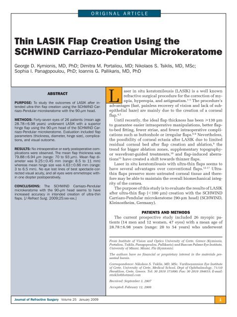
ORIGINAL ARTICLE<br />
<strong>Thin</strong> <strong>LASIK</strong> <strong>Flap</strong> <strong>Creation</strong> <strong>Using</strong> <strong>the</strong><br />
<strong>SCHWIND</strong> <strong>Carriazo</strong>-<strong>Pendular</strong> Microkeratome<br />
George D. Kymionis, MD, PhD; Dimitra M. Portaliou, MD; Nikolaos S. Tsiklis, MD, MSc;<br />
Sophia I. Panagopoulou, PhD; Ioannis G. Pallikaris, MD, PhD<br />
ABSTRACT<br />
PURPOSE: To study <strong>the</strong> outcomes of <strong>LASIK</strong> after intended<br />
ultra-thin fl ap creation using <strong>the</strong> <strong>SCHWIND</strong> <strong>Carriazo</strong>-<strong>Pendular</strong><br />
microkeratome with <strong>the</strong> 90-µm head.<br />
METHODS: Forty-seven eyes of 26 patients (mean age<br />
28.786.98 years) underwent <strong>LASIK</strong> with a superior<br />
hinge fl ap using <strong>the</strong> 90-µm head of <strong>the</strong> <strong>SCHWIND</strong> <strong>Carriazo</strong>-<strong>Pendular</strong><br />
microkeratome. Evaluation included fl ap<br />
parameters (thickness, diameter, hinge size), complications,<br />
and visual outcome.<br />
RESULTS: No intraoperative or early postoperative complications<br />
were observed. The mean fl ap thickness was<br />
79.886.94 µm (range: 70 to 93 µm). Mean fl ap diameter<br />
was 9.250.45 mm (range: 8.5 to 11 mm)<br />
whereas mean hinge size was 4.630.66 mm (range:<br />
3 to 6.5 mm). No eye lost lines of best spectacle-corrected<br />
visual acuity, and all eyes were emmetropic within<br />
one diopter postoperatively.<br />
CONCLUSIONS: The <strong>SCHWIND</strong> <strong>Carriazo</strong>-<strong>Pendular</strong><br />
microkeratome with <strong>the</strong> 90-µm head seems to have<br />
increased accuracy in intended creation of ultra-thin<br />
fl aps. [J Refract Surg. 2009;25:xxx-xxx.]<br />
L<br />
aser in situ keratomileusis (<strong>LASIK</strong>) is a well known<br />
refractive surgical procedure for <strong>the</strong> correction of myopia,<br />
hyperopia, and astigmatism. 1-3 The procedure’s<br />
advantages (fast, painless recovery of vision and lack of subepi<strong>the</strong>lial<br />
haze) are mainly due to <strong>the</strong> creation of a corneal<br />
flap. 4,5<br />
Until recently, <strong>the</strong> ideal flap thickness has been 130 µm<br />
to guarantee easier intraoperative manipulations, better flapto-bed<br />
fitting, fewer striae, and fewer intraoperative complications<br />
such as buttonhole or irregular flaps. 6-8 Never<strong>the</strong>less,<br />
<strong>the</strong> possibility of corneal ectasia after <strong>LASIK</strong> due to limited<br />
residual corneal bed after flap creation and ablation, 9 <strong>the</strong><br />
trend for bigger ablation zones, supplementary topographyor<br />
wavefront-guided treatments, 10 and flap-induced aberrations<br />
11 have created a shift towards thinner flaps.<br />
Laser in situ keratomileusis with ultra-thin flaps seems to<br />
have several advantages over conventional flaps. 12-17 Ultrathin<br />
flaps preserve more untreated corneal tissue and <strong>the</strong>refore<br />
may be able to maintain <strong>the</strong> overall biomechanical integrity<br />
of <strong>the</strong> cornea.<br />
The purpose of this study is to evaluate <strong>the</strong> results of <strong>LASIK</strong><br />
after ultra-thin flap (100 µm) creation with <strong>the</strong> <strong>SCHWIND</strong><br />
<strong>Carriazo</strong>-<strong>Pendular</strong> microkeratome (90-µm head) (<strong>SCHWIND</strong>,<br />
Kleinos<strong>the</strong>im, Germany).<br />
PATIENTS AND METHODS<br />
The current prospective study included 26 myopic patients<br />
(14 men and 12 women, 47 eyes) with a mean age of<br />
28.786.98 years (range: 20 to 54 years) who underwent<br />
From Institute of Vision and Optics University of Crete, Greece (Kymionis,<br />
Portaliou, Tsiklis, Panagopoulou, Pallikaris); and Bascom Palmer Eye Institute,<br />
University of Miami, Miami, Fla (Kymionis).<br />
The authors have no financial or proprietary interest in <strong>the</strong> materials presented<br />
herein.<br />
Correspondence: Nikolaos S. Tsiklis, MD, MSc, Vardinoyannion Eye Institute<br />
of Crete, University of Crete, Medical School, Dept of Ophthalmology, 71110<br />
Heraklion, Crete, Greece. Tel: 30 2810 371800; Fax: 30 2810 394653; E-mail:<br />
ntsiklis@hotmail.com<br />
Received: September 3, 2007<br />
Accepted: February 12, 2008<br />
Journal of Refractive Surgery Volume 25 January 2009<br />
1<br />
JRS0109KYMIONIS.indd 1<br />
12/4/2008 1:14:22 PM
Ultra-thin <strong>Flap</strong> <strong>LASIK</strong>/Kymionis et al<br />
<strong>LASIK</strong> with <strong>the</strong> <strong>SCHWIND</strong> <strong>Carriazo</strong>-<strong>Pendular</strong> microkeratome<br />
using a 90-µm head. Based on <strong>the</strong> keratometric<br />
measurements, rings 9 or 10 were used in 10<br />
and 16 patients, respectively. According to <strong>the</strong> manufacturer’s<br />
nomogram, in patients with keratometric<br />
values 42.00 diopters (D), ring 9 was used, and for<br />
values 42.00 D, ring 10 was used. The Table demonstrates<br />
patient refractive and demographic data. A<br />
written informed consent in accordance with <strong>the</strong> institutional<br />
guidelines and <strong>the</strong> Declaration of Helsinki<br />
was obtained by all participants.<br />
PREOPERATIVE MEASUREMENTS<br />
Preoperative examination included uncorrected visual<br />
acuity (UCVA) (decimal scale), best spectacle-corrected<br />
visual acuity (BSCVA) (decimal scale), cycloplegic<br />
refraction, slit-lamp examination with fundus<br />
evaluation, and corneal topography (C-Scan; Technomed,<br />
Baesweiler, Germany).<br />
Exclusion criteria were active anterior segment disease;<br />
residual, recurrent, or active ocular disease; and<br />
previous intraocular or corneal surgery, history of herpetic<br />
eye disease, corneal scarring, glaucoma, severe<br />
dry eye, and topographic evidence of keratoconus.<br />
SURGICAL TECHNIQUE<br />
Laser in situ keratomileusis procedures were performed<br />
in a standardized manner by <strong>the</strong> same surgeon<br />
(G.D.K.) using <strong>the</strong> ALLEGRETTO WAVE excimer laser<br />
(WaveLight Technologies, Erlangen, Germany).<br />
A drop of proparacaine hydrochloride 0.5% (Alcaine;<br />
Alcon Laboratories Inc, Ft Worth, Tex) was instilled in<br />
each eye 5 minutes before and in <strong>the</strong> beginning of <strong>the</strong><br />
procedure, followed by a povidone-iodine (Betadine;<br />
Lavipharm, Peania, Greece) preparation of <strong>the</strong> lids.<br />
2<br />
TABLE<br />
Demographic and Preoperative<br />
Refractive Data of 47 Eyes That<br />
Underwent <strong>LASIK</strong> With <strong>the</strong> <strong>SCHWIND</strong><br />
<strong>Carriazo</strong>-<strong>Pendular</strong> Microkeratome<br />
Demographic<br />
MeanStandard Deviation (Range)<br />
No. of eyes/patients 47/26<br />
Sex (male/female) 14/12<br />
Age (y) 28.786.98 (20 to 54)<br />
Corneal pachymetry (µm) 565.6327.10 (501 to 616)<br />
Steepest K (D) 43.461.56 (39.93 to 46.44)<br />
Sphere (D)<br />
4.572.66 (8.75 to 2.25)<br />
Cylinder (D)<br />
1.361.19 (4.75 to 0.25)<br />
Eyelashes were isolated by a drape, and a speculum<br />
with suction was placed in <strong>the</strong> operative eye. The cornea<br />
was marked with a corneal marker using gentian<br />
violet staining. The <strong>SCHWIND</strong> <strong>Carriazo</strong>-<strong>Pendular</strong> microkeratome<br />
with <strong>the</strong> 90-µm head was used in all cases<br />
to create a superior hinge. A new blade was used for<br />
each eye.<br />
<strong>Flap</strong> thickness was measured by performing intraoperative<br />
ultrasound pachymetry (DGH 5100; DGH Technology<br />
Inc, Exton, Pa) using <strong>the</strong> subtraction technique<br />
(central corneal thickness minus stromal bed thickness).<br />
All measurements were performed after inserting<br />
<strong>the</strong> speculum and drying <strong>the</strong> conjunctival fornices.<br />
Three measurements were obtained and averaged (all<br />
measurements had to be within 10 µm of one ano<strong>the</strong>r;<br />
o<strong>the</strong>rwise, a fourth measurement was obtained and <strong>the</strong><br />
three measurements within 10 µm were included for<br />
mean measurement calculation). After flap creation,<br />
any fluid present in <strong>the</strong> bed was wiped with a dry<br />
sponge prior to measuring stromal bed thickness. Three<br />
measurements were taken and averaged for <strong>the</strong> final<br />
recorded measurement. If <strong>the</strong>y varied by more than 10<br />
µm, a fourth measurement was taken (as <strong>the</strong> pre-keratectomy<br />
measurements). The difference between <strong>the</strong>se<br />
two mean measurement sets was recorded as <strong>the</strong> flap<br />
thickness. The flap was floated back into position after<br />
<strong>the</strong> ablation, and <strong>the</strong> stromal bed was irrigated with<br />
a balanced salt solution. <strong>Flap</strong> alignment was checked<br />
using gentian violet preoperative corneal markings and<br />
a striae test was performed to ensure proper flap adherence.<br />
The flap was <strong>the</strong>n allowed to dry for 2 minutes.<br />
A soft contact lens was applied in all eyes as a bandage<br />
and was removed on <strong>the</strong> first postoperative day.<br />
All patients were examined 60 minutes postoperatively<br />
to check flap adherence. They were given flurbiprofen<br />
sodium 0.03% drops (Ocuflur; Allergan, Irvine,<br />
Calif) four times daily for 2 days, dexamethasone 0.1%–<br />
tobramycin 0.3% drops (TobraDex, Alcon Laboratories<br />
Inc) four times daily for 2 weeks, and sodium hyaluronate<br />
0.18% drops (Vismed; TRB Chemedica, Newcastle<br />
under Lyme, United Kingdom) hourly for 1 month.<br />
POSTOPERATIVE FOLLOW-UP<br />
Patients were instructed to wear protective eye<br />
shields at night and to return <strong>the</strong> following day, on<br />
postoperative day 3, and 1 month after <strong>the</strong> procedure.<br />
RESULTS<br />
VISUAL OUTCOMES<br />
All eyes were emmetropic within 1.00 D at <strong>the</strong> 1-<br />
month postoperative examination (mean spherical<br />
equivalent refraction 0.250.48 D [range: 0.75 to<br />
journalofrefractivesurgery.com<br />
JRS0109KYMIONIS.indd 2<br />
12/4/2008 1:14:23 PM
Ultra-thin <strong>Flap</strong> <strong>LASIK</strong>/Kymionis et al<br />
0.50 D]). Mean UCVA significantly improved from<br />
counting fingers to 0.800.21 (range: 0.2 to 1.275) on<br />
<strong>the</strong> first postoperative day and 0.950.21 (range: 0.3 to<br />
1.27) on <strong>the</strong> third postoperative day. The mean UCVA<br />
at 1 month was 0.990.22 (range: 0.425 to 1.27) whereas<br />
mean BSCVA was 1.000.16 (range: 0.7 to 1.27). No<br />
eye lost lines of BSCVA.<br />
FLAP CHARACTERISTICS<br />
The mean flap thickness was 79.886.94 µm (range: 70<br />
to 93 µm). Mean flap diameter was 9.250.45 mm (range:<br />
8.5 to 11 mm), and mean hinge size was 4.630.66 mm<br />
(range: 3 to 6.5 mm).<br />
COMPLICATIONS AND ADVERSE EFFECTS<br />
No intraoperative complications occurred in this<br />
series. Few interface particles were observed on postoperative<br />
slit-lamp examination in four eyes whereas<br />
two eyes presented with microstriae. No surgical intervention<br />
was performed in <strong>the</strong>se eyes as <strong>the</strong> flap microstriae<br />
were out of <strong>the</strong> visual axis and did not affect<br />
UCVA or BSCVA or induced irregular astigmatism.<br />
No dislocations or corneal haze occurred. On <strong>the</strong> first<br />
postoperative day, one eye presented with diffuse lamellar<br />
keratitis stage I and was treated successfully<br />
with corticosteroids.<br />
Journal of Refractive Surgery Volume 25 January 2009<br />
DISCUSSION<br />
Laser in situ keratomileusis alters <strong>the</strong> organization<br />
of <strong>the</strong> collagen fibrils and <strong>the</strong> long-term structural integrity<br />
of <strong>the</strong> cornea. Normally, 300 to 500 lamella run<br />
from limbus to limbus with angular offsets; this orientation<br />
becomes increasingly dense in <strong>the</strong> anterior stroma<br />
where significantly more oblique branching and interweaving<br />
are noted. 18,19 Cutting <strong>the</strong> corneal lamellae<br />
during flap creation leads to biomechanical alterations<br />
that may predispose to cornea ectasia.<br />
<strong>Flap</strong> thickness is one of <strong>the</strong> most important parameters<br />
in <strong>LASIK</strong> surgery. Barraquer first proposed in<br />
1950 that 250 µm of residual corneal tissue is a safe<br />
limit for <strong>the</strong> long-term biomechanical stability of <strong>the</strong><br />
cornea. 20 Since <strong>the</strong>n, corneal ectasia after <strong>LASIK</strong> in patients<br />
with even 300 µm of untreated corneal tissue<br />
has been reported. 21,22 The need for higher attempted<br />
corrections with less risk of ectasia has led surgeons to<br />
prefer flaps thinner than 100 µm even though <strong>the</strong>y are<br />
difficult to manage and <strong>the</strong>refore might increase <strong>the</strong><br />
risk of flap striae and irregular astigmatism. 6-8<br />
The purpose of this study was to evaluate <strong>the</strong> results<br />
of ultra-thin flap <strong>LASIK</strong> with <strong>the</strong> <strong>SCHWIND</strong> <strong>Carriazo</strong>-<br />
<strong>Pendular</strong> microkeratome 90-µm head. A new sterilized<br />
head and blade were used for every eye, minimizing<br />
<strong>the</strong> probability of contamination by microbial pathogens<br />
and a possible thinner second cut or a buttonhole<br />
flap. 9 No intraoperative or early postoperative complications<br />
(needing additional surgical intervention) occurred.<br />
Sekundo et al 23 reported that placement of a<br />
bandage soft contact lens after <strong>LASIK</strong> does not exert<br />
any positive effect on microstriae and macrostriae incidence<br />
and worsens <strong>the</strong> first-day UCVA insignificantly.<br />
This study did not include patients with ultra-thin<br />
flaps. Due to previous flap dislocations after ultra-thin<br />
flap <strong>LASIK</strong>, a bandage contact lens was applied to all<br />
patients in this series during <strong>the</strong> first 24 hours. No flap<br />
dislocation was observed. Despite <strong>the</strong> complications<br />
(irregular astigmatism and striae) reported by o<strong>the</strong>r<br />
authors, 6-8 we did not observe such complications in<br />
this series (two eyes presented with microstriae that<br />
were out of <strong>the</strong> visual axis and did not affect UCVA or<br />
BSCVA or induced irregular astigmatism). The application<br />
of a bandage contact lens seems to decrease <strong>the</strong><br />
incidence of such complications after ultra-thin flap<br />
<strong>LASIK</strong>.<br />
An issue with thin flap creation is <strong>the</strong> long-term decrease<br />
of corneal thickness, which is estimated at approximately<br />
11 µm per year, which may lead to flap<br />
deterioration. 24 On <strong>the</strong> contrary, long-term studies<br />
after <strong>LASIK</strong> 3 demonstrated absence of late postoperative<br />
complications with stable visual performance.<br />
The deeper corneal stromal layers showed normal keratocytes,<br />
which indicated decreased wound healing<br />
response probably due to thin flap creation.<br />
A few potential limitations are apparent in this<br />
study such as <strong>the</strong> small sample size of treated eyes,<br />
limited follow-up, lack of a comparative group (o<strong>the</strong>r<br />
mechanical microkeratome or femtosecond group),<br />
and absence of multiple peripheral corneal thickness<br />
measurements with anterior segment optical coherence<br />
tomography (OCT). 25 Future studies should include<br />
evaluation of anterior segment OCT to characterize<br />
<strong>the</strong> uniformity and shape of <strong>the</strong> flaps, in addition to<br />
biomechanical studies to assess <strong>the</strong> viscoelasticity of<br />
<strong>the</strong> cornea after ultra-thin microkeratome flaps.<br />
REFERENCES<br />
1. Pallikaris IG, Papatzanaki ME, Stathi EZ, Frenschock O,<br />
Georgiadis A. Laser in situ keratomileusis. Lasers Surg Med.<br />
1990;10:463-468.<br />
2. Pallikaris IG, Papatzanaki ME, Siganos DS, Tsilimbaris MK. A<br />
corneal flap technique for laser in situ keratomileusis. Human<br />
studies. Arch Ophthalmol. 1991;109:1699-1702.<br />
3. Kymionis GD, Tsiklis NS, Astyrakakis N, Pallikaris AI, Panagopoulou<br />
SI, Pallikaris IG. Eleven-year follow-up of laser in situ<br />
keratomileusis. J Cataract Refract Surg. 2007;33:191-196.<br />
4. Tahzib NG, Bootsma SJ, Eggink FA, Nabar VA, Nuijts RM. Functional<br />
outcomes and patient satisfaction after laser in situ keratomileusis<br />
for correction of myopia. J Cataract Refract Surg.<br />
2005;31:1943-1951.<br />
3<br />
JRS0109KYMIONIS.indd 3<br />
12/4/2008 1:14:23 PM
Ultra-thin <strong>Flap</strong> <strong>LASIK</strong>/Kymionis et al<br />
5. Solomon KD, Fernández de Castro LE, Sandoval HP, Bartholomew<br />
LR, Vroman DT. Refractive surgery survey 2003.<br />
J Cataract Refract Surg. 2004;30:1556-1569.<br />
6. Jacobs JM, Taravella MJ. Incidence of intraoperative flap complications<br />
in laser in situ keratomileusis. J Cataract Refract Surg.<br />
2002;28:23-28.<br />
7. Tham VM, Maloney RK. Microkeratome complications of laser<br />
in situ keratomileusis. Ophthalmology. 2000;107:920-924.<br />
8. Lin RT, Maloney RK. <strong>Flap</strong> complications associated with lamellar<br />
refractive surgery. Am J Ophthalmol. 1999;127:129-136.<br />
9. Pallikaris IG, Kymionis GD, Astyrakakis N. Corneal ectasia induced<br />
by laser in situ keratomileusis. J Cataract Refract Surg.<br />
2001;27:1796-1802.<br />
10. Kymionis GD, Panagopoulou SI, Aslanides IM, Plainis S, Astyrakakis<br />
N, Pallikaris IG. Topographically supported customized<br />
ablation for <strong>the</strong> management of decentered laser in situ<br />
keratomileusis. Am J Ophthalmol. 2004;137:806-811.<br />
11. Pallikaris IG, Kymionis GD, Panagopoulou SI, Siganos CS, Theodorakis<br />
MA, Pallikaris AI. Induced optical aberrations following<br />
<strong>the</strong> formation of a laser in situ keratomileusis flap. J Cataract<br />
Refract Surg. 2002;28:1737-1741.<br />
12. Lin RT, Lu S, Wang LL, Kim ES, Bradley J. Safety of laser in<br />
situ keratomileusis performed under ultra-thin corneal flaps.<br />
J Refract Surg. 2003;19:S231-S236.<br />
13. Cobo-Soriano R, Calvo MA, Beltrán J, Llovet FL, Baviera J. <strong>Thin</strong><br />
flap laser in situ keratomileusis: analysis of contrast sensitivity,<br />
visual, and refractive outcomes. J Cataract Refract Surg.<br />
2005;31:1357-1365.<br />
14. Yeo HE, Song BJ. Clinical feature of unintended thin corneal flap<br />
in <strong>LASIK</strong>: 1-year follow-up. Korean J Ophthalmol. 2002;16:63-69.<br />
15. Vesaluoma M, Pérez-Santonja J, Petroll WM, Linna T, Alió J,<br />
Tervo T. Corneal stromal changes induced by myopic <strong>LASIK</strong>.<br />
Invest Ophthalmol Vis Sci. 2000;41:369-376.<br />
16. Aslanides IM, Tsiklis NS, Astyrakakis NI, Pallikaris IG, Jankov<br />
MR. <strong>LASIK</strong> flap characteristics using <strong>the</strong> Moria M2 microkeratome<br />
with <strong>the</strong> 90-micron single use head. J Refract Surg.<br />
2007;23:45-49.<br />
17. Kymionis GD, Tsiklis N, Pallikaris AI, Diakonis V, Hatzithanasis<br />
G, Kavroulaki D, Jankov M, Pallikaris IG. Long-term results<br />
of superficial laser in situ keratomileusis after ultrathin flap creation.<br />
J Cataract Refract Surg. 2006;32:1276-1280.<br />
18. Komai Y, Ushiki T. The three-dimensional organization of collagen<br />
fibrils in <strong>the</strong> human cornea and sclera. Invest Ophthalmol<br />
Vis Sci. 1991;32:2244-2258.<br />
19. Radner W, Zehetmayer M, Aufreiter R, Mallinger R. Interlacing<br />
and cross-angle distribution of collagen lamellae in <strong>the</strong> human<br />
cornea. Cornea. 1998;17:537-543.<br />
20. Barraquer JI. Queratomileusis y Queratofaquia. Bogota, Columbia:<br />
Instituto Barraquer de America; 1980:340-342, 405-406.<br />
21. Rabinowitz YS. Ectasia after laser in situ keratomileusis. Curr<br />
Opin Ophthalmol. 2006;17:421-426.<br />
22. Seiler T, Koufala K, Richter G. Iatrogenic keratectasia after laser<br />
in situ keratomileusis. J Refract Surg. 1998;14:312-317.<br />
23. Sekundo W, Dick HB, Meyer CH. Benefits and side effects of<br />
bandage soft contact lens application after <strong>LASIK</strong>: a prospective<br />
randomized study. Ophthalmology. 2005;112:2180-2183.<br />
24. Patel SV, Erie JC, McLaren JW, Bourne WM. Confocal microscopy<br />
changes in epi<strong>the</strong>lial and stromal thickness up to 7<br />
years after <strong>LASIK</strong> and photorefractive keratectomy for myopia.<br />
J Refract Surg. 2007;23:385-392.<br />
25. Stahl JE, Durrie DS, Schwendeman FJ, Boghossian AJ. Anterior<br />
OCT analysis of thin IntraLase femtosecond flaps. J Refract<br />
Surg. 2007;23:555-558.<br />
AUTHOR QUERIES<br />
The title was changed per Dr Waring. Okay as edited?<br />
Page 2, left column: Which ring was used in patients with a keratometric value of exactly 42.00 D (ie, ...>42<br />
ring 9,...


