Acta Medica Martiniana - Univerzita Komenského
Acta Medica Martiniana - Univerzita Komenského
Acta Medica Martiniana - Univerzita Komenského

Create successful ePaper yourself
Turn your PDF publications into a flip-book with our unique Google optimized e-Paper software.
A C T A M E D I C A M A R T I N I A N A 2 0 1 1 1 1 / 1 23<br />
biotin (30 minutes) (Universal detection kit LSAB+KIT/HRP, Dako). Next, the sections were<br />
visualized with DAB (3.3’- diaminobenzidine tetrahydrochloride) at a concentration of 0.5 mg/<br />
ml in Tris buffer, pH 7.6 and 0.015 % H2O2. Slides were stream-rinsed with tap water, counterstained<br />
with Mayer’s hematoxylin for 2 minutes, washed in tap water, dried, mounted and<br />
coverslipped. Sections processed with omission of primary antibody served as control.<br />
Semiquantitative evaluation<br />
Immunostaining was assessed by two independent observers blinded to animal<br />
characteristics.<br />
Expression of Pgp/MDR1 was evaluated separately using the following scale: 3+ =<br />
high level (91-100 % of positive cells), 2+ = medium level (11-90 % of positive cells), 1+ =<br />
low level (up to 10 % of positive cells), – = negative cells (0 % of positive cells). Samples<br />
with high [3+] and medium level [2+] of proteins expression were considered as positive.<br />
Samples scored as [1+] and [–] were considered as negative.<br />
RESULtS<br />
Kidney of rat cubs<br />
Using monoclonal antibody mouse anti-MDR1 clone C219 we have detected moderate<br />
immunopositivity for Pgp/MDR1 in all developmental stages (D1, D7, D14, D21). Employing<br />
semiquantitative evaluation of tissue samples we have found no differences in<br />
expression of Pgp/MDR1 in epithelial cells of proximal tubules. The remaining structures<br />
(epithelial cells of distal tubules, glomerulus and glomerular capsule) did not show any<br />
positivity for Pgp/MDR1.<br />
Using monoclonal antibody mouse anti-MDR1 UIC2 – clone C494 we have detected in all developmental<br />
stages the same spatial protein distribution but its signal was significantly weaker.<br />
Kidney of rat adults<br />
Using both monoclonal antibodies (mouse anti-MDR1:clone C219 and mouse anti-<br />
MDR1:UIC2 – clone C494) we have observed expression of Pgp/MDR1 in epithelial cells of<br />
proximal tubules in adult kidney, too. Employing semiquantitative evaluation we have found<br />
no differences in expression of Pgp/MDR1 in all tissue samples. The immunoreactivity for<br />
this protein was strongly restricted to the apical membrane and weakly to the cytoplasm of<br />
proximal tubular cells. No signal for this protein in the other cells of rat kidney was found.<br />
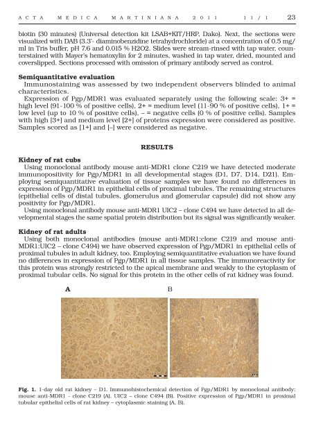
A<br />
B<br />
Fig. 1. 1-day old rat kidney – D1. Immunohistochemical detection of Pgp/MDR1 by monoclonal antibody:<br />
mouse anti-MDR1 - clone C219 (A), UIC2 – clone C494 (B). Positive expression of Pgp/MDR1 in proximal<br />
tubular epithelial cells of rat kidney – cytoplasmic staining (A, B).