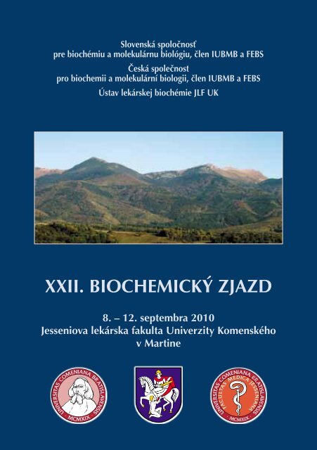
XXII. BIOCHEMICKÝ ZJAZD - Jesseniova lekárska fakulta
XXII. BIOCHEMICKÝ ZJAZD - Jesseniova lekárska fakulta
XXII. BIOCHEMICKÝ ZJAZD - Jesseniova lekárska fakulta

You also want an ePaper? Increase the reach of your titles
YUMPU automatically turns print PDFs into web optimized ePapers that Google loves.
Slovenská spoločnosť<br />
pre biochémiu a molekulárnu biológiu, člen IUBMB a FEBS<br />
Česká společnost<br />
pro biochemii a molekulární biologii, člen IUBMB a FEBS<br />
Ústav lekárskej biochémie JLF UK<br />
<strong>XXII</strong>. <strong>BIOCHEMICKÝ</strong> <strong>ZJAZD</strong><br />
8. – 12. septembra 2010<br />
<strong>Jesseniova</strong> <strong>lekárska</strong> <strong>fakulta</strong> Univerzity Komenského<br />
v Martine
Proceedings<br />
from <strong>XXII</strong>. Biochemistry Congress<br />
held in Martin September 8 – 12, 2010<br />
Chairman:<br />
Vicechairman:<br />
Members:<br />
Honorary advisory board<br />
J. Turňa<br />
V. Pačes<br />
D. Dobrota, A. Hrnčiar, D. Mištuna, J. Pastorek<br />
Program Committee<br />
I. Barák A. Breier A. Drgová Z. Ďuračková<br />
P. Griač P. Kaplán J. Korduláková J. Kormanec<br />
O. Križanová Ľ. Lacinová J. Lehotský M. Mareková<br />
K. Mikušová P. Račay S. Stuchlík Z. Sulová<br />
Ľ. Škultéty J. Turňa Ľ. Varečka<br />
Organizing Committee<br />
E. Babušíková M. Bittšanský D. Dobrota A. Drgová<br />
A. Evinová J. Hatok M. Chomová J. Jurečeková<br />
P. Kaplán R. Kirschnerová M. Kovalská S. Kuka<br />
J. Lehotský L. Letková T. Matáková M. Pavlíková<br />
P. Račay M. K. Sivoňová A. Štefaníková Z. Tatarková<br />
Edited by: E. Babušíková, D. Dobrota, J. Hatok, J. Lehotský<br />
ISBN 978-80-88866-83-1<br />
© Comenius University in Bratislava, Jessenius Faculty of Medicine in Martin,<br />
Department of Medical Biochemistry<br />
<strong>XXII</strong>. Biochemistry Congress, Martin<br />
1
<strong>XXII</strong>. Biochemistry Congress is supported by:<br />
MaIN SPONSOrs<br />
BIOMEDICA SLOVAKIA s.r.o.<br />
BIOTECH s.r.o.<br />
ROCHE SLOVENSKO s.r.o.<br />
KRD molecular technologies s.r.o.<br />
K-TRADE<br />
LAMBDA LIFE a.s.<br />
MERCK spol. s r.o.<br />
SHIMADZU SLOVAKIA o.z.<br />
SIEMENS s.r.o.<br />
SIGMA-ALDRICH®<br />
TRIGON s.r.o.<br />
SPONSOrs<br />
Beckman Coulter<br />
Bio-Rad<br />
E-Colli s.r.o.<br />
Ecomed<br />
Eppendorf Czech & Slovakia s.r.o.<br />
Fermentas<br />
GeneTiCA s.r.o.<br />
Hermes LabSystems Ltd.<br />
Merci Slovakia s.r.o.<br />
Mettler-Toledo s.r.o.<br />
MGP spol s r.o.<br />
Millipore<br />
Randox s.r.o.<br />
Scintila s.r.o.<br />
THANK YOU.<br />
2 <strong>XXII</strong>. Biochemistry Congress, Martin
TOPICS<br />
I. BIOCHEMISTRy AND MOLECULAR BIOLOGy OF NERVOUS sySTEM<br />
II.<br />
III.<br />
IV.<br />
BIOTECHNOLOGY<br />
BIOINFORMATICS<br />
GENOMICS<br />
V. CELL REGULATIONS aND SIGNAL TRANSDUCTION<br />
VI.<br />
VII.<br />
GLYCOMICS<br />
mEMBRANE BIOCHEMISTRY AND BIOENERGETICS<br />
VIII. NEW METHODOLOGIC PROCEDURES<br />
IX.<br />
PATHOBIOCHEMISTRY AND TRANSLATIONAL MEDICINE<br />
X. PROTEOMICS AND ENZYMOLOGY<br />
XI.<br />
XII.<br />
REACTIVE SPECIES IN BIOMEDICINE<br />
TEACHING OF BIOCHEMISTRY AND MOLECULAR BIOLOGY<br />
XIII. XENOBIOCHEMISTRY<br />
<strong>XXII</strong>. Biochemistry Congress, Martin<br />
3
Wednesday, 8 September 2010<br />
14:00 - 18:00 Registration<br />
CC<br />
18:00 - 23:00<br />
WELCOME RECEPTION<br />
CC<br />
Thursday, 9 September 2010<br />
8:15 - 8:45 OPENING CEREMONY<br />
CC<br />
8:45 - 9:00 WINNER'S CEREMONY CC<br />
9:00 - 9:45<br />
PLENARY LECTURE I.<br />
CC<br />
9:45 - 10:15<br />
COFFEE BREAK<br />
10:15 - 11:55<br />
11:55 - 12:15<br />
12:15 - 12:40<br />
12:40 - 13:45<br />
13:45 - 14:10<br />
14:10 - 15:25<br />
15:25 - 15:45<br />
R<br />
e<br />
g<br />
i<br />
s<br />
t<br />
r<br />
a<br />
t<br />
i<br />
o<br />
n<br />
Cell regulations and signal transduction<br />
CC<br />
Beckman lecture – cell biology<br />
Sigma lecture – biochemistry/biology<br />
Commercial lecture - Biotech<br />
CC<br />
CC<br />
CC<br />
Reactive species in biomedicine<br />
LUNCH & POSTER VIEWING V, VI, VII, XI<br />
Membrane biochemistry and bioenergetics I.<br />
CC<br />
PLENARY LECTURE II.<br />
COFFEE BREAK<br />
Glycomics<br />
A<br />
A<br />
15:45 - 16:35<br />
16:35 - 17:00<br />
CC<br />
Membrane biochemistry and bioenergetics I.<br />
CC<br />
Glycomics<br />
A<br />
17:00 - 21:15<br />
19:00 - 21:00<br />
Museum<br />
Slovak chamber theatre<br />
Aquapark Turčianské Teplice<br />
Friday, 10 September 2010<br />
8:30 - 9:15 PLENARY LECTURE III.<br />
CC<br />
9:15 - 9:35<br />
9:35 - 11:15<br />
11:15 - 11:40<br />
11:40 - 12:00<br />
12:00 - 13:00<br />
13:00 - 13:45<br />
13:45 - 14:10<br />
R<br />
e<br />
g<br />
i<br />
s<br />
t<br />
r<br />
a<br />
t<br />
i<br />
o<br />
n<br />
Pathobiochemistry and translational<br />
medicine I.<br />
Commercial lecture - KRD<br />
LUNCH<br />
COFFEE BREAK<br />
CC<br />
CC<br />
POSTER VIEWING II, III, IV, IX, XIII<br />
PLENARY LECTURE IV.<br />
Applied molecular biology<br />
Beckman lecture - Genomics A<br />
A<br />
KRD workshop A<br />
14:10 - 15:10<br />
15:10 - 15:30<br />
Xenobiochemistry<br />
Membrane biochemistry and bioenergetics II.<br />
CC<br />
A<br />
COFFEE BREAK<br />
15:30 - 16:30<br />
CC<br />
Xenobiochemistry<br />
CC<br />
Membrane biochemistry and bioenergetics II.<br />
A<br />
17:00 - 18:00<br />
18:00 - 19:00<br />
19:00 - 20:00<br />
Concert: Cantica Collegium Musicum<br />
4 <strong>XXII</strong>. Biochemistry Congress, Martin
10:15 - 10:35<br />
10:35 9:15 --10:15<br />
11:35<br />
10:15 11:35 - 10:35 12:00<br />
10:35 11:35<br />
12:00 9:15 --10:15<br />
13:00<br />
10:15 11:35 - 10:35 12:00<br />
13:00 - 14:00<br />
10:35<br />
12:00<br />
- 11:35<br />
13:00<br />
14:00 - 14:55<br />
11:35 - 12:00<br />
13:00 14:00<br />
14:55 - 15:15<br />
12:00 13:00<br />
14:00 - 14:55<br />
13:00 15:15 - 14:00 15:55<br />
14:55 - 15:15<br />
14:00 15:55 - 14:55 16:35<br />
15:15 - 15:55<br />
14:55 16:45 - 15:15 17:00<br />
15:55 - 16:35<br />
15:15<br />
17:00 -<br />
15:55<br />
18:00<br />
16:45 17:00<br />
15:55 16:35<br />
17:00<br />
18:00 -<br />
18:00<br />
22:00<br />
16:45 - 17:00<br />
18:00 - 22:00<br />
17:00 - 18:00<br />
18:00 - 22:00<br />
g<br />
i<br />
s<br />
t<br />
Rr<br />
ea<br />
gt<br />
i<br />
so<br />
t<br />
rR<br />
n<br />
ae<br />
CC<br />
tg<br />
i<br />
os<br />
nt<br />
rCC<br />
a<br />
t<br />
i<br />
o<br />
n<br />
CC<br />
Saturday, 11 September COFFEE 2010 BREAK<br />
Proteomics<br />
Proteomics<br />
and<br />
and<br />
enzymology<br />
enzymology<br />
CC<br />
Teaching of biochemistry and<br />
Beckman lecture Saturday, - proteomics<br />
11 September COFFEE 2010 BREAK molecular biology<br />
CC<br />
A<br />
Proteomics<br />
Proteomics<br />
and<br />
and<br />
enzymology<br />
enzymology LUNCH<br />
CC Teaching of biochemistry and<br />
Beckman lecture - proteomics COFFEE molecular biology<br />
CC BREAK<br />
A<br />
POSTER VIEWING I, VIII, X, XII<br />
Proteomics and enzymology<br />
LUNCH<br />
CC<br />
Pathobiochemistry and translational Biochemistry Teaching of and biochemistry molecular biology and<br />
Beckman lecture medicine - II. proteomics CC<br />
molecular of nervous biology system A<br />
POSTER VIEWING CC I, VIII, X, XII<br />
A<br />
COFFEE<br />
Pathobiochemistry and translational LUNCH<br />
BREAK<br />
Biochemistry and molecular biology<br />
medicine II.<br />
CC<br />
of nervous system A<br />
Pathobiochemistry and translational<br />
medicine POSTER II. VIEWING CC I, VIII, X, XII<br />
COFFEE BREAK<br />
Biochemistry and molecular biology<br />
Pathobiochemistry and translational Biochemistry of nervous and molecular system biology<br />
Pathobiochemistry medicine and II. translational CC<br />
of nervous system<br />
A<br />
medicine II.<br />
CC Biochemistry and molecular biology<br />
Closing COFFEE ceremony BREAK of nervous system CC<br />
A<br />
Pathobiochemistry and translational<br />
medicine II.<br />
CC<br />
Closing ceremony<br />
Biochemistry and molecular biology<br />
CC<br />
of nervous system<br />
Farewell party<br />
A<br />
Teaching of biochemistry and molecular biology<br />
A<br />
Teaching of biochemistry and molecular biology<br />
A<br />
Closing ceremony<br />
CC<br />
Farewell party<br />
Farewell party<br />
Sunday, 12 September 2010<br />
8:00 - 12:00<br />
8:00 - 12:00<br />
13:00<br />
13:00<br />
8:00 - 12:00<br />
13:00<br />
Sunday, 12 September 2010<br />
Orava Castle<br />
Orava Castle<br />
Sunday, 12 September Departure 2010<br />
Departure<br />
Orava Castle<br />
Departure<br />
Šútovo waterfall<br />
Šútovo waterfall<br />
Šútovo waterfall<br />
<strong>XXII</strong>. Biochemistry Congress, Martin<br />
5
Program in details: Wednesday<br />
PROGraM IN DETAILS<br />
WeDNESDAY, 8 September 2010<br />
Jessenius Faculty of Medicine – Convention Centre<br />
14.00 - 18.00 REGISTRATION<br />
18.00 - 22.00 WELCOME rECEPTION<br />
6 <strong>XXII</strong>. Biochemistry Congress, Martin
Program in details: Thursday<br />
ThURSDAy, 9 September 2010<br />
Jessenius Faculty of Medicine – Convention Centre<br />
8.00 - 17.00 REGISTRATION<br />
8.15 - 8.45 Opening Ceremony<br />
8.45 - 9.00 WINNER´S CEREMONY<br />
9.00 - 9.45 PLENarY LECTURE I.<br />
Chairs:<br />
Ján Turňa, Dušan Dobrota<br />
Mathias Sprinzl: ELECTROCHEMICAL DETECTION OF NUCLEIC ACIDS<br />
ON BIOSENSORS<br />
9.45 - 10.15 CoffeE BREAK<br />
10.15 - 12.40 CELL rEGULaTIONS aND SIGNal traNSDUCTION<br />
Chairs:<br />
Ján Kormanec, Imrich Barák<br />
10.15 - 10.35 Ján Kormanec, Renáta Nováková, Ľubica Fecková, Peter Kutaš,<br />
Alena Reháková: REGULATION OF AURICIN BIOSYNTHESIS IN<br />
STREPTOMYCES AUREFACIENS CCM 3239<br />
10.35 - 10.55 Imrich Barák, Katarína Muchová, Naďa Pavlendová, Ján Jamroškovič:<br />
LIPID HELICES FORMATION IN BACILLUS SUBTILIS CELL MEMBRANE<br />
10.55 - 11.15 Veronika Benson, Valeria Grobárová, Katarína Hulíková, Jan<br />
Svoboda, Daniel Rozbeský, Daniel Kavan, Alan Kádek, Karel Křenek,<br />
Anna Fišerová, Vladimír Křen, Karel Bezouška: HIGH AFFINITY<br />
CARBOHYDRATE AND NON-CARBOHYDRATE LIGANDS FOR LECTIN-<br />
TYPE ACTIVATION RECEPTORS OF NATURAL KILLER CELLS REGULATE<br />
EFFECTOR FUNCTION THROUGH PI3K PATHWAY, AND GENERATE<br />
PERMANENT IMMUNE PROTECTION AGAINST MELANOMAS<br />
11.15 - 11.35 Radim Černý, Elerin Kärner, Christian Unger, Mikael Wendel: OSTERIX<br />
OVER-EXPRESSION IN HUMAN EMBRYONIC STEM CELLS AND ITS<br />
EFFECT ON CELL DIFFERENCIATION<br />
<strong>XXII</strong>. Biochemistry Congress, Martin<br />
7
Program in details: Thursday<br />
11.35 - 11.55 Katarína Kršková, Daniela Ježová, Lucia Gajdošechová, Miroslava<br />
Eckertová, Štefan Zorad: THE ROLE OF ANGIOTENSIN II AND OXYTOCIN<br />
IN REGULATION OF ADIPOCYTE CELL SIZE<br />
LECTUre - BECKMan CZE: CELL BIOLOGY<br />
11.55 - 12.15 Jana Morová, Radim Osička, Jiří Mašín, Peter Šebo: RTX CYTOTOXINS<br />
RECOGNIZE β2 INTEGRIN RECEPTORS THROUGH N-LINKED<br />
OLIGOSACHARIDES<br />
LECTUre - SIGMA ALDRICH CZE<br />
12.15 - 12.30 Lucie Piterková, Jana Kučerová, Karel Indrák, Vladimír Divoký:<br />
INTRONIC LINE-1 INSERTION IN β–GLOBIN GENE CAUSE β–TALASEMIA<br />
DUE TO ABERRANT SPLICING, NONSENSE-MEDIATED DECAY AND<br />
DECREASED RATE OF β–GLOBIN L1<br />
ALLELE TRANSCRIPTION<br />
COMMERCIAL LECTURE - BIOTECH<br />
12.30 - 12.40 Martin Meluzín: MODERNÍ TRENDY ZOBRAZOVACÍCH METOD<br />
Jessenius Faculty of Medicine – Lecture Hall A<br />
10.15 - 12.40 rEaCTIve SPECIES IN BIOMEDICINE<br />
Chairs:<br />
Zdeňka Ďuračková, Peter Kaplán<br />
10.15 - 10.50 Karl-Heinz Wagner, Oliver Neubauer: HOW CAN OXIDATIVE STRESS<br />
AND DNA STABILITY BE INFLUENCED BY DIET AND PHYSICAL ACTIVITY<br />
10.50 - 11.15 Oľga Pecháňová, Andrej Barta, Stanislava Vranková, Jana Parohová,<br />
Mária Kovácsová: THE CROSS-TALK OF NITRIC OXIDE AND NUCLEAR<br />
factor kappa B in EXPERIMENTAL hypertension<br />
11.15 - 11.40 Jan Borovanský, Adéla Lipšová, Jiří Vachtenheim: FREE RADICAL<br />
SITUATION IN PIGMENT CELLS<br />
11.40 - 11.55 Jan Pláteník, Juraj Gáll, Jan Škrha, Jr., Richard Buchal, Eva Sedláčková,<br />
Karina Verébová: INDUCTION OF MITOCHONDRIAL PERMEABILITY<br />
TRANSITION BY MICROMOLAR IRON<br />
8 <strong>XXII</strong>. Biochemistry Congress, Martin
Program in details: Thursday<br />
11.55 - 12.10 Zuzana Tatarková, Eva Babušíková, Stanislav Kuka, Ján Lehotský,<br />
Peter Račay, Dušan Dobrota, Peter Kaplán: OXIDATIVE STRESS AND<br />
HEART AGING<br />
12.10 - 12.25 Eva Babušíková, Miloš Jeseňák, Peter Bánovčin, Dušan Dobrota:<br />
BRONCHIAL ASTHMA AND EFFECT OF OXIDATIVE STRESS ON ITS<br />
DEVELOPMENT<br />
12.25 - 12.40 Jana Muchová, Zuzana Nagyová, Iveta Ondrejovičová, Zdeňka<br />
Ďuračková: oxidative risk in atherosclerosis<br />
12.40 - 13.30 LUNCH<br />
13.00 - 13.45 POSTER VIEWING, Section V, VI, VII, XI (Convention Centre)<br />
Jessenius Faculty of Medicine – Convention Centre<br />
13.45 - 14.10 PLENarY LECTURE II: WINNEr of „DrOBNICOv MEMOriÁl“<br />
Chair:<br />
Albert Breier<br />
Mária Balážová, Peter Griač: IDENTIFICATION OF<br />
PHOSPHATIDYLGLYCEROL SPECIFIC PHOSPHOLIPASE C IN YEAST<br />
SACCHAROMYCES CEREVISIAE<br />
14.10 - 16.35 MEMBraNE BIOCHEMISTry aND BIOENErGETICS I.<br />
Chairs:<br />
Peter Griač, Ľubica Lacinová<br />
14.10 - 14.50 Anton Horváth, Ingrid Škodová, Anna Gnipová, Alena Zíková, Vladislava<br />
Benkovičová, Zdeněk Verner, Zdeněk Paris, Július Lukeš: SPECIALITIES<br />
OF OXIDATIVE PHOSPHORYLATION OF TRYPANOSOMATIDS AND<br />
EUGLENAS<br />
14.50 - 15.25 Peter Šmigáň, Monika Vidová, Janette Bobalova, Zuzana Nováková:<br />
ENERGETIC ASPECTS OF A MODIFICATION OF THE Na + /H +<br />
ANTIPORTER ACTIVITY IN A HARMALINE RESISTANT MUTANT OF<br />
METHANOTHERMOBACTER THERMAUTOTROPICUS<br />
15.25 - 15.45 Coffee BREAK<br />
<strong>XXII</strong>. Biochemistry Congress, Martin<br />
9
Program in details: Thursday<br />
15.45 - 16.00 Katarína Poloncová, Roman Holič, Peter Griač: IS<br />
PHOSPHATIDYLINOSITOL TRANSFER ACTIVITY ESSENTIAL FOR THE<br />
FUNCTION OF Pdr16p?<br />
16.00 - 16.15 Andrea Faltinová, Jana Gaburjáková, Ľubica Urbániková, Matúš<br />
Hajduk, Nataša Tomášková, Marián Antalík, Alexandra Zahradníková:<br />
EFFECT OF DOMAIN PEPTIDES OF THE CARDIAC ryANODINE RECEPtor<br />
ON THE STABILITy OF BILAYER LIPID MEMBRANES AND ON RyR2<br />
ACTIVITY<br />
16.15 - 16.35 Iveta Waczulíková, Oľga Uličná, Oľga Vančová, Jarmila Kucharská,<br />
Veronika Ilovská, Libuša Šikurová: ATORVASTATIN CHANGES<br />
MEMBRANE LIPID FLUIDITY IN MITOCHONDRIA ISOLATED FROM<br />
VARIOUS TISSUES OF RATS<br />
16.35 - 16.50 Roman Oros: ADVANCED TECHNOLOGY FOR METABOLIC<br />
INVESTIGATIONS<br />
Jessenius Faculty of Medicine – Lecture Hall A<br />
14.10 - 17.00 GLYCOMICS<br />
Chairs:<br />
Zdenka Sulová, Katarína Mikušová<br />
14.10 - 14.35 Igor Tvaroška: MOLECULAR MODELING INSIGHT INTO CATALYTIC<br />
MECHANISMS OF GLYCOSYLTRANSFERASES<br />
14.35 - 15.00 Marek Baráth, Igor Tvaroška, Ján Hirsch: SYNTHESIS OF GlcNAc-TS<br />
MIMETICS AS A POTENT INHIBITORS OF GLYCOSYLTRANSFERASES<br />
15.00 - 15.25 Michaela Wimmerová: LECTINS FROM PATHOGENS: MYSTERY OF<br />
LIFE<br />
15.25 - 15.45 Coffee BREAK<br />
15.45 - 16.10 Vladimír Farkaš: TRANSGLYCOSYLATION - A UNIVERSAL PRINCIPLE<br />
IN TAILORING THE PLANT AND FUNGAL CELL WALLS<br />
16.10 - 16.35 Mária Vršanská, Katarína Šuchová, Vladimír Puchart, Peter Biely:<br />
DIFFERENCIES BETWEEN TWO ENDOXYLANASES FROM GH5<br />
10 <strong>XXII</strong>. Biochemistry Congress, Martin
16.35 - 17.00 Zuzana Svetlíková, Marcelo E. Guerin, Mary Jackson, Jana Korduláková,<br />
Katarína Mikušová: MyCOBACTERIAL MANNOSyl TRANSFERASE PimA<br />
as A TARGET FOR the DEVELOPMENT of NEW ANTITUBERCULAR<br />
DRUGS<br />
16.45 - 21.15 SOCIal evENTS<br />
Program in details: Thursday<br />
17.30 - 18.30 Slovak National Museum<br />
16.45 - 21.15 Aquapark Turčianske Teplice<br />
19.00 - 21.00 Slovak Chamber Theatre, Martin<br />
<strong>XXII</strong>. Biochemistry Congress, Martin<br />
11
Program in details: Friday<br />
friday, 10 September 2010<br />
Jessenius Faculty of Medicine – Convention Centre<br />
8.30 - 9.15 PLENarY LECTURE III.<br />
Chairs:<br />
Ján Lehotský<br />
Peter Biely: MICROBIAL XYLANASES: PROPERTIES AND APPLICATIONS<br />
09.15 - 09.35 Coffee BREAK<br />
9.35 - 11.15 PaTHOBIOCHEMISTry aND traNSLaTIONal MEDICINE I.<br />
Chairs:<br />
Peter Račay, Oľga Križanová<br />
09.35 - 10.00 Juraj Kopáček, Jaromír Pastorek, Silvia Pastoreková: Molecular<br />
mechanisms involved in response to hypoxia<br />
10.00 - 10.25 Ľubomíra Lenčešová, Marta Sírová, Lucia Csáderová, Marcela Lauková,<br />
Zdena Sulová, Richard Kvetňanský, Oľga Križanová: CHANGES AND<br />
ROLE OF ADRENOCEPTORS IN PC12 CELLS AFTER PHENYLEPHRINE<br />
ADMINISTRATION AND APOPTOSIS INDUCTION<br />
10.25 - 10.50 Miroslav Barančík, Petra Šimončíková, Monika Ivanová: EFFECTS OF<br />
doxorubicin TREATMENT on MATRIX METALLOPROTEINASES IN<br />
RATS<br />
10.50 - 11.15 Attila Ziegelhöffer, Jana Mujkošová, Oľga Uličná, Iveta Waczulíková,<br />
Miroslav Ferko, Norbert Vrbjar, Štefan Polák, Tanya Ravingerová,<br />
Adriana Adameová: FUNCTION AND PROPERTIES OF HEART AND<br />
KIDNEY MITOCHONDRIA (MIT) IN SPONTANEOUSLY HYPERTENSIVE<br />
(HYP) RATS: INFLUENCE OF CAPTOPRIL AND NIFEDIPINE<br />
LECTUre - KRD<br />
11.40 - 11.50 Jiří Vašák: LIVING COLOURS: ILLUMINATE yOUR ASSAys WITH<br />
Clontech<br />
12 <strong>XXII</strong>. Biochemistry Congress, Martin
Program in details: Friday<br />
Jessenius Faculty of Medicine – Lecture Hall A<br />
9.35 - 12.00 aPPLIED MOLECULar BIOLOGY<br />
Chairs:<br />
Stanislav Stuchlík, Ján Turňa<br />
09.35 - 10.00 Peter Májek, Vladimír Špitalský, Gabriel Minárik, Tomáš Szemes:<br />
A METHOD FOR AUTOMATED DETECTION OF HETEROZYGOUS<br />
INSERTION-DELETION MUTATIONS<br />
10.00 - 10.25 Anna Hrabovska, Veronique Bernard, Eric Krejci: PROTEIN<br />
IMMUNIZATION OF MUTANT MOUSE – AN EFFICIENT WAY TO<br />
GENERATE SELECTIVE AND SENSITIVE ANTIBODIES<br />
10.25 - 10.50 Michal Kaliňák, Tibor Liptaj: NEW POSSIBILITIES FOR THE STUDY OF<br />
METABOLISM IN SLOVAKIA<br />
10.50 - 11.15 Martin Benej, Martina Poturnajová: TOXCAT METHOD: APPLICATION<br />
IN MOLECULAR ONCOLOGY<br />
11.15 - 11.40 Laco Kačáni: FROM BASIC BIOMEDICAL RESEARCH TO BIOTECHNOLOGy<br />
LECTUre - BECKMan CZE: GENOMICS<br />
11.40 - 12.00 Ondřej Mihola, Zdeněk Trachtulec, Jiří Forejt: The IDENTIFICATION<br />
and CHARACTERIZATION OF THE FIRST VERTEBRATE hyBRID STERILity<br />
gene (HST1/PRDM9)<br />
WorKSHOP<br />
12.00 - 13.00 KRD Molecular Technologies, s.r.o.<br />
12.00 - 13.00 LUNCH<br />
13.00 - 13.45 POSTER VIEWING, Sections II, III, IV,IX, XIII (Convention Centre)<br />
<strong>XXII</strong>. Biochemistry Congress, Martin<br />
13
Program in details: Friday<br />
Jessenius Faculty of Medicine – Convention Centre<br />
13.45 - 14.10 PLENaRY LECTURE Iv: WINNEr of „KoŠTířova CENA“<br />
Chair:<br />
Václav Pačes<br />
Dana Douděrová, Pavel Martásek: STRUCTURAL-FUNCTIONAL<br />
CORRELATIONS OF HYDROXYMETHYLBILANE SYNTHASE<br />
14.10 - 16.30 XENOBIOCHEMISTry<br />
Chairs:<br />
Albert Breier, Ľudovít Varečka<br />
14.10 - 14.30 Zdena Sulová, Mário Šereš, Miroslav Barančík, Lenka Gibalová,<br />
Branislav Uhrík, Lenka Poleková, Albert Breier: DOES EXISTS ANY<br />
RELATION BETWEEN P-GLYCOPROTEIN MEDIATED MULTIDRUG<br />
RESISTANCE AND INTRACELLULAR CALCIUM HOMEOSTASIS<br />
14.30 - 14.50 Pavlína Janů, Markéta Thimová, Petra Lovecká, Martina Macková,<br />
Kateřina Demnerová: EVALUATION OF TOXICITY AND GENOTOXICITY<br />
OF ORGANOHALOGEN PESTICIDES<br />
14.50 - 15.10 Monika Kmeťová Sivoňová, Dušan Dobrota, Tatiana Matáková, Zuzana<br />
Tatarková, Mária Kovalská, Martina Pavlíková, Róbert Dušenka, Ján<br />
Kliment: XENOBIOTIC-METABOLIZING ENZYMES POLYMORPHISMS<br />
AND CANCER RISK<br />
15.10 - 15.30 COFFEE BREAK<br />
15.30 - 15.50 Helena Mertlíková-Kaiserová, Antonín Holý, Ivan Votruba: Origin<br />
of ACqUIRED RESISTANCE TO cyTOTOXIC ACyCLIC NUCLEOSIDE<br />
phosphonates<br />
15.50 - 16.10 Marie Stiborová, Eva Frei: CYTOCHROME P450- AND PEROXIDASE-<br />
MEDIATED OXIDATION OF ELLIPTICINE DICTATES ITS ANTI-TUMOR<br />
EFFICIENCY<br />
16.10 - 16.30 Vladimíra Tomečková: SYNTHETIC CYCLIC CHALCONE ANALOGUES<br />
AS NOVEL BIOLOGICALLY ACTIVE DYES<br />
14 <strong>XXII</strong>. Biochemistry Congress, Martin
Program in details: Friday<br />
Jessenius Faculty of Medicine – Lecture Hall A<br />
14.10 - 16.10 MEMBraNE BIOCHEMISTry aND BIOENErGETICS II.<br />
Chairs:<br />
Peter Griač, Ľubica Lacinová<br />
14.10 - 14.50 Milan Štengl, František Barták, Roman Sýkora, Jiří Chvojka, Jan<br />
Beneš, Aleš Kroužecký, Ivan Novák, Jitka Švíglerová, Jitka Kuncová,<br />
Martin Matějovič: CARDIAC L-TYPE CALCIUM CURRENT IN SEPSIS<br />
14.50 - 15.05 Viera Kominková, Zuzana Tomášková, Ľubica Máleková, Karol<br />
Ondriaš: EFFECT OF ADENINE NUCLEOTIDES AND Mg 2+ IONS ON<br />
mitochondrial chloride channels<br />
15.05 - 15.30 COFFEE BREAK<br />
15.30 - 16.00 Ľubica Lacinová, Mária Karmažínová: GATING OF THE T-TYPE CALCIUM<br />
CHANNELS<br />
16.00 - 16.15 Mária Karmažínová, Edward Perez-Reyes, Ľubica Lacinová: GATING<br />
OF THE NEURONAL CA V<br />
3.3 CHANNEL<br />
19.00 - 20.00 CONCERT: CanTICa COLLEGIUM MUSICUM (EVANGELIC CHURCH)<br />
20.30 - 21.30 Biznis meeting: Members of committees Slovak and Czech biochemical<br />
societes<br />
<strong>XXII</strong>. Biochemistry Congress, Martin<br />
15
Program in details: Saturday<br />
SATURDAY, 11 September 2010<br />
Jessenius Faculty of Medicine – Convention Centre<br />
9.15 - 12.00 PrOTEOMICS aND ENZYMOLOGY<br />
Chairs:<br />
Ľudovít Škultéty, Peter Račay<br />
09.15 - 09.35 Daniela Krajčíková, Denisa Mullerová, Wan Qiang, Per Bullogh, Jilin<br />
Tang, Imrich Barák: ASSEMBLY OF BACILLUS SUBTILIS SPORE COAT:<br />
INVESTIGATION OF PROTEIN-PROTEIN INTERACTIONS AMONG THE<br />
SPORE COAT PROTEINS OF BACILLUS SUBTILIS<br />
09.35 - 09.55 Katarína Bíliková, Jozef Šimúth: PROTEOMICS OF MULTIFUNCTIONAL<br />
ROYAL JELLY PROTEINS<br />
09.55 - 10.15 Andrea Antošová, Katarína Šipošová, Martina Koneracká, Vlasta<br />
Závišová, Peter Kopčanský, Zuzana Gažová: INHIBITION OF INSULIN<br />
AMYLOID AGGREGATION WITH ALBUMIN FUNCTIONALIZED<br />
MAGNETIC FLUID<br />
10.15 - 10.35 COFFEE BREAK<br />
10.35 - 10.55 H. Mrazek, L. Weignerova, D. Manglova, D. Kavan , , V. Kren, K. Bezouska:<br />
FUNGAL α–N-ACETYLGALACTOSAMINIDASE FROM ASPERGILLUS<br />
NIGER: CLONING AND EXPRESSION IN YEAST<br />
10.55 - 11.15 Gabriela Flores Ramírez, Zuzana Bílková, Pavol Vadovič, Ľudovít<br />
Škultéty: IDENTIFICATION OF Surface PROTEINS OF THE OBLIGATE<br />
INTRACELLULAR bacterium CoxIELLA BURNETII<br />
11.15 - 11.35 Martin Hajduch, Katarína Klubicová, Maksym Danchenko, Ľudovít<br />
Škultéty, Namik Rashydov, Anna Preťová: TWENTy FOUR yEARS<br />
since Chernobyl DISASTER: WHAT seed PROTEIN can tell us?<br />
LECTUre - BECKMan CZE: prOTEOMICS<br />
11.35 - 12.00 Pavel Bouchal, Monika Mudrochová, Eva Budinská, Zbyněk Bortlíček,<br />
Iva Struhárová, Lenka Hernychová, Theodoros Roumeliotis, Spiros D.<br />
Garbis, Roman Hrstka, Petr Müller, Rudolf Nenutil, Bořivoj Vojtěšek:<br />
BIOMARKERS OF lyMPH NODE METASTASIS IN LOW-GRADE BREAST<br />
cancer: An integrated, proteomics-based approach<br />
16 <strong>XXII</strong>. Biochemistry Congress, Martin
Program in details: Saturday<br />
Jessenius Faculty of Medicine – Lecture Hall A<br />
9.15 - 12.00 TeaCHING of BIOCHEMISTry aND MOLECULar BIOLOGY<br />
Chairs:<br />
Anna Drgová, Mária Mareková, Jana Korduláková<br />
09.15 - 09.35 Ľubomír Tomáška: WHEN MORE IS LESS: A DILEMMA OF A BIOMEDIcal<br />
educator<br />
09.35 - 09.55 Zuzana Kostecká: TEACHING BIOCHEMISTRY AT THE UNIVERSITY OF<br />
VETERINARY MEDICINE AND PHARMACY IN KOŠICE<br />
09.55 - 10.15 Jiří Hudeček: DO WE TEACH BIOCHEMISTRY IN A LOGICAL WAY?<br />
REMARKS CONCERNING THE CONTENTS AND LEARNING APPROACH<br />
10.15 - 10.35 COFFEE BREAK<br />
10.35 - 10.55 Mária Kožurková, Marián Antalík, Dušan Podhradský: TEACHING<br />
BIOCHEMISTRY AT THE FACULTY OF SCIENCE IN KOŠICE<br />
10.55 - 11.15 Daniel Rajdl, Jaroslav Racek, Marie Šolcová: E-LEARNING – FRIEND<br />
OF FOE?<br />
11.15 - 11.35 Zdeňka Ďuračková, Zuzana Országhová: HISTORY AND PRESENT OF<br />
SCIENTIFIC AND PEDAGOGIC CONFERENCES OF TEACHERS FROM<br />
CHEMICAL INSTITUTES AND DEPARTMENTS OF SLOVAK AND CZECH<br />
MEDICAL FACULTIES<br />
11.35 - 11.55 Mária Mareková, Jana Mašlanková, Peter Urban, Juraj Guzy:<br />
BIOCHEMISTRY IN THE PICTURES – INTERACTIVE BIOCHEMISTRY<br />
12.00 - 13.00 LUNCH<br />
13.00 - 14.00 POSTER vIEWING, Sections: I, VIII, X (Convention Centre)<br />
<strong>XXII</strong>. Biochemistry Congress, Martin<br />
17
Program in details: Saturday<br />
Jessenius Faculty of Medicine – Convention Centre<br />
14.00 - 16.00 PaTHOBIOCHEMISTry aND traNSLaTIONal MEDICINE II.<br />
Chairs:<br />
Peter Račay, Oľga Križanová<br />
14.00 - 14.35 Nadežda Lukáčová, Alexandra Dávidová, Ľudmila Capková, Andrea<br />
Kucharíková: SPINAL CORD INJURY: PATHOGENESIS AND TREATMENT<br />
14.35 - 14.55 Peter Račay, Jozef Hatok, Mária Chomová, Jana Jurečeková, Peter<br />
Chudý, Juraj Chudej, Andrea Štefániková, Dušan Dobrota: APOPTOSIS<br />
– DOUBLE EDGED SWORD<br />
14.55 - 15.15 COFFEE BREAK<br />
15.15 - 15.35 Jozef Hatok, Jana Jurečeková, Peter Chudý, Pavol Hollý, Anton<br />
Dzian, Eduard Huľo, Eva Fabianová, Tatiana Matáková, Peter Račay:<br />
APOPTOSIS IN RELATION TO the DEVELOPMENT of CANCER AND<br />
RESISTANCE of cancer cells to cytostatics<br />
15.35 - 15.55 Lenka Surdeníková, Fei Ru, Marian Kollárik: UTILITY OF SINGLE<br />
CELL RT-PCR (scRT-PCR) FOR THE STUDY OF PRIMARY AFFERENT<br />
NEURONS - PRELIMINARY VALIDATION<br />
Jessenius Faculty of Medicine – Lecture Hall A<br />
14.00 - 16.00 BIOCHEMISTry aND MOLECULar BIOLOGY of NErvOUS<br />
system<br />
Chairs:<br />
Ján Lehotský, Dušan Dobrota<br />
14.00 - 14.35 Jozef Michalik, Egon Kurča: BIOCHEMICAL MARKERS OF MULTIPLE<br />
SCLEROSIS<br />
14.35 - 14.55 Eva Babušíková, Dušan Dobrota, Anthony J. Turner, Natalia N.<br />
Nalivaeva: PROCESSING OF AMYLOID PRECURSOR PROTEIN AFTER<br />
IN VIVO INDUCED ISCHEMIA<br />
14.55 - 15.15 COFFEE BREAK<br />
18 <strong>XXII</strong>. Biochemistry Congress, Martin
Program in details: Saturday<br />
15.15 - 15.35 Dušan Dobrota, Michal Bittšanský: MAGNETIC RESONANCE<br />
SPECTROSCOPY IN DIAGNOSTIC PROTOCOL OF THE BRAIN DISEASES<br />
15.35 - 15.55 Michal Bittšanský, Veronika Husárová, Igor Ondrejka, Valéria<br />
Kerná, Pavol Adamík, Huber Poláček, Dušan Dobrota: CHANGES<br />
IN NEURONAL METABOLITES MEASURED BY PROTON MAGNETIC<br />
RESONANCE SPECTROSCOPY IN DEPRESSED PATIENTS DURING<br />
TREATMENT<br />
15.55 - 16.15 Ján Lehotský, Mária Chomová, Andrea Evinová, Mária Kovalská,<br />
Martina Pavlíková, Zuzana Tatarková, Peter Kaplán, Peter Račay:<br />
INDUCTION OF ISCHEMIC TOLERANCE IN SENSITIVE NEURONS:<br />
coordinated role of multiple mechanisms<br />
16.15 - 16.35 Rostislav Skrabana, Radovan Dvorsky, Branislav Kovacech, Jozef<br />
Sevcik,Michal Novak: STRUCTURAL ANALySIS OF TAU PROTEIN, THE<br />
constituent of NEUROFIBRILLARy PATHOLOGy IN ALZHEIMER›s<br />
DISEASE<br />
16.45 - 17.00 CLOSING CEREMONY<br />
18.00 - 22.00 FAREWELL PARTy (Open-air Museum of Slovak Village, Martin)<br />
<strong>XXII</strong>. Biochemistry Congress, Martin<br />
19
Program in details: Sunday<br />
SUNDAY, 12 September 2010<br />
08.00 - 12.00 GUIDED TOUrs<br />
Orava Castle<br />
Šútovo waterfall<br />
13:00 DEPARTURE<br />
20 <strong>XXII</strong>. Biochemistry Congress, Martin
POSTER VIEWING<br />
<strong>XXII</strong>. Biochemistry Congress, Martin<br />
21
Program in details: Poster viewing<br />
V.<br />
POSTER vIEWING 1., SECTION V, VI, VII, XI<br />
THUrSDay, 9 SEPTEMBEr 2010,<br />
13.00 - 13.45<br />
29. Lucia Gajdošechová, Miroslava Eckertová, Katarína Kršková, Štefan Zorad: THE ROLE<br />
OF 14-3-3 PROTEIN IN REGULATION OF GLUCOSE TRANSPORTER GLUT4 TRANSLOCATION<br />
TO ADIPOCyTE PLASMA MEMBRANE<br />
30. Dana Grebeňová, Michaela Pluskalová, Zbyněk Hrkal, Kateřina Kuželová: CHANGES<br />
in COFILIN PHOSPHORyLATION DURING THE APOPTOSIS OF LEUKEMIC JURL-MK1 CELLS<br />
31. Dagmar Homerová, Bronislava Řežuchová, Henrieta Škovierová, Ján Kormanec:<br />
CHARACTERIZATION OF A GENE ENCODING A SMALL REGULATORy RPOE – DEPENDENT<br />
RNA IN SALMONELLA ENTERICA SEROVAR tyPHIMURIUM<br />
32. Iva Jelínková, Olga Vondálová Blanářová, Jiřina Hofmanová, Petr Sova, Alois Kozubík,<br />
Alena Vaculová: MOLECULAR MECHANISMS INVOLVED IN POTENT PLATINUM (IV) COMPLEXmediated<br />
COLON CANCER CELL SENSITIZATION TO TRAIL-INDUCED APOPTOSIS<br />
33. Lenka Kočí, Martina Hýžďalová, Alena Vaculová, Jiřina Hofmanová, Alois Kozubík:<br />
TRAIL-INDUCED APOPTOSIS CAUSES ACTIVATION OF PRO-SURVIVAL PATHWAys IN NON-<br />
ADHERENTLy GROWING COLON CANCER CELLS<br />
34. Gabriel Kollárovič, Miroslava Kretová, Lucia Lichá, Peter Baráth, Katarína Luciaková:<br />
PREPARATION AND FUNCTIONAL ANALySIS OF PHOSPHORyLATION MUTANT FORMS OF<br />
THE TRANSCRIPTION FACTOR NFI<br />
35. Soňa Kontseková, Anna Repič, Monika Baráthová, Katarína Polčicová, Jaromír Pastorek:<br />
GENERATION AND CHARACTERIZATION MONOCLONAL ANTIBODIES AGAINST ENDOSIALIN,<br />
the POTENTIAL MARKER OF TUMOR ANGIOGENESIS<br />
36. Miroslava Kretová, Ľudmila Šabová,Katarína Luciaková: THE ROLE OF NF1 IN p21 GENE<br />
expression<br />
37. Peter Kutaš, Ľubomíra Fecková, Alena Reháková, Renáta Nováková, Ján Kormanec:<br />
STRICT CONTROL OF AURICIN PRODUCTION IN STREPTOMyCES AUREOFACIES CCM 3239<br />
INVOLVES A FEEDBACK MECHANISM<br />
38. Ingrid Lajdová, Viera Spustová, Adrián Okša, Dušan Chorvát Jr.: ALTERED CALCIUM<br />
signaling IN PERIPHERAL BLOOD MONONUCLEAR CELLS OF CHRONIC KIDNEy DISEASE<br />
PATIENTS<br />
39. Ľubica Ondrušová, Jiří Vachtenheim: EXPRESSION OF MICROPHTHALMIA-ASSOCIATED<br />
TRANSCRIPTION FACTOR CRITICALLy REqUIRES ACTIVE SWI/SNF CHROMATIN REMODEL-<br />
ING COMPLEX<br />
22 <strong>XXII</strong>. Biochemistry Congress, Martin
Program in details: Poster viewing<br />
40. Barbora Brodská, Petra Otevřelová, Aleš Holoubek: MCL-1 AS REGULATOR OF APOPTOSIS<br />
in CML CELL LINE AND PERIPHERAL BLOOD MONONUCLEAR CELLS<br />
41. Jana Plšíková, Ján Kovaľ, Jaromír Mikeš, Mária Kožurková, Peter Fedoročko, Ladislav<br />
Janovec, Ján Ungvarský, Danica Sabolová: NOVEL GUANIDINE DERIVATIVES AND EVALUA-<br />
TION OF THEIR DNA BINDING AFFINITIES AND POSSIBLE ANTICANCER EFFECT<br />
42. Michaela Pluskalová, Dana Grebeňová, Zbyněk Hrkal, Kateřina Kuželová: HISTONE<br />
acetyLATION OF LEUKEMIC JURL-MK1 CELLS WITHIN SAHA TREATMENT<br />
43. Jiřina Procházková, Lenka Umannová, Alois Kozubík, Miroslav Machala, Jan Vondráček:<br />
REGULATION OF PLAKOGLOBIN EXPRESSION, A KEy DESMOSOMAL CONSTITUENT, by ARyl<br />
hyDROCARBON RECEPTOR AND cAMP SIGNALING<br />
44. Alena Reháková, Renáta Nováková, Ľubomíra Fecková, Peter Kutaš, Ján Kormanec:<br />
CHARAKTERIZATION OF SARP REGULATORy GENE INVOLVED IN POSITIVE REGULATION OF AN<br />
angucyCLINE-LIKE POLyKETIDE ANTIBIOTICS AURICIN GENE CLUSTER IN STREPTOMYCES<br />
aureofaciens CCM 3239<br />
45. Bronislava Řežuchová, Beatrica Ševčíková, Dagmar Homerová, Ján Kormanec: THE COMplex<br />
NETWORK REGULATORy CIRCUITS IN THE REGULATION OF SIGMA FACTORS INVOLVED<br />
in DIFFERENTIATION AND STRESS RESPONSE IN STREPTOMYCES COELICOLOR A3(2)<br />
VI.<br />
46. Tatiana Kurucová, Helena Kavcová, Kristína Rogozanová, Lucia Messingerová, Danica<br />
Mislovčová, Albert Breier, Zdena Sulová: DIFFERENCES IN INTERACTION OF LECTINS SPEcifically<br />
RECOGNIZING SIALIC ACID RESIDUES WITH SURFACE OF P-GP NEGATIVE OR<br />
positive L1210 CELLS<br />
47. Zdena Sulová, Peter Ditte, Tatiana Kurucová, Eva Poláková, Kristína Rogozanová Lucia<br />
Škvarková, Ján Sedlák, Jaromír Pastorek, Albert Breier: THE PRESENCE OF P-GLyCOPROTEIN<br />
in L1210 CELLS DIRECTLy INDUCES DOWN-REGULATION OF CELL SURFACE SACCHARIDE-<br />
TARGETS OF CONCANAVALIN A<br />
VII.<br />
48. Jana Antalíková, Jana Jankovičová, Katarína Michalková, Michal Simon, Ľubica Horovská:<br />
EPITOP OF IVA-520 MONOCLONAL ANTIBODy ON THE BOVINE SPERM CD46 MOLECULE<br />
49. Cagala M. , Lencesova L., Hudecova S., Csaderova L., Sirova M., Cholujova D., Kopacek<br />
J., Pastorekova S., Krizanova O.: DIMETHyLOXALLyl gyCINE MODULATES GENE EXPRESION<br />
and PROTEIN LEVELS OF THE SODIUM CALCIUM EXCHANGER IN HEK 293 CELL LINE<br />
<strong>XXII</strong>. Biochemistry Congress, Martin<br />
23
Program in details: Poster viewing<br />
50. Peter Kohút, Martin Valachovič, Lucia Hronská, Ivan Hapala: DEHyDROERGOSTEROL<br />
ELUCIDATES STEROL UPTAKE PROCESS IN yEAST S. CEREVISIAE<br />
51. Jan Madacki, Katarína Mikušová, Mary Jackson, Jana Korduláková: MyCOBACTERIAL<br />
EPOXIDEHyDROLASE EPHD IS INVOLVED IN FATTy ACID METABOLISM<br />
52. Boris Lakatoš, Lucia Bialešová, Eva Harnišová: ISOFORMS OF AMP-ACTIVATED PROTEIN<br />
kinase SUBUNITS IN lyMPHOCyTES AND OBESITy<br />
53. Katarína Michalková, Michal Simon, Jana Antalíková, Ľubica Horovská: DISTRIBUTION<br />
AND BIOCHEMICAL CHARACTERIZATION OF CD52-LIKE MOLECULE IN BULL EPIDIDyMIS<br />
54. Hana Rauchová, Martina Vokurková, ,<br />
Tomáš Soukup: IDEBENONE ACTIVATION OF<br />
glyCEROL-3-PHOSPHATE OXIDATION IN LIVER MITOCHONDRIA FROM CONTROL AND<br />
hyPERTHyROID RATS<br />
55. Oľga Uličná, Oľga Vančová, Jarmila Kucharská, Peter Božek, Iveta Waczulíková, Libuša<br />
Šikurová: EFFECT OF ATORVASTATIN ON BIOENERGETICS OF THE LIVER MITOCHONDRIA<br />
ON A HIGH LIPID DIET<br />
56. Oľga Vančová, Oľga Uličná, Katarína Šebeková, Magdalena Labieniec, Cezary Watala:<br />
EFFECT OF PAMAM G4 DENDRIMER ON LIVER MITOCHONDRIA OXIDATIVE PHOSPHORy-<br />
LATION AND LONG-TERM MARKERS OF hyPERGLyCAEMIA IN EXPERIMENTAL DIABETES<br />
57. Monika Vidová, Zuzana Nováková, Peter Šmigáň: BIOCHEMICAL AND MOLECULAR ANALySIS<br />
of NITRATE-RESISTANT MUTANT OF METHANOTHERMOBACTER THERMAUTOTROPHICUS<br />
XI.<br />
90. Lucia Andrezálová, Zuzana Országhová, Jana Muchová, Zdeňka Ďuračková: EFFECT OF<br />
OMEGA-3 FATTy ACIDS ON PON 1 ARyLESTERASE AND LACTONASE ACTIVITy IN CHILDREN<br />
SUFFERING FROM hyPERCHOLESTEROLEMIA<br />
91. Ima Dovinová, Zuzana Pakanová, Stanislava Vranková, Oľga Pecháňová, Soňa Čačányiová,<br />
František Kristek, Helena Paulíková: ANTIOXIDANT RESPONSE TO TREATMENT WITH NATUral<br />
COMPOUNDS AND AN NO DONOR IN EXPERIMENTAL hyPERTENSION<br />
92. Marián Koláček, Jana Muchová, Eva Uhlíková, Viera Kupčová, Ladislav Turecký: WILSON´s<br />
disease AND OXIDATIVE STRESS<br />
93. Stanislav Kuka, Zuzana Tatarková, Peter Račay, Ján Lehotský, Dušan Dobrota, Peter<br />
Kaplán: AGE-RELATED CHANGES IN ACTIVITIES OF MITOCHONDRIAL ELECTRON TRANSPORT<br />
chain COMPLEXES IN THE RAT HEART<br />
94. Daniela Mokrá, Anna Drgová, Rudolf Pullmann st., Andrea Čalkovská: SELECTIVE PHOSphodiesterase-3<br />
INHIBITOR OLPRINONE ALLEVIATES OXIDATIVE LUNG INJURy INDUCED<br />
by MECONIUM<br />
24 <strong>XXII</strong>. Biochemistry Congress, Martin
Program in details: Poster viewing<br />
95. Zuzana Országhová, Zuzana Paduchová, Ingrid Žitňanová, Cezary Watala, Jana Muchová,<br />
Zdeňka Ďuračková: METyLNICOTINAMIDE AND DNA OXIDATION DAMAGE IN RATS WITH<br />
STREPTOZOTOCINE INDUCED DIABETES MELLITUS<br />
96. Eliška Procházková, Petr Jansa, Lucie Čechová, Ivan Votruba, Helena Mertlíková-Kaiserová:<br />
ANTIOXIDANT CAPACITy OF SELECTED ANALOGS OF NUCLEIC ACID COMPONENTS: COM-<br />
PARISON OF CELL-FREE AND CELL-BASED ASSAys<br />
97. Beáta Veliká, Ivan Kron: EFFECTIVENESS OF PHENOLS AS ANTIOXIDANTS AGAINST<br />
superoxide RADICAL<br />
98. Martina Vokurková, Hana Rauchová, Stanislav Pavelka, Tomáš Soukup, Narcis Tribulová:<br />
EFFECT OF n-3 POLyUNSATURATED FATTy ACIDS SUPPLEMENTATION ON RAT LIVER IN<br />
different THyROID STATUS<br />
99. Adéla Zdařilová, Alena Rajnochová Svobodová, Jana Zapletalová, Pavel Štrebl, Josef<br />
Zadražil, Jitka Vostálová: EFFECT OF RENAL TRANSPLANTATION ON OXIDATIVE STRESSrelated<br />
BIOMARKERS<br />
POSTER VIEWING 2., SECTION II, III, IV,IX, XIII<br />
FrIDay, 10 SEPTEMBEr 2010,<br />
13.00 - 13.45<br />
II.<br />
8. Hind Al Alami, Ľubomíra Tóthová, Michal Kajsík, Jana Gajdošová, Hana Drahovská, Ján<br />
Turňa: CHARACTERIZATION OF BACTERIOPHAGES INFECTING CRONOBACTER STRAINS<br />
9. Andrea Balažová, Víťazoslava Blanáriková, Jindra Valentová, František Bilka, Ivana<br />
Holková: EFFECT OF METHyl JASMONATE ON THE PRODUCTION OF SANGUINARINE AND<br />
ITS PRECURSORS IN OPIUM POPPy SUSPENSION CULTURES<br />
10. František Bilka, Andrea Balažová, Andrea Bilková, Víťazoslava Blanáriková, Ivana Holková,<br />
Marián Vanko: COMPARISON OF SANGUINARINE PRODUCTION OF PAPAVERACEAE FAMILy<br />
plants IN VITRO CULTURES<br />
11. Michaela Kandričáková ,<br />
Stanislav Stuchlík, Ján Turňa: DESIGN OF AN EXPRESSION system<br />
FOR THE PRODUCTION OF RECOMBINANT HUMAN THROMBIN IN ESCHERICHIA COLI<br />
12. Tatiana Kraková, Jozef Šimúth, Katarína Bíliková: HETEROLOGOUS EXPRESSION OF<br />
royAL JELLy APALBUMINS IN E. COLI<br />
13. Mahesh Madyagol, Stanislav Stuchlík, Ján Turňa: EXPRESSION, PURIFICATION AND<br />
functional CHARACTERIZATION OF TWO FORMS OF AGROBACTERIUM SP. STRAIN CP4<br />
EPSPS GENE IN ESCHERICHIA COLI FOR HORIZONTAL GENE TRANSFER STUDIES<br />
<strong>XXII</strong>. Biochemistry Congress, Martin<br />
25
Program in details: Poster viewing<br />
14. Jozef Parnica, Lukáš Kandráč, Marián Antalík: CONFORMATION TRANSITIONS OF cy-<br />
TOCHROME c IN DEEP EUTECTIC SOLVENTS<br />
15. Lucia Pánčiová, Zdenko Levarski, Pavol Utekal, Stanislav Stuchlík, Ján Turňa: PRODUCTION<br />
of RECOMBINANT PROTEINS USING HIGH CELL DENSITy CULTURES OF ESCHERICHIA COLI<br />
in BIOREACTOR<br />
16. Michaela Šimšíková, Marián Antalík: SyNTHESIS AND SURFACE MODIFICATION OF ZINC<br />
oxide NANOPARTICLES<br />
17. Csaba Tóth, Roland Pálffy, Juraj Gašperík, Stanislav Stuchlík, Ján Turňa: CLONING, EX-<br />
PRESSION AND ANTIMICROBIAL ACTIVITy OF THE HUMAN CATHELICIDIN LL-37<br />
18. Ľubomíra Tóthová, Hind Al Alami, Jana Lintnerová, Hana Drahovská, Ján Turňa:<br />
CHARACTERIZATION OF BACTERIOPHAGES INFECTING SALMONELLA ENTERICA<br />
19. Pavol Utekal, Lucia Pánčiová, Stanislav Stuchlík, Ján Turňa: PRODUCTION OF TWO<br />
recombinant ALCOHOLDEHyDROGENASES SUITABLE FOR BIOTRANSFORMATION OF C-6<br />
aldehyDES INTO CORESPONDING ALCOHOLS<br />
20. Ondřej Vaněk, Petra Celadová, Jan Bláha, Daniel Kavan, Petr Pompach, Karel Bezouška:<br />
EUKARyOTIC EXPRESSION AS AN INDISPENSABLE TOOL FOR PREPARATION OF NATIVE<br />
dimeric FORMS OF NK CELL C-tyPE LECTIN-LIKE RECEPTORS<br />
III.<br />
21. Matej Stano, Ľuboš Kľučár: phiGENOME - A WEB-BASED GENOME BROWSER INTENDED<br />
for DISPLAy OF PHAGE GENOMES<br />
IV.<br />
22. Jarmila Farkašovská, Andrej Godány: SITE-SPECIFIC INTEGRATION OF BACTERIOPHAGE<br />
µ1/6 INTO THE STREPTOMYCES AUREOFACIENS CHROMOSOME<br />
23. Jana Gajdošová, Natália Kamodyová, Kristína Benedikovičová, Hana Drahovská,<br />
Eva Kaclíková, Ján Turňa: STUDy OF THERMOTOLERANCE ISLAND IN CRONOBACTER<br />
spp.<br />
24. Lucia Letková, Tatiana Matáková, Erika Halašová, Anna Drgová, Dušan Dobrota: DNA<br />
POLyMORPHISMS OF SELECTED REPAIR GENES AND RISK OF LUNG CANCER<br />
25. Eva Lincová/Slabáková, Zuzana Pernicová, Eva Slavíčková, Alois Kozubík, Karel Souček:<br />
EXPRESSION OF TRANSCRIPTION FACTORS AND microRNAs IN TGF-β1-INDUCED EMT OF<br />
BENIGN pROSTATE EPITHELIAL CELLS<br />
26 <strong>XXII</strong>. Biochemistry Congress, Martin
Program in details: Poster viewing<br />
26. Silvia Mahmood, Tatiana Matáková, Lucia Letková, Monika Kmeťová Sivoňová, Jozef<br />
Hatok, Dušan Dobrota: THE ASSOCIATION BETWEEN EGF 61 G/A POLYMORPHISM AND<br />
COLORECTAL CANCER DEVELOPMENT<br />
27. Milena Matejovičová, Michaela Králíková, Jitka Melounová, Martina Vodová, Jana Žáková,<br />
Igor Crha: SPERM DNA INTEGRITy ASSESMENT USING DIFFERENT COMET ASSAy PROTOCOLS<br />
28. Marika Matoušová, Ivan Votruba, Miroslav Otmar, Helena Mertlíková-Kaiserová:<br />
COMPARATIVE STUDy OF hyPOMETHyLATING ACTIVITIES OF 5-AZACyTIDINE CONGENERS<br />
IX.<br />
60. Soňa Bálentová, Eva Hajtmanová, Yvetta Mellová, Ivana Kinclová, Marián Adamkov:<br />
EFFECT OF FRACTIONATED DOSES OF GAMA RAys ON THE ROSTRAL MIGRATORy STREAM<br />
OF ADULTS RATS<br />
61. Ľudmila Capková, Alexandra Dávidová, Andrea Kucháriková, Nadežda Lukáčová: Is<br />
respiratory PATHWAy ACTING THROUGH NO-sGC?<br />
62. Monika Dvořáková, Jana Muchová, Branislav Trebatický, Ján Breza, Zdeňka Ďuračková:<br />
THE EFFECT OF NATURAL POLyPHENOLS ON ADIPONECTINE LEVEL IN PATIENTS SUFFERING<br />
from ERECTILE dySFUNCTION<br />
63. Jana Jurečeková, Jozef Hatok, Andrea Štefániková, Dušan Dobrota, Peter Račay: STUDy<br />
of ANTIAPOPTOTIC PROTEINS RESPONSIBLE FOR DEVELOPMENT OF DRUG RESISTANCE<br />
IN ACUTE LEUKEMIA<br />
64. Vlastimil Kulda, Martin Pešta, Ondřej Topolčan, Lukáš Řehoř, Martin Svatoň, Václav<br />
Liška, Václav Babuška, Luboš Holubec, Radim Černý: PROGNOSTIC SIGNIFICANCE OF MIR-<br />
21 AND MIR-143 EXPRESSION IN TISSUE SAMPLES OF COLORECTAL CARCINOMA AND<br />
colorectal LIVER METASTASES<br />
65. Erika Moravčíková, Evžen Křepela, Jan Prochádzka, Jan Čermák, Kamila Benková: THE<br />
functionality OF APOPTOSOME APPARATUS AND THE EXPRESSION OF ITS REGULATORS<br />
in NON-SMALL CELL LUNG CARCINOMA<br />
66. Iveta Ondrejovičová, Jana Muchová, Zuzana Paduchová, Zuzana Nagyová, Zdeňka<br />
Ďuračková: EFFECT OF OMEGA-3 PUFA ON LIPID PROFILE AND OXIDATIVE STRESS IN hy-<br />
PERCHOLESTEROLEMIC CHILDREN<br />
67. Blanka Stibůrková, Makoto Hosoyamada, Kimiyoshi Ichida, Ivan Šebesta: ANALySIS OF<br />
urate TRANSPORTERS SLC22A12 AND SLC2A9 IN PATIENTS WITH RENAL hyPOURICEMIA<br />
in CZECH POPULATION<br />
68. Andrea Štefániková, Jozef Hatok, Jana Jurečeková, Ivana Plameňová, Dušan Dobrota,<br />
Peter Račay: STUDy OF THE EFFECT OF HISTONE DEACETyLASE INHIBITOR ON THE SENSI-<br />
TIVITy OF LEUKAEMIC CELLS TO THE cyTOSTATICS<br />
<strong>XXII</strong>. Biochemistry Congress, Martin<br />
27
Program in details: Poster viewing<br />
69. Ladislav Vaško, Janka Vašková: CONTENT OF FATTy ACIDS IN FOOD AND HEALTH STATUS<br />
70. Janka Vašková, Ladislav Vaško: Effect of humic acids in vivo<br />
XIII.<br />
100. Hana Bártíková, Jana Firbasová, Ivan Vokřál, Lenka Skálová, Jiří Lamka, Vladimír<br />
Kubíček, Barbora Szotáková: BIOTRANSFORMATION OF SELECTED ANTHELMINTICS IN RAT<br />
tapeworm hyMENOLEPIS DIMINUTA<br />
101. Iva Boušová, Zuzana Průchová, Lucie Trnková, Jaroslav Dršata: INHIBITORy EFFECT OF<br />
natural FLAVONOIDS ON eqUINE LIVER GLUTATHIONE S-TRANSFERASE<br />
102. Lenka Gibalová, Ján Sedlák, Alena Reháková, Martina Labudová, Zdena Sulová,<br />
Albert Breier: MULTIDRUG RESISTANT P-GLyCOPROTEIN POSITIVE CELLS ARE ALSO CROSS-<br />
RESISTANT TO CISPLATIN<br />
103. Veronika Hanušová, Lenka Vildová, Věra Králová, Ladislava Schröterová, Lenka<br />
Trilecová, Alena Pakostová, Lenka Skálová: THE EFFECTIVENESS OF ORACIN IN ENHANC-<br />
ING THE cyTOTOXICITy OF DOXORUBICIN THROUGH THE INHIBITION OF DOXORUBICIN<br />
DEACTIVATION IN BREAST CANCER CELL LINE MCF-7<br />
104. Věra Kotrbová, Barbora Mrázová, Eva Frei, Marie Stiborová: CyTOCHROME b 5<br />
POTENtiates<br />
ACTIVITIES OF cyTOCHROMES P450 1A1 AND 1A2 TO OXIDIZE ANTICANCER DRUG<br />
ELLIPTICINE TO PHARMACOLOGICALLy EFFICIENT METABOLITES<br />
105. Věra Králová, Emil Rudolf: SELENITE-INDUCED CELL DEATH IN HUMAN COLON CAN-<br />
CER CELLS<br />
106. Jitka Křížková, Kamila Burdová, Petr Hodek, Marie Stiborová: EFFECTS OF FLAVONOIDS<br />
on cyTOCHROMES P450 AFTER PERORAL SINGLE DOSE ADMINISTRATION TO MALE RATS<br />
107. Tamara Lasotová, Hana Bártíková, Ivan Vokřál, Barbora Szotáková, Vladimír Kubíček,<br />
Jiří Lamka, Marián Várady, Lenka Skálová: ACTIVITIES OF DRUG-METABOLIZING AND AN-<br />
TIOXIDANT ENZyMES IN HEAMONCHUS CONTORTUS STRAINS RESISTANT OR SENSITIVE<br />
TO ANTHELMINTICS<br />
108. Kateřina Levová, Jana Šístková, Eva Frei, Volker M. Arlt, David H. Phillips, Heinz H.<br />
Schmeiser, Marie Stiborová: CyTOCHROMES P450 1A1/2 OXIDIZE CARCINOGENIC ARIStolochic<br />
ACID I FORMING ITS DETOXICATION METABOLITE AND DECREASING LEVELS OF<br />
AA-DNA ADDUCTS IN VIVO<br />
109. Anna Sobeková, Ľuboslava Lohajová, Peter Javorský: THE EFFECT OF BENDIOCARB ON<br />
antioxidant PARAMETERS IN MALE AND FEMALE ORGANS OF RABBIT<br />
110. Miroslava Štefanišinová, Mária Kožurková, Vladimíra Tomečková, Mária Mareková:<br />
INTERACTION OF PLASMID DNA AND MITOCHONDRIA WITH cyCLIC CHALCONE ANALOGUES<br />
28 <strong>XXII</strong>. Biochemistry Congress, Martin
Program in details: Poster viewing<br />
111. Tatiana Matáková, Erika Halašová, Lucia Letková, Anton Dzian, Dušan Dobrota:<br />
ASSOCIATION POLyMORPHISMS OF GST, HOGG1 AND XRCC1 GENES WITH LUNG<br />
adenocarcinoma<br />
112. Jana Mizerovská, Helena Dračínská, Volker M. Arlt, Heinz H. Schmeiser, Eva Frei, Marie<br />
Stiborová: OXIDATION OF 3-AMINOBENZANTRONE, A HUMAN METABOLITE OF CARCINO-<br />
GENIC 3-NITROBENZANTHRONE, by HUMAN AND RAT cyTOCHROMES P450<br />
113. Michaela Moserová, Miroslav Šulc, Volker M. Arlt, David H. Phillips, Eva Frei, Marie<br />
Stiborová: METABOLIC ACTIVATION OF CARCINOGENIC BENZO[a]pyRENE by cyTOCHROME<br />
P450 1A1 IS DICTATED by COMPOSITION OF THE MIXED-FUNCTION-MONOOXyGENASE sySTEM<br />
114. Barbora Mrázová, Eva Martínková, Radek Indra, Eva Frei, Marie Stiborová: THE<br />
study ON THE cyTOCHROME b 5<br />
–MEDIATED STIMULATION OF ELLIPTICINE OXIDATION<br />
by cyTOCHROME P450 3A4 TO ITS PHARMACOLOGICALLy MORE EFFICIENT METABOLITES<br />
115. Miloslava Netopilová, Libuše Černá, Lucie Škarydová, Vladimír Wsol: IMMUNODETECTION<br />
of 11β-hyDROXySTEROID DEHyDROGENASE DURING PURIFICATION OF A NEW HUMAN<br />
MEMBRANE-BOUND CARBONyl REDUCING ENZyME<br />
116. Radka Podlipná, Petra Šídlová, Kotyza Jan, Tomáš Vaněk: PhyTOREMEDIATION –<br />
the PROMISING METHOD FOR THE REMOVAL OF PHARMACEUTICAL RESIDUES FROM<br />
wastewater<br />
117. Jitka Poljaková, Tomáš Eckschlager, Eva Frei, Marie Stiborová: ELLIPTICINE cyTOTOXICity<br />
TO HUMAN THyROID CANCER CELL LINES<br />
118. Alena Rajnochová Svobodová, Adéla Zdařilová, Dana Kylarová, Bohumil Zálešák, Jitka<br />
Vostálová: qUALITy AND TIME-STABILITy OF HUMAN SKIN EXPLANTS<br />
119. Anna Sobeková, Katarína Holovská, Peter Javorský: OXIDATIVE STRESS IN TURKEys<br />
CAUSED by CHRONIC CADMIUM EXPOSURE<br />
120. Martina Svobodová, Markéta Martínková, Helena Dračínská, Marie Stiborová:<br />
SIMILARITy BETWEEN RAT AND HUMAN ENZyMES INVOLVED IN OXIDATION 2-NITROPHE-<br />
NOL, A METABOLITE OF CARCINOGENIC 2-NITROANISOLE<br />
121. Mário Šereš, Eva Poláková, Oľga Križanová, Zdena Sulová, Albert Breier: OVEREXPRESSION<br />
of P-GLyCOPROTEIN IN L1210/VCR CELLS IS ASSOCIATED WITH CHANGES IN SEVERAL EN-<br />
DOPLASMIC RETICULUM PROTEINS<br />
122. Lenka Umannová, Miroslav Machala, Alois Kozubík, Jan Vondráček: ENVIRONMENTAL<br />
pollutants AS FACTOR MODULATING THE INFLAMMATORy RESPONSE AND FUNCTIONS<br />
of LUNG CELLS<br />
123. Zuzana Vantová, Helena Paulíková, Mária Kožurková, Danica Sabolová, Ima Dovinová,<br />
Pavol Kristián, Ján Imrich, Ján Ungvarský, Ladislav Janovec: THE MECHANISM OF cyTOTOXIC<br />
effect OF NOVEL ACRIDINE INTERCALATORS<br />
<strong>XXII</strong>. Biochemistry Congress, Martin<br />
29
Program in details: Poster viewing<br />
124. Jiří Vrba, Jitka Ulrichová: RETINOIC ACID-INDUCED DIFFERENTIATION MODULATES<br />
the APOPTOTIC EFFECT OF SODIUM VALPROATE IN HL-60 CELLS<br />
POSTER VIEWING 3., SECTION I, VIII, X<br />
SaTUrday, 11 SEPTEMBEr 2010,<br />
13.00 - 14.00<br />
I.<br />
1. Daniel Čierny, Stanislav Celec, Mária Kovalská, Peter Kaplán, Ivan Ondrejka, Egon Kurča,<br />
Ján Lehotský: LABORATORy BIOMARKERS IN ISCHEMIC STROKE AND DEPRESSION IN HU-<br />
MAN PATIENTS<br />
2. Monika Ďurfinová, Marta Brechtlová, Ľubica Procházková, Peter Kukumberg, Ľubomír<br />
Kuračka, Branislav Líška: Is IT POSSIBLE TO IMPROVE DEMyELINATION DISEASES MONItoring<br />
by DETERMINATION OF SOME ENZyME ACTIVITIES CHARACTERISTIC FOR THE<br />
central NERVOUS sySTEM?<br />
3. Andrea Evinová, Eva Babušíková, Pavol Adamík, Ivan Ondrejka, Egon Kurča, Milan Grófik,<br />
Ján Lehotský: SELECTED GENE POLyMORPHISMS IN ISCHEMIC STROKE AND DEPRESSED<br />
HUMAN PATIENTS FROM CENTRAL SLOVAKIA<br />
4. Mária Chomová, Peter Račay: An ANALySIS OF THE IMPACT OF CNS ISCHEMIA ON MI-<br />
TOCHONDRIAL RESPIRATORy COMPLEXES<br />
5. Mária Kovalská, Martina Pavlíková, Zuzana Tatarková, Peter Kaplán, Dušan Dobrota,<br />
Marián Adamkov, Ján Lehotský: THE ROLE OF MAP-KINASE PATHWAy IN GLOBAL ISCHEMIA/<br />
reperfusion INJURy OF RAT BRAIN AFTER INDUCED hyPERHOMOCySTEINEMIA<br />
6. Marcela Martončíková, Kamila Lievajová, Juraj Blaško, Judita Orendáčová, Enikő<br />
Račeková: ANATOMICAL DISTRIBUTION OF dyING CELLS WITHIN ADULT RATS ROSTRAL<br />
migratory STREAM<br />
7. Martina Pavlíková, Mária Kovalská, Monika Sivoňová, Zuzana Tatarková, Ján Lehotský:<br />
SECRETORy PATHWAys SPCA1-Ca 2+ PUMP EXPRESSION AS A PART OF ISCHEMIC PRECON-<br />
DITIONING IN RAT FOREBRAIN<br />
VIII.<br />
58. Katarína Mrvová, Anna Hrabovská: DEVELOPMENT OF A DETECTION TOOL TO FOLLOW<br />
the SPECIFIC ACTIVITy OF BUTyryLCHOLINESTERASE IN HUMAN PATIENTS<br />
59. Dominika Neuschlová, Anna Hrabovská: OPTIMALIZATION OF ELLMAN´s ASSAy TO<br />
study THE KINETICS OF CHOLINESTERASES<br />
30 <strong>XXII</strong>. Biochemistry Congress, Martin
X.<br />
Program in details: Poster viewing<br />
71. Vladimír Pevala, Jacob A. Bauer, Javier García-Nafría, Gabriela Ondrovičová, Ľuboš<br />
Ambro, Elena Blagova, Vladimir M. Levdikov, Anthony J. Wilkinson, Keith S. Wilson, Eva<br />
Kutejová: HEXAMER FORMATION TRIGGERS A SWITCH FROM AN INACTIVE TO AN ACTIVE<br />
CONFORMATION IN HUMAN MITOCHONDRIAL LON PROTEASE<br />
72. Milo Bystrický, Martina Beláňová, Mary Jackson, Katarína Mikušová, Jana Korduláková:<br />
BIOCHEMICAL CHARACTERIZATION OF RV1459C PROTEIN – PUTATIVE GT-C GLyCOSyltransferase<br />
FROM myCOBACTERIA<br />
73. Ľubomír Borko, Vladena Bauerová-Hlinková, Eva Hostinová, Juraj Gašperík, Jozef Ševčík:<br />
THE STUDy OF RyANODINE RECEPTOR 2 N-TERMINAL REGION RESPONSIBLE FOR HEART<br />
ARRyTHMIAS AND HEART FALIURE<br />
74. Petronela Dianišková, Jana Korduláková, Henrieta Škovierová, Devinder Kaur, Mary<br />
Jackson, Patrick J. Brennan, Katarína Mikušová: THE FUNCTIONAL CHARACTERIZATION<br />
of THE PUTATIVE myCOBACTERIAL ABC TRANSPORTER MSMEG_6366 - MSMEG_6369<br />
75. Veronika Doubnerová, Lucia Miedzińska, Jana Dobrá, Radomíra Vaňková, Helena Ryšlavá:<br />
EFFECT OF DROUGHT ON THE METABOLISM OF TOBACCO PLANTS (NICOTIANA TABACUM L.)<br />
76. Diana Fedunová, Zuzana Flachbartová, Jaroslava Bágeľová, Zuzana Gažová, Marián<br />
Antalík: THERMAL STABILITy OF cyTOCHROME c AND α-LACTALBUMIN COMPLEXES<br />
77. Peter Grones, Zuzana Odnogová, Jozef Grones: REP 34<br />
PROTEIN ENCODE by PLASMID<br />
pGP2 FROM ACETOBACTER<br />
78. Hana Kiňová Sepová, Andrea Bilková, František Bilka, Lýdia Bezáková: PRODUCTION OF<br />
3-hyDROXyPROPIONALDEHyd by THE STRAINS OF LACTOBACILLUS REUTERI<br />
79. Michaela Koháryová, Marta Kollárová: THIOREDOXIN sySTEM OF STREPTOMyCETES<br />
80. Mária Kožurková, Danica Sabolová, Slávka Hamuľáková, Jana Janočková, Jana Plšíková,<br />
Pavol Kristian, Ján Imrich, Ondrej Holas, Miroslav Pohanka, Kamil Kuča: STUDIES OF NOVEL<br />
bivalent TACRINE DERIVATIVES TARGETING CHOLINESTERASES<br />
81. Lucia Lichardusová, Jaroslav Kušnír, Mária Mareková: APPLICATION OF CONCENTRATION<br />
fluorescence MATRICES TO THE DETECTION OF FLUORESCENCE METABOLITES IN URINE<br />
82. Marián Mazáň, Noelia Blanco, Javier Arroyo, Vladimír Farkaš: CRH TRANSGLyCOSyLASES<br />
catalyZE INTER-POLyMERIC LINKAGES IN FUNGAL CELL WALL<br />
83. Ľuboš Nižnanský, Svetlana Kryštofová, Ľudovít Varečka: DELETION OF GLUTAMATE<br />
decarboxyLASE GENE FROM TRICHODERMA VIRIDE F-534 STRAIN<br />
84. Helena Ryšlavá, Veronika Doubnerová, Robert Valenta, Kateřina Kloudová, Jana Trefancová,<br />
Helena Synková, Noemi Čeřovská: CHARACTERIZATION OF β-N-ACETyLHEXOSAMINIDASE<br />
IN LEAVES OF TOBACCO PLANTS<br />
<strong>XXII</strong>. Biochemistry Congress, Martin<br />
31
Program in details: Poster viewing<br />
85. Danica Sabolová, Lucia Krajňáková, Jana Plšíková, Mária Kožurková: DNA BINDING<br />
study OF 9-OXO-9,10-DIHyDROACRIDINCARBOXyhyDRAZIDES AS A POTENT TOPOISOM-<br />
ERASE I INHIBITORS<br />
86. Jana Schubertová Aradská, Dušan Blaškovič, Ján Turňa: IN VIVO cross-LINKING FOR<br />
IDENTIFICATION OF TELLURITE RESISTANCE-ASSOCIATED PROTEINS<br />
87. Martin Šimkovič, Anita Gdovinová, Zuzka Zemková, Ľudovít Varečka: MULTIPLE PRO-<br />
TEASES ARE SECRETED by VEGETATIVE TRICHODERMA VIRIDE myCELIUM CULTIVATED<br />
WITH PROTEIN INDUCER<br />
88. Katarína Šipošová, Andrea Antošová, Peter Kutschy, Zuzana Daxnerová, Zuzana Gažová:<br />
PhyTOALEXINS REDUCE INSULIN AMyLOID AGGREGATION<br />
89. Barbora Vidová, Michal Chotár, Andrej Godány: THE LysM DOMAIN IN SURFACE IM-<br />
MUNOGENIC PROTEIN (SIP) AND ITS INFLUENCE ON ELICITATION OF IMMUNITy AGAINST<br />
STREPTOCOCCUS AGALACTIAE<br />
32 <strong>XXII</strong>. Biochemistry Congress, Martin
BOOK OF ABSTraCTS<br />
<strong>XXII</strong>. Biochemistry Congress, Martin<br />
33
34 <strong>XXII</strong>. Biochemistry Congress, Martin
PLENarY LECTURES<br />
<strong>XXII</strong>. Biochemistry Congress, Martin<br />
35
Plenary lectures<br />
IDENTIFICATION OF PHOSPHATIDYLGLYCEROL SPECIFIC PHOSPHOLIPASE<br />
C IN YEAST SACCHarOMYCES CEREVISIAE<br />
Mária Balážová and Peter Griač<br />
Institute of Animal Biochemistry and Genetics SAV, Ivanka pri Dunaji<br />
Phosphatidylglycerol (PG) is a metabolic precursor to the unique anionic mitochondrial<br />
phospholipid, cardiolipin (CL). CL and PG are phospholipids with important functions in<br />
promoting cell growth, anaerobic metabolism, mitochondrial functions and biogenesis.<br />
Considering their important role in eukaryotic cell physiology, little is known about the<br />
mechanisms by which PG membrane composition is controlled.<br />
Product of the open reading frame YPL206c, Pgc1p, of the yeast Saccharomyces cerevisiae<br />
is homologous to bacterial and mammalian glycerophosphodiester phosphodiesterases.<br />
Deletion of PGC1 causes accumulation of PG, which was evident especially under the<br />
conditions of inositol limitation. To test if the product of PGC1 has an effect on degradation<br />
of PG, an in vitro assay was devised. Data obtained from this assay indicated that<br />
in the strain without Pgc1p, production of NBD-diacylglycerol (NBD-DAG) is significantly<br />
decreased compared to the wild type strain. In addition, NBD-DAG production was highly<br />
increased in the strain with overexpression of the Pgc1p. Two localizations of GST tagged-<br />
Pgs1p were observed: mitochondrion and lipid particles. However, in vitro phospholipase<br />
C activity of Pgc1 protein was detected only using mitochondrial protein extract. Based<br />
on these results we suggest that the product of YPL206c encodes mitochondrial PG<br />
specific phospholipase C (Pgc1p) involved in regulation of PG levels.<br />
Acknowledgement: Work was supported by LPP-0291-09 and VVCE-0064-07 grants.<br />
36 <strong>XXII</strong>. Biochemistry Congress, Martin
Plenary lectures<br />
MICROBIAL XYLANASES: PROPERTIES AND APPLICATIONS<br />
Peter Biely<br />
Institute of Chemistry, Center of Glycomics, Slovak Academy of Sciences, Bratislava<br />
Considerable attention of current research is devoted to development of environmentally<br />
friendly processes for utilization of renewable resources. This effort includes also<br />
bioconversion of the major plant hemicellulose, xylan, after cellulose, the second most<br />
abundant polysaccharide in nature. Xylan is a heteropolysaccharide with a main chain<br />
built of b-1,4-liked xylopyranosyl residues. Depending on a plant source the main chain is<br />
decorated with uronic acids, arabinofuranose or esterified with acetic acid. Decomposition<br />
of xylan in nature by microorganisms is a part of the carbon cycle and involves concerted<br />
action of several enzymes. The enzymes attacking the xylan main chain are the depolymerizing<br />
endo-b-1,4-xylanases and xylose-releasing b-xylosidases. The acetyl groups and<br />
carbohydrate substituents of the main chain and are liberated with so called accessory<br />
enzymes. The group led by the author contributed significantly to current knowledge<br />
on the production of xylanolytic enzymes, mode of their action, substrate structure<br />
requirements and diversity of endoxylanases and xylosidases. Important impact had<br />
the discovery of hemicellulolytic deacetylases and introduction of efficient assays of<br />
xylanolytic enzymes. Partial amino acid sequences of novel accessory enzymes enabled<br />
isolation of the corresponding genes, their expression and the search for homologous<br />
sequences in known microbial genomes. This work resulted in establishment of new<br />
glycoside hydrolase and carbohydrate esterase families (http://www.cazy.org) with important<br />
synthetic and biotechnological potential. Microbial enzymes hydrolyzing xylan<br />
to oligosaccharides and fermentable sugars, and decreasing viscosity of xylan solutions<br />
became important industrial enzymes. They found applications in the pulp and paper<br />
industry, food industry and animal feed.<br />
<strong>XXII</strong>. Biochemistry Congress, Martin<br />
37
Plenary lectures<br />
STRUCTUraL-FUNCTIONAL COrrELATIONS OF HYDROXYMETHYLBILANE<br />
SYNTHASE<br />
Dana Douděrová and Pavel Martásek<br />
Department of Pediatrics, 1 st Faculty of Medicine, Charles University in Prague<br />
Acute intermittent porphyria (AIP) is an autosomal dominantly inherited disorder, classified<br />
as acute hepatic porphyria. It is characterized by a deficiency of hydroxymethylbilane<br />
synthase (HMBS, EC 4.3.1.8), the third enzyme in heme biosynthesis. Clinical features<br />
include gastrointestinal, neurologic and cardiovascular symptoms, but the most common<br />
clinical presentation is abdominal pain caused by neurovisceral crises.<br />
The purpose of this study was first to perform molecular analysis of the AIP patients. In<br />
each affected family, this becomes an important tool for individualised medicine, allowing<br />
for careful drug prescription; in addition, it is very important for the asymptomatic<br />
carriers to be warned of precipitating factors, thus avoiding an acute attack.<br />
The proper DNA diagnostics can be achieved by a combination of a robust and effective<br />
pre-screening method and a confirmatory DNA sequencing step. We decided to<br />
establish a new generation pre-screening method, which will be highly sensitive and<br />
relatively time- and cost-effective. Our method of choice was high-resolution melting<br />
(HRM) analysis using the LightScanner instrument.<br />
Another important aspect of this project was to study the molecular heterogeneity of<br />
AIP in relation to the HMBS protein. We aimed at characterisation of the impact of the<br />
HMBS gene mutation on the structure and function of the enzyme, and demonstration<br />
of how this aids the interpretation of clinical, biochemical and genetic data in establishing<br />
an AIP diagnosis. To demonstrate this, we used expression and characterisation of<br />
mutant HMBS enzymes in the prokaryotic system together with the use of predictive<br />
computer-assisted structure-function correlation studies.<br />
38 <strong>XXII</strong>. Biochemistry Congress, Martin
Plenary lectures<br />
“BIOMacrOMOLECULar INTEraCTIONS ON ELECTrICaLY<br />
readaBLE MICrOCHIPS”<br />
Mathias Sprinzl<br />
Laboratorium für Biochemie, Universität Bayreuth, Germany<br />
Transduction of specific biochemical interactions to electrically readable signals is the<br />
main objective of the investigations. The aim is to develop analytical devices (biochips)<br />
for use in diagnostics, biotechnology and environmental analysis.<br />
Specific biomacromolecular interactions direct a reporter enzyme (1) for binding to gold<br />
electrodes where an electrochemically detectable molecule is enzymatically synthetized.<br />
Biomacromolecular reactions used for electrically readable biochips can work on different<br />
principles e.g. DNA/DNA, DNA/protein, protein/protein or RNA/protein interactions.<br />
RNA-aptamers, ribozymes and riboswitches can be also used for construction of biochips<br />
and used for sensitive and simple identification of whole cells, proteins, metabolites and<br />
small organic molecules with hand-hold, electrically readable, instruments. Examples<br />
for detection of bacteria by hybridisation with 16S RNA, amino acid analysis in physiological<br />
fluids, detection of toxic substances in water and analysis of micro RNA (2,3)<br />
will be presented.<br />
Supported by the Deutsche Forschungsgemeinschaft Sp 243/1-1/2, Forschungskreis der<br />
Ernährungsindustrie AiF 230 ZN and Siemens AG-CT, Erlangen, Germany<br />
1) Wang Y, Stanzel M, Gumbrecht W, Humenik M, Sprinzl M. (2007)Esterase<br />
2-oligodeoxynucleotide conjugates as sensitive reporter for electrochemical detection of nucleic<br />
acid hybridization. Biosens Bioelectron. 15, 1798-806.<br />
2) Pöhlmann C, Wang Y, Humenik M, Heidenreich B, Gareis M, Sprinzl M. (2009)Rapid, specific<br />
and sensitive electrochemical detection of foodborne bacteria. Biosens Bioelectron. 15, 2766-71.<br />
3) Humenik M, Pöhlmann C, Wang Y, Sprinzl M. (2008) Enhancement of electrochemical signal on<br />
gold electrodes by polyvalent esterase-dendrimer clusters. Bioconjug Chem. 19, 2456-61.<br />
<strong>XXII</strong>. Biochemistry Congress, Martin<br />
39
LECTURES<br />
40 <strong>XXII</strong>. Biochemistry Congress, Martin
Lectures<br />
PROCESSING OF AMYLOID PRECURSOR PROTEIN afTER<br />
IN VIVO INDUCED ISCHEMIA<br />
Eva Babušíková 1 , Dušan Dobrota 1 , Anthony J. Turner 2 and Natalia N. Nalivaeva 2<br />
1<br />
Department of Medical Biochemistry, Comenius University in Bratislava, Jessenius<br />
Faculty of Medicine in Martin, Martin, Slovakia, 2 Institute of Molecular and Cellular<br />
Biology, University of Leeds, Leeds, United Kingdom<br />
Ischemia stroke results from a transient or permanent reduction in cerebral blood flow.<br />
In recent years it has been suggested that neurological disorder in elderly human population<br />
as Alzheimer’s disease (AD) is linked to certain brain pathologies, which promote its<br />
development and progression via accumulation of toxic amyloid peptide (Aβ) deposits<br />
in the brain. In the present study we determined the effect of the global ischemia (the<br />
four-vessel occlusion model) on the amount of amyloid precursor protein (APP) and<br />
some amyloid peptide degrading metalloproteinases. We observed that ischemia result<br />
in increased amyloidogenic processing of APP in hippocampus and cortex as well. Levels<br />
of APP increased significantly after ischemia as well as the amount of sAPPPβ soluble<br />
fragment produced by APP cleavage by β-secretase (BACE). Levels of BACE were significant<br />
increase. Amounts of Aβ degrading enzymes neprilysin and endothelin-converting enzyme<br />
decreased significantly after ischemia. We observed oxidative damage after ischemia.<br />
Oxidative modifications of proteins were demonstrated by significant accumulation of<br />
dityrosines and formation of lysine conjugates with the lipid peroxidation end products.<br />
After ischemia levels of conjugated dienes increased significantly. Concentrations of free<br />
sulfhydryl groups and thiobarbituric acid-reactive substances did not change during<br />
ischemic insult. Our results suggest that global ischemia may lead to amyloid peptide<br />
deposits accumulation and promote Alzheimer’s disease, which in turn might induce<br />
protein and lipid oxidation and reactive oxygen species formation.<br />
<strong>XXII</strong>. Biochemistry Congress, Martin<br />
41
Lectures<br />
LIPID HELICES formaTION IN BaCILLUS SUBTILIS CELL MEMBraNE<br />
Imrich Barák, Katarína Muchová, Nada Pavlendová and Ján Jamroškovič<br />
Institute of Molecular Biology, Slovak Academy of Sciences, Dúbravská cesta 21,<br />
845 51 Bratislava, Slovakia<br />
The domains of different lipid composition are present in eukaryotic and prokaryotic cell<br />
membranes. Using membrane binding fluorescent dyes, we demonstrate previously, the<br />
presence of lipid spirals extending along the long-axis of cells of the rod-shaped bacterium<br />
B. subtilis. These data indicate a higher level of membrane lipid organization than<br />
previously observed. Little is known however of the origin of these helical structures.<br />
Principally, there are at least three main specifically localized molecular structures in<br />
the membrane or close proximity to it what can help to form or influence the formation<br />
of lipid helixes. In our work we have focused on analyzing these lipid structures in correlation<br />
with other above mentioned helical structures in the cell membrane or its close<br />
proximity. We were analyzing lipid domains by using lipid specific dyes in protoplasted<br />
cells, in Mbl, MreB and MreBH mutant strains. We have used FRAP and FRET experiments<br />
to determine dynamics of lipid domains and co-localization of lipid dyes with GFP fused<br />
proteins, respectively.<br />
We have also studied the role of lipid helices in cell division by directing the Min system<br />
to the helices from pole to pole. We inspected cell division when E. coli Min-system was<br />
introduced into B. subtilis cells. We show that MinD Ec<br />
can partially substitute function of<br />
its B. subtilis protein counterpart. Additionally, we observed dynamic behavior of MinD Ec<br />
and MinE in B. subtilis when expressed together. All these findings indicate that these<br />
two Min systems resemble each other more than was thought previously<br />
42 <strong>XXII</strong>. Biochemistry Congress, Martin
Lectures<br />
effECTS of DOXOrUBICIN treaTMENT ON MatrIX<br />
METaLLOPrOTEINaSES IN raTS<br />
Miroslav Barančík, Petra Šimončíková and Monika Ivanová<br />
Institute for Heart Research CEKVY SAS, Bratislava<br />
The anthracycline doxorubicin (DOX) is an effective chemotherapeutic agent which is<br />
frequently used in the treatment of many types of malignancies. Limitation of its use is<br />
a cardiotoxicity associated with the development of cardiomyopathy and chronic heart<br />
failure. Matrix metalloproteinases (MMPs) are enzymes that play an important role in<br />
degradation and remodeling of extracellular matrix under physiological and pathological<br />
conditions. Especially MMP-2 and MMP-9 are suggested to play an important role also in<br />
pathogenesis of several cardiovascular diseases. The aim of the study was to investigate<br />
the involvement of MMPs in the responses of rats to prolonged doxorubicin treatment.<br />
In the study, male Wistar rats were used. DOX was administered to rats by intraperitoneal<br />
injections of 7 doses in 3-day’s intervals (total cumulative dose of DOX was 15 mg<br />
per kg of body weight). The control animals were treated with saline. The samples of<br />
tissue or plasma were collected 4, 8 and 12 weeks after application of last dose of DOX.<br />
The protein levels were determined by immunoblot assay and MMPs activities were<br />
measured by gelatin zymography. Determined were also blood pressure, body weight<br />
and weight of several organs (heart, brain, liver, kidney) and the parameters obtained<br />
in DOX-treated rats were compared with parameters of control animals. The investigation<br />
of changes associated with action of DOX revealed that prolonged exposure of rats<br />
to DOX led to changes in MMPs activation in heart tissue. Moreover, the effects of DOX<br />
were connected with time-dependent changes in plasma MMPs activities. Our results<br />
suggest that MMPs are involved in the responses of rat hearts to chronic DOX treatment.<br />
Acknowledgement: Supported by VEGA SR 2/0205/09, APVV 51-027404<br />
<strong>XXII</strong>. Biochemistry Congress, Martin<br />
43
Lectures<br />
SYNTHESIS OF GLCNaC-TS MIMETICS AS a POTENT INHIBITORS OF<br />
GLYCOSYLTraNSFEraSES<br />
Marek Baráth, Igor Tvaroška and Ján Hirsch<br />
Institute of Chemistry, Slovak Academy of Sciences, Dúbravská cesta 9, 845 38<br />
Bratislava, Slovakia<br />
Glysosyltransferases are enzymes that catalyst transfer of monosacharidic unit from an<br />
activated sugar phosphate to an acceptor molecule, usually an alcohol. The result of<br />
glycosyl transfer can be a monosaccharide glycoside, an oligosaccharide or polysaccharide.<br />
Many functions have been implicated for protein glycosylation, including promoting<br />
protein folding or stabilizing cell-surface glycoproteins.<br />
Different strategies have been used in order to identify potent inhibitors of glycosyltransferases.<br />
The main goal of this contribution is to synthesized of the transition state<br />
(TS) analogs starting from the donor UDP-GlcNAc. A leading idea of all these TS analogs<br />
is a „ 1-thio“ linker between a mimetic of GlcNAc in TS geometry and a mimetic of the<br />
acceptor bearing the β-D-psicofuranose, β-D-tagatofuranose and backbones with the<br />
key substitution on position C-1 by the phosphate group and on position C-2 by the<br />
thiophenyl group, which has been designed.<br />
Couple of potential inhibitors have been synthesized bearing β-D-psico and β-D-tagato<br />
configuration using a multi step synthesis. The β-D-psico analogs were as well as utilized<br />
for the synthesis of N-acetylated β-D-fructo analogs as their epimers in three more steps<br />
using a Walden inversion at C-3 position.<br />
Acknowledgement: This work was partially supported by the grants: VEGA 2/6129/27,<br />
2/0128/08 and Centre of Excelence- GLYCOMED.<br />
44 <strong>XXII</strong>. Biochemistry Congress, Martin
Lectures<br />
CHANGES IN NEURONAL METABOLITES MEASURED BY PROTON<br />
MAGNETIC RESONANCE SPECTROSCOPY IN DEPRESSED PATIENTS<br />
DURING TREATMENT<br />
Michal Bittšanský, Veronika Husárová, Igor Ondrejka, Valéria Kerná, Pavol Adamík,<br />
Hubert Poláček and Dušan Dobrota<br />
Jessenius Faculty of Medicine, Comenius University, Martin, Slovakia<br />
Previous works have shown that the symptoms of depressive disorder can be correlated<br />
to the concentrations of proton MR metabolites. In our study, 20 depressive patients<br />
were at the admission to the hospital and after the hospital treatment clinically examined<br />
using MADRS scale (The Montgomery-Åsberg Depression Rating Scale) and using 1 H<br />
MR-spectroscopy (1.5 Tesla, single-voxel spectroscopy) in both hippocampi. We evaluated<br />
the absolute and relative signals of N-acetyl aspartate (NAA), total creatine (Cre),<br />
total cholines (Cho), myo-inositol (mI) measured with short (30 ms) and long (135 ms)<br />
time echo using calibrated LCModel. We observed statistically significant correlations of<br />
MRS-observable metabolites to the clinical MADRS score, and also significant differences<br />
between the patient groups before and after treatment. Some of the changes seem to<br />
reflect changes in the relaxation times of the metabolites.<br />
Acknowledgement: This work was supported by the grant 2007/57-UK-17 of Slovak<br />
Ministry of Health and by project “CREATING A NEW DIAGNOSTIC ALGORITHM FOR<br />
SELECTED CANCER DISEASES” co-financed from EU sources and European Regional<br />
Development Fund.<br />
<strong>XXII</strong>. Biochemistry Congress, Martin<br />
45
Lectures<br />
TOXCAT METHOD: APPLICATION IN MOLECULar ONCOLOGY<br />
Martin Benej 1 and Martina Poturnajová 2<br />
1<br />
Department of Molecular Biology UK Bratislava,<br />
2<br />
Cancer Research Institute SAS Bratislava<br />
Understanding the molecular mechanisms of cancer onset is one of the goals of contemporary<br />
cancer research. Individual approach to each disease is of crucial importance<br />
in this issue. Moreover, not only each disease, but also each patient carrying causative<br />
mutation in his/her genome requires individual approach. Thus, a need of a whole set<br />
of methods for characterization of causative mutations has arisen. ToxCAT is a method<br />
simulating the natural lipid bilayer environment, enabling the study of transmembrane<br />
(TM) domain interactions in vitro. The method is based on introduction of chimaeric constructs<br />
containing a TM domain of interest into periplasmic region of E.coli MM39 strain.<br />
Interactions of the chimerae result in expression of chloramphenicol acetyltransferase<br />
(CAT) reporter gene. The quantity of CAT expression corresponds with the strength of TM<br />
domain association. We illustrate the ToxCAT method application on the specific model<br />
of our interest – Medullary thyroid carcinoma (MTC). MTC is caused predominantly by<br />
single point mutations in six RET proto-oncogene exons. RET protein, the product of the<br />
RET gene is a tyrosine kinase cell-surface receptor. Mutations in extracellular domain of<br />
the receptor result in dimerization of the RET protein with other mutant RET molecule,<br />
thus enabling ligand-independent permanent activation of the RET receptor kinase. For<br />
this interaction, the strength of transmembrane domain oligomerization of the two RET<br />
molecules is responsible. We focus on the impact of RET TM domain mutations of Slovak<br />
MTC patients on the strength of TM domain oligomerization, to investigate a possible<br />
correlation with the age of onset and aggressiveness of the disease.<br />
46 <strong>XXII</strong>. Biochemistry Congress, Martin
Lectures<br />
HIGH affINITY carBOHYDraTE aND NON-carBOHYDraTE LIGaNDS<br />
for LECTIN-TYPE aCTIvaTION rECEPTOrs of naTUral KILLEr CELLS<br />
rEGULaTE effECTOr fUNCTION THrOUGH PI3K paTHWay, aND<br />
GENEraTE PErmaNENT IMMUNE prOTECTION agaINST MELaNOMas<br />
Veronika Benson 1 , Valeria Grobárová, 1 Katarína Hulíková 1 Jan Svoboda 1 ,<br />
Daniel Rozbeský 1,2 , Daniel Kavan 1,2 , Alan Kádek 1,2 , Karel Křenek 1 , Anna Fišerová 1 ,<br />
Vladimír Křen 1 and Karel Bezouška 1,2<br />
1<br />
Institute of Microbiology v.v.i., Academy of Sciences of Czech Republic and<br />
2<br />
Department of Biochemistry, Faculty of Science, Charles University Prague, Praha,<br />
Czech Republic<br />
Our laboratories are interested in understanding of complex interactions between activation<br />
receptors of natural killer (NK) cells, their target structures at tumor cell surface, and<br />
intracellular activation pathways resulting in the activation of NK cell effector functions at<br />
molecular and cellular level. To identify high afinity ligands, we produce stable recombinant<br />
soluble forms of NK cell receptors such as NKR-P1, CD69, and NKG2D and use them<br />
in binding, inhibition and precipitation studies based on standard biochemical assays and<br />
oligosaccharide arrays. High affinity ligand mimetics are constructed by attachment of the<br />
active compounds to polyamidoamine or calix arene cores, or by dimerization of the ligand<br />
through a defined chemical linker. GlcNAc-coated polyamidoamine dendrimers induce<br />
upregulation of antibody formation that triggered by their interaction with mNKR-P1C.<br />
GlcNAc-coated calyx arene downregulated the expression of GlcNAc transferases MGAT3<br />
and MGAT 5, increased the susceptibility of tumor cells to natural killing, and increased<br />
the expression of mNKG2D through the activation of PI3K–ERK but not phospholipase C-γ-<br />
JNK pathway. GlcNAc dimers can provide permanent protection in 70 % of mice bearing<br />
syngeneic B16S melanomas. This is due to activation of NKT lymphocytes, and subsequent<br />
infiltration of tumors by CD8 + cytotoxic lymphocytes. The exceptional signaling efficiency<br />
of GlcNAc dimers is explaned by sequential cooperative engagement of mNKR-P1A leading<br />
to the formation of large signaling complexes of about 20 MDa containing G proteins, ß-<br />
arrestin, phosphorylated dynamin, Src kinase, Vav, Rac1, Grb2, and Ras. Use of combined<br />
ligand mimetics results in engagement of several target receptors, and efficient activation<br />
of NK cell effector functions due to effective receptor cross-talk.<br />
Supported by grants by Ministry of Education of Czech Republic (MSM_21620808<br />
and 1M0505), by the Institutional Research Concept for the Institute of Microbiology<br />
(AVOZ50200510), by Czech Science Foundation (303/09/0477 and 305/09/H008), Grant<br />
Agency of Academy of Sciences of the Czech Republic nebo (ASCR) IAA500200620, and by<br />
the European Commission (Project Spine 2 Complexes, contract LSHG-CT-2006-031220).<br />
<strong>XXII</strong>. Biochemistry Congress, Martin<br />
47
Lectures<br />
PROTEOMICS OF MULTIFUNCTIONAL ROYAL JELLY PROTEINS<br />
Katarína Bíliková and Jozef Šimúth<br />
Department of Molecular Apidology, Institute of Molecular Biology,<br />
SAS, Bratislava<br />
Presented study demonstrated how neofunctionalization results from various posttranslational<br />
modifications of maternal proteins of honeybee royal jelly (RJ). We have purified<br />
a minority protein of RJ, named apalbumin2a. Characterization of apalbumin2a by LC-MALDI<br />
TOF/TOF MS revealed it as a homologue of major basic royal jelly protein apalbumin2,<br />
carrying two fully occupied N-glycosylation sites, one with high-mannose structure,<br />
HexNAc2Hex9, and other carrying complex type antennary structures, HexNAc4Hex3 and<br />
HexNAc5Hex4, while the maternal protein, apalbumin2, contained only high-mannose<br />
N-linked glycans. We have found that apalbumin2a inhibit growth of Paenibacillus<br />
larvae, the primary honeybee pathogen of American foulbrood disease, similarly to RJ<br />
peptide royalisin. In spite of a single gene in honeybee genome for apalbumin2, presence<br />
of various forms of the protein, having different N-terminal sequences could be<br />
a result of specific proteolytic degradation of mature protein, alternative splicing or<br />
heterogonous transcription start sites by „leakage“ of the RNA transcription machinery.<br />
Obtained data call attention for functional plasticity of RJ proteins with potential impact<br />
on fundamental research, namely studies of novel mechanisms of action of antibacterial<br />
proteins, as well as on the field of drug development and therapeutic application of RJ<br />
proteins as antibiotics.<br />
Acknowledgments: This work was supported by Max-Planck Society for Partner Group<br />
of Slovak Academy of Sciences and by 6RP EU-BeeShop No.: 022568.<br />
48 <strong>XXII</strong>. Biochemistry Congress, Martin
Lectures<br />
FREE raDICAL SITUATION IN PIGMENT CELLS<br />
Jan Borovanský, Adéla Lipšová and Jiří Vachtenheim<br />
Institute of Biochemistry & Experimental Oncology, 1 st Faculty of Medicine,<br />
Charles University, Prague<br />
Biochemical specificity of pigment cells consists in their capacity to synthesize specific<br />
metabolic products – cytoprotective eumelanins and cytotoxic phaeomelanins in the<br />
process of melanogenesis. Melanogenesis represents a potential threat for the pigment<br />
cell because the intermediates belong to cytotoxic species – quinones, semiquinones and<br />
the synthesis of melanins is accompanied by the production of superoxide anions and<br />
H 2<br />
O 2<br />
. For that reason melanogenesis is strictly compartmentalized to melanosomes. The<br />
free radical situation is quite complex because superoxide anions are tyrosinase substrate<br />
and melanin polymer behaves as a pseudosuperoxide dismutase producing H 2<br />
O 2<br />
. In 1991<br />
we demonstrated the presence of aberrant melanosomes with membrane defects (with<br />
subsequent leakage of cytotoxic species) as a common phenomenon in melanoma cells.<br />
Pigment cells are protected by scavenging mechanisms (a) intramelanosomal binding of<br />
cytotoxic species to proteins which can be illustrated by our finding of protein-bound dopa<br />
in melanosomal proteins; b) conversion of quinones into adducts with cysteine and GSH<br />
into cysteinyldopa which is excreted via urine; c) prevention of diphenol conversion into<br />
quinones by COMT. If the capacity of scavenging mechanisms is overcome, pathological<br />
reactions ensue which can be exploited in melanoma therapy: a) Trojan horse approach =<br />
administration of tyrosine and DOPA analogues that are converted by tyrosinase specifically<br />
in pigment cells to cytotoxic molecules; b) inhibition of scavenging mechanisms = using<br />
COMT inhibitors we were able to inhibit proliferation of melanoma cells in vitro but not<br />
in vivo. Tumour proliferation is often free radical burden for the host. To our surprise the<br />
growth of B16 and S91 melanomas in mice and MeLiM melanoma in minipigs was not accompanied<br />
with signs of free radical damage. For that reason we compared the activities<br />
of antioxidant enzymes in tumour cells between 7 human melanoma cell lines and human<br />
osteosarcoma, glioma, colorectal carcinoma, lung carcinoma and neuroblastoma cell lines.<br />
The comparison showed significantly higher (p=0,004) catalase activity in melanoma lines<br />
compared to nonmelanoma lines, whereas there were no significant differences as for<br />
glutathione peroxidase, SOD and γ-glutamyltransferase. The total antioxidant status (TAS)<br />
of melanoma cells was also significantly higher than in nonmelanoma cells (p=0.004).<br />
Correlation between catalase activity and TAS (R=0.909, p level = 0,00004) confirmed that<br />
the defense of melanoma was based on catalase activity. Taking into account the role of H 2<br />
O 2<br />
in cell proliferation, angiogenesis, invasion and metastasizing, apoptosis, the manipulation<br />
of catalase can be promising tool in experimental melanoma therapy.<br />
Ackowledgement: Supported by VZ MSM 21620808.<br />
<strong>XXII</strong>. Biochemistry Congress, Martin<br />
49
Lectures<br />
BIOMarKErs of LYMPH NODE METaSTaSIS IN LOW-graDE breaST<br />
caNCEr: aN INTEGraTED, prOTEOMICS-baSED aPProaCH<br />
Pavel Bouchal 1,2 , Monika Mudrochová 1,2 , Eva Budinská 3 , Zbyněk Bortlíček 3 ,<br />
Iva Struhárová 1,2 , Lenka Hernychová 4 , Theodoros Roumeliotis 5 , Spiros D. Garbis 5 ,<br />
Roman Hrstka 1 , Petr Müller 1 , Rudolf Nenutil 1 and Bořivoj Vojtěšek 1<br />
1<br />
Masaryk Memorial Cancer Institute, Brno; 2 Masaryk University, Faculty of Science, Brno;<br />
3<br />
Masaryk University, Institute of Biostatistics and Analyses, Brno;<br />
4<br />
University of Defence, Faculty of Military Health Sciences, Hradec Králové,<br />
5<br />
Academy of Athens, Greece<br />
Our effort has been focused on biomarker discovery in the set of 96 breast cancer tumors<br />
divided into groups according to grade and presence or absence of lymph node metastases.<br />
Three proteomics approaches were involved in complex protein analysis of tissue<br />
lysates: (i) SELDI-TOF MS enabled us to quantify 130 intact protein peaks. One protein<br />
peak correlating with lymph node metastases was detected and identified. (ii) Selected<br />
set of tumors was analyzed using iTRAQ-2DLC-MS/MS approach. This approach provides<br />
a simultaneous identification and quantitation of more than 600 proteins. Differentially<br />
expressed proteins belong to several functional groups which will be described in the<br />
presentation. (iii) Set of selected tumor lysates was analyzed also using 2-D SDS-PAGE.<br />
Additionally, the expression variability of 26 selected gene products was evaluated using<br />
qRT-PCR.<br />
Generally, our results indicate the differential expression of (i) cytoskeletal proteins, (ii)<br />
anterior gradient protein family members and (iii) proteins involved in heme biosynthesis<br />
between the two studied groups.<br />
Acknowledgments: This work was supported by Czech Science Foundation (Project No.<br />
304/10/0868), by Czech Ministry of Health (Project No. MZMOU2005) and by Czech<br />
Ministry of Education (MSM0021622413).<br />
50 <strong>XXII</strong>. Biochemistry Congress, Martin
Lectures<br />
DOES EXISTS ANY RELATION BETWEEN P-GLYCOPROTEIN MEDIATED<br />
MULTIDRUG RESISTANCE AND INTraCELLULar CALCIUM HOMEOSTASIS<br />
Zdena Sulová 1 , Mário Šereš 1 , Miroslav Barančík 2 , Lenka Gibalová 1 , Branislav Uhrík 1 ,<br />
Lenka Poleková 1 and Albert Breier 1<br />
1<br />
Institute of Molecular Physiology and Genetics, Centre of Excellence of the Slovak<br />
Research and Development Agency „BIOMEMBRANES2008”, Slovakia, 2 Institute<br />
for Heart Research, SAV, Bratislava<br />
Multidrug resistance (MDR) of neoplastic tissue represents real obstacle in effective<br />
chemotherapy of cancer. Several mechanisms of MDR were identified, from which<br />
over-expression and efflux activity of P-glycoprotein (P-gp) – plasma membrane ATPase<br />
(ABCB1 member of ABC transporter family) – represent most common observed reason<br />
of neoplastic diseases chemotherapy malfunction. Process of P-gp mediated MDR<br />
seems to be related to intracellular calcium homeostasis at least indirectly because: i.<br />
substances blocking calcium influx through L-type of calcium channels like verapamil<br />
were often found to antagonize P-gp mediated MDR; ii. calcium signal abnormalities<br />
were observed in cells over-expressing P-gp; iii. cells with P-gp mediated MDR were often<br />
resistant to thapsigargin; iv. several differences in intracellular calcium localization were<br />
observed when P-gp negative and P-gp positive cells were compared; v. differences in<br />
contents of several proteins of endoplasmic reticulum involved in calcium homeostasis<br />
were observed to be associated with P-gp over-expression. Current study represents an<br />
attempt to summarize knowledge about possible relations between P-gp mediated MRD<br />
and intracellular calcium homeostasis.<br />
Acknowledgments: This work was supported by: APVV-0084-07, VVCE-0064-07, VEGA-<br />
2/0123/10, VEGA-2/0155/09<br />
<strong>XXII</strong>. Biochemistry Congress, Martin<br />
51
Lectures<br />
OSTERIX OVER-EXPRESSION IN HUMAN EMBRYONIC STEM CELLS AND<br />
ITS EffECT ON CELL DIffERENCIATION<br />
Radim Černý 1 , Elerin Kärner 2 , Christian Unger 3 and Mikael Wendel 2<br />
1<br />
Department of Biochemistry, LFUK Plzeň, 2 Center for Oral Biology and 3 Department<br />
of Medicine, Karolinska Institutet, Stockholm, Sweden<br />
Osterix (Osx) is a recently identified zinc finger-containing transcription factor encoded<br />
by the Sp7 gene, which regulates gene expression in committed osteoblastic precursor<br />
cells, acting downstream of Runx2 (Nakashima et al.: Cell 108, 143, 2002). We have overexpressed<br />
Osx after lentiviral transfer of Osx cDNA recombined with enhanced green<br />
fluorescent protein (EGFP) into the genome of human embryonic stem cells (HESC) line<br />
H9. We obtained two HESC subpopulations expressing two significantly different levels<br />
of Osx. Both subpopulations exhibited spontaneous differentiation and reduced expression<br />
of markers characteristic of the pluripotent phenotype, such as SSEA3, Tra I-60, and<br />
Nanog. The high level of Osx expression, compared to endogenous levels found in primary<br />
human osteoblasts, did not enhance osteogenic differentiation, and did not up-regulate<br />
collagen I expression. Instead, the high Osx levels induced the commitment towards<br />
the hematopoietic-endothelial lineage by up-regulating the expression of CD34 and<br />
Gata I. However, low levels of Osx expression up-regulated collagen I, bone sialoprotein<br />
and osteocalcin production. Conversely, forced high level expression of the homeobox<br />
transcription factor HoxB4, a known regulator for early hematopoiesis, promoted osteogenesis<br />
in HESCs, while low levels of HoxB4 lead to hematopoietic gene expression. We<br />
conclude that for an enhanced osteogenesis originating from in vitro cultured HESCs,<br />
the correct levels of ectopic transcription factors need to be established. Our data also<br />
highlight the notion of close relationship between early blood and bone development.<br />
52 <strong>XXII</strong>. Biochemistry Congress, Martin
Lectures<br />
HISTORY AND PRESENT OF SCIENTIFIC AND PEDAGOGIC CONFERENCES<br />
OF TEACHERS frOM CHEMICAL INSTITUTES AND DEParTMENTS OF<br />
SLOvaK AND CZECH MEDICAL faCULTIES<br />
Zdeňka Ďuračková and Zuzana Országhová<br />
Institute of Medical Chemistry, Biochemistry and Clinical Biochemistry,<br />
Medical School, Comenius University, Bratislava, Slovakia;<br />
The regular meetings of Slovak and Czech teachers from institutes and departments<br />
providing education of chemical and biochemical disciplines at the Medical faculties<br />
are organized more than 50 years. The first meeting of teachers assembled by Prof. A.F.<br />
Richter in Prague was already in 1952.<br />
Primarily these meetings were focused only to the problems of education of chemistry and<br />
biochemistry. The way of interviewing and entrance tests, level of candidates, postgradual<br />
study as well as contemporary methods of education (e-learning) are also discussed.<br />
In the last years our conferences are also place for presentation of research activities<br />
of institutes. The discussions at the meetings and reciprocal deal with practical experiences<br />
are the big contribution to the work of teachers, scientists and doctorands of<br />
participated institutes.<br />
From 20 th to 21 st May 2010 pedagogical conference of Slovak and Czech chemical and biochemical<br />
teachers was organized in Modra by Institute of Medical Chemistry, Biochemistry<br />
and Clinical biochemistry, Medical School, Comenius University in Bratislava. This conference<br />
gave the chance to young colleagues to present the scientific programmes of<br />
individual institutes as well as parts of their own research.<br />
<strong>XXII</strong>. Biochemistry Congress, Martin<br />
53
Lectures<br />
MAGNETIC RESONANCE SPECTROSCOPY IN DIAGNOSTIC PROTOCOL OF<br />
THE BraIN DISEASES<br />
Dušan Dobrota and Michal Bittšanský<br />
Jessenius Faculty of Medicine in Martin, Comenius University in Bratislava,<br />
Slovak Republic<br />
Magnetic resonance spectroscopy (MRS allowed study of some biochemical changes,<br />
and metabolic pathway in vitro and in vivo. MRS is a physical technique that has been<br />
used as an analytical method, predominantly by chemists to describe the structure of<br />
molecules in a specific solution. In biological application the method allowed to study<br />
low molecular compounds containing atoms that have magnetic properties (e.g. 1 H, 31 P,<br />
13<br />
C, 19 F etc..Proton magnetic resonance spectroscopy ( 1 H MRS) can measure levels of<br />
cerebral metabolites with the low molecular weight such as N-acetylaspartate (NAA),<br />
choline (Cho), creatine (Cre), lactate (Lac) and some others. 1 H MRS application in vivo in<br />
the diagnostic protocol some brain diseases (brain tumors, epilepsy, neurodegenerative<br />
diseases and schizophrenia) was the study’s focus. In vivo magnetic resonance spectra<br />
were obtained from the different parts of the brain using clinical scanner Siemens<br />
Symphony (1,5T) and standard protocols. 1 H magnetic resonance spectroscopy (MRS)<br />
provides valuable information about the changes in the concentration of above mentioned<br />
metabolites and their ratios, which are typical for each brain diseases. The great<br />
advantage of MRS in vivo study is that we are allowed study of these biochemical events<br />
in real time without any disturbance of the tissue. We may conclude, that in vitro 1 H MRS<br />
provides good information about biochemical differences in various type of human brain<br />
pathologies and could be used in the standard diagnostic protocol.<br />
Acknowledgement: This work was supported by the Ministry of Education Grant 048-<br />
010UK-8/2008 and by project “CENTER OF TRANSLATIONAL MEDICINE” co-financed from<br />
EU sources and European Regional Development Fund.<br />
54 <strong>XXII</strong>. Biochemistry Congress, Martin
Lectures<br />
EffECT of DOMaIN PEPTIDES OF THE carDIac ryaNODINE rECEPTOr<br />
ON THE STaBILITY of BILAYER LIPID MEMBraNES<br />
AND ON rYr2 ACTIVITY<br />
Andrea Faltinová 1 , Jana Gaburjáková 1 , Ľubica Urbániková 2 , Matúš Hajduk 2 ,<br />
Nataša Tomášková 3 , Marián Antalík 3 and Alexandra Zahradníková 1<br />
1<br />
Institute of Molecular Physiology and Genetics SAS, Bratislava, Slovakia,<br />
2<br />
Institute of Molecular Biology SAS, Bratislava, Slovakia,<br />
3<br />
Institute of Experimental Physics SAS, Košice, Slovakia<br />
The cardiac ryanodine receptor (RyR2) contains one N-terminal, one central and two<br />
C-terminal domains where mutations related to the cardiac arrhythmia, CPVT, tend to be<br />
clustered. It is assumed that interaction between the N-terminal and the central domain<br />
plays a role in forming the “domain switch” that regulates the stability of the resting<br />
(closed) state of the RyR2. The aim of our study was to test whether mutation-prone<br />
regions of the RyR2 suppress the stability of the closed conformation.<br />
We constructed two peptides, DPcpvtN2 and DPcpvtC, corresponding to the N-terminal<br />
and central part of the RyR2 with the highest occurrence of CPVT mutations. We examined<br />
their effect on the resting activity of the RyR2. DPcpvtC (20 – 30 M) moderately<br />
increased the RyR2 open probability, in accordance with the hypothesis. However, before<br />
an effect on the RyR2 activity could be observed, DPcpvtN2 interacted with the BLM. In<br />
the concentration range of 0.5 – 2.0 M the peptide perforated the BLM regardless of<br />
the presence of the RyR2. Secondary structure analysis of DPcpvtN2 using bioinformatics,<br />
CD-spectroscopy and mapping on the known tertiary structure of the IP3R ligand-binding<br />
domain that is homological with the distal part of the N-terminal domain has shown<br />
a high incidence of αhelix (45 - 76 %) as well as ascending hydrophobicity gradient in<br />
the DPcpvtN2. These properties might explain the observed effect of DPcpvtN2 on the<br />
stability of BLM.<br />
Supported by grants APVV-0139-06, APVV-0441-09, VEGA 02/0190/10 and by the European<br />
Union Contract No. LSHM-CT-2005-018833/EUGeneHeart.<br />
<strong>XXII</strong>. Biochemistry Congress, Martin<br />
55
Lectures<br />
TraNSGLYCOSYLATION - a UNIVERSAL PRINCIPLE IN TAILORING<br />
THE PLANT AND FUNGAL CELL WALLS<br />
Vladimír Farkaš<br />
Institute of Chemistry, Center for Glycomics, Slovak Academy of Sciences, Department<br />
of Glycobiology, Dúbravská cesta 9, 84538 Bratislava, Slovakia<br />
Plant and fungal cell walls are composite structures composed of polysaccharides and<br />
protein-polysaccharides mutually cross-linked by non-covalent interactions and covalent<br />
bonds. Individual wall polymers are being synthesized separately, either intracellularly<br />
or at the plasma membrane and exported into the cell wall. The final stage of cell wall<br />
formation involves the formation of cross-links between the individual polymer molecules,<br />
either of the same or of the diverse types. The enzymes catalyzing the latter<br />
type of reactions are transglycosylases. They are either GPI-anchored to the plasma<br />
membrane or embedded in the cell wall. As an example from the plant kingdom, the<br />
enzyme xyloglucan endotransglycosylase (XET) will be presented. The enzyme catalyzes<br />
cleavage of xyloglucan molecules and transferring the cleaved fragments to other xyloglucan<br />
molecules in the plant cell walls. Transglycosylases operate also in the fungal<br />
cell walls. As the examples can serve the β-1,3-glucan elongases of the Gas family or<br />
the chitin endotransglycosylases of the Crh family from yeast. Biochemical properties<br />
of these enzymes heterologously expressed in Pichia were determined in vitro using<br />
specially devised assays. In these assays, soluble polysaccharide derivatives were used as<br />
the glycosyl donors and diverse fluorescently labeled oligosaccharides as the acceptors.<br />
As the measure of enzyme activity served the amount of the fluorescence incorporated<br />
into the polymer.<br />
56 <strong>XXII</strong>. Biochemistry Congress, Martin
Lectures<br />
INHIBITION OF INSULIN AMYLOID AGGREGATION WITH ALBUMIN<br />
FUNCTIONALIZED MAGNETIC FLUID<br />
Andrea Antošová 1 , Katarína Šipošová 2 , Martina Koneracká 1 , Vlasta Závišová 1 ,<br />
Peter Kopčanský 1 and Zuzana Gažová 1<br />
1<br />
Department of Biophysics, Department of Magnetism, Institute of Experimental<br />
Physics SAS, Kosice, Slovakia, 2 Department of Biochemistry, Faculty of Science,<br />
P. J. Safarik University, Kosice, Slovakia<br />
Amyloid-related diseases, such as Alzheimer’s disease or diabetes type II, are associated<br />
with self assembly of protein into amyloid aggregates. Recently, there are only few reports<br />
dealing with the effect of nanomaterials on the amyloid aggregation. We investigated<br />
effect of magnetic fluid consists of Fe 3<br />
O 4<br />
nanoparticles sterically stabilized by sodium<br />
oleate with adsorbed BSA (MF-BSA) on amyloid aggregation of insulin. The ability of MF-<br />
BSA to inhibit formation of insulin amyloid aggregates (Iagg) in vitro was studied by ThT<br />
assay and TEM. We have found that MF-BSA is able to prevent formation of aggregates,<br />
the extent of amyloid formation depends on MF-BSA concentration with extensive 70%<br />
inhibiting activity for ratio Iagg:MF-BSA = 1:7. The obtained results indicate that presence<br />
of MF-BSA led to the inhibition of insulin amyloid aggregation. Our findings make<br />
MF-BSA of potential interest as therapeutic agents against amyloid-related diseases.<br />
Ackowledgement: This work was supported within the projects Nos. 26220220005,<br />
26220120033 and 26220120021 in frame of SF EU, Centre of Excellence of SAS Nanofluid,<br />
VEGA 0079, 0056, 0077 and VVGS PF 13/2010/ Ch.<br />
<strong>XXII</strong>. Biochemistry Congress, Martin<br />
57
Lectures<br />
TWENTY fOUr YEars SINCE CHErNOBYL DISaSTEr:<br />
What SEED prOTEIN can TELL US?<br />
Martin Hajduch 1 , Katarína Klubicová 1 , Maksym Danchenko 1, 3 , Ludovit Škultéty 2 ,<br />
Namik Rashydov 3 and Anna Preťová 1<br />
1<br />
Department of Reproduction and Developmental Biology, Institute of Plant Genetics<br />
and Biotechnology, Slovak Academy of Sciences, Nitra, Slovakia<br />
2<br />
Center for Molecular Medicine, BITCET, Institute of Virology, Slovak Academy<br />
of Sciences, Bratislava, Slovakia, 3 Department of Biophysics and Radiobiology,<br />
Institute of Cell Biology and Genetic Engineering, National Academy of Sciences<br />
of Ukraine, Kyiv, Ukraine<br />
The explosion of one of the four reactors of Chernobyl nuclear power plant (CNPP) on<br />
26 April 1986 caused the worst environmental nuclear disaster in the history. Huge<br />
amounts of radioactivity were released not only to the close surroundings of the power<br />
plant but also to large parts of Europe. Despite the fact that since 1986 radiation levels<br />
in the affected environment have declined several hundred folds, dangerous long-living<br />
isotopes such as 137 Cs and 90 Sr remains as main contaminants. Now, 24 years after the<br />
accident, the question how plants in radio-contaminated Chernobyl were able to adapt<br />
is still open, and needs to be fully answered. Plants are stationary and thus must adapt<br />
to extreme conditions in order to survive. The main objective of our research is to<br />
characterize quantitative differences on protein levels between soybean (Glycine max)<br />
and flax (Linum usitatissimum) grown in contaminated (~5 km from CNPP) and control<br />
(~10 km from CNPP) experimental fields in order to elucidate molecular mechanisms<br />
plants used for adaptation. To acquire complex proteome information seed proteins<br />
are quantitatively analyzed using two-dimensional gel electrophoresis and identified by<br />
tandem mass spectrometry.<br />
58 <strong>XXII</strong>. Biochemistry Congress, Martin
Lectures<br />
Apoptosis IN rELaTION TO THE DEvELOPMENT of caNCEr<br />
aND rESISTaNCE of caNCEr CELLS TO CYTOSTaTICS<br />
Jozef Hatok 1 , Jana Jurečeková 1 , Peter Chudý 2 , Pavol Hollý 2 , Anton Dzian 3 ,<br />
Eduard Huľo 3 , Eva Fabianová 1 , Tatiana Matáková 1 and Peter Račay 1<br />
1<br />
Department of Medical Biochemistry, 2 Clinic of Hematology and Transfusiology,<br />
3<br />
Clinic of Surgery – JFM and MFH in Martin, CU in Bratislava, Slovakia<br />
Apoptosis plays an important role in development and homeostasis of the multicellular<br />
organisms and its deregulation may result in many serious diseases, including cancer.<br />
In addition, dysregulation of apoptosis is associated with resistance of cancer cells to<br />
cytostatics. We studied the expression of mRNA of apoptotic proteins (p53, Bax, Bcl-2<br />
and Bcl-X L<br />
) in samples from cancer patients. In addition, we focused on their correlation<br />
with the results of chemoresistance testing. We examined 102 samples from patients<br />
with haematological malignancies (60) and solid tumors (42). To determine the levels of<br />
mRNA we used the RT-PCR. The in vitro chemoresistance of leukaemic cells was evaluated<br />
by MTT assay. Statistically significant differences of mRNA expression of all investigated<br />
proteins between the group of leukaemia samples and leukocytes from healthy volunteers<br />
were determined (p
Lectures<br />
SPECIALITIES OF OXIDATIVE PHOSPHORYLATION OF TRYPANOSOMATIDS<br />
AND EUGLENAS<br />
Anton Horváth 1 , Ingrid Škodová 1 , Anna Gnipová 1 , Alena Zíková 2 ,<br />
Vladislava Benkovičová 1 , Zdeněk Verner 2 , Zdeněk Paris 2 and Július Lukeš 2<br />
1<br />
Department of Biochemistry FNS UK, Bratislava, Slovak republic, 2 Institute<br />
of Parasitology, Czech Academy of Sciences, České Budějovice, Czech Republic<br />
Trypanosomatids and euglenas are protists that belong to common phylum Euglenozoa.<br />
Despite their relatively close relations they differ quite strongly. Euglenas usually live in<br />
freshwater environments and contain chloroplasts with functional photosynthetic apparatus.<br />
In contrast with that all trypanosomatids are obligatory parasites and they are<br />
the only known eukaryotes with glycolysis separated from cytosol to special organelles<br />
- glycosomes. Euglenas and trypanosomatids are able suppress and again activate their<br />
oxidative phosphorylation depending on changing living conditions and they both have<br />
some alternative pathways to classical respiratory chain. So they are good models for<br />
study individual enzyme of oxidative phosphorylation and their importance for metabolism<br />
in different living conditions. In our lab we work with 4 different trypanosomatids and<br />
with Euglena gracilis and its mutants that are not able photosynthesis. Here we report<br />
about our two lines of our research:<br />
1. Localization and activity of two different FAD dependent glycerol-3-phosphate dehydrogenases<br />
and functions of several polypeptides associated with cytochrome C oxidase<br />
in Trypanosoma brucei<br />
2. General characterization of enzymes of respiratory chain in Euglena gracilis and their<br />
significance in cells grown on light and dark<br />
60 <strong>XXII</strong>. Biochemistry Congress, Martin
Lectures<br />
PROTEIN IMMUNIZATION OF MUTANT MOUSE – AN EffICIENT WAY<br />
TO GENEraTE SELECTIVE AND SENSITIVE ANTIBODIES<br />
Anna Hrabovska 1,2 , Veronique Bernard 1 and Eric Krejci 1<br />
1<br />
Centre d’Etude de la Sensori-Motricité, Université Paris Descartes- CNRS- UMR8194,<br />
45 rue des Saints Pères, 75006 Paris, France<br />
2<br />
Dpt. of pharmacology and toxicology, Faculty of Pharmacy, Comenius University,<br />
Odbojarov 10, 832 32 Bratislava<br />
Despite a long history, the successful generation of specific and selective antibodies is<br />
still a task fraught with considerable uncertainty.<br />
We examine a simple, fast, and highly efficient strategy to produce an antibody, which<br />
utilizes immunization of mutant mouse strains with antigens that the host strains themselves<br />
have been genetically targeted to be deficient for. To test this strategy we choose<br />
butyrylcholinesterase, an antigen that has been considered to be difficult to generate<br />
antibody against. Antigens of different origins all provided a strong immune response,<br />
while the characteristic of the resulting antibodies depended on the preparation for the<br />
antigen prior to the immunization.<br />
This method, introduced previously but since neglected until now due probably to the<br />
lack of specific resources, should at this time, based on our data presented here, be<br />
considered a reasonable and reliable choice for antibody production.<br />
Acknowledgement: Work was supported by grants AFM; grant ANR Neuroscience and<br />
APVV grants (SK-FR-0031-09 a SK-CZ-0028-09).<br />
<strong>XXII</strong>. Biochemistry Congress, Martin<br />
61
Lectures<br />
DO WE TEACH BIOCHEMISTRY IN a LOGICAL WAY? REMarKS<br />
CONCERNING THE CONTENTS AND LEarNING APPROACH<br />
Jiří Hudeček<br />
Department of Biochemistry, Faculty of Science, Charles University<br />
Hlavova 2030/8, 128 40 Praha 2, Czech Republic<br />
I was always puzzled with the fact that among many of the most motivated and gifted<br />
students of “pure” chemical disciplines, there was a sort of depreciatory attitude towards<br />
biochemistry. (Occasionally, I could feel a similar attitude also among teachers.) At the<br />
same time, biochemical problems are often “invading” the field of the “pure” chemical<br />
disciplines, and one can say that at present a considerable percentage of all research in<br />
our chemical Departments has a strong connection with biochemistry. Certainly, part of<br />
the reasons for this situation might be historical, but there is a tendency for reproduction<br />
of these feelings. After conducting some interviews with students, I understand now that<br />
for the more logically thinking students, biochemistry is often a discipline too “biological”<br />
(in the sense “historical”), just describing things and showing some a posteriori explanations.<br />
Additionally, the traditional approach to teaching (I call it “synthetical”) forces<br />
students to learn a lot of facts, for long time seemingly without much logical coherence.<br />
They sometimes have a difficulty to understand the reasons why to learn, say, details of<br />
the citrate cycle. As the overall view of the intermediate metabolism comes only later,<br />
they may lose the enthusiasm long before coming close to get any sense of the beauty<br />
of the discipline. In the present contribution, I would like to discuss several possibilities<br />
how the situation might be improved: (1) using the “analytical” concept (an overall<br />
view later completed with details), (2) stressing the “chemical logic” in certain parts of<br />
biochemistry (structure and properties of biopolymers), (3) strong connection between<br />
the description (results) and experimental background for it, etc.<br />
62 <strong>XXII</strong>. Biochemistry Congress, Martin
Lectures<br />
EvaLUATION OF TOXICITY AND GENOTOXICITY Of ORGANOHALOGEN<br />
PESTICIDES<br />
Pavlína Janů 1 , Markéta Thimová 1 , Petra Lovecká 1 ,<br />
Martina Macková 1 and Kateřina Demnerová 1<br />
Department of Biochemistry and Microbiology, Faculty of Food and Biochemical<br />
Technology, ICT Prague, Technická 5, 166 28 Prague, Czech Republic<br />
This work is focused on toxicity and genotoxicity analysis of the worldwide commonly<br />
used benzonitrile herbicides dichlobenil, chloroxynil, bromoxynil and ioxynil and their<br />
metabolites 2,6-dichlorobenzamid, 2,6-dichlorobenzoic acid, 3,5-dichloro-4-hydroxybenzoic<br />
acid, 3,5-dibromo-4-hydroxybenzoic acid, 3,5-diiodo-4-hydroxybenzoic acid. The toxicity<br />
was also determined for restricted organochlorine pesticides (fungicide HCB, insecticide<br />
γ-HCH, 4,4’-DDT and its metabolites 4,4’-DDA and 4,4’-DDE).<br />
Sea luminescent bacteria Vibrio fischeri, gramnegative bacteria Escherichia coli, grampositive<br />
sporulating bacteria Bacillus subtilis and microorganism isolated from contaminated<br />
soil (Burkholderia glathei) were chosen as the prokaryotic model systems for investigation<br />
of the acute toxicity. The eukaryotic model systems were represented by the seeds<br />
of Lactuca sativa, var. capitata and Avena sativa, by the „hairy root“ culture of Solanum<br />
nigrum and by the human cell line HEK 293T. Genotoxicity was determinated using<br />
the Ames test with bacteria Salmonella typhimurium His - TA98 and TA100 and the comet<br />
assay with the HEK 293T cell line.<br />
Our data point to that toxicity of pesticides is higher than their corresponding metabolites<br />
practically in all used systems. The toxic effect of studied compounds was similar in the<br />
context of used model system. The comet assay confirmed genotoxicity of pesticides<br />
ioxynil, bromoxynil, hexachlorobenzene and DDE, the metabolite of DDT.<br />
Acknowledgements: The authors thank for the support of grants Tandem FT –TA4/101,<br />
FT-TA5/043 a MSM 6046137305.<br />
<strong>XXII</strong>. Biochemistry Congress, Martin<br />
63
Lectures<br />
BRONCHIAL ASTHMA AND EffECT OF OXIDATIVE STRESS ON ITS<br />
DEVELOPMENT<br />
Eva Babušíková 1 , Miloš Jeseňák 2 , Peter Bánovčin 2 and Dušan Dobrota 1<br />
1<br />
Department of Medical Biochemistry, 2 Institute of Children and Adolescents,<br />
Comenius University in Bratislava, Jessenius Faculty of Medicine in Martin,<br />
Martin, Slovakia<br />
Bronchial asthma (BA) is associated with increased oxidative stress. Oxidative stress is<br />
increasing due to the shift of the balance between pro-oxidant and antioxidant to side of<br />
pro-oxidant. Bronchial asthma is a complex chronic inflammatory disorder of the airways<br />
and as a complex disease is multifactorial, with many candidate genes suspected as being<br />
important in its development. In our study we analyzed oxidative damage of proteins<br />
and lipids and polymorphism of glutathione-S-transferase (GST: GSTT1, GSTM1) which<br />
is a BA candidate gene due to its role in protection against oxidative stress. The total<br />
content of sulfhydryl groups was decreased significantly (p < 0.001) in asthmatic patients<br />
compared to healthy children. Concentrations of thiobarbituric acid-reactive substances<br />
were increased significantly (p < 0.001) in asthma patients. The GSTT1 and GSTM1 null<br />
genotypes were more frequent (OR 1.63, respectively OR 1.18) among the asthmatic<br />
patients. The null genotype for both representatives of glutathione-S-transferase family<br />
represented higher risk for development of BA (OR 2.23) than present of one of them.<br />
Asthmatic children with null genotypes had higher oxidative damage as well. These results<br />
suggested that increased oxidative stress may play role in the asthma pathogenesis.<br />
Oxidative stress and changed antioxidant defense are included in the asthma pathology<br />
and therefore elimination of oxidative stress could be potentially an appropriate strategy<br />
for treatment of bronchial asthma.<br />
Acknowledgements: This work was supported by Ministry of Health of the Slovak Republic<br />
2007/47-UK-12.<br />
64 <strong>XXII</strong>. Biochemistry Congress, Martin
Lectures<br />
FrOM baSIC BIOMEDICal rESEarCH TO BIOTECHNOLOGY<br />
Laco Kačáni<br />
CEMIT – Center of Excellence in Medicine &IT GmbH, Innsbruck, Austria<br />
Recent business success of the biotech and pharma industry is mainly based on the commercialization<br />
of basic research discoveries done by researchers at public universities.<br />
As a result of these new economic developments, the strict division between basic and<br />
applied research in life sciences has weakened, thereby linking the research capacities<br />
of universities with the commercial capabilities of industry. The protection of research<br />
results in the life sciences by means of intellectual property rights is a prerequisite for<br />
commercial exploitation of research results in biotechnology. However, increased awareness<br />
among academic researchers for the commercialization of their research is even<br />
more important for the transfer of new biotechnologies to the business sector.<br />
The way, in which academic researchers cooperate and build partnerships with businesses,<br />
as well as the importance of academic research for biotech and pharma industry,<br />
changed dramatically in recent years. Consequently, new models of technology transfer<br />
and collaboration with biotech industry emerged in the last decade, some stimulated by<br />
state interventions and others formed directly between universities and biotech companies.<br />
These models enable a rapid translation of new knowledge into technologies,<br />
reduce the overall costs of R&D and facilitate the launching of new products or services<br />
onto the market place.<br />
In this talk, various models of technology transfer and collaboration with biotech industry<br />
will be analyzed and exemplified from different points of view, using examples of successful<br />
commercialization of academic biomedical research in biotechnology.<br />
<strong>XXII</strong>. Biochemistry Congress, Martin<br />
65
Lectures<br />
NEW POSSIBILITIES FOR THE STUDY OF METABOLISM IN SLOvaKIA<br />
Michal Kaliňák 1 and Tibor Liptaj 2<br />
1<br />
Department of Biochemistry and Microbiology, 2 Department of NMR and MS,<br />
Faculty of Chemical and Food Technology Slovak University of Technology,<br />
Radlinského 9, 812 37 Bratislava<br />
NMR solves chemical structures for decades, but only after some laboratories in Slovakia<br />
were equipped with new NMR spectrometers and appropriate software it is possible to<br />
use metabonomic approach to tackle various biological questions. We have summarized<br />
various aspects of sample preparation, measurement setup and post-acquisition processing<br />
needed to be followed to obtain results giving biologically relevant information.<br />
Restrictions and disadvantages of NMR in terms of sensitivity, signal overlap and sample<br />
volume are presented.<br />
The metabonomic 1 H NMR data of germination, growth and conidiation of filamentous<br />
fungus Trichoderma viride are shown as an example of a study in fungal microbiology.<br />
The onset of metabolic activity can be observed during germination. Different cultivation<br />
conditions can be discerned on the basis of multivariate statistical analysis without the<br />
need to identify individual metabolites.<br />
Acknowledgement: This work was supported by Slovak Grant Agency VEGA 1/0462/08<br />
and APVV 0642-07. NMR experimental part of this work was facilitated by the support<br />
of Slovak National Research and Development Program No. 2003SP200280203.<br />
66 <strong>XXII</strong>. Biochemistry Congress, Martin
Lectures<br />
GATING OF THE NEURONAL CA V<br />
3.3 CHANNEL<br />
Mária Karmažínová 1 , Edward Perez-Reyes 2 and Ľubica Lacinová 1<br />
1<br />
Institute of Molecular Physiology and Genetics, Slovak Academy of Sciences,<br />
Bratislava, Slovakia, 2 Department of Pharmacology, University of Virginia,<br />
Charlottesville, Virginia 22908, USA<br />
Low-voltage activated Ca V<br />
3 Ca 2+ channels have activation threshold about -60 mV. Kinetics<br />
of their activation at membrane voltages just above activation threshold is much slower<br />
that the activation kinetics of other VDCC. It was demonstrated that intracellular loop<br />
connecting repeats I and II of all three Ca V<br />
3 channels contains so-called “gating brake”.<br />
Disruption of this brake yielded channels that activated at even more hyperpolarized<br />
potentials with significantly accelerated kinetics. We have compared gating of a wild type<br />
Ca V<br />
3.3 channel and a mutated ID12 channel, in which putative gating brake at proximal<br />
part of the I-II loop was removed. The whole cell Ca 2+ current was measured using the<br />
HEKA-10 patch clamp amplifier. Holding potential (HP) in all experiments was -100 mV.<br />
Gating currents were measured by 50 ms long depolarizing pulses to membrane potentials<br />
between -90 mV and +70 mV.<br />
Voltage dependence of gating current activation was shifted by 18.5 mV towards more<br />
hyperpolarized potentials in ID12 channel. Kinetics of the on-charge activation was<br />
significantly accelerated. Kinetics of the off-charge was not altered. Value of maximal<br />
on-charge normalized in respect to maximal inward current amplitude was doubled.<br />
We concluded that the putative gating brake in I-II loop hinders not only opening of<br />
the conducting pore but also the activating movement of voltage sensing S4 segments<br />
stabilizing the channel in its closed state.<br />
Acknowledgements: Supported by VVCE-0064-07 and VEGA 2/0195/10.<br />
<strong>XXII</strong>. Biochemistry Congress, Martin<br />
67
Lectures<br />
MOLECULar MECHaNISMS INvOLvED IN rESPONSE TO HYPOXIa<br />
Juraj Kopáček, Jaromír Pastorek and Silvia Pastoreková<br />
Institute of Virology, Slovak Academy of Sciences, Dúbravská cesta 9,<br />
845 05 Bratislava<br />
Cellular responses to diminished supply of oxygen include growth arrest, apoptosis,<br />
anaerobic glycolysis, angiogenesis etc. The primary response to the lack of oxygen at<br />
the molecular level is the stabilization of a subunit of HIF-1 transcriptional complex,<br />
a key regulator of the genes involved in adaptation to the hypoxic stress. In normoxia,<br />
HIF-1a undergoes hydroxylation that is required for its interaction with the product of<br />
the with type von Hippel-Lindau (VHL) tumor suppressor gene. This interaction results in<br />
fast ubiquitilation and proteasome degradation of HIF-1a. Loss or mutation in VHL, the<br />
main negative regulator of the hypoxic pathway, leads to development of the hypoxic<br />
phenotype also under normoxic conditions. In addition, stabilization of HIF-1a could be<br />
achieved by signal transduction through the pathways regulated by activated oncogenes<br />
that can contribute to or amplify the effects of of HIF-1 transcriptional complex. HIF-1 is<br />
a key regulator of a broad range of cellular and systemic responses to hypoxia and acts<br />
in all mammalian cells. HIF-1 activity is dependent upon the availability of the HIF-1α<br />
subunit, which is in turn regulated by cellular oxygen levels.<br />
This work was supported by the Research & Development Operational Programme funded<br />
by the ERDF „TRANSMED” and by VEGA 2/0194/09.<br />
68 <strong>XXII</strong>. Biochemistry Congress, Martin
Lectures<br />
rEGULaTION of aurICIN BIOSYNTHESIS IN<br />
StrePTomyces aureofaCIens CCM 3239<br />
Ján Kormanec, Renáta Nováková, Ľubica Fecková, Peter Kutaš and Alena Reháková<br />
Institute of Molecular Biology, Slovak Academy of Sciences, Dubravska cesta 21,<br />
845 51 Bratislava, Slovakia<br />
Gram-positive bacteria Streptomycetes are the main producers of bioactive natural products<br />
including many antibiotics. The polyketides, which are synthesized by multifunctional<br />
enzymes called polyketide synthase (PKS), belong to the most important classes of antibiotics.<br />
We previously identified a type II polyketide synthase (PKS) gene cluster, aur1, in<br />
Streptomyces aureofaciens CCM3239 with the highest similarity to angucycline polyketide<br />
subgroup of PKS clusters. Deletion of two critical biosynthetic genes resulted in a lack of<br />
antibiotic that was named auricin. However, its purification has been hampered by very<br />
low yields. This cryptic phenotype has been recently described for several homologous<br />
angucycline gene clusters, indicating their tight regulation. Sequence analysis of the<br />
whole aur1 cluster revealed a huge number of regulatory genes, including six genes for<br />
homologues of TetR family repressors, five genes for homologues of specific Streptomyces<br />
Antibiotic Regulatory Proteins (SARP family), a single gene for homologue of AraC/<br />
XylS-family regulators, a single genes for homologue of response regulators of bacterial<br />
two-component signal transduction systems, and two genes for gama-butyrolactone<br />
autoregulator-receptor system. Their partial characterization indicated that several of<br />
them play a positive or negative role in auricin production in a cascade scheme.<br />
Acknowledgements: This work was supported by the Slovak Research and Development<br />
Agency under the contract No. APVV-0017-07.<br />
<strong>XXII</strong>. Biochemistry Congress, Martin<br />
69
Lectures<br />
TEACHING BIOCHEMISTRY AT THE UNIVERSITY OF VETERINarY<br />
MEDICINE AND PHarMACY IN KOŠICE<br />
Zuzana Kostecká<br />
Institute of Biochemistry, Department of Chemistry, Biochemistry and Biophysics,<br />
The University of Veterinary Medicine and Pharmacy in Košice<br />
Implementation of credit system in the study at the University of Veterinary Medicine<br />
and Pharmacy in Košice was coupled with innovation of curricula of all obligatory study<br />
subjects, creating of new compulsory optional and optional subjects including biochemistry.<br />
Nowadays the teachers of biochemistry institute participate in teaching of obligatory<br />
subjects: Biochemistry for the study pogramme General veterinary medicine (SP GVM) and<br />
SP Food hygiene (SP FH), General Biochemistry for SP Pharmacy, Aplied Biochemistry for<br />
SP Safety of food and feed and Biochemistry for SP GVM in English language; compulsory<br />
optional study subjects: Molecular mechanisms of metabolic and inherent diseases for<br />
SP GVM and SP FH, Molecular mechanisms of metabolic and inherent diseases for SP<br />
GVM in English, Clinical biochemistry for SP GVM and SP FH and Clinical biochemistry<br />
for SP GVM in English. The obligatory subject Clinical and pathological biochemistry for<br />
SP Pharmacy is provided by external teachers (teachers of Faculty of Medicine of Pavol<br />
Jozef Šafárik University in Košice).<br />
The aim of this presentation is to inform about changes in teaching biochemistry and<br />
to evaluate the positives and negatives according to our experiences. Lecture shows<br />
the advantages and disadvantages of credit system of study, including the questions of<br />
actual problems in field of biochemistry teaching at our university, e.g. organisation of<br />
teaching, position of study subjects in curriculum, continuity of study subjects, using of<br />
multimedia technology, etc. In conclusion, considering the possible solutions and seeking<br />
answers to questions will be resulted in successful educational process.<br />
70 <strong>XXII</strong>. Biochemistry Congress, Martin
Lectures<br />
TEACHING BIOCHEMISTRY AT THE faCULTY OF SCIENCE IN KOŠICE<br />
Mária Kožurková, Marián Antalík and Dušan Podhradský<br />
Department of Biochemistry, Institute of Chemistry, Faculty of Science,<br />
P. J. Šafárik University, Košice, Slovakia<br />
We have been teaching biochemistry for 20 year. Our department provides an education<br />
of Biochemistry for students of the bachelor and master degree, according to the credit<br />
system of study at Faculty of Science.<br />
The subject Biochemistry was broken up into part I and II. Biochemistry I taught for 2 hour<br />
per week (for students of bachelor study) and will give a survey of new technologies and<br />
materials. The lectures are suitable for both scientific and pedagogically oriented students.<br />
Biochemistry II (for students of master study) will focus of structures and functions of<br />
saccharides and lipids, metabolism, regulation of metabolic pathways, basic metabolic<br />
processes and principle of bioenergetic. The subject is taught for 2 hours per week and<br />
is finished by final written test. Biochemistry III - Modern trends in biochemistry (for<br />
students of master study) provide biochemical view into the modern trends: evolution<br />
of energetic metabolism, protein splicing, protein sequencing, protein - DNA interaction,<br />
problems of DNA replication, cell division, viruses, and apoptosis. Students are eligible<br />
to graduate with a title of Master of Science upon passing of a state examination in the<br />
areas of Biochemistry, Molecular Biology, Biophysical Chemistry, Bioorganic Chemistry,<br />
Clinical Biochemistry and Biotechnology.<br />
The department is also an educational workplace for post-graduate students in the field<br />
of the structure and functions of biomolecules.<br />
Acknowledgement. This work was supported by the Grant Agency VEGA 1/0053/08 and<br />
KEGA 3/6301/08.<br />
<strong>XXII</strong>. Biochemistry Congress, Martin<br />
71
Lectures<br />
Assembly of BaCILLus suBTILIS SPOre COat: INvESTIGaTION of<br />
prOTEIN-prOTEIN INTEraCTIONS aMONG THE SPOre COat prOTEINS<br />
of BaCILLLUS SUBTILIS<br />
Daniela Krajčíková 1 , Denisa Mullerová 1 , Wan Qiang 2 , Per Bullogh 2 ,<br />
Jilin Tang 3 and Imrich Barák 1<br />
1<br />
Institute of Molecular Biology, Slovak Academy of Sciences, Bratislava, Slovakia;<br />
2<br />
Krebs Institute for Biomolecular Research, Department of Molecular Biology<br />
and Biotechnology, University of Sheffield, United Kingdom;<br />
3<br />
State Key Laboratory of Electroanaytical Chemistry, Changchun Institute of Applied<br />
Chemistry Chinese Academy of Sciences, Changchun, P. R. China<br />
Spores, dormant cell types of Bacillus subtilis, are incased in thick proteinaceous multilayered<br />
shell, called the coat, with significant protective role from the environment. While<br />
providing high level of resistance, the coat allows the spore to respond to the renewed<br />
presence of nutrients and start the cell growth in the process called germination. The<br />
unique properties of the coat are determined by the architecture of complex spore coat<br />
structure. Being formed by more than 70 different proteins, two main layer of coat are<br />
easily distinguished – the lamellar inner coat and thick striated outer coat. The order of<br />
assembly and final destination of the coat structural components rely mainly on specific<br />
protein-protein interactions and on the action of small group of morphogenetic proteins<br />
which guide the deposition of rest of the coat components onto the spore surface. Since<br />
the process of assembly is still poorly understood, by searching for direct protein-protein<br />
interactions we want to gradually generate the data to obtain the entire picture of whole<br />
coat formation event.<br />
In our studies we implemented yeast two hybrid system and other genetic and biochemical<br />
methods to examine protein interactions among a group of morphogenetic coat<br />
proteins and proteins of coat insoluble fraction. Investigation of properties of individual<br />
recombinant coat proteins showed, that they frequently form high molecular weight<br />
oligomeric structures in vitro. We employed AFM and EM microscopy to analyze these<br />
structures in detail.<br />
72 <strong>XXII</strong>. Biochemistry Congress, Martin
Lectures<br />
CHANGES AND ROLE OF ADRENOCEPTORS IN PC12 CELLS afTER<br />
PHENYLEPHRINE ADMINISTraTION AND APOPTOSIS INDUCTION<br />
Ľubomíra Lenčešová 1,2 , Marta Sírová 1 , Lucia Csáderová 2 , Marcela Lauková 3 ,<br />
Zdena Sulová 1 , Richard Kvetňanský 3 and Oľga Križanová 1<br />
1<br />
Institute of Molecular Physiology and Genetics, Center of Excellence<br />
for Cardiovascular Research, Slovak Academy of Sciences, Bratislava, Slovak Republic,<br />
2<br />
Molecular Medicine Center, Slovak Academy of Sciences, Bratislava, Slovak Republic,<br />
3<br />
Institute of Experimental Endocrinology, Slovak Academy of Sciences,<br />
Bratislava, Slovak Republic<br />
Adrenergic regulation might modulate the effect of apoptosis. Therefore we studied,<br />
whether a1-adrenergic receptor’s agonist phenylephrine (PE) can affect or induce apoptosis<br />
in rat pheochromocytoma (PC12) cells. We have shown that PE treatment did not<br />
increase level of the apoptosis, or level of the caspase 3 mRNA. When apoptosis was<br />
induced in the presence of PE, caspase 3 mRNA was significantly increased, while the<br />
percentage of apoptotic cells remained unchanged compared to apoptotic group without<br />
PE. During this process, a1D-, b2- and b3-adrenergic receptors (ARs) were upregulated.<br />
Since all these three types of ARs are differently localized in the cell, we assume that<br />
mutual communication of all three ARs is crucial to participate in this signaling and during<br />
development of apoptosis, some of these systems might translocate. Another inportant<br />
system in handling noradrenaline during apoptosis might be noradrenaline transporter<br />
(NET), since it was downregulated in apoptotic cells treated with PE, compared to untreated<br />
apoptotic cells. However, precise mechanism of mutual communication among<br />
all these systems remains to be elucidated.<br />
This work was supported with scientific grants APVV 51/0397 and VEGA 2/0049/10.<br />
<strong>XXII</strong>. Biochemistry Congress, Martin<br />
73
Lectures<br />
GATING OF THE T-TYPE CALCIUM CHANNELS<br />
Ľubica Lacinová and Mária Karmažínová<br />
Institute of Molecular Physiology and Genetics, SAV, Bratislava, Slovak Republic<br />
T-type calcium channels are distinguished by relatively low voltage threshold for an activation<br />
and steep voltage dependence of activation and inactivation kinetics just above<br />
the activation threshold. Further, while macroscopic current kinetics of Ca V<br />
3.1 and Ca V<br />
3.2<br />
channels are virtually identical, kinetics of the Ca V<br />
3.3 channel is almost one order more<br />
slow. Kinetics and voltage dependence of macroscopic inward calcium current through<br />
Ca V<br />
3 channels was described in a detail. In contrast, very little information is available<br />
on gating current of these channels. Therefore we compared gating currents measured<br />
from all three Ca V<br />
3.1, Ca V<br />
3.2 and Ca V<br />
3.3 channels.<br />
Voltage dependencies of macroscopic current activation are similar for all three Ca V<br />
3<br />
channels. While gating kinetics of macroscopic calcium current is virtually identical for<br />
Ca V<br />
3.1 and Ca V<br />
3.2 channels it is about one order slower for the Ca V<br />
3.3 channel. Voltage<br />
dependencies of charge movement differ dramatically from those for macroscopic current.<br />
First, their slope factors are several-fold bigger that slope factors of macroscopic<br />
current activation. Second, activation mid-point for Ca V<br />
3.3 channels on-gating is shifted<br />
to more positive membrane potentials by about 20 mV compare to Ca V<br />
3.1 and Ca V<br />
3.2<br />
channels, whose activation mid-points are similar. The same is truth for off-gating voltage<br />
dependences. Kinetics of both on- and off-gating is remarkably faster for Ca V<br />
3.1 and<br />
Ca V<br />
3.2 channels compare to Ca V<br />
3.3 channels. Further, more charge is moved per unit of<br />
macroscopic current amplitude in Ca V<br />
3.3 channels compare to Ca V<br />
3.1 and Ca V<br />
3.2 channels.<br />
Acknowledgement: Supported by VVCE-0064-07 and VEGA 2/0195/10.<br />
74 <strong>XXII</strong>. Biochemistry Congress, Martin
Lectures<br />
INDUCTION of ISCHEMIC TOLEraNCE IN SENSITIve NEUrONS:<br />
COOrDINaTED rOLE of MULTIPLE MECHaNISMS<br />
Ján Lehotský, Mária Chomová, Andrea Evinová, Mária Kovalská, Martina Pavlíková,<br />
Zuzana Tatarková, Peter Kaplán and Peter Račay<br />
Comenius University, Jessenius Faculty of Medicine, Department of Medical<br />
Biochemistry, Martin, Slovakia<br />
Ischemic brain injuries are among the most common and important causes of disability<br />
and death worldwide. Sublethal ischemia, ischemic preconditioning (IPC), triggers<br />
endogenous responses that protect the brain against a subsequent severe ischemic<br />
insult, a phenomenon known as tolerance. These treatments can initiate and amplify<br />
the endogenous adaptive/restorative processes in brain. The aim of this study was to<br />
determine whether altered interplay between intracellular Ca2 + stores resulting in<br />
the apoptotic and unfolded protein response is linked with the neuronal adapation<br />
induced by ischemic preconditioning. We refer here that ischemic/reperfusion injury<br />
(IRI) is manifested by: i) functional mitochondrial ganges, and ii) altered gene expression<br />
and translation of endoplasmic reticular (ER) key UPR proteins. Tissue response to<br />
ischemic preconditioning includes changes in the: i) level of initiation and execution of<br />
apoptosis ii) activation of inhibition of p53 translocation to mitochondria, iii) changes<br />
of endoplasmic reticular (ER) expression of Ca 2+ binding GRP78 and ATF6 proteins, and<br />
iv) effects on Secretory Pathways Calcium Pump (SPCA) gene expression and partial recovery<br />
of depressed SPCA activity. The results suggest that adaptation process /ischemic<br />
tolerance induced by preischemic challenge includes interplay between intracellular<br />
Ca 2+ stores, mitochondria, ER and Golgi apparatus, and have potential to translate into<br />
novel strategies for the treatment of ischemic stroke. In addition, we present here an<br />
overview of pathways which eventually maturates in tolerant phenotype of neuronal<br />
cells in affected brain areas.<br />
Acknowledgements: This work was supported by the VEGA grant No. 49/09, VVCE 64/07,<br />
55 UK-16/2007 and by project “Center of translational medicine” co-financed from EC<br />
sources and European regional development fund.<br />
<strong>XXII</strong>. Biochemistry Congress, Martin<br />
75
Lectures<br />
SPINAL CORD INJURY: PATHOGENESIS AND TREATMENT<br />
Nadežda Lukáčová, Alexandra Dávidová, Ľudmila Capková and Andrea Kucharíková<br />
Institute of Neurobiology, Slovak Academy of Sciences, Košice<br />
Transversal spinal cord lesions interrupt a neuronal pathways which provide an inhibitory<br />
effect on reflex activity. Subsequently, spasticity develops below the injury site. The aim of<br />
this study was to find out whether neuronal degeneration correlates with up-regulation<br />
of nitric oxide synthase (NOS) forming nitric oxide, and whether these neurons are protected<br />
by parvalbumin (PV), buffering free intracellular calcium. Fluoro-Jade B was used<br />
to detect dying neurons. 7 and 14 days after spinal cord injury both the level of nNOS<br />
protein and nNOS mRNA level were significantly increased in segments below the site of<br />
injury. We noted strong nNOS upregulation in motoneurons and in neurons of laminae<br />
VII. However, a-motoneurons were not Fluoro-Jade B positive. While PV-IR was increased<br />
in a number of small neurons in rat, rabbit’s a-motoneurons exhibited very strong PV<br />
fluorescent staining. The results indicate the participation of PV in motor control. After<br />
spinal injury the animals were treated with GABA B<br />
receptor agonist Baclofen (3 µg/2 x<br />
daily from 7th day), or with NNLA (an inhibitor of neuronal NOS dosed at 20 mg/b.w.)<br />
independently, or combined together. Intrathecal treatment with Baclofen for three days<br />
restored both the NO synthase and PV levels almost to control value. NNLA or Baclofen<br />
decreased the level of nNOS mRNA in spinal cord, but combined use of both drugs was<br />
not effective.<br />
Acknowledgements: Supported by APVV 0314-06 and by VEGA 2/0015/08.<br />
76 <strong>XXII</strong>. Biochemistry Congress, Martin
Lectures<br />
A method for aUTOMaTED DETECTION of HETErOZYGOUS<br />
INSErTION-DELETION MUTaTIONS<br />
Peter Májek 1 , Vladimír Špitalský 1 , Gabriel Minárik 2 and Tomáš Szemes 2<br />
1<br />
ADINIS s.r.o., Bratislava<br />
2<br />
Department of Molecular Biology, FNS, Comenius University in Bratislava<br />
Structural and insertion-deletion (indel) variants are of considerable importance, mostly<br />
because of their phenotypic consequences. Indels shorter than 30 bp currently constitute<br />
about 24 % of all disease causing mutations reported in Human Gene Mutation Database.<br />
However, typical tools for analysis of sequence traces with indels obtained from diploid<br />
samples do not have sufficient accuracy and therefore extensive manual review of such<br />
samples is needed.<br />
We present here a new algorithm, implemented in software ADINIS IndelFinder, for<br />
automated detection of indel mutations from diploid sequence traces. The algorithm<br />
identifies 95% of indel mutations selected by a human expert with 5% false positives<br />
rate on a set of 39 sequence traces of exon 11 of KIT gene of subjects diagnosed with<br />
gastrointestinal cancer.<br />
The algorithm is based on a parametric digitalization of the electropherogram signal to<br />
a discrete set of nucleotide peaks followed by three rounds of the Needleman-Wunsch<br />
(NW) algorithm. The parameters of electropherogram digitalization as well as the scoring<br />
matrices used in the NW are optimized by a Monte-Carlo sampling to automatically find<br />
the best performing set of parameters.<br />
<strong>XXII</strong>. Biochemistry Congress, Martin<br />
77
Lectures<br />
BIOCHEMISTry IN THE PICTUrES - INTEraCTIve BIOCHEMISTry<br />
Mária Mareková, Jana Mašlanková, Peter Urban and Juraj Guzy<br />
Department of Medical Chemistry, Biochemistry and Clinical Biochemistry,<br />
UPJŠ - Faculty of Medicine, Tr. SNP 1, 040 01 Košice, Slovak Republic<br />
Modern informatics technologies (IT) including internet, essentially change the classical<br />
teaching methods. Traditional book illustrations cannot compete to electronic assigns,<br />
which provides except high-quality image documentation with zoom support also video<br />
recordings, animations or comments. Electronic form provides quick searching and intermediate<br />
updates. Our aim is to prepare interactive atlas of biochemistry – biochemistry<br />
in the pictures, with pictures, schemes, brief texts and the testing system for student`s<br />
own control. This upcoming atlas will be adjusted also for multimedia support of teaching,<br />
f.e. for it`s using as e-learning component, which leads students to more active form of<br />
studying. For the development of interactive atlas is also necessary cooperation between<br />
teachers and IT professionals. We are searching for help with preparation and servicing<br />
of internet electronical atlas on internet web servers, using administrator’s rights with<br />
possibility to manage the personal user accounts and with creation of copyright protection<br />
for multimedia content.<br />
Acknowledgments: This work was supported by grant project KEGA3/7130/09.<br />
78 <strong>XXII</strong>. Biochemistry Congress, Martin
Lectures<br />
OrIGIN of aCQUIrED rESISTaNCE TO CYTOTOXIC aCYCLIC<br />
NUCLEOSIDE PHOSPHONaTES<br />
Helena Mertlíková-Kaiserová, Antonín Holý and Ivan Votruba<br />
Institute of Organic Chemistry and Biochemistry, Academy of Sciences of the Czech<br />
Republic, Gilead Sciences & IOCB Research Center, 166 10 Prague, Czech Republic<br />
Acquired resistance to chemotherapy upon repeated administration of the cytotoxic drugs<br />
remains a serious clinical issue. Disclosing the mechanisms that lead to its development is<br />
necessary for introduction of successful prevention/reversal strategies. In our laboratory,<br />
we have prepared CCRF-CEM leukemic cells resistant to high concentrations of cytotoxic<br />
nucleotide analogs PMEG and PMEDAP. Employing [8- 3 H] radiolabeled compounds we<br />
aimed to describe changes in membrane transport and/or intracellular metabolism of<br />
the compounds. We found that the uptake of both PMEG and PMEDAP was unaffected in<br />
resistant cells. The resistance is therefore not due to decreased intracellular concentrations<br />
of the parent compounds. However, their metabolites PMEG/PMEDAP phosphate<br />
and PMEG/PMEDAP diphosphate were only present in sensitive but not resistant cells.<br />
As only the latter two can be incorporated into the DNA and act as chain terminators it<br />
is clear that the origin of resistance lies in the phosphorylation step catalyzed by relevant<br />
nucleoside monophosphate kinases - guanylate kinase (GUK) for PMEG and mitochondrial<br />
adenylate kinase (AK2) for PMEDAP. Expression of these proteins was indeed decreased<br />
in resistant cells. It is possible that apart from lower amount of GUK and AK2 protein,<br />
catalytic activity of these enzymes might also be affected. This will further be explored.<br />
Acknowledgments: Research project of the IOCB #OZ40550506; Project #1M0508 by the<br />
Ministry of Education, Youth and Sports of the Czech Republic.<br />
<strong>XXII</strong>. Biochemistry Congress, Martin<br />
79
Lectures<br />
THE IDENTIfICaTION aND CHaraCTErIZaTION of THE firST<br />
verTEBraTE HYBrID STErILITY GENE (HST1/PrDM9)<br />
Ondřej Mihola, Zdeněk Trachtulec and Jiří Forejt<br />
Department of Mouse Molecular Genetics, Institute of Molecular Genetics, Academy<br />
of Sciences of the Czech Republic, Videnska 1083, 142 20 Prague, Czech Republic<br />
The mouse Hybrid sterility 1 (Hst1) gene participates in a breakdown of spermatogenesis<br />
in the F1 offspring of crosses between some laboratory strains predominantly of Mus m.<br />
domesticus origin (e.g., B6 or B10) and certain mice of Mus m. musculus (sub)species,<br />
such as of the PWD strain. Other hybrid males, e.g. (PWD x C3H), are fertile. The Hst1 gene<br />
has been mapped on a high-resolution ((B10 xC3H) x B10) backcross and by transgenesis.<br />
The Hst1 candidate region was narrowed down to a single gene, PR-domain 9 (Prdm9)<br />
or Meisetz, encoding a histone 3 K4 trimethyltransferase. The gene was confirmed as<br />
Hst1 by comparing the phenotypes of its null allele with the phenotypes of the sterile<br />
hybrids. To learn about the mechanisms regulating the germ-cell development through<br />
PRDM9, the expression profile of fertile and sterile hybrid testes differing only in the<br />
allele of Prdm9 were analysed by microarrays and real-time qRT-PCR. Furthemore, the<br />
localization of PRDM9 protein in germ cells was studied by indirect immunofluorescence.<br />
Our data suggest that Prdm9 is one of the master transactivators regulating meiotic<br />
epigenetic events.<br />
80 <strong>XXII</strong>. Biochemistry Congress, Martin
Lectures<br />
BIOCHEMICAL MarKERS OF MULTIPLE SCLEROSIS<br />
Jozef Michalik and Egon Kurča<br />
Clinic of Neurology, Jessenius Faculty of Medicine and Faculty Hospital in Martin<br />
Multiple sclerosis is an autoimmune, inflammatory, demyelinating disease of the central<br />
nervous system. Several pathophysiological mechanisms such as alteration of the<br />
immune system, disruption of blood-brain barrier, inflammation, oxidative stress and<br />
excitotoxicity, demyelination, axonal, neuronal damage, gliosis, remyelination and repair,<br />
cortical reorganisation are involved in the pathogenesis of the disease. Because of the<br />
heterogenity in clinical presentations and courses, a subtyping of patients by clinical,<br />
neuroradiological, genetical, neuroimmunological, biochemical parameters is necessary<br />
at present.<br />
This paper discusses the potential applicability of some biological markers for the diagnosis,<br />
disease activity, prediction of clinical courses and response to disease modifying therapies.<br />
There are some immunological markers, biomarkers of neuronal and axonal damage,<br />
biomarkers of demyelination, oxidative stress and excitotoxicity, gliosis (cytokines and<br />
their receptors, adhesion molecules, matrix metalloproteinases, 24S-hydroxycholesterol,<br />
antibodies to myelin oligodendrocyte glycoprotein, axonal antigens, amyloid precursor<br />
protein, 14-3–3 protein, MxA protein, B cell activating factor of the TNF family - BAFF).<br />
<strong>XXII</strong>. Biochemistry Congress, Martin<br />
81
Lectures<br />
RTX CYTOTOXINS rECOGNIZE β 2<br />
INTEGrIN rECEPTOrs THrOUGH<br />
N-LINKED OLIGOSaCCHarIDES<br />
Jana Morová, Radim Osička, Jiří Mašín and Peter Šebo<br />
Institute of Microbiology of the Academy of Sciences of the Czech Republic,<br />
Czech Republic<br />
Bordetella pertussis Adenylate cyclase toxin-hemolysin (CyaA, ACT, or AC-Hly) is a bifunctional<br />
protein belonging to the RTX (Repeat in ToXin) family of bacterial cytotoxins.<br />
CyaA delivers into target cells an adenylate cyclase domain, which catalyzes uncontrolled<br />
conversion of ATP to cAMP, a key signaling molecule subverting cell functions. The toxin<br />
utilizes as a specific cellular receptor, the CD11b/CD18 integrin (α M<br />
β 2<br />
, Mac-1, or CR3)<br />
that is heavily Nglycosylated. Here, we demonstrate that deglycosylation of cell surface<br />
proteins by glycosidases completely abolished CyaA binding to CD11b-expressing cells.<br />
Moreover, cAMP intoxication of the deglycosylated cells exposed to the toxin was significantly<br />
reduced, suggesting a requirement of CD11b/CD18 glycosylation. Similar results<br />
were obtained, when N-glycosylation of de novo synthesized cellular proteins was inhibited<br />
by the antibiotic tunicamycin. Moreover, binding of CyaA to CD11b-expressing cells<br />
was significantly inhibited in the presence of excess of free saccharides found in building<br />
units of the oligosaccharide complex of the integrin. On the other hand, saccharides<br />
not occurring in integrin oligosaccharide chains were unable to inhibit CyaA binding to<br />
CD11b/CD18 to any significant extent, showing that CyaA selectively recognizes the sugar<br />
residues of N-linked oligosaccharides of the integrin. Furthermore, the requirement for<br />
integrin glycosylation could be demonstrated also for binding of another RTX protein, the<br />
leukotoxin of Aggregatibacter actinomycetemcomitans (LtxA), which specifically binds<br />
to target cells via another receptor of the β 2<br />
integrin family, CD11a/CD18. These results<br />
demonstrate that glycosylation of β 2<br />
integrin receptors facilitates CyaA and LtxA binding<br />
to and killing of various target cells and set a new paradigm for action of RTX cytotoxins.<br />
82 <strong>XXII</strong>. Biochemistry Congress, Martin
Lectures<br />
Fungal ɑ-N-aCETYLGalaCTOSaMINIDaSE frOM aSPergILLus niger:<br />
CLONING aND EXPrESSION IN YEaST<br />
H.Mrázek 1,2 , L. Weignerová 2 , D. Manglová 1,2 , D. Kavan 1,2 , V. Křen 2 and K. Bezouška 2<br />
1<br />
Department of Biochemistry, Faculty of Science, Charles University in Prague, Prague,<br />
Czech Republic, 2 Institute of Microbiology v.v.i., Academy of Sciences<br />
of Czech Republic, Prague, Czech Republic<br />
Alpha-N-acetylgalactosaminidase is an exoglycosidase specific for the hydrolysis of terminal<br />
ɑ-linked N-acetylgalactosamine in various sugar chain. A large screening study of extracellular<br />
ɑ-N-acetylgalactosaminidase activity of a library of filamentous fungi (42 strains), led<br />
to the identification of the best constitutive producer Aspergillus niger CCIM K2.They occur<br />
widely in microorganisms, plants and animals, and have considerable potential in various<br />
industrial application. Stability and activity at high temperatures are important properties<br />
of ɑ-N-acetylgalactosaminidases. This enzyme from Aspergillus niger has certain unique<br />
properties, and it was thus interesting to clone it for further structural investigations.<br />
Alpha-N-acetylgalactosaminidase from Aspergillus niger CCIM K2 was partially sequenced<br />
by Edman degradation and MALDI MS. A gene fragment encoding a putative part of the<br />
ɑ-N-acetylgalactosaminidase was amplified using cDNA prepared from Aspergillus niger<br />
CCIM K2. Degenerated PCR primers were designed according to amino acids found in ɑ-Nacetylgalactosaminidase<br />
from Aspergillus niger CCIM K2. The full-length coding sequence<br />
of ɑ-N-acetylgalactosaminidase was cloned into pPICZɑ and the recombinant protein was<br />
expressed in yeast. The cloned DNA consists of 1450 base pair, and the deduced amino acid<br />
sequence (480 amino acid residues with molecular mass 54,814kDa) is almost identical to that<br />
of purified enzymes (as determined by SDS-PAGE). Comparison of this amino acid sequence<br />
with the GenBank database revealed significant homology (96% identity) with sequence of<br />
ɑ-galactosidase (EC3.2.1.22) from Aspergillus niger. Analysis of the identified sequences<br />
shows that ɑ-N-acetylgalactosaminidase is composed of three domains.<br />
The ɑ-N-acetylgalactosaminidase from Aspergillus niger CCIM K2 was expressed in yeast. In<br />
further experiments we would like to express the individual domains of this complex enzyme,<br />
and to evaluate, if there is an active carbohydrate-binding (lectin) domain within this enzyme.<br />
Acknowledgements: This work was supported, by Grant agency of Charles University in<br />
Prague (19309) and the Institutional Research Concept for the Institute of Microbiology<br />
(AVOZ5020051), and grants from the Czech Science Foundations<br />
(203/05/0172, 204/06/0771, I)<br />
<strong>XXII</strong>. Biochemistry Congress, Martin<br />
83
Lectures<br />
oxidaTIve rISK IN aTHErOSCLErOSIS<br />
Jana Muchová 1 , Zuzana Nagyová 2 , Iveta Ondrejovičová 1 and Zdeňka Ďuračková 1<br />
1<br />
Institute of Medical Chemistry, Biochemistry and Clinical Biochemistry,<br />
Medical School, Comenius University, Bratislava, Slovakia;<br />
2<br />
Juvenalia, Paediatric Center, Dunajská Streda, Slovakia;<br />
Atherosclerosis is the most common pathological process that leads to cardiovascular<br />
diseases and is known to be associated with inflammation, oxidative stress and endothelial<br />
dysfunction. In the vasculature reactive oxidant species may oxidatively modify<br />
lipids and proteins with deleterious consequences for vascular function. The ROS are<br />
common by-products of many oxidative biochemical and physiological processes. They<br />
can be released by increased activation of xanthine oxidase, NAD(P)H oxidase, lipoxygenases,<br />
mitochondria, or the uncoupling of nitric oxide synthase in vascular cells, as<br />
well as decreased cellular antioxidant capacity. ROS mediate various signaling pathways<br />
that underlie vascular inflammation in atherogenesis. The dysfunctional vasculature is<br />
characterized by lipid peroxidation and aberrant lipid deposition, inflammation, immune<br />
cells cell activation, platelet activation, thrombus formation, and disturbed hemodynamic<br />
flow. Each of these pathological states is associated with increasing of free radical<br />
species-derived oxidation products and, thereby, implicates increased oxidant stress in<br />
the pathogenesis of vascular diseases.<br />
84 <strong>XXII</strong>. Biochemistry Congress, Martin
Lectures<br />
AdvaNCED TECHNOLOGY for METaBOLIC INvESTIGaTIONS<br />
Roman Oros<br />
Shimadzu Austria HmbH, Korneuburg, Laaer Strasse 7-9, A-2100, Austria<br />
This lecture presents the use of MS n measurment with prediction software tool to identify<br />
the formulas and structures in fields such as impurity analysis of pharmacological active<br />
substances, metabolic profiling and biomarker research.<br />
Discerning the chemical formula or structure of unknowns is a difficult task that can be<br />
partially alleviated by acquiring high mass accuracy data, however, data interpretation<br />
is tedious and time consuming. By using fragmentation spectra collected from LCMS-IT-<br />
TOF(a hybrid ion-trap time-of-flight mass spectrometer) along with enhanced formula<br />
prediction software, samples are rapidly analyzed to identify chemical formulas and<br />
structures.<br />
The composition prediction is based on three main steps:a) composition calculated from<br />
mass, b)using isotopic pattern, c)MS n spectral filtering<br />
The LCMS n data for metabolite structural detection in a metabolomic field will be presented.<br />
<strong>XXII</strong>. Biochemistry Congress, Martin<br />
85
Lectures<br />
AcrIDINES – USEfUL MUTaGENS<br />
Helena Paulíková<br />
Department of Biochemistry and Microbiology, Faculty of Chemical and Food<br />
Technology, Slovak Technical University, SK-81237 Bratislava<br />
Acridine based compounds comprise of an important class of DNA-intercalating anticancer<br />
drugs, and are structurally characterised by the presence of a planar chromophore<br />
capable of intercalation into DNA. However acridines are a double-edged sword, they<br />
are known as substances cause mutation and cancer. Most notable among their mutagenic<br />
effects is the induction of frameshift mutations. In spite of their mutagenicity,<br />
DNA affinity makes them attractive for the design of antitumor drug targeting DNA. Four<br />
new classes of acridine derivatives have been investigated by our research group and<br />
their anticancer potential has been assessed. Intercalation into DNA was confirmed by<br />
a variety of spectroscopic and electrophoresis techniques and anti-proliferative activity<br />
against three tumor cell lines (L1210, HL-60; A2780) was observed. The mechanism of<br />
antitumor activity of acridines involves intercalator-dependent formation of irreparable<br />
DNA double or single strand breaks arising from the inhibiting of DNA topoisomerases.<br />
Although acridines are also known as mutagens, the data obtained from the literature<br />
and our results showed that not all acridine intercalators are mutagens and it seems that<br />
the presence of electrophilic functional groups is nescessary for mutagenicity.<br />
Acknowledgements: Supported by Slovak Grant Agency, grants No 1/0097/10 and<br />
1/0053/08.<br />
86 <strong>XXII</strong>. Biochemistry Congress, Martin
Lectures<br />
THE CROSS-TALK OF NITRIC OXIDE AND NUCLEar faCTOr kaPPa B<br />
IN EXPERIMENTAL HYPErTENSION<br />
Olga Pecháňová, Andrej Barta, Stanislava Vranková,<br />
Jana Parohová and Mária Kovácsová<br />
Institute of Normal and Pathological Physiology and Centre of Excellence<br />
for Cardiovascular Research, Slovak Academy of Sciences, Bratislava, Slovak Republic<br />
Recently we have demonstrated involvement of NF-κB in the upregulation of endothelial<br />
nitric oxide synthase (eNOS) in hypertension induced by N G -nitro-L-arginine methyl<br />
ester (L-NAME). Thus, the goal of our study was to analyze an effect of NF-κB inhibitor,<br />
lactacystin, in L-NAME-induced hypertension. Adult 12-week-old male Wistar rats were<br />
subjected to treatment with L-NAME (40 mg/kg/day) for seven weeks (n=14). Half of<br />
the rats received lactacystin together with L-NAME for last three weeks. Next 16-weekold<br />
male Wistar rats received lactacystin only for 3 weeks (n=7). Blood pressure was<br />
measured by tail-cuff plethysmography every week. Total NOS activity was determined<br />
by measuring the formation of L-[ 3 H] citrulline from L-[ 3 H] arginine. Endothelial NOS<br />
and NF-κB (p65) protein expressions were determined immunohistochemically and by<br />
Western blot analysis. Membrane oxidative damage was analyzed by conjugated diene<br />
(CD) level determination. Lactacystin treatment did not affect the blood pressure (103±6<br />
mmHg), while 7-week-L-NAME treatment increased blood pressure (141±3 mmHg) by 38%<br />
comparing the age-matched untreated animals (104±5 mmHg). Addition of lactacystin to<br />
the L-NAME increased blood pressure significantly (159±4 mmHg) by 54% comparing the<br />
untreated control group and by 12% comparing the L-NAME group. L-NAME treatment<br />
led to increased NF-κB expression followed by elevation of both eNOS protein expression<br />
and total NOS activity in the aorta, heart and kidney. Addition of lactacystin blocked,<br />
however, elevated eNOS protein expression in all tissues investigated. CD concentration<br />
was increased by lactacystin treatment.<br />
We hypothesized that NF-κB is responsible for upregulation of eNOS protein expression<br />
which may represent one of the counterregulatory mechanisms activated to compensate<br />
decreased NO production and increased blood pressure after long-term L-NAME treatment.<br />
Acknowledgements: This study was supported by the grants VEGA 2/0178/09 and<br />
APVV-0538-07.<br />
<strong>XXII</strong>. Biochemistry Congress, Martin<br />
87
Lectures<br />
INTrONIC LINE-1 INSErTION IN THE β-gloBIn GENE caUSES<br />
β-THalaSSEMIa DUE TO aBErraNT SPLICING, NONSENSE-MEDIaTED<br />
DECay aND DECreaSED raTE of β-gloBIn L1<br />
aLLELE traNSCrIPTION<br />
Lucie Piterková 1 , Jana Kučerová 1 , Karel Indrák 1,2 and Vladimír Divoký 1,2<br />
1<br />
Department of Biology, Faculty of Medicine, UP Olomouc<br />
2<br />
Department of Hemato-Oncology, University Hospital Olomouc<br />
ß-thalassemia is a common hereditary hemoglobin disorder characterized by quantitative<br />
reduction of functional ß globin chains. The patients, mother and daughter of Ukrainian<br />
descent exhibited typical laboratory features of ß -thalassemia trait. Molecular analyses<br />
revealed that the full-length (6 kb) retrotransposon L1 was inserted in the antisense<br />
orientation into the intron-2 of the ß-globin gene (collaboration with Dr. J. Prchal, Salt<br />
Lake City, UT). The total level of expression of the affected ß-globin gene transcript was<br />
reduced to 10-15% of the total ß-globin mRNA and thus leading to ß + -thalassemia. Based<br />
on recently published data we hypothesize that RNA production of mutated gene was<br />
affected by the combination of several events. We demonstrated that the observed<br />
reduction in steady-state level of ß-globin mRNA is partially caused by aberrant splicing<br />
followed by activation of nonsense-mediated decay (NMD) pathway, leading to increased<br />
degradation of aberrant ß-globin mRNA variants. Reduction in expression of ß-globin<br />
mRNA from ß -globin L1<br />
allele comes also from altered rate of transcription. We performed<br />
PCR-based nuclear run-on assay and forty minutes of in vitro transcription revealed 30%<br />
decrease in ß-globin L1<br />
allele transcription rate compared to wild-type ß-globin allele.<br />
We also observed the ß-globin L1<br />
3’ enhancer sequence was fully methylated. However,<br />
treatment with a demethylating agent did not increase the expression of the ß-globin<br />
transcript of the ß-globin L1<br />
gene. Therefore the methylation of the ß-globin L1<br />
3’ enhancer<br />
sequence is only a secondary event probably associated with enhancer displacement by<br />
L1 insertion. The other known mechanisms of intronic L1-mediated gene disruption as<br />
premature polyadenylation and gene breaking were not detected in our case.<br />
88 <strong>XXII</strong>. Biochemistry Congress, Martin
Lectures<br />
INDUCTION OF MITOCHONDRIAL PERMEABILITY<br />
TraNSITION BY MICROMOLar IRON<br />
Jan Pláteník, Juraj Gáll, Jan Škrha, Jr., Richard Buchal,<br />
Eva Sedláčková and Karina Verébová<br />
Institute of Medical Biochemistry, First Faculty of Medicine,<br />
Charles University in Prague<br />
Mitochondrial permeability transition (MPT) plays an important role in necrotic and<br />
apoptotic cell death. MPT is induced by calcium and promoted by oxidative stress. In<br />
vivo the oxidative stress is often catalyzed by iron. In this study we investigated ability of<br />
micromolar iron to induce MPT in isolated mitochondria. According to literary data, Fe(II)<br />
over-loads antioxidant defense that shifts NAD(P)H/NAD(P) to oxidation, and the loss of<br />
reduced NAD(P)H promotes MPT opening. Iron can also be imported to mitochondrial<br />
matrix by calcium uniporter. In this study, isolated rat liver mitochondria were initially<br />
stabilized with EDTA and bovine serum albumin. They were energized by succinate or<br />
malate/pyruvate, and for MPT induction stimulated by addition of Ca or Fe(II). We measured<br />
mitochondrial swelling (light scatter), the inner membrane potential (fluorescent<br />
probe JC-1) and NAD(P)H oxidation (autofluorescence 340/465 nm). Both Ca and Fe(II)<br />
could induce cyclosporin A-inhibitable depolarization and swelling (MPT). Fe(II) induced<br />
MPT only in the presence of some residual EDTA that formed an iron complex catalyzing<br />
rapid oxidation of NAD(P)H. Effect of iron also required membrane potential and<br />
could be prevented by post-addition of membrane permeant, but not impermeant iron<br />
chelators. Iron was apparently needed only for induction of MPT, while its propagation<br />
continued through calcium release/reuptake. We conclude that both iron import and<br />
NAD(P)H oxidation must occur simultaneously for MPT to occur. This observation can<br />
help to elucidate mechanism of iron toxicity, which may be involved in pathogenesis of<br />
several liver diseases where cytosolic iron sequestration fails, e.g. hemochromatosis<br />
and alcoholic liver disease.<br />
Acknowledgements: Supported by GAUK 43/2006/C/1.LF and MSM0021620807.<br />
<strong>XXII</strong>. Biochemistry Congress, Martin<br />
89
Lectures<br />
Is PHOSPHaTIDYLINOSITOL traNSfer aCTIvITY ESSENTIal for<br />
THE fUNCTION of Pdr16p?<br />
Katarína Poloncová, Roman Holič and Peter Griač<br />
Institute of Animal Biochemistry and Genetics SAV, Ivanka pri Dunaji<br />
A common feature of phosphatidylinositol transfer proteins (PITPs) is their ability to<br />
transport phosphatidylinositol (PI) between membranes in vitro. The main PITP of the<br />
yeast Saccharomyces cerevisiae is Sec14p. It is an essential protein that participates in<br />
the vesicular transport from the Golgi membranes. Two point mutations in sites invariably<br />
conserved among all Sec14p homologues present in yeast led to the loss of PI transfer<br />
activity of the Sec14p (Phillips et al. 1999, Molecular Cell 4, p. 187).<br />
Pdr16p is one of the Sec14p homologues with 24% homology of the primary amino acid<br />
sequence to Sec14p. Though the exact function of this protein is yet to be described, it is<br />
known that deletion of the PDR16 gene leads to increased sensitivity to azole antifungals.<br />
Importantly, clinical isolates of Candida albicans and Candida lusitanie resistant to azoles<br />
with increased expression of Pdr16p were identified.<br />
To determine whether the PI transfer activity is essential for the Pdr16p function, we<br />
prepared a mutant that is predicted to be deficient in PI transfer activity of the Pdr16p.<br />
Yeast cells with PI transfer deficient Pdr16p as a sole Pdr16p showed increased sensitivity<br />
to azole antifungals similar to the pdr16Δ cells. Sterol composition of this mutant<br />
was also changed compared to the wt and resembles sterol composition of the pdr16Δ<br />
cells. These data indicate that PI transfer activity of the Pdr16p has to be present for this<br />
protein to fulfill its cellular function.<br />
Acknowledgements: This work was supported by VEGA 2/0077/10 and VVCE-0064-07<br />
grants.<br />
90 <strong>XXII</strong>. Biochemistry Congress, Martin
Lectures<br />
Apoptosis – DOUBLE EDGED SWORD<br />
Peter Račay 1 , Jozef Hatok 1 , Mária Chomová 1 , Jana Jurečeková 1 , Peter Chudý 2 ,<br />
Juraj Chudej 2 , Andrea Štefániková 1 and Dušan Dobrota 1<br />
1<br />
Department of Medical Biochemistry,<br />
2<br />
Clinic of Haematology and Transfusiology, JLF UK Martin<br />
Apoptosis is an evolutionarily conserved process that is crucial for development and homeostasis<br />
of tissues of multicellular organisms. Its deregulation can elicit inappropriate<br />
cell death associated with different diseases (e. g. neurodegenerative diseases, diabetes,<br />
etc.) or is associated with survival of undifferentiated or transformed cells and can lead<br />
to development of cancer.<br />
Here we present results documenting initiation of apoptosis after global brain ischemia as<br />
well as dysfunction of apoptosis execution associated with acute myeloblastic leukaemia.<br />
We have documented that global brain ischemia induces transcription independent p53-<br />
mediated mitochondrial apoptosis. Execution of apoptosis was associated with genomic<br />
DNA fragmentation and death of pyramidal neurones in CA1 layer of rat hippocampus.<br />
On the other hand, RT-PCR analysis documented significant increase of mRNA level of<br />
apoptotic protein Bax in leukaemic cells in comparison to normal leukocytes, indicating<br />
transcription dependent initiation of p53-mediated apoptosis. Despite apoptosis initiation,<br />
survival of leukaemic cells is probably associated with inhibition of apoptosis execution<br />
mediated by increased transcription of both bcl-X and bcl-2 genes.<br />
Our results showed that detailed study of mechanism of apoptosis might by useful in<br />
search for new effective treatment of diseases associated with either cell death or cell<br />
survival due to deregulation of apoptosis.<br />
Acknowledgement: This work was supported by grant VVCE 0064-07<br />
<strong>XXII</strong>. Biochemistry Congress, Martin<br />
91
Lectures<br />
E-LEarNING – FrIEND of fOE?<br />
Daniel Rajdl, Jaroslav Racek and Marie Šolcová<br />
Institution of Clinical Biochemistry and Hematology, Medical Faculty,<br />
Charles University in Pilsen and Charles University Hospital in Pilsen<br />
E-learning as a way of teaching and learning is still encountering controversial reactions.<br />
Opponents argue that authoring e-learning materials is very complicated and dedicated<br />
to computer specialists. Furthermore, depersonalization, lack of dialogue among students<br />
plus teachers and poor quality and usability of many published e-learning materials add<br />
further strong arguments to opponents. On the other hand, e-learning fans appreciate<br />
its time flexibility, versatility, better control of outputs and emphasize enhanced possibilities<br />
of individual approach even in the large groups of students.<br />
In the presentation, we will focus on our personal experience with authoring e-learning<br />
materials and tutoring e-learning courses. We will emphasize Learning Mangement System<br />
Moodle and its usage especially in blended-learning (e-learning support for presence<br />
classes) and testing of students (exam tests). Furthermore, basic examples of Adobe<br />
Captivate (interactive presentations with multimedia), GoogleDocs (on-line office suite)<br />
and Adobe Connect Pro (with emphasis on videoconferencing) utilization in teaching will<br />
also be discussed. Moreover, some tips for technical equipment useful for interactive<br />
teaching (voting systems, interactive whiteboards) will be presented.<br />
92 <strong>XXII</strong>. Biochemistry Congress, Martin
Lectures<br />
IDENTIfICaTION of SurfaCE PrOTEINS of THE OBLIGaTE<br />
INTraCELLULar baCTErIUM CoXIeLLa burneTII<br />
Gabriela Flores Ramírez 1 , Zuzana Bílková 2 , Pavol Vadovič 1 and Ľudovít Škultéty 1,3<br />
1<br />
Department of Rickettsiology. Institute of Virology, SAS, 845 05 Bratislava, Slovakia.<br />
2<br />
Department of Biological and Biochemical Sciences, Faculty of Chemical Technology,<br />
University of Pardubice, Pardubice, Czech Republic, 3 Centre of Molecular Medicine,<br />
Slovak Academy of Sciences, Bratislava, Slovakia.<br />
Coxiella burnetti is an obligatory, intracellular bacterium that causes Q fever in humans.<br />
Due to the important role of surface proteins in adhesion, invasion, and intracellular<br />
survival of pathogens in the host, these proteins represent the crucial bacterial antigens,<br />
drug targets or biomarkers for diagnostic purposes. The aim of our investigation was to<br />
extract and identify the surface proteins of C. burnetii in virulent phase I.<br />
Two different strategies were applied. The first is based on protein extraction by detergent<br />
Triton X-114 followed by separation of the extracted proteins by SDS-PAGE and tryptic<br />
digestion. The second one is the surface shaving procedure employing magnetic beads<br />
with immobilized trypsin. The tryptic peptides were then separated by nanocapillary liquid<br />
chromatography and the surface proteins were identified by tandem mass spectrometry.<br />
Each of the presented methods has some advantages over those conventional ones and<br />
they allowed us to identify high numbers of proteins predicted to be surface localized. In<br />
addition to those lipoproteins, ribosomal proteins and metabolic enzymes were identified.<br />
Acknowledgements: This contribution/publication is the result of the project implementation:<br />
„TRANSMED“ supported by the Research & Development Operational Programme<br />
funded by the ERDF.<br />
<strong>XXII</strong>. Biochemistry Congress, Martin<br />
93
Lectures<br />
XENOBIOTIC-METABOLIZING ENZYMES POLYMORPHISMS<br />
AND CANCER RISK<br />
Monika Kmeťová Sivoňová 1 , Dušan Dobrota 1 , Tatiana Matáková 1 , Zuzana Tatarková 1 ,<br />
Mária Kovalská 1 , Martina Pavlíková 1 , Róbert Dušenka 2 and Ján Kliment 2<br />
1<br />
Department of Medical Biochemistry, Comenius University, Jessenius Faculty<br />
of Medicine, Martin, 2 Department of Urology, Comenius University, Jessenius Faculty<br />
of Medicine and University Hospital, Martin<br />
Research in the past few years has shown that genetic, socioeconomic and environmental<br />
factors, particularly diet and lifestyle can affect the process of carcinogenesis.<br />
It is assumed that increased exposure to procarcinogens and carcinogens contained in<br />
tobacco smoke, debris, fermented food, polluted water, air etc., is implicated in multistage<br />
carcinogenesis. Xenobiotic-metabolizing enzymes play a key role in the metabolism of<br />
drugs and environmental chemicals and in the metabolic activation and detoxification<br />
of procarcinogens. Phenotyping analyses have revealed an association between these<br />
enzymes activities and the risk of developing several forms of cancer. The gene polymorphism<br />
of xenobiotic-metabolizing enzymes can cause interindividual differences in<br />
the activation/inactivation of anticancer agents/carcinogens and understanding of the<br />
contribution of these enzymes gene polymorphisms and their interactions with other<br />
relevant factors may improve screening diagnostic assays for cancer.<br />
Acknowledgements: This work was supported by grants MH SR 2007/45-UK-10, MH SR<br />
2007/57-UK-17, MVTS Bil/ČR/SR/UK/06, AV 4/0013/05 and project “Center of Translational<br />
Medicine” co-financed from EC sources and European Regional Development Fund.<br />
94 <strong>XXII</strong>. Biochemistry Congress, Martin
Lectures<br />
StrUCTUral anaLYSIS of tau prOTEIN, THE CONSTITUENT of<br />
NEUrofIBrILLary paTHOLOGY IN aLZHEIMEr’s DISEaSE<br />
Rostislav Skrabana 1,2 , Radovan Dvorsky 3 , Branislav Kovacech 1,2 ,<br />
Zuzana Flachbartova 1 , Ondrej Cehlar 1 , Jozef Sevcik 4 and Michal Novak 1,2<br />
1<br />
Institute of Neuroimmunology, Slovak Academy of Sciences, Bratislava, Slovakia;<br />
2<br />
Axon Neuroscience GmbH, Vienna, Austria; 3 Max Planck Institute for Molecular<br />
Physiology, Dortmund, Germany; 4 Institute of Molecular Biology, Slovak Academy<br />
of Sciences, Bratislava, Slovakia<br />
Neuronal protein tau is one of the prominent microtubule-associated proteins in the<br />
brain. Tau shows typical features of an intrinsically disordered protein including low sequence<br />
complexity and large proportion of polar and charged amino acids. Tau protein<br />
amino acid composition implicates a high net charge at physiologic pH and a low mean<br />
hydrophobicity, increased hydrodynamic volume and a negligible content of secondary<br />
structure. Tau protein has an important role for the development of axons; on the other<br />
hand it is implicated in misfolded paired helical filaments (PHF) occurring in Alzheimer’s<br />
disease and other tauopathies. Traditionally, tau protein flexibility posed substantial<br />
limitations for assessing its structure using spectroscopic and/or diffraction techniques.<br />
However, recent technology advancements allowed deeper insight into tau protein<br />
conformational freedom. Using monoclonal antibodies as surrogate tau binding partners<br />
it was possible to stabilize a distinct tau protein conformation which can be studied by<br />
X-ray crystallography. Monoclonal antibody MN423, which recognizes a conformation<br />
of tau protein assembled into Alzheimer PHF core could be used as a molecular mould<br />
for studying PHF fold using disordered molecule of recombinant tau and adopting X-ray<br />
crystallography, biophysical techniques and molecular dynamics methods. Similarly,<br />
other antibodies directed against distinct parts of tau protein molecule, provided data<br />
about conformational space of tau.<br />
Acknowledgement: This work was supported by the Slovak Research and Development<br />
Agency under the contracts Nos. APVV-0471-06, APVV-0634-07, LPP-0038-09, by the<br />
Slovak Grant Agency VEGA grants Nos. 2/6172/26, 2/0162/10, 2/0217/10 and by the<br />
International Centre for Genetic Engineering and Biotechnology grant No. CRP/SVK05-01.<br />
<strong>XXII</strong>. Biochemistry Congress, Martin<br />
95
Lectures<br />
ENErGETIC aSPECTS of a MODIfICaTION of THE Na + /H +<br />
aNTIPOrTEr aCTIvITY IN a HarmaLINE rESISTaNT MUTaNT of<br />
MethanothermobaCTer thermautotrophICus<br />
Peter Šmigáň 1 , Monika Vidová 1 , Janette Bobalová 2 and Zuzana Nováková 1<br />
1<br />
Institute of Animal Biochemistry and Genetics, SAS, Ivanka pri Dunaji,<br />
2<br />
Institute of Analytical Chemistry, Academy of Sciences of the Czech Republic, Brno<br />
One of the most remarkable features of methanogenic archaea is the coexistence of two<br />
+<br />
primary ion gradients, Δμ~ H+<br />
and Δμ~ Na<br />
participating in ATP synthesis. Na + /H + antiport is<br />
uniquely able to balance these electrochemical gradients. However, physiological functions<br />
and protein components responsible for Na + /H + remain hypothetical. In this study<br />
a spontaneous mutant of M. thermautotrophicus resistant to the Na + /H + antiporter inhibitor<br />
harmaline was isolated. The Na + /H + exchanger activity in the mutant cells was remarkably<br />
decreased in comparison with wild -type cells. ATP synthesis driven by methanogenic<br />
electron transport was significantly enhanced in the mutant cells. To define the protein<br />
basis of harmaline resistance, the composition of membrane-associated proteins was<br />
partially characterized and compared with that of the wild- type strain. The experimental<br />
data revealed the differential expression in this mutant of A flavoprotein and Molybdenum<br />
–containing formylmethanofuran dehydrogenase 1 subunit C which play a direct role in<br />
flavin based electron bifurcation. The overexpression of these proteins might contribute<br />
to harmaline resistance. Taken together the results indicate that harmaline resistance<br />
in this mutant has arisen as a consequence of mutation(s) in an antiporter gene(s) or<br />
a protein(s) that links or influences an antiporter activity.<br />
Acknowledgements: This investigation was supported in part by the Science and Technology<br />
Assistance Agency (Slovak Republic) APVT-51- 024904, APVV-VVCE 0064-07 and by Research<br />
Grants VEGA 2/0015/09 from the Slovak Academy of Sciences and the Institutional<br />
Research Plan AV0Z40310501 of the Institute of Analytical Chemistry of the ASCR, v.v.i<br />
96 <strong>XXII</strong>. Biochemistry Congress, Martin
Lectures<br />
CarDIAC L-TYPE CALCIUM CUrrENT IN SEPSIS<br />
Milan Štengl 1 , František Barták 1 , Roman Sýkora 2 , Jiří Chvojka 2 , Jan Beneš 3 ,<br />
Aleš Kroužecký 2 , Ivan Novák 2 , Jitka Švíglerová 1 , Jitka Kuncová 1 and Martin Matějovič 2<br />
1<br />
Department of Physiology, 2 First Medical Department, 3 Department of Anesthesiology<br />
and Critical Care Medicine, Charles University in Prague, Faculty of Medicine<br />
and Teaching Hospital in Plzen, Plzen, Czech Republic<br />
Myocardial depression is a well recognized manifestation of sepsis and septic shock. We<br />
hypothesize that reduced L-type calcium current (ICaL) with consequent shortening of<br />
cardiac repolarization contributes to the myocardial depression in a clinically relevant<br />
porcine model of hyperdynamic septic shock. In anesthetized, mechanically ventilated<br />
and instrumented pigs (n=22), sepsis was induced by bacteremia (central venous infusion<br />
of live P. aeruginosa) and continued for 22 hours. Electrocardiogram was recorded before<br />
and 22 hours after induction of bacteremia. In vitro, action potentials were recorded<br />
in right ventricular trabeculae. ICaL was measured in isolated ventricular myocytes. RR,<br />
QT and QTc intervals were significantly shortened by sepsis. Action potential durations<br />
(APD) were shortened in septic preparations. TNF-α did not influence APD. Peak ICaL<br />
density was reduced in myocytes from septic animals (8.3±0.4 pA/pF vs. 11.2±0.6 pA/pF<br />
in control). The voltage dependence of both ICaL activation and inactivation was shifted<br />
to more negative potentials in myocytes from septic animals. Action potential-clamp<br />
experiments revealed that the contribution of ICaL to the septic action potential was<br />
significantly diminished. In cardiac myocytes incubated with TNF-α ICaL was not further<br />
affected. We conclude that in a clinically relevant porcine model, hyperdynamic septic<br />
shock induced reduction of ICaL and shortening of ventricular repolarization.<br />
Acknowledgement: Supported by the Research Project MSM 0021620819, Replacement<br />
of and support to some vital organs, from the Ministry of Education, Czech Republic.<br />
<strong>XXII</strong>. Biochemistry Congress, Martin<br />
97
Lectures<br />
CYTOCHrOME P450- aND PErOXIDaSE-MEDIaTED OXIDaTION of<br />
ELLIPTICINE DICTaTES ITS aNTI-TUMOr effICIENCY<br />
Marie Stiborová 1 and Eva Frei 2<br />
1<br />
Department of Biochemistry, Faculty of Science, Charles University in Prague,<br />
2<br />
Division of Molecular Toxicology, German Cancer Research Center,<br />
Heidelberg, Germany<br />
An antineoplastic alkaloid ellipticine is a prodrug, whose pharmacological efficiency is<br />
dependent on its cytochrome P450 (CYP)- and/or peroxidase-mediated activation in target<br />
tissues. The CYP-mediated ellipticine metabolites 9-hydroxy- and 7-hydroxyellipticine<br />
and the product of ellipticine oxidation by peroxidases, the ellipticine dimer, are the<br />
detoxication metabolites of this compound. Two carbenium ions, ellipticine-13-ylium and<br />
ellipticine-12-ylium, derived from two activation ellipticine metabolites, 13-hydroxyellipticine<br />
and 12-hydroxyellipticine, generate two major deoxyguanosine adducts in DNA<br />
found in the human breast adenocarcinoma MCF-7 cells, leukemia HL-60 and CCRF-CEM<br />
cells, neuroblastoma IMR-32, UKF-NB-3 and UKF-NB-4 cells and glioblastoma U87MG<br />
cells in vitro and in rat breast carcinoma in vivo. Formation of these covalent DNA adducts<br />
by ellipticine is the predominant mechanism of its cytotoxicity and anti-tumor activity<br />
to these cancer cell lines. Ellipticine is also an inducer of CYP1A, 1B1 and 3A4 enzymes<br />
in the cancer cells and/or in vivo in rats exposed to this compound, thus modulating<br />
its own pharmacological efficiencies. The study forms the basis to further predict the<br />
susceptibility of human cancers to ellipticine and suggests this alkaloid for treatment<br />
in combination with CYP and/or peroxidase gene transfer increasing the anticancer<br />
potential of this prodrug.<br />
Acknowledgements: Supported by GACR (P301/10/0356) and Czech Ministry of Education<br />
(MSM0021620813 and 1M0505).<br />
98 <strong>XXII</strong>. Biochemistry Congress, Martin
Lectures<br />
UTILITY OF SINGLE CELL RT-PCR (SCrT-PCR) FOR THE STUDY OF<br />
PRIMarY affERENT NEURONS - PRELIMINarY vaLIDATION<br />
Lenka Surdeníková 1 , Fei Ru 2 and Marian Kollárik 1,2#<br />
1<br />
Pathophysiology, Jessenius Medical School, 2 Medicine, Johns Hopkins School<br />
of Medicine, # correspondence: kollarik@jhmi.edu<br />
We addressed the hypothesis that scRT-PCR detection of selected receptors in primary<br />
sensory neurons provides information predictive of their functional response mediated<br />
by these receptors. scRT-PCR was performed on individual mouse or guinea pig primary<br />
sensory neurons. Functional response of the neurons or their putative peripheral nerve<br />
terminals was evaluated by calcium imaging, whole cell patch clamp or single nerve fiber<br />
recordings. scRT-PCR detection of MrgPrA 3<br />
receptor in neurons correlated with their<br />
responsiveness to the MrgPrA 3<br />
selective agonist chloroquine (1mM) in calcium imaging<br />
studies. MrgPrA 3<br />
mRNA was detected in 8 of 9 chloroquine-responsive neurons but not<br />
in 11 chloroquine -unresponsive neurons. Similarly, detection of TRPV1 receptor correlated<br />
with the responsiveness to the TRPV1 agonist capsaicin (1microM) in patch clamp<br />
studies (n=9). Detection of the purinergic receptors P2X 2<br />
/P2X 3<br />
in the nodose nociceptive<br />
neurons correlated with the P2X 2<br />
/P2X 3<br />
current signature in these neurons in patch<br />
clamp studies. scRT-PCR detection of the adenosine A 1<br />
and A 2A<br />
receptors, and the TRPA1<br />
receptor in the vagal nodose nociceptive neurons correlated with the responsiveness of<br />
their putative nerve terminals to the selective adenosine A 1<br />
and A 2A<br />
receptor agonists<br />
and TRPA1 agonists, respectively, in single fiber recording studies. We conclude that<br />
scRT-PCR detection of all receptors evaluated thus far in our studies in primary sensory<br />
neurons correlated with the functional response mediated by these receptors. Our data<br />
indicate that scRT-PCR on the individual primary sensory neurons provides information<br />
predictive of their functional response and is a suitable complementary tool for the<br />
study of these neurons.<br />
<strong>XXII</strong>. Biochemistry Congress, Martin<br />
99
Lectures<br />
MYCOBaCTErial maNNOSYL traNSferaSE PIMa as a TarGET for<br />
THE DEvELOPMENT of NEW aNTITUBErCULar drUGS<br />
Zuzana Svetlíková 1 , Marcelo E. Guerin 2 , Mary Jackson 3 ,<br />
Jana Korduláková 1 and Katarína Mikušová 1<br />
1<br />
Comenius University, Faculty of Natural Sciences, Bratislava, Slovakia;<br />
2<br />
University of the Basque Country, Unit of Biophysics, Bilbao, Spain;<br />
3<br />
Colorado State University, Department of Microbiology, Immunology and Pathology,<br />
Fort Collins, USA<br />
Tuberculosis still remains one of the most serious infectious diseases in the world with<br />
several million new cases each year. At present there are more than 2 billion people<br />
infected with Mycobacterium tuberculosis, the causative agent of tuberculosis, with<br />
a chance of one in ten for the development of the active disease. Increased prevalence<br />
of multidrug-resistant and extensively drug-resistant strains of M. tuberculosis diminishes<br />
the number of effective drugs available for the treatment of infections they cause. For<br />
this reason there is a need for drugs with new mode of action. The unique mycobacterial<br />
cell envelope provides an array of novel drug targets since its integrity is necessary<br />
for bacterial viability. Phosphatidylinositol mannosides (PIM) are important part of this<br />
crucial structure. They appear to be involved in host-pathogen interactions and serve as<br />
a basis for the biosynthesis of other key molecules - mycobacterial lipopolysaccharides<br />
lipoarabinomannan and lipomannan. Build up of PIM is initiated by the action of mannosyl<br />
transferase PimA catalyzing the transfer of the mannose residue from GDP-mannose to<br />
phosphatidylinositol. Since the reaction is essential for survival of M. tuberculosis, PimA<br />
represents an attractive target for the drug development.<br />
Acknowledgment: This work was supported by European Comission under contract<br />
LSHP-CT-2005-018923”NM4TB“<br />
100 <strong>XXII</strong>. Biochemistry Congress, Martin
Lectures<br />
oxidaTIve STrESS aND HEart aGING.<br />
Zuzana Tatarková, Eva Babušíková, Stanislav Kuka, Ján Lehotský, Peter Račay,<br />
Dušan Dobrota and Peter Kaplán<br />
Department of medical biochemistry JLF UK Martin<br />
Aging plays in our life uniquely role and oxidative stress (OS) is considered by many<br />
authors as a major factor in this process. The causes and consequences of aging involve<br />
the interaction of many processes. Mitochondria are the main sources of ROS (reactive<br />
oxygen species) during normal metabolism. ROS production by mitochondria is important<br />
because it underlies oxidative damage in a lot of pathologies and is characterized<br />
by destruction of structural integrity of membrane, leading to a decrease in membrane<br />
fluidity and enzyme activities. Therefore, the objective of our study was to determine<br />
the specific relationship between heart aging and changes in the level of OS, lipid peroxidation<br />
and OS-sensitive enzymes activities. We used three different age groups (6-,<br />
15- and 26-months old rats) which represented adult, old and senescence. The activities<br />
of all respiratory enzymes significantly decreased with the highest activity losses (37%)<br />
in complex IV. Inhibition of citric acid cycle enzymes, a-KGDH and SDH in the hearts of<br />
senescent rats, is also due to reaction of ROS with the thiol groups of these proteins. For<br />
this reason we studied total thiol group content, which decreases by 30%. The whole aging<br />
process was associated with elevation in basal levels of lipid peroxidation-end products,<br />
MDA and HNE. Cells are continually challenged by conditions that cause acute or chronic<br />
stress and there are many antioxidant mechanisms that cope with this situation. Activity<br />
of the Mn-SOD was together with cytosolic CuZn-SOD continuously depressed with age,<br />
but mitochondria creates higher level of MnSOD protein, which possibly could help to<br />
another antioxidant enzymes fight against OS. In all this reasons, OS in mitochondria<br />
may drive myocardial aging. But there are still many processes that we need investigate.<br />
Acknowledgements: This work was supported by grants VEGA 1/0027/08, VEGA 1/0049/09,<br />
VVCE-0064-07.<br />
<strong>XXII</strong>. Biochemistry Congress, Martin<br />
101
Lectures<br />
WHEN MOre IS LESS: a DILEMMa of a BIOMEDICal EDUCaTOr<br />
Ľubomír Tomáška<br />
Department of Genetics, Comenius University, Bratislava<br />
The exponential growth of data due to a dramatic increase in the number of active<br />
researchers as well as development of advanced and powerful technologies results in<br />
a general, yet difficult question related to science education: How and what to teach the<br />
future generation of biomedical scientists? Whereas 30 years ago the classical textbooks<br />
covered essentially the whole fields by means of their presentation in a form of highly<br />
readable and entertaining recapitulation of major discoveries, the modern textbooks, due<br />
to their factual nature, do not provide the reader with an opportunity to „participate“ in<br />
the exciting experiments. It seems logical that in 1976 the key experiment in molecular<br />
genetics describing the fine structure of a gene [1] is elaborated on 15 pages [2], while<br />
the most recent edition of the same textbook barely mentions its main message [3]. The<br />
changes of textbooks in both the richness of their content and style should reflect the<br />
content of undergraduate curricula. Or should they, really? Should the student have an<br />
overview of everything considered important by the major textbooks and thus naturally<br />
gain a shallow and superficial knowledge? Or should we employ key, yet technologically<br />
outdated experiments to catalyze creative thinking risking that the student will not have<br />
comprehensive information about the state-of-the-art in the corresponding discipline?<br />
We need to look for means to dissect this dilemma and think about the changes in the<br />
curricula [4] and ultimately in re-definition of goals of higher biomedical education [5].<br />
[1] Benzer, S. (1955). Proc. Natl. Acad. Sci. USA 41: 344-354.<br />
[2] Watson, J.D. (1976). Molecular biology of the gene. 3 rd Edition, W.A. Benjamin.<br />
[3] Watson, J.D. et al. (2008). Molecular biology of the gene. 6 th Edition, CSHL Press.<br />
[4] http://www.asbmb.org/CareersAndEducation.aspx?id=432<br />
[5] Connelly. T. et al. (2008). A new biology for the 21 st century. The NAS Press.<br />
102 <strong>XXII</strong>. Biochemistry Congress, Martin
Lectures<br />
EffECT of aDENINE NUCLEOTIDES aND Mg 2+ IONS ON<br />
MITOCHONDrial CHLOrIDE CHaNNELS<br />
Viera Kominková, Zuzana Tomášková, Ľubica Máleková and Karol Ondriaš<br />
Institute of molecular physiology and genetics, Slovak Academy of Sciences,<br />
Bratislava, Slovak Republic<br />
Chloride channels are involved in many physiological and pathological processes such<br />
as the apoptosis, stress, reperfusion injury or cardioprotection. The cells in ischemic<br />
tissues are depleted of oxygen and lose their capacity for ATP production. Due to this<br />
energy deprivation, destructive processes are activated which lead to cell injury or death.<br />
Molecular mechanisms of these processes are still not fully understood. The aim of our<br />
work was to study the possible role of mitochondrial chloride channels in these processes.<br />
We report the effect of adenine nucleotides (ATP, ADP and AMP-PNP) and Mg 2+ on the<br />
activity of mitochondrial chloride channels. Submitochondrial vesicles isolated from<br />
rat heart were incorporated into bilayer lipid membrane and single chloride channel<br />
currents were measured. We found that ATP inhibited chloride channels and affected<br />
both kinetics and current amplitude. This inhibitory effect was not dependent on phosphorylation<br />
of the channel. Furthermore, ADP did not affect the channel activity but<br />
only decreased the current amplitude. When the effect of ATP was studied in Mg 2+ free<br />
solutions, a hyperbolic current-voltage relationship was observed after addition of ATP.<br />
Addition of MgCl 2<br />
(up to 5mmol/L) partially relieved this effect of ATP.<br />
ATP, within the range of physiological concentrations, inhibits the mitochondrial chloride<br />
channels, which invokes a hypothesis that these channels have a role during pathological<br />
processes connected with ATP depletion.<br />
Acknowledgements: This work was supported by VEGA 2/0150/10 and VVCE-0064-07.<br />
<strong>XXII</strong>. Biochemistry Congress, Martin<br />
103
Lectures<br />
SYNTHETIC CYCLIC CHALCONE ANALOGUES AS NOVEL BIOLOGICALLY<br />
ACTIVE DYES<br />
Vladimíra Tomečková<br />
Department of Medical Chemistry, Biochemistry, Clinical Biochemistry<br />
and LABMED a.s. UPJŠ Košice<br />
This study demonstrates the behaviour of synthetic fluorescent substituted chalcone<br />
(1) and its cyclic chalcone analogues: indanone (2), tetralone (3), benzosuberone (4)<br />
with dimethylamino substituent in para position in the presence of BSA proteins, egg<br />
yolk lecithine vesicules and cells studied by fluorescence spectroscopy. These dyes with<br />
anticancer properties are yellow and have protein, lipid and cell staining properties.<br />
From the spectral results we can assume that all studied compounds interacted with<br />
lipids and proteins by hydrophobic interactions. Tetralone and benzosuberone showed<br />
the best hydrophobic interaction with lipids and proteins. In the presence of proteins<br />
all compounds showed batochromic shift of the spectra and increase of fluorescence<br />
intensity. Biologically the most active dye (4) was the least fluorescent and the least biologically<br />
active dye (1) was the most fluorescent. Interaction of these unpolar substances<br />
with proteins was concentration dependent and it was caused by hydrophobic bonding.<br />
From these measurements we have calculated the binding constant of these dyes with<br />
BSA protein, which was in order 4 > 3 > 2 > 1. The application of these biologically active<br />
dyes (1, 2, 3, 4) on breast cancer cell lines showed that these fluorescent dyes were<br />
able to stain all cellular components (cytoplasm, nucleus, lipid vesicules). The result<br />
fluorescence images of breast cancer cells staining were aqua tyrkys fluorescence studied<br />
by fluorescence microscope. Nucleus we have costained by DAPI dye, specific for DNA<br />
staining. Dye (3) as well as dye (4) destroyed nuclei of breast cancer cells after 3 hours<br />
and all studied dyes have cytotoxic effect on studied breast cancer cells after 24 hours.<br />
104 <strong>XXII</strong>. Biochemistry Congress, Martin
Lectures<br />
MOLECULar MODELING INSIGHT INTO CATALYTIC MECHANISMS OF<br />
GLYCOSYLTraNSFEraSES<br />
Igor Tvaroška<br />
Institute of Chemistry, Slovak Academy of Sciences, 845 38 Bratislava, Slovakia<br />
N- and O-linked oligosaccharide chains of glycoproteins play a crucial role in a number<br />
of biological processes. These compounds are found throughout biological systems and<br />
have been implicated in molecular recognition events such as bacterial, viral, and cellcell<br />
adhesion, inflammation, and tumor invasion. Numerous glycosyltransferases, that<br />
catalyze the addition of a specific glycosyl residue from sugar nucleotide to an acceptor<br />
substrate, were validated as prime targets for therapeutic intervention in human diseases.<br />
Effective inhibitors of these enzymes are not yet available and development of inhibitors<br />
for a specific glycosyltransferase is, therefore, of great interest. Though transition state<br />
analogs are valued tools for drug discovery as potent and specific inhibitors of enzymes,<br />
up to date, the ability to generate transition state analogs of glycosyltransferases has<br />
lagged behind. A transition state analog is a stable compound that structurally resembles<br />
the three-dimensional structure and charge distribution of a substrate(s) portion of<br />
the unstable transition state of an enzymatic reaction. Knowledge of the geometry and<br />
charge distribution of transition state provides blueprint for the design of the transition<br />
state analog inhibitors. It is obvious, that design of transition state analog inhibitors of<br />
an enzyme requires knowledge of the mechanism of the enzymatic reaction and the<br />
structure of transition state. Therefore, we have investigated the catalytic mechanism for<br />
inverting and retaining glycosyltransferases employing high level ab initio, DFT, and hybrid<br />
QM/MM calculations [1-6]. These results provided detailed insight into the mechanism<br />
of the monosaccharide transfer catalyzed by glycosyltransferases and revealed the main<br />
structural features of the transition states. The purpose of this paper is to summarize<br />
the structural insights into the catalytic mechanism of glycosyltransferases inferred from<br />
these calculations.<br />
Acknowledgements: This work was supported by the grants from the Slovak Research<br />
and Development Agency No. APVV-0607-07 and the Slovak Grant Agency VEGA No.<br />
2/0128/08.<br />
<strong>XXII</strong>. Biochemistry Congress, Martin<br />
105
Lectures<br />
LivING COLOUrs: ILLUMINaTE YOUr aSSaYS WITH CLONTECH<br />
Jiří Vašák<br />
KRD molecular technologies s.r.o., Saratovská 26, 841 02 Bratislava, Slovakia<br />
With the Living ColorsR Fluorescent Proteins, you have access to the largest selection<br />
of fluorescent proteins on the market. Choose from 20 fluorescent proteins in<br />
6 distinct colors: far red, red, orange, yellow, green, and cyan. The Living Colors fluorescent<br />
proteins provide a valuable, noninvasive approach for investigating biological<br />
events in living cells and tissues. They are some of the most widely used reporters<br />
in biological research. Ideal for monitoring gene expression and protein localization<br />
in vivo, in situ, and in realtime. Excellent reporters to monitor promoter activity.<br />
Effective markers to visualize specific tissues, whole cells, or subcellular organelles.<br />
Most of our fluorescent proteins mature rapidly in vivo, permitting detection within hours<br />
of transfection. Each emits a distinct wavelength which is easily detected by fluorescence<br />
microscopy or flow cytometry. This makes it easy to perform multiplex experiments - that<br />
is, combine multiple fluorescent proteins for the simultaneous detection of two or more<br />
events in the same cell or cell population. With the appropriate filters and excitation<br />
sources, you can resolve as many as three, and in some cases four, fluorescent proteins<br />
at the same time.<br />
106 <strong>XXII</strong>. Biochemistry Congress, Martin
Lectures<br />
DIffERENCIES BETWEEN TWO ENDOXYLANASES frOM GH5<br />
Mária Vršanská, Katarína Šuchová, Vladimír Puchart and Peter Biely<br />
Department of enzymology of Carbohydrates, Institute of Chemistry,<br />
Center for Glycomics, SAS, Bratislava<br />
Endoxylanase A(XynA) produced by plant pathogen Erwinia chrysanthemi and endoxylanase<br />
IV(XynIV) isolated from Trichoderma reesei RUT C-30 are classified into glycoside<br />
hydrolase family 5 (GH5). A detailed study of both endoxylanases showed on differencies<br />
in their mode of action on variety of plant glucuronoxylans and linear xylooligosaccharides.<br />
The hydrolytic action of XynA was found to be absolutely dependend on the presence of<br />
4-O-methyl-D-glucuronosyl (MeGlcA) side residues in both oligosaccharides and polysaccharides.<br />
Neutral linear xylooligosaccharides are resistant towards enzymatic action of<br />
XynA. As a rule, the enzyme attacked the second glycosidic linkage following the MeGlcA<br />
branch towards the reducing end. The productive complex of XynA with glucuronoxylan<br />
is formed in such a way that the MeGlcA residue is bound to the xylopyranosyl residue<br />
accommodated at the hypothetical subsite -2. Depending on the distribution of MeGlcA<br />
residues on glucuronoxylan main chain, XynA generated series of shorter and longer<br />
aldouronic acids in which the MeGlcA is linked exclusively to the second xylopyranosyl<br />
residues from the reducing end. Endoxylanase Trichoderma reesei XynIV also from GH5<br />
do not recognize uronic acid chains as substrate determinants. XynIV showed lover afinity<br />
toward deacetylated glucuronoxylan than to acetylated beechwood glucuronoxylan<br />
or to rhodymenan, a sea alga β-1,3-β-1,4-xylan. The behaviour on polymeric substrates<br />
suggested that XynIV prefers xylan with irregularities in the main chain such as the replacement<br />
of β-1,4-linkages by β-1,3-linkages as it is in rhodymenan, or substitution of<br />
xylopyranosyl residues as it is in acetylglucuronoxylan. [1- 3 H]-Linear xylooligosaccharides<br />
are exclusively cleaved by XynIV at the first glycosidic linkage from the reducing end, to<br />
give [1- 3 H]-xylose as the only radioactive product. All experimental evidence suggests<br />
that the substrate-binding site of XynIV is composed of three subsites -II, -I and +I with<br />
a serious steric barrier at the position of the hypothetical subsite +II. Our data point to<br />
a great diversity among endoxylanases belonging to the GH5 family.<br />
<strong>XXII</strong>. Biochemistry Congress, Martin<br />
107
Lectures<br />
ATOrvaSTATIN CHANGES MEMBraNE LIPID FLUIDITY IN<br />
MITOCHONDRIA ISOLATED frOM varIOUS TISSUES OF raTS<br />
Iveta Waczulíková 1 , Oľga Uličná 2 , Oľga Vančová 2 , Jarmila Kucharská 2 ,<br />
Veronika Ilovská 1 and Libuša Šikurová 1<br />
1<br />
Faculty of Mathematics, Physics and Informatics, 2 Pharmacobiochemical Lab.,<br />
3 rd Med. Clinic, Faculty of Medicine, Comenius University, Bratislava, Slovakia<br />
Atorvastatin is a potent cholesterol-lowering agent with well-described benefits and<br />
adverse effects manifested in clinical practice. Hypercholesterolemia leads to alterations<br />
in lipid composition and fluidity of blood cell plasma membranes and atorvastatin was<br />
reported to reverse these alterations. Little is known how the hypercholesterolemic<br />
condition and its therapy may influence mitochondrial membrane properties. We have<br />
examined the effect of two selected doses of atorvastatin on membrane lipid fluidity<br />
and function of mitochondria isolated from liver, heart and skeletal muscles of control<br />
and hypercholesterolemic rats. Wistar rats were on a high cholesterol diet for 8 weeks.<br />
Atorvastatin was administered per os either at a low or high dose (10/80 mg/kg/day, resp.)<br />
for 4 weeks. Fluidity was assessed spectrofluorometrically and functional parameters of<br />
mitochondria with a Clark oxygen electrode.<br />
Hypercholesterolemic condition affected the investigated properties of mitochondria in<br />
a tissue-dependent manner. This is likely the reason of an ambiguous action of atorvastatin<br />
on membrane fluidity. Atorvastatin seems to eliminate certain types of membrane<br />
damage induced by hypercholesterolemia, if it is controlled by an appropriate dosage.<br />
However, on average, it tended to fluidize the membranes and compromise capacity of<br />
mitochondrial respiratory chain as well as the rate of ATP formation in mitochondria in<br />
both, healthy and hypercholesterolemic rats.<br />
Acknowledgement: Supported by the grants: VEGA 1/0328/10 and 1/0293/08.<br />
108 <strong>XXII</strong>. Biochemistry Congress, Martin
Lectures<br />
HOW CAN OXIDATIVE STRESS AND DNA STABILITY BE INFLUENCED<br />
BY DIET AND PHYSICAL ACTIVITY?<br />
Karl-Heinz Wagner and Oliver Neubauer<br />
Department of Nutritional Sciences, Emerging Field Oxidative Stress and DNA Stability,<br />
University of Vienna, Austria<br />
Oxidative stress-induced DNA damage and insufficient DNA repair may play an important<br />
role in the etiology of cancer, diabetes and arteriosclerosis. Participation in regular<br />
physical activity as well as plant based diet reduces the risk of developing such and<br />
other lifestyle-dependent diseases. Clear evidence for protective effects was observed<br />
for several foods although no correlations between oxidative/antioxidative parameter<br />
and DNA relevant biomarker can be observed.<br />
Interestingly, acute and strenuous exercise may induce oxidative stress via enhanced<br />
formation of reactive oxygen (ROS) and nitrogen species (RNS) leading to oxidatively<br />
modified lipids, proteins and nucleic acids and possibly to lifestyle dependent diseases.<br />
Exercise-induced DNA damage might either be a consequence of oxidative stress or<br />
inflammatory processes or it is causally involved in inflammation and immunological<br />
alterations after strenuous prolonged exercise (e.g. by inducing lymphocyte apoptosis and<br />
lymphocytopenia). Currently, only a few data have investigated the influence of exercise<br />
on DNA stability and damage with conflicting results, small study groups and the use of<br />
different sample matrices or methods and result units. The presentation will address<br />
results of food based studies as well as the effects of exercise of various intensities and<br />
durations on DNA stability, focusing on human population studies. It will consider the<br />
type of exercise conducted (field or laboratory based) and the intensity performed (i.e.<br />
competitive ultra/endurance exercise or maximal tests until exhaustion). The findings<br />
suggest that competitive ultra-endurance exercise (>4 h) does not induce persistent DNA<br />
damage. However, when considering the effects of endurance exercise (
Lectures<br />
LECTINS frOM PATHOGENS: MYSTERY OF LIFE<br />
Michaela Wimmerová<br />
National Centre for Biomolecular Research & Department of Biochemistry, Faculty<br />
of Science, Masaryk University, Brno, Czech Republic<br />
Protein/carbohydrate interactions play a crucial role in host cell recognition by pathogens.<br />
Many of them have at disposal a wide arsenal of proteins that selectively recognize host<br />
cabohydrate epitops. In addition, while a general protein/saccharide binding is usually<br />
very weak, pathogens have evolved proteins that can bind even monosaccharides with<br />
unusually high affinities.<br />
Contribution will be focused on some examples of lectins from pathogens, which can<br />
mediate recognition and adhesion to host cells so that they could be important virulence<br />
factors. The combination of binding experiments (isothermal titration microcalorimetry,<br />
surface plasmon resonance,...) and X-ray crystallography approaches is used to decipher<br />
the thermodynamical and structural basis for high affinity binding of these lectins to host<br />
carbohydrates. Site-directed mutagenesis in combination with structural and functional<br />
studies is used for understanding particular amino acids for sugar specificity and preference.<br />
110 <strong>XXII</strong>. Biochemistry Congress, Martin
Lectures<br />
FUNCTION AND PROPERTIES OF HEarT AND KIDNEY MITOCHONDRIA<br />
(MIT) IN SPONTANEOUSLY HYPERTENSIVE (HYP) raTS: INFLUENCE OF<br />
CAPTOPRIL AND NIFEDIPINE<br />
Attila Ziegelhöffer 1 , Jana Mujkošová 1 , Oľga Uličná 2 , Iveta Waczulíková 3 , Miroslav ferko 1 ,<br />
Norbert Vrbjar 1 , Štefan Polák 4 , Tanya Ravingerová 1 and Adriana Adameová 5<br />
1<br />
Inst Heart Res SAS, 2 Pharmacobioch Lab III rd Med Clin MF UK, 3 Dept Biomed Physics<br />
FMFI UK, 4 Inst Histol Embryol MF UK, 5Inst Pharmacol Toxicol, PaF UK, Bratislava<br />
There are only scarce data available about HYP-induced changes in function and properties<br />
of heart MIT and multi-organ studies also involving the effect of anti-HYP treatment<br />
with captopril (CAP) or captopril + nifedipine (CAPNI) are missing completely. In present<br />
study we complete this lack by estimating O 2<br />
consumption and oxidative phosphorylation<br />
(Oxygraph Gilson), DNP stimulated Mg-ATPase activity and membrane fluidity<br />
(fluorescence probe DPH) in MIT isolated from heart and kidney of 16 weeks old HYP<br />
rats treated for the next 4 weeks with CAP (80 mg.kg -1 daily) or CAPNI (80 CAP+10 NI<br />
mg.kg -1 daily). HYP induced a moderate elevation of MIT ATP production in response to<br />
HYP-induced increase in energy demands (ED) of the heart. By removing the HYP, both<br />
treatments also decreased the ED and normalized the MIT ATP production. In kidney MIT<br />
HYP decreased the ATP production by depressing the values of OPR. Both treatments,<br />
particularly that with CAPNI even deepened this damage.<br />
Acknowledgements: Grants: VEGA 1/0620/10; 2/0173/08..<br />
<strong>XXII</strong>. Biochemistry Congress, Martin<br />
111
Lectures<br />
THE ROLE OF ANGIOTENSIN II AND OXYTOCIN IN REGULATION OF<br />
ADIPOCYTE CELL SIZE<br />
Katarína Kršková, Daniela Ježová, Lucia Gajdošechová,<br />
Miroslava Eckertová and Štefan Zorad<br />
Institute of Experimental Endocrinology, Slovak Academy of Sciences, Bratislava<br />
It is well recognized that adipocytes produce a number of proteins collectively named<br />
adipokines and thus adipose tissue plays a central role in development of cardiovascular<br />
and metabolic disorders. It has been shown that hypertrofic adipocytes produce more<br />
proinflammatory and insulin sensitivity decreasing adipokines (Zorad et al. 2006). This<br />
study presents treatment with angiotensin II receptor type I blocker (ARB) or with oxytocin<br />
in order to manipulate adipose tissue morphology with stimulated production of<br />
insulin sensitizing adipokines.<br />
Chronic ARB treatment reduced adipose tissue mass, adipocyte size, leptin and TNFα<br />
gene expression and leads to an elevation of serum adiponectin concetration, adiponectin<br />
and PPARγ gene expression in adipose tissue without a change in food intake. Oxytocin<br />
treatment did not affect the adipose tissue mass and food intake in rats. Despite that, the<br />
adipocyte diameter was significantly decreased and it was accompanied by a significant<br />
elevation of gene expression of aP2, PPARγ, GLUT4, leptin, ACE and CD31.<br />
Our results demonstrate positive effects of renin-angiotensin system inhibition on fat<br />
tissue remodeling and insulin sensitivity. Our oxytocin data suggests a new approach to<br />
modulate adipose tissue morphology in order to stimulate expression of insulin sensitivity<br />
markers. We assume that oxytocin increases both adipogenesis and angiogenesis<br />
in adipose tissue.<br />
This work was supported by grant VEGA 2/0162/08.<br />
112 <strong>XXII</strong>. Biochemistry Congress, Martin
POSTERS<br />
<strong>XXII</strong>. Biochemistry Congress, Martin<br />
113
Posters<br />
I. BIOCHEMISTry aND MOLECULar BIOLOGY of<br />
NErvOUS SYSTEM<br />
114 <strong>XXII</strong>. Biochemistry Congress, Martin
Posters<br />
1.<br />
LABOraTORY BIOMarKERS IN ISCHEMIC STROKE AND<br />
DEPRESSION IN HUMAN PATIENTS<br />
Daniel Čierny 1 , Stanislav Celec 1 , Mária Kovalská 1 , Peter Kaplán 1 , Igor Ondrejka 2 ,<br />
Egon Kurča 3 and Ján Lehotský 1<br />
1<br />
Department of Medical Biochemistry, 2 Psychiatric clinic, 2 Neurological clinic,<br />
Jessenius Fac Med, Comenius University, Martin, 03601, Slovakia<br />
Due to the extreme complex pathogenesis of ischemic stroke, prognostic value of<br />
laboratory parameters is still matter of debate. A number of brain biomarkers for the<br />
diagnosis of ischemic stroke have been evaluated for clinical use in the past several<br />
years. Neurobiological basis of depression is not yet clarified and it is not yet clear the<br />
etipathogenesis of poststroke occurred depression. We present here laboratory examinations<br />
of plasma derievd analytes in the group of ischemic stroke patients, in the group<br />
of patients with depression compared with the group of apparently healthy subjects.<br />
Laboratory results indicate for a time dependent association between ischemic damage<br />
and biochemical parameters in postischemic plasma. It also suggests for their use as an<br />
additional predictors of tissue damage progression.<br />
Supported by: VEGA 0049/09, COST B30, VVCE 0064/07, MZ 2007/55UK-16.<br />
<strong>XXII</strong>. Biochemistry Congress, Martin<br />
115
Posters<br />
2.<br />
IS IT POSSIBLE TO IMPROVE DEMYELINATION DISEASES MONITORING BY<br />
DETERMINATION OF SOME ENZYME ACTIVITIES CHaraCTERISTIC FOR<br />
THE CENTraL NErvOUS SYSTEM?<br />
Monika Ďurfinová 1 , Marta Brechtlová 1 , Ľubica Procházková 2 , Peter Kukumberg 2 ,<br />
Ľubomír Kuračka 1 and Branislav Líška 1<br />
1<br />
Department of Chemistry, Biochemistry and Clinical Biochemistry, LF UK Bratislava,<br />
2<br />
2 nd Department of Neurology, LF UK Bratislava<br />
Multiple sclerosis (MS) is a chronic inflammatory disease of the central nervous system<br />
(CNS). It is the most common neurological disease characterized by recurrent relapses<br />
and/or progression that are attributable to multifocal inflammation, demyelination and<br />
axonal pathology within the CNS. MS lesions are typically located in the periventricular<br />
white matter of the brain as well as in superficial areas of the spinal cord. Since MS<br />
lesions are not routinely biopsied, the cerebrospinal fluid (CSF) is used for measurements<br />
of various soluble markers. We have started determination of enzyme activities<br />
in the CSF of MS patients, which have some relationship to the function of the CNS<br />
tissue – phosphodiesterase 3 , ,5 , -cAMP and K + -paranitrophenyl-phosphatase. These<br />
enzyme activities were at first monitored in model experiments in vitro. We suppose<br />
that K + -paranitrophenylphosphatase might have a connection with the nerve impulse<br />
transmission. Interestingly, the part of phosphodiesterase 3 , ,5 , -cAMP activity, which is<br />
calcium-calmodulin dependent, was inhibited by the conditions of activated oxidative<br />
stress. However, several questions still remain to be elucidated, e.g.: if this CNS inflamatory<br />
damage could infuence releasing of these enzymes into CSF and if these enzymes<br />
can reflect the intensity of the patological process.<br />
116 <strong>XXII</strong>. Biochemistry Congress, Martin
Posters<br />
3.<br />
SELECTED GENE POLYMORPHISMS IN ISCHEMIC STrOKE aND<br />
DEPRESSED HUMan paTIENTS frOM CENTraL SLOvaKIA<br />
1<br />
Andrea Evinová, 1 Eva Babušíková, 2 Pavol Adamík, 2 Igor Ondrejka, 3 Egon Kurča,<br />
3<br />
Milan Grófik and 1 Ján Lehotský<br />
Comenius University, Jessenius Faculty of Medicine, Department of Medical<br />
Biochemistry 1 , Psychiatry Clinic 2 , Neurological Clinic 3 , Martin, Slovakia<br />
The neurobiological basis of ischemic stroke and postischemic depression is not yet clarified.<br />
We have focused on several genes which could be connected with the pathogenesis<br />
of ischemic stroke and depression. Angiotensin converting enzyme (ACE), as a part of<br />
the renin-angiotensin system, is important factor in blood pressure regulation. Incidence<br />
of D allele is associated with higher protein level and its activity. Serotonin-transporterlinked<br />
promoter (HTTLPR) is a d egenerate polymorphic region in the gene that codes the<br />
serotonin transporter. Its polymorphism is connected with neuropsychiatric disorders.<br />
Likewise, S allele is associated with the functional reduction as compared to L allele.<br />
Glutathione-S-tranferases (GST) detoxify a broad range of xenobiotics and carcinogens.<br />
The most studied polymorphisms are in families of GSTM1, GSTT1 and GSTP1. In our<br />
study we tested the gene polymorphisms in three groups of patients, with: i) ischemic<br />
stroke ii) depression, and iii) healthy controls. In the DD polymorphism of ACE gene, the<br />
depressed pacients exhibit higher frequency of DD genotypes in comparison to controls.<br />
Stroke patients exhibit only non-significantly lower frequency in comparison to controls.<br />
So far, we did not observe any significant differences in allelic frequency for HTTLPR<br />
gene neither for depressed nor ischemic groups in comparison to controls. Our results<br />
suggest that ACE DD genotype is increased in depressed pacients. Analysis of selected<br />
polymorphisms of GSTM1 and GSTT1 genes show higher frequency of GSTM1 null and<br />
GSTT1 wild genotypes.<br />
This work was supported by the VEGA 49/09 and MZ-2007/55-UK-16<br />
<strong>XXII</strong>. Biochemistry Congress, Martin<br />
117
Posters<br />
4.<br />
An anaLYSIS of THE IMPaCT of CNS ISCHEMIa ON MITOCHONDrial<br />
rESPIraTOry COMPLEXES<br />
Mária Chomová and Peter Račay<br />
Institute of Medical Biochemistry, Jessenius Faculty of Medicine in Martin,<br />
Comenius University in Bratislava<br />
Mitochondria are responsible for aerobic respiration and ATP synthesis by oxidative<br />
phosphorylation. In addition to being crucial for energy production and metabolic<br />
pathways, they also play key roles in the signal cell network. Ischemia/reperfusion is<br />
a multifactorial process that leads to disturbance of key mitochondrial functions and can<br />
initiate a cascade of events, which result in tissue damage and finally in the cell death.<br />
So the existence of mitochondrial signal network evokes numerous responses and effects,<br />
which depend on the signal nature, cell environment and the individual ability of<br />
the cell to deal with the signals. In this regard, the goal of the work was to contribute to<br />
understanding the different processes in mitochondria after ischemic attack, particularly<br />
in connection with mitochondrial respiratory complexes I and IV. The impact of 15 min<br />
global brain ischemia followed by 1, 3, 24 and 72 hours reperfusion on complexes I and<br />
IV in cortex and hippocampus was studied by spectrophotometric determination of their<br />
enzymatic activities by using two electron acceptors, decylubichinone and ferricyanide<br />
and two ways of membrane permeabilisation (sonification vs detergent). The noticed<br />
variations were analyzed by electrophoretic methods SDS PAGE and two- dimensional<br />
native BN PAGE/SDS PAGE. The posttranslation modifications of selected structural<br />
subunits of respiratory complexes were monitored by Western blotting and immunodetection<br />
of 3-nitrotyrosines as markers of oxidative damage to proteins. Add to this,<br />
the interest was focused on processes of the p53-mitochondrial pathway of apoptosis<br />
where the levels of p53 protein as well as pro- and antiapoptotic members of Bcl 2 family<br />
were monitored. A possible involvement of the complex I structural subunit GRIM-19 in<br />
apoptotic processes was also studied.<br />
118 <strong>XXII</strong>. Biochemistry Congress, Martin
Posters<br />
5.<br />
THE ROLE OF MAP-KINASE PATHWAY IN GLOBAL ISCHEMIA/<br />
REPErfUSION INJURY OF raT BraIN afTER INDUCED<br />
HYPERHOMOCYSTEINEMIA<br />
Mária Kovalská 1 , Martina Pavlíková 1 , Zuzana Tatarková 1 , Peter Kaplán 1 ,<br />
Dušan Dobrota 1 , Marian Adamkov 2 and Ján Lehotský 1<br />
1<br />
Department of Medical Biochemistry, 2 Department of Histology and Embryology<br />
Comenius University, Jessenius Fac Med Martin<br />
An altered cross-talk in intracellular signaling pathways is presumed in the mechanisms<br />
in ischemic injury. The MAPK/ERK is a signal transduction route that couples intracellular<br />
responses to cell surface receptors. ERK protein is part of the cascade leading to survival<br />
of neurons after injury. Tolerance induced by preconditioning (IPC) is tissue adaptation<br />
to sub-lethal ischemia. Hyperhomocysteinemia (hHcy) is one of the risk factor, which<br />
could have negative impact on the onset/progression of ischemia/reperfusion injury<br />
(IRI). In these experiments, we have studied changes in MAPK pathways and related<br />
enzymes after global IRI in forebrain homogenate. In addition, the effects of: i) ischemic<br />
preconditioning (IPC) and ii) hyperhomocysteinemia (hHcy) on IRI-associated alternations<br />
of protein levels of MAPK pathway was determined. Global forebrain ischemia was<br />
induced by 4-vessels occlusion. Rats were preconditioned by 5 min of sub-lethal ischemia<br />
and 2 days later, 15 min of lethal ischemia with reperfusion period of 1h, 3h, 24h and<br />
72h was induced. hHCy was induced by twice a day subcutaneous injection of homocysteine<br />
(0.45 µmol/g). Immunohistochemical as well as Western blot analysis identified<br />
MAPK/ERK protein in injured areas. The highest level of the protein was detected in the<br />
reperfusion time after IPC. Converse effect was observed in the reperfusion time after<br />
induced hHcy. This suggests that adaptive mechanisms in the MAPK signal transduction<br />
machinery might have a potential role in tissues response subjected to IRI and in the<br />
phenomenon of tolerance.<br />
Acknowledgments: Supported by VEGA 0049/09, VVCE 0064/07, UK-16/2007 and<br />
UK/10/2010.<br />
<strong>XXII</strong>. Biochemistry Congress, Martin<br />
119
Posters<br />
6.<br />
ANATOMICAL DISTRIBUTION OF DYING CELLS WITHIN ADULT raTS<br />
ROSTraL MIGraTORY STREAM<br />
Marcela Martončíková, Kamila Lievajová, Juraj Blaško,<br />
Judita Orendáčová and Enikő Račeková<br />
Institute of Neurobiology, Slovak Academy of Sciences, Košice, Slovak Republic<br />
The rostral migratory stream (RMS) is a pathway along which newborn cells originated<br />
from the subventricular zone migrate toward the olfactory bulb where they differentiate<br />
into interneurons. This migratory pathway consists of three parts in caudo-rostral<br />
direction: the vertical arm, the elbow and the horizontal arm. The precursor cells are<br />
able to proliferate during migration in the RMS. It has been reported that these cells may<br />
also undergo apoptotic cell death. Dying cells has been observed along entire extent<br />
of the RMS. The number and distribution of dying cells in the RMS varies according to<br />
the methodology used. Commonly applied methodology for identification of apoptotic<br />
cells is a terminal dUTP nick-end-labelling (TUNEL). Another marker for detection of dying<br />
cells is a fluorochrome, Flouro Jade-C (FJ-C), which stains all degenerating neurons,<br />
regardless of mechanism of cell death. Studies dealing with the cell death in the RMS<br />
assess the total number of positive cells mainly in whole extent of the RMS. In this<br />
study we focused on the distribution of FJ-C positive cells in individual anatomical parts<br />
of the RMS. We found that the number of dying cell is the highest in the caudal part<br />
of the RMS – in the vertical arm. The lowest number of dying cells was observed in the<br />
middle part of the RMS –in the elbow. In the rostral part of the RMS- in the horizontal<br />
arm the number of these cells was higher than in the elbow. Differences in the number<br />
of dying cells in the individual parts of the RMS are probably associated with different<br />
developmental origin of these parts.<br />
Acknowledgments: Supported by VEGA grants: 2/0147/09; 2/058/08.<br />
120 <strong>XXII</strong>. Biochemistry Congress, Martin
Posters<br />
7.<br />
SECRETORY PATHWAYS SPCA1- CA2+ PUMP EXPRESSION AS ParT OF<br />
ISCHEMIC PRECONDITIONING IN raT FOREBraIN<br />
Martina Pavlíková, Mária Kovalská, Monika Kmeťová Sivoňová,<br />
Zuzana Tatarková and Ján Lehotský<br />
Department of Medical Biochemistry Jessenius Faculty of Medicine,<br />
Comenius University, Martin, Slovakia<br />
Neural cells of the brain have important secretory functions. They secrete many neurotransmitters<br />
and secretory proteins. The Golgi apparatus, as part of secretory pathways<br />
(SP), is recognized Ca2+ store, which regulates secretion by SPCA- Ca2+ATPase. In addition,<br />
SP are involved in stress sensing and transduction of apoptotic signals. Ischemic<br />
preconditioning (IPC) is an important phenomenon of adaptation of tissue to sub-lethal<br />
short-term ischemia, which results in increased tolerance of tissue to the lethal ischemia.<br />
In this study we have investigated the changes in SPCA1 expression after global<br />
ischemic injury (IRI) in hippocampal and cortical areas. In addition, the effects of IPC on<br />
IRI associated alternations of mRNA and protein levels of SPCA1 were determined. Rats<br />
were preconditioned by 5 min of sub-lethal ischemia and 2 days later, 15 min of lethal<br />
ischemia followed by reperfusion for 1h, 3h and 24h. RT-PCR and Western blot analysis<br />
clearly detected expression of SPCA gene in injured area after IRI insult. In addition,<br />
tissue response on the level of mRNA was maximal in the reperfusion period in both<br />
paradigms. IPC did not change significantly the expression profile, however magnitude<br />
of cell response was elevated. Protein level of SPCA was highest in the reperfusion time.<br />
Our results showed that IPC affects gene expression and SPCA translation in forebrain<br />
induced by ischemia. This suggests for a potential role of SPCA regulated secretory processes<br />
in adaptation of neuronal circuits as response to preischemic challenge.<br />
Acknowledgement: This work was supported by grants VEGA 0049/09, UK 10/2010,<br />
VVCE 0064/07.<br />
<strong>XXII</strong>. Biochemistry Congress, Martin<br />
121
Posters<br />
II. BIOTECHNOLOGY<br />
122 <strong>XXII</strong>. Biochemistry Congress, Martin
Posters<br />
8.<br />
CHaraCTERIZATION OF BACTERIOPHAGES<br />
INFECTING CronoBACTer STraINS<br />
Hind Al Alami, Ľubomíra Tóthová, Michal Kajsík, Jana Gajdošová,<br />
Hana Drahovská and Ján Turňa<br />
Department of Molecular Biology, Faculty of Natural Sciences, Comenius University,<br />
Mlynská dolina 1, 842 15 Bratislava, Slovensko<br />
Cronobacter spp. (previously known as Enterobacter sakazakii) is an opportunistic<br />
pathogen, implicated in food-borne diseases especially in neonates and infants. Although<br />
the microorganism is widely distributed in the environment, dried infant milk formula<br />
has been implicated as the vehicle of transmission in many clinical manifestations. The<br />
current threat of antibiotic-resistant bacteria has renewed the interest in exploring<br />
bacteriophages as biocontrol agents.<br />
The aim of the our study was to isolate set of bacteriophages infecting Cronobacter<br />
strains and to assess their suitability to be used as antimicrobial agents in food industry.<br />
Six bacteriophages infecting Cronobacter were isolated from wastewater samples of<br />
three Bratislava wastewater treatment plants (Petržalka, Vrakuňa and Devínska Nová<br />
Ves) by propagating on indicator strains. We have determined the host specificity of<br />
bacteriophages on the collection of 16 strains. We have observed, that bacteriophages<br />
were able to infect 3-13 strains, three phages has specificity restricted to C. sakazakii<br />
species, the other three bacteriophages posses the broader host range specificity; they<br />
were able to inhibit growth of 11-13 strains of all Cronobacter species. By restriction<br />
mapping we observed different profiles for each phage.<br />
<strong>XXII</strong>. Biochemistry Congress, Martin<br />
123
Posters<br />
9.<br />
EffECT of METHYL jaSMONaTE ON THE prODUCTION of<br />
saNGUINarINE aND ITS prECUrSOrs IN OPIUM POPPY SUSPENSION<br />
CULTUrES<br />
Andrea Balažová 1 , Víťazoslava Blanáriková 1 , Jindra Valentová 2 ,<br />
František Bilka 1 and Ivana Holková 1<br />
1<br />
Department of cellular and molecular biology of drugs, FaF UK Bratislava<br />
2<br />
Department of chemical theory of drugs, FaF UK Bratislava<br />
Suspension cultures of opium poppy are not able to produce morphinan alkaloids but<br />
synthesize and store sanguinarine within a single cells. The morphinans and sanguinarine<br />
share common biosynthetical pathway, leading to (S)-reticuline as a crutial intermediate.<br />
The biosynthesis of these alkaloids begins with the conversion of tyrosine to both<br />
dopamine and 4-hydroxyphenylacetaldehyde. The suspension cultures of opium poppy<br />
enhenced their sanguinarine production after treating with three different concetrationes<br />
of methyl jasmonate (10, 100, 1000 μmol/l). The presence of elicitor in nutrient medium<br />
of suspension cultures also led to the changes in levels of sanguinarine precursors (DOPA,<br />
dopamine, tyrosine and tyramine). The suspension cultures were exposed to elicitror<br />
treatment for 2, 4, 6, 8, 10, 24 and 48 h. The maximum content of sanguinarine was<br />
detected after 8 h exposure to elicitor at concetration 100 μmol/l (5.8-fold increase over<br />
the control). The suspension cultures produced the highest amount of DOPA between<br />
2-6 h after elicitation with all three concetrationes of methyl jasmonate. The level of<br />
dopamine increased gradually in elicited cultures. The maximal content of dopamine was<br />
detected after 8 h exposure to elicitor at concentration 10 μmol/l (3.24-fold increase).<br />
The level of tyrosine started to increase later than the levels of DOPA and dopamine. The<br />
accumulation of tyrosine was positively affected mainly by methyl jamonate at concetrationes<br />
10 and 100 μmol/l. The levels of tyramine have not been changed significantly<br />
in elicited suspension cultures.<br />
124 <strong>XXII</strong>. Biochemistry Congress, Martin
Posters<br />
10.<br />
COMParISON OF SANGUINarINE PRODUCTION OF PAPavEraCEAE<br />
faMILY PLANTS IN VITro CULTURES<br />
František Bilka, Andrea Balažová, Andrea Bilková, Víťazoslava Blanáriková,<br />
Ivana Holková and Marián Vanko<br />
Department of cellular and molecular biology of drugs, Faculty of Pharmacy,<br />
Comenius University in Bratislava<br />
Intact plants of Papaveraceae family are producers of the whole range of benzylisoquinoline<br />
alkaloids, which are used in pharmaceutical industry. In vitro cultures derived from<br />
plants of Papaveraceae do not have the ability to produce so broad spectrum of alkaloids<br />
– active is only biosynthetic pathway leading to sanguinarine. We derived in vitro cultures<br />
of Papaver somniferum, Eschscholtzia californica, Chelidonium majus and Macleaya<br />
cordata. Their sanguinarine production abilities were tested and compared. Also the<br />
effect of elicitation on the sanguinarine production was studied. The lowest amounts of<br />
sanguinarine from all cultures tested were accumulated in suspension cultures of opium<br />
poppy (0.45 – 0.55 mg in 1 g of fresh weight). Eschscholtzia calicornica, Chelidonium majus<br />
and Macleaya cordata cultures produced similar amounts of sanguinarine (18.0 – 22.7 mg;<br />
20.5 – 26.3 mg; 15.4 – 20.3 mg in 1 g of fresh weight, resp.). In elicitation studies we used<br />
biotic (Botrytis cinerea hydrolysate) and abiotic (CuSO 4<br />
, methyljasmonate) stressors. In<br />
all cultures treated the increase in sanguinarine accumulation was observed. Among the<br />
elicitors used the most effective was B. cinerea hydrolysate. From all cultures tested the<br />
most intensive response was observed in opium poppy cultures (increse to almost 9 mg<br />
of sanguinarine in 1 g of fresh weight), although the amount of sanguinarine in elicited<br />
poppy cultures was lower than in non-elicited cultures of the other cultures.<br />
<strong>XXII</strong>. Biochemistry Congress, Martin<br />
125
Posters<br />
11.<br />
DESIGN of an EXPrESSION SYSTEM for THE PRODUCTION OF<br />
RECOMBINANT HUMAN THROMBIN IN ESCherICHIA COLI<br />
Michaela Kandričáková 1 , Stanislav Stuchlík1 and Ján Turňa 1,2<br />
1<br />
Comenius University, Faculty of Natural Sciences, Department of Molecular Biology,<br />
2<br />
Department of Molecular Biology SAV Bratislava<br />
Thrombin is a plasmatic protease, which plays a key role in blood coagulation. This form of<br />
active prothrombin is characterised by many biological functions including procoagulation<br />
and anti coagulation effects. The polyfunctionality of thrombin has received increasing<br />
interest in biofarmaceutical and biomedical research. One of the possible ways to gain<br />
recombinant human thrombin is to use expression systems based on E.coli cells. E.coli<br />
offers many adventages over other host organisms and therefore remains the most<br />
commonly used host for the production of heterologous proteins. Prethrombin-2, an<br />
activation intermediate of thrombin, is often expressed as a precursor of prothrombin.<br />
Presumably, the use of this gene assures higher transcription and translation efficiency<br />
compared to prothrombin gene mainly because of the smaller size of the mRNA transcript.<br />
During the preparation of prethrombin-2 syntetic gene the known sequence will be used<br />
with optimized E.coli codon usage ensuring higher expression yields.<br />
The aim of this work is the design of proper expression system for the production of<br />
recombinant human thrombin in the E.coli host cells. The study is focused on selection of<br />
proper gene sequence with codon usage optimized for E.coli, selection of suitable expression<br />
system and in the next step we would like to optimise physiological conditions for the<br />
foreign protein expression and subsequent verify expression of recombinant thrombin.<br />
126 <strong>XXII</strong>. Biochemistry Congress, Martin
Posters<br />
12.<br />
HETEROLOGOUS EXPRESSION OF ROYAL JELLY APALBUMINS IN E. COLI<br />
Tatiana Kraková, Jozef Šimúth and Katarína Bíliková<br />
Department of Molecular Apidology, Institute of Molecular Biology, SAS Bratislava<br />
The honeybee royal jelly (RJ) contains many valuable components and biologically active<br />
proteins and peptides. Major proteins of RJ, designated as apalbumins belong to a protein<br />
family consisting of nine members with M r<br />
of 49-87 kDa. Apalbumin1, apalbumin2 and<br />
apalbumin3 account for 90% of the RJ protein content and are 88% identical in amino<br />
acid sequence. Minor RJ proteins are mainly homologues of apalbumins, antimicrobial<br />
peptides and enzymes. RJ proteins display multifunctional features. For example, apalbumin1,<br />
apalbumin2 stimulated tumor necrosis factor alpha release. We have focused our<br />
attention to the molecular characterization of apalbumin2, the second most abundant<br />
protein of RJ. Apalbumin2 is a basic protein of 52.7 kDa with eight isoelectric variants in<br />
the range of pI 7.5-8.5. For first time we purified royal jelly and characterized minority<br />
homologue of apalbumin2 named as apalbumin2a (48.6 kDa) with antibiotics activity<br />
against P. larvae, the inducer of American foulbrood of honeybee colony.<br />
The aim of our study was a biotechnological preparation of apalbumin1 and apalbumin2<br />
and their homologues by heterologous expression in E. coli with the goal of study their<br />
antibiotic and immunostimulatory properties. For these purposes we cloned appropriate<br />
cDNA or their of PCR fragments into pQE30 and pET28b(+) expression vectors and<br />
transformed into E. coli DH5α and/or BL21-CodonPlus(DE3)-RIL. We present the molecular<br />
properties of recombined apalbumins and their homologues as well.<br />
<strong>XXII</strong>. Biochemistry Congress, Martin<br />
127
Posters<br />
13.<br />
EXPRESSION, PURIFICATION AND FUNCTIONAL CHaraCTERIZATION<br />
OF TWO FORMS OF agroBACTerIUM SP. STraIN CP4 EPSPS GENE IN<br />
ESCherICHIA COLI FOR HORIZONTAL GENE TraNSFER STUDIES<br />
Mahesh Madyagol, Stanislav Stuchlík and Jan Turňa<br />
Comenius University, Department of Molecular Biology, Faculty of Natural Sciences,<br />
Bratislava, SLOVAKIA<br />
The Agrobacterium sp. strain CP4 aroA gene encodes an enzyme called 5-enol-pyruvylshikimate-3-phosphate<br />
(EPSP) synthase. The enzyme is widely involved in glyphosate<br />
tolerant transgenic plants because it is the primary target of the nonselective herbicide<br />
glyphosate. We have cloned this gene and constructed a system for the high level expression<br />
of a recombinant form of this enzyme by amplifying the aroA gene from the<br />
genetically modified maize genomic DNA and the coding sequence of EPSPS gene was<br />
successfully subcloned in a plasmid-Escherichia coli system. Furthermore, the truncated<br />
form of CP4 EPSPS synthase has been created in order to compare in vitro glyphosate<br />
sensitivity between the two forms of enzymes. The cells containing the plasmid carrying<br />
both forms of EPSPS gene exhibited enhanced tolerance to herbicide glyphosate,<br />
compared to the control. The resulting plasmids produced the EPSP synthase in large<br />
quantities which has been purified to homogeneity and enzyme activity assay has been<br />
carried out. The study of horizontal gene transfer (HGT) is planned in the future work.<br />
128 <strong>XXII</strong>. Biochemistry Congress, Martin
Posters<br />
14.<br />
CONFORMATION TraNSITIONS OF CYTOCHROME C IN DEEP EUTECTIC<br />
SOLVENTS<br />
Jozef Parnica 1 , Lukáš Kandráč 1 and Marián Antalík 1, 2<br />
1<br />
Department of Biochemistry, Faculty of Science, P.J. Safarik University, Kosice,<br />
Slovak Republic, 2 Institute of Experimental Physics, SAS, Kosice, Slovak Republic<br />
Original deep eutectic mixtures (DES) were formed by mixing two components together in<br />
different molar ratios that had a melting point much lower than any of the constituents,<br />
until homogenous, colorless liquid was formed. Deep eutectic solvents were also included<br />
in the advanced ionic liquids. They have recently attracted much attention as “green”<br />
alternatives to conventional organic solvents in various fields including biochemistry and<br />
separation processes due to their unique properties such as high thermal stability, negligible<br />
vapor pressure and non-flammability. In our work, we applied DES’s as alternative<br />
solvents to determinate the stability and conformation transitions of a model protein,<br />
ferricytochrome c. We used urea, malonic acid, and sorbitol as hydrogen bond donors<br />
by UV-Vis spectroscopy and circular dichroism spectroscopy in mixtures of substituted<br />
quaternary ammonium salts e.g. hydroxyethyltrimethyl-ammonium (choline) chloride.<br />
We have found DES’s as suitable solvents for characterization new types of conformation<br />
states of ferricytochrome c that demonstrate their advantages and unique properties,<br />
and suggest opportunities for new applications.<br />
Acknowledgement: This work was supported within the projects Nos., 26220120001,<br />
26220220005, 2622022033 in frame of SF EU, Centre of Excellence of SAS Nanofluid and<br />
VEGA 0056, 0038 and 0079.<br />
<strong>XXII</strong>. Biochemistry Congress, Martin<br />
129
Posters<br />
15.<br />
PrODUCTION of rECOMBINaNT prOTEINS USING HIGH CELL DENSITY<br />
CULTUrES of ESCHErICHIa COLI IN BIOreaCTOrs<br />
Lucia Pánčiová, Zdenko Levarski, Pavol Utekal, Stanislav Stuchlík and Ján Turňa<br />
Comenius University Faculty of Natural science, Department of Molecular Biology<br />
Mathematical modeling of processes, information techniques and database generation<br />
have received increasing interest of biotech industry in recent years. For the generation<br />
of predictable mathematical models and databases of fermentation processes a vast<br />
amount of data is required to precisely characterize the growth dynamics and microorganism<br />
metabolism. These data are generated using systematic approaches. Systems<br />
of metabolic engineering are an ideal way for the development of bacterial strains<br />
producing bioproducts or aminoacids. Production of recombinant proteins using high<br />
cell density cultures of Escherichia coli became one of the most often used methods in<br />
biotech industry, mainly because of the high volumetric productivity associated with this<br />
method, but also because of the vast amount of data available regarding to host genetics<br />
and physiology. Application of different cultivation techniques results in achievement<br />
of cell densities higher than 100 g of dry cell weight per liter of cultivation media. The<br />
most common problems during cultivation of cells to high densities are limited oxygen<br />
solubility in cultivation medium, formation of growth inhibitory by-products, accumulation<br />
of acetate and CO 2<br />
and generation of heat by the cells. The basic parameter of the<br />
growth kinetics observation is the specific growth rate, which can be different for every<br />
microorganism, type of the cultivation media or cultivation technique used. In this work,<br />
we have focused on the construction of recombinant strains of E.coli, suitable cultivation<br />
media selection, definition of crucial cultivation parameters, optimization of gene<br />
expression, monitoring of growth kinetics and finally determination of cell viability and<br />
biomass concentration.<br />
130 <strong>XXII</strong>. Biochemistry Congress, Martin
Posters<br />
16.<br />
SYNTHESIS AND SUrfaCE MODIFICATION OF ZINC OXIDE<br />
naNOParTICLES<br />
Michaela Šimšíková 1 and Marián Antalík 1,2<br />
1<br />
Department of Biochemistry, Faculty of Science, P.J. Safarik University, Kosice,<br />
Slovak Republic; 2 Institute of Experimental Physics, SAS, Kosice, Slovak Republic<br />
Zinc oxide nanostructures are very attractive for their unique properties and biocompatibility<br />
and in consequence of this have great potencial for applications in biosystems,<br />
for example biolabeling, biosensoring, delivery systems and others, which can be used<br />
in genetics, pathology, criminology, safety of food and many other industries. For these<br />
bioapplications are necessary surface modifications, for example with water soluble<br />
thiols like stabilizating agents, which can made to the nanostructures to better suit their<br />
integration with biological systems, leading to such interesting properties as enhanced<br />
aqueous solubility, bio-recognition or applicability for biological systems..<br />
For synthesis of ZnO nanoparticles in aqueous solution we used three different concentrations<br />
of 11-mercapto-undecanoic acid MUA (0.1; 0.01 and 0.005 M) as stabilizing<br />
agent. The coating of nanoparticles with MUA could allow their solubility in a variety of<br />
organic solvent and the covalent binding through hydroxyl groups present in its structure.<br />
The PL spectra indicated that the optical properties of ZnO nanoparticles were changed<br />
with the insertion of MUA, and showed significant influence of this surface modification<br />
on intensity.<br />
Acknowledgements: This work was financially supported by the research grants VEGA<br />
No. 0038, 7055, 0056; VVGS PF 12/2010/CH: Slovak Academy of Science in frame of CEX<br />
NANOFLUID, projects ESF 26220120021 and NFP 26220220005.<br />
<strong>XXII</strong>. Biochemistry Congress, Martin<br />
131
Posters<br />
17.<br />
CLONING, EXPrESSION aND aNTIMICrOBIal aCTIvITY of THE HUMan<br />
caTHELICIDIN LL-37<br />
Csaba Tóth 1 , Roland Pálffy 1 , Juraj Gašperík 2 , Stanislav Stuchlík 1 and Ján Turňa 1<br />
1<br />
Department of molecular biology, Faculty of Natural Sciences, Comenius University,<br />
2<br />
Institute of Molecular Biology, SAS Bratislava<br />
Antimicrobial peptides are short polypeptides involved in the innate immunity system<br />
and they can be found among all classes of life. They demonstrate antimicrobial activity<br />
against Gram-negative and Gram-positive bacteria, viruses, fungal pathogens and cancerous<br />
cells. LL-37 is a multifunctional peptide and the only antimicrobial peptide from<br />
the cathelicidin family found in human. It is mainly expressed in myeloid cells, where it<br />
is located in specific granules, but it was also described in inflamed skin, testis, wound<br />
fluid, lung epithelia, sweat and saliva. Apart from its’ wide spectrum of bactericidal activity,<br />
LL-37 also plays an important role in the regulation of the inflammatory response,<br />
neutralization of LPS and the promotion of wound healing.<br />
As the therapeutical and commercial importance of these peptides is rising, simple<br />
expression and purification systems will be needed for their large-scale production.<br />
We have designed and prepared a cost-effective and efficient expression system for<br />
the production of LL-37 in fusion with ketosteroid isomerase in E. coli cells. After the<br />
purification and activation of the peptide, we have tested its’ biological activity against<br />
various bacterial pathogens.<br />
132 <strong>XXII</strong>. Biochemistry Congress, Martin
Posters<br />
18.<br />
CHaraCTERIZATION OF BACTERIOPHAGES INFECTING<br />
SALMoneLLA enTerICA<br />
Ľubomíra Tóthová, Hind Al Alami, Jana Lintnerová, Hana Drahovská and Ján Turňa<br />
Department of Molecular Biology, Faculty of Natural Sciences, Comenius University,<br />
Bratislava, Slovakia<br />
Food-borne infections caused by Salmonella enterica represent a major healthcare concern<br />
worldwide. Bacteriophages are viruses infecting specifically bacteria. With regard<br />
to their characteristics, they can be considered as a potential antibacterial agents. In<br />
these applications bacteriophages are suitable to prevent or reduce colonization and<br />
diseases in livestock, to decontaminate carcasses and other raw products, to disinfect<br />
equipment and contact surfaces, etc.<br />
The aim of our study was to isolate bacteriophages infecting Salmonella, which can be<br />
used for elimination of these pathogenic bacteria in food technology. Bacteriophages<br />
were isolated from wastewater samples of three Bratislava wastewater treatment<br />
plants (Petržalka, Vrakuňa and DevínskaNováVes) by propagating on indicator strains.<br />
Six bacteriophages infecting Salmonella were isolated and used for further investigation.<br />
We have determined the host specificity of bacteriophages on the collection of 16<br />
strains belonging to different salmonella serotypes. Two different testing methods, the<br />
cross test and the colorimetric microplate test, were used rendering similar results.<br />
We have observed, that bacteriophages were able to infect 4-14 indicator strains and<br />
that all strains were sensitive to at least one bacteriophage. The chromosomal DNA of<br />
bacteriophages was isolated and characterized by restriction cleavadge. We observed<br />
different restriction profiles for each phage.<br />
<strong>XXII</strong>. Biochemistry Congress, Martin<br />
133
Posters<br />
19.<br />
PrODUCTION of TWO rECOMBINaNT aLCOHOLDEHYDrOGENaSES<br />
SUITaBLE for BIOTraNSformaTION of C-6 aLDEHYDES INTO<br />
COrESPONDING aLCOHOLS<br />
Pavol Utekal, Lucia Pánčiová, Stanislav Stuchlík and Ján Turňa<br />
Department of Molecular biology Prif UK Bratislava<br />
C-6 aldehydes and alcohols contribute to the fresh green odor in plants and are widely<br />
used in perfumes and in food technology. Important member of this family is trans-2-<br />
hexenol. Carbonyl compounds such as aldehydes and alcohols are reduced by chemical<br />
methods in industry but it is not appropriate for production of compounds used in<br />
food industry. Therefore, in recent decades biocatalysis is used for these purposes. The<br />
enzymes suitable for reduction of aldehydes are oxidoreductases, which catalyze the<br />
reduction of carbonyl groups of aldehydes and alcohols. The most suitable enzyme from<br />
this class is alcoholdehydrogenase (ADH), which is the last enzyme involved in lipoxygenase<br />
pathway in several species of higher plants, which converts natural trans-2-hexenal<br />
to trans-2-hexenol. In the case of redox reactions catalyzed by oxidoreductases, which<br />
often require stoichiometric oxidation or reduction of costly coenzymes such as NAD(P)<br />
and FAD, efficient coenzyme recycling must be accomplished if a given application is to<br />
be practical from an economic standpoint. Formate dehydrogenase (FDH) is one of the<br />
frequently used biocatalysts for NADH regeneration. This work is focused on the cloning,<br />
expression, purification and measurements of enzyme activity of two enzymes - ADH<br />
from Saccharomyces cerevisiae and FDH from Candida boidinii.<br />
134 <strong>XXII</strong>. Biochemistry Congress, Martin
Posters<br />
20.<br />
EUKarYOTIC EXPrESSION as an INDISPENSaBLE TOOL for<br />
prEParaTION of naTIve DIMErIC forMS of NK CELL C-TYPE LECTIN-<br />
LIKE rECEPTOrs<br />
Ondřej Vaněk 1 , Petra Celadová 1 , Jan Bláha 1 , Daniel Kavan 2 ,<br />
Petr Pompach 2 and Karel Bezouška 1,2<br />
1<br />
Department of biochemistry PřF UK Praha, 2 Department of immunology<br />
and gnotobiology MBÚ AVČR Praha<br />
Natural killer cells are able to recognize and kill a variety of tumor and infected cells. The<br />
recognition is mediated by wide repertoire of cell surface receptors,<br />
both activation and inhibitory, belonging to two main structural families: immunoglobulinlike<br />
and C-type lectin-like. While ligands for the Ig-like receptors were shown to be MHC<br />
gp I proteins, ligands only for some of the lectin-like receptors are known up to date.<br />
Both families share relatively weak binding characteristics and in the case of lectin-like<br />
receptors, in vitro oligomerization clearly improves binding through increase in avidity.<br />
However, these lectin-like receptors were described mostly as dimeric in vivo and this<br />
minimal oligomer would be also the best for structural studies. Moreover, there is no<br />
crystal structure known of extracellular domain of any of these receptors in its full-length<br />
natural dimeric form; probably because prokaryotic expression of native dimers is rather<br />
difficult. Here we present successful design, cloning and production of dimeric forms<br />
of some mouse, rat and human NK cell lectin-like receptors belonging to NKRP1 and Clr<br />
families. Full-length extracellular receptor parts were expressed via transient transfection<br />
of HEK293 cell lines in suspension culture either alone or as a cleavable fusion with Fc<br />
fragment of human IgG1 to promote native dimerization. This fusion strategy resulted in<br />
cleaved pure covalent dimers of e.g. rat Clrb and purely dimeric Fc fusion of rat NKRP1B<br />
proteins and their structural characterization is currently under way.<br />
<strong>XXII</strong>. Biochemistry Congress, Martin<br />
135
Posters<br />
III. BIOINFORMATICS<br />
136 <strong>XXII</strong>. Biochemistry Congress, Martin
Posters<br />
21.<br />
phiGENOME – a WEB-baSED GENOME brOWSEr INTENDED FOR<br />
DISPLAY OF PHaGE GENOMES<br />
Matej Stano and Ľuboš Kľučár<br />
Institute of Molecular Biology, SAS Bratislava<br />
Graphical representations of genomes convey quick insight into organization of coding<br />
sequences and other functional regions within genomes. Applications intended for generation<br />
of graphical representations of genomes are genome browsers.<br />
We present phiGENOME, a web-based genome browser providing highly intuitive and<br />
interactive maps of phage genomes stored in phiSITE – database of gene regulation in<br />
bacteriopages. It provides dynamic genome maps built on the Adobe Flash platform as<br />
well as an accessory semi-graphics based on precisely formatted HTML tags. phiGENOME<br />
is a integral part of phiSITE web interface, therefore it was optimized for visualization of<br />
phage genomes with emphasized display of cis-regulatory elements. Graphical content<br />
is generated dynamically; required annotation data are retrieved promptly from the<br />
MySQL back-end. The data transfer from the database to the genome browser is supplied<br />
via XML files.<br />
Genome maps acquired from phiGENOME allow graphically subtle and lucid inspection<br />
of phage genomes. Individual genome features are displayed as coloured boxes; genes:<br />
blue, promoters: red, operators: green, terminators: yellow. All actions in this browser<br />
can be comfortably performed with mouse. Mouse drag enables continuous sliding along<br />
genome; mouse scroll zooms in (up to the level of nucleotide sequence) and out; mouse<br />
over a feature displays tool-tip with feature details; mouse click highlights selected feature<br />
and opens options menu for further actions. phiGENOME is an innovatory application<br />
according to the mode and purpose how the Flash technology was employed. It is freely<br />
accessible at phiSITE web interface: http://www.phisite.org.<br />
<strong>XXII</strong>. Biochemistry Congress, Martin<br />
137
Posters<br />
IV. GENOMICS<br />
138 <strong>XXII</strong>. Biochemistry Congress, Martin
Posters<br />
22.<br />
SITE SPECIFIC INTEGraTION OF BACTERIOPHAGE µ1/6 INTO THE<br />
STrePTOMYCES aureoFACIenS CHROMOSOME<br />
Jarmila Farkašovská and Andrej Godány<br />
Institute of Molecular Biology, Slovak Academy of Sciences, Bratislava<br />
Phage integrases are enzymes that mediate unidirectional site-specific recombination<br />
between two recognition sequences, the phage attachment site, attP, and the bacterial<br />
attachment site, attB. Site-specific recombinases can be classified into two major families<br />
based on amino acid sequence homology and catalytic residues, either tyrosine or<br />
serine. The properties of site-specificity and efficiency make phage integrases excellent<br />
tools for the manipulation of DNA.<br />
The phage µ1/6 integrates site-specifically into the chromosome of tetracycline producing<br />
Streptomyces aureofaciens strains. The region of µ1/6 genome involved in this process<br />
have been identified. The deduced 416 amino acid protein encoded by orf5 displays<br />
significant similarity with site-specific recombinases of the tyrosine family (sometimes<br />
referred to as integrase family). The phage attachment site (attP) was localized downstream<br />
of the putative integrase gene. The junctions (attL and attR sites) of the prophage<br />
with the host genome have been determined, allowing identification of the host attachment<br />
site (attB), which has a common 45-nucleotide core region with attP. In attB<br />
this core sequence consists of the 3 ’ end of the putative tRNA (Thr) (ACA) gene. To prove<br />
the functionality of the putative integrase gene (orf5), an integrative vector pCTint was<br />
constructed. This integration plasmid, consisting of the µ1/6 putative integrase gene<br />
and the phage µ1/6 attP, and a selectable marker (thiostrepton resistance gene for<br />
streptomycete) inserted stably into the chromosome of S. aureofaciens and S. lividans.<br />
The µ1/6 integrase with a deduced molecular mass of 47,5 kDa was overproduced in<br />
Escherichia coli and further analyzed.<br />
<strong>XXII</strong>. Biochemistry Congress, Martin<br />
139
Posters<br />
23.<br />
STUDY OF THERMOTOLEraNCE ISLAND IN CronoBACTer SPP<br />
Jana Gajdošová 1 , Natália Kamodyová 1 , Kristína Benedikovičová 1 , Hana Drahovská 1 ,<br />
Eva Kaclíková 2 and Ján Turňa 1<br />
1<br />
Department of Molecular Biology, Faculty of Natural Sciences, Comenius University,<br />
Mlynská dolina 1, 842 15 Bratislava, Slovensko<br />
2<br />
Food Research Institute, Priemyselná 4, Bratislava<br />
Cronobacter spp. is an opportunistic pathogen associated with sporadic cases of infections<br />
in infants. Dried infant milk formula has been identified as a potential source of the<br />
microorganism and the mode of transmission. The spread of Cronobacter in dried foods<br />
is promoted by its higher resistance than other Enterobacteriaceae to environmental<br />
stresses, including heating, drying or osmotic stress.<br />
The aim of our work was to characterize DNA region, which is present in some Cronobacter<br />
strains and which contributes to their elevated survival at 58°C. The thermotolerance<br />
island was sequenced in C. sakazakii LMG 5740, the length of the region was 19 kbp<br />
and 22 orfs with the size 141 – 2850 bp were detected. The greatest part of the region<br />
contained a cluster of conservative genes, most of them have significant homologies with<br />
bacterial proteins involved in some type of stress response, including heat, oxidation<br />
and acid stress. Similar organization of the region was observed in all thermotolerant<br />
strains. By rt-PCR approach we detected high expression throughout all thermotolerance<br />
gene cluster in both stationary and exponentially grown bacteria. Part of the thermotolerance<br />
genomic island covering orfD-G was cloned to pUC21 under lac promoter and<br />
transformed to E. coli host strain. Recombinant strain showed 80% increased D 58<br />
value<br />
comparing with control strain containing the vector only.<br />
140 <strong>XXII</strong>. Biochemistry Congress, Martin
Posters<br />
24.<br />
DNA POLYMOrPHISMS of SELECTED rEPair GENES aND rISK of LUNG<br />
caNCEr<br />
Lucia Letková 1 , Tatiana Matáková 1 , Erika Halašová 2 ,<br />
Anna Drgová 1 and Dušan Dobrota 1<br />
1<br />
Department of Medical Biochemistry; 2 Department of Medical Biology,<br />
Comenius University, Jessenius Faculty of Medicine, Martin, Slovakia<br />
The presence of polymorphisms of repair genes contributes to individual susceptibility<br />
to the development of lung cancer. We investigated the association between polymorphisms<br />
in DNA repair genes hOGG1 (Ser326Cys) and XRCC1 (Arg399Gln), which play an<br />
important role in DNA base excision repair. In this case-control study, we recruited 309<br />
lung cancer cases and 339 healthy controls. Genomic DNA was extracted from peripheral<br />
blood by phenol/chloroform extraction. The genotypes of hOGG1 and XRCC1genes were<br />
determined by PCR-RFLP method. The differences in the distribution of genotypes and<br />
alleles between cases and controls were analysed using x 2 -test. The odds ratio (OR) and<br />
95% confidence intervals (95% Cl) were adjusted for the risk of developing lung cancer,<br />
alone polymorphisms or their combinations. The presence of polymorphisms of genes<br />
hOGG1 and XRCC1 in women represents elevated risk of lung cancer compared to men.<br />
The frequency of the genotype XRCC1 Gln/Gln (OR=2,32; P=0,19) represented 2-fold<br />
higher risk of lung cancer compared with genotype Arg/Arg in women. Genotypes Arg/<br />
Arg+Ser/Cys (OR= 2,45; P=0,19), Arg/Gln+Ser/Ser (OR=2,3; P=0,08), Arg/Gln+Cys/Cys<br />
(OR=2,45; P=0,76) had a 2-fold increase risk of lung cancer compared with Arg/Arg+Ser/<br />
Ser genotype. The frequency of the Gln/Gln+Ser/Ser (OR=7,36; P=0,012) genotypes was<br />
statistically significantly higher compared with Arg/Arg+Ser/Ser genotype. In summary,<br />
variants alleles Cys326 and Gln399 are associated with reduce enzyme activity of genes<br />
and DNA repair capacity. The presence of these alleles leads to increase susceptibility<br />
at risk of developing lung cancer.<br />
<strong>XXII</strong>. Biochemistry Congress, Martin<br />
141
Posters<br />
25.<br />
EXPrESSION of traNSCrIPTION faCTOrs aND MICrOrNaS<br />
IN TGF-SS1–INDUCED EMT of BENIGN prOSTaTE EPITHELIal CELLS<br />
E. Lincová/Slabáková 1,2 , Zuzana Pernicová 1,2 , Eva Slavíčková 1,2 ,<br />
Alois Kozubík 1,2 and Karel Souček 1<br />
1<br />
Dept. of Cytokinetics, Institute of Biophysics AS CR, Brno, Czech Republic.<br />
2<br />
Dept of Exp. Biology, Masaryk University, Faculty of Science, Brno, CZ.<br />
Epithelial-mesenchymal transition (EMT), in which epithelial cells become motile mesenchymal<br />
cells, is viewed as an essential early step facilitating dissemination of tumour<br />
cells. MicroRNAs (miRNAs) are small non-protein-coding regulators of gene expression<br />
that play an important role in tumorigenesis and, depending on their targets, can<br />
function as tumour suppressors or oncogenes. Members of the miR-200 family, which<br />
regulate expression of ZEB transcription factors, have been found downregulated during<br />
EMT, suggesting an important role in inhibition of EMT. The aim of this study is to<br />
analyze the kinetics of expression of selected transcription factors and miRNAs in the<br />
EMT of prostate cells in order to identify key molecules responsible for tumorigenicity<br />
and invasivity of prostate cancer.<br />
In our experimental system, we induced EMT by TGF-ß1 treatment in BPH-1 cells derived<br />
from non-tumorigenic prostate epithelial cell lines. Among the genes analyzed, SNAI2/<br />
Slug was rapidly and strongly upregulated. Changes in expression levels of ZEB1 and<br />
ZEB2 mRNA and miRNA of miR-200 family are observed after extended periods of TGF-ß1<br />
treatment and correlate with E-cadherin expression and progression of EMT but may not<br />
be responsible for rapid upregulation of EMT-driving transcription factors.<br />
Acknowledgements: This work was supported by grants No. 310/07/0961 and 303/09/H048<br />
of the Czech Science Foundation, IGA MZD 9956-4/2008, IGA MZD 9600-4/2008, by the AS<br />
CR, grants no. AV0Z50040507 and AV0Z50040702 and by grant no. CZ.1.07/2.3.00/09.0020.<br />
142 <strong>XXII</strong>. Biochemistry Congress, Martin
Posters<br />
26.<br />
THE ASSOCIATION BETWEEN EGF 61 G/A POLYMORPHISM AND<br />
COLORECTAL CANCER DEVELOPMENT<br />
Silvia Mahmood, Tatiana Matáková, Lucia Letková, Monika Kmeťová Sivoňová,<br />
Jozef Hatok and Dušan Dobrota<br />
Department of Medical Biochemistry, Jessenius Faculty of Medicine,<br />
Comenius University, Martin, Slovakia<br />
The epidermal growth factor (EGF) gene has been demonstrated to participate in the<br />
pathogenesis or maintenance of several human cancers of epithelial origin. The purpose<br />
of the present case-control study was to investigate the association of the single-nucleotide<br />
polymorphism of EGF + 61 G/A with the susceptibility to colorectal cancer (CRC)<br />
in a population in the north of Slovakia. We have analyzed the genotype distribution of<br />
this polymorphism in 120 patients (65 [54,2%] men and 55 [45,8%] women, mean age<br />
64,2 years) and 110 healthy subjects, using the PCR-RFLP technique. Differences in allele<br />
and genotype frequencies were evaluated by the Chi-square (c 2 ) and Fisher exact tests.<br />
Our data suggests that EGF +61 G/A polymorphism may be used as a genetic susceptibility<br />
marker for CRC. With the aim to inhibit cancer growth and to reduce the risk of<br />
metastasis we adapt an avascular tumour growth model to simulate the effects of drug<br />
application on multicell spheroid (MCS) [1, 2].<br />
Acknowledgement: This work was supported by project “Creating a new diagnostic algorithm<br />
for selected cancer diseases” co-financed from EC sources and European Regional<br />
Development Fund.<br />
References<br />
(1) Mahmood, M., Mahmood, S. Dobrota, D. (2010): A numerical algorithm for avascular<br />
tumor growth model. Math. Comp. Sim., Vol. 80 (6): 1269-1277.<br />
(2) Ward JP, King JR (2003): Mathematical modelling of drug transport in tumour MCS<br />
and monolayer cultures. Math. Biosci., 181(2):177-207.<br />
<strong>XXII</strong>. Biochemistry Congress, Martin<br />
143
Posters<br />
27.<br />
SPErm DNA INTEGrITY aSSESMENT USING DIfferENT COMET aSSay<br />
prOTOCOLS<br />
Milena Matejovičová 1 , Michaela Králíková 1 , Jitka Melounová 1 , Martina Vodová 1 ,<br />
Jana Žáková 2 and Igor Crha 2<br />
1<br />
Department of Biochemistry, Medical Faculty MU, Brno, 2 Center for Reproductive<br />
Medicine, Dept. of Obstetrics and Gynecology, Brno<br />
DNA fragmentation in the individual cells can be measured using comet assay. The<br />
method is based on the electrophoresis of nuclear DNA immobilized in agarose gel that<br />
follows after the cell lysis and DNA denaturation. Cells containing damaged DNA have<br />
appearance of the comet: round „head“ representig unfragmented DNA and elongated<br />
„tail“, which intensity and lenght depends on both the extent of DNA fragmentation and<br />
fragment size. Specificity of the comet assay application on sperm consists in particullar<br />
organization of sperm chromatine, which is much more compact than in somatic cells<br />
and it contains a lot of disulfide bonds between DNA and protamines. For assesing DNA<br />
dammage in sperm cells, effective lysis and DNA denaturation without background<br />
damaging effect to DNA are crucial.<br />
A method of modified alkaline/neutral comet assay was used. We studied basal DNA<br />
damage in sperms from subjects undergoing semen analysis in the Center of Assisted<br />
Reproduction (CAR). Semen was liquified for 1 hour at room temperature and 1 ml aliquotes<br />
were frozen in liquid nitrogen. DNA damage level of frozen sperm samples was<br />
compared with the damage in a fresh samples, analyzed on a day of collection. No differences<br />
between fresh and frozen material were found. Next, we compared different<br />
methods of cell lysis. Two different protocol types were applied: 1) proteinase K digestion<br />
during lysis and 2) lithium diodosalicylate (LIS) as a detergent for cell membrane and<br />
chromatine protein core destruction. In both protocols, dithiothreitol (DTT) was used<br />
for disulfide bonds reduction. In the protocol with proteinase K, we have found higher<br />
level of DNA damage when using DTT than without this agent. The least damaged DNA<br />
was observed in assays with LIS and DTT.<br />
144 <strong>XXII</strong>. Biochemistry Congress, Martin
Posters<br />
28.<br />
COMParaTIve STUDY of HYPOMETHYLaTING<br />
aCTIvITIES of 5-azaCYTIDINE CONGENErs<br />
Marika Matoušová, Ivan Votruba, Miroslav Otmar and Helena Mertlíková-Kaiserová<br />
Institute of Organic Chemistry and Biochemistry, Academy of Sciences of the Czech<br />
Republic, Gilead Sciences & IOCB Research Center, 166 10 Prague, Czech Republic<br />
Cancer cells often display aberrant hypermethylation which leads to transcriptional<br />
inactivity of various gene groups (such as tumor suppressor genes) and is therefore<br />
responsible for genomic instability. The use of hypomethylating agents as anticancer<br />
drugs is intended for reactivate methylation-silenced genes by decreasing the overall<br />
methylation level. Since epigenetic therapy is expected to be more specific, less toxic<br />
and more effective than standard chemotherapy, the search for new hypomethylating<br />
agents is a top-priority task.<br />
The goal of this study was to compare hypomethylating activity of a series of 5-azacytidine<br />
congeners vs. decitabine and zebularine, the well-established DNA methyltransferase<br />
inhibitors. Quantitative methylation-specific PCR was employed to detect the efficiency<br />
of individual agents on thrombospondin-1 and cyclin-dependent kinase inhibitor 2B<br />
hypermethylated gene loci. Overall changes in DNA methylation level were quantified<br />
by direct detection of 5-methylcytosines by HPLC using digested genomic DNA.<br />
2´-Deoxy-5,6-dihydro-5-azacytidine was identified as a promising drug candidate for<br />
epigenetic therapy. It has similar hypomethylating activity as decitabine, the most effective<br />
hypomethylating drug tested, and is less cytotoxic.<br />
Acknowledgments: Research project of the Institute #OZ40550506; Project #1M0508 by<br />
the Ministry of Education, Youth and Sports of the Czech Republic.<br />
<strong>XXII</strong>. Biochemistry Congress, Martin<br />
145
Posters<br />
V. CELL REGULATIONS aND SIGNAL<br />
TraNSDUCTION<br />
146 <strong>XXII</strong>. Biochemistry Congress, Martin
Posters<br />
29.<br />
THE ROLE OF 14-3-3 PROTEIN IN REGULATION OF GLUCOSE<br />
TraNSPORTER GLUT4 TraNSLOCATION TO ADIPOCYTE PLASMA<br />
MEMBraNE<br />
Lucia Gajdošechová, Miroslava Eckertová, Katarína Kršková and Štefan Zorad<br />
Institute of Experimental Endocrinology, SAS, Bratislava, Slovakia<br />
Basic mechanism of insulin action is translocation of GLUT4 transport vesicles (GTV) from<br />
inner cell compartments to plasma membrane. Defect of this mechanism leads to development<br />
of insulin resistance and diabetes. Beta isoform of 14-3-3 protein binds to Thrphosphorylated<br />
AS160, the substrate of insulin-activated Akt protein kinase, thereby reducing<br />
of AS160 GTP-ase activity and subsequently activating GTV translocation in adipose tissue.<br />
The main purpose of the present study was to evaluate GLUT4 and GLUT1 transporters<br />
protein content in epididymal adipose tissue plasma membranes of young obese Zucker<br />
rats and correlate it with the amount of 14-3-3 protein in fat tisssue total homogenate.<br />
Zucker rats represent a model of obesity and insulin resistance with mild hyperglycemia.<br />
Based on unchanged GLUT1 and elevated GLUT4 content in plasma membrane fraction<br />
of obese animals we assume normal insulin sensitivity with regard to glucose transporter<br />
translocation in adipose tissue of 3-month-old obese Zucker rats. Augmented GLUT4<br />
translocation seems to be due to an enormous increase in 14-3-3 protein which might play<br />
a crucial role in glucose transporter activation. Surprisingly, the serum concentration of<br />
adiponectin was significantly elevated in obese animals despite of decreased mRNA level<br />
in white adipose tissue. The elucidation of 14-3-3 protein regulation, e.g. by adiponectin,<br />
await for further investigation.<br />
Ackowledgement: This work was supported by grant VEGA 2/0162/08.<br />
<strong>XXII</strong>. Biochemistry Congress, Martin<br />
147
Posters<br />
30.<br />
ChaNGES IN COfILIN PHOSPHOrYLaTION DUrING THE aPOPTOSIS of<br />
LEUKEMIC JURL-MK1 CELLS<br />
Dana Grebeňová, Michaela Pluskalová, Zbyněk Hrkal and Kateřina Kuželová<br />
Institute of Hematology and Blood Transfusion, Prague, Czech Republic<br />
Cofilin is a key mediator of actin dynamics which is involved in processes requiring changes<br />
in the cytoskeleton structure, e. g. cell migration, cell division and adhesion to the extracellular<br />
matrix. It promotes actin filament severing and depolymerization, facilitating the<br />
breakdown of existing filaments and the enhancement of filament growth from newly<br />
created barbed ends. Cofilin phosphorylation at Ser3 is known to prevent its activity.<br />
We explored the effect of two antileukemic drugs (suberoylanilide hydroxamic acid and<br />
imatinib mesylate) on JURL-MK1 cell adhesivity to fibronectin and on cofilin activity.<br />
Under certain conditions, both compounds were able to enhance the cellular adhesivity<br />
and cofilin phosphorylation (inactivation) increased as expected. To the contrary, higher<br />
drug doses induced the apoptosis which was accompagnied by a decrease in cellular<br />
adhesivity to fibronectin and by F-actin disassembly. Surprisingly, cofilin phosphorylation<br />
at Ser3 markedly increased even in these conditions. This increase could be at least<br />
partly inhibited by the apoptosis inhibitor Q-VD-OPh, but not by the inhibitor of ROCK,<br />
one of the main regulators of cofilin activity. We speculate that some prominent actin<br />
structures have to be protected from cofilin-mediated destruction in order to assure<br />
the cell disintegration into apoptotic bodies. The phosphorylated (inactive) cofilin was<br />
concentrated in small distinct spots distributed throughout the cytoplasm.<br />
Ackowledgement: This work was supported by the grant No 301/09/1026 from the Grant<br />
Agency of the Czech Republic.<br />
148 <strong>XXII</strong>. Biochemistry Congress, Martin
Posters<br />
31.<br />
CHaraCTERIZATION OF a GENE ENCODING a SMALL REGULATORY RPOE<br />
– DEPENDENT RNA IN SALMoneLLA enTerICA SEROvar TYPHIMURIUM<br />
Dagmar Homerová, Bronislava Řežuchová, Henrieta Škovierová and Ján Kormanec<br />
Institute of Molecular Biology, Slovak Academy of Sciences, Bratislava<br />
Gene regulation of bacteria is a complex process in which proteins have been believed<br />
as the only relevant regulators for a long time. In recent years, the family of small noncoding<br />
RNA (sRNA) with the important role especially in post-transcription control<br />
was described. Enterobacterial pathogen Salmonella enterica serovar Typhimurium<br />
(S. Typhimurium) is an attractive model for studying sRNAs, since the research could<br />
clarify many processes affecting the regulation of bacterial pathogenicity. So far, more<br />
than 70 sRNAs with various roles in control of gene expression have been identified in S.<br />
Typhimurium. Among them, small antisense RNA MicA, activated by sigma factor RpoE,<br />
strongly represses the mRNAs of two porins, OmpA and LamB. To understand the role<br />
of MicA we have investigated its expression and role in virulence of S. Typhimurium. In<br />
vitro transcriptional analysis revealed presence of a single promoter, micAp, with the<br />
high similarity with the consensus RpoE promoter sequence showing clear dependence<br />
upon this sigma factor. Activity of micAp increased towards stationary phase and was<br />
induced by several stresses including heat shock, cold shock, acid stress, oxidative stress,<br />
ethanol, polymixin B, and by degS-specific stress elicited by unfolded C-terminal outer<br />
membrane protein OmpA. Lack of micA elicited an RpoE-dependent envelope stress<br />
response. Although in vitro phenotypic analysis revealed no significant differences between<br />
wild type and micA mutant strains, in vivo studies showed that the micA mutant<br />
is more virulent in the mouse model.<br />
Ackowledgement: This work was supported by the VEGA grant 2/0104/09 from Slovak<br />
Academy of Sciences.<br />
<strong>XXII</strong>. Biochemistry Congress, Martin<br />
149
Posters<br />
32.<br />
MOLECULar MECHaNISMS INvOLvED IN POTENT PLaTINUM (IV)<br />
COMPLEX-MEDIaTED COLON caNCEr CELL SENSITIZaTION TO TraIL-<br />
INDUCED aPOPTOSIS<br />
Iva Jelínková 1,2 , Olga Vondálová Blanářová 1,2 , Jiřina Hofmanová 1,2 , Petr Sova 3 ,<br />
Alois Kozubík 1,2 and Vaculová Alena 1,2<br />
1<br />
Department of Cytokinetics, Institute of Biophysics, AS CR, v.v.i., Brno; 2 Faculty of<br />
Science, Masaryk University Brno; 3 R&D, Pliva-Lachema, a.s., Brno, Czech Republic<br />
Platinum (II) complexes such as cisplatin are used in the therapy of many solid cancers,<br />
but their application is limited due to serious side effects and/or cancer cell resistance.<br />
Recently, platinum (IV) adamantylamine ligand-containing complex LA-12 has been introduced<br />
and shown as highly effective in many cancer cells including colon. TRAIL (TNFrelated<br />
apoptosis inducing ligand), a cytokine that belongs to the TNF family, is a potent<br />
and promising anticancer agent that triggers apoptosis in many cancer but not in most<br />
normal cells. However, some cancer cells are resistant to its effects. We demonstrated<br />
that LA-12 was very effective in potentiation of TRAIL-induced apoptosis in colon cancer<br />
cells. Molecular mechanisms involved in cell sensitization were investigated, with a particular<br />
focus on the death receptors, caspases, and mitochondria. We demonstrated that<br />
LA-12 was responsible for significant up-regulation of surface and total TRAIL receptor<br />
2 (DR5) protein level. Combined treatment of colon cancer with both agents resulted in<br />
enhanced caspase activation, and mitochondrial apoptotic pathway engagement. Our<br />
results showed that LA-12 is a very promising agent to be used in combined cancer therapy<br />
with TRAIL, and suggested the intracellular targets for the therapeutic interventions.<br />
Acknowledgements: This work was supported by grants No. 1QS500040507 IGA AS CR<br />
and 301/07/1557 GACR.<br />
150 <strong>XXII</strong>. Biochemistry Congress, Martin
Posters<br />
33.<br />
TraIL-INDUCED aPOPTOSIS caUSES aCTIvaTION of pro-SUrvival<br />
paTHWaYS IN NON-aDHErENTLY grOWING COLON caNCEr CELLS<br />
Lenka Kočí, Martina Hýžďalová, Alena Vaculová,<br />
Jiřina Hofmanová and Alois Kozubík<br />
Department of Cytokinetics, Institute of Biophysics, Academy of Sciences of the Czech<br />
Republic, v.v.i., Kralovopolska 135, Brno 61265, Czech republic<br />
Resistance of transformed epithelial cells to the deachment-induced apoptosis (anoikis)<br />
promotes cancer cell invasion and metastasis. We studied the effects of TNF-related<br />
apoptosis inducing ligand (TRAIL) on cytokinetic parameters and adhesive properties of<br />
the cell lines derived from human foetal (FHC cells) and human adenocarcinoma (HT-29<br />
cells) colon tissues in association with anoikis induction.<br />
We detected the significant decrease of TRAIL-induced apoptosis in the non-adherently<br />
growing HT-29 cells in comparison with the adherent cultivation. Based on this finding we<br />
focused our attention to detail mechanisms of the cell survival under TRAIL treatment.<br />
We confirmed our hypothesis of activation of pro-survival pathways, actually PI3K/Akt<br />
and MAPK/ERK, which are connected with focal adhesion kinase (FAK) phosphorylation.<br />
Increased phosphorylation of Akt and ERK kinases and also enhanced expression of FLIP<br />
and Mcl-1 proteins as downstream molecules of PI3K/Akt pathway were observed during<br />
non-adherent cultivation.<br />
Taken together, our data suggested that the decrease of the TRAIL-mediated apoptosis<br />
of colon epithelial cells induced by non-adherent type of cultivation is connected with<br />
activation of pro-survival pathways.<br />
Acknowledgements: This work was supported by grants 305/09/1526 GACR, 303/09/<br />
H048 GACR, and 524/07/1178 GACR.<br />
<strong>XXII</strong>. Biochemistry Congress, Martin<br />
151
Posters<br />
34.<br />
PrEParaTION aND fUNCTIONal anaLYSIS of PHOSPHOrYLaTION<br />
MUTaNT forMS of THE traNSCrIPTION faCTOr NFI<br />
Gabriel Kollárovič, Miroslava Kretová, Peter Baráth and Katarína Luciaková<br />
Laboratory of Molecular Biology, Cancer Research Institute, SAS, Bratislava<br />
Nuclear factors I (NFI) are transcription factors and has been implicated in many cellular<br />
processes such as embryonic development, cell differentiation and transformation<br />
processes. In vertebrates, NFI proteins are encoded by four isogenes and exist<br />
in multiple splice variant and can act both as activators and repressors. Recent data<br />
from the functional proteomic studies identify NFIs as a substrate for ATM/ATR kinase<br />
in response to DNA damage. Such genomic instability results in activation of a protein<br />
cascade(s) that leads to the cell cycle arrest. Regarding our previous data on the role of<br />
NFI in growth-regulated gene expression, a logical question arises whether NFI proteins<br />
indeed play a role in the ATM/ATR signaling pathway. For this purpose specific mutations<br />
in the ATM/ATR phosphorylation sites in the NFI was prepared to study its effects on<br />
the expression of target genes. The serine to alanine point mutations were prepared by<br />
PCR. Simultaneously restriction site was introduced for restriction enzyme SacI for rapid<br />
screening of clones bearing the mutated forms of NFI.<br />
Acknowledgements: This work was supported by VEGA grant No. 2/0074/08.<br />
152 <strong>XXII</strong>. Biochemistry Congress, Martin
Posters<br />
35.<br />
GENEraTION aND CHaraCTErIZaTION MONOCLONal aNTIBODIES<br />
agaINST ENDOSIaLIN, THE POTENTIal marKEr of TUMOr<br />
aNGIOGENESIS<br />
Soňa Kontseková, Anna Repič, Monika Baráthová,<br />
Katarína Polčicová and Jaromír Pastorek<br />
Institute of Virology, SAS, Dúbravská cesta 9, 84505 Bratislava<br />
Endosialin, also known as TEM1, or CD248, was first discovered with the monoclonal<br />
antibody (mAb) Fb5 as an experimental approach to identify new targets for anti-cancer<br />
strategies. It was described as a transmembrane glycoprotein selectively expressed on<br />
tumor endothelium, but its expression is barely detectable in normal human tissue.<br />
Recent experiments have challenged the endothelial expression of endosialin and suggested<br />
an expression by activated fibroblasts and pericytes. Subsequent studies have<br />
also confirmed that endosialin is upregulated in blood vessels in wide range of tumors<br />
and its expression is restricted to capillaries, which means that endosialin is probably<br />
involved in vascular reorganisation. It has been functionally implicated in angiogenesis,<br />
a process characterized by vascular branching and sprouting, which is necessary for tumor<br />
expansion and progression. In this work, we generated monoclonal antibodies against<br />
endosialin, characterized their isotypes and binding epitopes. We also analysed the<br />
activity of antibodies in a set of imunological assays and characterized the most stabile<br />
antibody VIII-16 with potential usage in next studies of endosialin function.<br />
Acknowledgements: This work was supported by VEGA 2/0210/09 and TRANSMED.<br />
<strong>XXII</strong>. Biochemistry Congress, Martin<br />
153
Posters<br />
36.<br />
THE ROLE OF NFI IN P21 GENE EXPRESSION<br />
Miroslava Kretová, Ľudmila Šabová and Katarína Luciaková<br />
Laboratory of Molecular Biology, Cancer Research Institute SAS Bratislava<br />
p21 protein was originally identified as an inhibitor of cell cycle dependent kinases (CDK).<br />
p21 plays an important role in cell cycle arrest at the G1/ S checkpoint in response to<br />
DNA damage. p21 also modulates various processes such as cell growth, differentiation<br />
and apoptosis. p21 gene expression is mainly regulated at transcription level. The key<br />
regulator of p21 gene expression is tumor suppressor protein p53. However, expression<br />
of p21 may be either p53-dependent or p53-independent, which results in a network of<br />
control mechanisms influencing the cell cycle. Transcription factor NFI (nuclear factor-I) is<br />
a repressor of p21 transription. NFI and Sp1-binding sites, which play an important role<br />
in p21 gene expression, were identified in the p21 proximal promoter. The transforming<br />
growth factor β (TGF-β) activates p21 gene in G1 phase of the cell cycle. Molecular<br />
mechanism of the stress-induced expression of p21 is not well understood. TGF-β<br />
may activate signaling pathways affecting the target gene expression either directly<br />
by phosphorylation of Smad proteins or indirectly by activation of the MAPK signaling<br />
pathway. The aim of our work is to identify the signaling pathway(s) playing a role in the<br />
regulation of p21 gene in serum starved cells and in TGF-β-treated cells. Transfection of<br />
recombinant constructs bearing mutations and deletion of NFI binding sites in the p21<br />
promoter was used to measure the level of p21 expression in HaCaT, HCT-116 and JEG-3<br />
cells during cellular stress.<br />
Acknowledgements: This work was supported by VEGA grant No. 2/0074/08.<br />
154 <strong>XXII</strong>. Biochemistry Congress, Martin
Posters<br />
37.<br />
StrICT CONTrOL of aurICIN prODUCTION IN StrePTomyces<br />
aureofaCIens CCM 3239 INvOLvES a fEEDBaCK MECHaNISM<br />
Peter Kutaš, Ľubomíra Fecková, Alena Reháková,<br />
Renáta Nováková and Ján Kormanec<br />
Institute of Molecular Biology, Slovak Academy of Sciences, Dubravska cesta 21,<br />
845 51 Bratislava, Slovakia<br />
In Streptomyces aureofaciens CCM 3239, we have previously identified a type II polyketide<br />
synthase gene cluster, aur1, responsible for production of the angucycline-like antibiotic<br />
auricin. We found out, that auricin was produced at very specific stage and afterwards<br />
it was degraded to a non-active metabolite(s). Two regulatory genes, aur1P and aur1R,<br />
whose deduced products share significant similarity with two different types of bacterial<br />
regulatory proteins, were investigated in order to reveal tight regulation in S. aureofaciens<br />
CCM 3239. Expression of the auricin biosynthetic genes is under control of the<br />
pathway-specific positive regulator Aur1P that belongs to the family of response regulators<br />
of bacterial two-component signal transduction systems. Transcriptional analysis<br />
revealed that the activity of the identified aur1Ap promoter is dramatically decreased<br />
in later stages of stationary phase and the promoter is directly activated by the auricin<br />
pathway specific activator Aur1P at the entry to stationary phase. Aur1P was shown to<br />
bind specifically the aur1Ap promoter and this binding was abolished by the presence<br />
of auricin and/or its intermediates. In addition, the aur1Pp promoter was negatively<br />
regulated by the TetR family Aur1R repressor, and its binding to the promoter was also<br />
obolished by the presence of an auricin intermediate(s). The results indicate specific<br />
feed-back mechanism of auricin production in S. aureofaciens CCM 3239.<br />
Acknowledgements: This work was supported by the Slovak Research and Development<br />
Agency under the contract No. APVV-0017-07.<br />
<strong>XXII</strong>. Biochemistry Congress, Martin<br />
155
Posters<br />
38.<br />
ALTERED CALCIUM SIGNALING IN PERIPHEraL BLOOD MONONUCLEar<br />
CELLS OF CHRONIC KIDNEY DISEASE PATIENTS<br />
Ingrid Lajdová 1 , Viera Spustová 1 , Adrián Okša 1 and Dušan Chorvát Jr. 2<br />
1<br />
Slovak Medical University, Department of Clinical and Experimental Pharmacotherapy,<br />
2<br />
International Laser Centre, Department of Biophotonics, Bratislava<br />
A rise in the intracellular free calcium concentration ([Ca 2+ ] i<br />
) is a key signal in the initialization<br />
of wide range of cellular events. The elevation of [Ca 2+ ] i<br />
have been found in association<br />
with chronic kidney disease (CKD). The aim of this study was to investigate the [Ca 2+ ] i<br />
,<br />
intracellular calcium reserves, capacitative calcium entry and function of purinergic P2X 7<br />
receptors in peripheral blood mononuclear cells (PBMCs) of early-stage CKD patients.<br />
The study involved 22 healthy volunteers and 22 patients in CKD stage 2-3. The peripheral<br />
blood mononuclear cells (PBMCs) were separated by the Ficoll gradient centrifugation,<br />
and free intracellular calcium was measured using the Fluo-3 AM fluorimetry. The P2X 7<br />
pore function was evaluated by using a fluorescent dye ethidium bromide.<br />
Our results demonstrate that 1) [Ca 2+ ] i<br />
, intracellular calcium stores and the capacitative<br />
calcium entry were significantly increased already in early stages of CKD; 2) purinergic<br />
P2X 7<br />
receptors participate in intracellular calcium homeostasis regulation in CKD.<br />
Acknowledgements: Supported by grants No. APVT-21-033002 and VG SZU 19-90-07<br />
156 <strong>XXII</strong>. Biochemistry Congress, Martin
Posters<br />
39.<br />
EXPRESSION OF MICROPHTHALMIA-ASSOCIATED TraNSCRIPTION<br />
faCTOR CRITICALLY REQUIRES ACTIVE SWI/SNF CHROMATIN<br />
REMODELING COMPLEX<br />
Ľubica Ondrušová and Jiří Vachtenheim<br />
Laboratories of Molecular and Cell Biology, University Hospital Bulovka,<br />
Prague 8, Czech Republic<br />
Transcription factor MITF (microphthalmia-associated transcription factor) plays a central<br />
role in the expression of melanocyte-specific genes, lineage determination, and survival<br />
of embryonic, adult and malignant melanocytes. Here we show that the expression of<br />
MITF requires the presence of active SWI/SNF chromatin remodeling complex, which<br />
contains either Brg1 or Brm as a catalytic subunit. The SWI/SNF components were expressed<br />
in melanoma cell lines and only one cell line with Brg1 loss was found (SK-MEL-5<br />
cells). In several Brm/Brg1-expressing melanoma cell lines, knockdown of Brg1 severely<br />
compromised MITF expression with a concomitant downregulation of MITF targets and<br />
decreased cell proliferation. Although Brm was able to substitute for Brg1, sequential<br />
knockdown of both Brm and Brg1 in 501mel resulted in loss of proliferation and viability.<br />
Binding of Brg1 or Brm to the promoter of MITF was confirmed by chromatin immunoprecipitation.<br />
Furthermore, microarray analysis revealed that osteopontin, IGF1 and<br />
survivin, the proteins known to be associated with tumor progression, were reduced in<br />
Brg1-depleted 501mel cells, suggesting that loss of SWI/SNF function negatively affects<br />
survival pathways besides the MITF cascade. Our results demonstrate an essential role<br />
of SWI/SNF for MITF expression in melanoma cells and suggest that a tissue-aimed inactivation<br />
of the SWI/SNF complex might become an effective approach in the therapy<br />
of melanoma.<br />
<strong>XXII</strong>. Biochemistry Congress, Martin<br />
157
Posters<br />
40.<br />
MCL-1 as a rEGULaTOr of aPOPTOSIS IN CML CELL LINE aND<br />
PErIPHEral BLOOD MONONUCLEar CELLS<br />
Barbora Brodská, Petra Otevřelová and Aleš Holoubek<br />
Institute of Hematology and Blood Transfusion, U Nemocnice 1,<br />
128 20 Prague 2, Czech Republic<br />
Mcl-1 is a Bcl-2 family protein which can act as an apical molecule in apoptosis control,<br />
promoting cell survival by interfering at an early stage in a cascade of events leading to<br />
release of cytochrome c from mitochondria. Mcl-1 has a short half life and is a highly<br />
regulated protein. We investigated changes in the expression of this protein during<br />
combined treatment with demethylating agent decitabine (DAC) and histone deacetylase<br />
inhibitor SAHA (Vorinostat). These drugs represent epigenetic forces regulating<br />
gene and protein expression in cells, affecting cell cycle and cell death. In our experiments,<br />
DAC alone causes substantial increase of p21WAF1 expression, higher levels of<br />
proteins p53 and Puma were also detected, but the viability of the cells, as measured<br />
by MTT test, decreased only minimally, and we did not detect any remarkable apoptotic<br />
effect. While in CML-T1 cells we observed a slight decrease in Mcl-1 expression and<br />
PARP fragmentation, none of these effects have been found in lymphocytes. Addition<br />
of SAHA simultaneously with DAC supressed DAC-induced p21WAF1 up-regulation and<br />
substantial down-regulation of Mcl-1 protein expression together with mitochondrial<br />
membrane (MM) depolarization and PARP fragmentation was also detected. The extent<br />
of MM depolarization and the cell viability decrease observed in both CML-T1 cells and<br />
PBMC after DAC+SAHA treatment implies that this combination has a synergistic effect<br />
on apoptosis and Mcl-1 seems to play a crucial role in this process.<br />
Acknowledgement: This work was supported by the grants IGA NS 9637-4 and IGA<br />
MZOUHKT2005 from the MHCR.<br />
158 <strong>XXII</strong>. Biochemistry Congress, Martin
Posters<br />
41.<br />
NovEL GUaNIDINE DErivaTIvES aND evaLUaTION of THEIr DNA<br />
BINDING affINITIES aND POSSIBLE aNTICaNCEr effECT<br />
Jana Plšíková 1 , Ján Kovaľ 2 , Jaromír Mikeš 2 , Mária Kožurková 1 , Peter Fedoročko 2 ,<br />
Ladislav Janovec 3 , Ján Ungvarský 3 and Danica Sabolová 1<br />
1<br />
Department of Biochemistry UPJŠ Košice,<br />
2<br />
Department of Biology and Ecology UPJŠ Košice,<br />
3<br />
Department of Organic chemistry UPJŠ Košice<br />
Acridine derivatives represent a well known class of multi-faceted anticancer agents that<br />
generally interfere with DNA and inhibit important regulatory enzymes such as topoisomerases.<br />
In order to identify novel anticancer drugs, we evaluated the mechanism<br />
of action of a novel series of 1`,1``-(acridin–3,6–diyl)–3`,3``-dialkyldiguanidines (etyl<br />
to hexyl). The affinity of studied compounds with DNA was investigated by a variety of<br />
techniques including UV-VIS, fluorescence, CD spectroscopy and electrophoresis. Binding<br />
constants for the DNA-drug complexes were determined from spectrofluorimetric titrations,<br />
(K = 1,25 – 5,26 ×10 5 M -1 ). Moreover, these compounds were capable of inhibiting<br />
topoisomerase I. Biological activities of studied derivatives were determined using flow<br />
cytometric methods after 24, 48 and 72 h co-incubation with leukemic cancer cell line<br />
HL-60. The most profound effect on different cell parameters was observed after 72 h<br />
incubation with the pentyl and hexyl derivate. We detected a significant increase in caspase-3<br />
activity, increase of percentage of cells with dissipated mitochondrial membrane<br />
potential, cell accumulation in G1 phase of cell cycle and enhanced DNA fragmentation.<br />
In summary, the studied derivatives are capable of interacting with ctDNA thought<br />
intercalation and inhibiting topoisomerase I. They also display a significant anticancer<br />
effect in HL 60 cells.<br />
Acknowledgement: This work was supported by VEGA 1/0053/08, 1/0097/10 and by the<br />
Slovak Research and Development Agency under contract no. VVCE-0001-07.<br />
<strong>XXII</strong>. Biochemistry Congress, Martin<br />
159
Posters<br />
42.<br />
HISTONE aCETYLaTION of LEUKEMIC Jurl-MK1 CELLS WITHIN SAHA<br />
treaTMENT<br />
Michaela Pluskalová, Dana Grebeňová, Zbyněk Hrkal and Kateřina Kuželová<br />
Institute of Hematology and Blood Transfusion, Prague, Czech Republic<br />
The basic unit of chromatin, the nucleosome, is formed by about 146 bp of DNA wrapped<br />
around the histone octamer. The histones play an important role in the arrangement of<br />
nucleosomes. N-terminal tails of histones undergo multiple reversible posttranslational<br />
modifications, which have been proposed to form the so-called ´histone code´. This code<br />
(epigenetic information) regulates the binding of transcription complexes and determines<br />
gene transcription rate. The best understood histone modification is the lysine acetylation<br />
which occurs as a result of mutually opposed activities of histone acetyltransferases<br />
(HAT) and histone deacetylases (HDAC).<br />
Suberoylanilide hydroxamic acid (SAHA) is the first member of the group of HDAC inhibitors<br />
which is used for treating patients with cutaneous T-cell lymphoma. While it is<br />
also evaluated in clinical trials for the treatment of other oncological and hematological<br />
diseases, the mechanism of its action is largely unexplained. We found that the effect<br />
of SAHA on the leukemic cell line JURL-MK1 are strongly dose-dependent: an increase<br />
in the cellular adhesivity to fibronectin and cell cycle arrest was observed for subtoxic<br />
SAHA concentration while the apoptosis was triggered at higher doses. Using specific<br />
antibodies against the individual acetylation sites, we identified dose-dependent changes<br />
in the acetylation of histones H2A, H2B and H4 induced by SAHA treatment. We also<br />
detected changes in the cellular localization of these histones (transfer from nuclear to<br />
cytoplasmic fraction) due to SAHA.<br />
Acknowledgement: This work was supported by grant 301/09/1026 from the Grant<br />
Agency of the Czech Republic.<br />
160 <strong>XXII</strong>. Biochemistry Congress, Martin
Posters<br />
43.<br />
RegulaTION of EXPrESSION of PLaKOGLOBIN, a KEY DESMOSOMal<br />
CONSTITUENT, BY arYL HYDrOCarBON rECEPTOr aND CaMP<br />
SIGNaLING<br />
Jiřina Procházková 1,2 , Lenka Umannová 1,2 , Alois Kozubík 1 ,<br />
Miroslav Machala 2 and Jan Vondráček 1,2<br />
1<br />
Department of Cytokinetics, Insitute of Biophysics AS CR, Brno, 2 Department of<br />
toxicology, pharmacology and immunology, Veterinary Research Insitute, Brno<br />
Aryl hydrocarbon receptor (AhR) has been originally described as a transcription factor<br />
mediating the toxic effects of dioxin-like compounds, but its signaling can be triggered<br />
also by endogenous agents, such as second messenger cyclic adenosine monophosphate<br />
(cAMP). Using an in vitro model of liver progenitor cells, participating both in liver<br />
regeneration and in hepatocarcinogenesis, we first investigated the potency of cAMP<br />
and another protein kinase A activator, forskolin, to activate AhR signaling pathway,<br />
as analyzed at the level of: i) AhR nuclear uptake; ii) AhR protein degradation; and iii)<br />
induction of model target gene Cyp1a1. We further analyzed the potency of cAMP and<br />
forskolin, alone or in combination with dioxin to modulate expression of Jup gene encoding<br />
plakoglobin protein, which plays a key role in establishment of intercellular junctions.<br />
Using WB-F344 cells, we found that cAMP and forskolin may transiently activate AhR<br />
signaling. As both compounds were able to modulate TCDD-induced changes in Cyp1a1<br />
and Jup expression, our results support the existence of a possible link between cAMPactivated<br />
signaling and AhR in regulation of both structural and signaling functions of<br />
desmosomal and adherent junctions.<br />
Acknowledgement: This work was funded by the Czech Science Foundation, grants No.<br />
524/09/1337 and 305/09/1526].<br />
<strong>XXII</strong>. Biochemistry Congress, Martin<br />
161
Posters<br />
44.<br />
CharaCTErIZaTION of SarP rEGULaTOry GENE INvOLvED IN<br />
POSITIve rEGULaTION of an aNGUCYCLINE-LIKE POLYKETIDE<br />
aNTIBIOTICS aurICIN GENE CLUSTEr IN StrePTomyces aureofaCIens<br />
CCM 3239<br />
Alena Reháková, Renáta Nováková , Ľubomíra Fecková,<br />
Peter Kutaš and Ján Kormanec<br />
Institute of Molecular Biology, Slovak Academy of Sciences, Dubravska cesta 21,<br />
845 51 Bratislava, Slovakia<br />
Streptomyces aureofaciens CCM 3239 is gram-positive soil bacterium undergoing a complex<br />
developmental life cycle. During aerial mycelium growth S. aureofaciens CCM 3239<br />
produces their secondary metabolites including antibiotic auricin. The auricin gene cluster<br />
was named aur1. Synthesis of auricin is strictly regulated due to toxicity of auricin and<br />
its regulation is under the complex control of several regulatory genes. The product<br />
of one of these regulatory genes (sa9) belongs to the family of Streptomyces antibiotic<br />
regulatory proteins (SARPs). We characterized this gene and its impact on auricin<br />
production. By using the S1-nuclease mapping we identified a single promoter, sa9p,<br />
which expressed in a narrow stage during growth in rich liquid Bennet medium. The sa9<br />
gene was disrupted by a homologous recombination in S. aureofaciens CCM3239. The<br />
resulting mutant strain had dramatically decreased auricin production. In addition we<br />
confirmed a direct effect of the aur1P regulatory gene upon sa9 expression. The auricin<br />
pathway-specific activator Aur1P directly interacted with the sa9p promoter, indicating<br />
a cascade regulation of auricin production.<br />
Acknowledgement: This work was supported by the Slovak Research and Development<br />
Agency under the contract No. APVV-0017-07.<br />
162 <strong>XXII</strong>. Biochemistry Congress, Martin
Posters<br />
45.<br />
THE COMLEX NETWORK REGULATORY CIRCUITS IN THE REGULATION<br />
OF SIGMA faCTORS INVOLVED IN DIffERENTIATION AND STRESS<br />
RESPONSE IN STrePTOMYCES CoeLICOLor A3(2)<br />
Bronislava Řežuchová, Beatrica Ševčíková, Dagmar Homerová and Ján Kormanec<br />
Institute of Molecular Biology, Slovak Academy of Sciences, Dubravska cesta 21,<br />
845 51 Bratislava, Slovakia<br />
Genome of the gram-positive soil bacterium Streptomyces coelicolor is a twice as large as<br />
the E. coli genome and contains 7 825 genes including 66 genes encoding sigma factors<br />
of RNA polymerase that is the largest number among the so far examined bacteria. In<br />
B. subtilis a general stress response is under the control of a single sigma factor, SigB. Its<br />
gene is located in the operon together with rsbW and rsbV genes. The product of rsbW is<br />
an anti sigma factor of SigB, which serves as serine kinase that binds and phosphorylates<br />
its negative regulator so-called anti-anti sigma factor RsbV. However, genome sequence<br />
analysis in S. coelicolor has shown nine sigB homologues, as well as putative 48 anti-sigma<br />
factors (RsbW homologues) and 18 antagonists of anti-sigma factors (RsbV homologues).<br />
Bacterial two hybrid system was used to investigate and characterize interaction between<br />
nine known homologues SigB and 15 suggested anti-sigma factors, and between these<br />
anti-sigma factors and 9 selected anti-anti sigma factors. This method allows detect even<br />
relatively weak protein-protein interaction. The efficiency of interactions was quantified<br />
by measuring β-galactosidase activities in liquid cultures. The interactions were<br />
confirmed by native PAGE. The results revealed very complex way of regulation, where<br />
several sigma factors interact with different anti-sigma factors that additionally interact<br />
with several subclasses of anti-sigma factor antagonists.<br />
Acknowledgement: This work was supported by the VEGA grant 2/0104/09 from Slovak<br />
Academy of Sciences.<br />
<strong>XXII</strong>. Biochemistry Congress, Martin<br />
163
Posters<br />
vI. GLYCOMICS<br />
164 <strong>XXII</strong>. Biochemistry Congress, Martin
Posters<br />
46.<br />
DifferENCES IN INTEraCTION of LECTINS SPECIfICaLLY rECOGNIZING<br />
SIaLIC aCID rESIDUES WITH SUrfaCE of P-GP NEGaTIve or POSITIve<br />
L1210 CELLS<br />
Tatiana Kurucová, Helena Kavcová, Kristína Rogozanová, Lucia Messingerova,<br />
Danica Mislovičová 1 , Albert Breier and Zdena Sulová<br />
Institute of Molecular Physiology and Genetics SAS, Bratislava, Slovakia<br />
1<br />
Institute of Chemistry SAS, Bratislava, Slovakia<br />
Multidrug resistance (MDR) of mouse leukemic cell line L1210/VCR (R) obtained by<br />
adaptation of L1210 cells (S) to vincristine (VCR) is accompanied by overexpression of<br />
P-glycoprotein (P-gp). Selection with VCR that confers overexpression of P-gp is also inducing<br />
various kinds of metabolic alteration. We described recently several differences<br />
in cell surface saccharides between R and S cells.<br />
Aim of this study was to resolve in exist differences in interaction of Triticum vulgaris<br />
lectin (WGA), Maackia amurensis lectin (MAA), Sambucus nigra lectin (SNA) with cell<br />
surface of S, R cells and L1210 cells that express P-gp due to transfection of cells with<br />
plasmid containing human gene encoding this protein (T cells). While MAA was found to<br />
be nontoxic for S, R and T cells and SNA is exerting small cell damage effect, WGA induced<br />
concentration depended cytotoxic effect. Agglutination of cells by this lectin is more massive<br />
to cells expressed P-gp. WGA lectin interact more potently with P-gp positive cells<br />
R and T as with P-gp negative S cells. Spectrum of cell membrane glycoprotein targets<br />
of lectins were assessed by lectin blot methods. Our data indicated that application of<br />
lectins enable to study the differences cell surface saccharides.<br />
Acknowledgements: This work was supported by: APVV-0084-07, VVCE-0064-07, VEGA-<br />
2/0123/10, VEGA-2/0155/09.<br />
<strong>XXII</strong>. Biochemistry Congress, Martin<br />
165
Posters<br />
47.<br />
THE prESENCE of P-GLYCOPrOTEIN IN L1210 CELLS DIrECTLY INDUCES<br />
DOWN-rEGULaTION of CELL SUrfaCE saCCHarIDE-tarGETS of<br />
CONCanavaLIN A<br />
Zdena Sulová 1 , Peter Ditte 2 , Tatiana Kurucová 1 , Eva Poláková 1 , Kristína Rogozanová 1<br />
Lucia Škvarková 1 , Ján Sedlák 3 , Jaromír Pastorek 2 and Albert Breier 1<br />
1<br />
Institute of Molecular Physiology and Genetics, 2 Institute of Virology, 3 Istitute of<br />
Experimental Oncology, SAS, Bratislava, Slovak Republic<br />
Overexpression of P-glycoprotein (P-gp), a plasma membrane drug transporter (an<br />
ABCB1 member of the ABC transporter family), is the most prevalent cause of multidrug<br />
resistance in cancer tissues. Lectin Concanavalin A (ConA) induces massive cell death of<br />
L1210 leukemia cells (S). Cell sublines of L1210 in which P-gp overexpression was induced<br />
by selection with vincristine (R) or by stable transfection with a plasmid encoding fulllength<br />
human Pglycoprotein (T) were less sensitive to ConA. Both P-glycoprotein-positive<br />
cell lines exhibited typical P-glycoprotein-mediated multidrug resistance. Resistance of<br />
R and T cells to ConA was associated with lower binding of ConA as compared to S cells<br />
when analyzed by the following methods: i) SDS PAGE and electrobloting of proteins in<br />
the crude membrane fraction followed by detection with biotinylated ConA and avidinperoxidase;<br />
ii) fluorescent cytometry or confocal microscopy of the intact cells with<br />
surfaces labeled by FITC-ConA. These data indicated that the presence of P-glycoprotein<br />
in L1210 cells independently on mode of its expression induced down-regulation of cell<br />
surface saccharide-targets of ConA. Therefore, this feature may be considered as a secondary<br />
cellular response on P-glycoprotein expression.<br />
Acknowledgements: This work was supported by: APVV-0084-07, VVCE-0064-07, VEGA-<br />
2/0123/10, VEGA-2/0155/09.<br />
166 <strong>XXII</strong>. Biochemistry Congress, Martin
Posters<br />
viI. mEMBraNE BIOCHEMISTRY AND<br />
BIOENERGETICS<br />
<strong>XXII</strong>. Biochemistry Congress, Martin<br />
167
Posters<br />
48.<br />
EPITOP of Iva-520 MONOCLONal aNTIBODY ON THE<br />
BovINE SPErm CD46 MOLECULE<br />
Jana Antalíková, Jana Jankovičová, Katarína Michalková,<br />
Michal Simon and Ľubica Horovská<br />
Institute of Animal Biochemistry and Genetics, Slovak Academy of Sciences,<br />
Ivanka pri Dunaji, Slovak Republic, Centre of Excellence of Slovak Research and<br />
Development Agency „Biomembranes 2008“.<br />
Membrane cofactor protein (MCP/CD46) serves as a cofactor for the factor I-mediated<br />
cleavage of C3b and C4b complement components deposited on self tissues and probably<br />
plays also a role in reproduction. In Human and also New World monkeys the unique<br />
pattern of sperm CD46, different from other cells and tissues were detected. The bovine<br />
sperm isoform remains unclear. Using the monoclonal antibody (mAb) IVA-520 against<br />
bovine CD46 (obtained after intrasplenic immunisation of BALB/c mice with intact bull<br />
spermatozoa) the expression of CD46 on bovine spermatozoa has been detected. By SDS-<br />
PAGE of detergent solubilised sperm proteins and immunoblotting with mAb IVA - 520<br />
we found out two bands with molecular weight of 53 and 43 kDa - different from common<br />
blood cells and tissue isoforms (46-52 lower band, 60-68 upper band), resistant to<br />
digestion with N-glycosidase F and disappearing after neuraminidase treatment (not in<br />
other cells and tissues). The treatment of suspension of whole spermatozoa with neuraminidase<br />
decreased the binding ability of mAb IVA-520 on sperm CD46 molecule. We<br />
suppose the posttranslational modification of the molecule are sperm-specific, the part<br />
of bovine CD46 epitope recognised by mAb IVA - 520 is formed probably by the serine,<br />
threonine and-proline rich region (STP) of MCP molecule and the sialic acid probably<br />
takes part in an epitope conformation of mAb IVA-520.<br />
Acknowledgement: This work was supported by grant VEGA-2/0001/09 and in part by<br />
the grant VVCE-0064-07.<br />
168 <strong>XXII</strong>. Biochemistry Congress, Martin
Posters<br />
49.<br />
DIMETHYL-OXALYLGYCINE MODULATES GENE EXPRESION AND PROTEIN<br />
LEVELS OF THE SODIUM CALCIUM EXCHANGER IN HEK 293 CELL LINE<br />
Cagala M. 1 , Lencesova L. 1,2 , Hudecova S. 1 , Csaderova L. 2 , Sirova M. 1 ,<br />
Cholujova D. 3 , Kopacek J. 4 , Pastorekova S. 2,4 and Krizanova O. 1,3<br />
1<br />
IMPG, Center of Excellence for Cardiovascular Research, SAS, Vlarska 5,<br />
833 34 Bratislava, Slovak Republic 2 MMC, SAS, Vlarska 3-7, 831 01 Bratislava,<br />
Slovak Republic 3 IEO, SAS, Vlarska 7, 833 91, Bratislava, Slovak Republic 4 IV,<br />
Center of Excellence for Cardiovascular Research, SAS, Dubravska cesta 9,<br />
845 05 Bratislava, Slovak Republic<br />
Up to now a little is known about the effect of hypoxia on the NCX1 expression and<br />
function. Therefore we studied, how dimethyl-oxalylglycine (DMOG), an activator and<br />
stabilizator of the HIF-1α could affect expression of the NCX1 in HEK 293 cell line. We also<br />
tried to determine, whether this activation can result in the development of apoptosis in<br />
HEK 293 cells. We have found that DMOG treatment for 3 hours significantly increased<br />
gene expression and also protein levels of the NCX1. This increase was accompanied<br />
with the decrease in intracellular pH. Wash-out of DMOG did not result in decrease of<br />
NCX1 mRNA and protein to original, control levels, although pH returned to control<br />
values. In the promoter region of the NCX1 we did not find consensus sequence for<br />
HRE, but we have found consensus NF-kB sequence in this region of the NCX1. Using<br />
luciferase reporter assay we observed increase in NCX1 promoter activity after DMOG<br />
treatment, which suggests that NF-kB is involved in DMOG induced upregulation of<br />
the NCX1. Moreover, we also showed that this upregulation might be involved, at least<br />
partially, in the induction of the process of apoptosis. Nevertheless, further experiments<br />
are needed to clarify this issue.<br />
Ackowledgement: This work was supported by grants APVV 51-0397-07, VEGA 2/0082/10<br />
and TRANSMED.<br />
<strong>XXII</strong>. Biochemistry Congress, Martin<br />
169
Posters<br />
50.<br />
DEHYDROERGOSTEROL ELUCIDATES STEROL UPTAKE PROCESS IN YEAST<br />
S. CereVISIae<br />
Peter Kohút 1 , Martin Valachovič 1,2 , Lucia Hronská 1 and Ivan Hapala 1<br />
1<br />
Institute of Animal Biochemistry and Genetics, Slovak Academy of Sciences,<br />
Moyzesova 61, 90028 Ivanka pri Dunaji, Slovakia<br />
2<br />
Medical University Vienna, Christian Doppler Laboratory for Infection Biology,<br />
Max F. Perutz Laboratories, Vienna, Austria<br />
Yeast Saccharomyces cerevisiae is a facultative anaerobic organism - it can grow either<br />
in the presence or absence of oxygen. However, during anaerobic growth it is unable to<br />
synthesize unsaturated fatty acids and ergosterol, which have to be supplied externally.<br />
Uptake of external sterol in yeast consists of three steps: 1) passage through the cell<br />
wall, 2) entry into plasma membrane and 3) integration into metabolism, but molecular<br />
mechanisms of these individual steps are largely unknown. Sterol uptake can be efficiently<br />
studied with fluorescent sterol probes. Dehydroergosterol (DHE) is a naturally<br />
fluorescent sterol and structural similarity between DHE and ergosterol – native yeast<br />
sterol – ensures that the data acquired using DHE as a probe in the study of sterol uptake<br />
in yeast reflect metabolic processes taking place under physiological conditions. In our<br />
experiments we used DHE to analyze molecular details of sterol uptake in the yeast S.<br />
cerevisiae, particularly to reveal the role of Aus1p and Pdr11p proteins in this process.<br />
These proteins are members of the ABC transporter family and they were previously<br />
identified in a wide-scale screen as proteins involved in sterol uptake in S. cerevisiae. We<br />
used mutants in Aus1p and Pdr11p putative sterol transporters to separate the first two<br />
steps in sterol uptake process. Fluorescent properties of DHE enabled us to distinguish<br />
between membrane-incorporated and cell wall-associated sterol. Our results indicate<br />
that Aus1p and Pdr11p are required for entry of sterols into the plasma membrane and<br />
not for internalization of sterols as suggested in some studies.<br />
This work was supported by grants APVT-51-029504 and VVCE-0064-07<br />
170 <strong>XXII</strong>. Biochemistry Congress, Martin
Posters<br />
51.<br />
MYCOBaCTErial EPOXIDEHYDrOLaSE EPHD IS INvOLvED IN<br />
faTTY aCID METaBOLISM<br />
Jan Madacki 1 , Katarína Mikušová 1 , Mary Jackson 2 and Jana Korduláková 1<br />
1<br />
Department of Biochemistry, Faculty of Natural Sciences, Comenius University,<br />
Bratislava, Slovakia; 2 Department of Microbiology, Immunology, and Pathology,<br />
Colorado State University, Fort Collins, CO, USA<br />
The cell envelope of mycobacteria is a crucial determinant of virulence and drug resistance<br />
in mycobacteria. The major components of the mycobacterial envelope are mycolic<br />
acids – very long branched α-alkyl-, β-hydroxy- fatty acids, which play an important role<br />
in the integrity of the mycobacterial cell, as well as in pathogenicity of mycobacteria.<br />
Experimental observations, accumulated during several decades allowed to draw a general<br />
scheme for the mycolic acid biogenesis. However, many key questions regarding the<br />
molecular basis of the biosynthetic routes leading to the mature mycolates still have to<br />
be answered.<br />
We investigated the function of the protein EphD in the non pathogenic strain of<br />
Mycobacterium smegmatis. The recombinant protein was produced in E. coli and using<br />
the generic substrate we showed that EphD displays the epoxide hydrolase activity in<br />
vitro. Phenotypic analysis of M. smegmatis carrying the disrupted copy of ephD orthologue<br />
revealed that this protein is involved in the fatty acid metabolism, particularly in<br />
the biogenesis of mycolic acids.<br />
Acknowledgement: This work was supported by Slovak Grant Agency VEGA (grant No.<br />
1/0223/08)<br />
<strong>XXII</strong>. Biochemistry Congress, Martin<br />
171
Posters<br />
52.<br />
ISOFORMS OF AMP-ACTIvaTED PROTEIN KINASE SUBUNITS IN<br />
LYMPHOCYTES AND OBESITY<br />
Boris Lakatoš, Lucia Bialešová and Eva Harnišová<br />
Institute of Biochemistry, Nutrition and Health Protection, Department of Biochemistry<br />
and Microbiology, Faculty of Chemical and Food Technology, Slovak University of<br />
Technology, Radlinského 9, 812 37 - Bratislava, Slovakia<br />
Obesity becomes to be a real problem in developed countries and it seems that its<br />
prevalence increases. One of consequences of obesity is the increasing incidence of<br />
metabolic syndrome - a state of metabolic dysregulation characterized besides obesity<br />
also with resistance to insulin, changes in blood lipids level, early atherosclerosis, changes<br />
in blood tension, which can result to the appearance of civilization diseases. Although<br />
the obesity could be caused by different factors, studies in last decades pointed out on<br />
fundamental mechanisms leading to the defects in lipid metabolism, which are regulating<br />
energy fluxes in animal metabolism. It is known that 5´-AMP-activated protein kinase<br />
(AMPK) a sensor of metabolic status plays the key regulatory role. Structurally is AMPK<br />
heterotrimeric complex consisting of catalytic α subunit and regulatory β and γ subunits<br />
existing in several isoforms (α 1<br />
, α 2<br />
, β 1<br />
, β 2<br />
, γ 1<br />
, γ 2<br />
, γ 3<br />
). AMPK is widely distributed in all body<br />
tissues. In this work we followed expression of AMPK subunits isoforms in human lymphocytes<br />
isolated from blood of undergraduate students of FCHFT SUT. Simultaneously<br />
we measured basic biochemical parameters of lipid, glucose and energy metabolism.<br />
The obtained values were used to create correlation dependence with body mass index<br />
(BMI) and physical activity.<br />
We found expression of genes for α 2<br />
, β 2<br />
and γ 3<br />
subunits in all blood samples regardless of<br />
gender, BMI or physical activity of probands. Sequential analysis of obtained PCR products<br />
for α 2<br />
subunit of AMPK did not show any polymorphisms. Biochemical parameters<br />
showed rather weak, if any, correlation with BMI.<br />
Acknowledgements: This work was supported by the grant VEGA, nr. 1/0589/08.<br />
172 <strong>XXII</strong>. Biochemistry Congress, Martin
Posters<br />
53.<br />
DISTRIBUTION AND BIOCHEMICAL CHaraCTERIZATION OF CD52-LIKE<br />
MOLECULE IN BULL EPIDIDYMIS<br />
Katarína Michalková, Michal Simon, Jana Antalíková and Ľubica Horovská<br />
Institute of Animal Biochemistry and Genetics SAS Ivanka pri Dunaji<br />
The aim of this study was to analyze the distribution and biochemical properties of the<br />
antigen in bull genital tract using monoclonal antibody IVA-543 produced in IABG SAS.<br />
CD52 is a GPI linked protein with a molecular weight of about 25 to 29 kDa, it is expressed<br />
on all lymphocytes, and in male genital tract. Except humans, CD52 has been described<br />
in mice, rats, chimps and dogs. Characteristic common to all species is very similar nature<br />
of expression in cells of the epididymal epithelium, the protein in the genital tract<br />
is produced post-testicular and constitutes the main surface glycopeptide of the sperm<br />
from the corpus and cauda epididymis. Our results revealed that CD52-like molecule in<br />
the reproduction system of bull is produced by epididymal epithelial cells and secreted<br />
into the lumen. Immunohistochemical analysis confirmed that the antigen is synthesized<br />
in the epididymis downstream, in the largest quantities it can be detected in the cauda<br />
epididymal tissue. The similar results we obtained by western blot analysis of the cauda<br />
epididymal fluid. FACS analysis and the indirect immunofluorescence of sperm from of<br />
epididymis confirmed these results, in the proximal part of epididymis CD52-like molecule<br />
is present on 4.73 % of sperm and in the distal part it is present on up to 99.45 % of the<br />
sperm. Bovine antigen expression in the distal part of the epididymis points out that<br />
the bull sperm acquire CD52-like molecule on their surface during their transit through<br />
the epididymis, especially in the distal part of this organ. Western blot analysis showed<br />
that the molecular weight of this bull epididymal antigen derived from tissue, fluid or<br />
from sperm is ranging from 22 to 25 kDa.<br />
Acknowledgments: This work was supported by grants VEGA-2/6023/27 and<br />
APVV-VVCE-0064-07.<br />
<strong>XXII</strong>. Biochemistry Congress, Martin<br />
173
Posters<br />
54.<br />
IDEBENONE ACTIvaTION OF GLYCEROL-3-PHOSPHATE OXIDATION IN<br />
LIVER MITOCHONDRIA frOM CONTROL AND HYPERTHYROID raTS<br />
Hana Rauchová 1,2 , Martina Vokurková 1,2 and Tomáš Soukup 1<br />
1<br />
Institute of Physiology, Academy of Sciences of the Czech Republic,<br />
2<br />
Centre for Cardiovascular Research, Prague, Czech Republic<br />
A synthetically prepared analog of coenzyme Q (CoQ) with lower hydrophobicity, idebenone<br />
(IDE; hydroxydecyl-ubiquinone), was found to be very effective in animal experiments and<br />
human replacement therapy when the synthesis of CoQ was decreased. In our previous<br />
studies, we found that mitochondrial FAD-linked glycerol-3-phosphate dehydrogenase<br />
(GPDH; EC 1.1.99.5) from brown adipose tissue is activated by IDE. However, in most<br />
mammalian tissues expression of GPDH is highly depressed and enzyme activity is very<br />
low. Thyroid hormones are known to have a marked influence on GPDH activity; they<br />
especially induce GPDH activity in liver. The aim of our study was to test to what extent<br />
IDE can activate glycerol phosphate (GP) oxidation measured as oxygen uptake and GP<br />
cytochrome c oxidoreductase activity in mitochondria from control and hyperthyroid rat<br />
liver. We found the significant increase of GP-dependent oxygen uptake as well as the<br />
activation of enzyme activity. Our results indicate that IDE may be used for the activation<br />
of the GP shuttle catalyzing the interconversion between dihydroxyaceton phosphate and<br />
GP (formed by GPDH together with its cytosolic NADH-dependent counterpart) through<br />
which NADH from cytosol can be oxidized by the mitochondrial respiratory chain to contribute<br />
to the maintenance of the high rate of glycolysis. It might be important in the case<br />
when mitochondrial Complex I is impaired and energy production must be maintained.<br />
Acknowledgement: This work was supported by the GACR (303/09/0570) and Ministry<br />
of Education, Youth and Sport (1M6798582302 and AV0Z 50110509).<br />
174 <strong>XXII</strong>. Biochemistry Congress, Martin
Posters<br />
55.<br />
EffECT OF ATOrvaSTATIN ON BIOENERGETICS OF THE LIVER<br />
MITOCHONDRIA ON a HIGH-LIPID DIET<br />
Oľga Uličná 1 , Oľga Vančová 1 , Jarmila Kucharská 1 , Peter Božek 2 ,<br />
Iveta Waczulíková 3 and Libuša Šikurová 3<br />
1<br />
Pharmacobiochemical laboratory 3rd Dept of Int Medicine Med Fac UK Bratislava,<br />
2<br />
Dept of Clin Biochem and Hematol St Michal Hospital Bratislava, 3 Dept of Nuclear<br />
Physic and Biophysic Math Phys Inf Fac UK Bratislava<br />
Statins impair hepatocellular cholesterol production by inhibiting the synthesis of mevalonate.<br />
Mevalonate is a precursor not only of cholesterol but also of ubiquinone, which<br />
is an important carrier of the mitochondrial respiratory chain.<br />
The aim of the study was to evaluate the oxidative phosphorylation in liver mitochondria<br />
in rats on a high-lipid diet. We investigated the toxicity of atorvastatin on the liver<br />
mitochondria. Male Wistar rats (b.w. 210-270g) were divided into four groups: 1. control<br />
group on the Larsen´s diet, 2. hypercholesterolemic (HCh) on a high-lipid diet (Larsen´s<br />
diet containing 4% of cholesterol and 10% of a saturated fat) 3. and 4. HCh treated with<br />
atorvastatin at the dose of 10 and 80 mg.kg -1 , respectively. After 8 weeks, total cholesterol<br />
(tChol) and triacylglycerols (TAG) were determined in the plasma and liver tissues.<br />
Liver mitochondria were isolated by differential centrifugation. Parameters of oxidative<br />
phosphorylation were measured on an oxygraph Gilson 5/6H. We found an increased<br />
content of TAG and tChol in the plasma and liver tissues of HCh group. Atorvastatin at the<br />
high dose lowered tChol and TAG in the plasma and liver tissues. Parameters of oxidative<br />
phosphorylation were significantly impaired in the liver mitochondria of HCh rats. The<br />
high dose of atorvastatin worsened bioenergetics of the liver mitochondria of HCh rats,<br />
but the low dose had no effect. Our results suggest that administration of low doses of<br />
atorvastatin does not exert a negative effect on liver under the high-fat dietary conditions.<br />
Acknowledgements: Supported by the grants VEGA 1/0328/10 and 1/0293/08.<br />
<strong>XXII</strong>. Biochemistry Congress, Martin<br />
175
Posters<br />
56.<br />
EffECT OF PAMAM G4 DENDRIMER ON LIVER MITOCHONDRIA<br />
OXIDATIVE PHOSPHORYLATION AND METABOLIC CONTROL IN<br />
EXPERIMENTAL DIABETES<br />
Oľga Vančová 1 , Oľga Uličná 1 , Katarína Šebeková 2 ,<br />
Magdalena Labieniec 3 and Cezary Watala 3<br />
1<br />
Pharmacobiochemical laboratory 3rd Dept of Int Medicine LFUK Bratislava,<br />
2<br />
Department of Preventive and Clinical Medicine SZU Bratislava, 3 Department of<br />
Haemostasis and Haemostatic Dis Med Univ of Lodz, Poland<br />
Prolonged exposure to hyperglycaemia causes non-enzymatic glycation of proteins and<br />
can lead to production of reactive oxygen species.<br />
Polyamidoamine dendrimer PAMAM G4, a strong nucleophilic molecule with 64 free<br />
primary amino groups at its surface appears as an effective scavenger of excessive<br />
glucose in diabetes.<br />
The aim was to study the ability of PAMAM G4 to lower the concentration of plasma<br />
glucose, to suppress long-term parameters of hyperglycaemia and to improve oxidative<br />
phosphorylation in the liver mitochondria.<br />
Experimental diabetes mellitus was evoked by an injection of streptozotocin in male Wistar<br />
rats. After 7 days half the rats were administered PAMAM 64 i.p. daily. After 8 weeks the<br />
concentration of glucose, the end-products of advanced glycation and the end-products<br />
of advanced protein oxidation in plasma and glycated haemoglobin in blood were determined.<br />
Liver tissue was used for mitochondria isolation and parameters of oxidative<br />
phosphorylation were measured using volt-amper method with a Clark oxygen electrode.<br />
Our results, for the first time in vivo experiment, show that PAMAM G4, regardless of<br />
its known cytotoxicity, significantly reduced hyperglycaemia and all the measured longterm<br />
markers of hyperglycaemia in diabetic rats. This positive effect is not sufficient to<br />
restore the impaired mitochondrial function in experimental diabetes.<br />
Acknowledgement: Supported by grant VEGA 1/0328/10.<br />
176 <strong>XXII</strong>. Biochemistry Congress, Martin
Posters<br />
57.<br />
BIOCHEMICAL AND MOLECULar ANALYSIS OF NITraTE-RESISTANT<br />
MUTANT OF MethanothermobaCTer thermautotrophICus<br />
Monika Vidová, Zuzana Nováková and Peter Šmigáň<br />
Institute of Animal Biochemistry and Genetics SAV Bratislava<br />
In spite of many studies over the past decade, the processes of energy conservation<br />
in methanoarchaea have not yet been satisfactorily elucidated. To contribute to the<br />
solution of this complex problem, we started with a systematic genetic approach to the<br />
problem of energy conservation in methanoarchaea. This work presents a microbiological,<br />
biochemical and molecular analysis of a spontaneous mutant of Methanothermobacter<br />
thermautotrophicus resistant to nitrate. Nitrate inhibits the A 1<br />
cytoplasmatic domain of<br />
A 1<br />
A 0<br />
-ATP synthase.<br />
Nitrate inhibits methanogenesis in the wild-type cells in the presence of 30 mM nitrate;<br />
however, the nitrate-resistant mutant exhibited two times higher methanogenesis, even<br />
in the presence of 70 mM nitrate. While nitrate profoundly inhibited ATP synthesis driven<br />
by methanogenic electron transport in the wild type, only a slight inhibition was observed<br />
in the mutant strain. These results suggested a modification in the ATP-synthesizing<br />
system of the mutant strain. The sequence of the complete A 1<br />
A 0<br />
-ATP synthase operon<br />
(MTH952 – MTH961) in the wild-type and mutant strains was determined and compared.<br />
Two mutations leading to amino acid substitutions in two A 1<br />
A 0<br />
-ATP synthase subunits<br />
were identified – Ala 337<br />
Val in subunit A and Ala 292<br />
Ser in subunit B. Moreover, this study<br />
revealed the differential expression of several proteins that may contribute to nitrate<br />
resistance. The results imply that changes of nitrate sensitivities of nitrate-resistant<br />
mutant is due to mutational substitutions in the A 1<br />
A 0<br />
-ATP synthase operon.<br />
<strong>XXII</strong>. Biochemistry Congress, Martin<br />
177
Posters<br />
vIII. NEW METHODOLOGIC PROCEDURES<br />
178 <strong>XXII</strong>. Biochemistry Congress, Martin
Posters<br />
58.<br />
DEVELOPMENT OF a DETECTION TOOL TO FOLLOW THE SPECIFIC<br />
ACTIVITY OF BUTYRYLCHOLINESTEraSE IN HUMAN PATIENTS<br />
Katarína Mrvová and Anna Hrabovská<br />
Dpt. of pharmacology and toxicology, Faculty of Pharmacy, Comenius University,<br />
Odbojárov 10, 832 32 Bratislava<br />
Many hypotheses have been proposed in afford to explain a butyrylcholinesterase (BChE)<br />
inter-individual variability in humans, e.g., different expression levels; different catalytic<br />
properties. Yet, it has been impossible to study this topic due to the lack of an efficient<br />
detection tool. However, we have recently generated a selective and specific antibody<br />
against human BChE which allows us to address this problem. The aim of the project<br />
was to develop an ELISA assay for detection of BChE activity and use this tool to study<br />
the inter-individual BChE activity variation in humans.<br />
Human plasma was prepared from the capillary blood of 86 healthy human volunteers<br />
(age 20 – 23; BMI = 17,6-28,4). The Ellman’s assay was used at the conditions determined<br />
in our laboratory (see the abstract of D. Neuschlova). ELISA has been used to determine<br />
specific activity. All samples were tested in triplicates.<br />
In the first step we determined the conditions for the ELISA assay. The lowest saturating<br />
dilution of the primary antibody and the experimental serum dilution were determined<br />
from the saturation curves. In the second step, human sera were tested for the total<br />
BChE activity (Ellman’s assay) and for the specific BChE activity in excess plasma (ELISA)<br />
and compared. Our results suggest that an inter-individual BChE activity in humans is<br />
a caused by both, higher expression level and different catalytic properties.<br />
Acknowledgement: The project was supported by APVV grant SK-CZ-0028-09.<br />
<strong>XXII</strong>. Biochemistry Congress, Martin<br />
179
Posters<br />
59.<br />
OPTIMALIZATION OF ELLMAN’S ASSAY TO STUDY<br />
THE KINETICS OF CHOLINESTEraSES<br />
Dominika Neuschlová and Anna Hrabovská<br />
Dpt. of pharmacology and toxicology, Faculty of Pharmacy, Comenius University,<br />
Odbojárov 10, 832 32 Bratislava<br />
Ellman’s assay (EA) has been widely used in experimental research and clinical practice.<br />
The limitations of this method are however a high background in biological samples,<br />
instability of the dissolved substrate (thiocholine) and its sensitivity to the light exposure.<br />
The aim of this project was to determine the favorable conditions for EA in order<br />
to lower the background and the reagent instability and thus allow detecting even very<br />
low activities of cholinesterases.<br />
Human butyrylcholinesterase was chosen to study the conditions of EA. Butyrylthiocholine<br />
iodide was used as a substrate. Phosphate, HEPES and Ringer buffers were used at pH<br />
values 7,0; 7,5; 8,0; and 8,5. Stability of the substrate dissolved in each buffer was tested<br />
over the time. The full spectrum was followed in each buffer. Velocity of the color product<br />
production was followed as a function of time (v/t curve) and substrate concentration (v/s).<br />
The substrate was the most stable in the presence of HEPES buffer. The charts of full<br />
spectrums and the velocity dependences suggested the same kinetics of butyrylcholinesterase-catalyzed<br />
reaction of butyrylthiocholine in both phosphate and HEPES buffers.<br />
Based on our results we can conclude that Ellman’s assay performed in HEPES buffer, in<br />
contrary to phosphate buffer, is more suitable for measuring of cholinesterase activity.<br />
This is due to the lower background (raising from substrate instability) and unaffected<br />
kinetics.<br />
Acknowledgement: The project was supported by APVV grant SK-CZ-0028-09.<br />
180 <strong>XXII</strong>. Biochemistry Congress, Martin
IX. PATHOBIOCHEMISTRY AND TraNSLATION<br />
MEDICINE<br />
Posters<br />
<strong>XXII</strong>. Biochemistry Congress, Martin<br />
181
Posters<br />
60.<br />
EffECT OF fraCTIONATED DOSES OF GAMMA raYS ON THE ROSTraL<br />
MIGraTORY STreaM Of aDULT raTS<br />
Soňa Bálentová 1 , Eva Hajtmanová 2 , Yvetta Mellová 3 ,<br />
Ivana Kinclová 2 and Marián Adamkov 1<br />
1<br />
Institute of Histology and Embryology, Jessenius Faculty of Medicine in Martin,<br />
CU, Martin, Slovakia, 2 Department of Radiotherapy and Oncology, Martin Faculty<br />
Hospital, Martin, Slovakia, 3 Institute of Anatomy, Jessenius Faculty of Medicine<br />
in Martin, CU, Martin, Slovakia<br />
Ionizing radiation commonly used in the radiotherapy of brain tumours can cause adverse<br />
side effects to surrounding normal brain tissue. The adult mammalian subventricular<br />
zone (SVZ) of the brain lateral ventricles (LV) and their subsequent lateral ventricular<br />
extension, the rostral migratory stream (RMS), is one of the few areas, which retains the<br />
ability to generate new neurons and glial cells throughout life. Take into account the fact,<br />
that ionizing radiation is one of the strongest exogenous factors affecting cell proliferation,<br />
the aim of the present study was to investigate the occurence of radiation-induced<br />
alterations of apoptosis in the forebrain’s RMS using animal model of radiosurgery. Adult<br />
male Wistar rats were investigated 7, 14 or 21 days after whole-body irradiation with<br />
fractionated doses of gamma rays (the total dose of 3 Gy). Radiation-induced apoptotic<br />
cell death was determined by in situ labeling of DNA nick ends (TUNEL) and light microscopy<br />
evaluation of TUNEL-positive cells. However, the data from quantitative analysis of<br />
the numbers of apoptotic cells are still under evaluation, our preliminary results showed,<br />
that ionizing radiation clearly affect this neurogenic region.<br />
Acknowledgement: This work was supported by AV4/2026/08 Grant.<br />
182 <strong>XXII</strong>. Biochemistry Congress, Martin
Posters<br />
61.<br />
IS RESPIraTORY PATHWAY ACTING THROUGH NO-sGC?<br />
Ľudmila Capková, Alexandra Dávidová, Andrea Kucháriková and Nadežda Lukáčová<br />
Institute of Neurobiology, Slovak academy of Sciences, Košice<br />
Most effects of the nitric oxide (NO) are mediated by stimulation of soluble guanylyl<br />
cyclase (sGC) and subsequent increase in cyclic guanosine monophophate (cGMP) formation.<br />
NO/sGC/cGMP signalization was identified in neuronal pathways of the brain,<br />
but little is known about this signaling pathway in the spinal cord. The aim of our study<br />
was to find out, whether bulbospinal respiratory pathway is acting through NO-sGC<br />
signalization. This pathway begins in medulla and project to the phrenic motoneurons<br />
controlling diaphragm activity. The distribution of the neuronal nitric oxide synthase<br />
(nNOS), β1 subunit of soluble guanylyl cyclase (β1sGC) and synaptophysin (SYN) was<br />
explored in control animals and after C2-C3 spinal cord hemisection in upper part of the<br />
respiratory pathway. Retrograde tracer Fluorogold (FG) was used for identification of<br />
respiratory neurons in medulla. Two days after injection of FG into the phrenic nucleus<br />
(PN) at C4 level revealed amount of FG fluorescent neurons in the ventral respiratory<br />
group (VRG) mostly on contralateral side. We showed intense punctate nNOS and SYNpositive<br />
terminals of respiratory neurons in neuropile of PN in control animals and strong<br />
depletion of these terminals on contralateral side and almost entire depletion of nNOS<br />
and SYN-positive terminals on ipsilateral side of lesioned animals. Eight days after the<br />
hemisection we have revealed lightly ß1sGC fluorescent motoneurons of PN and around<br />
them a few nNOS fluorescent boutons on contralateral side. Almost no sign of ß1sGC<br />
immunopositivity could be seen ipsilaterally to the hemisection. These results together<br />
suggest that bulbospinal respiratory pathway is acting through NO-sGC.<br />
Ackowledgement: The experimental work was supported by VEGA Grant 2/0015/08.<br />
<strong>XXII</strong>. Biochemistry Congress, Martin<br />
183
Posters<br />
62.<br />
THE effECT of naTUral POLYPHENOLS ON aDIPONECTINE LEvEL IN<br />
paTIENTS SUfferING frOM erECTILE DYSfUNCTION<br />
Monika Dvořáková 1 , Jana Muchová 1 , Branislav Trebatický 2 ,<br />
Ján Breza 2 and Zdeňka Ďuračková 1<br />
1<br />
Department of Chemistry, Biochemistry and Clinical Biochemistry, Medical Faculty,<br />
Bratislava, Slovakia, 2 Department of Urology, Faculty of Medicine,<br />
Comenius University, Bratislava, Slovakia<br />
As patients suffering from erectile dysfunction (ED) are considered patients with temporary or<br />
permanent impotence. In the pathophysiology of ED psychological as well as organic factors<br />
(or both together) can take a part. The main role in the erection process plays a relaxation<br />
of smooth muscles inside the arterial system and cavernous tissue. It is controlled by spinal<br />
reflex and can be initialized by visual, osmatic or imaginary stimulation. The most important<br />
blood vessels relaxant is NO. By effect of free radicals, NO becomes more inactivated, what<br />
leads to lowered tissue relaxation caused by change in cGMP/cAMP ratio. Pycnogenol ®<br />
(Pyc) as a mixture of natural polyphenols and polyphenolic extract from gingko biloba leafs<br />
(EGb761) stimulate a constitutive NO synthase, induce vasodilatation and improve the<br />
microcirculation in the blood. Adiponectin is a protein hormone that modulates glucose<br />
regulation and fatty acid catabolism. Levels of the hormone are inversely correlated with body<br />
fat percentage. Levels of adiponectin are reduced in diabetics compared to non-diabetics.<br />
Weight reduction significantly increases circulating levels. Hypoadiponectinemia is an<br />
independent risk factor for developing of e.g. metabolic syndrome, cardiovascular disease<br />
and diabetes mellitus, which are often found as disorders associated to ED. To our double<br />
blind, randomized, placebo controlled study 52 patients suffering from ED were included.<br />
Patients were investigated before, one and three months after supplements administration<br />
and after termination of drugs supplementation. In addition, ED group of patients was split<br />
to diabetes and nondiabets groups to compare adiponectin levels. No change after Pyc or<br />
Egb 761 administration has been found in comparison to placebo group. Similarly, there has<br />
not been found a difference in ED patients with diabetes and ED patients without diabetes<br />
mellitus. ED patients have decreased levels of adiponectine compared to reference values.<br />
But what is the reason of this decrease is hard to say because of the complexity of the disease.<br />
Ackowledgement: This study was supported by VEGA grants of Ministry of Education of the<br />
Slovak Republic, and Mind and Health, civil association.<br />
184 <strong>XXII</strong>. Biochemistry Congress, Martin
Posters<br />
63.<br />
STUDY OF ANTIAPOPTOTIC PROTEINS RESPONSIBLE FOR DEVELOPMENT<br />
OF DRUG RESISTANCE IN ACUTE LEUKEMIA<br />
Jana Jurečeková, Jozef Hatok, Andrea Štefániková, Dušan Dobrota and Peter Račay<br />
Comenius University in Bratislava, Jessenius Faculty of Medicine in Martin,<br />
Department of Medical Biochemistry<br />
Deregulation of apoptosis disrupts the complex and delicate balance between cell proliferation,<br />
survival and cell death and plays a major role in the development of diseases<br />
such as cancer, and particularly acute leukemia. Malignant cells that have alterations in<br />
proteins involed in cell death signaling are very frequently resistant to chemotherapy<br />
and are difficult to treat with chemotherapeutic agents that primarily act by inducing<br />
apoptosis. Structural analysis of antiapoptotic proteins together with studies of their<br />
biochemical mechanisms have outlines strategies for generation of drugs, resulting in<br />
numerous novel chemical entities with mechanism-based activity.<br />
In presented study, the influence of ABT-737, the synthetic inhibitor of antiapoptotic<br />
proteins Bcl-2 and Bcl-xL, on induction of apoptosis and viability of leukemic cell lines<br />
HL-60 and K-562 was tested. Higher sensitivity of HL-60 (EC 50<br />
= 4,5 µM) probably correlates<br />
with mildly lower expression of Mcl-1, which overexpression may be responsible<br />
for resistance to ABT-737. Fragmentation of DNA was observed already after 3 hours of<br />
cultivation with ABT-737, so we confirmed that ABT-737 acts via induction of apoptosis.<br />
ABT-737 was also found to enhance the effects of chemotherapy in HL-60.<br />
Understanding of the core components of the apoptotic machinery at the molecular and<br />
structural levels may lead to creating a new era in cancer therapy where the intrinsic and<br />
acquired resistance of malignant cells to apoptosis can be pharmacologically reversed,<br />
reinstating natural pathways of cell suicide.<br />
Acknowledgements: This work was supported by the Grant UK/38/2010.<br />
<strong>XXII</strong>. Biochemistry Congress, Martin<br />
185
Posters<br />
64.<br />
PROGNOSTIC SIGNIFICANCE OF MIr-21 AND MIr-143 EXPRESSION IN<br />
TISSUE SAMPLES OF COLORECTAL CarCINOMA AND COLORECTAL<br />
LIVER METASTASES<br />
Vlastimil Kulda 1 , Martin Pešta 2 , Ondřej Topolčan 2 , Lukáš Řehoř 1 , Martin Svatoň 1 ,<br />
Václav Liška 3 , Václav Babuška 1 , Luboš Holubec 2 and Radim Černý 1<br />
1<br />
Department of biochemistry LF UK Plzeň, 2 Department of internal medicine<br />
II LF UK Plzeň, 3 Department of surgery LF UK Plzeň<br />
Some of the microRNAs, which are the endogenously expressed regulatory small<br />
noncoding RNA molecules, have an altered expression in colorectal cancer. The aim of<br />
our study was to assess the relationship between miR-21 and miR-143 expression and<br />
prognostic/clinicopathological features of colorectal carcinoma (CRC) and colorectal<br />
liver metastases (CLM) as well. The estimation was performed in 46 paired (tumor and<br />
control) tissue samples of CRC. Further we studied 30 tissue samples of CLM. MiR-21<br />
and miR-143 expressions were quantified using the qRT-PCR method. Relation of miR-21<br />
and miR-143 expression to DFI (disease free interval) (Wilcoxon; p=0.0026 and p=0.0191,<br />
respectively) was recorded. There was shorter DFI in patients with a higher expression<br />
of miR-21 and surprisingly also in patients with a higher expression of miR-143, which<br />
is a putative tumor suppressor. There was a higher expression of miR-21 and lower<br />
expression of miR-143 in CRC tissue in comparison with adjacent normal colon tissue<br />
(p
Posters<br />
65.<br />
THE FUNCTIONALITY of aPOPTOSOME APParaTUs aND THE<br />
EXPRESSION OF ITS rEGULaTOrS IN NON-SMaLL CELL LUNG<br />
caRCINOMA<br />
Erika Moravčíková 1 , Evžen Křepela 1 , Jan Procházka 1 ,<br />
Jan Čermák 1 and Kamila Benková 2<br />
1<br />
Department of Pneumology and Thoracic Surgery, 2 Department of Pathology,<br />
University Hospital Bulovka, Prague, Czech Republic<br />
Deficient signaling in the apoptosome pathway may contribute to tumorigenesis and<br />
neoplastic progression of malignant tumors as well as to their chemo- and radioresistance.<br />
In the present study, we investigated the functionality of the apoptosome apparatus<br />
(AA), the expression of mRNAs encoding the activatable and inactivatable Apaf1<br />
protein variants, and the expression of AA regulators, including nucling/UACA, APIP, and<br />
procaspase-2 (PC-2) in human non-small cell lung carcinoma (NSCLC) cells and tissues.<br />
First, the enzymatic analysis showed that in 2 of 6 examined NSCLC cell lines and in 18<br />
of 59 NSCLC tissues (from surgically treated patients) the cytochrome-c (cyt-c) plus dATP<br />
could trigger a significant increase in cytosolic caspase-3-like activity. Furthermore, the<br />
endogenous as well as the (cyt-c + dATP)-induced caspase-3-like activities were higher<br />
in NSCLC tissues as compared to matched lungs. Both in the tumors and the lungs, the<br />
expression of mRNAs encoding the activatable Apaf1-XL and -LC variants was higher<br />
than the expression of mRNAs encoding the inactivatable Apaf1-LN and -S variants.<br />
Interestingly, the expression of both nucling/UACA and APIP was downregulated in<br />
NSCLC tissues as compared to the lungs, but it did not correlate with the AA activation<br />
in NSCLC cell lines and tissues and lungs. The expression of PC-2 mRNA in the tumors<br />
was higher as compared to the lungs, but there was no correlation between PC-2 mRNA<br />
and the endogenous caspase-3-like activity in NSCLC tissues. The results of this study<br />
indicate that the expression of nucling/UACA, APIP and PC-2 did not importantly affect<br />
the functionality of AA in NSCLC.<br />
Acknowledgments: Supported by research projects (MZO 00064211 and NS/9715-3) from<br />
Ministry of Health, Czech Republic.<br />
<strong>XXII</strong>. Biochemistry Congress, Martin<br />
187
Posters<br />
66.<br />
EffECT OF OMEGA-3 PUfa ON LIPID PROFILE AND OXIDATIVE STRESS IN<br />
HYPERCHOLESTEROLEMIC CHILDREN<br />
Iveta Ondrejovičová 1 , Jana Muchová 1 , Zuzana Paduchová 1 ,<br />
Zuzana Nagyová 2 and Zdeňka Ďuračková 1<br />
1<br />
Institute of medical chemistry, biochemistry and clinical biochemistry,<br />
Medical Faculty, Comenius University, Bratislava, Slovak Republic<br />
2<br />
Juvenalia, s.r.o., Pediatric center, Dunajská Streda, Slovak Republic<br />
Hypercholesterolemia is defined as an impairment of lipid metabolism. It is characterized<br />
by an increased level of cholesterol above 6.2 mmol/L in adults and above 4.2 mmol/L in<br />
children. Several studies have proved that every fourth child in Slovakia older than 11 – 12<br />
years has increased cholesterol levels. The aim of our clinical study was to monitor changes<br />
in lipid profile in 35 children with a mild hypercholesterolemia (average age 16 ± 3.34 years),<br />
after daily supplementation with a nutritional product containing omega-3 polyunsaturated<br />
fatty acids (PUFA) (1000 mg EPA/DHA) and phytosterol esters (1300 mg) during 16<br />
weeks period. Oxidative stress also participates in pathogenesis of hypercholesterolemia<br />
and therefore we have monitored the effect of this product on markers of oxidative damage<br />
to lipids (lipid hydroperoxides, 8-isoprostanes) and the total antioxidative status. We<br />
collected samples before and after 8 and 16 weeks of administration of the supplement.<br />
Serum/plasma was prepared by a standard procedure and was stored at -20 ºC. The total<br />
cholesterol (TCH), LDL-cholesterol (LDL), HDL-cholesterol (HDL), triacylglycerols (TAG),<br />
C-reactive protein, lipoprotein-A, glucose, uric acid, as well as parameters of oxidative stress<br />
were analyzed in serum/plasma samples. After 16 weeks of supplement administration we<br />
have found out that levels of TCH (5.24–4.86 mmol/L, p
Posters<br />
67.<br />
ANALYSIS OF UraTE TraNSPORTERS SLC22A12 aND SLC2A9 IN PATIENTS<br />
WITH RENAL HYPOURICEMIA IN CZECH POPULATION<br />
Blanka Stibůrková 1 , Makoto Hosoyamada 3 , Kimiyoshi Ichida 4 and Ivan Šebesta 1,2<br />
1<br />
Charles University in Prague, First Faculty of Medicine, Institute of Inherited Metabolic<br />
Disorders and 2 Institute of Clinical Biochemistry and Laboratory Medicine; 3 Division of<br />
Pharmacotherapeutics, Faculty of Pharmacy, Keio University, Tokyo; 4 Department of<br />
Pathophysiology, Tokyo University of Pharmacy and Life Sciences, Japan<br />
Renal hypouricemia is a heterogeneous inherited disorder characterised by impaired<br />
tubular uric acid transport, reabsorption insufficiency and/or acceleration of secretion<br />
(OMIM #220150) with severe complications such as acute renal failure and nephrolithiasis.<br />
The most causative genes are SLC22A12 and SLC2A9. We have selected 14 patients from<br />
10 families for analysis of the SLC22A12 and SLC2A9 from the group of 569 hypouricemic<br />
cases. Sequence analysis of SLC22A12 revealed three transitions G366R, T467M, R477H and<br />
one deletion A416_L418del in seven heterozygotes, three compound hetero-zygotes and<br />
four homozygotes. Sequence analysis of SLC2A9 revealed three unpublished transitions<br />
G216R, D281H, N333S, one insertion with frame shifting change p.[I118HfsX27] and four<br />
published sequence variants G25R, V282I, R294H and P350L in two heterozygotes, five<br />
compound heterozygotes and two homozygotes. The function and immunohistochemistry<br />
analysis in Xenopus laevis oocytes including subcellular localization, colocalization and<br />
processing dynamics and transport of proteins are in process.<br />
Our finding supports the prediction that intact function of both SLC22A12 and SLC2A9<br />
transporters is necessary for normal urate reabsorption. Further detailed studies could<br />
clarify genotype/phenotype relations in hypo/hyper-uricemia and gout and in conditions<br />
related to hyperuricemia as well.<br />
Acknowledgement: Acknowledgement: Supported by grants MSM0021620806 and IGA<br />
MZ 11322 – 4 /2010, Czech Republic.<br />
<strong>XXII</strong>. Biochemistry Congress, Martin<br />
189
Posters<br />
68.<br />
STUDY OF THE EffECT OF HISTONE DEACETYLASE INHIBITOR ON THE<br />
SENSITIVITY OF LEUKAEMIC CELLS TO THE CYTOSTATICS<br />
Andrea Štefániková 1 , Jozef Hatok 1 , Jana Jurečeková 1 , Ivana Plameňová 2 ,<br />
Dušan Dobrota 1 and Peter Račay 1<br />
1<br />
Department of Medical Biochemistry, 2 Clinic of Haematology and Transfusiology,<br />
JLF UK Martin<br />
One of the possibilities how to overcome a chemoresistance of tumor cells is a combination<br />
of classic cytostatics with substances possessing a potential to block proliferation<br />
or induce apoptosis of these cells. Histone deacetylases are a group of enzymes that<br />
catalyze removing acetic acid residue from histones. This epigenetic process leads to<br />
condensation of chromatin and suppresion of transcription. The influence of histone<br />
deacetylases inhibitors is intensively studied nowadays, especially because of their connection<br />
with antiproliferative and antineoplastic effect. There has long been known an<br />
inhibitor of histone deacetylases class I - sodium butyrate. It is a short chain fatty acid<br />
that has effects at the molecular, cellular, and tissue level. By performing of in vitro MTT<br />
test, we have revealed polyresistance of leukaemic blasts isolated from blood of AML<br />
patient to almost all cytostatics tested. Addition of sodium butyrate in concentration of<br />
2mM led to significantly increased chemosensitivity of blasts. In search for molecular<br />
mechanism of butyrate action we have performed analysis of leukaemic cell line HL60<br />
treated with sodium butyrate. Our results open possibility that addition of butyrate<br />
increases chemosensitivity of blasts by alteration of expression of proteins involved in<br />
apoptosis initiation.<br />
Acknowledgement: This work was supported by grant UK/223/2010 to Andrea Štefaniková.<br />
190 <strong>XXII</strong>. Biochemistry Congress, Martin
Posters<br />
69.<br />
CONTENT OF faTTY ACIDS IN FOOD AND HEALTH STATUS<br />
Ladislav Vaško and Janka Vašková<br />
Department of Medical Chemistry, Biochemistry and Clinical Biochemistry, Faculty of<br />
Medicine, Pavol Jozef Šafárik University in Košice, 040 66 Košice<br />
Change in blood and tissue fatty acid composition join and trigger quite a few of pathological<br />
variations. Even incorrect lipid nutrition could be negatively affecting the health<br />
status. Know edge of fatty acid content and ratio is important to ensure suitable lipid<br />
nutrition depending on organism physiological needs to prevent morbidity. In case of<br />
congenital afflictions determination of fatty acid content elaborates diagnostics and<br />
predestinates diet treatment with defined fatty acids. Demand of cognizance the food<br />
fatty acid composition, either their influence on health state or prevention infers possible<br />
solution for right lipid nutrition including direct n-6 and n-3 polyunsaturated fatty acids<br />
ratio. In feeding trial on laying hens the addition of flax oil in first treatment group and<br />
fish oil in the second treatment group led to outstanding increase of polyunsaturated<br />
fatty acid content. It concerned, especially, n-3 acid content in blood, eggs and although<br />
in fat tissue of laying hens. Results attained n-6: n-3 ratio making possible for consumers<br />
to improve existing adverse n-6: n-3 ratio by eating such improved white meat and eggs.<br />
Acknowledgement: Study was supported by Slovak grant agency for Science VEGA<br />
1/0799/09.<br />
<strong>XXII</strong>. Biochemistry Congress, Martin<br />
191
Posters<br />
70.<br />
EffECT of HUMIC aCIDS in VIVo<br />
Janka Vašková and Ladislav Vaško<br />
Department of Medical Chemistry, Biochemistry and Clinical Biochemistry, Faculty of<br />
Medicine, Pavol Jozef Šafárik University in Košice, 040 66 Košice<br />
Humic acids were described as prospective compounds utilized to ensure sufficient quantity<br />
of food for human population at a high economic profitability of agriculture production<br />
and moreover protection of the environment. In feeding trial on broiler chickens the<br />
effects of humic acids as feeding additives on production parameters and activities of<br />
antioxidant enzymes in blood were tested. 14 800 chickens from treatment group were<br />
compared to 142000 untouched control chickens. Treatment group showed significant<br />
decrease in mortality (0.95%) in comparison to control (3.5%), better of feed conversion<br />
(13%) and faster growth. Comparison of the results from blood sampling on days 14 and<br />
35 of treatment period showed enhanced cooperation of antioxidant enzymes as glutathione<br />
peroxidase, glutathione reductase, superoxiddismutase leading to overall lower<br />
levels of substrate peroxidation. Addition of humic acids to feed in treatment group in<br />
comparison to control showed better protection against radical generation.<br />
Acknowledgement: Study was supported by Slovak grant agency for Science VEGA<br />
1/0799/09.<br />
192 <strong>XXII</strong>. Biochemistry Congress, Martin
Posters<br />
X. PROTEOMICS AND ENZYMOLOGY<br />
<strong>XXII</strong>. Biochemistry Congress, Martin<br />
193
Posters<br />
71.<br />
HEXaMEr formaTION trIGGErs a SWITCH frOM an INaCTIve TO an<br />
aCTIve CONformaTION IN HUMan MITOCHONDrial LON prOTEaSE<br />
Vladimír Pevala 1 , Jacob A. Bauer 1 , Javier García-Nafría 2 , Gabriela Ondrovičová 1 ,<br />
Ľuboš Ambro 1 , Elena Blagova 2 , Vladimir M. Levdikov 2 , Anthony J. Wilkinson 2 ,<br />
Keith S.Wilson 2 and Eva Kutejová 1<br />
1<br />
Institute of Molecular Biology, Department of Biochemistry, SAS, Dúbravská cesta 21,<br />
845 51 Bratislava, Slovakia,<br />
2<br />
Structural Biology Laboratory, Department of Chemistry, University of York,<br />
York YO10 5YW, UK<br />
Although Lon is one of the least complicated ATP-dependent proteases, a structure<br />
of the full-length protein still has not been determined. At present, structures have<br />
been solved of a fragment of the N-terminus, the small α-domain and the proteolytic<br />
domain. We determined the crystal structure of the proteolytic domain of the human<br />
mitochondrial Lon protease. Although the overall structure is very similar to the EcLon<br />
one, the conformation around the active site more closely resembled that seen in the<br />
Methanococcus jannaschii Lon structure. A detailed analysis of these three structures led<br />
us to propose a mechanism by which hexamer formation is coupled to a conformational<br />
transition at the active site, which converts the inactive conformation seen in the hLon<br />
structure to one resembling that seen in the EcLon one. To better understand the roles<br />
of the proteolytic domain in the overall functions of human Lon protease, we designed<br />
several point mutations in this domain based on the known Lon protease crystal structures.<br />
We then tested their influence on protease, peptidase, and ATPase activity as well<br />
as on oligomer formation and stability.<br />
Acknowledgement: This work was supported by VEGA 2/0141/08, APVV-0024-07 and<br />
by European Commission funding SPINE2–COMPLEXES project LSHG–CT–2006–031220.<br />
We thank the staff at the ESRF (beamline ID14-4) for provision of synchrotron facilities,<br />
and Johan Turkenberg and Sam Hart for assistance with data collection.<br />
194 <strong>XXII</strong>. Biochemistry Congress, Martin
Posters<br />
72.<br />
BIOCHEMICal CHaraCTErIZaTION of Rv1459c prOTEIN PUTaTIve<br />
GT-C GLYCOSYLTraNSferaSE frOM MYCOBaCTEria<br />
Milo Bystrický 1 , Martina Beláňová 1 , Mary Jackson 2 ,<br />
Katarína Mikušová 1 and Jana Korduláková 1<br />
1<br />
Department of Biochemistry, Faculty of Natural Sciences, Comenius University,<br />
Bratislava, Slovakia; 2 Department of Microbiology, Immunology, and Pathology,<br />
Colorado State University, Fort Collins, CO, USA<br />
Mycobacterium tuberculosis and related species of the order Actinomycetales carry<br />
a significant number of glycosyltransferases of the GT-C class. This group of glycosyltransferases<br />
comprises integral membrane proteins with dependency on polyprenylphosphate-linked<br />
sugar donors. No protein structure of the GT-C superfamily has yet<br />
been solved. In mycobacteria GT-C glycosyltransferases are believed to be involved in<br />
the later biosynthetic steps of key polysaccharides in the mycobacterial cell envelope.<br />
We focused on the functional characterization of the mycobacterial protein Rv1459c,<br />
putative GT-C glycosyltransferase encoded by the gene located in the cluster of the<br />
genes rv1459c-rv1456c. Homologues of the genes rv1459c - rv1456c are present in all<br />
mycobacterial species sequenced so far. Similar gene clusters were identified also in<br />
corynebacteria and nocardia - organisms with the cell wall structures very similar to that<br />
of mycobacteria. The glycosyltransferase Rv1459c was produced in E. coli in the form<br />
of soluble protein fused with the maltose binding protein. Heterologous production of<br />
the soluble Rv1459c allowed us to isolate this protein for crystallography purposes, as<br />
well as for the investigation of the interactions between Rv1459c and the subunits of<br />
the ABC transporter Rv1458c/Rv1457c/Rv1456c.<br />
Acknowledgement: This work was supported by Slovak Research and Development<br />
Agency (grant No. APVV-0499-07)<br />
<strong>XXII</strong>. Biochemistry Congress, Martin<br />
195
Posters<br />
73.<br />
THE STUDY of rYaNODINE rECEPTOr 2 N-TErMINal rEGION<br />
rESPONSIBLE for HEart arrYTHMIas aND HEart faILUre<br />
Ľubomír Borko 1,2 , Vladena Bauerová-Hlinková 2 , Eva Hostinová 2 ,<br />
Juraj Gašperík 2 and Jozef Ševčík 2<br />
1<br />
Department of Molecular Biology, PRIF UK, Mlynská dolina, 842 15 Bratislava<br />
2<br />
Institute of Molecular Biology, SAV, Dúbravská cesta 21, 845 51 Bratislava<br />
Ryanodine receptor (RyR) is a homotetramer composed of four subunits with a molecular<br />
weight of ~560 kDa. In humans there are three isoforms of RyR: RyR1, RyR2 and RyR3.<br />
RyR1 (expressed mostly in sceletal muscle) and RyR2 (in myocardium) are suggested to<br />
be of a vital importance. RyRs are localized in the membrane of sarcoplasmatic reticulum.<br />
The role of RyR is to transport Ca 2+ from sarcopasmatic reticulum to the myoplasm.<br />
The main RyR2 gating regulation mechanism is considered to be the domain-domain<br />
interaction between the N – terminal (aa ~ 1-600) and central region (aa ~ 2100-2500).<br />
A hypothesis was proposed that mutations in these regions cause regulation failure and<br />
thus lead to nonspecific Ca 2+ release which results in several heart diseases. Ryanodine<br />
receptor 2, has a domain structure, so we decided to prepare DNA fragments coding<br />
for residues 384-606, 391-606, 409-606, 1-606 and 1-655. These fragments were cloned<br />
into appropriate pET expression vectors for heterologous expression in E. coli strains.<br />
Recombinant polypeptides RyR2 409-606, NusA_409-606, Trx_409-606, Trx_391-606,<br />
Trx_384-606, 1-606 and 1-655 were obtained in high expression levels. Because of high<br />
rate of aggregation and instability of these recombinant proteins it was necessary to<br />
use appropriate detergents, pH values, buffers and salts. Isolation and purification of<br />
recombinant polypeptides was optimized to obtain soluble monomeric products. To<br />
study stability and folding we prepared a number of N-terminal mutations (1-606 fragment),<br />
which are connected to heart arrhythmias and failures. Our work is oriented to<br />
study domain-domain and domain-ligand interactions and structure determination of<br />
interacting regions by X-ray crystallography.<br />
196 <strong>XXII</strong>. Biochemistry Congress, Martin
Posters<br />
74.<br />
THE fUNCTIONal CHaraCTErIZaTION of THE PUTaTIve<br />
MYCOBaCTErial ABC traNSPOrTer MSMEG_6366 - MSMEG_6369<br />
Petronela Dianišková 1 , Jana Korduláková 1 , Henrieta Škovierová 1 , Devinder Kaur 2 ,<br />
Mary Jackson 2 , Patrick J. Brennan 2 and Katarína Mikušová 1<br />
1<br />
Department of Biochemistry, Faculty of Natural Sciences, Comenius University,<br />
Bratislava, Slovakia; 2 Mycobacterial Research Laboratories, Department of<br />
Microbiology, Immunology and Pathology, Colorado State University,<br />
Fort Collins, Colorado, USA<br />
The mycobacterial cell envelope differs substantially from the cell walls of other bacteria.<br />
This unique structure accounts for its unusually low permeability and resistance towards<br />
common antibiotics and thus enables mycobacteria to survive and multiply within the<br />
host. The main covalently linked structural element of mycobacterial cell wall consists of<br />
three entities – peptidoglycan, heteropolymeric arabinogalactan and highly hydrophobic<br />
mycolic acids. During the last years a number of enzymes involved in the biosynthesis<br />
of these components have been identified. In spite of this fact, a great challenge is to<br />
decipher the mechanism of the assembly of this complex structure including the transport<br />
of the cell wall intermediates across the plasma membrane. In our effort to identify the<br />
transport proteins that could be involved in this process we focused on a putative ABC<br />
transporter Rv3781-Rv3783 from M. tuberculosis H37Rv. Rv3781 and Rv3783 genes are<br />
located around glfT1, the gene encoding galactosyl transferase responsible for the initiation<br />
of the galactan biosynthesis. Here we demonstrate that orthologs of all three genes are<br />
co-transcribed in M. smegmatis mc 2 155 and we have also confirmed that recombinant<br />
Rv3781 ortholog (MSMEG_6366) and GlfT1 (MSMEG_6367) from M. smegmatis mc 2 155<br />
interact with each other suggesting that they form a complex that could be involved in<br />
the biosynthesis of the cell wall in mycobacteria.<br />
Ackowledgement: This work was supported by the Slovak Research and Development<br />
Agency under the contract No. RPEU-0012-06, by European Comission under contract<br />
LSHP-CT-2005-018923”NM4TB“, and by the grant AIDS-FIRCA TW 006487 from NIH, NIAID.<br />
<strong>XXII</strong>. Biochemistry Congress, Martin<br />
197
Posters<br />
75.<br />
EffECT OF DROUGHT ON THE METABOLISM OF TOBACCO PLANTS<br />
(NICOTIana TABACUM L.)<br />
Veronika Doubnerová 1 , Lucia Miedzińska 1 , Jana Dobrá 2 ,<br />
Radomíra Vaňková 2 and Helena Ryšlavá 1<br />
1<br />
Department of Biochemistry, Faculty of Natural Science, Charles University in Prague,<br />
2<br />
Institute of Experimental Botany AS CR<br />
Drought is one of the most significant types of abiotic stress worldwide, due to almost<br />
a third of the Earth surface is arid or semi-arid area. In this study the changes in enzyme<br />
activities of NADP-malic enzyme (EC 1.1.1.40; NADP-ME), phosphoenolpyruvate carboxylase<br />
(EC 4.1.1.31; PEPC) and pyruvate, phosphate dikinase (EC 2.7.9.1; PPDK) in tobacco<br />
plants (Nicotiana tabacum L., cv. W38) after drought were investigated. Enzyme activities<br />
in tobacco leaves were significantly increased during 11 days of stress, PEPC 2-fold, PPDK<br />
3,3-fold and NADP-ME 4-fold compared to control plants. The regulation of NADP-ME and<br />
PEPC activities were studied on transcriptional level by the real-time PCR method and<br />
on translational level-immunochemically. The amount of NADP-ME protein and mRNA<br />
for chloroplast NADP-ME isoform was increased, but mRNA for cytosolic isoform was<br />
not affected. The amount of PEPC protein was unchanged; amount of mRNA for PEPC<br />
was little decreased. Also regulation of PEPC activity was studied, because this enzyme is<br />
regulated at many levels, especially by phosphorylation. We suppose that the decreased<br />
activity after alkaline phosphatase treatment, activation by D-glucose-6-phosphate and<br />
increased ratio of activity at pH 7.1 and 8.1 means enhanced phosphorylation level of<br />
PEPC molecule in stressed plants.<br />
Acknowledgements: This work was supported by the grants of Ministry of Education of<br />
the Czech Republic (grants MSM0021620808 and 1M0505).<br />
198 <strong>XXII</strong>. Biochemistry Congress, Martin
Posters<br />
76.<br />
THERMAL STABILITY OF CYTOCHROME C AND α-LACTALBUMIN<br />
COMPLEXES<br />
Diana Fedunová 1 , Zuzana Flachbartová 2 , Jaroslava Bágeľová 1 ,<br />
Zuzana Gažová 1 and Marián Antalík 1,2<br />
1<br />
Department of Biophysics, Institute of Experimental Physics SAS, Kosice, Slovakia<br />
2<br />
Institute of Chemical Sciences, Faculty of Science, P. J. Safarik University,<br />
Kosice, Slovakia<br />
Effective activation of apoptosis is one of the important tools for tumor cell treatment.<br />
Cytochrome c (cyt c) plays relevant role during early phase of apoptosis after release<br />
from mitochondria in vivo. A new approach is oriented to the study of the ability of cyt<br />
c to induce apoptosis by its transport from extracellular location. The active transport<br />
of cyt c to the cells requires complexation with compounds supporting this process.<br />
We have studied interactions of cyt c with α-lactalbumin (α-LA) in order to find optimal<br />
conditions for their complexation necessary for using these complexes in induction of<br />
programmed cell death. We have found that α-LA has only negligible effect on cyt c heme<br />
pocket within wide pH interval. The complex formation is accompanied by turbidity increase<br />
even at low protein concentrations. Thermal stability of the complex depends on<br />
protein concentration ratio and absolute concentration value. The properties of studied<br />
complexes depend strongly on ionic strength.<br />
Acknowledgements: This work was supported within the projects Nos. 26220120021,<br />
26220120001, 26220220005, 2622022033 in frame of SF EU, Centre of Excellence of SAS<br />
Nanofluid and VEGA 0056, 0038 and 0079.<br />
<strong>XXII</strong>. Biochemistry Congress, Martin<br />
199
Posters<br />
77.<br />
Rep 34<br />
prOTEIN ENCODE BY PLaSMID pGP2 frOM aCetobaCTer<br />
Peter Grones, Zuzana Odnogová and Jozef Grones<br />
Comenium University, Department of Molecular Biology, Bratislava<br />
The Acetobacter estunensis Rep 34<br />
protein participates in the replication of bacterial small<br />
cryptic plasmid pGP2. The Rep 34<br />
protein (213 aa, 23.65 kDa) from Acetobacter plasmid<br />
pGP2, was cloned to the expression vector, that ensure fusion with a His-tag sequence<br />
(Rep 34<br />
His-tagged), over-expressed in Escherichia coli and purified by metal-affinity<br />
chromatography to yield a highly purified and active protein. On this purified protein<br />
number different activities and motifs were detected. DNA band-shift assays showed that<br />
the Rep 34<br />
His-tagged protein bound to the regulation region for replication on the linear<br />
double-stranded DNA. In the protein was determined phosphatase activity, ATPase activity<br />
and protein is possible to unwind double strand DNA. After fusion with GFP protein the<br />
accruement of protein expression with confocal microscopy was proven.<br />
200 <strong>XXII</strong>. Biochemistry Congress, Martin
Posters<br />
78.<br />
PRODUCTION OF 3HYDROXYPROPIONALDEHYD BY THE STraINS OF<br />
LACTOBACILLUS reuTerI<br />
Hana Kiňová Sepová, Andrea Bilková, František Bilka and Lýdia Bezáková<br />
Department of Cell and Molecular Biology of Drugs FaF UK Bratislava<br />
Eight bacterial strains were isolated from stomach mucus of breast fed lamb (C, D, and<br />
E) and kid (KO4b, KO4m, KO5, KG1, KG4). Four of these strains were identified by sequencing<br />
of 16S rDNA as Lactobacillus reuteri (E, KO4b, KO4m, and KO5) and tested for<br />
production of antimicrobial substance 3hydroxypropionaldehyd (3HPA). For this purpose<br />
PCR primers targeting gene of large subunit of glycerol dehydratase (GD), key enzyme<br />
involved in 3HPA synthesis, were designed. Presence of this gene-fragment was detected<br />
in the case of all four strains and the reference strain L. reuteri ATCC 55730. Sequencing<br />
of PCR product proved that it is fragment of gene of large subunit of GD. The ability to<br />
produce 3HPA was determined spectrophotometrically, positive reaction was noticed<br />
in the case of L. reuteri E and reference strain. Quantative analysis of 3HPA production<br />
by L. reuteri E and L. reuteri ATCC 55730 in aerobic and anaerobic conditions was determined<br />
spectrophotometrically by tryptophan method. The amount of 3HPA produced<br />
aerobically was in both strains higher than those produced anaerobically. In the case of<br />
L. reuteri E it was aerobically 1.60 mmol/L, anaerobically 0.58 mmol/L and in the case of<br />
L. reuteri ATCC 55730 it was aerobically 184.51 mmol/L and anaerobically 5.71 mmol/L.<br />
Strain L. reuteri E produced lower amounts of 3-HPA than probiotic strain L. reuteri<br />
ATCC 55730. However this does not exclude it from the group of potential probiotics;<br />
it has other important attributes that allow L. reuteri E to be a good probiotic strain for<br />
veterinary use.<br />
<strong>XXII</strong>. Biochemistry Congress, Martin<br />
201
Posters<br />
79.<br />
THIOrEDOXIN SYSTEM IN StrEPTOMYCES<br />
Michaela Koháryová and Marta Kollárová<br />
Department of Biochemistry, Faculty of Natural Sciences, Comenius University,<br />
Mlynská dolina CH-1, 842 15 Bratislava, Slovak Republic<br />
Thiol-disulphide bond balance is generally under control of Trx system and/or GSH/GSHR<br />
system in bacteria. Both systems are the main regulators of redox homeostasis, which<br />
can modulate biological activity of many proteins and also participate in protection<br />
against oxidative stress. S. coelicolor A3(2) (like other Streptomycetes) is a suitable model<br />
organism for studying Trx system, because lacks GSH/Grx system (Newton et al., 1996).<br />
In the most prokaryotic and eukaryotic organisms are multiple functions accomplished<br />
by single thioredoxin. The complete genome sequence of S. coelicolor A3(2) (Bentley et<br />
al., 2000) revealed several possible thioredoxin genes: three genes for thioredoxin, two<br />
for putative thioredoxins, one for thioredoxin reductase and two for putative thioredoxin.<br />
Their high sequence similarity can predicted similar physiological functions. All genes<br />
encode proteins with putative oxidoreductase activities, however their biological roles<br />
are not determined yet. It seems, that S. coelicolor has a very complex redox system, what<br />
can be responsible for multicellular development of Streptomyces. We prepared E. coli<br />
strains with overproduced thioredoxins (A, A2, A3) and also thioredoxin reductase TrxB.<br />
We purified the proteins and performed their functional analysis. After first screening<br />
we obtained TrxB protein crystals.<br />
Acknowledgements: This work was supported by project “BIOMAKRO1 ITMS:26240120003”,<br />
“BIOMAKRO2 ITMS:26240120027”, thanks to operating program Research and progression<br />
financed by Europe fund of regional development and also thanks to VEGA grant 1/03/09.<br />
202 <strong>XXII</strong>. Biochemistry Congress, Martin
Posters<br />
80.<br />
STUDIES OF NOVEL BIvaLENT TACRINE DERIvaTIVES TarGETING<br />
ACETYLCHOLINESTEraSES<br />
Mária Kožurková 1 , Danica Sabolová 1 , Slávka Hamuľaková 1 , Jana Janočková 1 ,<br />
Jana Plšíková 1 , Pavol Kristian 1 , Ján Imrich 1 , Ladislav Janovec 1 , Ondrej Holas 2 ,<br />
Miroslav Pohanka 2 and Kamil Kuča 2<br />
1<br />
Institute of Chemistry, Faculty of Sciences, P.J. Šafárik University, Košice, SK<br />
2<br />
Faculty of Military Health Sciences, University of Defence, Hradec Králové, CZ<br />
Derivatives of acridine/tacrine are effective drugs against Alzheimer disease (AD) which<br />
is associated with the progressive memory loss and other cognitive impairments. The<br />
primary approach to treating AD is delaying of symptoms of disease and increasing the<br />
acetylcholinesterases levels in the brain by using acetylcholinesterase inhibitors. The<br />
cholinesterase inhibitors have been identified according to their binding mode.<br />
Eleven compounds of acridine/tacrine derivatives were synthesized and their properties<br />
have been studied by UV-Vis, fluorescence spectrophotometry and circular dichroism.<br />
The most promising inhibitor of acetylcholine from the newly prepared compounds were<br />
derivative N-{2-[4-(acridine-9-yl) piperazino]etyl}-N-(1,2,3,4-tetrahydroacridine-9-yl)<br />
amine (IC 50<br />
= 0.003 ± 0.0006 µM) and N-(1,2,3,4-tetra-hydroacridine-9-yl)-N´-[2-(1,2,3,4-<br />
tetrahydroacridine-9-ylamino)butyl]thiourea (IC 50<br />
= 0.002 ± 0.0004 µM). These compounds<br />
were two-fold stronger inhibitors compared to standards (tacrine and 7-MEOTA).<br />
Acknowledgement: This work was supported by the Scientific Grant Agency VEGA<br />
1/0053/08 and 1/0097/10 (Slovak Republic) and Ministry of Defence– OVUOFVZ200805<br />
(Czech Republic).<br />
<strong>XXII</strong>. Biochemistry Congress, Martin<br />
203
Posters<br />
81.<br />
ApplicaTION of CONCENTraTION fLUOrESCENCE matrICES TO THE<br />
DETECTION of fLUOrESCENCE METaBOLITES IN urINE<br />
Lucia Lichardusová, Jaroslav Kušnír and Mária Mareková<br />
Institute of Chemistry, Biochemistry, Medical Biochemistry and LABMED a.s.,<br />
UPJŠ Košice<br />
Urine contains a variety of organic and inorganic chemicals, including a number of<br />
natural fluorophores, most of which are tryptophan metabolites. The alteration in the<br />
autofluorescence of urine could result from both physiological and pathological changes.<br />
The synchronous fluorescence spectrum (SFS) is considered to be the characteristic “fingerprint“,<br />
because it is unique for a given mixture and it characterizes it graphically as one<br />
unit. By introducing concentration parameter, the SFS form concentration fluorescence<br />
matrices which provide the information of the precise mixture ratios of the urine samples.<br />
In this work we present application of concentration fluorescence matrices to the detection<br />
of urine fluorophores, particularly tryptophan and tyrosine metabolites. The SFS<br />
include autofluorescence of individual fluorophores and their interaction. Every alternation<br />
of one fluorophor disturbs balance of fluorophores interaction and this is shown by<br />
alternation graphic record. The CSMF spectra were formed by the SFS at various Δλ. As<br />
the urine is a complex multi-component system, the shape and intensity of the spectra<br />
obtained under different Δλ changed distinctly and contained rich and varied information.<br />
Acknowledgement: This study was supported by VEGA 1/0402/10.<br />
204 <strong>XXII</strong>. Biochemistry Congress, Martin
Posters<br />
82.<br />
CRH TraNSGLYCOSYLASES CATALYZE INTER POLYMERIC LINKAGES IN<br />
FUNGAL CELL WALL<br />
Marián Mazáň 1 , Noelia Blanco 2 , Javier Arroyo 2 and Vladimír Farkaš 1<br />
1<br />
Institute of Chemistry - Center for Glycomics, Slovak Academy of Sciences,<br />
Dúbravská cesta 9, 84538 Bratislava, Slovakia<br />
2<br />
Departamento de Microbiología II, Facultad de Farmacia,<br />
Universidad Complutense de Madrid, 28040 Madrid, Spain<br />
The cell wall of Saccharomyces cerevisiae is an elastic structure that provides osmotic<br />
and physical protection and determines the shape of the cell. Crh1 and Crh2 are GPIlinked<br />
cell wall proteins required for the formation of linkages between chitin and β-1,3-<br />
glucose branches of β-1,6-glucan in the cell wall of budding yeast. According to the CAZY<br />
database, they belong to the glycoside hydrolase family 16.<br />
We developed a fluorescent in vitro assay system for determination of the transglycosylating<br />
activity of Crh proteins. His-tagged Crh1p and Crh2p of S. cerevisiae were heterologously<br />
expressed in Pichia pastoris, purified on Ni-column and their basic biochemical<br />
characteristics were investigated.<br />
The following properties of Crh proteins were determined: pH and temperature optima,<br />
donor and acceptor substrate specificity, effectors, thermal stability and mode of action.<br />
Acknowledgement: This work was supported by grant no. 2/0011/09 from the Slovak<br />
Grant Agency for Science VEGA.<br />
<strong>XXII</strong>. Biochemistry Congress, Martin<br />
205
Posters<br />
83.<br />
DELETION of GLUTamaTE DECarBOXYLaSE GENE frOM trICHODErma<br />
virIDE F-534 STraIN<br />
Ľuboš Nižnanský, Svetlana Kryštofova and Ľudovít Varečka<br />
Department of biochemistry and microbiology, Faculty chemical and food technology<br />
Enzyme glutamate decarboxylase (GAD) is present in eukaryotic and prokaryotic organisms,<br />
where is playing role in different processes. GAD catalyses the conversion of L-glutamate<br />
to a non-coded amino acid 4-aminobutyrate (GABA). In yeasts, GAD is important for<br />
oxidative stress response to acid environment. In this study, we used electroporation<br />
to transfer the gad gene disruption cassette with hygromycin resistance into T. viride<br />
conidia and obtained 21 transformants. Deletion of gad gene was proved by GAD activity<br />
measurements and Southern analysis in two transformants (mutants 10 and 21). Mutants<br />
lacking GAD have delayed conidiation. Submerged mycelia formed pellet-shaped mycelia<br />
during submerged cultivation unlike of wild strain. Microscopic analysis of Δgad10<br />
and Δgad21 mutants revealed the increase in forming chlamydospores and thinning of<br />
hyphae. In submerged cultivation with sucrose as carbon source, growth rates of these<br />
mutants were lower than that of wild-type strain, while the growth on GABA as a carbon<br />
source showed the opposite pattern. Growth of Δgad10 was more rapid than that of the<br />
wild-type strain using GABA as carbon source but L-glutamate had an opposite effect.<br />
Respiratory quotients of these mutants were higher than that of wild-type strain. Thus,<br />
the gad gene seems to influence basic bioenergetic processes in this fungus with the<br />
impact on growth and conidiation.<br />
Acknowledgement: This work was supported by grant APVV nr. 0642-07<br />
206 <strong>XXII</strong>. Biochemistry Congress, Martin
Posters<br />
84.<br />
CHaraCTERIZATION OF β-N-ACETYLHEXOSAMINIDASE IN LEavES OF<br />
TOBACCO PLANTS<br />
Helena Ryšlavá 1 , Veronika Doubnerová 1 , Robert Valenta 1 , Kateřina Kloudová 1 ,<br />
Jana Trefancová 1 , Helena Synková 2 and Noemi Čeřovská 2<br />
1<br />
Department of Biochemistry, Faculty of Natural Science, Charles University in Prague,<br />
2<br />
Institute of Experimental Botany AS CR<br />
Plant glycosidases were characterized in seeds, but there is little information about these<br />
enzymes in leaves. In tobacco plants (Nicotiana tabacum L., cv. Petit Havana, SR1) the<br />
activity of α-glucosidase (EC 3.2.1.20), β-glucosidase (EC 3.2.1.21), α-galactosidase (EC<br />
3.2.1.22), β-galactosidase (EC 3.2.1.23), α-mannosidase (EC 3.2.1.24) and activity of β-Nacetyl-hexosaminidase<br />
(EC 3.2.1.52) were determined. The highest activity was found<br />
for β-N-acetyl-hexosaminidase, this enzyme was able to hydrolyze both chromogenic<br />
substrates p-NP-GlcNAc and p-NP-GalNAc. The products of the reaction were found to<br />
be non-competitive inhibitors of the reactions catalyzed by β-N-acetyl-hexosaminidase.<br />
The hydrolysis of another substrate -chitobiose was studied by capillary electrophoresis.<br />
The size of β-N-acetyl-hexosaminidase molecule was estimated at 245 000. Under biotic<br />
stress caused by viral infection enhanced activity of β-N-acetyl-hexosaminidase was found.<br />
Acknowledgements: This work was supported by the grants of Ministry of Education of<br />
the Czech Republic (grants MSM0021620808 and 1M0505).<br />
<strong>XXII</strong>. Biochemistry Congress, Martin<br />
207
Posters<br />
85.<br />
DNA BINDING STUDY of 9-OXO-9,10-DIHYDroacrIDINcarBOXYHYDraZIDES<br />
as POTENT TOPOISOMEraSE I INHIBITOrs<br />
Danica Sabolová, Lucia Krajňáková, Jana Plšíková and Mária Kožurková<br />
Department of Biochemistry, Institute of Chemistry, Faculty of Science,<br />
P.J. Šafárik University, Moyzesova 11, SK-04167 Košice<br />
DNA, the basic genetic material, is one of the most enigmatic biomolecules and efforts<br />
to understand the subtleties of its behaviour from a structural and energetic perspective<br />
have not yet been fully rewarded. The pivotal role played by DNA in the synthesis<br />
of proteins as well as its own replication makes it an extremely important potential<br />
target for drugs, especially for anticancer, antibiotic and antiviral action. Regions of DNA<br />
involved in vital processes like origin of replication, promotion of transcription etc. are<br />
of particular interest as targets for such drugs.<br />
In this work, the interactions of acridone derivates 9-oxo-9,10-dihydroacridine-carbohydrazides<br />
with DNA were studied by a variety of spectroscopic techniques including<br />
UV-Vis spectrophotometry, fluorimetric titrations and circular dichroism. From spectrofluorimetric<br />
titrations the binding constants for DNA - drug complexes were determined<br />
(K= 1.2 × 10 5 M -1 – 5.1 × 10 5 M -1 ). The effect of investigated compounds on the thermal<br />
denaturation profiles of calf thymus DNA were also studied. By electrophoretic methods<br />
was determined, that the new drugs were able to inhibit the topoisomerase I.<br />
Acknowledgements: This work was supported by the Slovak Grant Agency VEGA, No.<br />
1/0053/08 and 1/0097/10.<br />
208 <strong>XXII</strong>. Biochemistry Congress, Martin
Posters<br />
86.<br />
IN VIVO CROSS-LINKING FOR IDENTIFICATION OF TELLURITE<br />
RESISTANCE-ASSOCIATED PROTEINS<br />
Jana Schubertová Aradská 1 , Dušan Blaškovič 1 and Ján Turňa 1,2<br />
1<br />
Department of molecular biology PriF UK Bratislava, 2 Department of molecular<br />
biology SAV Bratislava<br />
In spite of the fact that a number of tellurite-resistance determinants have been<br />
characterized, little is known about the mechanism responsible for this resistance.<br />
Determinant encoding resistance against pottasium tellurite was discovered in clinical<br />
isolate of Escherichia coli KL53 on a large conjugative plasmid pTE53. Analyses showed<br />
that genes terB, terC, terD and terE are essential for conservation of the resistance. In<br />
order to understand this mechanism we are looking for tellurite resistance associated<br />
proteins. For initial investigation of protein-protein interactions we decided to use in vivo<br />
chemical cross-linkings to effectively capture and identify interacting proteins. Chemical<br />
cross-linking stabilizes interactions through covalent bond formation, allowing the detection<br />
of protein-protein interactions in native cells that are weak and/or transient. The<br />
most commonly used cross-linking reagent is formaldehyde, a water-soluble and cell<br />
membrane-permeable molecule, which provides fast and reversible cross-linking reaction.<br />
With this in mind, we decided to investigate the effects of formaldehyde cross-linking.<br />
All four genes were cloned to pET expression system to produce His-tagged proteins<br />
and constucts were transformed to E. coli BL21(DE3). These strains were subequently<br />
co-transformed with plasmid pLK18 which contains the functional part of the tellurite<br />
resistance operon, encompasing terBCDEF. Strains were cultivated on medium with tellurite<br />
to induce oxidative stress and living cells were treated with formaldehyde. Crosslinked<br />
TerB, TerC, TerD and TerE proteins were purificated by Ni-NTA resin and analyzed by<br />
SDS-PAGE. In the next step we would like to identify associated proteins by MS analyses.<br />
<strong>XXII</strong>. Biochemistry Congress, Martin<br />
209
Posters<br />
87.<br />
MULTIPLE prOTEaSES are SECrETED BY vEGETaTIve TrIChoderma<br />
VIride MYCELIUM CULTIvaTED WITH prOTEIN INDUCEr<br />
Martin Šimkovič, Anita Gdovinová, Zuzka Zemková and Ľudovít Varečka<br />
Department of biochemistry and microbiology, Faculty of chemical and food<br />
technology, Slovak Univerzity of Technology, Radlinského 9, 81237 Bratislava<br />
The cultivation of Trichoderma viride mycelium with purified protein substrate (bovine<br />
serum albumin, ovalbumin) resulted in the secretion of protease activity into the medium<br />
under submerged conditions. The secretion began within 30 h and the greatest secretion<br />
activity was observed after 72 h cultivation. The secretion continued upon the prolonged<br />
cultivation (up to 8 d) with lower secreted proteolytic activity. Gelatine zymography of<br />
the secreted protease revealed high-molecular weight protease(s) (~200 kDa) with high<br />
autolytic activity as the only secretory product. Low-molecular weight protease(s) involved<br />
in the mycoparasitic activity of Trichoderma spp. was seen after prolonged cultivation<br />
only, as a band with m.w. about 30 kDa. Expression of known Trichoderma spp. genes<br />
encoding secreted proteases Prb1, ProA showed that only Prb1 was expressed after 3-4<br />
days of cultivation, i.e., after fading out the early secretion phase. The secretory activity<br />
of the earlier phase was impaired by tunicamycin and brefeldin A and was significantly<br />
stimulated by uncoupler. The existence of biphasic fungal secretory response represents<br />
a yet not recognized process. The structure and the biological role of secreted proteases<br />
should be elucidated in the future.<br />
Acknowledgements: This work was supported by the VEGA Grant Agency project (No.<br />
1/0434/08) and by the Science and Technology Assistance Agency under the contracts<br />
Nos. APVV-0642-07, APVT-20-003904 and VVCE-0064-07.<br />
210 <strong>XXII</strong>. Biochemistry Congress, Martin
Posters<br />
88.<br />
PHYTOALEXINS REDUCE INSULIN AMYLOID AGGREGATION<br />
Katarína Šipošová 1 , Andrea Antošová 2 , Peter Kutschy 1 ,<br />
Zuzana Daxnerová 3 and Zuzana Gažová 2<br />
1<br />
Institute of Chemical Sciences, 3 Institute of Biology and Ecology, Faculty of Science,<br />
P. J. Šafárik University, Košice, Slovakia, 2 Department of Biophysics, Institute of<br />
Experimental Physics SAS, Košice, Slovakia<br />
Amyloid aggregation, a generic behavior of proteins, is related to incurable human pathologies<br />
(amyloid-related diseases) associated with formation of amyloid deposits in<br />
the body. Insulin amyloid deposits have been observed in patients with diabetes after<br />
repeated subcutaneous insulin injection. The recent data confirm the toxic effect of aggregates<br />
on the cells, however, it was found that reduction of amyloid aggregates plays<br />
important role in prevention as well as therapy of amyloidosis. We investigated capability<br />
of phytoalexin derivatives to affect the insulin amyloid aggregation. Phytoalexins are<br />
low molecular weight secondary metabolites produced by plants after their exposure to<br />
biological or physical stress. By ThT and ANS fluorescence assays, the efficiency of derivatives<br />
to inhibit formation of amyloid aggregates and depolymerize pre-formed amyloid<br />
fibrils was studied. We identified very effective phytoalexin derivates, benzocamalexin<br />
and cyclobrassinin, with significant inhibiting and depolymerizing activities. For these<br />
derivatives, the determined IC50 and DC50 values are at low micromolar concentrations.<br />
Our data suggest the potential therapeutic use of the most effective phytoalexins in the<br />
reduction of insulin amyloid aggregation.<br />
Acknowledgements: This work was supported within the projects Nos. 26220220005,<br />
26220120033 and 26220120021 in frame of SF EU, Centre of Excellence of SAS Nanofluid,<br />
VEGA 0079, 0056 and VVGS PF 13/2010/Ch.<br />
<strong>XXII</strong>. Biochemistry Congress, Martin<br />
211
Posters<br />
89.<br />
THE LysM DOMaIN IN SUrfaCE IMMUNOGENIC prOTEIN (Sip) aND ITS<br />
INfLUECE ON ELICITaTION of IMMUNITY agaINST StrePTocoCCus<br />
agalaCTIae<br />
Barbora Vidová, Michal Chotár and Andrej Godány<br />
Institute of Molecular Biology, SAS Bratislava<br />
The microbial pathogens are characterized by adapting their surface proteins in order to<br />
protect themselves against the host immune system; accordingly, there is a possibility<br />
of some variability also within the sip gene. This work was focused on determining of<br />
potential variability within the sip gene in the collection of bovine isolates of S. agalactiae<br />
by multiplex PCR method and characterizing the protein Sip on its nucleotide level,<br />
with the aim to study its probable variability and prepare the basis for future study<br />
of its epitope responsible for immunity elicitation. Concurrently, we also examined<br />
the presence of a peptidoglycan-binding anchor, the LysM domain motif. Because the<br />
identification of both variability and anchor motif is important for the identification of<br />
a potential epitope within the Sip, the immune responses to whole and partial Sip were<br />
also tested in the mice model.<br />
Acknowledgements: This work was supported by the VEGA grant no. 2/0121 of the Slovak<br />
Academy of Sciences.<br />
212 <strong>XXII</strong>. Biochemistry Congress, Martin
Posters<br />
XI. REACTIVE SPECIES IN BIOMEDICINE<br />
<strong>XXII</strong>. Biochemistry Congress, Martin<br />
213
Posters<br />
90.<br />
effECT of OMEGa-3 faTTY aCIDS ON PON 1 arYLESTEraSE<br />
aND laCTONaSE aCTIvITY IN CHILDrEN SUfferING frOM<br />
HYPErCHOLESTErOLEMIa<br />
Lucia Andrezálová, Zuzana Országhová, Jana Muchová and Zdeňka Ďuračková<br />
Institute of Medical Chemistry, Biochemistry and Clinical Biochemistry,<br />
Faculty of Medicine, Comenius University Bratislava, Slovakia<br />
Hypercholesterolemia is characterized by an increased level of blood cholesterol and give<br />
rise to the development of atherosclerosis. The atherosclerosis process begins already in<br />
childhood and is the main risk factor that contributes to the pathology of cardiovascular<br />
diseases in adulthood.<br />
Paraoxonase 1 (Pon 1), HDL-associated enzyme with antioxidative properties, plays an<br />
important role in the antiatherogenic processes. The aim of this study was to determine<br />
PON 1 arylesterase and lactonase activity in 35 moderately hypercholesterolemic children<br />
(average age 16 ± 3.34) and study the changes in its activity during the daily administration<br />
of nutritional supplement (CW) comprising of omega-3 fatty acids (1000 mg EPA/<br />
DHA) and phytosterol esters (1300 mg) over a 16 week period PON 1 arylesterase and<br />
lactonase activities were determined spectophotometrically towards phenylacetate and<br />
dihydrocoumarin in serum samples. CW supplementation did not significantly affect PON 1<br />
activities. While arylesterase activity was within the physiological range, lactonase activity<br />
was moderately reduced compared to healthy subjects. PON 1 activities were correlated<br />
with other biochemical parameters as well as with lipid peroxides and homocysteine.<br />
Acknowledgement: Authors wish to thank for partial financial support to Obsidian Research<br />
Limited, Pork Talbot (Great Britain), Mind and Health, civil association and MVTS and<br />
VEGA grants of Ministry of Education SR.<br />
214 <strong>XXII</strong>. Biochemistry Congress, Martin
Posters<br />
91.<br />
ANTIOXIDANT RESPONSE TO TREATMENT WITH NATUraL COMPOUNDS<br />
AND AN NO DONOR IN EXPERIMENTAL HYPERTENSION<br />
Ima Dovinová 1 , Zuzana Pakanová 2 , StanislavaVranková 1 , Oľga Pecháňová 1 ,<br />
Soňa Čačányiová 1 , František Kristek 1 and Helena Paulíková 2<br />
1<br />
Institute of Normal and Pathological Physiology, SAS, Bratislava;<br />
2<br />
Department of Biochemistry and Microbiology, Faculty of Chemical and Food<br />
Technology STU, Bratislava<br />
SOD enzymes play an important role in cardiovascular tissue by protecting NO against<br />
oxidative inactivation by superoxide. Both NO and superoxide are free radicals with<br />
unpaired electrons and can react with one another. Studies with pharmacologically inhibited<br />
SOD1 and SOD3 showed that NO cannot be released from endothelium without<br />
oxidative degradation, that is, SOD enzymes play an important role in vasodilatation and<br />
in protection of NO in blood vessel walls.<br />
In this study, we observed the effect of flavonoids (Alibernet red wine extract), melatonin,<br />
and an NO-donor, PETN, on antioxidant response of blood vessels and the heart in<br />
animal hypertension models, SHR and SHR-cp.<br />
The experiments show that in young hypertensive rats the Alibernet extract increased<br />
the activity of SOD and NOS in the left ventricle of SHR and the total antioxidant status<br />
in plasma.<br />
In adult hypertensive rats, the NO donor, PETN, increased SOD and GPx activities and<br />
decreased left ventricle damage.<br />
The results indicate that the most remarkable effects of the Alibernet red wine extract<br />
in young hypertensive rats, and of the PETN NO donor in adult hypertensive rats are<br />
related to protective effects of SOD enzymes in the heart.<br />
Ackowledgement: The work was supported by VEGA Grant No 2/0066/08.<br />
<strong>XXII</strong>. Biochemistry Congress, Martin<br />
215
Posters<br />
92.<br />
WILSON’s DISEaSE aND OXIDaTIve STrESS<br />
Marián Koláček 1 , Jana Muchová 1 , Eva Uhlíková 1 ,<br />
Viera Kupčová 2 and Ladislav Turecký 1<br />
1<br />
Institute of medical chemistry, biochemistry a clinical biochemistry<br />
Faculty of medicine Comenius University, Bratislava<br />
2<br />
3 th internal clinic Faculty of medicine Comenius University<br />
Wilson’s disease (hepatolenticular degeneration) is autosomal hereditary recessive<br />
disorder of copper metabolism with disorder of copper excretion into the bile and its<br />
excessive storage in some organs. Disorder is caused by mutation of the gene encoding<br />
specific copper transport protein – ATP7B for ATPase type P. Mutated ATP7B inhibits<br />
copper excretion into the bile, stimulates its direct release into the blood and its storage<br />
in organs and this leads to increased free radicals production. Decreased production<br />
of functional ceruloplasmin increases iron toxicity and thus oxidative stress becomes<br />
further increased.<br />
Our goal was to find out how are selected parameters of oxidative stress affected by<br />
Wilson’s disease. The studied group consisted of 17 patients with diagnosed Wilson’s<br />
disease (9 women and 8 men). We determined total antioxidant capacity of blood plasma<br />
(TEAC), activity of a superoxide dismutase (SOD) in erythrocytes and nitrotyrosine –<br />
marker of damage of proteins by free radicals derived from nitrogen.<br />
TEAC, as well as SOD activity in erythrocytes of patients with Wilson’s disease were higher<br />
compared to control group (2,51 mmol/l vs. 1,22 mmol/l and 11,7 U/g Hb vs. 9,2 U/g<br />
Hb). We saw no significant difference in levels of nitrotyrosine of patients and controls<br />
(54,7 nmol/l vs. 53,2 nmol/l).<br />
The increase of TEAC of blood plasma in patients with Wilson’s disease may be a compensatory<br />
response to the decline of ferooxidase activity of ceruloplasmin in serum as<br />
well as a response to increased oxidative stress in liver.<br />
216 <strong>XXII</strong>. Biochemistry Congress, Martin
Posters<br />
93.<br />
AGE-RELATED CHANGES IN ACTIVITIES OF MITOCHONDRIAL ELECTRON<br />
TraNSPORT CHAIN COMPLEXES IN THE raT HEarT.<br />
Stanislav Kuka, Zuzana Tatarková, Peter Račay, Ján Lehotský,<br />
Dušan Dobrota and Peter Kaplán<br />
Department of Medical Biochemistry Jessenius Faculty of Medicine,<br />
Comenius University, Martin<br />
Oxidative damage of tissues by reactive oxygen species (ROS) has been implicated to be<br />
a major factor of aging process. Mitochondria are considered to be the main intracellular<br />
sources of ROS as well as the major target for their damaging effects.<br />
In our study we examined age-related changes in activities of electron transport chain<br />
complexes in cardiac mitochondria of adult (6 months), old (15 months) and senescent<br />
(26 months) male Wistar rats. Our results display non-uniform changes in activities of<br />
particular ETC complexes. While old rats showed no significant changes in activities of<br />
complexes I and II compared with adult control rats, in senescent rats the activities of<br />
both complexes were significantly decreased (P < 0.01). On the other hand, complex III<br />
activity was significantly lower already at 15 months (P < 0.05) and further decreased at<br />
age of 26 months (P < 0.01). The most pronounced age-related changes were observed<br />
in complex IV activity. Compared to adult, complex IV activity decreased by 21% (P <<br />
0.01) at old age and by 37% (P
Posters<br />
94.<br />
SELECTIve PHOSPHODIESTEraSE-3 INHIBITOr OLPrINONE aLLEviaTES<br />
OXIDaTIve LUNG INJUry INDUCED BY MECONIUM<br />
Daniela Mokrá 1 , Anna Drgová 2 , Rudolf Pullmann st. 3 and Andrea Čalkovská 1<br />
1<br />
Department of Physiology, 2 Department of Biochemistry, 3 Department of Clinical<br />
Biochemistry, Jessenius Faculty of Medicine, Comenius University, Martin<br />
In the pathophysiology of neonatal meconium aspiration syndrome, inflammation and<br />
oxidation play a key role. This study evaluated effects of phosphodiesterase (PDE)-3<br />
inhibitor olprinone on meconium-induced lung injury.<br />
Oxygen-ventilated adult rabbits received intratracheally meconium or saline (Sal, n=7).<br />
Thirty minutes after meconium instillation, animals were treated by olprinone (0.2 mg/kg<br />
i.v., Mec+Olp, n=7) or were left without treatment (Mec, n=8). All animals were oxygenventilated<br />
for additional 5 hours. Left lungs were saline-lavaged and differential WBC in<br />
the sediment was estimated. Right lungs were used to determine lung edema by wet/<br />
dry weight ratio and oxidative damage by estimation of thiol content, conjugated dienes,<br />
thiobarbituric acid-reactive substances (TBARS), dityrosines and lysine-lipid peroxidation<br />
products concentrations, cytochrome c oxidase activity (COX) in mitochondria of lung,<br />
and total antioxidant status (TAS) in lung homogenate. In blood plasma, TBARS and TAS<br />
were determined.<br />
Meconium instillation increased lung neutrophils and edema formation and concentrations<br />
of oxidation markers and decreased concentration of TAS. Olprinone reduced the lung<br />
edema, lung neutrophils and several oxidation markers and increased TAS concentrations.<br />
Selective PDE-3 inhibitor olprinone effectively reduced oxidative lung injury in meconiuminstilled<br />
animals.<br />
Acknowledgments: Supported by Project “Center of Excellency in Perinatology” No.<br />
26220120016 co-financed by EC, and by Grant VEGA No. 1/0061/08.<br />
218 <strong>XXII</strong>. Biochemistry Congress, Martin
Posters<br />
95.<br />
METYLNICOTINAMIDE AND DNA OXIDATION DAMAGE IN raTS WITH<br />
STREPTOZOTOCINE INDUCED DIABETES MELLITUS<br />
Zuzana Országhová 1 , Zuzana Paduchová 1 , Ingrid Žitňanová 1 , Cezary Watala 2 ,<br />
Jana Muchová 1 and Zdeňka Ďuračková 1<br />
1<br />
Institute of Medical Chemistry, Biochemistry and Clinical Biochemistry,<br />
Medical School, Comenius University, Bratislava, Slovakia;<br />
2<br />
Department of Haemostasis and Haemostatic Disorders,<br />
Medical University of Lodz, Poland<br />
Increased oxidative and glycooxidative stresses contribute to the development and progression<br />
of diabetes-associated complications. Under conditions of DM many important<br />
biomolecules including DNA, lipids or proteins are affected. N(1)methylnicotin-amide<br />
chloride (MNA), which is a derivative of nicotinamide, is an intensively studied agent<br />
possessing remarkable anti-inflammatory as well as antidiabetic properties. This effect is<br />
associated with scavenging of reactive forms of oxygen, in particular superoxide radical<br />
anion and hydroxyl radical. In this study we have studied the level of oxidative damage to<br />
DNA in lymphocytes and liver tissue as well as markers of glycation (AGEs) and oxidation<br />
(carbonyls) damage of proteins in streptozotocine induced diabetic rats. Administration<br />
of MNA in dose 200 mg/kg of weight during 7 weeks significantly decreased DNA damage<br />
in lymphocytes and level of protein carbonyls. All doses of MNA decreased DNA damage<br />
in liver tissue. Advanced glycation of proteins were not affected with MNA. Our results<br />
show possible beneficial protective role of MNA against oxidative stress and damage of<br />
important biomolecules under the conditions of DM.<br />
Acknowledgement: This study was partially supported by MVTS and VEGA grants.<br />
<strong>XXII</strong>. Biochemistry Congress, Martin<br />
219
Posters<br />
96.<br />
AntioxidaNT capaCITY of SELECTED anaLOGS of NUCLEIC aCID<br />
COMPONENTS: COMParISON of CELL-frEE aND CELL-baSED aSSaYS<br />
Eliška Procházková, Petr Jansa, Lucie Čechová, Ivan Votruba and<br />
Helena Mertlíková-Kaiserová<br />
Institute of Organic Chemistry and Biochemistry, Academy of Sciences of the Czech<br />
Republic, Gilead Sciences & IOCB Research Center, 166 10 Prague, Czech Republic<br />
Overproduction of reactive oxygen species (ROS) is a phenomenon accompanying a number<br />
of human diseases as well as drug toxicity. Pharmacological potential of the analogs<br />
of nucleic acid components is of considerable interest and so is their ability to interfere<br />
with ROS production. In this study we used a series of poly-substituted pyrimidines<br />
and purines in order to explore their antioxidant capacity. Their ability of direct radical<br />
scavenging was first determined with use of the trolox equivalent antioxidant capacity<br />
(TEAC) assay. Consequently, the effects of the tested compounds on Fe 2+ /ascorbateinduced<br />
lipid peroxidation in rat liver microsomes were studied. Finally, ROS cellular<br />
levels were traced in compound-pretreated HepG2 cells exposed to oxidative stress by<br />
means of a redox-sensitive fluorescein-based probe CM-H 2<br />
DCFDA. Although remarkable<br />
antioxidant capacity of several compounds was detected in individual assays, we<br />
observed that the results largely depend on the method used and the data therefore<br />
have to be interpeted with caution. We suggest that these variations could be due to<br />
different physico-chemical properties of the compounds.<br />
Acknowledgement: This work was supported by the Research project of the IOCB<br />
OZ40550506 and the Project #1M0508 by the Ministry of Education, Youth and Sports<br />
of the Czech Republic.<br />
220 <strong>XXII</strong>. Biochemistry Congress, Martin
Posters<br />
97.<br />
EffECTIVENESS OF PHENOLS AS ANTIOXIDANTS AGAINST SUPEROXIDE<br />
raDICAL<br />
Beáta Veliká and Ivan Kron<br />
Department of Medical Chemistry, Biochemistry, Clinical Biochemistry and<br />
LABMED a.s, Faculty of Medicine, PJ Šafárik University in Košice, Slovakia<br />
Phenolic antioxidants are included in the category of free radical terminators. It is known,<br />
that phenols reduce rates of oxidation of organic mater by transferring a hydrogen atom<br />
from OH groups to the chain carrying ROO° radicals (Foti, 2007). It was proved, that the<br />
intramolecular hydrogen bonding has an important effect on the antioxidant properties<br />
of phenols. Various ortho-substituents may function as H-bond acceptors (Foti, 2007).<br />
For the study of antioxidant properties of selected compounds (resorcinol, hydroquinone,<br />
pyrocatechol and pyrogallol) we used a generator of superoxide radical (Beauchamp and<br />
Fridovich, 1971) with NBT as a detector after 5, 10, 15 and 20 min of UV illumination. These<br />
compounds are able to capture and acquit an electron, what depends on the structure<br />
of used compounds. The antioxidant activity of phenols is related to the number and<br />
position of hydroxyl groups on benzene skeleton. The best antioxidant activity showed<br />
dihydroxyderivatives in para- and ortho- position. On the other hand, prooxidant properties<br />
were recorded for resorcinol and pyrogallol in time and concentration dependance.<br />
Our study confirms the relationship between antioxidant properties and structure.<br />
Acknowledgement: VEGA 1/0624/08.<br />
<strong>XXII</strong>. Biochemistry Congress, Martin<br />
221
Posters<br />
98.<br />
EffECT OF N-3 POLYUNSATUraTED faTTY ACIDS SUPPLEMENTATION ON<br />
raT LIVER IN DIffERENT THYROID STATUSES<br />
Martina Vokurková 1,2 , Hana Rauchová1,2 , Stanislav Pavelka 1 ,<br />
Tomáš Soukup 1 and Narcis Tribulová3<br />
1<br />
Institute of Physiology, Academy of Science of the Czech Republic, 2 Centre for<br />
Cardiovascular Research, Prague, Czech Republic, 3 Institute for Heart Research,<br />
Slovak Academy of Sciences, Bratislava, Slovak Republic<br />
It is known that thyroid hormones influence lipid metabolism and oxidative stress.<br />
Epidemiological studies demonstrate that n-3 polyunsaturated fatty acids (PUFAs) consumption<br />
is associated with a reduced risk of atherosclerosis and hyperlipidemia. The aim<br />
of our study was to test how PUFAs supplementation in different thyroid statuses of rat<br />
can affect lipid metabolism and parameters of oxidative stress in the liver. The different<br />
thyroid statuses of the experimental groups (euthyroid, hyperthyroid and hypothyroid)<br />
were defined by the levels of the thyroid hormones in the plasma and an activity of liver<br />
mitochondrial glycerol-3-phosphate dehydrogenase. In this study, the hyperthyroid rats<br />
had significantly decreased levels of total plasma cholesterol and LDL-cholesterol than<br />
the hypothyroid rats. The levels of HDL-cholesterol were increased in the hyperthyroid<br />
rats in comparison with the euthyroid rats. The concentrations of liver thiol groups differed<br />
according to the thyroid statuses (they were the highest in the hypothyroid group<br />
and the lowest in the hyperthyroid group). However, no differences in the content of<br />
conjugated dienes were observed among the experimental groups. PUFAs supplementation<br />
thus, in our experimental design, modified neither lipid metabolism nor parameters<br />
of the oxidative stress in the liver.<br />
Acknowledgements: This work was supported by the GACR (303/09/0570) and Ministry<br />
of Education, Youth and Sport (1M6798582302 and AV0Z 50110509).<br />
222 <strong>XXII</strong>. Biochemistry Congress, Martin
Posters<br />
99.<br />
EffECT OF RENAL TraNSPLANTATION ON OXIDATIVE STRESS-RELATED<br />
BIOMarKERS<br />
Adéla Zdařilová 1 , Alena Rajnochová Svobodová 1 , Jana Zapletalová 2 , Pavel Štrebl 3 ,<br />
Josef Zadražil 3 and Jitka Vostálová 1<br />
1<br />
Department of Medical Chemistry and Biochemistry, 2 Department of Medical<br />
Biophysics, Faculty of Medicine and Dentistry, Palacký University, Hněvotínská 3,<br />
775 15 Olomouc; 3 Department of Internal Medicine III University Hospital,<br />
I. P. Pavlova 6, 775 20 Olomouc, Czech Republic<br />
Patients after kidney transplantation (KT) have a high prevalence of oxidative stress (OS)<br />
that is associated with the excess of cardiovascular complications observed in this population.<br />
Determination of OS-related parameters together with specific kidney function<br />
markers is used to monitor graft function.<br />
Time-dependent changes in OS-related markers in patients (N = 39; male = 23; female<br />
= 16; mean age = 57 ± 10) before (day 0) and after KT (day 1, 7, 30, 90 and 180) were<br />
evaluated. Total antioxidant capacity (TAC), levels of advanced oxidation protein products<br />
(AOPP), lipid peroxidation as thiobarbituric acid-reactive substances (TBARS) and<br />
reduced glutathione (GSH), activities of glutathione peroxidase (GPX), catalase (CAT) and<br />
superoxide dismutase (SOD) and kidney function markers were measured.<br />
AOPP, TAC and TBARS were significantly decreased whereas GSH was significantly increased<br />
after KT. Antioxidant enzyme activities did not changed after KT during monitored period.<br />
Of specific kidney function markers glomerular filtration was significantly increased and<br />
creatinine level was significantly decreased after KT.<br />
Acknowledgements: This work was supported by grants MSM 6198959216 and MZ CR<br />
(NS/9964-4).<br />
<strong>XXII</strong>. Biochemistry Congress, Martin<br />
223
Posters<br />
XIII. XENOBIOCHEMISTRY<br />
224 <strong>XXII</strong>. Biochemistry Congress, Martin
Posters<br />
100.<br />
BIOTraNSformaTION of SELECTED aNTHELMINTICS IN raT<br />
taPEWOrm HYMENOLEPIS DIMINUTa<br />
Hana Bártíková, Jana Firbasová, Ivan Vokřál, Lenka Skálová, Jiří Lamka,<br />
Vladimír Kubíček and Barbora Szotáková<br />
Faculty of Pharmacy, Charles University, Hradec Králové, Czech Republic<br />
In all organisms, drug metabolising enzymes (DME) serve as an efficient defence against<br />
the potential negative action of xenobiotics. While human and mammalian DME have<br />
been intensively studied for many years, DME of helminth parasites have been relatively<br />
little investigated so far in spite of the fact that helminth´ DME can preserve the parasite<br />
against the anthelmintic drug and could contribute to drug-resistance development. The<br />
present project was designed to evaluate DME activities in rat tapeworm (Hymenolepis<br />
diminuta), This tapeworm, infecting mammals and causing hymenolepiasis, is often used<br />
as model species for investigation into Cestodes as the life cycle of H. diminuta is relatively<br />
simple, involves rodents (rats primarily) as the definitive host and beetles (Tribolium or<br />
Tenebrio) as the intermediate host. The implementation of H. Diminuta breeding was<br />
the first task of our project. Rats were experimentally infected with H. diminuta cysticercoids<br />
obtained from T. molitor and then adult worms were collected from rat small<br />
intestine. Activities of oxidation enzymes, carbonyl-reducing enzymes and conjugation<br />
enzymes were tested in subcellular fractions of H. diminuta homogenate. The results<br />
showed the ability of H. diminuta enzymes to reduce effectively the carbonyl group of<br />
xenobiotics, e.g. anthelmintics flubendazole and mebendazole. Catalase, peroxidases,<br />
UDP-glucuronosyl transferase and glutathione-S-transferase activities were detected as<br />
well. These results document the ability of H. diminuta to detoxify different xenobiotics<br />
including certain anthelmintics.<br />
Acknowledgements: This project was supported by the Czech Science Foundation, grant<br />
No. P502/10/0217 and by SVV-2010-261-003.<br />
<strong>XXII</strong>. Biochemistry Congress, Martin<br />
225
Posters<br />
101.<br />
INHIBITOry effECT of naTUral flavONOIDS ON EQUINE LIver<br />
GLUTatHIONE s-traNSferaSE<br />
Iva Boušová, Zuzana Průchová, Lucie Trnková and Jaroslav Dršata<br />
Department of Biochemical Sciences, Charles University in Prague, Faculty of<br />
Pharmacy, Hradec Králové<br />
Mammalian glutathione S-transferases (GST, EC 2.5.1.18), group of intracellular enzymes<br />
involved in detoxification of xenobiotics, belong to the most abundant cytosolic proteins.<br />
They catalyze conjugation of the thiol group of the glutathione (GSH) to electrophilic xenobiotics,<br />
and thereby defend cells against the mutagenic, carcinogenic, and toxic effects of<br />
these compounds. Several classes of GST isozymes differing in substrate specificity exist.<br />
Flavonoids are a group of polyphenolic compounds that are widely distributed in plant<br />
kingdom. They are known to possess antioxidant, anti-radical, antiviral, antibacterial,<br />
anti-inflammatory, and vasodilatory activity. Besides positive acting, flavonoids exert<br />
also pro-oxidative effects and are able to inhibit various enzymes (e.g. cyclooxygenase<br />
and glutathione peroxidase). We suppose that dietary flavonoids may inhibit GST and<br />
thus influence detoxification of xenobiotics.<br />
GST was incubated in the presence or absence of flavonoids (0-100 µM). Then its activity<br />
was measured using common spectrophotometric assay with 1chloro-2,4-dinitrobenzene<br />
adapted to 96-well plates. Inhibitory effect of studied flavonoids was characterized by IC 50<br />
values. The most pronounced inhibition was found in the case of gallocatechin gallate (IC50<br />
= 1.26 µM), which is the major polyphenolic constituent in the green tea. The obtained<br />
results indicated that GST can be a potential target for inhibitory effect of flavonoids.<br />
Acknowledgments: This study was supported by the Czech Science Foundation (GAČR,<br />
grant No. 524/09/P121).<br />
226 <strong>XXII</strong>. Biochemistry Congress, Martin
Posters<br />
102.<br />
MULTIDRUG RESISTANT P-GLYCOPROTEIN POSITIVE CELLS arE ALSO<br />
CROSS-RESISTANT TO CISPLATIN<br />
Lenka Gibalová 1 , Ján Sedlák 2 , Alena Reháková 1 , Martina Labudová 3 ,<br />
Zdena Sulová 1 and Albert Breier 1<br />
1<br />
Institute of Molecular Physiology and Genetics SAS Bratislava, 2 Cancer Research<br />
Institute SAS Bratislava, 3 Institute of Virology SAS Bratislava<br />
P-glycoprotein (P-gp, a drug transporter of the plasma membrane) mediated multidrug<br />
resistance of neoplastic cells represents a real problem in the chemotherapeutic treatment.<br />
Mouse leukemia cells L1210 (S) and two drug resistant cell lines, in which the<br />
overexpression of P-gp was induced by i. selection with vincristine (R) or by ii. transfection<br />
of S cells with human gene for P-gp (T) are our experimental models. Cisplatin (cisPt)<br />
is a substance untransportable by P-gp. We found that R and T cells are also resistant<br />
to cisPt. However, cisPt resistance in R and T cells could not be reversed by verapamil<br />
(known P-gp inhibitor), that excluding responsibility of P-gp transport activity for this<br />
resistance. CisPt induced more pronounced entry to apoptosis in S cells than in R or T<br />
cells. While similar levels of Bax and Bcl-2 proteins were observed in P-gp negative and<br />
positive cells, cisPt induced a more significant decrease of the Bcl-2 levels in S cells than<br />
in R and T cells. Consistent with this, cisPt induced a larger decrease of the Bcl-2 content<br />
in the Bcl-2/Bax heterooligomer in S cells than in R cells. Moreover, cisPt induced<br />
apoptotic DNA fragmentation was observed in all three cell sublines. All observations<br />
indicated that expression of P-gp in L1210 cells is directly associated with reduction of<br />
cisPt induced apoptosis and consequently with resistance to this drug that is not connected<br />
with P-gp induced cisPt efflux.<br />
Ackowledgement: This work was supported by: VVCE-0064-07, APVV-0084-07 and VEGA<br />
2/0123/10, 2/0155/09.<br />
<strong>XXII</strong>. Biochemistry Congress, Martin<br />
227
Posters<br />
103.<br />
THE effECTIvENESS of oraCIN IN ENHaNCING THE CYTOTOXICITY<br />
of DOXOrUBICIN THrOUGH THE INHIBITION of DOXOrUBICIN<br />
DEaCTIvaTION IN breaST caNCEr CELL LINE MCF-7<br />
Veronika Hanušová 1 , Lenka Vildová 1 , Věra Králová 2 , Ladislava Schröterová 2 ,<br />
Lenka Trilecová 3 , Alena Pakostová 1 and Lenka Skálová 1<br />
1<br />
Department of Biochemical Sciences, Faculty of Pharmacy, Charles University, Hradec<br />
Králové, Czech Republic, 2 Department of Biology, Faculty of Medicine,<br />
Charles University, Hradec Králové, Czech Republic 3 Veterinary Research Institute,<br />
Brno, Czech Republic<br />
Doxorubicin (DOX) has played a key role in the treatment of breast cancer, but the clinical<br />
utility of this agent is strictly limited by his associated toxicities, especially cardiotoxicity.<br />
In present project was studied the possibilities of inhibition of the DOX reduction in breast<br />
cancer cell line MCF7, one possible strategy to improve DOX efficacy. DOX is deactivated<br />
mainly by the action of carbonyl reductases in MCF-7. Oracin (ORC, 6-[2-(2-hydroxyethyl)<br />
aminoethyl]-5,11-dioxo-5,6-dihydro-11H-indeno[1,2-c] isoquinoline) significantly inhibited<br />
DOX reduction in MCF-7 cytosol. The cytotoxicity of DOX, ORC and DOX+ORC combinations<br />
in MCF7 cells was assayed using three different end-point tests of cell viability. In<br />
all DOX+ORC combinations, only non-cytotoxic ORC concentrations were used to avoid<br />
the possible additive toxic effect of ORC. After a 48-hour exposition, DOX together with<br />
ORC was able to kill 55 % more cells than DOX alone. All DOX+ORC combinations were<br />
more toxic in rapidly proliferating cells than in slowly proliferating cells. ORC significantly<br />
increases DOX efficacy in MCF-7 cells, probably due to the inhibition of DOX reductases.<br />
Results suggest that concomitant use of ORC and DOX may improve the therapeutic index<br />
of DOX in the managements of breast cancer.<br />
228 <strong>XXII</strong>. Biochemistry Congress, Martin
Posters<br />
104.<br />
CYTOCHrOME b 5<br />
POTENTIaTES aCTIvITIES of CYTOCHrOMES<br />
P450 1A1 aND 1A2 TO OXIDIZE aNTICaNCEr drUG ELLIPTICINE TO<br />
PHarmaCOLOGICaLLY effICIENT METaBOLITES<br />
Věra Kotrbová 1 , Barbora Mrázová 1 , Eva Frei 2 and Marie Stiborová 1<br />
1<br />
Department of Biochemistry, Faculty of Science, Charles University, Albertov 2030,<br />
128 40 Prague 2, Czech Republic; 2 Department of Molecular Toxicology,<br />
German Cancer Research Center, 69 120 Heidelberg, Germany<br />
Ellipticine is an alkaloid exhibiting significant antineoplastic activities. Its mode of action<br />
is based mainly on cytochrome P450 (CYP)-mediated formation of covalent DNA<br />
adducts. Many CYP-dependent reactions have been shown to be stimulated by another<br />
microsomal protein, cytochrome b 5<br />
(b 5<br />
). Five ellipticine metabolites, 9-OH-, 12-OH-,<br />
13-OH-, 7-OH-ellipticine and the ellipticine N 2 -oxide, are generated by CYP1A1/2. The<br />
patterns and amounts of ellipticine metabolites vary significantly when b 5<br />
is present<br />
in the incubations. The formation of detoxication products of its oxidation (7-OH- and<br />
9-OH-ellipticine) is decreased, while generation of 13-OH- and 12-OH-ellipticine, the<br />
metabolites responsible for formation of DNA adducts, is increased considerably. The<br />
enhanced generation of ellipticine-DNA adducts in the presence of b 5<br />
in the system was<br />
confirmed by 32 P postlabeling. The treatment of rats with ellipticine resulted in elevated<br />
expression of b 5<br />
. Other proteins with or without heme in their molecules are without<br />
such effects on ellipticine oxidation. Cytochrome b 5<br />
affects also the kinetics of ellipticine<br />
oxidation by CYP1A1/2. In the case of the CYP1A1, the presence of b 5<br />
changes the kinetics<br />
of ellipticine oxidation to 12-OH- and 13-OH-ellipticine from hyperbolic to sigmoidal,<br />
whereas no cooperativity was observed with CYP1A2.<br />
Acknowledgements: Supported by GACR (203/09/0812, P301/10/0356), the Czech Ministry<br />
of Education (MSM0021620808, 1M0505) and GAUK (127208).<br />
<strong>XXII</strong>. Biochemistry Congress, Martin<br />
229
Posters<br />
105.<br />
SELENITE-INDUCED CELL DEATH IN HUMAN COLON CANCER CELLS<br />
Věra Králová and Emil Rudolf<br />
Department of Medical Biology and Genetics, Faculty of Medicine in Hradec Králové<br />
Selenium compounds have been shown to play role in chemoprevention of tumours.<br />
Sodium selenite was reported to inhibit proliferation and induce different types of cell<br />
death in cancer cells in vitro. In our study we tested the effects of sodium selenite on<br />
cell proliferation and cell death in human colon cancer cell line HCT 116. Cell proliferation<br />
was measured by xCELLigence impedance method. Cell morfology was followed by<br />
time-lapse videomicroscopy. Caspase activity and mitochondrial membrane potential<br />
were determined by flow cytometry. Activation of DNA damage response was detected<br />
using western blotting and immunofluorescence. Sodium selenite at concentration of 10<br />
µM induced cell death in HCT 116 cells characterised by cell detachment and rounding,<br />
decrease in mitochondrial membrane potential and activation of caspases. Seleniteinduced<br />
cell death was prevented with antioxidant MnTMPyP and enhanced by depletion<br />
of reduced glutathion with BSO, which suggests role of oxidative stress. The process was<br />
accompanied by increase in ATM kinase and histone H2A.X phosphorylation.<br />
Acknowledgements: This work was supported by Charles University Grant Agency (GAUK)<br />
Research Project 129609.<br />
230 <strong>XXII</strong>. Biochemistry Congress, Martin
Posters<br />
106.<br />
EffECTS of flavONOIDS ON CYTOCHROMES P450 afTER Peroral<br />
SINGLE DOSE aDMINISTraTION TO maLE raTS<br />
Jitka Křížková, Kamila Burdová, Petr Hodek and Marie Stiborová<br />
Department of Biochemistry, Faculty of Science, Charles University in Prague,<br />
Hlavova 2030, Praha 2, 128 43<br />
In recent years, the consumption and use of dietary supplements containing concentrated<br />
phytochemicals (e.g. flavonoids) increased dramatically. Flavonoids, as foreign compounds<br />
(xenobiotics), have great potential to modulate the activity of cytochrome P450s<br />
(CYPs), xenobiotic-metabolizing enzymes involved in the activation and detoxification of<br />
food and environmental carcinogens. Thus, the aim of this study was to investigate the<br />
effects of selected flavonoids on CYPs in rat liver and small intestine, as the two main<br />
organs responsible for xenobiotic metabolism, after p.o. single dose (60 mg/kg b.w.)<br />
administration to male rats.<br />
The expression and specific activity of CYP1A and CYP2B were determined using Western<br />
blotting technique and specific activity assays with alkyl-resorufin derivatives. The rats<br />
were sacrificed 24h, 48h and 72h after the single dose treatment.<br />
To evaluate the effects of flavonoids on CYPs along small intestine, the tissue was dissected<br />
into proximal (near pylorus), middle and distal parts. Of the two natural compounds,<br />
isoquercitrin was the most efficient CYP1A1 inducer in the proximal part of small intestine<br />
72h after the single dose treatment. Obtained data demonstrate the different effects of<br />
selected flavonoids on CYP expression in rat liver and small intestine in dependence on<br />
the time after the administration. Since these phytochemicals are xenobiotics, and thus<br />
they can increase the human risk of cancer development, their consumption in large<br />
quantities should be carefully considered.<br />
Acknowledgements: This work was supported by the Czech Science Foundation (Grant<br />
No. 305/09/H008) and the Grant Agency of Charles University (Grant No. 4909).<br />
<strong>XXII</strong>. Biochemistry Congress, Martin<br />
231
Posters<br />
107.<br />
ActivITIES of drUG-METaBOLIZING aND aNTIOXIDaNT ENZYMES<br />
IN Haemonchus contortus STraINS rESISTaNT or SENSITIve TO<br />
aNTHELMINTICS<br />
Tamara Lasotová 1 , Hana Bártíková 1 , Ivan Vokřál 1 , Barbora Szotáková 1 ,<br />
Vladimír Kubíček 1 , Jiří Lamka 1 , Marián Várady 2 and Lenka Skálová 1<br />
1<br />
Faculty of Pharmacy, Charles University, Hradec Králové, Czech Republic<br />
2<br />
Institute of Parasitology, Slovak Academy of Sciences, Košice, Slovakia<br />
Haemonchus contortus is one of the most pathogenic parasites of small ruminants (e.g.,<br />
sheep and goat). The treatment of haemonchosis is complicated because of frequent<br />
resistance of H. contortus to common anthelmintics. The aim of this project was to<br />
compare the activities of antioxidant enzymes and biotransformation enzymes toward<br />
selected anthelmintics and model xenobiotics in three different strains of H. contortus:<br />
ISE strain (sensitive to common anthelmintics), BR strain (resistant to benzimidazole<br />
anthelmintics) and WR strain (resistant to all common anthelmintics). For this purpose,<br />
lambs were experimentally infected with H. contortus L3 larvae and then adults<br />
worms were collected from ovine abomasums. The biotransformation of anthelmintics<br />
albnedazole and flubendazole was tested in vitro (in subcellular fractions of H. contortus<br />
homogenate) as well as ex vivo (in living nematodes cultivated in flasks with medium).<br />
In vitro, the activities of several carbonyl-reducing enzymes, UDP-glucosyltransferase,<br />
glutathione-S-transferases, peroxidases, catalase, and superoxid dismutase toward model<br />
substrates were tested. The acquired data showed significant differences in tested activities<br />
between sensitive and resistant H. contortus strains. The altered activities of certain<br />
detoxifying enzymes could protect some parasites against the toxic effect of the drugs<br />
and contribute to drug-resistance of these parasites.<br />
Acknowledgement: This project was supported by the Czech Science Foundation, grant<br />
No. P502/10/0217.<br />
232 <strong>XXII</strong>. Biochemistry Congress, Martin
Posters<br />
108.<br />
cytochrOMES P450 1A1/2 OXIDIZE carCINOGENIC arISTOLOCHIC<br />
aCID I forMING ITS DETOXICaTION METaBOLITE aND DECreaSING<br />
LEvELS of aa-DNA aDDUCTS IN vivo<br />
Kateřina Levová 1 , Jana Šístková 1 , Eva Frei 2 , Volker M. Arlt 3 , David H. Phillips 3 ,<br />
Heinz H. Schmeiser 2 and Marie Stiborová 1<br />
1<br />
Department of Biochemistry, Faculty of Science, Charles University, Albertov 2030,<br />
128 40 Prague 2, Czech Republic; 2 German Cancer Research Center, 69 120 Heidelberg,<br />
Germany; 3 Institute of Cancer Research, Section of Molecular Carcinogenesis, Sutton,<br />
Surrey, SM2 5NG, United Kingdom<br />
Aristolochic acid (AA) causes the development of aristolochic acid nephropathy (AAN),<br />
the Balkan endemic nephropathy (BEN) and urothelial cancer. One of the common<br />
features of AAN and BEN is that not all individuals exposed to AA suffer from these diseases.<br />
We found that hepatic microsomes of wild-type (WT) mice oxidize AAI in vitro to<br />
detoxication metabolite, AAIa, while those of a HRN line were without this effect. Levels<br />
of AA-DNA adducts in organs of WT mice exposed to AAI were up to more than 10-fold<br />
lower than in those of HRN mice. To define the role of CYP enzymes in AAI oxidation, we<br />
used besides hepatic microsomes of these mouse models, also those of human and rat.<br />
Using correlations between the CYP catalytic activities provided with the set of human<br />
hepatic microsomes from 14 different human donors and the rates of formation of AAIa<br />
metabolite in the same set of human hepatic microsomes, we found a highly significant<br />
correlation coefficient between the rates of phenacetin O-deethylase, a marker for<br />
CYP1A2, and the levels of AAIa. The results demonstrate a major role of hepatic CYPs in<br />
AAI detoxication in vivo and that of CYP1A1/2 in this reaction in vitro.<br />
Acknowledgements: Supported by GACR (303/09/0472, 305/09/H008) and Ministry of<br />
Education (MSM 0021620808).<br />
<strong>XXII</strong>. Biochemistry Congress, Martin<br />
233
Posters<br />
109.<br />
THE EffECT OF BENDIOCarB ON ANTIOXIDANT ParaMETERS IN MALE<br />
AND FEMALE ORGANS OF raBBIT<br />
Anna Sobeková 1 , Ľuboslava Lohajová 1 and Peter Javorský 2<br />
1<br />
Department of Chemistry, Biochemistry and Biophysics UVLF in Košice<br />
2<br />
Institute of Animal Physiology SAV Košice<br />
The toxicity of bendiocarb is believed to be connected with the acetylcholine esterase<br />
inhibition. However, the toxic effect of pesticides is cumulated from all their metabolites.<br />
Male and female rabbits were orally administered bendiocarb at a dose 5 mg.kg -1 body<br />
weight for 30 days. Administration of bendiocarb resulted in a significant alterations of<br />
antioxidant and detoxifying enzymes activities (SOD-superoxide dismutase, CAT-catalase,<br />
GPx-glutathione peroxidase) in liver and kidney of male and female rabbits. In the male<br />
liver, the activities of SOD and CAT were not affected by bendiocarb, but changes in SOD<br />
isoenzymes pattern were observed in the experimental groups. The activities of GPx<br />
were significantly decreased on the 3rd and the 10th day of an experiment. Significantly<br />
higher levels of TBARS indicate the oxidative stress of the organ. The activity of GPx does<br />
not change in female liver. In the kidney, the activities of all followed enzymes changed<br />
without significant increasing of TBARS.<br />
The inhibition of antioxidant defence system, increased TBARS values and changes in<br />
SOD isoenzyme pattern showed that the toxic effect of bendiocarb is not only in the<br />
acetylcholine esterase inhibition, but probably also in ROS production. ROS can be produced<br />
by cyclic biotransformation of bendiocarb and its metabolites. Our results also<br />
show the alterations in the level of some antioxidant parameters regarding variation in<br />
the response of male and female animals.<br />
Acknowledgement: This work was supported by grant project VEGA 2/0021/09.<br />
234 <strong>XXII</strong>. Biochemistry Congress, Martin
Posters<br />
110.<br />
INTEraCTION of PLaSMID DNA aND MITOCHONDria WITH CYCLIC<br />
CHaLCONE anaLOGUES<br />
Miroslava Štefanišinová 1 , Mária Kožurková 2 , Vladimíra Tomečková 1 and<br />
Mária Mareková 1<br />
1<br />
Department of Medical Chemistry, Biochemistry and Clinical Biochemistry,<br />
UPJŠ - Faculty of Medicine, Tr. SNP 1, 040 01 Košice, Slovak Republic,<br />
2<br />
Department of Biochemistry, Institute of Chemistry, UPJŠ - Faculty of Science,<br />
Moyzesova 11, 040 66 Košice, Slovak Republic<br />
The binding of small molecules to DNA has been of great interest due to the understanding<br />
of the drug-DNA interaction. Substituted chalcone (1) and its cyclic chalcone analogues:<br />
indanone (2), tetralone (3), benzosuberone (4) with dimethylamino substituent in 2, 4<br />
position are interesting as drugs with antitumor activity. The purpose of this study was<br />
to investigate the interaction of DNA with new ligands (1 – 4) using by spectroscopy<br />
methods. Ligand - DNA binding affinities and constants (K) of the DNA-drug complexes<br />
were determined by UV-Vis and fluorescence spectrophotometric titration and CD spectroscopy.<br />
Generally, these compounds bound to DNA and show significant decrease of<br />
fluorescence and bathochromic shift of excitation and emission maxima compared to the<br />
spectral characteristics of the free form of ligands in solution phase. The strong hypochromism,<br />
extensive broadening, red-shifting and increased stability of DNA double helix<br />
were observed when these derivatives where bound to DNA. The compounds have no<br />
circular dichroism spectrum when are free in the solution but they induced CD spectrum<br />
when they are in the complex with DNA. The helical band at 246 nm corresponding to<br />
the DNA unwound extent exhibited decrease for all the compounds in the same order<br />
as that of the binding affinity. This phenomenon could be due to the stabilization of the<br />
right-handed B-form of DNA by intercalation.<br />
Acknowledgments: This work was supported by grant project VEGA 1/0402/10<br />
<strong>XXII</strong>. Biochemistry Congress, Martin<br />
235
Posters<br />
111.<br />
ASSOCIATION POLYMORPHISMS of GST, hOGG1 aND XRCC1 GENES<br />
WITH LUNG aDENOCarCINOMa<br />
Tatiana Matáková 1 , Erika Halašová 2 , Lucia Letková 2<br />
Anton Dzian 3 and Dušan Dobrota 1<br />
1<br />
Institute of Medical Biochemistry, JMF CU in Martin; 2 Institute of Medical Biology,<br />
JMF CU in Martin; 3 Clinic of Surgery, JMF CU and MFH in Martin; Slovak Republic<br />
A case-control study with 130 lung cancer cases and 290 controls was conducted. DNA<br />
was extracted from peripheral blood leukocytes, and the polymorphisms of GSTM1,<br />
GSTT1, GSTP1, XRCC1 and hOGG1 enzymes were determined by PCR-based methods.<br />
In this study we determined the genotype distribution of all these genes, and their combinations.<br />
Association between specific genotypes and the development of lung cancer<br />
were examined using logistic regression analysis to calculate odds ratios (OR) and 95% CI.<br />
The GSTT1 null genotype (OR=2.2; p=0.004) was associated with an elevated risk. The<br />
statistically significant correlation was found also for the combined genotypes of GSTT1<br />
null and GSTM1 positive (OR=2.2; p=0.04) and GSTT1 null and GSTP1 Ile/Val or Val/Val<br />
(OR = 2.9; p = 0.009). For hOGG1 and XRCC1 were no difference in the frequency of<br />
genotypes between matched cases and controls. The statistically significant correlation<br />
was found also for the combined genotypes of GSTT1 null and GSTM1 positive (OR=2.2;<br />
p=0.04) and GSTT1 null and GSTP1 Ile/Val or Val/Val (OR=2.9; p=0.009).<br />
The statistically significant correlation was found for the combined genotypes of GSTT1<br />
null and hOGG1 Ser/Ser (OR=2.2; p=0.02) and GSTT1 null and XRCC1 Arg/Arg (OR=2.7;<br />
p=0.03).<br />
The genotype of selected enzymes affects the risk for lung adenocarcinoma.<br />
Acknowledgments: This work was supported by Ministry of Health of the Slovak republic<br />
under the project No. 2007/48-UK-13 and project “Center of translational medicine”<br />
cofinanced from EC sources and European Regional Development Fund.<br />
236 <strong>XXII</strong>. Biochemistry Congress, Martin
Posters<br />
112.<br />
oxidaTION of 3-aMINOBENZaNTrONE, a HUMan METaBOLITE of<br />
carCINOGENIC 3-NITrOBENZaNTHrONE, BY HUMan aND<br />
RAT CYTOCHrOMES P450<br />
Jana Mizerovská 1 , Helena Dračínská 1 , Volker M. Arlt 2 , Heinz H. Schmeiser 3 ,<br />
Eva Frei 3 and Marie Stiborová 1<br />
1<br />
Department of Biochemistry, Faculty of Science, Charles University, Albertov 2030,<br />
128 40 Prague 2, Czech Republic, 2 Section of Molecular Carcinogenesis, Institute of<br />
Cancer Research, Brookes Lawley Building, Sutton, Surrey SM2 5NG, United Kingdom,<br />
3<br />
German Cancer Research Center, 69 120 Heidelberg<br />
3-Aminobenzanthrone (3-ABA) is the main reductive metabolite of the carcinogenic<br />
environmental pollutant 3-nitrobenzanthrone (3-NBA). 3-ABA was detected in the urine<br />
samples of salt mining workers exposed to diesel emissions, indicating that exposure to<br />
3-NBA can be detectable. In addition, both parental 3-NBA and its metabolite, 3-ABA,<br />
were found to induce biotransformation enzymes such as cytochrome P450 1A1 and<br />
NAD(P)H:quinone oxidoreductase (NQO1) in several tissues of rats i.p. treated with<br />
both compounds.<br />
Oxidation of 3-ABA was studied using human and rat recombinant CYP enzymes. The<br />
most potent enzymes oxidizing 3-ABA were identified to be CYP1A1 and CYP3A. Using<br />
these enzymes, the kinetic parameters such as K m<br />
and V max<br />
were determined. Here we<br />
also investigate the induction potential of 3-NBA to induce NQO1 and CYP1A1 in rats,<br />
after the 3-NBA intratracheal instillation. Expression of NQO1 and CYP1A1 protein and<br />
its enzymatic activity were induced by 3-NBA in liver, kidney and lung tissues of rats<br />
treated i.t. with a single dose of 0.2 and 2 mg/kg bw of 3-NBA.<br />
Acknowledgments: Supported by GACR (303/09/0472, 203/09/0812 and 305/09/H008)<br />
and the Czech Ministry of Education (MSM 0021620808).<br />
<strong>XXII</strong>. Biochemistry Congress, Martin<br />
237
Posters<br />
113.<br />
METaBOLIC aCTIvaTION of carCINOGENIC BENZO[a]PYrENE BY<br />
CYTOCHrOME P450 1A1 IS DICTaTED BY COMPOSITION of THE MIXEDfUNCTION-MONOOXYGENaSE<br />
SYSTEM<br />
Michaela Moserová 1 , Miroslav Šulc 1 , Volker M. Arlt 3 , David H. Phillips 3 ,<br />
Eva Frei 2 and Marie Stiborová 1<br />
1<br />
Department of Biochemistry, Charles University, Albertov 2030, 128 40 Prague 2; 2<br />
Department of Molecular Toxicology, German Cancer Research Center,<br />
69 120 Heidelberg; 3 Section of Molecular Carcinogenesis, Institute of Cancer Research,<br />
Brookes Lawley Building, Sutton, Surrey SM2 5NG, UK<br />
Carcinogenic benzo(a)pyrene (BaP) was investigated for its potential to generate DNA<br />
adducts and to induce cytochrome P450 (CYP)and NADPH:CYP reductase (POR) enzymes<br />
in livers.<br />
BaP induces expression of these enzymes in livers of experimental models, which leads to<br />
increase in their enzymatic activity. In addition, this compound is capable of generating<br />
DNA adducts, predominantly in livers of studied organisms. The stimulation effect was<br />
attributed to induction of CYP1A1/2 and/or enzymes, which are responsible for oxidative<br />
activation of BaP to the metabolites generating major DNA adducts in vitro. BaP is<br />
oxidized by hepatic microsomes from pretreated and control animals to six metabolites.<br />
BaP is also oxidized with purified CYP1A1 reconstituted with NADPH:CYP reductase<br />
generating only five metabolites, but forming two DNA adducts. Activation of BaP by<br />
CYP1A1 is dictated by composition of the mixed-function-monooxygenase system of the<br />
endoplasmic reticulum.<br />
Acknowledgments: Supported by Czech Science Foundation (303/09/0472, 305/09/<br />
H008), Grant Agency of Charles University (127208) and Ministry of Education, Youth<br />
and Sports (MSM 0021620808).<br />
238 <strong>XXII</strong>. Biochemistry Congress, Martin
Posters<br />
114.<br />
THE STUDY ON CYTOCHROME B 5<br />
–MEDIATED STIMULATION<br />
OF ELLIPTICINE OXIDATION BY CYTOCHROME P450 3A4 TO ITS<br />
PHarMACOLOGICALLY MORE EffICIENT METABOLITES<br />
Barbora Mrázová 1 , Eva Martínková 1 , Radek Indra 1 , Eva Frei 2 and Marie Stiborová 1<br />
1<br />
Department of Biochemistry, Faculty of Science, Charles University, Albertov 2030,<br />
128 43 Prague 2, Czech Republic<br />
2<br />
German Cancer Research Center, 69 120 Heidelberg, Germany<br />
Ellipticine alkaloid exhibiting significant antineoplastic activities. CYP3A4 was found to be<br />
the major enzyme responsible for oxidation of ellipticine to 13-hydroxy- and 12-hydroxyellipticine,<br />
two metabolites generating DNA adducts. Many CYP3A4-dependent reactions have<br />
been shown to be stimulated by another microsomal protein, cytochrome b 5<br />
(b 5<br />
). Beside<br />
12-hydroxy- and 13-hydroxyellipticine, other three ellipticine metabolites are generated<br />
by CYP3A4. Human b 5<br />
, purified rabbit hepatic b 5<br />
and apo-b 5<br />
reconstituted with heme were<br />
capable of stimulating oxidation of ellipticine by CYP3A4. The patterns and amounts of<br />
ellipticine metabolites vary significantly when b 5<br />
is present in the incubations. Whereas,<br />
the formation of detoxication product of ellipticine oxidation (9-hydroxyellipticine) is<br />
practically not influenced by b 5<br />
, generation of 13-hydroxy- and 12-hydroxyellipticine is<br />
increased significantly by these b 5<br />
proteins. The enhanced generation of ellipticine-DNA<br />
adducts in the presence of b 5<br />
in the system was confirmed by 32 P postlabeling. Other<br />
proteins with or without heme in their molecules are without such effects on ellipticine<br />
oxidation. The hyperbolic kinetics of ellipticine oxidation by CYP3A4 with or without b 5<br />
indicates no cooperativity. The results indicate that the presence of heme in b 5<br />
seems to<br />
be required for the stimulation of CYP3A4-mediated oxidation of ellipticine.<br />
Acknowledgments: Supported by GACR (P303/10/0356, 203/09/0812), the Czech Ministry<br />
of Education (MSM0021620808, 1M0505) and GAUK (1272080).<br />
<strong>XXII</strong>. Biochemistry Congress, Martin<br />
239
Posters<br />
115.<br />
IMMUNODETECTION OF 11SS-HYDROXYSTEROID DEHYDROGENASE<br />
DURING PURIFICATION OF a NEW HUMAN MEMBraNE-BOUND<br />
CarBONYL REDUCING ENZYME<br />
Miloslava Netopilová, Libuše Černá, Lucie Škarydová and Vladimír Wsol<br />
Department of Biochemical Sciences, Faculty of Pharmacy, Charles University,<br />
Hradec Králové , Czech Republic<br />
The 11ß-hydroxysteroid dehydrogenase type 1 (11ßHSD1) is a membrane-bound microsomal<br />
protein that mainly catalyzes the conversion of inactive glucocorticoid cortisone<br />
to active cortisol. However, this enzyme has a broad substrate specificity and can also<br />
metabolize various xenobiotics with carbonyl groups, for example the cytostatic drug<br />
oracin. Oracin is reduced by microsomal enzymes to two enantiomers of 11-dihydrooracin<br />
(DHO). Stereospecificity of the conversion depends on type of tissues and enzymes.<br />
While human liver microsomes reduce oracin to (-)-DHO and (+)-DHO in a ratio of 60:40,<br />
purified human liver 11ßHSD1 change oracin more specifically to (-)-DHO (76%). Because<br />
11ßHSD1 is only known microsomal enzyme reducing oracin, it is supposed that the liver<br />
microsomes contain at least one other oracin carbonyl reducing enzyme with stereospecific<br />
production of (+)-DHO. The presence of 11ßHSD1 was determinated by immunodetection<br />
using Western blotting. The blot was incubated with a primary polyclonal rabbit antibody<br />
against human 11ßHSD1(1:1000, Abcam) and then with a secondary polyclonal swine<br />
antibody anti-rabbit IgG coupled to horseradish peroxidase (1:1000, Dako). About 30<br />
fractions were prepared using separation of human microsomes on Q-Sepharose. The<br />
highest reducing activity was found in the flow through fractions Q2-Q4 and in the fractions<br />
Q10-Q14; 11ßHSD1 was detected only in fraction Q11-Q14. The selected fraction<br />
Q12 was separated on the Phenyl-Sepharose column and the highest activity was in the<br />
fractions P10-P12. In these fractions, 11ßHSD1 was not found.<br />
Acknowledgement: This project was supported by the Grant Agency of Charles University,<br />
Grant No. 45508/C/2008.<br />
240 <strong>XXII</strong>. Biochemistry Congress, Martin
Posters<br />
116.<br />
PHYTOREMEDIATION – THE PROMISING METHOD FOR THE REMOvaL OF<br />
PHarMACEUTICAL RESIDUES frOM WASTEWATER<br />
Radka Podlipná a , Petra Šídlová b , Kotyza Jan c and Tomáš Vaněk a<br />
a<br />
Laboratory of Plant Biotechnologies, Joint Laboratory of the Institute of Experimental<br />
Botany, Academy of Science of the Czech Republic and<br />
Research Institute of Crop Production, Prague<br />
b<br />
Department of Biochemical Sciences, Faculty of Pharmacy UK, Hradec Králové<br />
c<br />
Faculty of Environmental Technology, Institute of Chemical Technology, Prague<br />
Recently there is an increasing concern about contamination of the environment with<br />
pharmaceutical residues and their metabolites. These contaminants are excreted into<br />
a hospital or municipal wastewater in considerable amount; in addition many of them<br />
are not completely degraded in sewage treatment plants and remains in water effluent.<br />
Although the environmental concentrations of these compounds are usually on low level<br />
(
Posters<br />
117.<br />
ELLIPTICINE CYTOTOXICITY TO HUMan THYrOID caNCEr CELL LINES<br />
Jitka Poljaková 1 , Tomáš Eckschlager 2 , Eva Frei 1 and Marie Stiborová 1<br />
1<br />
Department of Biochemistry, Faculty of Science, Charles University in Prague,<br />
2<br />
Department of Pediatric Hematology and Oncology,<br />
2 nd Medical School, Charles University in Prague<br />
Ellipticines are plant alkaloids with an antineoplastic activity. The mode of action is based<br />
mainly on DNA intercalation, inhibition of topoisomerase II and formation of covalent<br />
DNA adducts mediated by cytochromes P450 (CYP) and peroxidases. Such ellipticine-<br />
DNA-adducts are formed in vitro, in human breast adenocarcinoma MCF-7, leukemia<br />
HL-60, CCRF-CEM, neuroblastoma IMR-32, UKF-NB-3, UKF-NB-4 cell lines and in vivo in<br />
rats exposed to ellipticine. Here, the cytotoxicity of ellipticine to human thyroid cancer<br />
cell lines BHT-101, B-CPAP and 8505-C was investigated. Furthermore, the effect of hypoxic<br />
conditions on the ellipticine toxicity to these cells was also examined. The toxicity<br />
of ellipticine to thyroid cancer cells cultivated under the standard conditions was higher<br />
than that to the cell lines grown under hypoxia (1% oxygen). Another target of this work<br />
was to evaluate the effect of ellipticine on expression of enzymes activating ellipticine,<br />
namely on CYP1A1, 1B1, 3A4, cytochrome b 5<br />
and peroxidases COX-1 and TPO.<br />
Acknowledgement: Supported by GACR (P301/10/0356) and Czech Ministry of Education<br />
(MSM0021620813).<br />
242 <strong>XXII</strong>. Biochemistry Congress, Martin
Posters<br />
118.<br />
QUALITY AND TIME-STABILITY OF HUMAN SKIN EXPLANTS<br />
Alena Rajnochová Svobodová 1 , Adéla Zdařilová 1 , Dana Kylarová 2 ,<br />
Bohumil Zálešák 3 and Jitka Vostálová 1<br />
1<br />
Department of Medical Chemistry and Biochemistry, Palacký University Olomouc,<br />
2<br />
Department of Histology and Embryology, Palacký University Olomouc,<br />
3<br />
Department of Plastic and Aesthetic Surgery, Faculty Hospital, Olomouc<br />
Human skin obtained from plastic surgery has been used for isolation of skin cells such<br />
as keratinocytes, fibroblasts, melanocytes or endothelial cells for several decades. Single<br />
cell cultures, co-cultures or 3D cultures of these cells have been utilized for dermatological<br />
studies including phototoxicity and photoprotection investigation. However, these<br />
very simple systems are very different from human skin. Therefore cultivation of human<br />
skin organ culture (ex vivo skin) has been developed as a more suitable model system.<br />
The aim of this study was to determine explant usage period (quality and stability<br />
during cultivation). Morphological structure and antioxidant parameters (activity of<br />
superoxide dismutase (SOD), glutathione peroxidase (GPX), glutathione S-transferase<br />
(GST) and catalase (CAT) and level of thiobarbituric acid-reactive substances (TBARS)<br />
and glutathione (GSH) were evaluated. Epidermis did not show any significant changes<br />
in its morphology until 72 h cultivation. In dermis moderate acute inflammatory reaction<br />
was observed (48 - 72 h) but it disappeared after 120 h. Dermal papillae started to be<br />
reduced after 96 h and these changes became more significant after 120 h cultivation.<br />
TBARS level together with GSH amount and activity of SOD have markedly increased<br />
for the cultivation period (0 - 144 h). Activities of GPX and CAT have grown for 72 - 96 h<br />
and then decreased. Changes in GST activity did not have any trend and varied between<br />
70 - 160 % of control (0 h).<br />
In conclusion human skin explants can be utilized for various dermatological applications<br />
including photoprotection or metabolic and xenobiochemic studies.<br />
Acknowledgement: This work was financially supported by the grant MSM 6198959216.<br />
<strong>XXII</strong>. Biochemistry Congress, Martin<br />
243
Posters<br />
119.<br />
OXIDATIVE STRESS IN TURKEYS CAUSED BY CHRONIC CADMIUM<br />
EXPOSURE<br />
Anna Sobeková 1 , Katarína Holovská 2 and Peter Javorský 3<br />
1<br />
Department of Chemistry, Biochemistry and Biophysics UVLF in Košice,<br />
2<br />
Department of Anatomy, Histology and Physiology UVLF in Košice,<br />
3<br />
Institute of Animal Physiology SAV Košice<br />
Heavy metals can cause oxidative stress by increasing the formation of reactive oxygen<br />
species (ROS), which render antioxidants incapable of defence against growing amounts<br />
of free radicals. The alterations of ROS scavenging enzyme activities are the consequences<br />
of this effect. The effect of single Cd and Cd+Zn on the antioxidant enzymes<br />
(SOD-superoxide dismutase, GPx-glutathione peroxidise, GR-glutathione reductase,<br />
GST-glutathione-S-transferase) and the content of thiobarbituric acid reactive species<br />
(TBARS) was evaluated in parenchymal tissues (liver, kidney) from turkeys.<br />
In the liver, the specific activity of SOD was significantly increased in Cd group. Mitochondria<br />
try to decrease intracellular ROS levels by decreasing to consume of oxygen via formation<br />
of „extremely swollen mitochondria“. Ultrastructural changes were confirmed by<br />
electron microscopy. TBARS values were not changed in Cd group. Antioxidant defence<br />
system were able to protect the hepatocytes against oxidative damage.<br />
Activity of SOD was significantly decreased in the kidney. Cadmium evoked the increase<br />
of activity of another determined antioxidant enzymes (GSHPx, GR, GST). Higher TBARS<br />
values indicate the oxidative stress in this organ. Zinc co-administration shows protective<br />
effect in both observed tissues.<br />
The alterations in the activities of antioxidant defence system and increased TBARS<br />
values showed that toxic effect of cadmium is not only in the high thiol group affinity,<br />
but also in ROS production.<br />
244 <strong>XXII</strong>. Biochemistry Congress, Martin
Posters<br />
120.<br />
SIMILarITY BETWEEN raT AND HUMAN ENZYMES INVOLVED IN<br />
OXIDATION 2-NITROPHENOL, a METABOLITE OF CarCINOGENIC<br />
2-NITROANISOLE<br />
Martina Svobodová, Markéta Martínková, Helena Dračínská and Marie Stiborová<br />
Department of Biochemistry, Faculty of Science, Charles University in Prague<br />
2-Nitrophenol (2-NP) is the main detoxication metabolite of 2-nitroanisole (2-NA) that<br />
is an important industrial pollutant and a potent carcinogen for rodents. The aim of this<br />
study was to investigate the efficiency of rat and human hepatic cytochromes P450 (CYPs)<br />
to metabolize 2-NP, to determine the metabolites formed during such a metabolism and<br />
to compare the efficiencies of rat CYPs with those of human. 2-NP is oxidized by both<br />
liver microsomes to one metabolite, 2,5-dihydroxynitrobenzene (2,5-DNB). To define<br />
the role of rat CYPs in 2-NP oxidation we investigated the modulation of this reaction<br />
by specific inducers and selective inhibitors of these enzymes. Using microsomes from<br />
Baculovirus transfected insect cells expressing recombinant rat and human CYP enzymes,<br />
we found that rat recombinant CYP2E1, 2C11, 2B1, 1A2, 1A1 and 3A4 were the most<br />
effective to oxidize 2-NP. Similarly, human CYP2E1, followed by CYP2A6, 2C6, 3A4 and<br />
2D6 were the most efficient to oxidize 2-NP. In addition, kinetics of oxidation of 2-NP by<br />
the most efficient enzyme catalyzing this reaction, CYP2E1, was investigated. The present<br />
study shows the similarity between human and rat hepatic enzymes metabolizing<br />
2-NP to 2,5-DNB and indicate that rat might serve as a suitable model to mimic the fate<br />
of 2-NP in human.<br />
Acknowledgements: Supported by GACR (303/09/0472) and the Czech Ministry of<br />
Education (MSM 0021620808).<br />
<strong>XXII</strong>. Biochemistry Congress, Martin<br />
245
Posters<br />
121.<br />
OVEREXPRESSION OF P-GLYCOPROTEIN IN L1210/VCR CELLS IS<br />
ASSOCIATED WITH CHANGES IN SEVEraL ENDOPLASMIC RETICULUM<br />
PROTEINS<br />
Mário Šereš, Eva Poláková, Oľga Križanová, Zdena Sulová and Albert Breier<br />
Institute of Molecular Physiology and Genetics SAS Bratislava<br />
Multidrug resistant cells R, which express membrane drug efflux transporter – P-glycoprotein<br />
(P-gp, a member of ABC transporter family), was found to be also less sensitive to thapsigargin,<br />
inhibitor of sarco(endo)plasmic reticulum calcium pump (SERCA), in contrast<br />
to parental drug-sensitive cells L1210 (S). R cells was obtained from S cells by selection<br />
with vincristine (VCR). We have studied differences among S and R cells in expression<br />
of endoplasmic reticulum proteins involved in the regulation of calcium homeostasis<br />
and calcium-dependent processes. Amounts of mRNA encoding RyR and IP 3<br />
-receptor<br />
channels were found to be at similar in S and R cells. However, mRNAs encoding IP 3<br />
R1<br />
or 2 were decreased in resistant cells cultivated in the presence of VCR, while mRNA<br />
encoding RyR remained unchanged. The amount of mRNA encoding SERCA2 was lower<br />
in R cells as in S cells. This decrease was more pronounced when R cells were cultivated<br />
in the presence of VCR. Calnexin was believed to be active in P-gp maturation, but surprisingly,<br />
calnexin was found to be less expressed at the protein level in R as in S cells.<br />
We have also studied thapsigargin effect on expression of some endoplasmic reticulum<br />
proteins and P-glycoprotein. P-gp expression was decreased in presence of thapsigargin.<br />
Thus, S and R cells differ in the expression of endoplasmic reticulum proteins involved<br />
in the control of intracellular calcium homeostasis or calcium-dependent processes.<br />
Acknowledgement: This work was supported by: VVCE-0064-07, APVV-0084-07 and<br />
VEGA 2/0123/10, 2/0155/09.<br />
246 <strong>XXII</strong>. Biochemistry Congress, Martin
Posters<br />
122.<br />
EnvirONMENTal POLLUTaNTS as faCTOrs MODULaTING THE<br />
INflaMMaTOry rESPONSE aND fUNCTIONS of LUNG CELLS<br />
Lenka Umannová 1,2 , Miroslav Machala 2 , Alois Kozubík 1 and Jan Vondráček 1,2<br />
1<br />
Laboratory of Cytokinetics, Institute of Biophysics, ASCR v.v.i., 612 65 Brno,<br />
Czech Republic<br />
2<br />
Veterinary Research Institute v.v.i., 621 00 Brno, Czech Republic<br />
Chronic inflammation plays a central role in pathogenesis of lung cancer or chronic obstructive<br />
pulmonary disease, which belong among leading causes of death. Air pollution<br />
represents a significant risk factor for lung cancer incidence. The polluted atmosphere<br />
contains various matrices with carcinogenic potential including vehicle exhaust, side-stream<br />
tobacco smoke, coal combustion effluents and transient metals. In the present study, we<br />
analyzed mutual deregulation of enzymes involved both in xenobiotic metabolism and<br />
inflammatory responses, caused by environmental pollutant and important tobacco smoke<br />
constituent benzo[a]pyrene (BaP) and TNF-a in rat lung RLE-6TN alveolar type II cell line.<br />
Our results show that BaP modulates the expression/activity of enzymes induced by<br />
TNF-a that are involved in inflammatory responses such as prostaglandin-endoperoxide<br />
synthase 2 (COX-2) or inducible nitric oxide synthase (iNOS), leading to enhanced release<br />
of their products such as prostaglandin E2. Moreover, BaP increases expression of inflammatory<br />
cytokines induced by TNF-a in target cells. Important role in increased expression<br />
may play oxidative stress and activation of p38 mitogen activated protein kinases. These<br />
results demonstrate that environmental pollutants affect signaling pathways related to<br />
inflammatory response and may thus contribute to deregulated inflammation and lung<br />
epithelial damage.<br />
Acknowledgement: Supported by the Czech Ministry of Agriculture, grant No. MZe<br />
0002716202.<br />
<strong>XXII</strong>. Biochemistry Congress, Martin<br />
247
Posters<br />
123.<br />
THE MECHANISM OF CYTOTOXIC EffECT OF NOVEL ACRIDINE<br />
INTERCALATORS<br />
Zuzana Vantová 1 , Helena Paulíková 1 , Mária Kožurková 1 , Danica Sabolová 1 ,<br />
Ima Dovinová 1 , Pavol Kristián 2 , Ján Imrich 2 , Ján Ungvarský 2 and Ladislav Janovec 2<br />
1<br />
Department of Biochemistry and Microbiology, STU Bratislava,<br />
2<br />
Department of Biochemistry, UPJŠ Košice<br />
Acridines are well-known chemotherapeutic agents in cancer therapy. New class of acridine<br />
derivatives were synthesized – 2´,2´´-[(acridin-3,6-diyl)]-3´,3´´-dialkyl-1,3-diimidazolidin-4-<br />
on hydrochloride (AcrDIMs). These compounds bearing imidazolidinone rings, are capable<br />
to interact with DNA, mainly by intercalation and to inhibit activity of topoisomerase I.<br />
Their biological activity to inhibit cell proliferation of leukaemia cells (HL-60 a L1210 cells)<br />
and adherent ovarian cancer cell line A2780 was confirmed. Despite the lowest affinity<br />
to DNA (K = 1.9×10 5 M -1 ), 2´,2´´-[(acridin-3,6-diyl)]-3´,3´´-dihexyl-1,3-diimidazolidin-4-on<br />
was the most active derivative with an IC 50<br />
of 4.61±2.08 µM on a HL 60 cell line, with IC 50<br />
of 5.52±1.08 µM on a L1210 cell line and with IC 50<br />
of 30,83 ± 2,63 µM on a A-2780 cells<br />
after 48h of incubation.. It has been discovered that the cytotoxicity of AcrDIMs correlates<br />
with their hydrophobicity (uptake into cells was monitored). Although apoptosis<br />
of treated cells has not been confirmed it has been shown that dihexyl-AcrDIM arrested<br />
cell cycle, and HL-60 cells were accumulated in G 1<br />
phase after 48 h incubation. Together,<br />
our results support the hypothesis that AcrDIMs exert cytostatic properties by interaction<br />
with DNA inducing arrest of cell cycle and oxidative stress in cancer cells.<br />
Acknowledgement: This work has been supported by VEGA grant 1/0097/10 and 1/0053/08.<br />
248 <strong>XXII</strong>. Biochemistry Congress, Martin
Posters<br />
124.<br />
RETINOIC ACID-INDUCED DIffERENTIATION MODULATES THE<br />
APOPTOTIC EffECT OF SODIUM vaLPROATE IN HL-60 CELLS<br />
Jiří Vrba and Jitka Ulrichová<br />
Department of medical chemistry and biochemistry UP Olomouc<br />
Differentiation of myeloid leukemia cells results in resistance to various apoptotic stimuli.<br />
We examined whether human leukemia HL-60 cells differentiating by all-trans retinoic<br />
acid (ATRA) into neutrophil-like phenotype became resistant to the apoptogenic activity<br />
of valproate, an inhibitor of histone deacetylases. In undifferentiated HL-60 cells, valproate<br />
induced the G0/G1 cell cycle arrest and apoptotic changes including dissipation<br />
of the inner mitochondrial membrane potential, loss of plasma membrane asymmetry<br />
and/or integrity, activation of caspase-9 and caspase-3, and appearance of sub-G1 DNA.<br />
Cotreatment with ATRA enhanced the apoptotic response of undifferentiated cells to<br />
valproate. On the other hand, in HL-60 cells pretreated for 72 h with ATRA, valproate in<br />
combination with ATRA induced lower dissipation of mitochondrial membrane potential<br />
and weaker apoptotic changes in the plasma membrane while the DNA fragmentation<br />
was not attenuated in comparison with undifferentiated cells. We conclude that ATRAinduced<br />
differentiation modulates but does not abrogate the apoptotic response of<br />
HL-60 cells to sodium valproate, and nuclei are preferentially affected during apoptosis<br />
of differentiated cells.<br />
Acknowledgements: This research was supported by the Ministry of Education, Youth<br />
and Sports of the Czech Republic (MSM 6198959216).<br />
<strong>XXII</strong>. Biochemistry Congress, Martin<br />
249
250 <strong>XXII</strong>. Biochemistry Congress, Martin
LIST of AUTHORS<br />
Names Pages E-mail address<br />
Adameová A. 111<br />
Adamkov M. 119, 182<br />
Adamík P. 45, 117<br />
Al Alami H. 123, 133 hendalalami@hotmail.com<br />
Ambro Ľ. 194 lubos.ambro@savba.sk<br />
Andrezálová L. 214 lucia.andrezalova@fmed.uniba.sk<br />
Antalík M. 55, 71, 129, 131, 199<br />
Antalíková J. 168, 173 Jana.Antalikova@savba.sk<br />
Antalová Ľ.<br />
Antošová A. 57, 211<br />
Arlt V.M. 233, 237, 238<br />
Arroyo J. 205<br />
Babušíková E. 41, 64, 101, 117 babusikova@jfmed.uniba.sk<br />
Babuška V. 186<br />
Bágeľová J. 199 bagelova@saske.sk<br />
Balažová A. 124, 125 balazova@fpharm.uniba.sk<br />
Balážová M. 36 simockova@gmail.com<br />
Bálentová S. 182 balentova@jfmed.uniba.sk<br />
Bánovčin P. 64<br />
Barák I. 42, 72 imrich.barak@savba.sk<br />
Barančík M. 43 Miroslav.Barancik@savba.sk<br />
Baráth M. 44 chemmbar@savba.sk<br />
Baráth P. 152<br />
Baráthová M. 153<br />
Barta A. 87<br />
Barták F. 97<br />
Bártíková H. 225, 232 hana.bartikova@faf.cuni.cz<br />
Bauer J.A. 194 jacob.bauer@savba.sk<br />
Bauerová-Hlinková V. 196<br />
Beláňová M. 195 belanova@fns.uniba.sk<br />
Benedikovičová K. 140<br />
Benej M. 46 martin.benej@gmail.com<br />
Beneš J. 97<br />
Benková K. 187<br />
Benkovičová V. 60<br />
Benson V. 47<br />
Bernard V. 61<br />
Bezáková L. 201<br />
<strong>XXII</strong>. Biochemistry Congress, Martin<br />
251
List of Authors<br />
Bezouška K. 47, 83, 135 bezouska@biomed.cas.cz<br />
Bialešová L. 172<br />
Biely P. 37, 107 chempbsa@savba.sk<br />
Bíliková K. 48, 127 katarina.bilikova@savba.sk<br />
Bilka F. 124, 125, 201 bilka@fpharm.uniba.sk<br />
Bílková A. 125, 201<br />
Bílková Z. 93<br />
Bittšanský M. 45, 54 michal@bittsansky.net<br />
Blagová E. 194<br />
Bláha J. 135<br />
Blanáriková V. 124, 125<br />
Blanco N. 205<br />
Blaško J. 120<br />
Blaškovič D. 209<br />
Bobalová J. 96<br />
Borko Ľ. 196 borko.l.jr@gmail.com<br />
Borovanský J. 49 jborov@lf1.cuni.cz<br />
Bortlíček Z. 50<br />
Bouchal P. 50 bouchal@mou.cz<br />
Boušová I. 226 Iva.Bousova@faf.cuni.cz<br />
Božek P. 175<br />
Brechtlová M. 116<br />
Breier A. 51, 165, 166, 227, 246 breier@up.upsav.sk<br />
Brennan P.J. 197<br />
Breza J. 184<br />
Brodská B. 158<br />
Budinská E. 50<br />
Buchal R. 89<br />
Bullogh P. 72<br />
Burdová K. 231<br />
Bystrický M. 195<br />
Capková Ľ. 76, 183 capkova@saske.sk<br />
Cagala M. 169 martin.cagala@savba.sk<br />
Cehlar O. 95<br />
Celadová P. 135<br />
Celec S. 115<br />
Crha I. 144<br />
Csáderová L. 73, 169<br />
Čačányiová S. 215<br />
Čalkovská A. 218<br />
Čechová L. 220<br />
Čermák J. 187<br />
252 <strong>XXII</strong>. Biochemistry Congress, Martin
List of Authors<br />
Černá L. 240<br />
Čeřovská N. 207<br />
Černý R. 52, 186 Radim.Cerny@lfp.cuni.cz<br />
Čierny D. 115<br />
Danchenko M. 58<br />
Dávidová A. 76, 183<br />
Daxnerová Z. 211<br />
Demnerová K. 63<br />
Dianišková P. 197 dianiskova@fns.uniba.sk<br />
Divoký V. 88<br />
Ditte P. 166<br />
Dobrá J. 198<br />
Dobrota D.<br />
41, 45, 54, 64, 91, 93, 101, 119, 141, 143,<br />
dobrota@jfmed.uniba.sk<br />
185, 190, 217, 236<br />
Doubnerová V. 198, 207 doubnerova@volny.cz<br />
Douděrová D. 38 dana.u@email.cz<br />
Dovinová I. 215, 248 ima.dovinova.@savba.sk<br />
Dračínská H. 237, 245<br />
Drahovská H. 123, 133, 140 drahovska@fns.uniba.sk<br />
Drgová A. 141, 218<br />
Dršata J. 226<br />
Ďuračková Z. 53, 84, 184, 188, 214, 219<br />
Ďurfinová M. 116 monika.durfinova@fmed.uniba.sk<br />
Dušenka R. 94<br />
Dvorsky R. 95<br />
Dvořáková M. 184 monika.dvorakova@gmail.com<br />
Dzian A. 59, 236<br />
Eckertová M. 112, 147<br />
Eckschlager T. 242<br />
Evinová A. 75, 117 peregrinova@jfmed.uniba.sk<br />
Fabianová E. 59<br />
Faltinová A. 55 andrea.faltinova@savba.sk<br />
Farkaš V. 56, 205 chemvfar@savba.sk<br />
Farkašovská J. 139 jarmila.farkasovska@savba.sk<br />
Fecková Ľ. 69, 155, 162<br />
Fedoročko P. 159<br />
Fedunova D. 199 fedunova@saske.sk<br />
Ferko M. 111<br />
Firbasová J. 225<br />
Fišerová A. 47<br />
Flachbartova Z. 95, 199<br />
Flores Ramirez G. 93 virugafl@savba.sk<br />
<strong>XXII</strong>. Biochemistry Congress, Martin<br />
253
List of Authors<br />
Forejt J. 80<br />
Frei E. 98, 229, 233, 237, 238, 239, 242<br />
Gaburjáková J. 55<br />
Gajdošechová L. 112, 147 lgajdosechova@azet.sk<br />
Gajdošová J. 123, 140 gajdosova7@gmail.com<br />
Gáll J. 89<br />
Garbis S.D. 50<br />
García-Nafría J. 194<br />
Gašperík J. 132, 196<br />
Gažová Z. 57, 199, 211 gazova@saske.sk<br />
Gdovinová A. 210<br />
Gibalová L. 51, 227 lenka.gibalova@savba.sk<br />
Gnipová A. 60<br />
Godány A. 139, 212<br />
Grebeňová D. 148, 160 grebenova@uhkt.cz<br />
Griač P. 36, 90 Peter.Griac@savba.sk<br />
Grobárová V. 47<br />
Grófik M. 117<br />
Grones J. 200 grones@fns.uniba.sk<br />
Grones P. 200<br />
Guerin M.E. 100<br />
Guzy J. 78<br />
Hajduk M. 55, 58 hajduch@savba.sk<br />
Hajtmanová E. 182<br />
Halašová E. 141, 236<br />
Hamuľáková S. 203<br />
Hanušová V. 228 Veronika.Hanusova@faf.cuni.cz<br />
Hapala I. 170<br />
Harnišová E. 172<br />
Hatok J. 59, 91, 143, 185, 190 hatok@jfmed.uniba.sk<br />
Hernychová L. 50<br />
Hirsch J. 44<br />
Hodek P. 231<br />
Hofmanová J. 150, 151<br />
Holas O. 203<br />
Holič R. 90<br />
Holková I. 124, 125<br />
Hollý P. 59,<br />
Holoubek A. 158<br />
Holovská K. 244<br />
Holubec L. 186<br />
Holý A. 79<br />
254 <strong>XXII</strong>. Biochemistry Congress, Martin
List of Authors<br />
Homerová D. 149, 163 dagmar.homerova@savba.sk<br />
Horovská Ľ. 168, 173<br />
Horváth A. 60 horvath@fns.uniba.sk<br />
Hosoyamada M. 189<br />
Hostinová E. 196<br />
Hrabovská A. 61, 179, 180 hrabovska@fpharm.uniba.sk<br />
Hrkal Z. 148, 160<br />
Hronská L. 170<br />
Hrstka R. 50<br />
Hudecová S. 169<br />
Hudeček J. 62 hudecek@natur.cuni.cz<br />
Hulíková K. 47<br />
Huľo E. 59<br />
Husárová V. 45<br />
Hýžďalová M. 151<br />
Cholujová D. 169<br />
Chomová M. 75, 91, 118 chomova@jfmed.uniba.sk<br />
Chorvát Jr. D. 156<br />
Chotár M. 212<br />
Chudej J. 91<br />
Chudý P. 59, 91<br />
Chvojka J. 97<br />
Ichida K. 189<br />
Ilovská V. 108<br />
Imrich J. 203, 248<br />
Indra R. 239<br />
Indrák K. 88<br />
Ivanová M. 43<br />
Jackson M. 43, 100, 171, 195, 197<br />
Jamroškovič J. 42<br />
Jankovičová J. 168<br />
Janočková J. 203<br />
Janovec L. 159, 203, 248<br />
Jansa P. 220<br />
Janů P. 63 janup@vscht.cz<br />
Javorský P. 234, 244<br />
Jelínková I. 150<br />
Jeseňák M. 64 jesenak@gmail.com<br />
Ježová D. 112<br />
Jurečeková J. 59, 91, 185, 190 jurecekova@jfmed.uniba.sk<br />
Kaclíková E. 140<br />
Kačáni L. 65 laco.kacani@cemit.at<br />
<strong>XXII</strong>. Biochemistry Congress, Martin<br />
255
List of Authors<br />
Kádek A. 47<br />
Kajsík M. 123<br />
Kaliňák M. 66 michal.kalinak@stuba.sk<br />
Kamodyová N. 140<br />
Kandráč L. 129<br />
Kandričáková M. 126 kandricakovam@gmail.com<br />
Kaplán P. 75, 101, 115, 119, 217<br />
Karmažínová M. 67, 74 maria.drigelova@savba.sk<br />
Kaur D. 197<br />
Kavan D. 47, 83, 135<br />
Kavcová H. 165<br />
Kärner E. 52<br />
Kerná V. 45<br />
Kinclová I. 182<br />
Kiňová Sepová H. 201 kinovasepova@fpharm.uniba.sk<br />
Kliment J. 94<br />
Kloudová K. 207<br />
Klubicová K. 58<br />
Kľučár Ľ. 137<br />
Kočí L. 151 koci@ibp.cz<br />
Koháryová M. 202 koharyova@fns.uniba.sk<br />
Kohút P. 170 peter.kohut@savba.sk<br />
Koláček M. 216 marian.kolacek@fmed.uniba.sk<br />
Kollárik M. 99<br />
Kollárová M. 202<br />
Kollárovič G. 152 gabriel.kollarovic@savba.sk<br />
Kominková V. 103<br />
Koneracká M. 57<br />
Kontseková S. 153 virusona@savba.sk<br />
Kopáček J. 68, 169 virukopa@savba.sk<br />
Kopčanský P. 57<br />
Korduláková J. 100, 171, 195, 197 kordulakova@fns.uniba.sk<br />
Kormanec J. 69, 149, 155, 162, 163 jan.kormanec@savba.sk<br />
Kostecká Z. 70 kostecka1@azet.sk<br />
Kotrbová V. 229 verakotrbova@centrum.cz<br />
Kotyza J. 241<br />
Kovacech B. 95<br />
Kovaľ J. 159<br />
Kovalská M. 75, 94, 115, 119, 121 kovalskaM@post.sk<br />
Kovácsová M. 87<br />
Kozubík A. 142, 150, 151, 161, 247<br />
Kožurková M. 71, 159, 203, 208, 235, 248 maria.kozurkova@upjs.sk<br />
256 <strong>XXII</strong>. Biochemistry Congress, Martin
List of Authors<br />
Krajčíková D. 72 daniela.krajcikova@savba.sk<br />
Krajňáková L. 208<br />
Kraková T. 127 tatiana.krakova@savba.sk<br />
Králíková M. 144<br />
Králová V. 228, 230 kralovav@lfhk.cuni.cz<br />
Krejci E. 61<br />
Kretová M. 152, 154 miroslava.kretova@savba.sk<br />
Kristek F. 215<br />
Kristián P. 203, 248<br />
Križanová O. 73, 169, 246 olga.krizanova@savba.sk<br />
Kron I. 221<br />
Kroužecký A. 97<br />
Kryštofová S. 206<br />
Kršková K. 112, 147 katarina.krskova@savba.sk<br />
Křen V. 47, 83<br />
Křenek K. 47<br />
Křepela E. 187<br />
Křížková J. 231 jitus.k@atlas.cz<br />
Kubíček V. 225, 232<br />
Kuča K. 203<br />
Kučerová J. 88<br />
Kucháriková A. 76, 183<br />
Kucharská J. 108, 175<br />
Kuka S. 101, 217 kuka@jfmed.uniba.sk<br />
Kukumberg P. 116<br />
Kulda V. 186 Vlastimil.Kulda@lfp.cuni.cz<br />
Kuncová J. 97<br />
Kupčová V. 216<br />
Kuračka Ľ. 116<br />
Kurča E. 81, 115, 117<br />
Kurucová T. 165, 166 tatiana.kurucova@savba.sk<br />
Kušnír J. 204<br />
Kutaš P. 69, 155, 162 peter.kutas@gmail.com<br />
Kutejová E. 194 eva.kutejova@savba.sk<br />
Kutschy P. 211<br />
Kuželová K. 148, 160<br />
Kvetňanský R. 73<br />
Kylarová D. 243<br />
Labienec M. 176<br />
Labudová M. 227<br />
Lacinová Ľ. 67, 74 lubica.lacinova@savba.sk<br />
Lajdová I. 156 ingrid.lajdova@szu.sk<br />
<strong>XXII</strong>. Biochemistry Congress, Martin<br />
257
List of Authors<br />
Lakatoš B. 172 boris.lakatos@stuba.sk<br />
Lamka J. 225, 232<br />
Lasotová T. 232<br />
Lauková M. 73<br />
Lehotský J. 75, 101, 115, 117, 119, 121, 217, lehotsky@jfmed.uniba.sk<br />
Lenčešová Ľ. 73, 169<br />
Letková L. 141, 143, 236 letkova@jfmed.uniba.sk<br />
Levarski Z. 130<br />
Levdikov V.M. 194<br />
Levová K. 233 katkalev@gmail.com<br />
Lievajová K. 120<br />
Lichá L. 152<br />
Lichardusová L. 204 lucialichardusova@gmail.com<br />
Lincová E. 142 lincova@ibp.cz<br />
Lintnerová J. 133<br />
Lipšová A. 49<br />
Liptaj T. 66<br />
Liška V. 186<br />
Líška B. 116<br />
Lohajová Ľ. 234 lohajova@uvlf.sk<br />
Lovecká P. 63<br />
Luciaková K. 152, 154<br />
Lukáčová N. 76, 183 lukacova@saske.sk<br />
Lukeš J. 60<br />
Macková M. 63<br />
Madacki J. 171<br />
Madyagol M. 128 ing.mahesh@gmail.com<br />
Mahmood S. 143 mahmood@jfmed.uniba.sk<br />
Machala M. 161, 247<br />
Májek P. 77 majek@adinis.sk<br />
Máleková Ľ. 103<br />
Manglová D. 83<br />
Mareková M. 78, 204, 235 maria.marekova@upjs.sk<br />
Martásek P. 38<br />
Martínková E. 239<br />
Martínková M. 245<br />
Martončíková M. 120 martoncikova@saske.sk<br />
Mašín J. 82<br />
Mašlanková J. 78<br />
Matáková T. 59, 94, 141, 143, 236 matakova@jfmed.uniba.sk<br />
Matějovič M. 97<br />
Matejovičová M. 144 matejovic@med.muni.cz<br />
258 <strong>XXII</strong>. Biochemistry Congress, Martin
List of Authors<br />
Matoušová M. 145 matousova@uochb.cas.cz<br />
Mazáň M. 205 marian.mazan @savba.sk<br />
Mellová Y. 182<br />
Melounová J. 144<br />
Mertlíková-Kaiserová H. 79, 145, 220 kaiserova@uochb.cas.cz<br />
Messingerová L. 165 lucia.messingerova@savba.sk<br />
Miedzińska L. 198<br />
Mihola O. 80 mihola@img.cas.cz<br />
Michalík J. 81 michalik@mfn.sk<br />
Michalková K. 168, 173 Katarina.Fabryova@savba.sk<br />
Mikeš J. 159<br />
Mikušová K. 100, 171, 195, 197 mikusova@fns.uniba.sk<br />
Minárik G. 77<br />
Mislovicová D. 165<br />
Mizerovská J. 237 janna.mizerovska@seznam.cz<br />
Mokrá D. 218 mokrá@jfmed.uniba.sk<br />
Moravčíková E. 187 moravcikerika@yahoo.com<br />
Morová J. 82 jana.m@volny.cz<br />
Moserová M. 238 moserova@seznam.cz<br />
Mrázek H. 83 Hynek.mrazek@biomed.cas.cz<br />
Mrázová B. 229, 239 barunka.mrazova@seznam.cz<br />
Mrvová K. 179 mrvova.kaja@gmail.com<br />
Mudrochová M. 50<br />
Muchová J. 84, 184, 188, 214, 216, 219 jana. muchova@fmed.uniba.sk<br />
Muchová K. 42<br />
Mujkošová J. 111<br />
Müller P. 50<br />
Mullerová D. 72<br />
Nagyová Z. 84, 188<br />
Nalivaeva N.N. 41<br />
Nenutil R. 50<br />
Netopilová M. 240 netopilova@faf.cuni.cz<br />
Neubauer O. 109<br />
Neuschlová D. 180 dominika.neuschlova@gmail.com<br />
Nižnanský Ľ. 206 lubosniznansky@gmail.com<br />
Novák I. 97, 162<br />
Novák M. 95<br />
Novaková R. 69, 155<br />
Nováková Z. 96, 177<br />
Odnogová Z. 200<br />
Okša A. 156<br />
Ondrejka I. 45, 115, 117<br />
<strong>XXII</strong>. Biochemistry Congress, Martin<br />
259
List of Authors<br />
Ondrejovičová I. 84, 188 iveta.ondrejovicova@fmed.uniba.sk<br />
Ondriaš K. 103<br />
Ondrovičová G. 194 gabriela.ondrovicova@savba.sk<br />
Ondrušová Ľ. 157 lubica.ondrusova@gmail.com<br />
Orendáčová J. 120<br />
Oros R. 85 Roman.Oros@shimadzu.eu.com<br />
Országhová Z. 53, 214, 219 zuzana.orszaghova@fmed.uniba.sk<br />
Osička R. 82<br />
Otevřelová P. 158 Petra.Otevrelova@uhkt.cz<br />
Otmar M. 145<br />
Paduchová Z. 188, 219<br />
Pakanová Z. 215<br />
Pakostová A. 228<br />
Pálffy R. 132<br />
Pánčiová L. 130, 134 lucia.panciova@gmail.com<br />
Paris Z. 60<br />
Parnica J. 129 jozef.parnica@gmail.com<br />
Parohová J. 87<br />
Pastorek J. 68, 153, 166<br />
Pastoreková S. 68, 169<br />
Paulíková H. 86, 215, 248 helena.paulikova@stuba.sk<br />
Pavlíková M. 75, 94, 119, 121 Martina.Pavlikova@jfmed.uniba.sk<br />
Pavelka S. 222<br />
Pavlendová N. 42<br />
Pecháňová O. 87, 215 olga.pechanova@savba.sk<br />
Perez-Reyes E. 67<br />
Pernicová Z. 142<br />
Pešta M. 186<br />
Pevala V. 194 vladimir.pevala@savba.sk<br />
Phillips D.H. 233, 238<br />
Piterková L. 88 lucie.piterkova@upol.cz<br />
Plameňová I. 190<br />
Pláteník J. 89 jan.platenik@lf1.cuni.cz<br />
Plšíková J. 159, 203, 208 janaplsikova@gmail.com<br />
Pluskalová M. 148, 160 pluskalova@uhkt.cz<br />
Podhradský D. 71<br />
Podlipná R. 241 podlipna@ueb.cas.cz<br />
Pohanka M. 203<br />
Poláček H. 45<br />
Polák Š. 111<br />
Poláková E. 166, 246<br />
Polčicová K. 153<br />
260 <strong>XXII</strong>. Biochemistry Congress, Martin
List of Authors<br />
Poleková L. 51<br />
Poljaková J. 242 Jitka.Poljakova@seznam.cz<br />
Poloncová K. 90 katarinapoloncova@hotmail.com<br />
Pompach P. 135<br />
Poturnajová M. 46<br />
Preťová A. 58<br />
Procházka J. 187<br />
Procházková E. 220 prochazkova@uochb.cas.cz<br />
Procházková J. 161 iris@ibp.cz<br />
Procházková Ľ. 116<br />
Průchová Z. 226<br />
Puchart V. 107<br />
Pullmann st. R. 218<br />
Qiang W. 72<br />
Racek J. 92<br />
Račay P. 59, 75, 91, 101, 118, 185, 190, 217 racay@jfmed.uniba.sk<br />
Račeková E. 120<br />
Rajdl D. 92 rajdl@fnplzen.cz<br />
Rajnochová Svobodová A. 223, 243 alf.svoboda@seznam.cz<br />
Rahydov N. 58<br />
Rauchová H. 174, 222 rauchova@biomed.cas.cz<br />
Ravingerová T. 111<br />
Reháková A. 69, 155, 162, 227 alenka.rehakova@gmail.com<br />
Repič A. 153<br />
Rogozanová K. 165, 166<br />
Roumeliotis T. 50<br />
Rozbeský D. 47<br />
Ru F. 99<br />
Rudolf E. 230<br />
Ryšlavá H. 198, 207 rysl@natur.cuni.cz<br />
Řehoř L. 186<br />
Řežuchová B. 149, 163<br />
Sabolová D. 159, 203, 208, 248 danica.sabolova@upjs.sk<br />
Sedláčková E. 89<br />
Sedlák J. 166, 227<br />
Sevcik J. 95<br />
Schmeiser H.H. 233, 237<br />
Schröterová L. 228<br />
Schubertová Aradská J. 209 jana.aradska@gmail.com<br />
Simon M. 168, 173<br />
Sivoňová M. 94, 121, 143 sivonova@jfmed.uniba.sk<br />
Skálová L. 225, 228, 232 skaloval@faf.cuni.cz<br />
<strong>XXII</strong>. Biochemistry Congress, Martin<br />
261
List of Authors<br />
Slavíčková E. 142<br />
Sobeková A. 234, 244 sobekova@uvlf.sk<br />
Souček K. 142<br />
Soukup T. 174, 222<br />
Sova P. 150<br />
Sprinzl M. 39 mathias.sprinzl@uni-bayreuth.de<br />
Spustová V. 156<br />
Stano M. 137 matej.stano@savba.sk<br />
Stiborová M. 98, 229, 231, 233, 237, 238, 239, 242, 245 stiborov@natur.cuni.cz<br />
Stibůrková B. 189 blanka.stiburkova@centrum.cz<br />
Struhárová I. 50<br />
Stuchlík S. 126, 128, 130, 132, 134 stuchlik@fns.uniba.sk<br />
Sulová Z. 51, 73, 165, 166, 227, 246 zdena.sulova@savba.sk<br />
Surdeníková L. 99 surdenikova@jfmed.uniba.sk<br />
Svatoň M. 186<br />
Svetlíková Z. 100 zsvetlik@gmail.com<br />
Svoboda J. 47<br />
Svobodová M. 245 svobodova.mar@seznam.cz<br />
Sýkora R. 97<br />
Synková H. 207<br />
Szemes T. 77<br />
Szotáková B. 225, 232 barbora.szotakova@faf.cuni.cz<br />
Šabová Ľ. 154<br />
Šebeková K. 176<br />
Šebesta I. 189<br />
Šebo P. 82<br />
Šereš M. 51, 246 mario.seres@savba.sk<br />
Ševčík J. 196<br />
Ševčíková B. 163<br />
Šídlová P. 241<br />
Šikurová L. 108, 175<br />
Šimkovič M. 210 martin.simkovic@stuba.sk<br />
Šimončíková P. 43<br />
Šimšíková M. 131 michaela.simsikova@gmail.com<br />
Šimúth J. 48, 127<br />
Šipošová K. 57, 211 katkasiposova@gmail.com<br />
Šírová M. 73, 169<br />
Šístková J. 233<br />
Škarydová L. 240<br />
Škodová I. 60<br />
Škovierová H. 149, 197<br />
Skrabana R. 95 Rostislav.Skrabana@savba.sk<br />
262 <strong>XXII</strong>. Biochemistry Congress, Martin
List of Authors<br />
Škrha Jr. J. 89<br />
Škultéty Ľ. 58, 93<br />
Škvarková L. 166<br />
Šmigáň P. 96, 177 peter.smigan@savba.sk<br />
Šolcová M. 92<br />
Špitalský V. 77<br />
Šťavíková V.<br />
csbmb@seznam.cz<br />
Štefaniková A. 91, 185, 190 stefanikova@jfmed.uniba.sk<br />
Štefanišinová M. 235<br />
Štengl M. 97 milan.stengl@lfp.cuni.cz<br />
Štrebl P. 223<br />
Šuchová K. 107<br />
Šulc M. 238<br />
Švíglerová J. 97<br />
Tang J. 72<br />
Tatarková Z. 75, 94, 101, 119, 121, 217 tatarkova@jfmed.uniba.sk<br />
Trefancová J. 207<br />
Thimová M. 63<br />
Tomáška Ľ. 102 tomaska@fns.uniba.sk<br />
Tomášková N. 55<br />
Tomášková Z. 103 zuzana.tomaskova@savba.sk<br />
Tomečková V. 104, 235 vladimira.tomeckova@upjs.sk<br />
Topolčan O. 186<br />
Tóth C. 132 toth@fns.uniba.sk<br />
Tóthová Ľ. 123, 133 tothoval@seznam.cz<br />
Trachtulec Z. 80<br />
Trebatický B. 184<br />
Tribulová N. 222<br />
Trilecová L. 228<br />
Trnková L. 226<br />
Turecký L. 216<br />
Turňa J. 123, 126, 128, 130, 132, 133, 134, 140, 209 turna@cvtisr.sk<br />
Turner A.J. 41<br />
Tvaroška I. 44, 105 chemitsa2@savba.sk<br />
Uhlíková E. 216<br />
Uhrík B. 51<br />
Uličná O. 108, 111, 175, 176 olga.ulicna@fmed.uniba.sk<br />
Ulrichová J. 249<br />
Umannová L. 161, 247 umi@ibp.cz<br />
Unger Ch. 52<br />
Ungvarský J. 159, 248<br />
Urban P. 78<br />
<strong>XXII</strong>. Biochemistry Congress, Martin<br />
263
List of Authors<br />
Urbániková Ľ. 55<br />
Utekal P. 130, 134 utekal@fns.uniba.sk<br />
Vaculová A. 150, 151<br />
Vadovič P. 93<br />
Vachtenheim J. 49, 157<br />
Valachovič M. 170<br />
Valenta R. 207<br />
Valentová J. 124<br />
Vančová O. 108, 175, 176 olgavancova@gmail.com<br />
Vaněk O. 135<br />
Vaněk T. 241 kenav3@seznam.cz<br />
Vanko M. 125<br />
Vaňková R. 198<br />
Vantová Z. 248 zuzana.vantova@gmail.com<br />
Várady M. 232<br />
Varečka Ľ. 206, 210<br />
Vašák J. 106 krd@krd.cz<br />
Vaško L. 191, 192 ladislav.vasko1@upjs.sk<br />
Vašková J. 191, 192 janka.vaskova@upjs.sk<br />
Veliká B. 221 bvelika@gmail.com<br />
Verébová K. 89<br />
Verner Z. 60<br />
Vidová B. 96, 212 barbora.vidova@savba.sk<br />
Vidová M. 177 monika.vidova@savba.sk<br />
Vildová L. 228<br />
Vodová M. 144<br />
Vojtěšek B. 50<br />
Vokřál I. 225, 232<br />
Vokurková M. 174, 222 martina@biomed.cas.cz<br />
Vondálová Blanářová O. 150<br />
Vondráček J. 161, 247<br />
Vostálová J. 223, 243<br />
Votruba I. 79, 145, 220<br />
Vranková S. 87, 215<br />
Vrba J. 249 vrbambv@seznam.cz<br />
Vrbjar N. 111<br />
Vršanská M. 107 Maria.Vrsanska@savba.sk<br />
Waczulíková I. 108, 111, 175 waczulikova@fmph.uniba.sk<br />
Wagner K.-H. 109 karl-heinz.wagner@univie.ac.at<br />
Watala C. 176, 219<br />
Weignerová L. 83<br />
Wendel M. 52<br />
264 <strong>XXII</strong>. Biochemistry Congress, Martin
List of Authors<br />
Wilkinson A.J. 194<br />
Wilson K.S. 194<br />
Wimmerová M. 110 michaw@chemi.muni.cz<br />
Wsol V. 240<br />
Zadražil J. 223<br />
Zahradníková A. 55<br />
Zálešák B. 243<br />
Zapletalová J. 223<br />
Závišová V. 57<br />
Zdařilová A. 223, 243 alfa.baba@seznam.cz<br />
Zemková Z. 210<br />
Ziegelhöffer A. 111 usrdzigy@savba.sk<br />
Zíková A. 60<br />
Zorad Š. 112, 147<br />
Žáková J. 144<br />
Žitňanová I. 219<br />
<strong>XXII</strong>. Biochemistry Congress, Martin<br />
265
MAIN SPONSORS<br />
266 <strong>XXII</strong>. Biochemistry Congress, Martin
R&D SYSTEMS<br />
Protilátky pre prietokovú cytometriu, Western blot, histochémiu a ELISA metodiku<br />
www.rndsystems.com<br />
Produkty pre imunologický výskum a bunkovú biológiu<br />
www.biolegend.com<br />
www.luminexcorp.com www.miraibio.com www.panomics.com<br />
Multiplexný analyzátor Luminex 200 je určený pre multiplexné molekulárne analýzy založené na tzv. xMAP<br />
technológii, tj. na princípe prietokovej cytometrie a špeciálne značených mikročastíc. Umožňuje identifikovať a<br />
kvantifikovať až 100 rôznych parametrov v malom objeme vzorky.<br />
Unikátna gene-chip technológia<br />
www.affymetrix.com<br />
www.promocell.com<br />
Primárne bunky, médiá a cytokíny, detekcia a eliminácia patogénov v kultúrach, fluorescencia<br />
<strong>XXII</strong>. Biochemistry Congress, Martin<br />
267
CZ: 800 124 683<br />
SK: 0800 124 683<br />
biotech@biotech.cz<br />
GL212PRO<br />
UV fluorescence, upgradovatelný pro detekci luminiscence<br />
barvené gely, bloty, destičky<br />
GL2200PRO<br />
UV fluorescence, luminiscence<br />
barvené gely, bloty, destičky<br />
IS4000MM Pro<br />
Multi-wavelength fluorescence, luminescence<br />
barvené gely, bloty, destičky, macroarrays<br />
snímání radioisotopů (volitelně)<br />
upgradovatelné pro In-Vivo aplikace<br />
In-vivo Multispectral<br />
Multi-wavelength fluorescence<br />
Multispectral Imaging, X-ray Imaging<br />
luminescence, barvené gely, bloty, destičky, macroarrays, snímání radioisotopů<br />
268 <strong>XXII</strong>. Biochemistry Congress, Martin<br />
Kontakt CR: Martin Meluzin, meluzin@biotech.cz, +420 731 426 049<br />
Kontakt SK: Miloš Čebík, cebik@biotech.sk, +421 905 791 502
cobas ® 8000 – modulárna línia analyzátorov<br />
Inteligentná sila v laboratóriu<br />
<strong>XXII</strong>. Biochemistry Congress, Martin<br />
Roche Slovensko, s.r.o., Diagnostics Division, Lazaretská 12, 811 08 Bratislava 1, www.roche.sk<br />
269
270 <strong>XXII</strong>. Biochemistry Congress, Martin
Váš špecialista<br />
pre laboratórium<br />
Molekulárna biológia<br />
Bunkové kultúry<br />
PCR diagnostika<br />
Imunológia<br />
Histológia<br />
Prístroje a zariadenia<br />
Od roku 1995 sme partnerom pre Vašu laboratórnu prax.<br />
Dodávame výrobky overených renomovaných značiek, pokrývajúce<br />
celé spektrum práce v biologickom, alebo biochemickom laboratóriu.<br />
Ponúkame individuálny prístup pri riešení Vašich požiadaviek.<br />
K – trade, Púpavová 31, 841 04 Bratislava, Tel/Fax: +421 2 5477 4494, Tel: +421 2 5 930 2422,<br />
<strong>XXII</strong>. Biochemistry Congress, ktrade@ktrade.sk,<br />
Martin<br />
www.ktrade.sk<br />
271
272 <strong>XXII</strong>. Biochemistry Congress, Martin
Merck Chemicals pre Bioscience. Výskum je úspešný, ked má<br />
možnosť využiť inovácie a výhody, ktoré súčasná veda prináša,<br />
tak ako výskum pod hlavičkou Merck Chemicals. Každý rok<br />
uvádzame stovky nových produktov pre Bioscience. Náš ciel?<br />
Pomôcť vám byt lepšími medzi najlepšími. Podporujeme výskum<br />
produktami prémiovej kvality. Ako najlepší z najlepších myslíme<br />
dopredu a novými technológiami dláždime cestu pre vás.<br />
www.merck-chemicals.com<br />
Ako Merck Chemicals podporuje úspech Bioscience?<br />
Je to jednoduché.<br />
Naše inovatívne technológie,<br />
prémiové produkty a profesionálna<br />
technická podpora vás dovedú<br />
k prelomovým riešeniam<br />
na svetovej úrovni.<br />
That’s what’s in it for you. Merck Chemicals<br />
<strong>XXII</strong>. Biochemistry Congress, Martin<br />
273
274 <strong>XXII</strong>. Biochemistry Congress, Martin
Bioselect.<br />
Sigma’s exclusive Prestige Antibodies ® are the most<br />
highly characterized in the industry.<br />
Sigma now offers over 8,300 Prestige Antibodies<br />
powered by Atlas Antibodies.<br />
2,200 new Prestige Antibodies are now available! Developed<br />
through the Human Protein Atlas Program, Prestige Antibodies<br />
are standardized in universal protocols to enhance the efficiency<br />
and effectiveness of your research. Each antibody is supported<br />
with over 700 immunohistochemistry, immunofluorescence and<br />
Western blot images that are all publicly available on the Human<br />
Protein Atlas website.<br />
wherebiobegins.com/biomolecules<br />
biomolecules<br />
<strong>XXII</strong>. Biochemistry Congress, Martin<br />
275
Ako zabezpečiť bezproblémovú<br />
prevádzku laboratória?<br />
Ak ide o rýchlosť a presnosť, nikto vám nedokáže<br />
poskytnúť lepšie nástroje.<br />
Siemens Healthcare Diagnostics veľmi dobre chápe neustály tlak na zefektívňovanie pracovných postupov, rýchlejšie poskytovanie<br />
výsledkov a elimináciu chýb. Naše pokročilé testovacie systémy, IT konektivita a automatizačné riešenia vám umožnia pracovať<br />
efektívnejšie tak, aby ste lekárom mohli poskytovať informácie, ktoré nevyhnutne potrebujú pre zabezpečenie lepšej starostlivosti<br />
o pacienta. Začnite už dnes:<br />
www.siemens.com/diagnostics-streamline<br />
Answers for life.<br />
276 <strong>XXII</strong>. Biochemistry Congress, Martin
Aby mohli lekári úspešne uzdravovať svojich<br />
pacientov, vyvíjame inovatívne medicínske<br />
prístroje pre lepšie diagnostikovanie a liečenie<br />
Siemens vyrába široké spektrum tých najmodernejších medicínskych prístrojov a techniky<br />
na určenie správnej diagnózy a poskytnutie tej najlepšej lekárskej starostlivosti. Do rôznych<br />
oblastí medicíny dodáva zariadenia počítačovej tomografie, magnetickej rezonancie,<br />
ultrazvuky, mamografy, prístroje nukleárnej medicíny, rádiodiagnostiku a rôzne ďalšie.<br />
Vďaka ich úspešnému a čoraz častejšiemu využívaniu pomáha tak Siemens ochraňovať ľudské<br />
životy a zdravie.<br />
www.siemens.sk/healthcare<br />
<strong>XXII</strong>. Biochemistry Congress, Martin<br />
277
...riešenie pre Vaše laboratórium<br />
PRÍPRAVA VZORIEK<br />
ANALÝZA VZORIEK<br />
SKLADOVANIE VZORIEK<br />
centrifúgy a ultracentrifúgy<br />
laminárne boxy, biohazardy, digestory<br />
chladiace, mraziace a hlbokomraziace boxy<br />
termostaty, sušiarne, autoklávy<br />
anaeróbne a hypoxické boxy<br />
laboratórne umývačky a sušičky skla<br />
PCR cykléry, PCR boxy, hybridizačné piecky<br />
mikroplatničkové fotometre, premývačky, dispenzory<br />
bio-imaging - gelová analýza a dokumentácia<br />
lyofilizátory, vákuové koncentrátory<br />
automatické počítače kolónií<br />
monitoring teploty a vlhkosti<br />
pipety FINNPIPETTE ® a špičky FINNTIPS ®<br />
purifikácia vody<br />
chovné boxy pre laboratórne zvieratá<br />
laboratórne plasty NUNC&NALGENE<br />
StAtE-Of-ARt DIZAjN<br />
uNIKátNA tEchNOLógIA<br />
SPOľAhLIVÝ SERVIS<br />
DLhOROČNá tRADÍcIA<br />
výhradné zastúpenie popredných svetových výrobcov<br />
THERMO SCIENTIFIC - Laboratory Equipment Division (Jouan/Heto - Holten/Heraeus/H+P/Hybaid/Forma/Labsystems/Savant/Sorvall/Barnstead)<br />
EVERmed - LANCER - RUSKINN - SYNGENE - SYNBIOSIS - TECNIPLAST<br />
TRIGON, s.r.o., Štefunkova 13, 821 03 Bratislava 2<br />
IČO: 31 340 954, IČ DPH: SK2020343490<br />
Bankové spojenie: Tatrabanka Bratislava, č.ú.: 2629002352/1100<br />
Registrácia na Okresnom súde BA I., odd. Sro, vložka č. 4225/B<br />
prevádzka: TRIGON, s.r.o., Goralská 40, Bratislava<br />
tel./fax: +421 2 635 31 241, +421 905 707 597 - 728 896<br />
tel./fax: +421 2 635 31 240, servis: +421 905 215 212<br />
e-mail: mail@trigon-plus.sk, www.trigon-plus.sk<br />
278 <strong>XXII</strong>. Biochemistry Congress, Martin
SPONSORS<br />
<strong>XXII</strong>. Biochemistry Congress, Martin<br />
279
There is no satisfactory<br />
substitution to excellence<br />
www.beckman.sk<br />
280 <strong>XXII</strong>. Biochemistry Congress, Martin
Produkty pre vedu a výskum<br />
Amlifikácia/PCR/Real-Time PCR<br />
● Súpravy pre izoláciu RNA a DNA<br />
● Prístrojové vybavenie pre PCR a Real-Time<br />
PCR<br />
- Termocyklér s duálnym blokom C1000<br />
- CFX96 Real-Time PCR<br />
- Softvérové vybavenie pre HRM<br />
● Reagencie a spotrebný materiál pre PCR<br />
- EvaGreen Supermix pre HRM aplikácie<br />
CFX96Real-Time PCR<br />
Analýza a separácia proteínov<br />
● Elektroforetické zariadenia a prístroje pre blotting<br />
- Prístroje a spotrebný materiál pre IEF a následnú 2-D elektroforézu<br />
- Zdroje napätia, aparatúry pre blotting a SD-blotting<br />
● Automatizovaná elektroforéza na čipe - Experion<br />
● Chromatografické zariadenia a spotrebný materiál<br />
Experion – Automatizovaná<br />
elektroforéza<br />
BioPlex 200 xPlex System<br />
Multiplexná analýza biomolekúl<br />
● BioPlex 200 Array System<br />
- Prístroj a softvér pre multiplexnú analýzu biomolekúl<br />
●Široká ponuka kitov a reagencií pre analýzu:<br />
- cytokínov<br />
- fosfokináz a signálnych dráh<br />
- panely pre diabetes, akútnu fázu, angiogenézu<br />
- možnosť vytvárania vlastných analytických súprav<br />
Proteínové interakcie<br />
● ProteOn XPR36 Array System<br />
- Prístroj a softvér pre analýzu proteínových interakcií<br />
- Unikátny princíp plazmovej rezonancie umožňuje:<br />
- 6x6 interakčný formát, až 36 interakciíí v jednom behu<br />
- kompletnú kinetickú analýzu na jednom čipe<br />
ProteOn XPR36 System<br />
_________________________________________________________________________________________________________________<br />
Mlynské Nivy 54, 821 05 Bratislava<br />
tel.: 02/ 43634 487<br />
fax: 02/ 43634 488<br />
mobil: 0905-531649-51<br />
<strong>XXII</strong>. Biochemistry Congress, Martin<br />
www.bio-rad.com<br />
butkova@bio-rad.sk<br />
halasa@bio-rad.sk<br />
noska@bio-rad.sk<br />
281
282 <strong>XXII</strong>. Biochemistry Congress, Martin
<strong>XXII</strong>. Biochemistry Congress, Martin<br />
283
eppendorf ® a Eppendorf Xplorer ® jsou registrované ochranné známky Eppendorf AG.<br />
Všechna práva vyhrazena, včetně grafiky a zobrazení. Copyright © 2009 by Eppendorf AG.<br />
Už jsi o ní také<br />
slyšela?<br />
Eppendorf Xplorer ® –<br />
nová elektronická pipeta.<br />
Jednoduché pipetování:<br />
‡ Intuitivní ovládání<br />
‡ Optimální ergonomie<br />
‡ Vysoká reprodukovatelnost<br />
www.eppendorf.de/xplorer<br />
Eppendorf Czech & Slovakia s.r.o. · Kolovratská 1476 · 251 01 Říčany u Prahy · Česká republika<br />
Tel./Fax.: +420 323 605 454 · E-mail: eppendorf@eppendorf.cz · www.eppendorf.cz<br />
NOVÁ<br />
pipeta!<br />
284 <strong>XXII</strong>. Biochemistry Congress, Martin
RESTRICTION DIGESTION<br />
REINVENTED<br />
Now Sold as<br />
Thermo Scientific<br />
Upgrade to FastDigest ® Enzymes<br />
FastDigest ® Enzymes are an advanced line of restriction enzymes<br />
for rapid DNA digestion. All FastDigest ® Enzymes are 100% active<br />
in the universal FastDigest ® Green Buffer (as opposed to complex<br />
multi-buffer systems) and are able to digest DNA in minutes.<br />
In addition, FastDigest ® buffer supports 100% activity of enzymes<br />
used for DNA blunting, dephosphorylation and ligation. Therefore,<br />
sequential digestions and buffer changes between experimental<br />
steps are eliminated.<br />
• 100% activity of all enzymes in universal buffer<br />
• Complete digestion in minutes<br />
• Direct loading on gels<br />
• 176 FastDigest ® Enzymes available<br />
M 1 2 3 4 M<br />
M – GeneRuler Express DNA Ladder, #SM1553<br />
1 – plasmid undigested control<br />
2 – plasmid digested with FastDigest ® EcoRI<br />
3 – plasmid double digested with FastDigest ® EcoRI<br />
and FastDigest ® KpnI<br />
4 – plasmid triple digested with FastDigest ® EcoRI,<br />
FastDigest ® KpnI and FastDigest ® SmaI<br />
www.thermoscientific.com/fermentas<br />
<strong>XXII</strong>. Biochemistry Congress, Martin<br />
Part of Thermo Fisher Scientific<br />
285<br />
_Ad_FDGreenBuf20100809.indd 1 09.08.2010 15:34:16
286 <strong>XXII</strong>. Biochemistry Congress, Martin
HERMES<br />
LABSYSTEMS Ltd.<br />
<strong>XXII</strong>. Biochemistry Congress, Martin<br />
287
PROH<br />
V á š p a r t n e r p r o l a b<br />
288<br />
V případě<br />
<strong>XXII</strong>.<br />
vrácení<br />
Biochemistry Congress, Martin
METTLER TOLEDO<br />
Mettler-Toledo s.r.o.<br />
Hattalova 12, 83103<br />
Tel: 02 4444 1220<br />
Fax: 02 4444 1223<br />
E-mail: predaj@mt.com<br />
www.mt.com<br />
ŠPIČKOVÉ PRODUKTY PRE BEŽNÉ<br />
AJ ŠPIČKOVÉ APLIKÁCIE<br />
Spoločnosť METTLER TOLEDO O je popredným svetovým výrobcom a dodávateľom<br />
elektronických váh, váhových h systémov a laboratórnych<br />
prístrojov. Z oblasti<br />
laboratórnych prístrojov ponúka širokú škálu produktov: titrátory, refraktometre,<br />
analyzátory vlhkosti, pH metre, pH, ISE a vodivostné elektródy, termickú analýzu,<br />
hustomery, pipety. METTLER TOLEDO Vám ponúka kompletné riešenia -<br />
od správneho výberu prístroja, inštalácie, vývoja metodiky, po kalibračný,<br />
záručný a pozáručný servis.<br />
<strong>XXII</strong>. Biochemistry Congress, Martin<br />
289
.<br />
.<br />
. .<br />
290 <strong>XXII</strong>. Biochemistry Congress, Martin
Millipore s.r.o.<br />
Říčanova 21, 169 00 Praha 6<br />
Zaregistrujte se<br />
a vyhrajte dárek!!!<br />
Široký sortiment<br />
produktů v oblasti:<br />
• Imunodetekce<br />
• Buněčné biologie<br />
• Úpravy vody (Milli-Q)<br />
• Laboratorní filtrace<br />
Srdečně Vás zvou:<br />
Ing. Radmila Sedlářová<br />
Mgr. Radka Šetková<br />
Ing. Kamila Handlová<br />
Millipore s.r.o.<br />
Tel: +420 220 513 841<br />
<strong>XXII</strong>. Biochemistry Congress, Martin<br />
291
RANDOX LABORATORIES OFFER<br />
A WIDE RANGE OF HIGH QUALITY<br />
DIAGNOSTIC SOLUTIONS FOR<br />
LABORATORIES WORLDWIDE.<br />
Biochip Array Technology<br />
The Evolution is the latest addition to the Evidence Family of<br />
immunoassay analyzers which comprises Evidence, Evidence<br />
Investigator and Evidence MultiStat. All four analyzers<br />
utilise the unique multiplexing Biochip Array Technology to<br />
simultaneously detect multiple analytes from a single sample.<br />
Quality Management Solutions<br />
The Acusera range of true third party quality controls covers<br />
over 220 parameters and guarantees excellent performance. In<br />
fact the Acusera range of controls is among the most consistent,<br />
most accurate and most stable on the market.<br />
RX series of Clinical Analyzers<br />
The RX series of fully automated bench top analyzers comprises<br />
the RX daytona and RX imola. Both offer straightforward<br />
operation, a comprehensive test panel, bar-coded reagents,<br />
integrated ISE unit and high throughput testing. Also available<br />
is the RX monza, a semi-automated analyzer offering all the<br />
benefits of the RX series on a smaller scale.<br />
Reagents<br />
Our range of clinical diagnostic reagents is constantly expanding to<br />
provide you with an increased choice of high quality reagents. With<br />
flexible pack sizes and a comprehensive list of analyzer applications<br />
available you are sure to find what you are looking for.<br />
Randox S.R.O.<br />
Vilová 2, 851 01 Bratislava, Slovakia.<br />
T +421 2 6381 3324 F +421 2 6381 2482<br />
292 <strong>XXII</strong>. Biochemistry Congress, Martin
ELEKTROFORÉZA DNA/RNA<br />
výborné rozlišení a vysoký výkon<br />
uniformní lineární elektrické pole<br />
automatická kontrola teploty<br />
homogenní cirkulace pufru<br />
rovné a ostré proužky (ne smiling efekt)<br />
READY-TO-USE gely<br />
Spreadex ®<br />
PCR CheckIT<br />
GMA<br />
Poly(NAT) ®<br />
Short Fragment<br />
Clearose RNA<br />
Excellence in Electrophoresis<br />
Elektroforéza nové generace<br />
zlatý standard aplikací v DNA/RNA analýze<br />
integrovaný chladící/ohřívací systém<br />
www.scintila.cz<br />
market@scintila.cz<br />
567 302 681<br />
<strong>XXII</strong>. Biochemistry Congress, Martin<br />
293
294 <strong>XXII</strong>. Biochemistry Congress, Martin
<strong>XXII</strong>. Biochemistry Congress, Martin<br />
295
296 <strong>XXII</strong>. Biochemistry Congress, Martin
ISBN 978-80-88866-83-1<br />
9 788088 866831