Gastrointestinal Stromal Tumor (GIST) Associated with ... - rjge.ro
Gastrointestinal Stromal Tumor (GIST) Associated with ... - rjge.ro
Gastrointestinal Stromal Tumor (GIST) Associated with ... - rjge.ro

You also want an ePaper? Increase the reach of your titles
YUMPU automatically turns print PDFs into web optimized ePapers that Google loves.
<st<strong>ro</strong>ng>Gast<strong>ro</strong>intestinal</st<strong>ro</strong>ng> <st<strong>ro</strong>ng>St<strong>ro</strong>mal</st<strong>ro</strong>ng> <st<strong>ro</strong>ng>Tumor</st<strong>ro</strong>ng> (<st<strong>ro</strong>ng>GIST</st<strong>ro</strong>ng>) <st<strong>ro</strong>ng>Associated</st<strong>ro</strong>ng> <st<strong>ro</strong>ng>with</st<strong>ro</strong>ng><br />
Synch<strong>ro</strong>nous Colon Adenocarcinoma – A Case Report<br />
Catalin Nemes 1 , Liliana Rogojan 2 , Teodora Surdea-Blaga 1 , Andrada Seicean 3 , Dan L. Dumitrascu 1 , Constantin Ciuce 4<br />
1) 2 nd Department of Internal Medicine; 2) Department of Pathology, Clinical County Emergency Hospital; 3) Regional<br />
Institute of Gast<strong>ro</strong>ente<strong>ro</strong>logy and Hepatology “P<strong>ro</strong>f. Dr. Octavian Fodor”, University of Medicine and Pharmacy Iuliu<br />
Hatieganu; 4) 1 st Surgical Clinic, University of Medicine and Pharmacy Iuliu Hatieganu, Cluj-Napoca, Romania<br />
Abstract<br />
<st<strong>ro</strong>ng>Gast<strong>ro</strong>intestinal</st<strong>ro</strong>ng> st<strong>ro</strong>mal tumors (<st<strong>ro</strong>ng>GIST</st<strong>ro</strong>ng>) are rare<br />
mesenchymal neoplasms of the gast<strong>ro</strong>intestinal tract <st<strong>ro</strong>ng>with</st<strong>ro</strong>ng><br />
a malignant potential and unpredictable behavior. In the<br />
literature a few cases of synch<strong>ro</strong>nous development of a <st<strong>ro</strong>ng>GIST</st<strong>ro</strong>ng><br />
and another neoplasia <st<strong>ro</strong>ng>with</st<strong>ro</strong>ng> different incidence, etiology,<br />
evolution and p<strong>ro</strong>gnostic have been described. We report a<br />
case of a 61 year old male <st<strong>ro</strong>ng>with</st<strong>ro</strong>ng> a simultaneous occurrence<br />
of a <st<strong>ro</strong>ng>GIST</st<strong>ro</strong>ng> and a colon adenocarcinoma.<br />
Key words<br />
<st<strong>ro</strong>ng>Gast<strong>ro</strong>intestinal</st<strong>ro</strong>ng> st<strong>ro</strong>mal tumors - <st<strong>ro</strong>ng>GIST</st<strong>ro</strong>ng> – adenocarcinoma<br />
– synch<strong>ro</strong>nous tumors – metallothioneins.<br />
Int<strong>ro</strong>duction<br />
<st<strong>ro</strong>ng>Gast<strong>ro</strong>intestinal</st<strong>ro</strong>ng> st<strong>ro</strong>mal tumors (<st<strong>ro</strong>ng>GIST</st<strong>ro</strong>ng>) are rare<br />
mesenchymal neoplasms of the gast<strong>ro</strong>intestinal tract <st<strong>ro</strong>ng>with</st<strong>ro</strong>ng><br />
an incidence of 1.5/100,000/year [1, 2] typically described<br />
in adults, <st<strong>ro</strong>ng>with</st<strong>ro</strong>ng> a peak incidence in the sixth and seventh<br />
decades [3].<br />
These tumors have malignancy potential, but their<br />
behavior has been difficult to predict and the co-existence<br />
of other primary gast<strong>ro</strong>intestinal malignancies and <st<strong>ro</strong>ng>GIST</st<strong>ro</strong>ng><br />
has been rarely reported in the literature [4]. We present a<br />
61 year old male <st<strong>ro</strong>ng>with</st<strong>ro</strong>ng> a simultaneous occurrence of a <st<strong>ro</strong>ng>GIST</st<strong>ro</strong>ng><br />
and a colon adenocarcinoma.<br />
He denied any associated gast<strong>ro</strong>intestinal symptoms such<br />
as nausea, vomiting, weight loss, diarrhea, constipation,<br />
melena or hematemesis.<br />
A duodenal ulcer, surgery for an umbilical hernia and<br />
ch<strong>ro</strong>nic anemia were mentioned in his medical history. The<br />
patient had as comorbidities essential hypertension, ischemic<br />
heart disease, stable angina pectoris and benign p<strong>ro</strong>state<br />
hypert<strong>ro</strong>phy.<br />
At physical examination, abdominal obesity <st<strong>ro</strong>ng>with</st<strong>ro</strong>ng> diffuse<br />
tenderness to deep palpation, leg edema, mucocutaneous<br />
pallor was detected. Laboratory tests revealed abnormal<br />
parameters (reference range in parentheses): i<strong>ro</strong>n deficiency<br />
anemia <st<strong>ro</strong>ng>with</st<strong>ro</strong>ng> hemoglobin of 7.5 g/dl (11.5–17.5 g/dl),<br />
hematocrit of 25.98% (35–52%), sideremia of 23 μg/dl (50<br />
- 175 μg/dl), reticulocyte count of 26 ‰ (5-20‰) and a<br />
positive hemoccult test.<br />
Upper gast<strong>ro</strong>intestinal (GI) endoscopy was performed,<br />
showing an ulcerated tumor, covered <st<strong>ro</strong>ng>with</st<strong>ro</strong>ng> normal gastric<br />
mucosa, localized on the posterior wall of the greater<br />
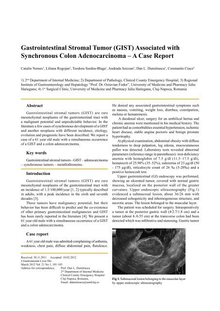
curvature. Upper endoscopic ultrasonography (Fig.1)<br />
evidenced a submucosal lesion, about 36/26 mm <st<strong>ro</strong>ng>with</st<strong>ro</strong>ng><br />
decreased echogenicity and inhomogeneous structure, and<br />
nec<strong>ro</strong>tic areas. The lesion belonged to the muscular layer.<br />
The patient was scheduled for surgery. Intraoperatively<br />
a tumor at the posterior gastric wall (4/2.7/1.8 cm) and a<br />
tumor (about 4./6.55 cm) at the transverse colon had been<br />
detected which was infiltrative and stenosing. Gastric tumor<br />
Case report<br />
A 61 year old male was admitted complaining of asthenia,<br />
weakness, chest pain, diffuse abdominal pain, flatulence.<br />
Received: 30.11.2011 Accepted: 10.02.2012<br />
J Gast<strong>ro</strong>intestin Liver Dis<br />
March 2012 Vol. 21 No 1, 101-103<br />
Address for correspondence: P<strong>ro</strong>f. Dan L. Dumitrascu<br />
2 nd Department of Internal Medicine<br />
Clinical County Emergency Hospital<br />
Cluj-Napoca, Romania,<br />
Email: ddumitrascu@umfcluj.<strong>ro</strong><br />
Fig 1. Submucosal lesion belonging to the muscular layer<br />
by upper endoscopic ultrasonography
102<br />
Nemes et al<br />
resection and enlarged right hemicolectomy <st<strong>ro</strong>ng>with</st<strong>ro</strong>ng> ileocolic<br />
anastomosis were performed.<br />
The histopathological diagnosis for the submucosal<br />
nodule was fusiform <st<strong>ro</strong>ng>GIST</st<strong>ro</strong>ng> low-grade (G1) pT2 (Fig 2),<br />
<st<strong>ro</strong>ng>with</st<strong>ro</strong>ng>out nec<strong>ro</strong>sis and atypia and the mitotic index (number<br />
of mitoses per 50 high-power fields) was 1/50 HPF (Fig 3).<br />
The immunohistochemistry indicated st<strong>ro</strong>ng staining for c-<br />
Kit/CD117 (Fig 4) and CD34 (Fig 5), while expression of<br />
S-100 were negative.<br />
The tumor in the colon was a well differentiated (G1)<br />
colon adenocarcinoma, <st<strong>ro</strong>ng>with</st<strong>ro</strong>ng> lower mucinous component<br />
pT3N1cMx (Fig 6).<br />
Fig 6. Colonic adenocarcinoma – mucinous areas<br />
(H&E x4).<br />
The patient was discharged f<strong>ro</strong>m the hospital and oral<br />
chemotherapy was initiated <st<strong>ro</strong>ng>with</st<strong>ro</strong>ng>out any complications.<br />
Fig 2. Fusiform low grade <st<strong>ro</strong>ng>GIST</st<strong>ro</strong>ng> (H&E stain x4).<br />
Fig 3. <st<strong>ro</strong>ng>GIST</st<strong>ro</strong>ng> – Ki-67 x10 (nuclear antigen associated<br />
<st<strong>ro</strong>ng>with</st<strong>ro</strong>ng> cell p<strong>ro</strong>liferation) p<strong>ro</strong>liferation index 3%.<br />
Fig 4. <st<strong>ro</strong>ng>GIST</st<strong>ro</strong>ng> – c-Kit/CD 117 (a type III ty<strong>ro</strong>sine<br />
kinase g<strong>ro</strong>wth factor receptor) positivity (x4).<br />
Fig 5. <st<strong>ro</strong>ng>GIST</st<strong>ro</strong>ng> – CD34 (myeloid p<strong>ro</strong>genitor cell<br />
antigen) positivity (x4).<br />
Discussion<br />
<st<strong>ro</strong>ng>GIST</st<strong>ro</strong>ng> and adenocarcinomas represent distinct oncogenic<br />
entities. <st<strong>ro</strong>ng>GIST</st<strong>ro</strong>ng>s are the most common mesenchymal tumors<br />
of the GI tract [5] and this g<strong>ro</strong>up of tumors represents about<br />
0.1% to 3% of all GI neoplasms.<br />
The mean age at presentation is over 40 and the clinical<br />
presentation is typically characterized by GI bleeding,<br />
abdominal pain, weight loss, anemia and a palpable mass<br />
[6]. The etiology may be represented by mutations in the Kit<br />
gene or PDGFRA (platelet derived g<strong>ro</strong>wth factor receptor<br />
alpha) gene and the origin of <st<strong>ro</strong>ng>GIST</st<strong>ro</strong>ng> is considered to be the<br />
interstitial cell of Cajal [7].<br />
The most common location for <st<strong>ro</strong>ng>GIST</st<strong>ro</strong>ng> is the stomach<br />
(52-60%), followed by small intestine (20% - 30%) and<br />
colorectum (10%) and the diagnosis is based on morphology<br />
and immunohistochemistry. Immunohistochemical stains<br />
like c-Kit/CD117 (a ty<strong>ro</strong>sine kinase inhibitor) – positive in<br />
95% [8], CD34 – positive in 40-50%, SMA (smooth muscle<br />
actin) – positive in 20-30% and S100 desmin – in about 10%,<br />
Ki67 - marker of cell p<strong>ro</strong>liferation [6] are necessary to make<br />
a precise diagnosis of <st<strong>ro</strong>ng>GIST</st<strong>ro</strong>ng> and to make the differential<br />
diagnosis between <st<strong>ro</strong>ng>GIST</st<strong>ro</strong>ng> and other mesenchymal tumors<br />
[7].<br />
Surgery is typically the first step in the treatment of<br />
<st<strong>ro</strong>ng>GIST</st<strong>ro</strong>ng>, inclusive in patients <st<strong>ro</strong>ng>with</st<strong>ro</strong>ng> resectable metastatic tumors.<br />
Recurrence, metastatic disease or unresectable tumors can<br />
be treated <st<strong>ro</strong>ng>with</st<strong>ro</strong>ng> Imatinib (stopping cell-p<strong>ro</strong>liferation actions<br />
of the KIT and PDGFR ty<strong>ro</strong>sine kinases) [9] .<br />
The p<strong>ro</strong>gnostic indicators are represented by tumor size,<br />
mitotic index, nec<strong>ro</strong>sis, infiltration and metastatic disease<br />
– variables that have an independent value for predicting<br />
the p<strong>ro</strong>gnosis of patients <st<strong>ro</strong>ng>with</st<strong>ro</strong>ng> <st<strong>ro</strong>ng>GIST</st<strong>ro</strong>ng> [7].<br />
The colorectal adenocarcinoma ranks as the fourth cause<br />
of cancer deaths worldwide and its development is a p<strong>ro</strong>cess<br />
of several stages influenced by complex interactions between<br />
host and envi<strong>ro</strong>nmental factors [10]. The histological
<st<strong>ro</strong>ng>GIST</st<strong>ro</strong>ng> associated <st<strong>ro</strong>ng>with</st<strong>ro</strong>ng> synch<strong>ro</strong>nous colon adenocarcinoma 103<br />
grade was demonstrated by many analyses to be a stageindependent<br />
p<strong>ro</strong>gnostic factor in colorectal cancer [11].<br />
The simultaneous occurrence of <st<strong>ro</strong>ng>GIST</st<strong>ro</strong>ng> and adenocarcinoma<br />
is uncommon in the literature, often the first one being<br />
detected incidentally at surgery [12].<br />
However, in one study of 783 patients, Pandurengan et<br />
al showed that app<strong>ro</strong>ximately 20% of patients <st<strong>ro</strong>ng>with</st<strong>ro</strong>ng> <st<strong>ro</strong>ng>GIST</st<strong>ro</strong>ng><br />
develop other types of cancers [13] but it remains unclear if<br />
this is just an incidental coexistence or these two are related<br />
by a causal relationship.<br />
Another study [14] demonstrated that synch<strong>ro</strong>nous<br />
colorectal adenocarcinoma and <st<strong>ro</strong>ng>GIST</st<strong>ro</strong>ng> have been more<br />
frequently reported. Due to the small number of cases, one<br />
cannot exclude an incidental relationship. Genetic pathways<br />
seem to be different for these two tumors.<br />
Yan-Jun Liu et al found that incidental <st<strong>ro</strong>ng>GIST</st<strong>ro</strong>ng> coexisted<br />
most commonly <st<strong>ro</strong>ng>with</st<strong>ro</strong>ng> esophageal (1.13%) and gastric tumors<br />
(0.53%), less <st<strong>ro</strong>ng>with</st<strong>ro</strong>ng> colorectal tumors (0.03%) and has a high<br />
prevalence in males [15]. The association of a <st<strong>ro</strong>ng>GIST</st<strong>ro</strong>ng> <st<strong>ro</strong>ng>with</st<strong>ro</strong>ng> a<br />
small bowel tumor has previously been reported [16].<br />
A possible explanation for the synch<strong>ro</strong>nous occurrence<br />
of these two entities is represented by the metallothioneins<br />
(MT), p<strong>ro</strong>teins <st<strong>ro</strong>ng>with</st<strong>ro</strong>ng> an increased affinity for heavy metal<br />
ions, coded by a family of 10 functional genes in human. The<br />
expression of these metallop<strong>ro</strong>teins has been associated <st<strong>ro</strong>ng>with</st<strong>ro</strong>ng><br />
p<strong>ro</strong>tection against DNA damage, apoptosis, cell survival,<br />
angiogenesis and oxidative stress [17]. Metallothioneins<br />
have been reported to be overexpressed in multiple neoplasia<br />
(such as breast, ovarian, uterus, oral cavity, lung, skin and<br />
pancreas) and down regulated in other types of cancers<br />
such as gastric, colorectal, liver and central nervous system<br />
tumors [18].<br />
Soo et al observed the nuclear expression of MT as<br />
determined by immunohistochemistry in all the <st<strong>ro</strong>ng>GIST</st<strong>ro</strong>ng>.<br />
Knowing that MT is correlated <st<strong>ro</strong>ng>with</st<strong>ro</strong>ng> cell p<strong>ro</strong>liferation, there is<br />
a possibility that MT may be involved in <st<strong>ro</strong>ng>GIST</st<strong>ro</strong>ng> p<strong>ro</strong>liferation<br />
[19]. Several studies have shown a direct correlation between<br />
MT and pathophysiology [20].<br />
The particularity of the present case is represented by the<br />
synch<strong>ro</strong>nous appearance of a gastric gast<strong>ro</strong>intestinal st<strong>ro</strong>mal<br />
tumor and a colon adenocarcinoma in a patient <st<strong>ro</strong>ng>with</st<strong>ro</strong>ng>out<br />
specific manifestations and also the incidental finding of a<br />
colon tumor during lapa<strong>ro</strong>tomy for <st<strong>ro</strong>ng>GIST</st<strong>ro</strong>ng>.<br />
References<br />
1. Theodosopoulos T, Dellaportas D, Psychogiou V, et al. Synch<strong>ro</strong>nous<br />
gastric adenocarcinoma and gast<strong>ro</strong>intestinal st<strong>ro</strong>mal tumor (<st<strong>ro</strong>ng>GIST</st<strong>ro</strong>ng>)<br />
of the stomach: a case report. World J Surg Oncol 2011;9:60.<br />
2. Casali PG, Blay JY; ESMO/CONTICANET/EUROBONET<br />
Consensus Panel of Experts. <st<strong>ro</strong>ng>Gast<strong>ro</strong>intestinal</st<strong>ro</strong>ng> st<strong>ro</strong>mal tumours:<br />
ESMO Clinical Practice Guidelines for diagnosis, treatment and<br />
follow-up. Ann Oncol 2010; 21 Suppl 5:v98-102.<br />
3. Miranda ME, Alberti LR, Tatsuo ES, Piçar<strong>ro</strong> C, Rausch M.<br />
<st<strong>ro</strong>ng>Gast<strong>ro</strong>intestinal</st<strong>ro</strong>ng> st<strong>ro</strong>mal tumor of the stomach in a child <st<strong>ro</strong>ng>with</st<strong>ro</strong>ng> a 3-<br />
year follow-up period—Case report. Int J Surg Case Rep 2011; 2:<br />
114–117.<br />
4. W<strong>ro</strong>nski M, Ziarkiewicz-W<strong>ro</strong>blewska B, Gornicka B, et al.<br />
Synch<strong>ro</strong>nous occurrence of gast<strong>ro</strong>intestinal st<strong>ro</strong>mal tumors and<br />
other primary gast<strong>ro</strong>intestinal neoplasms. World J Gast<strong>ro</strong>ente<strong>ro</strong>l<br />
2006;12:5360-5362.<br />
5. Miettinen M, Lasota J. <st<strong>ro</strong>ng>Gast<strong>ro</strong>intestinal</st<strong>ro</strong>ng> st<strong>ro</strong>mal tumors--definition,<br />
clinical, histological, immunohistochemical, and molecular genetic<br />
features and differential diagnosis. Virchows Arch 2001;438:1-12.<br />
6. Rabin I, Chikman B, Lavy R, et al. <st<strong>ro</strong>ng>Gast<strong>ro</strong>intestinal</st<strong>ro</strong>ng> st<strong>ro</strong>mal tumors:<br />
a 19 year experience. Isr Med Assoc J 2009;11:98-102.<br />
7. Kang YN, Jung HR, Hwang I. Clinicopathological and<br />
immunohistochemical features of gast<strong>ro</strong>intestinal st<strong>ro</strong>mal tumors.<br />
Cancer Res Treat 2010;42:135-143.<br />
8. Corless CL, Fletcher JA, Heinrich MC. Biology of gast<strong>ro</strong>intestinal<br />
st<strong>ro</strong>mal tumors. J Clin Oncol 2004;22:3813–3825.<br />
9. Neves LR, Oshima CT, Artigiani-Neto R, Yanaguibashi G, Lourenço<br />
LG, Fo<strong>ro</strong>nes NM.. Ki67 and p53 in gast<strong>ro</strong>intestinal st<strong>ro</strong>mal tumors<br />
– <st<strong>ro</strong>ng>GIST</st<strong>ro</strong>ng>. Arq Gast<strong>ro</strong>ente<strong>ro</strong>l 2009;46:116-120.<br />
10. Ibrahim KO, Anjorin AS, Afolayan AE, Badmos KB. Morphology<br />
of colorectal carcinoma among Nigerians: A 30-year review. Niger<br />
J Clin Pract 2011;14:432-435.<br />
11. Compton CC. Colorectal carcinoma: diagnostic, p<strong>ro</strong>gnostic, and<br />
molecular features. Mod Pathol 2003;16:376–388.<br />
12. Yamamoto D, Hamada Y, Tsubota Y, Kawakami K, Yamamoto C,<br />
Yamamoto M. Simultaneous development of adenocarcinoma and<br />
gast<strong>ro</strong>intestinal st<strong>ro</strong>mal tumor (<st<strong>ro</strong>ng>GIST</st<strong>ro</strong>ng>) in the stomach: Case report.<br />
World J Surg Oncol 2012;10:6.<br />
13. Pandurengan RK, Dumont AG, Araujo DM, et al. Survival of patients<br />
<st<strong>ro</strong>ng>with</st<strong>ro</strong>ng> multiple primary malignancies: a study of 783 patients <st<strong>ro</strong>ng>with</st<strong>ro</strong>ng><br />
gast<strong>ro</strong>intestinal st<strong>ro</strong>mal tumor. Ann Oncol 2010; 21: 2107-2111.<br />
14. Melis M, Choi EA, Anders R, Christiansen P, Fichera A. Synch<strong>ro</strong>nous<br />
colorectal adenocarcinoma and gast<strong>ro</strong>intestinal st<strong>ro</strong>mal tumor<br />
(<st<strong>ro</strong>ng>GIST</st<strong>ro</strong>ng>). Int J Colorectal Dis 2007;22:109-114.<br />
15. Liu YJ, Yang Z, Hao LS, Xia L, Jia QB, Wu XT. Synch<strong>ro</strong>nous<br />
incidental gast<strong>ro</strong>intestinal st<strong>ro</strong>mal and epithelial malignant tumors.<br />
World J Gast<strong>ro</strong>ente<strong>ro</strong>l 2009; 15: 2027–2031.<br />
16. Efremidou EI, Liratzopoulos N, Papageorgiou MS, Romanidis<br />
K. Perforated <st<strong>ro</strong>ng>GIST</st<strong>ro</strong>ng> of the small intestine as a rare cause of acute<br />
abdomen: surgical treatment and adjuvant therapy. Case Report. J<br />
Gast<strong>ro</strong>intestin Liver Dis 2006; 15: 297-299.<br />
17. Cherian MG, Jayasurya A, Bay BH. Metallothioneins in<br />
human tumors and potential <strong>ro</strong>les in carcinogenesis. Mutat Res<br />
2003;533:201-209.<br />
18. Pedersen MØ, Larsen A, Stoltenberg M, Penkowa M. The <strong>ro</strong>le<br />
of metallothionein in oncogenesis and cancer p<strong>ro</strong>gnosis. P<strong>ro</strong>g<br />
Histochem Cytochem 2009;44:29-64.<br />
19. Soo ET, Ng CT, Yip GW, et al. Differential expression of<br />
metallothionein in gast<strong>ro</strong>intestinal st<strong>ro</strong>mal tumors and gastric<br />
carcinomas. Anat Rec (Hoboken) 2011;294:267-272.<br />
20. Thirumoorthy N, Shyam Sunder A, Manisenthil Kumar K, Senthil<br />
Kumar M, Ganesh G, Chatterjee M. A review of metallothionein<br />
isoforms and their <strong>ro</strong>le in pathophysiology. World J Surg Oncol<br />
2011;9:54.