Localization of retrovirus in the central nervous system of a patient ...
Localization of retrovirus in the central nervous system of a patient ...
Localization of retrovirus in the central nervous system of a patient ...

Create successful ePaper yourself
Turn your PDF publications into a flip-book with our unique Google optimized e-Paper software.
Journal <strong>of</strong> NeuroVirology 7: 61±65 2001<br />
ã 2001 Taylor & Francis Ltd ISSN 1355±0284/01 $12.00+.00<br />
Case Report<br />
<strong>Localization</strong> <strong>of</strong> <strong>retrovirus</strong> <strong>in</strong> <strong>the</strong> <strong>central</strong> <strong>nervous</strong><br />
<strong>system</strong> <strong>of</strong> a <strong>patient</strong> co-<strong>in</strong>fected with HTLV-1 and HIV<br />
with HAM/TSP and HIV-associated dementia<br />
Michael C Lev<strong>in</strong>* ,1,2 , Marc K Rosenblum 3 , Cecil H Fox 4,5 and Steven Jacobson 2<br />
1<br />
De partment <strong>of</strong> Ne uro logy, La boratory <strong>of</strong> Viral and De myel<strong>in</strong>at<strong>in</strong>g Dise ases, University <strong>of</strong> Tenne ssee He alth Sciences<br />
Ce nte r, Me mphis, Te nne ssee, 38163, USA; 2 Ne uro immuno logy Branch, Na tiona l Institute <strong>of</strong> Ne uro logical Diseases and<br />
Stroke, Na tiona l Institutes <strong>of</strong> He alth, Be <strong>the</strong>sda, MD 20892, USA; 3 De partment <strong>of</strong> Pa thology (Neuro pathology), Me m orial<br />
Slo an Ke tter<strong>in</strong>g Ca ncer Ce nte r, Ne w Yo rk, NY 10021, USA; 4 Unive rsity <strong>of</strong> Arkansas Me dical Center, Little Ro ck , AR<br />
72205, USA and 5 Mo le cula r Histology La boratories, Ga i<strong>the</strong> rsburg, MD 20879, USA<br />
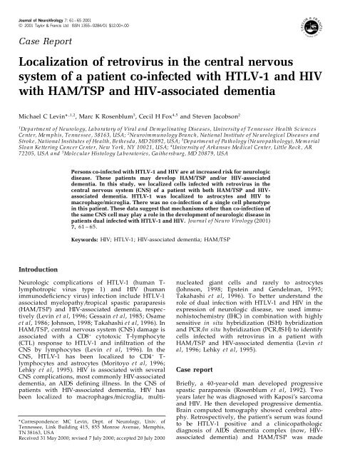
Persons co-<strong>in</strong>fected with HTLV-1 and HIV are at <strong>in</strong>creased risk for neurologic<br />
disease. These <strong>patient</strong>s may develop HAM/TSP and/or HIV-associated<br />
dementia. In this study, we localized cells <strong>in</strong>fected with <strong>retrovirus</strong> <strong>in</strong> <strong>the</strong><br />
<strong>central</strong> <strong>nervous</strong> <strong>system</strong> (CNS) <strong>of</strong> a <strong>patient</strong> with both HAM/TSP and HIVassociated<br />
dementia. HTLV-1 was localized to astrocytes and HIV to<br />
macrophage/microglia. There was no co-<strong>in</strong>fection <strong>of</strong> a s<strong>in</strong>gle cell phenotype<br />
<strong>in</strong> this <strong>patient</strong>. These data suggest that mechanisms o<strong>the</strong>r than co-<strong>in</strong>fection <strong>of</strong><br />
<strong>the</strong> same CNS cell may play a role <strong>in</strong> <strong>the</strong> development <strong>of</strong> neurologic disease <strong>in</strong><br />
<strong>patient</strong>s dual <strong>in</strong>fected with HTLV-1 and HIV. Journa l <strong>of</strong> Neuro Virology (2001)<br />
7, 61 ± 65.<br />
Keywords: HIV; HTLV-1; HIV-associated dementia; HAM/TSP<br />
Introduction<br />
* Correspondence: MC Lev<strong>in</strong>, Dept. <strong>of</strong> Neurology, Univ. <strong>of</strong><br />
Tennessee, L<strong>in</strong>k Build<strong>in</strong>g 415, 855 Monroe Avenue, Memphis,<br />
TN 38163, USA<br />
Received 31 May 2000; revised 7 July 2000; accepted 20 July 2000<br />
Neurologic complications <strong>of</strong> HTLV-1 (human T-<br />
lymphotropic virus type 1) and HIV (human<br />
immunode® ciency virus) <strong>in</strong>fection <strong>in</strong>clude HTLV-1<br />
associated myelopathy/tropical spastic paraparesis<br />
(HAM/TSP) and HIV-associated dementia, respectively<br />
(Lev<strong>in</strong> et al, 1996; Gessa<strong>in</strong> et al, 1985; Osame<br />
et al, 1986; Johnson, 1998; Takahashi et al, 1996). In<br />
HAM/TSP, <strong>central</strong> <strong>nervous</strong> <strong>system</strong> (CNS) damage is<br />
associated with a CD8 + cytotoxic T-lymphocyte<br />
(CTL) response to HTLV-1 and <strong>in</strong>® ltration <strong>of</strong> <strong>the</strong><br />
CNS by lymphocytes (Lev<strong>in</strong> et al, 1996). In <strong>the</strong><br />
CNS, HTLV-1 has been localized to CD4 + T-<br />
lymphocytes and astrocytes (Moritoyo et al, 1996;<br />
Lehky et al, 1995). HIV is associated with several<br />
CNS complications, most commonly HIV-associated<br />
dementia, an AIDS de® n<strong>in</strong>g illness. In <strong>the</strong> CNS <strong>of</strong><br />
<strong>patient</strong>s with HIV-associated dementia, HIV has<br />
been localized to macrophages/microglia, mult<strong>in</strong>ucleated<br />
giant cells and rarely to astrocytes<br />
(Johnson, 1998; Epste<strong>in</strong> and Gendelman, 1993;<br />
Takahashi et al, 1996). To better understand <strong>the</strong><br />
role <strong>of</strong> dual <strong>in</strong>fection with HTLV-1 and HIV <strong>in</strong> <strong>the</strong><br />
expression <strong>of</strong> neurologic disease, we used immunohistochemistry<br />
(IHC) <strong>in</strong> comb<strong>in</strong>ation with highly<br />
sensitive <strong>in</strong> situ hybridization (ISH) hybridization<br />
and PCR/<strong>in</strong> situ hybridization (PCR/ISH) to identify<br />
cells <strong>in</strong>fected with <strong>retrovirus</strong> <strong>in</strong> a <strong>patient</strong> with<br />
HAM/TSP and HIV-associated dementia (Lev<strong>in</strong> et<br />
al, 1996; Lehky et al, 1995).<br />
Case report<br />
Brie¯ y, a 40-year-old man developed progressive<br />
spastic paraparesis (Rosenblum et al, 1992). Two<br />
years later he was diagnosed with Kaposi’s sarcoma<br />
and HIV. He <strong>the</strong>n developed progressive dementia.<br />
Bra<strong>in</strong> computed tomography showed cerebral atrophy.<br />
Retrospectively, <strong>the</strong> <strong>patient</strong>’s serum was found<br />
to be HTLV-1 positive and a cl<strong>in</strong>icopathologic<br />
diagnosis <strong>of</strong> AIDS dementia complex (now, HIVassociated<br />
dementia) and HAM/TSP was made
62<br />
HIV and HTLV-1 co-<strong>in</strong>fection<br />
MC Lev<strong>in</strong><br />
(Rosenblum et al, 1992). Autopsy showed bra<strong>in</strong><br />
atrophy and HIV-1 encephalitis con® rmed by<br />
positive HIV-1 p24 cells. The sp<strong>in</strong>al cord showed<br />
nonvacuolar myelopathy with axonal loss and<br />
demyel<strong>in</strong>ation. Initial retroviral analysis for HIV<br />
and HTLV-1 by ISH was negative. Solution phase<br />
PCR <strong>of</strong> bra<strong>in</strong> showed HTLV-1 speci® c signal<br />
(Rosenblum et al, 1992).<br />
In this study, a comb<strong>in</strong>ation <strong>of</strong> immunocytochemistry,<br />
ISH and PCR/ISH were used to detect<br />
HIV and HTLV-1 and co-localize it to speci® c cell<br />
phenotypes <strong>in</strong> formal<strong>in</strong> ® xed, paraf® n embedded<br />
CNS and control tissues. For those experiments<br />
requir<strong>in</strong>g immunocytochemistry, it was performed<br />
® rst under RNASE free conditions. Primary antibodies<br />
<strong>in</strong>cluded: HAM-56 (Enzo Biochemical Co.,<br />
New York, NY, USA) for macrophage/microglia and<br />
glial ® brillary acidic prote<strong>in</strong> (GFAP) (Dako, Carp<strong>in</strong>teria,<br />
CA, USA) for astrocytes. Biot<strong>in</strong>ylated secondary<br />
antibodies (Vector, Burl<strong>in</strong>game, CA, USA) were<br />
used to detect primary antibodies. The antigenantibody-biot<strong>in</strong><br />
complex was conjugated to avid<strong>in</strong><br />
(ABC solution, Vector) and detected with a diam<strong>in</strong>obenzid<strong>in</strong>e<br />
(DAB) peroxide <strong>system</strong> <strong>in</strong> which<br />
positive sta<strong>in</strong><strong>in</strong>g is brown or by new fuchs<strong>in</strong> (Dako)<br />
<strong>in</strong> which positive sta<strong>in</strong><strong>in</strong>g is red.<br />
For HIV ISH, a full length HIV-1 LA1 cRNA probe<br />
( 35 S-dCTP, 2 ´ 10 6 d.p.m./ul, alkal<strong>in</strong>e hydrolyzed)<br />
was used <strong>in</strong> <strong>the</strong> anti-sense con® guration to detect<br />
HIV-RNA. A HIV <strong>in</strong>fected cell l<strong>in</strong>e (H-9) was used as<br />
positive control. For HTLV-1-RNA ISH, an antisense<br />
2.1 kb cRNA probe ( 35 S-dCTP, 2 ´ 10 6 d.p.m./<br />
ul, alkal<strong>in</strong>e hydrolyzed) <strong>of</strong> HTLV-1-tax was used.<br />
To detect HTLV-1-DNA, PCR/ISH with HTLV-1-tax<br />
speci® c primers were used to amplify HTLV-1 tax<br />
(Lev<strong>in</strong> et a l, 1996). Follow<strong>in</strong>g PCR, ISH was<br />
performed us<strong>in</strong>g <strong>the</strong> sense HTLV-1-tax probe to<br />
detect ampli® ed DNA. An HTLV-1<strong>in</strong>fected cell l<strong>in</strong>e<br />
(HUT-102) was used as a positive control. Un<strong>in</strong>fected<br />
peripheral blood lymphocytes (PBL) and<br />
normal CNS tissues were used for negative controls.<br />
Follow<strong>in</strong>g ISH, autoradiograms were prepared with<br />
photographic emulsion (Kodak). Slides were developed<br />
after ® ve days and countersta<strong>in</strong>ed with<br />
hematoxyl<strong>in</strong> and/or eos<strong>in</strong>.<br />
Conventional ISH with <strong>the</strong> antisense 35 S-labeled<br />
HTLV-1-tax probe was used to detect HTLV-1-tax<br />
RNA (Table 1). In HUT 102 cells (an HTLV-1<br />
<strong>in</strong>fected cell l<strong>in</strong>e that expresses multiple copies <strong>of</strong><br />
HTLV-1-RNA), <strong>the</strong>re was <strong>in</strong>tense silver gra<strong>in</strong><br />
localization (Figure 1a). There was no speci® c<br />
signal <strong>in</strong> un<strong>in</strong>fected PBL (Figure 1b) or <strong>in</strong> normal<br />
Table 1 Detection <strong>of</strong> HTLV-1 and HIV <strong>in</strong> normal and<br />
<strong>retrovirus</strong>-<strong>in</strong>fected tissues<br />
HTLV-1<br />
HIV<br />
Ð Ð<br />
Ð Ð<br />
Ð<br />
Ð<br />
Un<strong>in</strong>fected PBL ( ) ( )<br />
HTLV-1 (+) HUT 102 cells (+) RNA and DNA nd<br />
HIV (+) H9 cells nd (+)<br />
Normal, un<strong>in</strong>fected CNS ( ) ( )<br />
Dual (HTLV-1/HIV) <strong>in</strong>fected CNS (+) (+)<br />
GFAP positive astrocytes (+) RNA and DNA ( )<br />
Macrophage/microglia ( ) (+)<br />
nd, not done.<br />
Figure 1 ISH for HTLV-1-tax-RNA with <strong>the</strong> antisense 35 -S HTLV-1-tax probe (magni® cation 400 ´ , hematoxyl<strong>in</strong> and eos<strong>in</strong><br />
countersta<strong>in</strong> except where noted). (a) positive control, HUT-102 cells. Intense silver gra<strong>in</strong> localization <strong>in</strong> all cells; (b) negative control,<br />
un<strong>in</strong>fected PBL. No signal present with<strong>in</strong> cells; (c) normal bra<strong>in</strong>. No speci® c signal present; (d) dual <strong>in</strong>fected bra<strong>in</strong>. Multiple HTLV-1-<br />
tax-RNA positive cells are present; (e) dual <strong>in</strong>fected bra<strong>in</strong> sta<strong>in</strong>ed with GFAP (p<strong>in</strong>k) and countersta<strong>in</strong>ed with hematoxyl<strong>in</strong> only. HTLV-<br />
1-tax-RNA (silver gra<strong>in</strong>s) co-localized to a GFAP positive astrocyte (p<strong>in</strong>k).
HIV and HTLV-1 co-<strong>in</strong>fection<br />
MC Lev<strong>in</strong> 63<br />
CNS (Figure 1c). In <strong>the</strong> dual <strong>in</strong>fected case, <strong>the</strong>re<br />
were multiple positive HTLV-1-RNA <strong>in</strong>fected cells<br />
(Figure 1d). These cells were located <strong>in</strong> white<br />
matter and <strong>the</strong>re were no <strong>in</strong>¯ ammatory cells.<br />
Fur<strong>the</strong>rmore, HTLV-1-RNA was co-localized to<br />
GFAP-positive astrocytes (Figure 1e). This data is<br />
consistent with data seen <strong>in</strong> <strong>patient</strong>s with HAM/<br />
TSP alone, <strong>in</strong> which HTLV-1 RNA was localized to<br />
astrocytes <strong>in</strong> <strong>the</strong> CNS <strong>in</strong> areas devoid <strong>of</strong> <strong>in</strong>¯ ammation<br />
(Lehky e t a l, 1995). HTLV-1 RNA did not<br />
localize to macrophage/microglial cells (Table 1).<br />
PCR/ISH was used to detect HTLV-1-DNA (Table<br />
1). Us<strong>in</strong>g <strong>the</strong> sense 35 S-HTLV-1-tax probe, <strong>the</strong>re was<br />
no speci® c signal detected <strong>in</strong> HUT 102 cells (Figure<br />
2a). This is consistent with conventional ISH not<br />
be<strong>in</strong>g sensitive enough to detect HTLV-1-DNA <strong>in</strong><br />
low copy numbers (Lev<strong>in</strong> e t al, 1996). Follow<strong>in</strong>g<br />
PCR/ISH, <strong>the</strong>re was <strong>in</strong>tense silver gra<strong>in</strong> localization<br />
<strong>in</strong> HUT 102 cells (Figure 2b), but not <strong>in</strong> un<strong>in</strong>fected<br />
PBL (Figure 2c). Also, <strong>the</strong>re was no signal if Taq<br />
polymerase or tax speci® c primers were elim<strong>in</strong>ated<br />
from <strong>the</strong> PCR cocktail (not shown) (Lev<strong>in</strong> et al,<br />
1996). PCR/ISH <strong>of</strong> normal CNS for HTLV-1-tax DNA<br />
was negative (Figure 2d). In contrast, PCR/ISH <strong>of</strong><br />
dual <strong>in</strong>fected CNS showed multiple positive cells <strong>in</strong><br />
regions similar to that <strong>of</strong> conventional ISH for<br />
HTLV-1-RNA (Figure 2e), and some <strong>of</strong> <strong>the</strong>se cells<br />
co-localized to GFAP-positive astrocytes (Figure 2f).<br />
Conventional ISH with <strong>the</strong> antisense 35 S-labeled<br />
HIV-1 probe was used to detect HIV-RNA (Table 1).<br />
In H9 cells (an HIV <strong>in</strong>fected cell l<strong>in</strong>e that express<br />
HIV RNA), <strong>the</strong>re was <strong>in</strong>tense silver gra<strong>in</strong> localization<br />
(Figure 3a). There was no speci® c signal us<strong>in</strong>g<br />
un<strong>in</strong>fected PBL (Figure 3b). ISH for HIV-RNA was<br />
also negative <strong>in</strong> normal CNS (Figure 3c). In <strong>the</strong> dual<br />
<strong>in</strong>fected autopsy case, <strong>the</strong>re were multiple positive<br />
HIV-RNA <strong>in</strong>fected cells (Figure 3d). These cells<br />
were located <strong>in</strong> both gray and white matter.<br />
Fur<strong>the</strong>rmore, HIV-RNA localized to macrophage/<br />
microglial cells (Figure 3e) and to mult<strong>in</strong>ucleated<br />
giant cells (Figure 3f). This is consistent with <strong>the</strong><br />
localization <strong>of</strong> HIV <strong>in</strong> <strong>the</strong> CNS described previously<br />
(Takahashi et a l, 1996). There was no localization <strong>of</strong><br />
HIV-RNA to GFAP-positive astrocytes (Figure 3f).<br />
Figure 2 PCR/ISH for HTLV-1-tax DNA with <strong>the</strong> sense 35 -S<br />
HTLV-1-tax probe follow<strong>in</strong>g PCR ampli® cation (except where<br />
noted) with HTLV-1-tax speci® c primers (magni® cation 400 ´ ,<br />
hematoxyl<strong>in</strong> and eos<strong>in</strong> countersta<strong>in</strong> except where noted). (a)<br />
positive control HUT 102 cells without PCR ampli® cation. No<br />
silver gra<strong>in</strong> localization to show that ISH alone is not sensitive<br />
enough to detect HTLV-1-tax DNA; (b) positive control HUT 102<br />
cells follow<strong>in</strong>g PCR ampli® cation. Intense silver gra<strong>in</strong> localization<br />
<strong>in</strong> all cells; (c) negative control, un<strong>in</strong>fected PBL. No silver gra<strong>in</strong><br />
localization to cells; (d) normal bra<strong>in</strong>. No speci® c signal present;<br />
(e) dual <strong>in</strong>fected bra<strong>in</strong>. Positive HTLV-1-tax DNA positive cells<br />
present; (f) dual <strong>in</strong>fected bra<strong>in</strong> sta<strong>in</strong>ed with GFAP (brown) and<br />
countersta<strong>in</strong>ed with hematoxyl<strong>in</strong> only. HTLV-1-tax DNA (silver<br />
gra<strong>in</strong>s, arrow) co-localized to a GFAP positive astrocyte (brown).<br />
The <strong>in</strong>sert shows <strong>the</strong> identical cell with <strong>the</strong> silver gra<strong>in</strong>s <strong>in</strong> focus<br />
which are with<strong>in</strong> <strong>the</strong> emulsion layer <strong>of</strong> <strong>the</strong> autoradiogram. The<br />
full ® gure shows <strong>the</strong> focus on <strong>the</strong> immunocytochemistry.<br />
Discussion<br />
In this co-<strong>in</strong>fected <strong>patient</strong>, HTLV-1-RNA was<br />
localized to astrocytes. Infection was con® rmed by<br />
show<strong>in</strong>g that HTLV-1-DNA co-localized to astrocytes<br />
by PCR/ISH (Figure 1D). O<strong>the</strong>r studies<br />
localized HTLV-1 to <strong>in</strong>® ltrat<strong>in</strong>g CD4 + T-lymphocytes<br />
<strong>in</strong> <strong>the</strong> CNS <strong>of</strong> HAM/TSP <strong>patient</strong>s (Moritoyo et<br />
al, 1996). These data may not be mutually<br />
exclusive. HTLV-1 <strong>in</strong>fected CD4 + lymphocytes (<strong>the</strong><br />
source HTLV-1 <strong>in</strong> blood) may enter <strong>the</strong> CNS and<br />
<strong>in</strong>fect an astrocyte. Early <strong>in</strong> disease, CD4 + T-<br />
lymphocytes are plentiful and may act as an antigen<br />
present<strong>in</strong>g cell to stimulate a CD8 + CTL response,<br />
and upon <strong>the</strong> secretion <strong>of</strong> toxic levels <strong>of</strong> cytok<strong>in</strong>es,<br />
<strong>in</strong>directly damage CNS tissue. Later <strong>in</strong> disease, an<br />
HTLV-1 <strong>in</strong>fected astrocyte may act as a target for<br />
<strong>the</strong> CTL, which may result <strong>in</strong> neurologic damage.<br />
HIV-RNA localized to mult<strong>in</strong>ucleated giant cells<br />
and to macrophage/microglial cells, but not to<br />
astrocytes. O<strong>the</strong>r studies have shown low levels <strong>of</strong><br />
HIV-RNA expression <strong>in</strong> astrocytes, particularly <strong>in</strong><br />
children (Saito e t al, 1994). In <strong>the</strong> adult <strong>patient</strong> we<br />
studied, HIV-RNA was not detected <strong>in</strong> astrocytes<br />
(Figure 3f). This is consistent with reports <strong>in</strong> <strong>the</strong><br />
literature, <strong>in</strong> which HIV RNA expression is low or<br />
undetectable (Takahashi e t al, 1996). Current data<br />
suggests that HIV <strong>in</strong>fection <strong>of</strong> macrophages/micro-
64<br />
HIV and HTLV-1 co-<strong>in</strong>fection<br />
MC Lev<strong>in</strong><br />
Figure 3 ISH for HIV-RNA with <strong>the</strong> antisense 35 -S HIV-1 probe (magni® cation 400 ´ , hematoxyl<strong>in</strong> and eos<strong>in</strong> countersta<strong>in</strong> except<br />
where noted). (a) positive control, H9 cells. Intense silver gra<strong>in</strong> localization over <strong>the</strong> cell; (b) negative control, un<strong>in</strong>fected PBL. No<br />
silver gra<strong>in</strong> localization over cells; (c) normal bra<strong>in</strong>. No positive cell present; (d) dual <strong>in</strong>fected bra<strong>in</strong>. Multiple positive cells present, (e)<br />
dual <strong>in</strong>fected bra<strong>in</strong> sta<strong>in</strong>ed with HAM-56, a macrophage/microglia marker (p<strong>in</strong>k) and countersta<strong>in</strong>ed with hematoxyl<strong>in</strong> only. HIV-RNA<br />
(silver gra<strong>in</strong>s, arrow) co-localized to a HAM-56 positive cell <strong>of</strong> macrophage/microglia l<strong>in</strong>eage (p<strong>in</strong>k) (bright® eld microscopy). The<br />
<strong>in</strong>sert shows <strong>the</strong> identical cell us<strong>in</strong>g polarized light and shows <strong>in</strong>tense silver gra<strong>in</strong> localization with<strong>in</strong> <strong>the</strong> cell; (f) dual <strong>in</strong>fected bra<strong>in</strong><br />
sta<strong>in</strong>ed with GFAP (p<strong>in</strong>k) and countersta<strong>in</strong>ed with hematoxyl<strong>in</strong> only. HIV-RNA (silver gra<strong>in</strong>s, arrow) with<strong>in</strong> a mult<strong>in</strong>ucleated giant<br />
cell. The <strong>in</strong>sert shows <strong>the</strong> same cell us<strong>in</strong>g polarized light and shows <strong>in</strong>tense silver gra<strong>in</strong> localization. There was no co-localization <strong>of</strong><br />
HIV-RNA with<strong>in</strong> astrocytes (p<strong>in</strong>k).<br />
glia <strong>in</strong> <strong>the</strong> CNS <strong>of</strong> <strong>patient</strong>s with HIV-associated<br />
dementia may result <strong>in</strong> neuronal or astrocyte<br />
dysfunction through <strong>in</strong>direct pathways from release<br />
<strong>of</strong> cytok<strong>in</strong>es, tox<strong>in</strong>s or viral prote<strong>in</strong>s (Takahashi et<br />
al, 1996; Johnson, 1998).<br />
Cl<strong>in</strong>ical manifestations <strong>of</strong> dual <strong>in</strong>fected <strong>patient</strong>s<br />
with neuropathological correlation are <strong>of</strong> <strong>in</strong>terest<br />
because <strong>of</strong> <strong>the</strong> high rate <strong>of</strong> co-<strong>in</strong>fection with HTLV-<br />
1 <strong>in</strong> certa<strong>in</strong> HIV <strong>in</strong>fected populations. There have<br />
been several reports show<strong>in</strong>g that neurologic<br />
disease associated with co-<strong>in</strong>fection can result <strong>in</strong><br />
<strong>the</strong> cl<strong>in</strong>ical expression <strong>of</strong> ei<strong>the</strong>r HAM/TSP, HIVassociated<br />
dementia or both diseases (Rosenblum<br />
et al, 1992; Harrison et a l, 1997; Berger et al, 1991).<br />
Also, HIV <strong>in</strong>fected <strong>patient</strong>s can develop vacuolar<br />
myelopathy (VM), a sp<strong>in</strong>al cord disease that is<br />
dist<strong>in</strong>ct pathologically from HAM/TSP, but may<br />
present with similar symptoms. Recently, <strong>patient</strong>s<br />
<strong>in</strong>fected with both HTLV-1 and HIV have a greater<br />
risk for develop<strong>in</strong>g myelopathy than those with<br />
HIV alone (Harrison et a l, 1997). This is important<br />
<strong>in</strong> order for <strong>patient</strong>s <strong>in</strong> appropriate risk groups to<br />
be screened for both HTLV-1 and HIV. There seems<br />
to be a reciprocal relationship between HTLV-1<br />
and HIV. HTLV-1 may <strong>in</strong>crease <strong>the</strong> risk <strong>of</strong> AIDS<br />
and <strong>in</strong>crease HIV replication (Page et al, 1990;<br />
Cheng et a l, 1998). However, this may not be <strong>the</strong><br />
case <strong>in</strong> vivo, <strong>in</strong> which HIV viral load is similar<br />
between dual <strong>in</strong>fected <strong>patient</strong>s and <strong>patient</strong>s with<br />
HIV alone (Harrison et a l, 1996). Alternatively, HIV<br />
<strong>in</strong>creased HTLV-1 levels <strong>in</strong> dual <strong>in</strong>fected <strong>patient</strong>s<br />
compared to <strong>patient</strong>s with HTLV-1 alone (Beilke e t<br />
al, 1998). We did not ® nd evidence <strong>of</strong> overexpression<br />
<strong>of</strong> <strong>retrovirus</strong> <strong>in</strong> <strong>the</strong> CNS nor did we<br />
® nd cells <strong>of</strong> <strong>the</strong> same phenotype co-<strong>in</strong>fected with<br />
both HTLV-1 and HIV. Consider<strong>in</strong>g <strong>the</strong> dist<strong>in</strong>ct<br />
neuropathology <strong>of</strong> HIV-associated dementia and<br />
HAM/TSP, <strong>the</strong> reciprocal relationship between<br />
HTLV-1 and HIV may <strong>in</strong>crease <strong>the</strong> risk <strong>of</strong><br />
neurologic disease via alterations <strong>in</strong> <strong>the</strong> immune<br />
response or cytok<strong>in</strong>e expression <strong>in</strong> <strong>the</strong> CNS, ra<strong>the</strong>r<br />
than direct <strong>in</strong>fection <strong>of</strong> both <strong>retrovirus</strong>es on <strong>the</strong><br />
CNS.<br />
Acknowledgements<br />
MC Lev<strong>in</strong> was a recipient <strong>of</strong> a National Multiple<br />
Sclerosis Society Post-Doctoral Fellowship Grant<br />
and an AIDS National Loan Repayment Award.
HIV and HTLV-1 co-<strong>in</strong>fection<br />
MC Lev<strong>in</strong> 65<br />
References<br />
Beilke MA, Japa S, V<strong>in</strong>son DG (1998). HTLV-1 and<br />
HTLV-II virus expression <strong>in</strong>crease with HIV-1 co<strong>in</strong>fection.<br />
J Acquir Immune De ® c Synd romes Hum an<br />
Re trovirol 17: 391 ± 397.<br />
Berger JR, Raffanti S, Svenn<strong>in</strong>gsson A, McCarthy M,<br />
Snodgrass S, Resnick L (1991). The role <strong>of</strong> HTLV <strong>in</strong><br />
HIV-1 neurologic disease. Ne urology 41: 197 ± 202.<br />
Cheng H, Tarnok J, Parks WP (1998). Human immunode®<br />
ciency virus type 1 genome activation <strong>in</strong>duced by<br />
human T-cell leukemia virus type 1 tax prote<strong>in</strong> is<br />
through cooperation <strong>of</strong> NF-jB and Tat. J Virol 72(8):<br />
6911 ± 6916.<br />
Epste<strong>in</strong> LG, Gendelman HE (1993). Human immunode-<br />
® ciency virus type 1 <strong>in</strong>fection <strong>of</strong> <strong>the</strong> <strong>nervous</strong> <strong>system</strong>:<br />
pathogenetic mechanisms. Ann Neuro l 33: 429 ± 436.<br />
Gessa<strong>in</strong> A, Vernant J, Maurs L, Bar<strong>in</strong> F, Gout O,<br />
Calender A, The GD (1985). Antibodies to human T-<br />
lymphotropic virus I <strong>in</strong> <strong>patient</strong>s with tropical spastic<br />
paraparesis. La ncet 2: 407 ± 410.<br />
Harrison LH, Qu<strong>in</strong>n TC, Schechter M (1996). Human T<br />
cell lymphotropic virus type I does not <strong>in</strong>crease<br />
human immunode® ciency virus viral load <strong>in</strong> vivo. J<br />
Infect Dis 175: 438 ± 440.<br />
Harrison LH, Vaz B, Taveira DM, Qu<strong>in</strong>n TC, Gibbs CJ,<br />
deSouza SH, McArthur JC, Schechter M (1997).<br />
Myelopathy among Brazilians co<strong>in</strong>fected with juman<br />
T-cell lymphotropic virus type I and HIV. Ne uro logy<br />
48: 13 ± 18.<br />
Johnson RT (1998). Viral <strong>in</strong>fections <strong>of</strong> <strong>the</strong> Nervous<br />
System: ``Hum an immuno de® ciency virus’’. Philadelphia:<br />
Lipp<strong>in</strong>cott-Raven Publishers, Pp. 287 ± 314.<br />
Lehky T, Fox C, Koenig S, Lev<strong>in</strong> M, Flerlage N, Izumo S,<br />
Sato E, Ra<strong>in</strong>e C, Osame M, Jacobson S (1995).<br />
Detection <strong>of</strong> human T lymphotropic virus type I<br />
(HTLV-1) tax RNA <strong>in</strong> <strong>the</strong> <strong>central</strong> <strong>nervous</strong> <strong>system</strong> <strong>of</strong><br />
HTLB-1 associated myelopathy/tropical spastic paraparesis<br />
by <strong>in</strong> situ hybridization. Ann Ne urol 37: 246 ±<br />
254.<br />
Lev<strong>in</strong> MC, Fox R, Lehky T, Walter M, Fox CH, Flerlage<br />
N, Bamford R, Jacobson S (1996). PCR-<strong>in</strong> situ<br />
hybridization detection <strong>of</strong> human T-cell lymphotropic<br />
virus type I (HTLV-I) tax proviral DNA <strong>in</strong> peripheral<br />
blood lymphocytes <strong>of</strong> <strong>patient</strong>s with HTLV-I associated<br />
neurologic disease. J Virol 70: 924 ± 933.<br />
Moritoyo, T, Re<strong>in</strong>hart T, Moritoyo H, Sato E, Izumo S,<br />
Osame M, Haase A (1996). Human T-lymphotropic<br />
virus type I associated myelopathy and tax gene<br />
expression <strong>in</strong> CD4+ T lymphocytes. Ann Ne uro l 40:<br />
84 ± 90.<br />
Osame M, Usuku, K, Izumo S, Ijchi N, Amitani H, Igata<br />
A, Matsumoto M, Tara M (1986). HTLV-I associated<br />
myelopathy: a new cl<strong>in</strong>ical entity. La ncet 1: 1031 ±<br />
1032.<br />
Page JB, Lai S, Chitwood DD, Klimas NG, Smith PC,<br />
Fletcher MA (1990). HTLV-I/II seropositivity and<br />
death from AIDS among HIV-1 seropositive <strong>in</strong>travenous<br />
drug users. La ncet 335: 1439 ± 1441.<br />
Rosenblum MK, Brew BJ, Hahn B, Shaw G, Haase A,<br />
Maroushek S, Price RW (1992). Human T-lymphotropic<br />
virus type I-associated myelopathy <strong>in</strong> <strong>patient</strong>s<br />
with <strong>the</strong> acquired immunode® ciency syndrome. Human<br />
Pa thol 23: 513 ± 519.<br />
Saito Y, Sharer LR, Epste<strong>in</strong> LG, Michaels J, M<strong>in</strong>tz M,<br />
Louder M, Gold<strong>in</strong>g K, Cvetkovich TA, Blumberg BM<br />
(1994). Overexpression <strong>of</strong> nef as a marker for<br />
restricted HIV-1 <strong>in</strong>fection <strong>of</strong> astrocytes <strong>in</strong> postmortem<br />
pediatric <strong>central</strong> <strong>nervous</strong> tissues. Ne urology 44: 474 ±<br />
481.<br />
Takahashi K, Wessel<strong>in</strong>gh SL, Grif® n DE, McArthur JC,<br />
Johnson RT, Glass JD (1996). <strong>Localization</strong> <strong>of</strong> HIV-1 <strong>in</strong><br />
human bra<strong>in</strong> us<strong>in</strong>g polymerase cha<strong>in</strong> reaction/<strong>in</strong> situ<br />
hybridization and immunocytochemistry. Ann Neuro l<br />
39: 705 ± 711.