Anti - Titin - Antibody ELISA - DLD Diagnostika GmbH
Anti - Titin - Antibody ELISA - DLD Diagnostika GmbH
Anti - Titin - Antibody ELISA - DLD Diagnostika GmbH

Create successful ePaper yourself
Turn your PDF publications into a flip-book with our unique Google optimized e-Paper software.
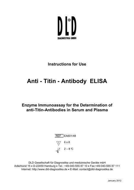
Instructions for Use<br />
<strong>Anti</strong> - <strong>Titin</strong> - <strong>Anti</strong>body <strong>ELISA</strong><br />
Enzyme Immunoassay for the Determination of<br />
anti-<strong>Titin</strong>-<strong>Anti</strong>bodies in Serum and Plasma<br />
REF EA601/48<br />
6 x 8<br />
2 – 8 °C<br />
<strong>DLD</strong> Gesellschaft für <strong>Diagnostika</strong> und medizinische Geräte mbH<br />
Adlerhorst 15 • D-22459 Hamburg • Tel.: +49-040-555 87 10 • Fax:+49-040-555 87 111<br />
Internet: http://www.dld-diagnostika.de • E-Mail: contact@dld-diagnostika.de<br />
January 2012
Page 2
Contents<br />
1. Introduction and Principle of the Test Page 4<br />
2. Precautions Page 5<br />
3. Storage and Stability Page 5<br />
4. Contents of the Test Page 5<br />
5. Preparation of Reagents and Samples Page 7<br />
6. Assay Procedure Page 8<br />
7. Calculation and Interpretation of Results Page 9<br />
8. Literature Page 11<br />
Pipetting Scheme Page 12<br />
Page 3
1. Introduction and Principle of the Test<br />
In about 80 % of patients with Myasthenia gravis alterations of the thymus<br />
can be detected. Approximately 10 % of these patients develop thymus<br />
neoplasia as thymic epithelial tumor (TET) or thymic carcinoma. An early<br />
diagnosis of the thymoma and subsequent thymectomy is decisive for the<br />
prognosis of these patients.<br />
Most of the affected patients develop acetylcholine receptor antibodies which<br />
can be detected with the ACHRAB ®-Assay for the diagnosis of myasthenia<br />
gravis, as well as autoantibodies against striated muscles, among others<br />
antibodies to titin.<br />
<strong>Titin</strong> is a protein of the striated muscles with an extremely high molecular<br />
weight. The immunogenic region of titin is located on a 30 kD protein<br />
fragment. <strong>Anti</strong>bodies against this fragment presumably crossreact with the<br />
epitopes of the acetylcholine receptors (paraneoplastic myasthenia gravis).<br />
The recombinant MGT30 peptide is used in the <strong>ELISA</strong> for the specific<br />
determination of anti-titin antibodies.<br />
The new <strong>ELISA</strong> assay is clearly superior to the immunofluorescence test on<br />
sections of striated human or monkey muscles.<br />
The <strong>Anti</strong>-<strong>Titin</strong> <strong>ELISA</strong> kit uses the microtitre plate format. Recombinant titin<br />
fragment (MGT30 peptide) is coated onto the surface of the microwells.<br />
Diluted serum specimens are incubated to allow antibodies to titin to bind to<br />
the plastic surface. After washing away unbound antibodies and serum<br />
constituents, the specific titin antibodies are detected by protein Aperoxidase.<br />
The TMB / peroxidase reaction is monitored at 450 nm.<br />
Page 4
2. Precautions<br />
• For in vitro research use only.<br />
• Do not eat, drink or smoke where immunodiagnostic materials are being<br />
handled. Do not pipette by mouth.<br />
• Some reagents contain sodium acid as preservative. Avoid skin contact.<br />
• Wear disposable gloves when handling immunodiagnostic material.<br />
• Some kit components are made with human sera. All sera used were<br />
tested for HIV I/II antibodies, HCV and HBsAg and found to be negative.<br />
However, because no test method can offer complete assurance that<br />
infectious agents are absent, these reagents should be handled as<br />
potential biohazardous material.<br />
• Material of animal origin used in the preparation of the kit has been<br />
obtained from animals certified as healthy but these materials should be<br />
handled as potentially infectious.<br />
3. Storage and Stability<br />
On arrival, store the kit at 2-8 °C. Once opened th e kit is stable until its expiry<br />
date. For stability of prepared reagents refer to Preparation of Reagents.<br />
Allow all reagents to reach room temperature before use.<br />
4. Contents of the Test<br />
4.1 MT-Strips STRIPS 6 strips<br />
8 wells each, break apart<br />
precoated with recombinant MGT30 peptide<br />
4.2 Enzyme Conjugate CONJ 1 vial<br />
6 ml, ready for use<br />
Protein-A-POD conjugate<br />
4.3 Calibrator CAL 1 vial<br />
1 ml serum, ready for use (1:101 prediluted)<br />
4.4 Negative Control CON - 1 vial<br />
1 ml serum, ready for use (1:101 prediluted)<br />
4.5 Positive Control CON + 1 vial<br />
1 ml serum, ready for use (1:101 prediluted)<br />
Page 5
4.6 Sample Diluent DIL 1 bottle<br />
55 ml, ready for use<br />
4.7 Wash Buffer WASH 1 vial<br />
50 ml, concentrated<br />
Dilute with dist. water to 500 ml.<br />
4.8 Substrate SUB 1 vial<br />
6 ml TMB solution, ready for use<br />
4.9 Stop Solution STOPP 1 vial<br />
6 ml, ready for use<br />
Contains 0.3M sulphuric acid, not corrosive<br />
Reagents and materials required but not provided:<br />
• Pipettes (20 µl, 100 µl, 1 ml)<br />
• Repeating dispenser 100 µl<br />
• Horizontal shaker<br />
• Microplate washing device<br />
• Microplate photometer<br />
• Distilled water<br />
Page 6
5. Preparation of Reagents and Samples<br />
Allow all reagents and required number of MT strips to reach room<br />
temperature.<br />
5.1 Specimen<br />
Fresh plasma or serum samples are suitable. Samples should be stored at<br />
-20 °C if necessary. Repeated freezing and thawing, however, can affect the<br />
results. Hemolytic samples should not be used.<br />
Samples must be diluted 1 : 101 (e.g. 10 µl serum plus 1 ml diluent) in<br />
Sample Diluent before assay. Diluted samples should be stored frozen for<br />
repeated measurement.<br />
5.2 MT-Strips STRIPS<br />
Keep the closed bag at room temperature for around 10 minutes. Take the<br />
strips that are not needed out of the frame and store them in the thoroughly<br />
closed bag (leave the desiccant in the bag).<br />
5.3 Wash Buffer WASH<br />
Prepare the Wash Buffer by diluting with distilled water to a final volume of<br />
500 ml. Diluted Wash Buffer is stable for two months at 2 - 8 °C.<br />
All other reagents are ready for use.<br />
Page 7
6. Assay Procedure<br />
6.1 Sample Incubation<br />
Dispense 100 µl of each ready for use Calibrator, ready for use Negative<br />
and Positive Control and diluted patient samples into the corresponding<br />
wells.<br />
Incubate 60 minutes at room temperature on a horizontal shaker.<br />
6.2 Washing<br />
Discard or aspirate the contents of the wells and wash thoroughly with<br />
each 300 µl Wash Buffer. Repeat the washing procedure 2 to 3 times.<br />
Remove residual liquid by tapping the inverted plate on clean absorbent<br />
paper.<br />
6.3 Conjugate Incubation<br />
Dispense 100 µl Enzyme Conjugate into each well.<br />
Incubate for 30 minutes at room temperature on a shaker.<br />
6.4 Washing<br />
Repeat the washing procedure as described in 6.2.<br />
6.5 Substrate Incubation<br />
Dispense each 100 µl Substrate in the wells and incubate for 15 to 25<br />
minutes on a shaker.<br />
6.6 Stopping<br />
Dispense each 100 µl Stop Solution into the wells in the same order<br />
sequence as the Substrate.<br />
6.7 Measurement<br />
Read the optical density at 450 nm (reference wavelength between 570<br />
and 650 nm) in a microplate photometer within 10 minutes.<br />
Samples with an OD higher than 2.5 at 450nm should be read again at<br />
405nm and should be evaluated against the calibrator read at 40nm.<br />
Page 8
7. Calculation and Interpretation of Results<br />
The measured optical density of the samples divided by the optical density of<br />
the Calibrator equals factor F:<br />
Typical example:<br />
Factor Sample =<br />
Page 9<br />
O.D.<br />
O.D.<br />
Sample<br />
Calibrator<br />
O. D. Sample<br />
O.D. Sample<br />
O.D. Calibrator<br />
Interpretation<br />
Calibrator 0.766 1.0<br />
Negative Control 0.126 0.2 –<br />
Positive Control 2.157 2.8 +<br />
Sample 1 0.391 0.5 –<br />
Sample 2 1.498 2.0 +<br />
Samples with an OD higher than 2.5 at 450nm should be read again at<br />
405nm and should be related to the calibrator read at 405nm.<br />
In the following table the results of the measurement of 100 sera of normal<br />
healthy blood donors are shown. The following picture shows the distribution<br />
of the normal values.<br />
Furthermore the table shows first data of control groups (patients with ANA or<br />
TRAb antibodies, respectively) to check the specificity.<br />
First measurements with sera from thymoma patients showed values from<br />
factor 2 up to 4.<br />
As a preliminary normal range owing to the obtained results we recommend<br />
concentrations < 1.0.
Results<br />
Normal ANA+ TRAb+<br />
N 100 12 6<br />
Min 0.09 0.27 0.20<br />
Max 1.60 1.46 0.38<br />
95%-Percentile 0.54 0.92 0.38<br />
Median 0.25 0.45 0.26<br />
Distribution of normal values<br />
N<br />
20<br />
15<br />
10<br />
5<br />
0<br />
0 0.1 0.2 0.3 0.4 0.5 0.6 0.7 0.8 0.9 1 1.1 1.2 1.3 1.4 1.5 1.6<br />
Page 10<br />
ODSample / ODCalibrator
8. Literature<br />
• E Lübke, A Freiburg, GO Skeie, B Kolmerer, S Labeit, JA Aarli, NE Gilhus,<br />
R Wollmann, M Wussling, JC Ruegg, WA Linke<br />
Striational autoantibodies in myasthenia gravis patients recognize I-band titin<br />
epitopes<br />
J Neuroimmunol 1998;81:98-108<br />
• RD Voltz, WC Albrich, A Nägele, F Schumm, M Wick, A Freiburg, M Gautel,<br />
HT Thaler, J Aarli, Th Kirchner, R Hohlfeld<br />
Paraneoplastic myasthenia gravis: Detection of anti-MGT30 (titin) antibodies<br />
predicts thymic epithelial tumor<br />
Neurology 1997;49:1454-1457<br />
• M Gautel, A Lakey, DP Barlow, Z Holmes, S Scales, K Leonard, S Labeit,<br />
A Mygland, NE Gilhus, JA Aarli<br />
<strong>Titin</strong> antibodies in myasthenia gravis: Identification of a major immunogenic<br />
region of titin<br />
Neurology 1993;43:1581-1585<br />
Page 11
Pipetting Scheme<br />
Calibrator 100 µl<br />
Controls (negative + positive) 100 µl<br />
Samples 1:101 diluted 100 μl<br />
↓<br />
60 minutes incubation at room temperature<br />
↓<br />
3 - 4 x washing<br />
Enzyme Conjugate 100 µl<br />
↓<br />
↓<br />
30 minutes incubation at room temperature<br />
↓<br />
3 - 4 x washing<br />
↓<br />
Substrate 100 µl<br />
↓<br />
15 to 25 minutes incubation at room temperature<br />
Stop Solution 100 µl<br />
↓<br />
↓<br />
Reading of absorbance at 450 nm<br />
Page 12