Using the Doppler Blood Pressure Monitor - Jorgensen Laboratories
Using the Doppler Blood Pressure Monitor - Jorgensen Laboratories
Using the Doppler Blood Pressure Monitor - Jorgensen Laboratories

Create successful ePaper yourself
Turn your PDF publications into a flip-book with our unique Google optimized e-Paper software.
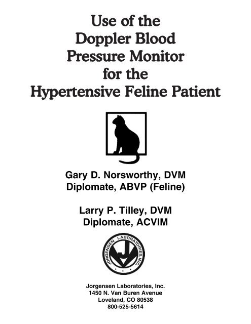
Use of <strong>the</strong><br />
<strong>Doppler</strong> <strong>Blood</strong><br />
<strong>Pressure</strong> <strong>Monitor</strong><br />
for <strong>the</strong><br />
Hypertensive Feline Patient<br />
Gary D. Norsworthy, DVM<br />
Diplomate, ABVP (Feline)<br />
Larry P. Tilley, DVM<br />
Diplomate, ACVIM<br />
<strong>Jorgensen</strong> <strong>Laboratories</strong>, Inc.<br />
1450 N. Van Buren Avenue<br />
Loveland, CO 80538<br />
800-525-5614
Definition<br />
Hypertension, commonly called high blood<br />
pressure, is a sustained elevation in ei<strong>the</strong>r<br />
systolic or diastolic arterial blood pressure<br />
above normal range. Several values are found<br />
in <strong>the</strong> current veterinary literature for what<br />
constitutes hypertension in cats. Some<br />
authors feel that systolic values above 160<br />
mmHg are abnormal. O<strong>the</strong>r authors define<br />
hypertension as systolic values above 200<br />
mmHg. It has been shown that stress and<br />
environmental factors can influence blood<br />
pressure readings; <strong>the</strong>refore, some of <strong>the</strong><br />
variation in <strong>the</strong>se values is likely due to <strong>the</strong><br />
conditions under which <strong>the</strong> readings were<br />
made. However, <strong>the</strong>re appears to be universal<br />
agreement that systolic readings above 200<br />
mmHg are abnormal.<br />
Causes<br />
Hypertension in Feline Patients<br />
Primary or essential hypertension is a<br />
common disease in humans. However, it is<br />
apparently a rare disease in <strong>the</strong> cat, if it<br />
occurs at all. Most cases of feline hypertension<br />
have been closely related to two disease<br />
mechanisms: 1) Diseases that increase<br />
peripheral resistance (chronic renal failure),<br />
and 2) Diseases that increase cardiac output<br />
(hyperthyroidism).<br />
Gary D. Norsworthy, DVM, DABVP<br />
Larry P. Tilley, DVM, DACVIM<br />
Chronic renal disease is <strong>the</strong> most common<br />
disease that increases peripheral resistance.<br />
A very simplified explanation states that aged,<br />
shrunken kidneys, that normally receive 20%<br />
of <strong>the</strong> cardiac output, are unable to<br />
accommodate that amount of blood.<br />
Therefore, blood is regurgitated into <strong>the</strong> aorta.<br />
However, many cats that are hypertensive<br />
only have a slight degree of azotemia and<br />
may have<br />
kidneys of relatively normal size.<br />
This observation has caused us to seek o<strong>the</strong>r<br />
explanations. Several rationales have been<br />
given for how hypertension is related to renal<br />
failure in human patients. These include failure<br />
to excrete a normal quantity of salt or fluid,<br />
stiffening of <strong>the</strong> venous capacitance system,<br />
alterations in adrenergic activity, activation of<br />
<strong>the</strong> renin-angiotensin-aldosterone axis with<br />
increased peripheral resistance and salt<br />
retention, stimulation of renopressor systems,<br />
and suppression of renodepressors or<br />
prostaglandins. However, none of <strong>the</strong>se has<br />
been proved. One study (JAAHA 11/94) found<br />
that 65% of cats with chronic renal disease<br />
were hypertensive.<br />
Hyperthyroidism is <strong>the</strong> most common disease<br />
that causes increased cardiac output.<br />
Thyrotoxic cardiomyopathy is a common finding<br />
in hypertensive cats. It has several effects<br />
on <strong>the</strong> feline heart, <strong>the</strong> sum of which result in<br />
tachycardia, left ventricular hypertrophy, and<br />
increased contractility. The heart is hyperkinetic,<br />
resulting in increased cardiac output. One<br />
study found that 23% of hyperthyroid cats<br />
were hypertensive.<br />
It is important to understand <strong>the</strong>se mechanisms<br />
and <strong>the</strong>ir likely causes. The clinical<br />
impact is that documented hypertensive cats<br />
should have a diagnostic workup that includes<br />
renal and thyroid tests.<br />
Clinical Signs<br />
The typical clinical presentation for hypertensive<br />
cats is sudden onset retinal blindness.<br />
Owners report that <strong>the</strong>ir cats have suddenly<br />
started walking into walls and furniture. Since<br />
this presentation is so dramatic, we are likely<br />
to see <strong>the</strong>se cats only a few hours after <strong>the</strong><br />
onset of blindness, a fortunate occurrence<br />
<strong>the</strong>rapeutically.
Retinal blindness associated with hypertension<br />
is usually due to retinal detachment. The<br />
detached and folded retina can be seen with a<br />
direct or indirect ophthalmoscope. However, in<br />
many cats, <strong>the</strong> changes are so dramatic that<br />
<strong>the</strong>y may be seen without special equipment.<br />
(Figures 1a, b)<br />
Ano<strong>the</strong>r clinical sign is retinal hemorrhage,<br />
which usually precedes retinal detachment.<br />
Most cats with retinal hemorrhage, but without<br />
detachment, are not blind. However, <strong>the</strong>se<br />
cats may exhibit reduced pupillary light reflex<br />
or, if unilateral, anisocoria. (Figure 2)<br />
Hypertension may cause arterial bleeding in<br />
o<strong>the</strong>r organs, including <strong>the</strong> brain, which may<br />
result in stroke-like signs of seizures, incoordination,<br />
circling, or excessive vocalization.<br />
(Figure 3)<br />
Figure 1b: Retinal detachment can be seen from <strong>the</strong><br />
11:00 to 5:00 position even without an opthalmoscope.<br />
Figure 2: The left pupil is larger than <strong>the</strong> right due to<br />
greater retinal hemorrhage. The cat’s systolic blood<br />
pressure was 230 mmHg, and it was hyperthyroid. The<br />
pupils equalized two months following thyroidectomy.<br />
Figure 1a: Fixed and dilated pupils are characteristic of<br />
cats that are blind due to hypertension.<br />
Figure 3: This cat was presented for circling which was<br />
due to a stroke. Its systolic blood pressure was 290<br />
mmHg.
Diagnosis<br />
When retinal blindness occurs in a geriatric<br />
cat, it is important to consider hypertension as<br />
<strong>the</strong> first differential. Rapid commencement of<br />
<strong>the</strong>rapy is extremely important in restoring<br />
vision, so blood pressure readings should be<br />
performed first. <strong>Blood</strong> pressure can be measured<br />
directly with intraarterial ca<strong>the</strong>terization<br />
and by using several indirect methods.<br />
However, <strong>Doppler</strong> technology is <strong>the</strong> one that<br />
is non-invasive, non-stressful, and reliable in<br />
cats. The technique is described below.<br />
O<strong>the</strong>r signs of hypertension may be detected.<br />
Palpation or auscultation of <strong>the</strong> heart usually<br />
reveals a pounding beat. Filling of a syringe<br />
during blood collection from a peripheral vein,<br />
such as <strong>the</strong> cephalic, is much faster. Both of<br />
<strong>the</strong>se should heighten one’s index of suspicion<br />
for hypertension. Clinical findings that<br />
may signal one of <strong>the</strong> causes of hypertension<br />
are decreased renal size, as determined by<br />
palpation, radiography, or ultrasound, and a<br />
palpable thyroid lobe.<br />
Appropriate diagnostic tests include a CBC,<br />
chemistry profile,T4, urinalysis, and abdominal<br />
and cardiac imaging (radiographs or ultrasound).<br />
(Figure 4) These tests usually reveal<br />
<strong>the</strong> presence of renal disease, thyroid disease<br />
or both.<br />
If your geriatric patient with retinal blindness is<br />
not hypertensive, o<strong>the</strong>r causes of retinal blindness<br />
should be considered. Tests for feline<br />
infectious peritonitis, FeLV antigen, toxoplasmosis,<br />
and fungal diseases are appropriate.<br />
******<br />
<strong>Using</strong> <strong>the</strong> <strong>Doppler</strong> <strong>Blood</strong> <strong>Pressure</strong> <strong>Monitor</strong><br />
The doppler blood pressure monitor detects<br />
<strong>the</strong> flow of blood in arteries. The radial, cranial<br />
tibial, and median coccygeal arteries are <strong>the</strong><br />
most practical to monitor. Our preference is<br />
<strong>the</strong> cranial tibial artery because <strong>the</strong> hair<br />
clipping that is required is least noticeable.<br />
However, <strong>the</strong> o<strong>the</strong>r two arteries are acceptable<br />
and preferred by some. When <strong>the</strong> median<br />
tibial artery is used, a 2-4 cm cuff is placed<br />
above <strong>the</strong> hock. The width of <strong>the</strong> cuff is critical<br />
when using oscillometric technology, but it is<br />
not as critical with <strong>Doppler</strong> technology.<br />
Stress is a significant factor that must be minimized<br />
to get meaningful blood pressure readings.<br />
Following an automobile ride to your<br />
hospital, allow your patient a few minutes to<br />
rest in a quiet place. Be sure your patient is<br />
not exposed to dogs and o<strong>the</strong>r cats. If possible,<br />
place your client and patient in a quiet<br />
examination room and have <strong>the</strong> owner hold<br />
and reassure it for a few minutes. The presence<br />
of <strong>the</strong> owner is very helpful in relieving<br />
your patient’s stress. Take advantage of this, if<br />
possible.<br />
Simple Steps for Success<br />
1) Assemble <strong>the</strong> <strong>Doppler</strong> unit before addressing<br />
<strong>the</strong> cat. Plug in <strong>the</strong> headphones and place<br />
<strong>the</strong> ear pieces over your ears.<br />
2) Have <strong>the</strong> owner hold <strong>the</strong> cat on your examination<br />
table. Placing a towel or blanket under<br />
<strong>the</strong> cat can help make it more comfortable and<br />
more relaxed.<br />
3) Clip a 1 x 2 cm patch of hair immediately<br />
proximal to <strong>the</strong> tarsal pad or carpal pad or on<br />
<strong>the</strong> ventral surface of <strong>the</strong> tail about 4-5 cm<br />
from <strong>the</strong> body, depending upon which artery is<br />
to be used. (Figure 5)<br />
4) Position <strong>the</strong> cuff proximal to <strong>the</strong> hock, proximal<br />
to <strong>the</strong> elbow, or at <strong>the</strong> base of <strong>the</strong> tail,<br />
depending upon which artery is to be used.<br />
5) Turn on <strong>the</strong> unit and set <strong>the</strong> volume in <strong>the</strong><br />
high range. If <strong>the</strong> volume is too low, arterial<br />
sound may not be detected.<br />
6) Apply a thin coat of coupling gel to <strong>the</strong> skin.
Figure 4:The small left kidney is typical of chronic<br />
renal disease in old cats.<br />
Figure 6: The transducer is held in place with <strong>the</strong> thumb.<br />
Firm pressure is required; however, too much pressure<br />
can close <strong>the</strong> artery.<br />
Figure 5: Hair is shaved over <strong>the</strong> cranial tibial artery so<br />
<strong>the</strong> transducer can make good contact with <strong>the</strong> skin.<br />
Figure 7: The position of <strong>the</strong> needle on <strong>the</strong> manometer is<br />
noted when <strong>the</strong> arterial swish returns.<br />
7) Apply coupling gel to <strong>the</strong> ventral surface of<br />
<strong>the</strong> transducer.<br />
8) Position <strong>the</strong> transducer over <strong>the</strong> artery. The<br />
median tibial artery is usually slightly medial to<br />
midline. It will be necessary to move <strong>the</strong> transducer<br />
until <strong>the</strong> swishing of <strong>the</strong> blood is heard<br />
in <strong>the</strong> artery. Hold it in place with your thumb.<br />
(Figure 6) It may be necessary to apply firm<br />
pressure to assure proper contact between<br />
<strong>the</strong> transducer and <strong>the</strong> skin.<br />
9) Squeeze <strong>the</strong> bulb on <strong>the</strong> manometer until<br />
<strong>the</strong> pressure in <strong>the</strong> cuff exceeds that of <strong>the</strong><br />
artery. At that point <strong>the</strong> swishing of blood in<br />
<strong>the</strong> artery will no longer be heard. (Figure 7)<br />
Note that filling <strong>the</strong> cuff too rapidly may<br />
frighten your patient.<br />
10) Slowly bleed off <strong>the</strong> pressure in <strong>the</strong> cuff by<br />
squeezing <strong>the</strong> manometer’s trigger. When <strong>the</strong><br />
pressure in <strong>the</strong> cuff falls below <strong>the</strong> arterial<br />
pressure, <strong>the</strong> arterial swishing sound can be<br />
heard again. This should be noted and recorded<br />
as <strong>the</strong> systolic pressure.<br />
11) The conventional approach is for seven<br />
measurements to be taken. Discard <strong>the</strong> high
and low readings and average <strong>the</strong> remaining<br />
five. This is considered <strong>the</strong> systolic blood<br />
pressure and is measured in mmHg.<br />
However, some stressed cats may relax during<br />
<strong>the</strong> course of <strong>the</strong> examination. These cats<br />
will have several readings at a higher level followed<br />
by several at a lower level. The former<br />
should be ignored, and <strong>the</strong> latter considered<br />
as <strong>the</strong> accurate ones.<br />
The diastolic pressure can be detected in<br />
some cats by closely watching <strong>the</strong> dial of <strong>the</strong><br />
manometer. It oscillates at <strong>the</strong> diastolic pressure<br />
level. However, a reproducible reading is<br />
not always possible, especially in cats that are<br />
stressed due to catecholamine influence (that<br />
makes <strong>the</strong> vessels more stiff than normal),<br />
cats that are cold, and very small patients.<br />
However, <strong>the</strong> important measurement is systolic<br />
blood pressure in <strong>the</strong> cat.<br />
Technique Tips<br />
Do not tape <strong>the</strong> transducer in place, as is recommended<br />
by some. The slightest movement<br />
of <strong>the</strong> cat’s foot or leg will often move <strong>the</strong><br />
transducer enough that <strong>the</strong> arterial sounds are<br />
lost. Instead, hold <strong>the</strong> transducer in place with<br />
your thumb. If <strong>the</strong> artery is lost, slight movement<br />
of your thumb can relocate it. This is<br />
much faster than having to untape and retape.<br />
<strong>the</strong>ir blood pressure. The headphones bypass<br />
<strong>the</strong> external speaker so <strong>the</strong> cat no longer<br />
hears <strong>the</strong> harsh sounds. (Figure 8)<br />
Comfortably extend <strong>the</strong> cat's leg. If it is flexed,<br />
<strong>the</strong> blood flow through <strong>the</strong> artery may be<br />
abnormally restricted giving no readings or<br />
falsely low readings. (Figures 9a, b)<br />
You may notice that <strong>the</strong> blood pressure readings<br />
decline as you are making <strong>the</strong> seven<br />
measurements. This means that <strong>the</strong> cat is<br />
more comfortable and less stressed. These<br />
are actually more accurate readings, so discard<br />
<strong>the</strong> ones that were initially made.<br />
The transducer is fragile (and expensive to<br />
replace). It may be damaged by dropping it or<br />
by striking it against something solid. Treat it<br />
with utmost care.<br />
The use of ultrasound gel is essential for making<br />
good contact between <strong>the</strong> transducer and<br />
<strong>the</strong> skin. However, do not use ECG paste or<br />
baby oil. These products may cause deterioration<br />
of <strong>the</strong> resin surface of <strong>the</strong> transducer.<br />
Ultrasound gel is water soluble. Remove it<br />
from <strong>the</strong> transducer after each use.<br />
Firm pressure is required to make adequate<br />
contact between <strong>the</strong> transducer and <strong>the</strong> skin.<br />
However, do not place too much pressure on<br />
<strong>the</strong> transducer with your thumb or you will<br />
mechanically shut off <strong>the</strong> artery with <strong>the</strong> transducer.<br />
Use <strong>the</strong> maximal volume that is comfortable to<br />
your ears. Too little volume may cause you to<br />
miss <strong>the</strong> sounds.<br />
Use <strong>the</strong> headphones. Although <strong>the</strong>y are<br />
somewhat cumbersome, <strong>the</strong>y minimize <strong>the</strong><br />
stress of <strong>the</strong> procedure. The sounds of <strong>the</strong><br />
external speaker frightens many cats, elevating<br />
Figure 8: Headphones are used to bypass <strong>the</strong> external<br />
speaker to prevent frightening <strong>the</strong> cat.
Treat <strong>the</strong> Underlying Disease<br />
Cats with hyperthyroidism should be started<br />
on methimazole (Tapazole). The average initial<br />
dose is 5 mg q12h PO. Definitive treatment<br />
options for this disease are long-term<br />
methimazole, thyroidectomy, and radioactive<br />
iodine <strong>the</strong>rapy. Most cats that are treated successfully<br />
for hyperthyroidism maintain a normotensive<br />
state without fur<strong>the</strong>r specific blood<br />
pressure <strong>the</strong>rapy.<br />
Cats with renal failure should be considered<br />
for fur<strong>the</strong>r diagnostics, including an ultrasound<br />
examination, renal biopsy, and excretory urogram,<br />
to fur<strong>the</strong>r delineate <strong>the</strong> etiology of <strong>the</strong><br />
renal disease. However, geriatric cats with<br />
very small kidneys are typically diagnosed<br />
with end-stage renal disease or idiopathic<br />
tubulointerstitial nephritis. Aggressive treatment<br />
for renal failure should commence. As<br />
<strong>the</strong> BUN and creatinine improve, blood pressure<br />
should be monitored. A few cats become<br />
normotensive and no longer need <strong>the</strong>rapy for<br />
hypertension on a long-term basis.<br />
Summary<br />
Hypertension is a serious threat to geriatric<br />
cats because it is associated with two common<br />
diseases of elderly cats; hypertension<br />
and chronic renal failure. <strong>Blood</strong> pressure<br />
determinations should be performed on all<br />
aged cats for <strong>the</strong> purpose of early detection of<br />
hypertension and on all cats with retinal disease.<br />
Hypertension causes retinal blindness<br />
and can easily complicate <strong>the</strong> signs of cardiac<br />
disease and systemic metabolic disorders.<br />
O<strong>the</strong>r Uses for <strong>Blood</strong> <strong>Pressure</strong> <strong>Monitor</strong>ing<br />
A <strong>Doppler</strong> blood pressure monitor can also be<br />
used in <strong>the</strong> following situations:<br />
1. As a surgical monitor: The transducer can<br />
be taped in place during surgery so <strong>the</strong> pulse<br />
can be audibly monitored in a hands-free fashion.<br />
2. For suspected thromboembolic disease:<br />
Thromboembolic disease most often occurs<br />
secondary to hypertrophic cardiomyopathy. If<br />
blood flow impairment is suspected to any<br />
limb, <strong>the</strong> <strong>Doppler</strong> can be used to determine<br />
<strong>the</strong> blood flow in <strong>the</strong> distal artery of <strong>the</strong> limb. If<br />
a thrombus is present, <strong>the</strong> blood pressure will<br />
be very low or zero.<br />
3. For suspected vascular compromise to a<br />
traumatized limb or tail: The <strong>Doppler</strong> can be<br />
used in a similar manner as above when vascular<br />
impairment is suspected due to trauma.<br />
4. For monitoring renal failure patients:<br />
Hypertension may be an ongoing disease in<br />
cats that are undergoing treatment for chronic<br />
renal disease. The only way to know if<br />
hypotensive <strong>the</strong>rapy is indicated and to properly<br />
adjust <strong>the</strong> dose is to perform serial blood<br />
pressure determinations.<br />
Suggested Readings<br />
Goodwin JK. Systemic Hypertension. In:<br />
Norsworthy GD, ed. The Feline Patient:<br />
Essentials of Diagnosis and Treatment.<br />
Philadelphia: Williams and Wilkins, 1998.<br />
Thornhill JA. Hypertension, Systemic. In: Tilley<br />
LP, Smith FWK, eds. The Five Minute Clinical<br />
Consult. Philadelphia: Williams and Wilkins,<br />
1997:706-707.
<strong>Jorgensen</strong> <strong>Laboratories</strong>, Inc.<br />
Loveland, Colorado