Pathophysiology of Migraine - Kompetenznetz Parkinson
Pathophysiology of Migraine - Kompetenznetz Parkinson
Pathophysiology of Migraine - Kompetenznetz Parkinson
You also want an ePaper? Increase the reach of your titles
YUMPU automatically turns print PDFs into web optimized ePapers that Google loves.
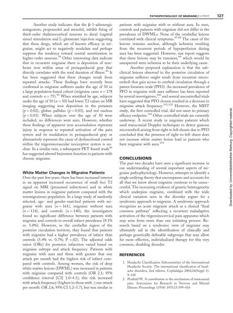
Another study indicates that the b-1-adrenergic<br />
antagonists, propranolol and atenolol, inhibit firing <strong>of</strong><br />
third-order thalamocortical neurons to dural (sagittal<br />
sinus) stimulation and L-glutamate injection suggesting<br />
that these drugs, which are <strong>of</strong> known efficacy in migraine,<br />
might act to negatively modulate and perhaps<br />
suppress the tendency toward central sensitization in<br />
higher-order neurons. 93 Other interesting data indicate<br />
that in recurrent migraine there is deposition <strong>of</strong> nonheme<br />
iron within areas <strong>of</strong> periaqueductal gray that<br />
directly correlates with the total duration <strong>of</strong> illness. 94 It<br />
has been suggested that these changes result from<br />
repeated attacks. These findings have recently been<br />
confirmed in migraine sufferers under the age <strong>of</strong> 50 in<br />
a large population-based cohort (migraine cases n ¼ 178<br />
and controls n ¼ 75). 95 When stratified by age, subjects<br />
under the age <strong>of</strong> 50 (n ¼ 50) had lower T2 values on MR<br />
imaging suggesting iron deposition in the putamen<br />
(p ¼ 0.02), globus pallidus (p ¼ 0.03), and red nucleus<br />
(p ¼ 0.03). When subjects over the age <strong>of</strong> 50 were<br />
included, no differences were seen. However, whether<br />
these findings <strong>of</strong> apparent iron accumulation represent<br />
injury in response to repeated activation <strong>of</strong> the pain<br />
system and its modulation in periaqueductal gray or<br />
alternatively represent the cause <strong>of</strong> dysfunctional control<br />
within the trigeminovascular nociceptive system is unclear.<br />
In a similar vein, a subsequent PET-based study 96<br />
has suggested altered brainstem function in patients with<br />
chronic migraine.<br />
White Matter Changes in <strong>Migraine</strong> Patients<br />
Over the past few years, there has been increased interest<br />
in an apparent increased occurrence <strong>of</strong> small foci T2<br />
signal on MRI (presumed infarctions) and in white<br />
matter lesions in migraine patients compared with the<br />
nonmigrainous population. In a large study <strong>of</strong> randomly<br />
selected, age- and gender-matched patients with migraine<br />
with aura (n ¼ 161), migraine without aura<br />
(n ¼ 134), and controls (n ¼ 140), the investigators<br />
found no significant difference between patients with<br />
migraine and controls in overall infarct prevalence (8.1%<br />
vs. 5.0%). However, in the cerebellar region <strong>of</strong> the<br />
posterior circulation territory, they found that patients<br />
with migraine had a higher prevalence <strong>of</strong> infarct than<br />
controls (5.4% vs. 0.7%; P =.02). The adjusted odds<br />
ratios (ORs) for posterior infarction varied based on<br />
migraine subtype and attack frequency. Patients with<br />
migraine with aura and those with greater that one<br />
attack per month had the highest risk <strong>of</strong> infarct compared<br />
with controls. Among women, the risk <strong>of</strong> deep<br />
white matter lesions (DWML) was increased in patients<br />
with migraine compared with controls (OR 2.1; 95%<br />
confidence interval [CI] 1.0–4.1); this risk increased<br />
with attack frequency (highest in those with one attack<br />
per month: OR 2.6; 95% CI 1.2–5.7), but was similar in<br />
patients with migraine with or without aura. In men,<br />
controls and patients with migraine did not differ in the<br />
prevalence <strong>of</strong> DWMLs. None <strong>of</strong> the cerebellar lesions<br />
correlated with clinical symptoms. 97,98 The cause <strong>of</strong> the<br />
lesions remains unclear, although ischemia resulting<br />
from the recurrent periods <strong>of</strong> hypoperfusion during<br />
aura has been suggested. However, one report suggests<br />
that these lesions may be transient, 99 which would be<br />
unexpected were ischemia to be their underlying cause.<br />
Another proposed explanation is that the subclinical<br />
lesions observed in the posterior circulation <strong>of</strong><br />
migraine sufferers might result from recurrent microemboli<br />
that gain access to cerebral circulation through a<br />
patent foramen ovale (PFO). An increased prevalence <strong>of</strong><br />
PFO in migraine with aura sufferers has been reported<br />
by several investigators, 100 and several uncontrolled trials<br />
have suggested that PFO closure resulted in a decrease in<br />
migraine attack frequency. 101,102 However, the MIST<br />
study, the first controlled trial, did not meet its primary<br />
efficacy endpoint. 103 Other controlled trials are currently<br />
underway. A recent study in migraine patients which<br />
used transcranial Doppler techniques to detect gaseous<br />
microemboli arising from right to left shunts due to PFO<br />
concluded that the presence <strong>of</strong> right-to-left shunt does<br />
not increase white matter lesion load in patients who<br />
have migraine with aura. 104<br />
CONCLUSIONS<br />
The past two decades have seen a significant increase in<br />
our understanding <strong>of</strong> several important aspects <strong>of</strong> migraine<br />
pathophysiology. However, attempts to identify a<br />
single unifying theory that encompasses and accounts for<br />
all that we know about migraine continue to be unsuccessful.<br />
The increasing evidence <strong>of</strong> genetic heterogeneity<br />
which underpins migraine, combined with the wide<br />
clinical variation seen in the disorder argues for a<br />
syndromic approach to migraine. A syndromic approach<br />
recognizes an acute migraine attack as a clinical ‘‘final<br />
common pathway’’ reflecting a recurrent maladaptive<br />
activation <strong>of</strong> the trigeminocervical pain apparatus which<br />
may arise from more than one initiating process. Research<br />
based on a syndromic view <strong>of</strong> migraine may<br />
ultimately aid in the identification <strong>of</strong> clinically and<br />
perhaps genetically definable subgroups that may allow<br />
for more effective, individualized therapy for this very<br />
common, disabling disorder.<br />
REFERENCES<br />
PATHOPHYSIOLOGY OF MIGRAINE/CUTRER 127<br />
1. Headache Classification Subcommittee <strong>of</strong> the International<br />
Headache Society. The international classification <strong>of</strong> headache<br />
disorders, 2nd edition. Cephalalgia 2004;24(Suppl 1):<br />
9–160<br />
2. Penfield W. A contribution to the mechanism <strong>of</strong> intracranial<br />
pain. Association for Research in Nervous and Mental<br />
Disease Proceedings (1934) 1935;15:399–416<br />
Downloaded by: Universitätsbibliothek Marburg. Copyrighted material.


