in vitro PHARMACOLOGY 2011 CATALOG - Cerep
in vitro PHARMACOLOGY 2011 CATALOG - Cerep
in vitro PHARMACOLOGY 2011 CATALOG - Cerep

Create successful ePaper yourself
Turn your PDF publications into a flip-book with our unique Google optimized e-Paper software.
<strong>in</strong> <strong>vitro</strong><br />
pharmacology<br />
<strong>2011</strong> catalog
❚ france<br />
[Laboratories]<br />
Le Bois l’Evêque<br />
86600 CELLE L’EVESCAULT<br />
tel. +33 (0)5 49 89 30 00<br />
sales@cerep.com<br />
❚ usa<br />
[Laboratories]<br />
15318 N.E. 95th Street<br />
Redmond, WA 98052<br />
tel. +1 (425) 895 8666<br />
sales@cerep.com<br />
❚ japan<br />
[Sales Office]<br />
Namiki Shoji Co., Ltd.<br />
Kenseish<strong>in</strong>juku Bldg. 5-5-3<br />
Sh<strong>in</strong>juku, Sh<strong>in</strong>juku-ku<br />
TOKYO, 160-0022<br />
tel. +81 (0)3 3354 4026<br />
ishimoto@namiki-s.co.jp<br />
❚ ch<strong>in</strong>a<br />
[Laboratories]<br />
上 海 张 江 高 科 技 区 爱 迪 生 路 326 号 302-1 室<br />
(326 Aidisheng Road, B 302-1)<br />
Zhangjiang High-Tech Park<br />
Shanghai 201203<br />
tel. +86 21 5132 0568<br />
sales@cerep-ltd.com<br />
❚ www.cerep.com<br />
•<br />
•<br />
• •
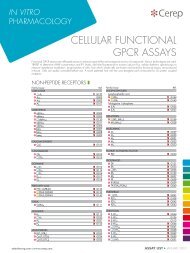
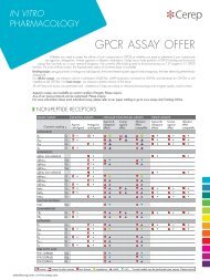
<strong>Cerep</strong> services p. 4<br />
Receptors<br />
[GPCrs - Nuclear receptors - Other receptors] p. 13<br />
Ion channels p. 93<br />
Transporters p. 103<br />
K<strong>in</strong>ases p. 107<br />
Epigenetic & DNA-related enzymes p. 157<br />
Other enzymes p. 163<br />
Specialized cellular assays p. 185<br />
Standard profiles p. 195<br />
Test<strong>in</strong>g conditions p. 203<br />
Order<strong>in</strong>g <strong>in</strong>formation p. 209<br />
Assay list& <strong>in</strong>dex p. 215
4 <strong>in</strong> <strong>vitro</strong> pharmacology <strong>2011</strong> catalog<br />
cerep services<br />
<strong>Cerep</strong> offers <strong>in</strong> <strong>vitro</strong> pharmacology, <strong>in</strong> <strong>vitro</strong> ADME-Tox and <strong>in</strong> vivo PK services, and provides solutions allow<strong>in</strong>g faster and<br />
cost-effective drug discovery by identify<strong>in</strong>g at early stages the most promis<strong>in</strong>g drug candidates as well as elim<strong>in</strong>at<strong>in</strong>g those<br />
compounds likely to fail <strong>in</strong> development.<br />
<strong>Cerep</strong>’s services benefit annually to more than 450 pharmaceutical and biotechnological companies worldwide <strong>in</strong>clud<strong>in</strong>g<br />
most of the top pharmaceutical firms.<br />
Both standard and custom research solutions are available.<br />
Standard research services <strong>in</strong>clude:<br />
compound management,<br />
high-throughput screen<strong>in</strong>g,<br />
<strong>in</strong> <strong>vitro</strong> safety profil<strong>in</strong>g,<br />
lead optimization (or SAR) profil<strong>in</strong>g,<br />
<strong>in</strong> <strong>vitro</strong> ADME profil<strong>in</strong>g,<br />
<strong>in</strong> vivo PK.<br />
Custom research services encompass assay development, profile design as well as data <strong>in</strong>terpretation and provide scientistto-scientist<br />
communications to understand and address client’s needs.<br />
Over the past 12 years, <strong>Cerep</strong> has developed BioPr<strong>in</strong>t ® , a unique database and related IT tools (see page 7), which allows<br />
the model<strong>in</strong>g of cl<strong>in</strong>ical effects of drug candidates from their molecular properties. BioPr<strong>in</strong>t ® may be used to <strong>in</strong>terpret complex<br />
profil<strong>in</strong>g data, prioritize lead or drug candidates or design lead optimization profiles.<br />
To ensure the highest quality and provide the most reproducible data, <strong>Cerep</strong> produces <strong>in</strong>-house biological material that are<br />
used <strong>in</strong> most <strong>Cerep</strong> assays and qualifies its suppliers for best quality reagents and plasticware.<br />
In 2010 <strong>Cerep</strong> has established a laboratory <strong>in</strong> Shanghai which is now fully operational to effectively support drug discovery<br />
projects that are run <strong>in</strong> Asia by provid<strong>in</strong>g the same high quality data that made <strong>Cerep</strong>’s reputation <strong>in</strong> the market.<br />
The success of this endeavor could only be achieved through the adaptation, set-up and implementation of processes which<br />
have proven their superiority <strong>in</strong> terms of quality, reproducibility, cost effectiveness and reduced timel<strong>in</strong>es.<br />
❚ broad profil<strong>in</strong>g/screen<strong>in</strong>g services<br />
<strong>Cerep</strong> cont<strong>in</strong>ues to expand and diversify the assay families, with an average of 100 assays added or updated each year.<br />
These assays are either <strong>in</strong>tegrated <strong>in</strong> the <strong>Cerep</strong> catalog, or are exclusively available to sponsor.<br />
The <strong>in</strong> <strong>vitro</strong> pharmacology and ADME-Tox & PK <strong>2011</strong> catalogs regroup about 1,300 assays, of which 473 GPCRs,<br />
<strong>in</strong>clud<strong>in</strong>g 134 cellular functional targets (agonist and antagonist effects), 255 biochemical k<strong>in</strong>ases and 32 cellular k<strong>in</strong>ase<br />
assays (activator and <strong>in</strong>hibitor effects), 51 ion channels, 61 CYPs (phenotyp<strong>in</strong>g, <strong>in</strong>hibition, <strong>in</strong>duction), 20 epigenetic and<br />
DNA-related enzymes, 14 PDEs, 24 phosphatases ...<br />
<strong>Cerep</strong>’s platforms cover a wide range of target classes <strong>in</strong>clud<strong>in</strong>g ADME-Tox related targets, GPCRs, various enzymes,<br />
transporters, nuclear receptors and ion channels; employ<strong>in</strong>g methods and assay types such as radioligand b<strong>in</strong>d<strong>in</strong>g assays,<br />
calcium mobilization and cAMP measurement us<strong>in</strong>g TR-FRET, transporter/uptake assays, and biolum<strong>in</strong>escent and other<br />
fluorescent-based assays.<br />
❚ assay design and development<br />
The customized assay development services <strong>in</strong>clude a full range of assays and technologies adapted to clients’ specific<br />
projects and needs.<br />
<strong>Cerep</strong> has an extensive and <strong>in</strong>tegrated suite of core competencies valuable to any drug discovery programs.<br />
The tool boxes shown below provide <strong>in</strong>formation about <strong>Cerep</strong>’s know-how and expertise <strong>in</strong> assay development. They are<br />
extendable to other studies, targets and technologies depend<strong>in</strong>g on your needs.<br />
Please contact us for a specific solution adapted to your specific request at: customresearch@cerep.com.
5<br />
❚ tool-box for cell-based assay development<br />
In order to study compounds activity <strong>in</strong> a liv<strong>in</strong>g cellular background, <strong>Cerep</strong> can design cellular assays for any target. For<br />
this purpose, <strong>Cerep</strong> offers a broad panel of cell-based assay solutions, for which pr<strong>in</strong>cipal families are represented here.<br />
These assays can be applied either on recomb<strong>in</strong>ant models or more physiologically relevant models such as primary<br />
cells.<br />
<br />
<strong>Cerep</strong><br />
services<br />
Ion<br />
channels<br />
G prote<strong>in</strong>-coupled<br />
receptors<br />
Growth factor<br />
receptors<br />
Cytok<strong>in</strong>e<br />
receptors<br />
Secretions<br />
ELISA<br />
Alphalisa<br />
Transporters<br />
Receptors<br />
Patch-clamp<br />
Ca 2+<br />
Fluorescence<br />
IP1<br />
TR-FRET<br />
Ca 2+<br />
PLC<br />
Fluorescence<br />
Lum<strong>in</strong>escence<br />
PKC<br />
Gq<br />
Gi<br />
AC<br />
cAMP<br />
TR-FRET<br />
Impedance<br />
CDS<br />
Gs<br />
G12/13<br />
Customized cellular assays developed at <strong>Cerep</strong> on non-recomb<strong>in</strong>ant models<br />
Rho<br />
ELISA<br />
AlphaScreen<br />
K<strong>in</strong>ases<br />
AlphaScreen<br />
Proliferation<br />
Lum<strong>in</strong>escence<br />
Radioactivity<br />
STATs<br />
AlphaScreen<br />
Cytotoxicity<br />
Lum<strong>in</strong>escence<br />
Imag<strong>in</strong>g<br />
Uptake<br />
Radioactivity<br />
SPA<br />
Ion<br />
channels<br />
Transporters<br />
K<strong>in</strong>ases<br />
Epigenetic &<br />
DNA-related<br />
enzymes<br />
EYES<br />
ECSC: x<br />
TM: x<br />
CSM: x<br />
Iris: x<br />
AIRWAY<br />
A7R5: NKCC1, L Ca channel<br />
BSMC: beta 2 , H 1<br />
NHBE: PAR1, ROCK<br />
CARDIOVASCULAR<br />
ECV304: OT<br />
HUVEC: H 1 , PAR1, VEGF, ROCK<br />
eEPC: CxCR<br />
HCAEC: PAR1<br />
HMVEC: PAR1<br />
PASMC: aCGRP<br />
SKIN<br />
A431: EGF<br />
B16F1: MC 1<br />
3T3: FGF, PDGF<br />
NHEK: beta 2 , MOP, MC 2<br />
NHDF: NKCC1<br />
Preadipocytes: x<br />
Adipocytes: x<br />
BLOOD CELLS<br />
HL60d: CxCR2<br />
THP1: CCR1, CCR2, CxCR4<br />
MM6: CxCR4<br />
Neutrophils: CXCR1/2<br />
PBMC: IL<br />
Macrophages: x<br />
Dendritic: x<br />
Platelets: x<br />
BONE<br />
SaOS2: PTH1<br />
hMSC: LTB 4<br />
Osteoblasts: LTB 4<br />
Osteoclasts: x<br />
HAIR<br />
HHDPC: x<br />
NEURAL<br />
PC12: A2 A, neurite outgrowth<br />
SK-N-MC: ETA, Y 1 , β3<br />
SK-N-SH: Na channel<br />
SH-SY-5Y: MOP, DOP<br />
NG108-15: DOP, AT 2 , CB 1<br />
IMR32: SST2<br />
Neurons: CB 1<br />
RC4BC: grehl<strong>in</strong><br />
U373MG: NK 1 , CB 1<br />
C6: 5-HT 2A<br />
1321N1: H 1 , beta 2 , M 3 , PAR1<br />
Astrocytes: mGluR5, beta 2 , ET A<br />
MAMMARY<br />
T47D: calciton<strong>in</strong><br />
MDA-MB-231: PAR1/2, IGF1, HGF, EGF<br />
PANCREATIC<br />
betaTC6: GLP-1<br />
HIT-T15: GPR119<br />
LIVER<br />
HepG2: GLP-1, INS, Tox<br />
Hepatocytes: INS, CYPs, BA transport<br />
KIDNEY<br />
HEK: TP, AT 1 , JNK1/3<br />
COLON<br />
HT29: VPAC 1<br />
UTERUS<br />
HELA: alpha 2A , beta 2 , H 1 , EGF<br />
OVARY<br />
CHO: 5-HT 1B , EP 2<br />
Red: Primary cells<br />
x: Undisclosed target (confi dentiality agreement)<br />
Other<br />
enzymes<br />
Specialized<br />
cellular<br />
assays<br />
Standard<br />
profiles<br />
Test<strong>in</strong>g<br />
conditions<br />
Order<strong>in</strong>g<br />
<strong>in</strong>formation<br />
Assay list<br />
& <strong>in</strong>dex
6 <strong>in</strong> <strong>vitro</strong> pharmacology <strong>2011</strong> catalog<br />
❚ assay design & development<br />
❚ tool-box for biochemical assays<br />
In addition to cellular assays, <strong>Cerep</strong> offers multiple biochemical assays to accurately study the <strong>in</strong>teraction of compounds<br />
with the target of <strong>in</strong>terest. These assays can be developed on recomb<strong>in</strong>ant material or native tissues.<br />
Targets B<strong>in</strong>d<strong>in</strong>g assays Functional assays<br />
GPCR Radioligand b<strong>in</strong>d<strong>in</strong>g GTPgS<br />
Ion channel Radioligand b<strong>in</strong>d<strong>in</strong>g –<br />
K<strong>in</strong>ases TR-FRET TR-FRET<br />
Other enzymes Radioligand b<strong>in</strong>d<strong>in</strong>g Multiple technologies<br />
Growth factor receptors Radioligand b<strong>in</strong>d<strong>in</strong>g –<br />
Cytok<strong>in</strong>e receptors Radioligand b<strong>in</strong>d<strong>in</strong>g –<br />
Nuclear receptors Radioligand b<strong>in</strong>d<strong>in</strong>g AlphaScreen<br />
Transporters Radioligand b<strong>in</strong>d<strong>in</strong>g –<br />
Customized b<strong>in</strong>d<strong>in</strong>g assays developed at <strong>Cerep</strong> on native animal tissues:<br />
Seroton<strong>in</strong> 5-HT 1A , 5-HT 2A <strong>in</strong> rat bra<strong>in</strong>, 5-HT 3 , 5-HT 6 , 5-HT 7 <strong>in</strong> rat cDNA<br />
Muscar<strong>in</strong>ic M 1 , M 3 <strong>in</strong> rat bra<strong>in</strong>, M 2 <strong>in</strong> rat heart<br />
Endothel<strong>in</strong> ET A <strong>in</strong> rat A-10 cells, ET B <strong>in</strong> rat cerebellum<br />
Cholecystok<strong>in</strong><strong>in</strong> CCK A , CCK B <strong>in</strong> rodent<br />
Dopam<strong>in</strong>e D 1 , D 2 <strong>in</strong> rat striatum<br />
Transporters chol<strong>in</strong>e, dopam<strong>in</strong>e and norep<strong>in</strong>ephr<strong>in</strong>e <strong>in</strong> rat striatum, 5-HT <strong>in</strong> rat bra<strong>in</strong><br />
...<br />
For a complete list, please contact us at customresearch@cerep.com.<br />
❚ tool-box for gpcrs<br />
Depend<strong>in</strong>g on the type of target, study and model, <strong>Cerep</strong> can recommend the most adapted technology.<br />
Studies Targets Gi Gq Gs G12/13<br />
Aff<strong>in</strong>ity<br />
Kon/Koff<br />
Agonist<br />
Antagonist<br />
Allosteric (PAM & NAM)<br />
Inverse agonist<br />
Weak agonist<br />
Slow agonist<br />
RLB 1<br />
RLB 1<br />
RLB 1<br />
RLB 1<br />
SPA 2 SPA 2 SPA 2 SPA 2<br />
GTPgS<br />
TR-FRET cAMP<br />
CDS 3<br />
Aequor<strong>in</strong> 4<br />
GTPgS<br />
TR-FRET cAMP<br />
CDS 3<br />
GTPgS<br />
TR-FRET cAMP<br />
CDS 3<br />
TR-FRET IP1<br />
Fluorescence<br />
CDS 3<br />
Aequor<strong>in</strong> 4<br />
TR-FRET IP1<br />
CDS 3<br />
TR-FRET IP1<br />
CDS 3<br />
TR-FRET cAMP<br />
CDS 3<br />
Aequor<strong>in</strong> 4<br />
ELISA<br />
CDS 3<br />
Aequor<strong>in</strong> 4<br />
TR-FRET cAMP<br />
CDS 3 CDS 3<br />
TR-FRET cAMP<br />
CDS 3 CDS 3<br />
Deorphanization CDS 3<br />
CDS 3<br />
CDS 3<br />
CDS 3<br />
Aequor<strong>in</strong> 4 Aequor<strong>in</strong> 4 Aequor<strong>in</strong> 4 Aequor<strong>in</strong> 4<br />
Coupl<strong>in</strong>g mechanism CDS 3 CDS 3 CDS 3 CDS 3<br />
K<strong>in</strong>ase phosphorylation AlphaScreen AlphaScreen AlphaScreen AlphaScreen<br />
1<br />
RLB: RadioLigand B<strong>in</strong>d<strong>in</strong>g<br />
2<br />
SPA: Sc<strong>in</strong>tillation Proximity Assay<br />
3<br />
CDS: Cellular Dielectric Spectroscopy<br />
4<br />
Expression of chimeric or promiscuous G prote<strong>in</strong>s may be required.
7<br />
❚ profile design<br />
<strong>Cerep</strong> offers a profile design service based on the experience and knowhow of its scientists for both content and execution<br />
of profiles. <strong>Cerep</strong> BioPr<strong>in</strong>t ® database (see below), various publicly available databases, and the scientific literature are<br />
used to design different types of profiles accord<strong>in</strong>g to your needs.<br />
<br />
<strong>Cerep</strong><br />
services<br />
❚ tool-box for profile design<br />
Type Content Applications<br />
Disease profile<br />
Side effect profile<br />
Predictive profile<br />
Targets that are<br />
associated to a<br />
particular disease<br />
Targets that are<br />
associated to a<br />
particular side effect<br />
Targets that are<br />
susceptible to be<br />
hit by a given<br />
compound<br />
- F<strong>in</strong>d a therapeutic <strong>in</strong>dication for a given compound or marketed drug (reposition<strong>in</strong>g)<br />
- F<strong>in</strong>d the target(s) responsible for the therapeutic effect (mechanism of action)<br />
- Lead optimization<br />
- Anticipate an unwanted sife effect<br />
- F<strong>in</strong>d the target(s) responsible for the therapeutic effect (mechanism of action)<br />
- Lead optimization<br />
- Assess the target selectivity of a given compound<br />
- F<strong>in</strong>d a therapeutic <strong>in</strong>dication for a given compound or marketed drug (reposition<strong>in</strong>g)<br />
- F<strong>in</strong>d the target(s) responsible for the therapeutic effect (mechanism of action)<br />
- Anticipate an unwanted sife effect<br />
- F<strong>in</strong>d the target(s) responsible for the therapeutic effect (mechanism of action)<br />
- Lead optimization<br />
Receptors<br />
Ion<br />
channels<br />
Transporters<br />
❚ biopr<strong>in</strong>t ® profile & services<br />
K<strong>in</strong>ases<br />
BioPr<strong>in</strong>t ® is a large, homogenous pharmacology and ADME<br />
database, which provides a unique resource for support<strong>in</strong>g<br />
decision mak<strong>in</strong>g <strong>in</strong> drug discovery. The database is composed<br />
of three ma<strong>in</strong> data sets:<br />
chemical descriptors (structures and chemical <strong>in</strong>formation,<br />
2D and 3D descriptors),<br />
<strong>in</strong>-house generated <strong>in</strong> <strong>vitro</strong> pharmacology and ADME<br />
data, also considered as compound descriptors,<br />
collected and curated <strong>in</strong> vivo effects of drugs.<br />
Chemical and pharmacophoric descriptors together form<br />
a data set for QSAR generation support<strong>in</strong>g organ safety<br />
model<strong>in</strong>g.<br />
BioPr<strong>in</strong>t ® positions a new drug candidate <strong>in</strong> the context of<br />
marketed drugs, anticipat<strong>in</strong>g potential <strong>in</strong> vivo liabilities,<br />
predict<strong>in</strong>g off-target activities, and ADME characteristics<br />
(Drug profile <strong>in</strong>terpretation). In another application, BioPr<strong>in</strong>t ®<br />
serves to identify secondary targets that are not genetically<br />
parented to a test target (target profile design). Both these<br />
applications are available as custom services.<br />
2,450 compounds<br />
chemical<br />
properties<br />
2D structures<br />
and 3D descriptors<br />
<strong>in</strong> <strong>vitro</strong> data<br />
consistent<br />
<strong>in</strong> <strong>vitro</strong><br />
pharmacology<br />
& ADME profiles<br />
on 159 assays<br />
QSAR<br />
cl<strong>in</strong>ical<br />
observations<br />
organ safety<br />
side effects<br />
PK<br />
therapeutic effects<br />
Epigenetic &<br />
DNA-related<br />
enzymes<br />
Other<br />
enzymes<br />
Specialized<br />
cellular<br />
assays<br />
Standard<br />
profiles<br />
❚ BIOPRINT ® PROFILE<br />
The BioPr<strong>in</strong>t ® profile <strong>in</strong>cludes the assays used to explore the properties of about 2,450 BioPr<strong>in</strong>t ® compounds (ma<strong>in</strong>ly<br />
marketed drugs and reference compounds), establish<strong>in</strong>g <strong>in</strong>dividual Pharma-ADME f<strong>in</strong>gerpr<strong>in</strong>ts for each compound.<br />
Offered on a standard basis, this profile is ma<strong>in</strong>ly based on target diversity and <strong>in</strong>cludes today 105 b<strong>in</strong>d<strong>in</strong>g assays (GPCRs,<br />
nuclear and other receptors, ion channels and transporters), 34 enzyme assays (<strong>in</strong>clud<strong>in</strong>g 10 k<strong>in</strong>ases, 10 proteases and<br />
5 phosphodiesterases), as well as 20 ADME-Tox assays (Solubility, Absorption, Metabolism and CYP-mediated drug-drug<br />
<strong>in</strong>teraction). More than 70% are human targets. The 159 assays of this profile represent a rationalized panel from a larger<br />
assay collection <strong>in</strong> the database, selected for highest <strong>in</strong>formation content.<br />
For list of assays <strong>in</strong>cluded <strong>in</strong> BioPr<strong>in</strong>t ® profile, please go to www.cerep.com catalog onl<strong>in</strong>e<br />
❚ BIOPRINT ® DRUG PROFILE INTERPRETATION<br />
Any compound <strong>in</strong> drug development, when run as a test compound on the BioPr<strong>in</strong>t ® profile, can be placed <strong>in</strong> the context<br />
of already marketed drugs, when similar <strong>in</strong> <strong>vitro</strong> profiles exist <strong>in</strong> the database. Hypotheses on cl<strong>in</strong>ical behavior of the test<br />
Test<strong>in</strong>g<br />
conditions<br />
Order<strong>in</strong>g<br />
<strong>in</strong>formation<br />
Assay list<br />
& <strong>in</strong>dex
8 <strong>in</strong> <strong>vitro</strong> pharmacology <strong>2011</strong> catalog<br />
❚ biopr<strong>in</strong>t ® profile & services [drug profile <strong>in</strong>terpretation]<br />
compounds can be drawn by comparison with data on BioPr<strong>in</strong>t ® compounds that have been collected and analyzed<br />
over the last twelve years, <strong>in</strong>clud<strong>in</strong>g:<br />
Cl<strong>in</strong>ical effects of drugs with a similar profile or sub-profile.<br />
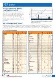
Adverse Drug Reactions (ADRs) correlated with <strong>in</strong> <strong>vitro</strong> assays: more than 5,000 significant statistical associations<br />
have been identified between the activity of molecules on a specific target and the occurrence of an ADR <strong>in</strong> man.<br />
Probability of occurrence of cl<strong>in</strong>ical effects or ADRs estimated with multivariate models us<strong>in</strong>g the complete <strong>in</strong> <strong>vitro</strong> profile:<br />
today more than 170 ADRs have been modeled.<br />
Deliverables – Phase A<br />
Primary screen<strong>in</strong>g + IC 50 s (with a 10% hits hypothesis) on BioPr<strong>in</strong>t ® profile<br />
If statistical neighbors are identified, then the follow<strong>in</strong>g deliverables will be provided as part of the report. In case no<br />
pharmacological neighbors exist, the client will be <strong>in</strong>formed at no extra charge.<br />
Deliverables – Phase B<br />
Individual hit analysis (similar hits of drugs on the market, known liabilities)<br />
Profile similarity analysis (nearest neighbor compounds by whole profile analysis, known liabilities)<br />
List of ADR associations (ADRs associated to hits of test compound and nearest neighbors)<br />
Strength of hits (with respect to a threshold of significance and PK comparison)<br />
Follow-up (suggestion on further assays and other tests)<br />
Option<br />
Neighbor compound % of <strong>in</strong>hibition and IC 50 s<br />
❚ BIOPRINT ® TARGET PROFILE DESIGN<br />
Any target, run as a test target on the BioPr<strong>in</strong>t ® compound library, can be compared to any other target <strong>in</strong> BioPr<strong>in</strong>t ® , based<br />
on its pharmacological profile (compounds that <strong>in</strong>teract with a given target). Analysis of cross-react<strong>in</strong>g compounds between<br />
targets (number and <strong>in</strong>tensity of common hits), allows to identify targets that are “pharmacophorically related” to the test target.<br />
Interest<strong>in</strong>gly, a profile of pharmacophorically related targets can differ significantly to a list of genetically related targets.<br />
BioPr<strong>in</strong>t ® target profile design represents highly valuable <strong>in</strong>formation, when sett<strong>in</strong>g up secondary screen<strong>in</strong>g tests, anticipat<strong>in</strong>g<br />
secondary targets and effectively support<strong>in</strong>g lead optimization to monitor potential secondary target related liability<br />
issues. If the test target is already part of BioPr<strong>in</strong>t ® assays, BioPr<strong>in</strong>t ® target profile design is limited to data-analysis and<br />
establishment of a profile of secondary targets. If the test target is not part of the BioPr<strong>in</strong>t ® assay panel, <strong>Cerep</strong> can develop<br />
the assay and run the BioPr<strong>in</strong>t ® compounds on an exclusive or shared data basis.<br />
Deliverables I – Analysis<br />
When relevant statistical correlations can be drawn, an analysis <strong>in</strong>clud<strong>in</strong>g the follow<strong>in</strong>gs will be performed (<strong>in</strong> case no<br />
strong correlations are identified, no charge will be <strong>in</strong>curred):<br />
Total number of compounds <strong>in</strong> BioPr<strong>in</strong>t ® that hit test target<br />
Number of compounds hitt<strong>in</strong>g secondary target<br />
Average number of BioPr<strong>in</strong>t ® targets hit by compounds react<strong>in</strong>g with test target<br />
Identity of up to 20 nearest neighbor targets (<strong>in</strong>clud<strong>in</strong>g closest CYP 450s)<br />
Prioritization of neighbor targets by number of cross-react<strong>in</strong>g compounds<br />
Digest, suggest<strong>in</strong>g a selected set of assays to cover major cross-reactivity issues<br />
Option<br />
Literature search for relevant associated targets<br />
Deliverables II – Test-target profil<strong>in</strong>g + Profile design (shared or exclusive data)<br />
Total number and ID of BioPr<strong>in</strong>t ® compounds hitt<strong>in</strong>g test target (% of <strong>in</strong>hibition, IC 50 s)<br />
Cross-react<strong>in</strong>g compound IDs, % of <strong>in</strong>hibition and IC 50 s<br />
Deliverables I <strong>in</strong>cluded<br />
REFERENCES<br />
The BioPr<strong>in</strong>t ® approach for the evaluation of ADMET properties: application to the prediction of cytochrome P450 2D6 <strong>in</strong>hibition - Gozalbes, R., et al. (2006) <strong>in</strong><br />
Pharmacok<strong>in</strong>etic profil<strong>in</strong>g <strong>in</strong> drug Research, VHCA, Zürich, WILEY-VCH, We<strong>in</strong>heim.<br />
Can we rationally design promiscuous drugs? - Hopk<strong>in</strong>s et al. (2006) et al. (2005), Current Op<strong>in</strong>ion <strong>in</strong> Structural Biology, 16:127-136<br />
Construction of a homogeneous and <strong>in</strong>formative <strong>in</strong> <strong>vitro</strong> profil<strong>in</strong>g database for anticipat<strong>in</strong>g the cl<strong>in</strong>ical effects of drugs - Froloff, N. et al. (2006) <strong>in</strong> Chemogenomics<br />
knowledge-based approaches to Drug discovery, Imperial College Press, London.<br />
QSAR model<strong>in</strong>g of <strong>in</strong> <strong>vitro</strong> <strong>in</strong>hibition of cytochrome P450 3A4 - Mao, B., et al. (2006) J. of Chem. Inf. and Model<strong>in</strong>g, 46: 2125-2134.<br />
Pharmacological and pharmaceutical profil<strong>in</strong>g: new trends - Hamon, V., et al. (2006) <strong>in</strong> The Process of New Drug Discovery and Development (2 nd Edition), Smith,<br />
C.G. & O’Donnell, J.T. (Eds.), Informa Healthcare, New York..<br />
Analysis of drug-<strong>in</strong>duced effect patterns to l<strong>in</strong>k structure and side effects of medic<strong>in</strong>es - Fliri, F. A. et al. (November 2005), Nature Chemical Biology<br />
Biological spectra analysis: L<strong>in</strong>k<strong>in</strong>g biological activity profiles to molecular structure - Fliri, F. A. et al. (2005), PNAS, 102: 261-266<br />
G-prote<strong>in</strong>-coupled receptor aff<strong>in</strong>ity prediction based on the use of a profil<strong>in</strong>g dataset: QSAR design, synthesis, and experimental validation - Rolland C., et al. (2005)<br />
Journal of Medic<strong>in</strong>al Chemistry, 48: 6563-6574.<br />
Prob<strong>in</strong>g drug action us<strong>in</strong>g <strong>in</strong> <strong>vitro</strong> pharmacological profiles - Froloff, N. (2005) Trends <strong>in</strong> Biotechnology, 23: 488-490.<br />
Predict<strong>in</strong>g ADME properties and side effects: The BioPr<strong>in</strong>t approach - Krejsa C.M., et al. (2003) Curr. Op<strong>in</strong>. Drug. Discov. Devel., 6: 470-480<br />
Neighborhood behavior of <strong>in</strong> silico structural spaces with respect to <strong>in</strong> <strong>vitro</strong> activity spaces – A novel understand<strong>in</strong>g of the molecular similarity pr<strong>in</strong>ciple <strong>in</strong> the context<br />
of multiple receptor b<strong>in</strong>d<strong>in</strong>g profiles - Horvath D. and Jeandenans C. (2003). J. Chem. Inf. & Comp. Sci., 43: 680-690.<br />
Neighborhood behavior of <strong>in</strong> silico structural spaces with respect to <strong>in</strong> <strong>vitro</strong> activity spaces – A benchmark for neighborhood behavior assessment of different <strong>in</strong> silico<br />
similarity metrics - Horvath D. and Jeandenans C. (2003) J. Chem. Inf. & Comp. Sci., 43: 691-698.<br />
Method of identification of leads or active compounds - Jean, T. and Chapela<strong>in</strong>, B. (1999) - EP 1018008B1.
9<br />
❚ data onl<strong>in</strong>e<br />
A powerful web-based, secure system to view your study <strong>in</strong>formation <strong>in</strong> real-time.<br />
<br />
<strong>Cerep</strong><br />
services<br />
Download your data and reports directly<br />
<strong>Cerep</strong>’s Data Onl<strong>in</strong>e service can be accessed us<strong>in</strong>g Internet Explorer 5+, Netscape Navigator 6+ and Mozilla.<br />
To experience the power and convenience of Data Onl<strong>in</strong>e, go to https://www.cerep.com/secure, and use “Demo” as<br />
log<strong>in</strong> and password.<br />
❚<br />
❚<br />
Two options<br />
- The real-time data option enables the sponsor to view study data as they are produced, and also to convert them <strong>in</strong>to<br />
MSExcel.<br />
- The f<strong>in</strong>al report option enables the sponsor to download complete study reports and other related documents as they<br />
are generated.<br />
Security<br />
- To deliver the world’s highest level of trust, a company specialized <strong>in</strong> provid<strong>in</strong>g High-Assurance SSL Certificates authenticates<br />
our organization, enabl<strong>in</strong>g end users to verify our site and communicate via state-of-the-art SSL encryption.<br />
- Our server supports all browser SSL encryption types (40 bit, 56 bit, and 128 bit strong encryption).<br />
- Passwords are not stored <strong>in</strong> any database; they are owned and encrypted by the operat<strong>in</strong>g system security mechanism.<br />
Only the sponsor has knowledge of the password.<br />
- You can safely enter your password and download files from the secure server.<br />
- Our network topology to access the Data Onl<strong>in</strong>e web server is also secured via state-of-the-art network security solutions.<br />
- Server operat<strong>in</strong>g systems and software are updated immediately upon receipt of the provider’s security bullet<strong>in</strong>.<br />
To experience the power and convenience of Data Onl<strong>in</strong>e, please go to www.cerep.com<br />
data onl<strong>in</strong>e<br />
Receptors<br />
Ion<br />
channels<br />
Transporters<br />
K<strong>in</strong>ases<br />
Epigenetic &<br />
DNA-related<br />
enzymes<br />
Other<br />
enzymes<br />
Specialized<br />
cellular<br />
assays<br />
❚ see also ADME-Tox & PK <strong>2011</strong> catalog<br />
ADME-Tox & PK<br />
<strong>2011</strong> <strong>CATALOG</strong><br />
IN VITRO ADME<br />
Solution properties<br />
Drug absorption/drug transport<br />
Drug metabolism<br />
Drug-drug <strong>in</strong>teractions<br />
IN VITRO TOXICITY<br />
Cardiac toxicity<br />
Cytotoxicity<br />
Genetic toxicity<br />
IN VIVO PK/BIOANALYTICAL<br />
In vivo PK<br />
Bioanalytical<br />
ASSAY SELECTION GUIDE<br />
STANDARD profiles<br />
APPLICATION NOTES<br />
Standard<br />
profiles<br />
Test<strong>in</strong>g<br />
conditions<br />
Order<strong>in</strong>g<br />
<strong>in</strong>formation<br />
Assay list<br />
& <strong>in</strong>dex
10 <strong>in</strong> <strong>in</strong> <strong>vitro</strong> pharmacology <strong>2011</strong> catalog
11<br />
<strong>in</strong> <strong>vitro</strong><br />
pharmacology<br />
assays<br />
Receptors<br />
[GPCrs]<br />
[Nuclear receptors]<br />
[Other receptors]<br />
p. 13<br />
p. 79<br />
p. 83<br />
Ion channels p. 93<br />
Transporters p. 103<br />
K<strong>in</strong>ases p. 107<br />
Epigenetic & DNA-related enzymes p. 157<br />
Other enzymes p. 163<br />
Specialized cellular assays p. 185
12 <strong>in</strong> <strong>vitro</strong> pharmacology <strong>2011</strong> catalog<br />
❚ notes<br />
❚ see also ADME-Tox & PK <strong>2011</strong> catalog<br />
ADME-Tox & PK<br />
<strong>2011</strong> <strong>CATALOG</strong><br />
IN VITRO ADME<br />
Solution properties<br />
Drug absorption/drug transport<br />
Drug metabolism<br />
Drug-drug <strong>in</strong>teractions<br />
IN VITRO TOXICITY<br />
Cardiac toxicity<br />
Cytotoxicity<br />
Genetic toxicity<br />
IN VIVO PK/BIOANALYTICAL<br />
In vivo PK<br />
Bioanalytical<br />
ASSAY SELECTION GUIDE<br />
STANDARD PROFILES<br />
APPLICATION NOTES
13<br />
<strong>Cerep</strong><br />
services<br />
receptors<br />
<br />
Receptors<br />
[GPCRs]<br />
[g prote<strong>in</strong>-coupled receptors]<br />
Ion<br />
channels<br />
❚ Adenos<strong>in</strong>e<br />
Transporters<br />
A 1 - antagonist radioligand<br />
b<strong>in</strong>d<strong>in</strong>g<br />
Ref. 0002<br />
Q 3 weeks<br />
Included <strong>in</strong>:<br />
ExpresS Profile<br />
High-throughput profile<br />
Diversity profile<br />
Source<br />
Ligand<br />
Kd<br />
Non specific<br />
Reference<br />
human recomb<strong>in</strong>ant (CHO cells)<br />
[ 3 H]DPCPX (1 nM)<br />
1.7 nM<br />
DPCPX (1 µM)<br />
DPCPX (IC 50 : 1 nM)<br />
Townsend-Nicholson, A. and Schofield, P.R. (1994) J. Biol. Chem., 269: 2373-2376<br />
specific b<strong>in</strong>d<strong>in</strong>g (% of control)<br />
100<br />
50<br />
0<br />
-11 -10 -9 -8 -7 -6 -5 -4<br />
log [drug] (M)<br />
DPCPX<br />
CPA<br />
NECA<br />
CGS 21680<br />
K<strong>in</strong>ases<br />
Epigenetic &<br />
DNA-related<br />
enzymes<br />
A 1 - agonist radioligand<br />
b<strong>in</strong>d<strong>in</strong>g<br />
Ref. 0442<br />
Q 3 weeks<br />
Included <strong>in</strong>:<br />
BioPr<strong>in</strong>t ® profile<br />
Organ safety profile<br />
Source<br />
Ligand<br />
Kd<br />
Non specific<br />
Reference<br />
human recomb<strong>in</strong>ant (CHO cells)<br />
[ 3 H]CCPA (1 nM)<br />
0.7 nM<br />
CPA (10 µM)<br />
CPA (IC 50 : 1.84 nM)<br />
specific b<strong>in</strong>d<strong>in</strong>g (% of control)<br />
100<br />
50<br />
0<br />
-11 -10 -9 -8 -7 -6 -5 -4<br />
CPA<br />
DPCPX<br />
CGS 21680<br />
log [drug] (M)<br />
Rivkees, S.A. et al. (1995) J. Biol. Chem., 270: 20485-20490.<br />
[CUSTOM OFFER] rat cerebral cortex model (Ref. 0001), please contact us at customresearch@cerep.com<br />
Other<br />
enzymes<br />
Specialized<br />
cellular<br />
assays<br />
A 1<br />
cellul ar<br />
Ref. 2814<br />
Ref. 2820<br />
Q 3 weeks<br />
Agonist effect<br />
Antagonist effect<br />
Source<br />
human recomb<strong>in</strong>ant (CHO cells)<br />
Measured product impedance<br />
Detection method cellular dielectric spectroscopy<br />
Agonist effect Control CPA (1 µM)<br />
Reference CPA (EC 50 : 1 nM)<br />
Antagonist effect Stimulant CPA (10 nM)<br />
Reference DPCPX (IC 50 : 39 nM)<br />
Cordeaux, Y. et al. (2004) Brit. J. Pharmacol., 143: 705-714.<br />
<br />
<br />
<br />
<br />
<br />
<br />
<br />
<br />
<br />
<br />
<br />
<br />
<br />
[Solvent] must be kept 0.1%<br />
<br />
<br />
<br />
<br />
<br />
<br />
<br />
Standard<br />
profiles<br />
Test<strong>in</strong>g<br />
conditions<br />
A 1 - <strong>in</strong>verse agonist effect<br />
cellul ar<br />
Ref. 2408<br />
Q 3 weeks<br />
Source<br />
Measured product cAMP<br />
Detection method HTRF<br />
human recomb<strong>in</strong>ant (CHO cells)<br />
Control<br />
CGS 15943 (10 µM)<br />
Reference CGS 15943 (EC 50 : 161 nM)<br />
[Solvent] must be kept ≤ 0.3%<br />
Shryock, J.C. et al. (1998) Mol. Pharmacol., 53: 886-893.<br />
<br />
<br />
<br />
<br />
<br />
<br />
<br />
<br />
<br />
<br />
<br />
<br />
<br />
<br />
<br />
<br />
<br />
<br />
<br />
<br />
<br />
Order<strong>in</strong>g<br />
<strong>in</strong>formation<br />
Assay list<br />
& <strong>in</strong>dex
14 <strong>in</strong> <strong>vitro</strong> pharmacology <strong>2011</strong> catalog<br />
❚ Adenos<strong>in</strong>e<br />
A 1<br />
tissue<br />
Ref. 0294<br />
Q 4 weeks<br />
Source<br />
Agonist CPA (pD 2 = 7.2)<br />
Antagonist DPCPX (pA 2 = 8.1)<br />
gu<strong>in</strong>ea-pig left atrium (electrically paced)<br />
Test concentrations 3 concentrations, n=2 (2 tissues)<br />
for both activities<br />
[Solvent] must be kept ≤ 0.1%<br />
Collis, M.G. (1983) Brit. J. Pharmacol., 78: 207-212.<br />
tension (% of control)<br />
100<br />
50<br />
-10 -9 -8 -7 -6 -5 -4<br />
log [agonist] (M)<br />
DPCPX<br />
none<br />
10 nM<br />
30 nM<br />
100 nM<br />
A 2A - agonist radioligand<br />
b<strong>in</strong>d<strong>in</strong>g<br />
Ref. 0004<br />
Q 3 weeks<br />
Included <strong>in</strong>:<br />
ExpresS Profile<br />
High-throughput profile<br />
Diversity profile<br />
BioPr<strong>in</strong>t ® profile<br />
Organ safety profile<br />
Source<br />
Ligand<br />
Kd<br />
Non specific<br />
Reference<br />
human recomb<strong>in</strong>ant (HEK-293 cells)<br />
[ 3 H]CGS 21680 (6 nM)<br />
27 nM<br />
NECA (10 µM)<br />
NECA (IC 50 : 28 nM)<br />
specific b<strong>in</strong>d<strong>in</strong>g (% of control)<br />
100<br />
50<br />
0<br />
-10 -9 -8 -7 -6 -5<br />
log [drug] (M)<br />
Luth<strong>in</strong>, D.R. et al. (1995) Mol. Pharmacol., 47: 307-313.<br />
[CUSTOM OFFER] rat striatum model (Ref. 0003), please contact us at customresearch@cerep.com<br />
NECA<br />
CGS 21680<br />
CPA<br />
DPCPX<br />
A 2A<br />
cellul ar<br />
Ref. 2055<br />
Ref. 2056<br />
Q 3 weeks<br />
Included <strong>in</strong>:<br />
Agonist effect<br />
Antagonist effect<br />
Cellular functional GPCR profile<br />
Source<br />
PC12 cells (endogenous)<br />
Measured product cAMP<br />
Detection method HTRF<br />
Agonist effect Control NECA (1 µM)<br />
Reference NECA (EC 50 : 18.7 nM)<br />
Antagonist effect Stimulant NECA (300 nM)<br />
Reference ZM 241385 (IC 50 : 1.6 nM)<br />
Gao, Z. et al. (2001) J. Pharmacol. Exp. Ther., 298: 209-218.<br />
<br />
<br />
<br />
<br />
<br />
<br />
<br />
<br />
<br />
<br />
<br />
<br />
[Solvent] must be kept 0.3%<br />
<br />
<br />
<br />
<br />
<br />
<br />
<br />
A 2A - agonist effect<br />
cellul ar<br />
Ref. 2527<br />
Q 3 weeks<br />
Source<br />
human recomb<strong>in</strong>ant (HEK-293 cells)<br />
Measured product impedance<br />
Detection method cellular dielectric spectroscopy<br />
Control<br />
NECA (1 µM)<br />
Reference NECA (EC 50 : 35 nM)<br />
[Solvent] must be kept ≤ 0.1%<br />
Gao, Z. et al. (2001) J. Pharmacol. Exp. Ther., 298: 209-218.<br />
<br />
<br />
<br />
<br />
<br />
<br />
<br />
<br />
<br />
<br />
<br />
<br />
<br />
<br />
<br />
<br />
<br />
A 2A - <strong>in</strong>verse agonist effect<br />
cellul ar<br />
Ref. 2409<br />
Q 3 weeks<br />
Source<br />
Measured product cAMP<br />
Detection method HTRF<br />
Control<br />
Reference<br />
[Solvent] must be kept ≤ 0.3%<br />
human recomb<strong>in</strong>ant (HEK-293 cells)<br />
ZM 241385 (300 nM)<br />
ZM 241385 (EC 50 : 6.33 nM)<br />
Kl<strong>in</strong>ger, M. et al. (2002) Naun.-Sch. Arch. Pharmacol., 366: 287-298.<br />
<br />
<br />
<br />
<br />
<br />
<br />
<br />
<br />
<br />
<br />
<br />
<br />
<br />
<br />
<br />
<br />
A 2A<br />
tissue<br />
Ref. 0295<br />
Q 4 weeks<br />
Source<br />
gu<strong>in</strong>ea-pig trachea<br />
(precontracted with 0.1 µM carbachol)<br />
Agonist NECA (pD 2 = 6.7)<br />
Antagonist CGS 15943A (pA 2 = 7.3)<br />
Test concentrations 3 concentrations, n=2 (2 tissues)<br />
for both activities<br />
[Solvent] must be kept ≤ 0.1%<br />
Ghai, G. et al. (1987) J. Pharmacol. Exp. Ther., 242: 784-790.<br />
tension (% of control)<br />
100<br />
<br />
50 <br />
<br />
0<br />
-9 -8 -7 -5 -6<br />
<br />
<br />
log [agonist] (M)<br />
CGS 15943A<br />
none<br />
0.1 µM<br />
0.3 µM<br />
1 µM
15<br />
Adenos<strong>in</strong>e ❚<br />
A 2B - antagonist radioligand<br />
b<strong>in</strong>d<strong>in</strong>g<br />
Ref. 0005<br />
Q 3 weeks<br />
Included <strong>in</strong>:<br />
BioPr<strong>in</strong>t ® profile<br />
Organ safety profile<br />
Source<br />
Ligand<br />
Kd<br />
Non specific<br />
Reference<br />
human recomb<strong>in</strong>ant (HEK-293 cells)<br />
[ 3 H]MRS 1754 (2 nM)<br />
1 nM<br />
NECA (100 µM)<br />
NECA (IC 50 : 1.5 µM)<br />
Ji, X. et al.(2001) Biochem. Pharmacol., 61: 657-663.<br />
<br />
<br />
<br />
<br />
<br />
<br />
<br />
<br />
<br />
<br />
<strong>Cerep</strong><br />
services<br />
<br />
Receptors<br />
[GPCRs]<br />
A 2B<br />
cellul ar<br />
Ref. 0452<br />
Ref. 0455<br />
Q 3 weeks<br />
Included <strong>in</strong>:<br />
Agonist effect<br />
Antagonist effect<br />
Cellular functional GPCR profile<br />
Source<br />
human recomb<strong>in</strong>ant (HEK-293 cells)<br />
Measured product cAMP<br />
Detection method HTRF<br />
Agonist effect Control NECA (10 µM)<br />
Reference NECA (EC 50 : 330 nM)<br />
Antagonist effect Stimulant NECA (1 µM)<br />
Reference XAC (IC 50 : 51 nM)<br />
Cooper, J. et al. (1997) Brit. J. Pharmacol., 122: 546-550.<br />
cAMP modulation (% of control)<br />
100<br />
100<br />
50<br />
50<br />
0<br />
0<br />
-9 -8 -7 -6 -5 -4<br />
-9 -8 -7 -6 -5 -4<br />
log [agonist] (M)<br />
log [antagonist] (M)<br />
NECA<br />
XAC<br />
adenos<strong>in</strong>e<br />
CGS 15943<br />
CPA<br />
DPCPX<br />
IB-MECA<br />
MRS 1754<br />
[Solvent] must be kept ≤ 0.3%<br />
Ion<br />
channels<br />
Transporters<br />
A 2B<br />
tissue<br />
Source<br />
gu<strong>in</strong>ea-pig aorta<br />
(precontracted with 1 µM phenylephr<strong>in</strong>e)<br />
Agonist NECA (pD 2 = 6.4)<br />
Antagonist 8-phenyltheophyll<strong>in</strong>e (pA 2 = 6.2)<br />
Test concentrations 3 concentrations, n=2 (2 tissues)<br />
for both activities<br />
tension (% of control)<br />
100<br />
50<br />
0<br />
8-phenyltheophyll<strong>in</strong>e<br />
none<br />
1 µM<br />
3 µM<br />
10 µM<br />
K<strong>in</strong>ases<br />
Ref. 0783<br />
Q 4 weeks<br />
[Solvent] must be kept ≤ 0.1%<br />
Gurden, M.F. et al. (1993) Brit. J. Pharmacol., 109: 693-698.<br />
-8 -7 -6 -5 -4<br />
log [agonist] (M)<br />
Epigenetic &<br />
DNA-related<br />
enzymes<br />
A 3 - agonist radioligand<br />
b<strong>in</strong>d<strong>in</strong>g<br />
Ref. 0006<br />
Q 3 weeks<br />
Included <strong>in</strong>:<br />
ExpresS Profile<br />
High-throughput profile<br />
Diversity profile<br />
BioPr<strong>in</strong>t ® profile<br />
Organ safety profile<br />
Source<br />
Ligand<br />
Kd<br />
Non specific<br />
Reference<br />
human recomb<strong>in</strong>ant (HEK-293 cells)<br />
[ 125 I]AB-MECA (0.15 nM)<br />
0.22 nM<br />
IB-MECA (1 µM)<br />
IB-MECA (IC 50 : 0,27 nM)<br />
Salvatore, C.A. et al. (1993) Proc. Natl. Acad. Sci. USA, 90: 10365-10369.<br />
<br />
<br />
<br />
<br />
<br />
<br />
<br />
<br />
<br />
<br />
Other<br />
enzymes<br />
Specialized<br />
cellular<br />
assays<br />
A 3<br />
cellul ar<br />
Ref. 0982<br />
Ref. 0983<br />
Q 3 weeks<br />
Included <strong>in</strong>:<br />
Agonist effect<br />
Antagonist effect<br />
Cellular functional GPCR profile<br />
Source<br />
human recomb<strong>in</strong>ant (CHO cells)<br />
Measured product cAMP<br />
Detection method HTRF<br />
Agonist effect Control IB-MECA (100 nM)<br />
Reference IB-MECA (EC 50 : 1.5 nM)<br />
Antagonist effect Stimulant IB-MECA (30 nM)<br />
Reference MRS 1220 (IC 50 : 125 nM)<br />
Yates, L. et al. (2006) Auton. Autacoid. Pharmacol., 26: 191-200.<br />
<br />
<br />
<br />
<br />
<br />
<br />
<br />
<br />
<br />
[Solvent] must be kept 0.3%<br />
<br />
<br />
<br />
<br />
<br />
<br />
<br />
<br />
<br />
<br />
<br />
<br />
<br />
<br />
<br />
❚ See other Adenos<strong>in</strong>e assays, page 103<br />
Standard<br />
profiles<br />
Test<strong>in</strong>g<br />
conditions<br />
<br />
❚ For radioligand b<strong>in</strong>d<strong>in</strong>g assays, how should I choose between the agonist and the antagonist models when both are<br />
available?<br />
<br />
For some b<strong>in</strong>d<strong>in</strong>g assays two models are available us<strong>in</strong>g either agonist or antagonist as radioligand. <br />
<br />
<br />
G-prote<strong>in</strong>-coupled receptors have both high-aff<strong>in</strong>ity and low-aff<strong>in</strong>ity states that are bound differently by agonists and antagonists. Whereas<br />
the antagonists b<strong>in</strong>d with an equal aff<strong>in</strong>ity to both aff<strong>in</strong>ity states, agonists b<strong>in</strong>d poorly to the low aff<strong>in</strong>ity state of the receptor. Therefore,<br />
it is advisable to use an antagonist radioligand to evaluate the b<strong>in</strong>d<strong>in</strong>g of antagonists know<strong>in</strong>g that this may fail to reveal the b<strong>in</strong>d<strong>in</strong>g<br />
of agonists. On the other hand, an assay us<strong>in</strong>g an agonist radioligand is suitable to evaluate both agonists and antagonists.<br />
The test<strong>in</strong>g of a compound <strong>in</strong> both assays and the comparison of its competition curves aga<strong>in</strong>st each radioligand may provide <strong>in</strong>formation<br />
about its functional activity at the receptor.<br />
Order<strong>in</strong>g<br />
<strong>in</strong>formation<br />
Assay list<br />
& <strong>in</strong>dex
16 <strong>in</strong> <strong>vitro</strong> pharmacology <strong>2011</strong> catalog<br />
❚ Adrenergic<br />
alpha 1 (non-selective) - antagonist radioligand<br />
b<strong>in</strong>d<strong>in</strong>g<br />
Ref. 0008<br />
Q 3 weeks<br />
Included <strong>in</strong>:<br />
ExpresS Profile<br />
High-throughput profile<br />
Diversity profile<br />
Source<br />
Ligand<br />
Kd<br />
Non specific<br />
Reference<br />
rat cerebral cortex<br />
[ 3 H]prazos<strong>in</strong> (0.25 nM)<br />
0.09 nM<br />
prazos<strong>in</strong> (0.5 µM)<br />
prazos<strong>in</strong> (IC 50 : 0.182 nM)<br />
Greengrass, P. and Bremner, R. (1979) Eur. J. Pharmacol., 55: 323-326.<br />
specific b<strong>in</strong>d<strong>in</strong>g (% of control)<br />
100<br />
50<br />
prazos<strong>in</strong><br />
WB 4101<br />
spiperone<br />
0<br />
phentolam<strong>in</strong>e<br />
-12 -11 -10 -9 -8 -7 -6 -5 -4<br />
log [drug] (M)<br />
alpha 1 (non-selective)<br />
tissue<br />
Ref. 0296<br />
Q 4 weeks<br />
Source<br />
rabbit aorta (endothelium-denuded)<br />
Agonist phenylephr<strong>in</strong>e (pD 2 = 6.5)<br />
Antagonist prazos<strong>in</strong> (pA 2 = 9.1)<br />
Test concentrations 3 concentrations, n=2 (2 tissues)<br />
for both activities<br />
[Solvent] must be kept ≤ 0.1%<br />
Docherty, J.R. et al. (1981) Naun.-Sch. Arch. Pharmacol., 317: 5-7.<br />
tension (% of max.)<br />
100<br />
50<br />
0<br />
-8 -7 -6 -5 -4<br />
log [agonist] (M)<br />
prazos<strong>in</strong><br />
none<br />
3 nM<br />
10 nM<br />
30 nM<br />
alpha 1A - antagonist radioligand<br />
b<strong>in</strong>d<strong>in</strong>g<br />
Ref. 2338<br />
Q 3 weeks<br />
Included <strong>in</strong>:<br />
BioPr<strong>in</strong>t ® profile<br />
Organ safety profile<br />
Source<br />
Ligand<br />
Kd<br />
Non specific<br />
Reference<br />
human recomb<strong>in</strong>ant (CHO cells)<br />
[ 3 H]prazos<strong>in</strong> (0.1 nM)<br />
0.1 nM<br />
ep<strong>in</strong>ephr<strong>in</strong>e (0.1 mM)<br />
WB 4101 (IC 50 : 0.36 nM)<br />
Schw<strong>in</strong>n, D.A. et al. (1990) J. Biol. Chem., 265: 8183-8189.<br />
<br />
<br />
<br />
<br />
<br />
<br />
<br />
<br />
<br />
<br />
alpha 1A - antagonist radioligand<br />
Source<br />
rat salivary glands<br />
Ligand<br />
[ 3 H]prazos<strong>in</strong> (0.06 nM)<br />
Kd<br />
0.066 nM<br />
Non specific phentolam<strong>in</strong>e (10 µM)<br />
b<strong>in</strong>d<strong>in</strong>g<br />
Reference WB 4101 (IC 50 : 0.12 nM)<br />
Ref. 0009<br />
Q 3 weeks<br />
Michel, A.D. et al. (1989) Brit. J. Pharmacol., 98: 883-889.<br />
specific b<strong>in</strong>d<strong>in</strong>g (% of control)<br />
100<br />
50<br />
0<br />
-12 -11 -10 -9 -8 -7 -6 -5<br />
log[drug] (M)<br />
WB 4101<br />
spiperone<br />
phentolam<strong>in</strong>e<br />
oxymetazol<strong>in</strong>e<br />
alpha 1A<br />
cellul ar<br />
Ref. 1500 Agonist effect<br />
Ref. 1501 Antagonist effect<br />
Q 3 weeks<br />
Included <strong>in</strong>:<br />
Cellular functional GPCR profile<br />
Source<br />
human recomb<strong>in</strong>ant (CHO cells)<br />
Measured product <strong>in</strong>tracellular [Ca 2+ ]<br />
Detection method fluorimetry<br />
Agonist effect Control ep<strong>in</strong>ephr<strong>in</strong>e (30 nM)<br />
Reference ep<strong>in</strong>ephr<strong>in</strong>e (EC 50 : 0.4 nM)<br />
Antagonist effect Stimulant ep<strong>in</strong>ephr<strong>in</strong>e (3 nM)<br />
Reference WB 4101 (IC 50 : 7 nM)<br />
Vicentic, A. et al. (2002) J. Pharmacol. Exp. Ther., 302: 58-65.<br />
Ca 2+ mobilization (% of control)<br />
100<br />
50<br />
0<br />
-10 -9 -8 -7<br />
-12 -11 -10 -9 -8 -7 -6 -5<br />
log [agonist] (M)<br />
ep<strong>in</strong>ephr<strong>in</strong>e<br />
A61603<br />
cirazol<strong>in</strong>e<br />
oxymetazol<strong>in</strong>e<br />
100<br />
[Solvent] must be kept ≤ 0.1%<br />
50<br />
0<br />
log [antagonist] (M)<br />
WB 4101<br />
prazos<strong>in</strong><br />
L765314<br />
BMY7378<br />
-11 -10<br />
-9 -8 -7 -6 -5 -4<br />
For further details and updated <strong>in</strong>formation on assays:<br />
❚ Please go to www.cerep.com catalog onl<strong>in</strong>e or contact us at sales@cerep.com<br />
-12 -11<br />
-10 -9 -8 -7<br />
-12 -11 -10 -9 -8 -7<br />
❚ Europe: +33 (0)5 49 89 30 00 – USA: +1 (425) 895 8666 – Japan: +81 (0)3 3354 4026 – Ch<strong>in</strong>a: +86 21 5132 0568<br />
-12 -11 -10 -9 -8 -7 -6 -5<br />
Assay developed <strong>in</strong> 2010 New assay conditions Human Q Standard turnaround time<br />
-9 -8 -7 -6 -5<br />
-10 -4<br />
-11 -10<br />
-9 -8 -7 -6 -5 -4
17<br />
alpha 1B - antagonist radioligand<br />
b<strong>in</strong>d<strong>in</strong>g<br />
Ref. 1633<br />
Q 3 weeks<br />
Included <strong>in</strong>:<br />
BioPr<strong>in</strong>t ® profile<br />
Source<br />
Ligand<br />
Kd<br />
Non specific<br />
Reference<br />
human recomb<strong>in</strong>ant (CHO cells)<br />
[ 3 H]prazos<strong>in</strong> (0.15 nM)<br />
0.055 nM<br />
phentolam<strong>in</strong>e (10 µM)<br />
prazos<strong>in</strong> (IC 50 : 0.25 nM)<br />
specific b<strong>in</strong>d<strong>in</strong>g (% of control)<br />
100<br />
50<br />
0<br />
-12<br />
-11 -10 -9 -8 -7 -6 -5 -4<br />
log [drug] (M)<br />
Ford, A.P.D.W. et al. (1997) Brit. J. Pharmacol., 121: 1127-1135.<br />
[CUSTOM OFFER] rat liver model (Ref. 0010), please contact us at customresearch@cerep.com<br />
adrenergic ❚<br />
prazos<strong>in</strong><br />
BMY7378<br />
oxymetazol<strong>in</strong>e<br />
phenylephr<strong>in</strong>e<br />
<strong>Cerep</strong><br />
services<br />
<br />
Receptors<br />
[GPCRs]<br />
alpha 1B<br />
cellul ar<br />
Ref. 1901<br />
Ref. 1902<br />
Q 3 weeks<br />
Included <strong>in</strong>:<br />
Agonist effect<br />
Antagonist effect<br />
Cellular functional GPCR profile<br />
Source<br />
human recomb<strong>in</strong>ant (CHO cells)<br />
Measured product cAMP<br />
Detection method HTRF<br />
Agonist effect Control ep<strong>in</strong>ephr<strong>in</strong>e (10 µM)<br />
Reference ep<strong>in</strong>ephr<strong>in</strong>e (EC 50 : 207 nM)<br />
Antagonist effect Stimulant ep<strong>in</strong>ephr<strong>in</strong>e (1 µM)<br />
Reference L765314 (IC 50 : 24 nM)<br />
Schw<strong>in</strong>n, D.A. et al. (1995) J. Pharmacol. Exp. Ther., 272: 134-142.<br />
cAMP modulation (% of control)<br />
100<br />
100<br />
50<br />
50<br />
0<br />
0<br />
-9 -8 -7 -6 -5 -4 -12 -11 -10 -9 -8 -7 -6 -5<br />
log [agonist] (M)<br />
log [antagonist] (M)<br />
ep<strong>in</strong>ephr<strong>in</strong>e<br />
L765314<br />
norep<strong>in</strong>ephr<strong>in</strong>e<br />
prazos<strong>in</strong><br />
M6434<br />
BMY7378<br />
phenylephr<strong>in</strong>e<br />
corynanth<strong>in</strong>e<br />
Ion<br />
channels<br />
Transporters<br />
alpha 1D - antagonist radioligand<br />
Source<br />
human recomb<strong>in</strong>ant (CHO cells)<br />
Ligand<br />
[ 3 H]prazos<strong>in</strong> (0.2 nM)<br />
b<strong>in</strong>d<strong>in</strong>g<br />
Kd<br />
0.15 nM<br />
Ref. 1942<br />
Non specific phentolam<strong>in</strong>e (10 µM)<br />
Q 3 weeks<br />
Reference prazos<strong>in</strong> (IC 50 : 0.38 nM)<br />
Included <strong>in</strong>:<br />
Organ safety profile<br />
Kenny, B.A. et al. (1995) Brit. J. Pharmacol., 115: 981-986.<br />
specific b<strong>in</strong>d<strong>in</strong>g (% of control)<br />
100<br />
50<br />
0<br />
-12 -11 -10 -9 -8 -7 -6 -5 -4<br />
log [drug] (M)<br />
prazos<strong>in</strong><br />
WB 4101<br />
BMY7378<br />
oxymetazol<strong>in</strong>e<br />
K<strong>in</strong>ases<br />
Epigenetic &<br />
DNA-related<br />
enzymes<br />
alpha 2 (non-selective) - antagonist radioligand<br />
b<strong>in</strong>d<strong>in</strong>g<br />
Ref. 0011<br />
Q 3 weeks<br />
Included <strong>in</strong>:<br />
ExpresS Profile<br />
High-throughput profile<br />
Diversity profile<br />
Source<br />
Ligand<br />
Kd<br />
Non specific<br />
Reference<br />
rat cerebral cortex<br />
[ 3 H]RX 821002 (0.5 nM)<br />
0.38 nM<br />
(-)ep<strong>in</strong>ephr<strong>in</strong>e (100 µM)<br />
yohimb<strong>in</strong>e (IC 50 : 58.7 nM)<br />
Uhlen, S. and Wikberg, J.E. (1991) Pharmacol. Toxicol., 69: 341-350.<br />
specific b<strong>in</strong>d<strong>in</strong>g (% of control)<br />
100<br />
50<br />
0<br />
-11 -10 -9 -8 -7 -6 -5<br />
log [drug] (M)<br />
yohimb<strong>in</strong>e<br />
RX 821002<br />
oxymetazol<strong>in</strong>e<br />
prazos<strong>in</strong><br />
Other<br />
enzymes<br />
Specialized<br />
cellular<br />
assays<br />
selected cerep assays<br />
❚ b<strong>in</strong>d<strong>in</strong>g assays<br />
B<strong>in</strong>d<strong>in</strong>g assays are developed accord<strong>in</strong>g to the competition assay pr<strong>in</strong>ciple. There are three assay formats used: filtration, sc<strong>in</strong>tillation<br />
proximity assay (SPA) and centrifugation. All formats utilize a radiolabeled ligand and a source of receptor (membranes, soluble/purified).<br />
Large batches of cells, from which prepared membranes are frozen and stored, are produced <strong>in</strong>-house. Membrane preparations are<br />
quality controlled before use.<br />
❚ cellular Functional GPCR assays<br />
Functional GPCR assays are cell-based assays to measure agonist-like, <strong>in</strong>verse agonist, antagonist and modulator activity of compounds.<br />
Various technologies are used: TR-FRET to determ<strong>in</strong>e cAMP concentration and IP1 levels, real time fluorescence to monitor calcium flux,<br />
cellular dielectric spectroscopy to measure impedance modulation. Large batches of cells, from which whole cells are frozen and stored<br />
for functional assays, are produced <strong>in</strong>-house. Cells are quality controlled before use. A novel patented host cell l<strong>in</strong>e was designed and<br />
constructed for Gi prote<strong>in</strong> coupled receptors.<br />
❚ tissue bioassays<br />
Tissue bioassays are functional assays designed to evaluate the agonist and antagonist activities of compounds at various receptors and<br />
ion channels <strong>in</strong> whole tissues. The tissues used are isolated from contractile cardiac and smooth muscles of the cardiovascular, respiratory,<br />
gastro<strong>in</strong>test<strong>in</strong>al and urogenital tracts of rodents, rabbit or human. The selected tissues are assayed <strong>in</strong> organ bath systems to record the<br />
contractile activity <strong>in</strong> conditions as selective as possible to avoid any potential <strong>in</strong>teraction with other receptors known to be present <strong>in</strong><br />
the tissue used.<br />
Standard<br />
profiles<br />
Test<strong>in</strong>g<br />
conditions<br />
Order<strong>in</strong>g<br />
<strong>in</strong>formation<br />
Assay list<br />
& <strong>in</strong>dex
18 <strong>in</strong> <strong>vitro</strong> pharmacology <strong>2011</strong> catalog<br />
❚ adrenergic<br />
alpha 2 (non-selective)<br />
tissue<br />
Ref. 0297<br />
Q 4 weeks<br />
Source<br />
rabbit ear ve<strong>in</strong><br />
Agonist clonid<strong>in</strong>e (pD 2 = 7.9)<br />
Antagonist yohimb<strong>in</strong>e (pA 2 = 8.1)<br />
Test concentrations 3 concentrations, n=2 (2 tissues)<br />
for both activities<br />
[Solvent] must be kept ≤ 0.1%<br />
Daly, C.J. et al. (1988) Brit. J. Pharmacol., 94: 1085-1090.<br />
tension (% of max.)<br />
100<br />
50<br />
0<br />
-10 -9 -8 -7 -6 -5<br />
log [agonist] (M)<br />
yohimb<strong>in</strong>e<br />
none<br />
30 nM<br />
100 nM<br />
300 nM<br />
alpha 2A - antagonist radioligand<br />
b<strong>in</strong>d<strong>in</strong>g<br />
Ref. 0013<br />
Q 3 weeks<br />
Included <strong>in</strong>:<br />
BioPr<strong>in</strong>t ® profile<br />
Source<br />
Ligand<br />
Kd<br />
Non specific<br />
Reference<br />
human recomb<strong>in</strong>ant (CHO cells)<br />
[ 3 H]RX 821002 (1 nM)<br />
0.8 nM<br />
(-)ep<strong>in</strong>ephr<strong>in</strong>e (100 µM)<br />
yohimb<strong>in</strong>e (IC 50 : 5.5 nM)<br />
Lang<strong>in</strong>, D. et al. (1989) Eur. J. Pharmacol., 167: 95-104.<br />
specific b<strong>in</strong>d<strong>in</strong>g (% of control)<br />
100<br />
50<br />
0<br />
-11 -10 -9 -8 -7 -6 -5<br />
log [drug] (M)<br />
yohimb<strong>in</strong>e<br />
prazos<strong>in</strong><br />
RX 821002<br />
oxymetazol<strong>in</strong>e<br />
alpha 2A - agonist radioligand<br />
Source<br />
human recomb<strong>in</strong>ant (CHO cells)<br />
Ligand<br />
[ 3 H](-)ep<strong>in</strong>ephr<strong>in</strong>e (1 nM)<br />
b<strong>in</strong>d<strong>in</strong>g<br />
Kd<br />
0.7 nM<br />
Ref. 1669<br />
Non specific RX 821002 (1 µM)<br />
Q 3 weeks<br />
Reference ep<strong>in</strong>ephr<strong>in</strong>e (IC 50 : 2.6 nM)<br />
Included <strong>in</strong>:<br />
Organ safety profile<br />
Senard, J.M. et al. (1989) Eur. J. Pharmacol., 162: 225-236.<br />
specific b<strong>in</strong>d<strong>in</strong>g (% of control)<br />
100<br />
50<br />
0<br />
-11 -10 -9 -8 -7 -6 -5<br />
log [drug] (M)<br />
ep<strong>in</strong>ephr<strong>in</strong>e<br />
oxymetazol<strong>in</strong>e<br />
RX 821002<br />
prazos<strong>in</strong><br />
alpha 2A<br />
cellul ar<br />
Ref. 2558<br />
Ref. 2559<br />
Q 3 weeks<br />
Agonist effect<br />
Antagonist effect<br />
Source<br />
human recomb<strong>in</strong>ant (CHO cells)<br />
Measured product impedance<br />
Detection method cellular dielectric spectroscopy<br />
Agonist effect Control ep<strong>in</strong>ephr<strong>in</strong>e (100 nM)<br />
Reference ep<strong>in</strong>ephr<strong>in</strong>e (EC 50 : 4 nM)<br />
Antagonist effect Stimulant ep<strong>in</strong>ephr<strong>in</strong>e (10 nM)<br />
Reference RX 821002 (IC 50 : 14 nM)<br />
Bavadekar, S.A. et al. (2006) J. Pharmacol. Exp. Ther., 31: 739-748.<br />
<br />
<br />
<br />
<br />
<br />
<br />
<br />
<br />
<br />
<br />
<br />
<br />
<br />
<br />
<br />
<br />
<br />
<br />
[Solvent] must be kept 0.1%<br />
alpha 2B - antagonist radioligand<br />
b<strong>in</strong>d<strong>in</strong>g<br />
Ref. 1344<br />
Q 3 weeks<br />
Included <strong>in</strong>:<br />
BioPr<strong>in</strong>t ® profile<br />
Organ safety profile<br />
Source<br />
Ligand<br />
Kd<br />
Non specific<br />
Reference<br />
human recomb<strong>in</strong>ant (CHO cells)<br />
[ 3 H]RX 821002 (2.5 nM)<br />
5 nM<br />
(-)ep<strong>in</strong>ephr<strong>in</strong>e (100 µM)<br />
yohimb<strong>in</strong>e (IC 50 : 5.2 nM)<br />
specific b<strong>in</strong>d<strong>in</strong>g (% of control)<br />
<br />
<br />
<br />
<br />
<br />
100<br />
<br />
<br />
<br />
<br />
<br />
<br />
50<br />
<br />
0<br />
<br />
<br />
-10<br />
<br />
<br />
<br />
<br />
<br />
<br />
<br />
-9 -8 -7 -6 -5<br />
<br />
yohimb<strong>in</strong>e<br />
RX 821002<br />
prazos<strong>in</strong><br />
oxymetazol<strong>in</strong>e<br />
log [drug] (M)<br />
Devedjian, J.-C. et al. (1994) Eur. J. Pharmacol., 252: 43-49.<br />
[CUSTOM OFFER] mouse NG108-15 cells model (Ref. 0014), please contact us at customresearch@cerep.com<br />
alpha 2B<br />
cellul ar<br />
Ref. 1813<br />
Ref. 0299<br />
Q 3 weeks<br />
Agonist effect<br />
Antagonist effect<br />
Source<br />
human recomb<strong>in</strong>ant (HEK-293 cells)<br />
Measured product cAMP<br />
Detection method HTRF<br />
Agonist effect Control dexmedetomid<strong>in</strong>e (1 µM)<br />
Reference dexmedetomid<strong>in</strong>e (EC 50 : 5.5 nM)<br />
Antagonist effect Stimulant dexmedetomid<strong>in</strong>e (30 nM)<br />
Reference yohimb<strong>in</strong>e (IC 50 : 320 nM)<br />
Eason, M.G. et al. (1992) J. Biol. Chem., 267: 15795-15801.<br />
<br />
<br />
<br />
<br />
<br />
<br />
<br />
<br />
<br />
<br />
<br />
[Solvent] must be kept 0.3%
19<br />
adrenergic ❚<br />
alpha 2C - antagonist radioligand<br />
b<strong>in</strong>d<strong>in</strong>g<br />
Ref. 0016<br />
Q 3 weeks<br />
Included <strong>in</strong>:<br />
BioPr<strong>in</strong>t ® profile<br />
Source<br />
Ligand<br />
Kd<br />
Non specific<br />
Reference<br />
human recomb<strong>in</strong>ant (CHO cells)<br />
[ 3 H]RX 821002 (2 nM)<br />
0.95 nM<br />
(-)ep<strong>in</strong>ephr<strong>in</strong>e (100 µM)<br />
yohimb<strong>in</strong>e (IC 50 : 1.9 nM)<br />
Devedjian, J.-C. et al. (1994) Eur. J. Pharmacol., 252: 43-49.<br />
specific b<strong>in</strong>d<strong>in</strong>g (% of control)<br />
100<br />
50<br />
0<br />
-10 -9 -8 -7 -6 -5<br />
log [drug] (M)<br />
yohimb<strong>in</strong>e<br />
RX 821002<br />
prazos<strong>in</strong><br />
oxymetazol<strong>in</strong>e<br />
<strong>Cerep</strong><br />
services<br />
<br />
Receptors<br />
[GPCRs]<br />
alpha 2C - agonist radioligand<br />
Source<br />
human recomb<strong>in</strong>ant (CHO cells)<br />
Ligand<br />
[ 3 H](-)ep<strong>in</strong>ephr<strong>in</strong>e (1 nM)<br />
b<strong>in</strong>d<strong>in</strong>g<br />
Kd<br />
0.9 nM<br />
Ref. 1682<br />
Non specific RX 821002 (1 µM)<br />
Q 3 weeks<br />
Reference ep<strong>in</strong>ephr<strong>in</strong>e (IC 50 : 2.87 nM)<br />
Included <strong>in</strong>:<br />
Organ safety profile<br />
Bylund, D.B. et al. (1992) Mol. Pharmacol., 42: 1-5.<br />
specific b<strong>in</strong>d<strong>in</strong>g (% of control)<br />
100<br />
50<br />
0<br />
-11 -10 -9 -8 -7 -6 -5<br />
log [drug] (M)<br />
ep<strong>in</strong>ephr<strong>in</strong>e<br />
oxymetazol<strong>in</strong>e<br />
RX 821002<br />
prazos<strong>in</strong><br />
Ion<br />
channels<br />
Transporters<br />
alpha 2C<br />
cellul ar<br />
Ref. 1736<br />
Ref. 1737<br />
Q 3 weeks<br />
Included <strong>in</strong>:<br />
Agonist effect<br />
Antagonist effect<br />
Cellular functional GPCR profile<br />
Source<br />
human recomb<strong>in</strong>ant (CHO cells)<br />
Measured product cAMP<br />
Detection method HTRF<br />
Agonist effect Control ep<strong>in</strong>ephr<strong>in</strong>e (1 µM)<br />
Reference ep<strong>in</strong>ephr<strong>in</strong>e (EC 50 : 2.6 nM)<br />
Antagonist effect Stimulant ep<strong>in</strong>ephr<strong>in</strong>e (100 nM)<br />
Reference rauwolsc<strong>in</strong>e (IC 50 : 55 nM)<br />
Regan, J.W. et al. (1988) Biochem., 85: 6301-6305.<br />
cAMP modulation (% of control)<br />
100<br />
50<br />
0<br />
-12 -11<br />
-10 -9 -8 -7 -6 -5<br />
log [agonist] (M)<br />
ep<strong>in</strong>ephr<strong>in</strong>e<br />
oxymetazol<strong>in</strong>e<br />
clonid<strong>in</strong>e<br />
UK 14,304<br />
100<br />
50<br />
0<br />
-9 -8 -7 -6 -5<br />
-10 -4<br />
log [antagonist] (M)<br />
rauwolsc<strong>in</strong>e<br />
yohimb<strong>in</strong>e<br />
prazos<strong>in</strong><br />
RX 821002<br />
K<strong>in</strong>ases<br />
Epigenetic &<br />
DNA-related<br />
enzymes<br />
beta 1 - agonist radioligand<br />
b<strong>in</strong>d<strong>in</strong>g<br />
Ref. 0018<br />
Q 3 weeks<br />
Included <strong>in</strong>:<br />
ExpresS Profile<br />
High-throughput profile<br />
Diversity profile<br />
BioPr<strong>in</strong>t ® profile<br />
Organ safety profile<br />
Source<br />
Ligand<br />
Kd<br />
Non specific<br />
Reference<br />
human recomb<strong>in</strong>ant (HEK-293 cells)<br />
[ 3 H](-)CGP 12177 (0.15 nM)<br />
0.39 nM<br />
alprenolol (50 µM)<br />
atenolol (IC 50 : 278 nM)<br />
specific b<strong>in</strong>d<strong>in</strong>g (% of control)<br />
-12 -11<br />
100<br />
-10 -9 -8 -7 -6 -5<br />
50<br />
-9 -8 -7 -6 -5<br />
-10 -4<br />
0<br />
-10 -9 -8 -7 -6 -5 -4<br />
log [drug] (M)<br />
Lev<strong>in</strong>, M.C. et al. (2002) J. Biol.Chem., 277: 30429-30435.<br />
[CUSTOM OFFER] rat heart model (Ref. 0017), please contact us at customresearch@cerep.com<br />
atenolol<br />
CGP 20712A<br />
propranolol<br />
ICI 118551<br />
Other<br />
enzymes<br />
Specialized<br />
cellular<br />
assays<br />
beta 1<br />
cellul ar<br />
Ref. 1605<br />
Ref. 1606<br />
Q 3 weeks<br />
Included <strong>in</strong>:<br />
Agonist effect<br />
Antagonist effect<br />
Cellular functional GPCR profile<br />
Source<br />
human recomb<strong>in</strong>ant (HEK-293 cells)<br />
Measured product cAMP<br />
Detection method HTRF<br />
Agonist effect Control isoproterenol (100 nM)<br />
Reference isoproterenol (EC 50 : 0.3 nM)<br />
Antagonist effect Stimulant isoproterenol (1 nM)<br />
Reference atenolol (IC 50 : 150 nM)<br />
Frielle, T. et al. (1987) Proc. Natl. Acad. Sci. USA, 84: 7920-7924.<br />
cAMP modulation (% of control)<br />
100<br />
100<br />
50<br />
50<br />
0<br />
0<br />
-12 -11 -10 -9 -8 -7 -6 -5 -4 -3 -9 -8 -7 -6 -5 -4<br />
log [agonist] (M)<br />
log [antagonist] (M)<br />
isoproterenol<br />
atenolol<br />
norep<strong>in</strong>ephr<strong>in</strong>e<br />
propranolol<br />
xamoterol<br />
CGP 20712A<br />
salbutamol<br />
ICI 118551<br />
[Solvent] must be kept ≤ 0.3%<br />
Standard<br />
profiles<br />
Test<strong>in</strong>g<br />
conditions<br />
beta 1 - <strong>in</strong>verse agonist effect<br />
cellul ar<br />
Ref. 2377<br />
Q 3 weeks<br />
Source<br />
human recomb<strong>in</strong>ant (HEK-293 cells)<br />
Measured product cAMP<br />
Detection method HTRF<br />
Control<br />
ICI 118551 (10 µM)<br />
Reference ICI 118551 (EC 50 : 93.8 nM)<br />
[Solvent] must be kept ≤ 0.3%<br />
Lattion, A.L. et al. (1999) FEBS Lett., 457: 302-306.<br />
<br />
<br />
<br />
-12 -11 -10 -9 -8 -7 -6 -5<br />
-9 -8 -7 -6 -5<br />
-10 -4<br />
<br />
<br />
<br />
<br />
<br />
Order<strong>in</strong>g<br />
<strong>in</strong>formation<br />
Assay list<br />
& <strong>in</strong>dex
20 <strong>in</strong> <strong>vitro</strong> pharmacology <strong>2011</strong> catalog<br />
❚ adrenergic<br />
beta 1<br />
tissue<br />
Ref. 0298<br />
Q 4 weeks<br />
Source<br />
gu<strong>in</strong>ea-pig right atrium<br />
Agonist isoproterenol (pD 2 = 8.3)<br />
Antagonist atenolol (pA 2 = 7.2)<br />
Test concentrations 3 concentrations, n=2 (2 tissues)<br />
for both activities<br />
[Solvent] must be kept ≤ 0.1%<br />
O’Donell, S.R. and Wanstall, J.C. (1979) Naun.-Sch. Arch. Pharm., 308: 183-190.<br />
frequency (% of max.)<br />
100<br />
50<br />
0<br />
-11 -10 -9 -8 -7 -6<br />
log [agonist] (M)<br />
atenolol<br />
none<br />
0.1 µM<br />
0.3 µM<br />
1 µM<br />
beta 2 - agonist radioligand<br />
b<strong>in</strong>d<strong>in</strong>g<br />
Ref. 0020<br />
Q 3 weeks<br />
Included <strong>in</strong>:<br />
ExpresS Profile<br />
High-throughput profile<br />
Diversity profile<br />
BioPr<strong>in</strong>t ® profile<br />
Organ safety profile<br />
Source<br />
Ligand<br />
Kd<br />
Non specific<br />
Reference<br />
human recomb<strong>in</strong>ant (CHO cells)<br />
[ 3 H](-)CGP 12177 (0.3 nM)<br />
0.15 nM<br />
alprenolol (50 µM)<br />
ICI 118551 (IC 50 : 0.8 nM)<br />
Joseph, S.S. et al. (2004) Naun.-Sch. Arch. Pharm., 369: 525-532.<br />
<br />
<br />
<br />
<br />
<br />
<br />
<br />
<br />
<br />
<br />
beta 2<br />
cellul ar<br />
Ref. 1976<br />
Ref. 1977<br />
Q 3 weeks<br />
Included <strong>in</strong>:<br />
Agonist effect<br />
Antagonist effect<br />
Cellular functional GPCR profile<br />
Source<br />
human recomb<strong>in</strong>ant (CHO cells)<br />
Measured product cAMP<br />
Detection method HTRF<br />
Agonist effect Control isoproterenol (0.1 µM)<br />
Reference isoproterenol (EC 50 : 0.83 nM)<br />
Antagonist effect Stimulant isoproterenol (3 nM)<br />
Reference ICI 118551 (IC 50 : 6 nM)<br />
Baker, J.G. (2005) Brit. J. Pharmacol., 144: 317-322.<br />
cAMP modulation (% of control)<br />
100<br />
50<br />
0<br />
-11 -10 -9 -8 -7 -6 -5<br />
log [agonist] (M)<br />
isoproterenol<br />
salbutamol<br />
salmeterol<br />
denopam<strong>in</strong>e<br />
100<br />
50<br />
0<br />
-11 -10 -9 -8 -7 -6 -5<br />
log [antagonist] (M)<br />
ICI 118551<br />
CGP 20712A<br />
SR 59230A<br />
propranolol<br />
beta 2 - <strong>in</strong>verse agonist effect<br />
cellul ar<br />
Ref. 2407<br />
Q 3 weeks<br />
Source<br />
human recomb<strong>in</strong>ant (CHO cells)<br />
Measured product cAMP<br />
Detection method HTRF<br />
Control<br />
ICI 118551 (1 µM)<br />
Reference ICI 118551 (EC 50 : 3.8 nM)<br />
[Solvent] must be kept ≤ 0.3%<br />
Baker, J.G. et al. (2003) Mol. Pharmacol., 64: 1357-1369.<br />
<br />
-12 -11<br />
<br />
<br />
-10 -9 -8 -7 -6 -5<br />
-9 -8 -7 -6 -5<br />
-10 -4<br />
<br />
<br />
<br />
<br />
<br />
beta 2<br />
tissue<br />
Source<br />
gu<strong>in</strong>ea-pig trachea<br />
(precontracted with 0.1 µM carbachol)<br />
Agonist salbutamol (pD 2 = 7.7)<br />
Antagonist ICI 118551 (pA 2 = 9.1)<br />
Test concentrations 3 concentrations, n=2 (2 tissues)<br />
for both activities<br />
tension (% of control)<br />
100<br />
<br />
50<br />
<br />
<br />
0<br />
ICI 118551<br />
none<br />
3 nM<br />
10 nM<br />
30 nM<br />
<br />
Ref. 0299<br />
Q 4 weeks<br />
[Solvent] must be kept ≤ 0.1%<br />
O’Donell, S.R. and Wanstall, J.C. (1979) Naun.-Sch.Arch. Pharm.,308: 183-190.<br />
-10 -9 -8 -7 -6 -5<br />
log [agonist] (M) <br />
beta 3 - antagonist radioligand<br />
b<strong>in</strong>d<strong>in</strong>g<br />
Ref. 0227<br />
Q 3 weeks<br />
Included <strong>in</strong>:<br />
BioPr<strong>in</strong>t ® profile<br />
Source<br />
Ligand<br />
Kd<br />
Non specific<br />
Reference<br />
SK-N-MC cells (endogenous)<br />
[ 125 I]CYP (0.6 nM) (+ 0.1 µM (-)propranolol)<br />
0.7 nM<br />
(-)propranolol (1 mM)<br />
cyanop<strong>in</strong>dolol (IC 50 : 37 nM)<br />
Curran, P.K. and Fishman, P.H. (1996) Cell Signal., 8: 355-364.<br />
specific b<strong>in</strong>d<strong>in</strong>g (% of control)<br />
100<br />
50<br />
0<br />
-10 -9 -8 -7 -6 -5 -4 -3<br />
log [drug] (M)<br />
cyonop<strong>in</strong>dolol<br />
(-)propranolol<br />
ICI 118551<br />
atenolol
adrenergic ❚<br />
21<br />
beta 3<br />
cellul ar<br />
Ref. 2189<br />
Ref. 0020<br />
Q 3 weeks<br />
Agonist effect<br />
Antagonist effect<br />
Source<br />
SK-N-MC cells (endogenous)<br />
Measured product cAMP<br />
Detection method HTRF<br />
Agonist effect Control isoproterenol (100 µM)<br />
Reference isoproterenol (EC 50 : 2.7 µM)<br />
Antagonist effect Stimulant isoproterenol (5 µM)<br />
Reference cyanop<strong>in</strong>dolol (IC 50 : 300 nM)<br />
Curran, P.K. and Fishman, P.H. (1996) Cell signal., 8: 355-364.<br />
<br />
<br />
<br />
<br />
<br />
<br />
<br />
<br />
<br />
<br />
<br />
<br />
[Solvent] must be kept 0.3%<br />
<br />
<br />
<br />
<br />
<br />
<br />
<strong>Cerep</strong><br />
services<br />
<br />
Receptors<br />
[GPCRs]<br />
beta 3<br />
tissue<br />
Ref. 0300<br />
Q 4 weeks<br />
Source<br />
rat colon (precontracted with 30 mM KCl)<br />
Agonist isoproterenol (pD 2 = 6.6)<br />
Antagonist (-)cyanop<strong>in</strong>dolol (pA 2 = 8.8)<br />
Test concentrations 3 concentrations, n=2 (2 tissues)<br />
for both activities<br />
[Solvent] must be kept ≤ 0.1%<br />
McLaughl<strong>in</strong>, D.P. and Macdonald, A. (1990) Brit. J. Pharmacol., 101: 569-574.<br />
tension (% of control)<br />
<br />
<br />
<br />
<br />
100<br />
<br />
<br />
50<br />
<br />
<br />
(-)cyanop<strong>in</strong>dolol<br />
none<br />
0.1 µM<br />
0<br />
1 µM<br />
10 µM<br />
-10 -9 -8 -7 -6 -5 -4 -3<br />
log [agonist] (M)<br />
Ion<br />
channels<br />
Transporters<br />
<br />
❚ Angiotens<strong>in</strong> II<br />
K<strong>in</strong>ases<br />
AT 1 - antagonist radioligand<br />
b<strong>in</strong>d<strong>in</strong>g<br />
Ref. 0024<br />
Q 3 weeks<br />
Included <strong>in</strong>:<br />
ExpresS Profile<br />
High-throughput profile<br />
Diversity profile<br />
BioPr<strong>in</strong>t ® profile<br />
Organ safety profile<br />
Source<br />
Ligand<br />
Kd<br />
Non specific<br />
Reference<br />
human recomb<strong>in</strong>ant (HEK-293 cells)<br />
[ 125 I][Sar 1 ,Ile 8 ]-AT-II (0.05 nM)<br />
0.05 nM<br />
angiotens<strong>in</strong>-II (10 µM)<br />
saralas<strong>in</strong> (IC 50 : 0.63 nM)<br />
Le, M.T. et al. (2005) Eur. J. Pharmacol., 513: 35-45.<br />
specific b<strong>in</strong>d<strong>in</strong>g (% of control)<br />
100<br />
50<br />
0<br />
-12 -11 -10 -9 -8 -7 -6 -5<br />
log [drug] (M)<br />
saralas<strong>in</strong><br />
ZD 7155<br />
angiotens<strong>in</strong>-II<br />
PD 123319<br />
Epigenetic &<br />
DNA-related<br />
enzymes<br />
Other<br />
enzymes<br />
AT 1<br />
cellul ar<br />
Ref. 1912<br />
Ref. 1913<br />
Q 3 weeks<br />
Agonist effect<br />
Antagonist effect<br />
Source<br />
human recomb<strong>in</strong>ant (HEK-293 cells)<br />
Measured product <strong>in</strong>tracellular [Ca 2+ ]<br />
Detection method fluorimetry<br />
Agonist effect Control angiotens<strong>in</strong>-II (100 nM)<br />
Reference angiotens<strong>in</strong>-II (EC 50 : 0.85 nM)<br />
Antagonist effect Stimulant angiotens<strong>in</strong>-II (3 nM)<br />
Reference saralas<strong>in</strong> (IC 50 : 16.8 nM)<br />
Mart<strong>in</strong>, M.M. et al. (2001) Mol. Endocr<strong>in</strong>ol. 15: 281-293.<br />
Ca 2+ mobilization (% of control)<br />
100<br />
50<br />
0<br />
-12 -11<br />
-10 -9 -8 -7 -6 -5<br />
log [agonist] (M)<br />
angiotens<strong>in</strong>-II<br />
angiotens<strong>in</strong>-I<br />
CGP 42112A<br />
100<br />
[Solvent] must be kept ≤ 0.1%<br />
50<br />
0<br />
-11 -10 -9 -8 -7<br />
-12 -6<br />
log [antagonist] (M)<br />
saralas<strong>in</strong><br />
ZD 7255<br />
[Sar 1 ,leu 8 ]-AT-II<br />
PD 123319<br />
Specialized<br />
cellular<br />
assays<br />
Standard<br />
profiles<br />
AT 1<br />
tissue<br />
Ref. 0301<br />
Q 4 weeks<br />
Source<br />
rabbit aorta<br />
Agonist angiotens<strong>in</strong>-II (pD 2 = 8.9)<br />
Antagonist saralas<strong>in</strong> (pA 2 = 9.7)<br />
Test concentrations 3 concentrations, n=2 (2 tissues)<br />
for both activities<br />
[Solvent] must be kept ≤ 0.1%<br />
Pendleton, G.G. et al. (1989) J. Pharmacol. Exp. Ther., 248: 637-643.<br />
tension (% of max.)<br />
-12 -11<br />
-12 -11<br />
-12 -11<br />
100-12 -11<br />
-10 -9 -8 -7<br />
-10 -9 -8 -7<br />
50<br />
-10 -9 -8 -7 -6 -5<br />
-10 -9 -8 -7 -6 -5<br />
-9 -8 -7 -6 -5<br />
-10 -4<br />
0<br />
-11 -10<br />
-10 -9 -8 -7<br />
log [agonist] (M)<br />
-11 -10 -9 -8 -7 -6 -5 -4 -3<br />
-9 -8 -7 -6 -5 -4<br />
saralas<strong>in</strong><br />
none<br />
3 nM<br />
10 nM<br />
30 nM<br />
Test<strong>in</strong>g<br />
conditions<br />
Order<strong>in</strong>g<br />
<strong>in</strong>formation<br />
-11 -10<br />
-9 -8 -7 -6 -5 -4<br />
For further details and updated <strong>in</strong>formation on assays:<br />
❚ Please go to www.cerep.com catalog onl<strong>in</strong>e or contact us at sales@cerep.com<br />
❚ Europe: +33 (0)5 49 89 30 00 – USA: +1 (425) 895 8666 – Japan: +81 (0)3 3354 4026 – Ch<strong>in</strong>a: +86 21 5132 0568<br />
Assay developed <strong>in</strong> 2010 New assay conditions Human Q Standard turnaround time<br />
Assay list<br />
& <strong>in</strong>dex
22 <strong>in</strong> <strong>vitro</strong> pharmacology <strong>2011</strong> catalog<br />
❚ angiotens<strong>in</strong> II<br />
AT 2 - agonist radioligand<br />
b<strong>in</strong>d<strong>in</strong>g<br />
Ref. 0026<br />
Q 3 weeks<br />
Included <strong>in</strong>:<br />
High-throughput profile<br />
Diversity profile<br />
BioPr<strong>in</strong>t ® profile<br />
Organ safety profile<br />
Source<br />
Ligand<br />
Kd<br />
Non specific<br />
Reference<br />
human recomb<strong>in</strong>ant (HEK-293 cells)<br />
[ 125 I]CGP 42112A (0.01 nM)<br />
0.01 nM<br />
angiotens<strong>in</strong>-II (1 µM)<br />
angiotens<strong>in</strong>-II (IC 50 : 0.14 nM)<br />
Tsuzuki, S., et al. (1994) Biochem. Biophys. Res. Commun., 200: 1449-1454.<br />
<br />
<br />
<br />
<br />
<br />
<br />
<br />
<br />
<br />
<br />
❚ Apel<strong>in</strong><br />
<br />
<br />
<br />
<br />
APJ (apel<strong>in</strong>) - agonist radioligand<br />
b<strong>in</strong>d<strong>in</strong>g<br />
Ref. 2154<br />
Q 3 weeks<br />
Included <strong>in</strong>:<br />
BioPr<strong>in</strong>t ® profile<br />
Source<br />
Ligand<br />
Kd<br />
Non specific<br />
Reference<br />
human recomb<strong>in</strong>ant (CHO cells)<br />
[ 125 I](Glp 65 ,Nle 75 ,Tyr 77 )-apel<strong>in</strong>-13 (0.03 nM)<br />
0.06 nM<br />
apel<strong>in</strong>-13 (1 µM)<br />
apel<strong>in</strong>-13, TFA (IC 50 : 0.2 nM)<br />
Katugampola, S.D. et al. (2001) Brit. J. Pharmacol., 132: 1255-1260.<br />
<br />
<br />
<br />
<br />
<br />
<br />
<br />
<br />
<br />
<br />
<br />
<br />
<br />
<br />
<br />
<br />
<br />
APJ (apel<strong>in</strong>)<br />
cellul ar<br />
Ref. 2844 Agonist effect<br />
Ref. 2850 Antagonist effect<br />
Q 3 weeks<br />
❚ BOMBESIN<br />
Source<br />
human recomb<strong>in</strong>ant (CHO cells)<br />
Measured product cAMP<br />
Detection method HTRF<br />
Agonist effect Control apel<strong>in</strong>-13 (30 nM)<br />
Reference apel<strong>in</strong>-13 (EC 50 : 0.07 nM)<br />
Antagonist effect Stimulant apel<strong>in</strong>-13 (1 nM)<br />
Reference unavailable<br />
Medhurst, A.D. et al. (2003) J. Neurochem., 84: 1162-1172.<br />
<br />
<br />
<br />
<br />
<br />
<br />
<br />
<br />
<br />
<br />
<br />
<br />
<br />
<br />
<br />
<br />
<br />
<br />
<br />
<br />
<br />
<br />
<br />
❚ For Atrial natriuretic peptide and Benzodiazep<strong>in</strong>e assays, see page 83<br />
<br />
<br />
<br />
<br />
<br />
BB (non-selective) - agonist radioligand<br />
Source<br />
rat cerebral cortex<br />
Ligand<br />
[ 125 I][Tyr 4 ]bombes<strong>in</strong> (0.01 nM)<br />
b<strong>in</strong>d<strong>in</strong>g<br />
Kd<br />
0.71 nM<br />
Ref. 0030<br />
Non specific bombes<strong>in</strong> (1 µM)<br />
Q 3 weeks<br />
Reference bombes<strong>in</strong> (IC 50 : 0.205 nM)<br />
Included <strong>in</strong>:<br />
High-throughput profile<br />
Guark, S. et al. (1993) Eur. J. Pharmacol., 240: 177-184.<br />
<br />
<br />
<br />
<br />
<br />
<br />
<br />
<br />
<br />
<br />
<br />
<br />
BB 1 - agonist radioligand<br />
Source<br />
human recomb<strong>in</strong>ant (CHO cells)<br />
Ligand<br />
[ 125 I][Tyr 4 ]bombes<strong>in</strong> (0.1 nM)<br />
b<strong>in</strong>d<strong>in</strong>g<br />
Kd<br />
0.1 nM<br />
Ref. 2193<br />
Non specific neuromed<strong>in</strong> B (1 µM)<br />
Q 3 weeks<br />
Reference neuromed<strong>in</strong> B (IC 50 : 0.04 nM)<br />
Included <strong>in</strong>:<br />
Organ safety profile<br />
Ryan, R.R. et al. (1999) J. Pharmacol. Exp. Ther., 290: 1202-1211.
-12 -11 -10 -9 -8 -7 -6 -5 -4 -3<br />
bombes<strong>in</strong> ❚<br />
23<br />
BB 1<br />
cellul ar<br />
Ref. 2846<br />
Ref. 2852<br />
Q 3 weeks<br />
Agonist effect<br />
Antagonist effect<br />
Source<br />
human recomb<strong>in</strong>ant (CHO cells)<br />
Measured product <strong>in</strong>tracellular [Ca 2+ ]<br />
Detection method fluorimetry<br />
Agonist effect Control neuromed<strong>in</strong> B (10 nM)<br />
Reference neuromed<strong>in</strong> B (EC 50 : 0.007 nM)<br />
Antagonist effect Stimulant neuromed<strong>in</strong> B (0.1 nM)<br />
Reference PD 168368 (IC 50 : 200 nM)<br />
Mason, S. et al. (2002) Eur. J. Pharmacol., 438: 25-34.<br />
<br />
<br />
<br />
<br />
<br />
<br />
<br />
<br />
<br />
<br />
<br />
[Solvent] must be kept 0.1%<br />
<br />
<br />
<br />
<br />
<br />
<strong>Cerep</strong><br />
services<br />
<br />
Receptors<br />
[GPCRs]<br />
BB 1<br />
tissue<br />
Ref. 0303<br />
Q 4 weeks<br />
Source<br />
rat ur<strong>in</strong>ary bladder<br />
Agonist bombes<strong>in</strong> (pD 2 = 7.9)<br />
Antagonist not available<br />
Test concentrations 3 concentrations, n=2 (2 tissues)<br />
for both activities<br />
[Solvent] must be kept ≤ 0.1%<br />
Rouissi, N. et al. (1991) Brit. J. Pharmacol., 103: 1141-1147.<br />
tension (% of max.)<br />
<br />
<br />
100<br />
<br />
<br />
<br />
50<br />
<br />
<br />
<br />
0<br />
<br />
-9 -7<br />
-10 -8<br />
log [agonist] (M)<br />
Ion<br />
channels<br />
Transporters<br />
BB 2 - agonist radioligand<br />
Source<br />
human recomb<strong>in</strong>ant (HEK-293 cells)<br />
Ligand<br />
[ 125 I][Tyr 4 ]bombes<strong>in</strong> (0.04 nM)<br />
b<strong>in</strong>d<strong>in</strong>g<br />
Kd<br />
0.04 nM<br />
Ref. 1986<br />
Non specific bombes<strong>in</strong> (1 µM)<br />
Q 3 weeks<br />
Reference GRP (IC 50 : 0.041 nM)<br />
Included <strong>in</strong>:<br />
Organ safety profile<br />
Ashnood, V. et al. (1998) Bio. Med. Chem. Lett., 8: 2589-2594.<br />
specific b<strong>in</strong>d<strong>in</strong>g (% of control)<br />
100<br />
50<br />
0<br />
-12 -11 -10 -9 -8 -7 -6 -5<br />
log [drug] (M)<br />
GRP<br />
bombes<strong>in</strong><br />
neuromed<strong>in</strong> B<br />
ICI 216,140<br />
K<strong>in</strong>ases<br />
Epigenetic &<br />
DNA-related<br />
enzymes<br />
BB 2<br />
cellul ar<br />
Ref. 1915<br />
Ref. 1916<br />
Q 3 weeks<br />
Agonist effect<br />
Antagonist effect<br />
Source<br />
human recomb<strong>in</strong>ant (HEK-293 cells)<br />
Measured product <strong>in</strong>tracellular [Ca 2+ ]<br />
Detection method fluorimetry<br />
Agonist effect Control GRP (3 nM)<br />
Reference GRP (EC 50 : 0.015 nM)<br />
Antagonist effect Stimulant GRP (0.1 nM)<br />
Reference ICI 216,140 (IC 50 : 510 nM)<br />
Mason, S. et al. (2002) Eur. J. Pharmacol., 438: 25-34.<br />
Ca 2+ mobilization (% of control)<br />
100<br />
100<br />
50<br />
50<br />
0<br />
0<br />
-13 -12 -11 -10 -9 -8 -7<br />
-9 -8 -7 -6 -5<br />
log [agonist] (M)<br />
log [antagonist] (M)<br />
GRP<br />
ICI 216,140<br />
Bn(6-14)<br />
bombes<strong>in</strong><br />
neuromed<strong>in</strong> B<br />
[Solvent] must be kept ≤ 0.1%<br />
Other<br />
enzymes<br />
Specialized<br />
cellular<br />
assays<br />
BB 3 - agonist radioligand<br />
b<strong>in</strong>d<strong>in</strong>g<br />
Ref. 0472<br />
Q 3 weeks<br />
Included <strong>in</strong>:<br />
BioPr<strong>in</strong>t ® profile<br />
Organ safety profile<br />
Source<br />
Ligand<br />
Kd<br />
Non specific<br />
Reference<br />
human recomb<strong>in</strong>ant (CHO cells)<br />
[ 125 I]Bn(6-14) (0.1 nM)<br />
0.16 nM<br />
Bn(6-14) (1 µM)<br />
Bn(6-14) (IC 50 : 8.5 nM)<br />
Mantey, S.A. et al. (1997) J. Biol. Chem., 272: 26062-26071.<br />
<br />
-11 -10<br />
<br />
-12 -11<br />
-12 -11<br />
-9 -8 -7 -6 -5 -4<br />
-10 -9 -8 -7<br />
-10 -9 -8 -7<br />
-12 -11<br />
-10 -9 -8 -7 -6 -5<br />
-9 -8 -7 -6 -5<br />
-10 -4<br />
<br />
<br />
-11 -10 -9 -8 -7 -6 -5 -4 -3<br />
-13 -12<br />
<br />
-11 -10 -9 -8 -7<br />
<br />
<br />
<br />
Standard<br />
profiles<br />
Test<strong>in</strong>g<br />
conditions<br />
BB 3<br />
cellul ar<br />
Ref. 1319<br />
Ref. 1764<br />
Q 3 weeks<br />
Agonist effect<br />
Antagonist effect<br />
Source<br />
human recomb<strong>in</strong>ant (CHO cells)<br />
Measured product <strong>in</strong>tracellular [Ca 2+ ]<br />
Detection method fluorimetry<br />
Agonist effect Control Bn(6-14) (1 µM)<br />
Reference Bn(6-14) (EC 50 : 9.7 nM)<br />
Antagonist effect Stimulant Bn(6-14) (30 nM)<br />
Reference unavailable<br />
Weber, D. et al. (2003) J. Med. Chem., 46: 1918-1930.<br />
Ca 2+ mobilization (% of control)<br />
100<br />
50<br />
0<br />
-10<br />
-9 -8 -7 -6 -5<br />
log [agonist] (M)<br />
Bn(6-14)<br />
bombes<strong>in</strong><br />
neuromed<strong>in</strong> B<br />
GRP<br />
[Solvent] must be kept ≤ 0.1%<br />
antagonist effect:<br />
no graph available<br />
Order<strong>in</strong>g<br />
<strong>in</strong>formation<br />
Assay list<br />
& <strong>in</strong>dex<br />
-11 -10<br />
-9 -8 -7 -6 -5 -4<br />
-12 -11<br />
-10 -9 -8 -7<br />
-11 -10<br />
-9 -8 -7 -6 -5 -4
24 <strong>in</strong> <strong>vitro</strong> pharmacology <strong>2011</strong> catalog<br />
❚ bradyk<strong>in</strong><strong>in</strong><br />
B 1 - agonist radioligand<br />
b<strong>in</strong>d<strong>in</strong>g<br />
Ref. 1189<br />
Q 3 weeks<br />
Included <strong>in</strong>:<br />
Diversity profile<br />
Organ safety profile<br />
Source<br />
Ligand<br />
Kd<br />
Non specific<br />
Reference<br />
human recomb<strong>in</strong>ant (CHO cells)<br />
[ 3 H]desArg 10 -KD (0.35 nM)<br />
0.085 nM<br />
desArg 9 [Leu 8 ]-BK (10 µM)<br />
desArg 10 -KD (IC 50 : 0.77 nM)<br />
Jones, C. et al. (1999) Eur. J. Pharmacol., 374: 423-433.<br />
specific b<strong>in</strong>d<strong>in</strong>g (% of control)<br />
100<br />
50<br />
0<br />
-12<br />
desArg 10 -KD<br />
desArg 9 [Leu 8 ]-BK<br />
desArg 9 -BK<br />
-11 -10 -9 -8 -7 -6 -5<br />
log [drug] (M)<br />
B 1<br />
cellul ar<br />
Ref. 1108<br />
Ref. 1109<br />
Q 3 weeks<br />
Agonist effect<br />
Antagonist effect<br />
Source<br />
human recomb<strong>in</strong>ant (CHO cells)<br />
Measured product <strong>in</strong>tracellular [Ca 2+ ]<br />
Detection method fluorimetry<br />
Agonist effect Control LysdesArg 9 -BK (30 nM)<br />
Reference LysdesArg 9 -BK (EC 50 : 0.81 nM)<br />
Antagonist effect Stimulant LysdesArg 9 -BK (3 nM)<br />
Reference LysdesArg 9 [Leu 8 ]-BK (IC 50 : 3 nM)<br />
Simpson, P.B. et al. (2000) Eur. J. Pharmacol., 392: 1-9.<br />
Ca 2+ mobilization (% of control)<br />
100<br />
50<br />
0<br />
-11 -10 -9 -8 -7 -6 -5<br />
-10 -9 -8 -7 -6 -5 -4<br />
log [agonist] (M)<br />
LysdesArg 9 -BK<br />
desArg 9 -BK<br />
Lys-BK<br />
bradyk<strong>in</strong><strong>in</strong><br />
100<br />
[Solvent] must be kept ≤ 0.1%<br />
50<br />
0<br />
log [antagonist] (M)<br />
LysdesArg 9 -[Leu 8 ]-BK<br />
desArg 9 -[Leu 8 ]-BK<br />
[desArg 10 ]-HOE140<br />
NPC 567<br />
B 1<br />
tissue<br />
Ref. 0935<br />
Q 4 weeks<br />
Source<br />
human umbilical ve<strong>in</strong><br />
Agonist LysdesArg 9 -BK (pD 2 = 8.5)<br />
Antagonist LysdesArg 9 -[Leu 8 ]-BK (pA 2 = 7.8)<br />
Test concentrations 3 concentrations, n=2 (2 tissues)<br />
for both activities<br />
[Solvent] must be kept ≤ 0.1%<br />
Gobeil, F et al. (1996) Brit. J. Pharmacol., 118: 289-294.<br />
tension (% of max.)<br />
-12 -11<br />
-11 -10 -9 -8 -7 -6 -5 -4<br />
-12 -11 -10 -9 -8 -7<br />
100<br />
-11 -10 -9 -8 -7 -6 -5 -4<br />
-10 -9 -8 -7<br />
-12 -11 50 -10 -9 -8 -7 -6 -5<br />
LysdesArg9-[Leu8]<br />
-10 -9 -8 -7 -6 -5 -4<br />
-BK<br />
none<br />
30 nM<br />
100 nM<br />
0<br />
300 nM<br />
-10 -9 -8 -7 -6 -5<br />
log [agonist] (M)<br />
B 1<br />
tissue<br />
Ref. 0304<br />
Q 4 weeks<br />
Source<br />
rabbit aorta (endothelium-denuded)<br />
Agonist desArg 9 -BK (pD 2 = 7.3)<br />
Antagonist desArg 9 -[Leu 8 ]-BK (pA 2 = 7.3)<br />
Test concentrations 3 concentrations, n=2 (2 tissues)<br />
for both activities<br />
[Solvent] must be kept ≤ 0.1%<br />
Rhaleb, N.-E. et al. (1990) Brit. J. Pharmacol., 99: 445-448.<br />
tension (% of max.)<br />
100<br />
50<br />
0<br />
-9 -8 -7 -6 -5<br />
log [agonist] (M)<br />
desArg 9-[Leu8]-BK<br />
none<br />
0.1 µM<br />
0.3 µM<br />
1 µM<br />
B 2 - agonist radioligand<br />
b<strong>in</strong>d<strong>in</strong>g<br />
Ref. 0033<br />
Q 3 weeks<br />
Included <strong>in</strong>:<br />
ExpresS Profile<br />
High-throughput profile<br />
Diversity profile<br />
BioPr<strong>in</strong>t ® profile<br />
Organ safety profile<br />
Source<br />
Ligand<br />
Kd<br />
Non specific<br />
Reference<br />
human recomb<strong>in</strong>ant (CHO cells)<br />
[ 3 H]bradyk<strong>in</strong><strong>in</strong> (0.2 nM)<br />
0.32 nM<br />
bradyk<strong>in</strong><strong>in</strong> (1 µM)<br />
NPC 567 (IC 50 : 18 nM)<br />
Pruneau, D. et al. (1998) Brit. J. Pharmacol., 125: 365-372.<br />
specific b<strong>in</strong>d<strong>in</strong>g (% of control)<br />
100<br />
50<br />
0<br />
-11 -10 -9 -8 -7 -6 -5<br />
log [drug] (M)<br />
NPC 567<br />
HOE 140<br />
bradyk<strong>in</strong><strong>in</strong><br />
desArg 9 -BK<br />
For further details and updated <strong>in</strong>formation on assays:<br />
❚ Please go to www.cerep.com catalog onl<strong>in</strong>e or contact us at sales@cerep.com<br />
❚ Europe: +33 (0)5 49 89 30 00 – USA: +1 (425) 895 8666 – Japan: +81 (0)3 3354 4026 – Ch<strong>in</strong>a: +86 21 5132 0568<br />
Assay developed <strong>in</strong> 2010 New assay conditions Human Q Standard turnaround time
adyk<strong>in</strong><strong>in</strong> ❚<br />
25<br />
B 2<br />
cellul ar<br />
Ref. 1263<br />
Ref. 1743<br />
Q 3 weeks<br />
Agonist effect<br />
Antagonist effect<br />
Source<br />
human recomb<strong>in</strong>ant (CHO cells)<br />
Measured product <strong>in</strong>tracellular [Ca 2+ ]<br />
Detection method fluorimetry<br />
Agonist effect Control bradyk<strong>in</strong><strong>in</strong> (10 nM)<br />
Reference bradyk<strong>in</strong><strong>in</strong> (EC 50 : 0.009 nM)<br />
Antagonist effect Stimulant bradyk<strong>in</strong><strong>in</strong> (0.1 nM)<br />
Reference HOE 140 (IC 50 : 85 nM)<br />
Simpson, P.B. et al. (2000) Eur. J. Pharmacol., 392: 1-9.<br />
Ca 2+ mobilization (% of control)<br />
100<br />
50<br />
0<br />
-13-12-11-10 -9 -8 -7 -6 -5 -4<br />
log [agonist] (M)<br />
bradyk<strong>in</strong><strong>in</strong><br />
desArg 9 -BK<br />
kallid<strong>in</strong><br />
desArg 10 -KD<br />
100<br />
[Solvent] must be kept ≤ 0.1%<br />
50<br />
0<br />
-12-11 -10 -9 -8 -7 -6 -5 -4<br />
log [antagonist] (M)<br />
HOE 140<br />
NPC 567<br />
LysdesArg 9 [Leu 8 ]-BK<br />
bradyzide<br />
<strong>Cerep</strong><br />
services<br />
<br />
Receptors<br />
[GPCRs]<br />
B 2<br />
tissue<br />
Ref. 0623<br />
Q 4 weeks<br />
Source<br />
human umbilical ve<strong>in</strong><br />
Agonist bradyk<strong>in</strong><strong>in</strong> (pD 2 = 8.5)<br />
Antagonist HOE 140 (pA 2 = 8.9)<br />
Test concentrations 3 concentrations, n=2 (2 tissues)<br />
for both activities<br />
[Solvent] must be kept ≤ 0.1%<br />
Gobeil, F et al. (1996) Brit. J. Pharmacol., 118: 289-294.<br />
<br />
-12 -11<br />
-12 -11<br />
-12 -11 -10 -9 -8 -7<br />
<br />
-11 -10 -9 -8 -7 -6 -5 -4<br />
-10 -9 -8 -7<br />
-10 -9 -8 -7 -6 -5<br />
-9 -8 -7 -6 -5<br />
-10 -4<br />
<br />
<br />
<br />
<br />
<br />
<br />
<br />
<br />
Ion<br />
channels<br />
Transporters<br />
-11 -10 -9 -8 -7 -6 -5 -4<br />
B 2<br />
tissue<br />
Ref. 0305<br />
Q 4 weeks<br />
Source<br />
rabbit jugular ve<strong>in</strong><br />
Agonist [Hyp 3 ,Tyr(Me) 8 ]-BK (pD 2 = 9.3)<br />
Antagonist D-Arg[Hyp 3 ,D-Phe 7 ,Leu 8 ]-BK (pA 2 = 8.4)<br />
Test concentrations 3 concentrations, n=2 (2 tissues)<br />
for both activities<br />
[Solvent] must be kept ≤ 0.1%<br />
Rhaleb, N.-E et al. (1990) Brit. J. Pharmacol., 99: 445-448.<br />
tension (% of max.)<br />
100<br />
50<br />
0<br />
-11 -10 -9 -8 -7<br />
log [agonist] (M)<br />
D-Arg[Hyp3,D-Phe7,<br />
Leu8]-BK<br />
none<br />
10 nM<br />
30 nM<br />
100 nM<br />
K<strong>in</strong>ases<br />
Epigenetic &<br />
DNA-related<br />
enzymes<br />
Other<br />
enzymes<br />
❚ calciton<strong>in</strong><br />
Specialized<br />
cellular<br />
assays<br />
CT (calciton<strong>in</strong>) - agonist radioligand<br />
Source<br />
T47D cells (endogenous)<br />
Ligand<br />
[ 125 I]calciton<strong>in</strong> (salmon) (0.05 nM)<br />
Kd<br />
0.045 nM<br />
Non specific calciton<strong>in</strong> (salmon) (0.5 µM)<br />
b<strong>in</strong>d<strong>in</strong>g<br />
Reference calciton<strong>in</strong> (salmon) (IC 50 : 0.5 nM)<br />
Ref. 0728<br />
Q 6 weeks<br />
Kurokawa, M. et al. (1991) J. Endocr<strong>in</strong>ol., 130: 321-326.<br />
specific b<strong>in</strong>d<strong>in</strong>g (% of control)<br />
100<br />
50<br />
0<br />
-11 -10 -9 -8 -7 -6 -5<br />
log [drug] (M)<br />
calciton<strong>in</strong><br />
salmon<br />
human<br />
eel<br />
porc<strong>in</strong>e<br />
Standard<br />
profiles<br />
Test<strong>in</strong>g<br />
conditions<br />
CT (calciton<strong>in</strong>)<br />
cellul ar<br />
Ref. 1964 Agonist effect<br />
Ref. 1965 Antagonist effect<br />
Q 3 weeks<br />
Source<br />
T47D cells (endogenous)<br />
Measured product cAMP<br />
Detection method HTRF<br />
Agonist effect Control human calciton<strong>in</strong> (1 µM)<br />
Reference human calciton<strong>in</strong> (EC 50 : 1.9 nM)<br />
Antagonist effect Stimulant human calciton<strong>in</strong> (30 nM)<br />
Reference salmon calciton<strong>in</strong> 8-32<br />
(IC 50 : 25.8 nM)<br />
Zimmermann, U. et al. (1997) J. Endocr<strong>in</strong>ol., 155: 423-431.<br />
cAMP modulation (% of control)<br />
100<br />
50<br />
0<br />
log [agonist] (M)<br />
human calciton<strong>in</strong><br />
salmon calciton<strong>in</strong><br />
amyl<strong>in</strong><br />
hCGRPα<br />
100<br />
-11 -10 -9 -8 -7 -6 -5 -11 -10 -9 -8 -7 -6 -5<br />
[Solvent] must be kept ≤ 0.3%<br />
50<br />
0<br />
log [antagonist] (M)<br />
salmon calciton<strong>in</strong> 8-32<br />
CGRP 8-37<br />
Order<strong>in</strong>g<br />
<strong>in</strong>formation<br />
Assay list<br />
& <strong>in</strong>dex<br />
-11 -10<br />
-9 -8 -7 -6 -5 -4<br />
-12 -11<br />
-10 -9 -8 -7<br />
-11 -10<br />
-9 -8 -7 -6 -5 -4
26 <strong>in</strong> <strong>vitro</strong> pharmacology <strong>2011</strong> catalog<br />
❚ calciton<strong>in</strong> gene-related peptide<br />
CGRP - agonist radioligand<br />
b<strong>in</strong>d<strong>in</strong>g<br />
Ref. 0373<br />
Q 3 weeks<br />
Included <strong>in</strong>:<br />
High-throughput profile<br />
Organ safety profile<br />
Source<br />
Ligand<br />
Kd<br />
Non specific<br />
Reference<br />
human recomb<strong>in</strong>ant (CHO cells)<br />
[ 125 I]hCGRPa (0.03 nM)<br />
0.06 nM<br />
hCGRPa (1 µM)<br />
hCGRPa (IC 50 : 0.032 nM)<br />
Aiyar, N. et al. (1996) J. Biol. Chem., 271: 11325-11329.<br />
specific b<strong>in</strong>d<strong>in</strong>g (% of control)<br />
100<br />
50<br />
0<br />
-13 - 12 -11 - 10 -9 -8 -7 -6<br />
log [drug] (M)<br />
hCGRPα<br />
CGRP 8-37<br />
adrenomedull<strong>in</strong><br />
human calciton<strong>in</strong><br />
CGRP<br />
cellul ar<br />
Ref. 1621<br />
Ref. 1622<br />
Q 3 weeks<br />
Agonist effect<br />
Antagonist effect<br />
Source<br />
human recomb<strong>in</strong>ant (CHO cells)<br />
Measured product cAMP<br />
Detection method HTRF<br />
Agonist effect Control hCGRPa (10 nM)<br />
Reference hCGRPa (EC 50 : 0.11 nM)<br />
Antagonist effect Stimulant hCGRPa (0.3 nM)<br />
Reference hCGRPa (8-37) (IC 50 : 26 nM)<br />
Aiyar, N. et al. (1996) J. Biol. Chem., 271: 11325-11329.<br />
cAMP modulation (% of control)<br />
100<br />
50<br />
0<br />
-13 -12 -11 -10 -9 -8 -7 -6 -5<br />
log [agonist] (M)<br />
hCGRPα<br />
adrenomedull<strong>in</strong><br />
amyl<strong>in</strong><br />
100<br />
50<br />
0<br />
-10 -9 -8 -7 -6 -5<br />
log [antagonist] (M)<br />
hCGRPα (8-37)<br />
CGRP<br />
tissue<br />
Ref. 0306<br />
Q 4 weeks<br />
Source<br />
rat vas deferens (field-stimulated)<br />
Agonist hCGRPa (pD 2 = 9)<br />
Antagonist hCGRPa (8-37) (pA 2 = 6.3)<br />
Test concentrations 3 concentrations, n=2 (2 tissues)<br />
for both activities<br />
[Solvent] must be kept ≤ 0.1%<br />
Longmore, J. et al. (1994) Eur. J. Pharmacol., 265: 53-59.<br />
tension (% of control)<br />
-12 -11<br />
-12 -11<br />
-11 -10<br />
-12 -11<br />
100<br />
-9 -8 -7 -6 -5 -4<br />
-10 -9 -8 -7<br />
-10 -9 -8 -7<br />
50 -10 -9 -8 -7 -6 -5<br />
-9 -8 -7 -6 -5<br />
-10 -4<br />
0<br />
-11 -10<br />
-11 -10 -9 -8 -7<br />
log [agonist] (M)<br />
-9 -8 -7 -6 -5 -4<br />
hCGRPα (8-37)<br />
none<br />
1 µM<br />
❚ calcium sens<strong>in</strong>g<br />
CaS<br />
cellul ar<br />
Ref. 2144<br />
Ref. 2146<br />
Q 3 weeks<br />
Agonist effect<br />
Antagonist effect<br />
Source<br />
human recomb<strong>in</strong>ant (HEK-293 cells)<br />
Measured product <strong>in</strong>tracellular [Ca 2+ ]<br />
Detection method fluorimetry<br />
Agonist effect Control neomyc<strong>in</strong> (100 µM)<br />
Reference neomyc<strong>in</strong> (EC 50 : 11 µM)<br />
Antagonist effect Stimulant neomyc<strong>in</strong> (30 µM)<br />
Reference unavailable<br />
Qu<strong>in</strong>n, S.J. et al. (1997) Am. J. Physiol., 273: 1315-1323.<br />
Ca 2+ mobilization (% of control)<br />
100<br />
50<br />
0<br />
-7 -6 -5 -4 -3 -2<br />
log [agonist] (M)<br />
neomyc<strong>in</strong><br />
gadol<strong>in</strong>ium (III) chloride hexahydrate<br />
sperm<strong>in</strong>e<br />
CaCl2<br />
[Solvent] must be kept ≤ 0.1%<br />
antagonist effect:<br />
no graph available<br />
-11 -10<br />
-9 -8 -7 -6 -5 -4<br />
-10 -9 -8 -7<br />
❚ For radioligand b<strong>in</strong>d<strong>in</strong>g assays, how should I choose between the agonist and the antagonist models when both are<br />
-11 -10 -9 -8 -7 -6 -5 -4<br />
available?<br />
-12 -11 -10 -9 -8 -7 -6 -5 -4 -3<br />
-12 -11 -10 -9 -8 -7<br />
For some b<strong>in</strong>d<strong>in</strong>g assays two models are available us<strong>in</strong>g either agonist or antagonist as radioligand.<br />
-12 -11 -10 -9 -8 -7 -6 -5<br />
G-prote<strong>in</strong>-coupled receptors have both high-aff<strong>in</strong>ity and low-aff<strong>in</strong>ity states that are bound differently by agonists and antagonists. Whereas<br />
-10 -9 -8 -7 -6 -5 -4<br />
the antagonists b<strong>in</strong>d with an equal aff<strong>in</strong>ity to both aff<strong>in</strong>ity states, agonists b<strong>in</strong>d poorly to the low aff<strong>in</strong>ity state of the receptor. Therefore,<br />
it is advisable to use an antagonist radioligand to evaluate the b<strong>in</strong>d<strong>in</strong>g of antagonists know<strong>in</strong>g that this may fail to reveal the b<strong>in</strong>d<strong>in</strong>g<br />
of agonists. On the other hand, an assay us<strong>in</strong>g an agonist radioligand is suitable to evaluate both agonists and antagonists.<br />
The test<strong>in</strong>g of a compound <strong>in</strong> both assays and the comparison of its competition curves aga<strong>in</strong>st each radioligand may provide <strong>in</strong>formation<br />
about its functional activity at the receptor.<br />
-12 -11
27<br />
❚ cannab<strong>in</strong>oid<br />
CB 1 - agonist radioligand<br />
b<strong>in</strong>d<strong>in</strong>g<br />
Ref. 0036<br />
Q 3 weeks<br />
Included <strong>in</strong>:<br />
ExpresS Profile<br />
High-throughput profile<br />
Diversity profile<br />
BioPr<strong>in</strong>t ® profile<br />
Organ safety profile<br />
Source<br />
Ligand<br />
Kd<br />
Non specific<br />
Reference<br />
human recomb<strong>in</strong>ant (CHO cells)<br />
[ 3 H]CP 55940 (0.5 nM)<br />
3.5 nM<br />
WIN 55212-2 (10 µM)<br />
CP 55940 (IC 50 : 0.58 nM)<br />
specific b<strong>in</strong>d<strong>in</strong>g (% of control)<br />
100<br />
50<br />
0<br />
-11 - 10 -9 -8 -7 -6 -5<br />
CP 55940<br />
WIN 55212-2<br />
AM 281<br />
anandamide<br />
log [drug] (M)<br />
R<strong>in</strong>aldi-Carmona, M. et al. (1996) J. Pharmaco. Exp. Ther., 278: 871-878.<br />
[CUSTOM OFFER] at cerebellum model (Ref. 0035), please contact us at customresearch@cerep.com<br />
<strong>Cerep</strong><br />
services<br />
<br />
Receptors<br />
[GPCRs]<br />
Ion<br />
channels<br />
CB 1<br />
cellul ar<br />
Ref. 1744<br />
Ref. 1745<br />
Q 3 weeks<br />
Agonist effect<br />
Antagonist effect<br />
Source<br />
human recomb<strong>in</strong>ant (CHO cells)<br />
Measured product cAMP<br />
Detection method HTRF<br />
Agonist effect Control CP 55940 (100 nM)<br />
Reference CP 55940 (EC 50 : 0.8 nM)<br />
Antagonist effect Stimulant CP 55940 (10 nM)<br />
Reference AM 281 (IC 50 : 100 nM)<br />
Felder, C.C. et al. (1995) Mol. Pharmacol., 48: 443-450.<br />
cAMP modulation (% of control)<br />
100<br />
100<br />
50<br />
50<br />
0<br />
0<br />
-12 -11 -10 -9 -8 -7 -6 -5 -4 -12 -11 -10 -9 -8 -7 -6 -5<br />
log [agonist] (M)<br />
log [antagonist] (M)<br />
CP 55940<br />
AM 281<br />
WIN 55212-2<br />
AM 251<br />
anandamide<br />
AM 630<br />
JWH 133<br />
[Solvent] must be kept ≤ 0.3%<br />
Transporters<br />
K<strong>in</strong>ases<br />
CB 1 - <strong>in</strong>verse agonist effect<br />
cellul ar<br />
Ref. 2378<br />
Q 3 weeks<br />
Source<br />
Measured product cAMP<br />
Detection method HTRF<br />
human recomb<strong>in</strong>ant (CHO cells)<br />
Control<br />
AM 281 (3 µM)<br />
Reference AM 281 (EC 50 : 33.2 nM)<br />
[Solvent] must be kept ≤ 0.3%<br />
Ross, R.A. et al. (1999) Brit. J. Pharmacol., 126: 665-672.<br />
<br />
-12 -11<br />
-11 -10<br />
-12 -11<br />
<br />
-12 -11<br />
-9 -8 -7 -6 -5 -4<br />
-10 -9 -8 -7<br />
-10 -9 -8 -7<br />
-10 -9 -8 -7 -6 -5<br />
-9 -8 -7 -6 -5<br />
-10 -4<br />
<br />
-11 -10<br />
<br />
-9 -8 -7 -6 -5 -4<br />
<br />
<br />
<br />
<br />
Epigenetic &<br />
DNA-related<br />
enzymes<br />
Other<br />
enzymes<br />
CB 1<br />
tissue<br />
Ref. 0307<br />
Q 4 weeks<br />
Source<br />
mouse vas deferens (field-stimulated)<br />
Agonist CP 55940 (pD 2 = 8.7)<br />
Antagonist AM 281<br />
Test concentrations 3 concentrations, n=2 (2 tissues)<br />
for both activities<br />
[Solvent] must be kept ≤ 0.1%<br />
R<strong>in</strong>aldi-Carmona, M. et al. (1994) FEBS Lett., 350: 240-244.<br />
tension (% of control)<br />
100<br />
<br />
50<br />
<br />
<br />
0<br />
-9 -7 -6<br />
-10 -8<br />
log [agonist] (M) <br />
CP 55940<br />
anandamide<br />
Specialized<br />
cellular<br />
assays<br />
Standard<br />
profiles<br />
<br />
CB 2 - agonist radioligand<br />
b<strong>in</strong>d<strong>in</strong>g<br />
Ref. 0037<br />
Q 3 weeks<br />
Included <strong>in</strong>:<br />
Diversity profile<br />
BioPr<strong>in</strong>t ® profile<br />
Organ safety profile<br />
Source<br />
Ligand<br />
Kd<br />
Non specific<br />
Reference<br />
human recomb<strong>in</strong>ant (CHO cells)<br />
[ 3 H]WIN 55212-2 (0.8 nM)<br />
1.5 nM<br />
WIN 55212-2 (5 µM)<br />
WIN 55212-2 (IC 50 : 1 nM)<br />
Munro, S. et al. (1993) Nature, 365: 61-65.<br />
<br />
<br />
<br />
<br />
<br />
<br />
<br />
<br />
Test<strong>in</strong>g<br />
conditions<br />
Order<strong>in</strong>g<br />
<strong>in</strong>formation<br />
For further details and updated <strong>in</strong>formation on assays:<br />
❚ Please go to www.cerep.com catalog onl<strong>in</strong>e or contact us at sales@cerep.com<br />
❚ Europe: +33 (0)5 49 89 30 00 – USA: +1 (425) 895 8666 – Japan: +81 (0)3 3354 4026 – Ch<strong>in</strong>a: +86 21 5132 0568<br />
Assay developed <strong>in</strong> 2010 New assay conditions Human Q Standard turnaround time<br />
Assay list<br />
& <strong>in</strong>dex
28 <strong>in</strong> <strong>vitro</strong> pharmacology <strong>2011</strong> catalog<br />
❚ cannab<strong>in</strong>oid<br />
CB 2<br />
cellul ar<br />
Ref. 0647<br />
Ref. 1746<br />
Q 3 weeks<br />
Agonist effect<br />
Antagonist effect<br />
Source<br />
human recomb<strong>in</strong>ant (CHO cells)<br />
Measured product cAMP<br />
Detection method HTRF<br />
Agonist effect Control WIN 55212-2 (100 nM)<br />
Reference WIN 55212-2 (EC 50 : 0.67 nM)<br />
Antagonist effect Stimulant WIN 55212-2 (10 nM)<br />
Reference AM 630 (IC 50 : 5.2 µM)<br />
Felder, C.C. et al. (1995) Mol. Pharmacol., 48: 443-450.<br />
cAMP modulation (% of control)<br />
100<br />
50<br />
0<br />
-12 -11 -10 -9 -8 -7 -6 -5 -4<br />
log [agonist] (M)<br />
WIN 55212-2<br />
CP 55940<br />
anandamide<br />
JWH 133<br />
100<br />
[Solvent] must be kept ≤ 0.3%<br />
50<br />
0<br />
-8 -7 -6 -5 -4 -3<br />
log [antagonist] (M)<br />
AM 630<br />
AM 281<br />
AM 251<br />
CB 2 - <strong>in</strong>verse agonist effect<br />
cellul ar<br />
Ref. 2379<br />
Q 3 weeks<br />
Source<br />
Measured product cAMP<br />
Detection method HTRF<br />
Control<br />
Reference<br />
[Solvent] must be kept ≤ 0.3%<br />
human recomb<strong>in</strong>ant (CHO cells)<br />
AM 630 (10 µM)<br />
AM 630 (EC 50 : 74.5 nM)<br />
Ross, R.A. et al. (1999) Brit. J. Pharmacol., 126: 665-672.<br />
<br />
-12 -11<br />
-12 -11<br />
-11 -10<br />
<br />
-12 -11<br />
-9 -8 -7 -6 -5 -4<br />
-10 -9 -8 -7<br />
-10 -9 -8 -7<br />
-10 -9 -8 -7 -6 -5<br />
-9 -8 -7 -6 -5<br />
-10 -4<br />
<br />
-11 -10<br />
-9 -8 -7 -6 -5 -4<br />
-13 -12 -11 -10 -9 -8 -7 -6<br />
<br />
<br />
-12 -11 -10 -9 -8 -7 -6 -5 -4<br />
<br />
<br />
<br />
<br />
❚ See other cannab<strong>in</strong>oid assays, page 103<br />
<br />
<br />
<br />
❚ chemok<strong>in</strong>es<br />
<br />
CCR1 - agonist radioligand<br />
b<strong>in</strong>d<strong>in</strong>g<br />
Ref. 0361<br />
Q 3 weeks<br />
Included <strong>in</strong>:<br />
ExpresS Profile<br />
High-throughput profile<br />
Source<br />
Ligand<br />
Kd<br />
Non specific<br />
Reference<br />
human recomb<strong>in</strong>ant (HEK-293 cells)<br />
[ 125 I]MIP-1a (0.01 nM)<br />
0.02 nM<br />
MIP-1a (100 nM)<br />
MIP-1a (IC 50 : 0.04 nM)<br />
Neote, K. et al. (1993) Cell, 72: 415-425.<br />
specific b<strong>in</strong>d<strong>in</strong>g (% of control)<br />
100<br />
50<br />
0<br />
-14 -13<br />
-12 -11 -10 -9 -8 -7 -6<br />
log [drug] (M)<br />
MIP-1α<br />
MIP-1β<br />
MCP-1<br />
RANTES<br />
(CCL5)<br />
CCR1<br />
cellul ar<br />
Ref. 2502 Agonist effect<br />
Ref. 2503 Antagonist effect<br />
Q 3 weeks<br />
Source<br />
human recomb<strong>in</strong>ant (CHO cells)<br />
Measured product impedance<br />
Detection method cellular dielectric spectroscopy<br />
Agonist effect Control MIP-1a (100 nM)<br />
Reference MIP-1a (EC 50 : 0.2 nM)<br />
Antagonist effect Stimulant MIP-1a (1 nM)<br />
Reference J 113863 (IC 50 : 7.5 nM)<br />
Chou, C.C. et al. (2002) Brit. J. Pharmacol., 137: 663-675.<br />
<br />
<br />
<br />
<br />
<br />
<br />
α<br />
<br />
<br />
<br />
<br />
[Solvent] must be kept 0.1%<br />
<br />
<br />
<br />
<br />
<br />
<br />
<br />
<br />
CCR2 - agonist radioligand<br />
b<strong>in</strong>d<strong>in</strong>g<br />
Ref. 0362<br />
Q 3 weeks<br />
Included <strong>in</strong>:<br />
BioPr<strong>in</strong>t ® profile<br />
Organ safety profile<br />
Source<br />
Ligand<br />
Kd<br />
Non specific<br />
Reference<br />
human recomb<strong>in</strong>ant (HEK-293 cells)<br />
[ 125 I]MCP-1 (0.01 nM)<br />
0.007 nM<br />
MCP-1 (10 nM)<br />
MCP-1 (IC 50 : 0.022 nM)<br />
Berkhout, T.A. et al. (1997) J. Biol. Chem., 272: 16404-16413.<br />
specific b<strong>in</strong>d<strong>in</strong>g (% of control)<br />
<br />
100<br />
<br />
50<br />
<br />
MCP-1<br />
MCP-4<br />
MIP-1α<br />
0<br />
MIP-1β<br />
-13 -12 -11 -10 -9 -8 -7 -6<br />
log [drug] (M)
29<br />
chemok<strong>in</strong>es ❚<br />
CCR2<br />
cellul ar<br />
Ref. 2497<br />
Ref. 2501<br />
Q 3 weeks<br />
Agonist effect<br />
Antagonist effect<br />
Source<br />
human recomb<strong>in</strong>ant (CHO cells)<br />
Measured product cAMP<br />
Detection method HTRF<br />
Agonist effect Control MCP-1 (100 nM)<br />
Reference MCP-1 (EC 50 : 2.7 nM)<br />
Antagonist effect Stimulant MCP-1 (30 nM)<br />
Reference MIP-II (IC 50 : 47 nM)<br />
Jarnag<strong>in</strong>, K. et al. (1999) Biochemistry, 38: 16167-16177.<br />
<br />
<br />
<br />
<br />
<br />
<br />
<br />
<br />
α<br />
β<br />
<br />
<br />
<br />
<br />
<br />
<br />
<br />
<br />
<strong>Cerep</strong><br />
services<br />
<br />
Receptors<br />
[GPCRs]<br />
CCR3 - agonist radioligand<br />
Source<br />
human recomb<strong>in</strong>ant (K562 cells)<br />
Ligand<br />
[ 125 I]eotax<strong>in</strong> (0.1 nM)<br />
Kd<br />
0.73 nM<br />
Non specific eotax<strong>in</strong> (0.3 µM)<br />
b<strong>in</strong>d<strong>in</strong>g<br />
Reference eotax<strong>in</strong> (IC 50 : 0.34 nM)<br />
Ref. 0481<br />
Q 3 weeks<br />
White, J.R. et al. (1997) J. Leuk. Biol., 62: 667-675.<br />
specific b<strong>in</strong>d<strong>in</strong>g (% of control)<br />
<br />
<br />
<br />
<br />
<br />
100<br />
<br />
<br />
<br />
<br />
50<br />
<br />
<br />
0<br />
<br />
<br />
<br />
<br />
<br />
<br />
<br />
<br />
log [drug] (M)<br />
<br />
-12 -11 -10 -9 -8 -7 -6<br />
<br />
[Solvent] must be kept 0.3%<br />
eotax<strong>in</strong><br />
Ion<br />
channels<br />
Transporters<br />
CCR3<br />
cellul ar<br />
Ref. 2136<br />
Ref. 2137<br />
Q 3 weeks<br />
Agonist effect<br />
Antagonist effect<br />
Source<br />
human recomb<strong>in</strong>ant (CHO cells)<br />
Measured product impedance<br />
Detection method cellular dielectric spectroscopy<br />
Agonist effect Control eotax<strong>in</strong> 2 (100 nM)<br />
Reference eotax<strong>in</strong> 2 (EC 50 : 5.9 nM)<br />
Antagonist effect Stimulant eotax<strong>in</strong> 2 (10 nM)<br />
Reference J 113863 (IC 50 : 5 nM)<br />
Daugherty, B.L. et al. (1996) J. Exp. Med. 183: 2349-2354.<br />
<br />
<br />
<br />
<br />
<br />
<br />
<br />
<br />
<br />
<br />
α<br />
<br />
[Solvent] must be kept 0.1%<br />
<br />
<br />
<br />
<br />
<br />
<br />
<br />
K<strong>in</strong>ases<br />
Epigenetic &<br />
DNA-related<br />
enzymes<br />
CXCR2 (IL-8B) - agonist radioligand<br />
b<strong>in</strong>d<strong>in</strong>g<br />
Ref. 0419<br />
Q 3 weeks<br />
Included <strong>in</strong>:<br />
ExpresS Profile<br />
High-throughput profile<br />
Organ safety profile<br />
Source<br />
Ligand<br />
Kd<br />
Non specific<br />
Reference<br />
human recomb<strong>in</strong>ant (HEK-293 cells)<br />
[ 125 I]IL-8 (0.025 nM)<br />
0.022 nM<br />
IL-8 (30 nM)<br />
IL-8 (IC 50 : 0.115 nM)<br />
White, J.R. et al. (1998) J. Biol. Chem., 273: 10095-10098.<br />
specific b<strong>in</strong>d<strong>in</strong>g (% of control)<br />
<br />
<br />
100<br />
<br />
<br />
<br />
50 <br />
<br />
<br />
<br />
IL-8<br />
IL-2<br />
IL-1α<br />
0<br />
GRO-α<br />
-12 -11 -10 -9 -8 -7<br />
log [drug] (M)<br />
Other<br />
enzymes<br />
Specialized<br />
cellular<br />
assays<br />
CXCR2 (IL-8B)<br />
cellul ar<br />
Ref. 2480 Agonist effect<br />
Ref. 2482 Antagonist effect<br />
Q 3 weeks<br />
Source<br />
human recomb<strong>in</strong>ant (HEK-293 cells)<br />
Measured product impedance<br />
Detection method cellular dielectric spectroscopy<br />
Agonist effect Control IL-8 (100 nM)<br />
Reference IL-8 (EC 50 : 1.2 nM)<br />
Antagonist effect Stimulant IL-8 (3 nM)<br />
Reference SB 225002 (IC 50 : 4.9 µM)<br />
White, J.R. et al. (1998) J. Biol. Chem., 273: 10095-10098.<br />
<br />
<br />
<br />
<br />
<br />
<br />
<br />
<br />
α<br />
β<br />
<br />
<br />
<br />
[Solvent] must be kept 0.1%<br />
<br />
<br />
<br />
<br />
<br />
<br />
Standard<br />
profiles<br />
Test<strong>in</strong>g<br />
conditions<br />
CXCR4 - agonist radioligand<br />
b<strong>in</strong>d<strong>in</strong>g<br />
Ref. 1022<br />
Q 3 weeks<br />
Included <strong>in</strong>:<br />
BioPr<strong>in</strong>t ® profile<br />
Organ safety profile<br />
Source<br />
Ligand<br />
Kd<br />
Non specific<br />
Reference<br />
human recomb<strong>in</strong>ant (CHO cells)<br />
[ 125 I]SDF-1a (0.01 nM)<br />
0.046 nM<br />
SDF-1a (10 nM)<br />
SDF-1a (IC 50 : 0.018 nM)<br />
Zhou, N. et al. (2002) J. Biol. Chem., 277:17476-17485.<br />
<br />
<br />
<br />
<br />
<br />
<br />
<br />
<br />
<br />
<br />
<br />
<br />
α<br />
<br />
<br />
<br />
<br />
<br />
<br />
<br />
<br />
<br />
Order<strong>in</strong>g<br />
<strong>in</strong>formation<br />
Assay list<br />
& <strong>in</strong>dex
30 <strong>in</strong> <strong>vitro</strong> pharmacology <strong>2011</strong> catalog<br />
❚ chemok<strong>in</strong>es<br />
CXCR4<br />
cellul ar<br />
Ref. 2464 Agonist effect<br />
Ref. 2466 Antagonist effect<br />
Q 3 weeks<br />
Source<br />
human recomb<strong>in</strong>ant (CHO cells)<br />
Measured product impedance<br />
Detection method cellular dielectric spectroscopy<br />
Agonist effect Control SDF-1a (30 nM)<br />
Reference SDF-1a (EC 50 : 0.43 nM)<br />
Antagonist effect Stimulant SDF-1a (1 nM)<br />
Reference MIP-II (IC 50 : 55 nM)<br />
Zhou, Y. et al. (2002) J. Biol. Chem., 277: 49481-49487.<br />
<br />
<br />
<br />
<br />
<br />
<br />
<br />
<br />
α<br />
<br />
<br />
<br />
<br />
<br />
<br />
[Solvent] must be kept 0.1%<br />
<br />
<br />
<br />
<br />
<br />
<br />
❚ cholecystok<strong>in</strong><strong>in</strong><br />
<br />
<br />
<br />
<br />
<br />
<br />
<br />
<br />
<br />
<br />
CCK 1 (CCK A ) - agonist radioligand<br />
b<strong>in</strong>d<strong>in</strong>g<br />
Ref. 0039<br />
Q 3 weeks<br />
Included <strong>in</strong>:<br />
ExpresS Profile<br />
High-throughput profile<br />
Diversity profile<br />
BioPr<strong>in</strong>t ® profile<br />
Organ safety profile<br />
Source<br />
Ligand<br />
Kd<br />
Non specific<br />
Reference<br />
human recomb<strong>in</strong>ant (CHO cells)<br />
[ 125 I]CCK-8s (0.08 nM)<br />
0.24 nM<br />
CCK-8s (1 µM)<br />
CCK-8s (IC 50 : 0.1 nM)<br />
Bignon, E. et al. (1999) J. Pharmacol. Exp. Ther., 289: 742-751.<br />
<br />
specific b<strong>in</strong>d<strong>in</strong>g (% of control)<br />
100<br />
50<br />
CCK-8s<br />
SR 27897B<br />
LY 225910<br />
0<br />
devazepide<br />
-12 -11 -10 -9 -8 -7 -6 -5 -4<br />
log [drug] (M)<br />
CCK 1 (CCK A )<br />
cellul ar<br />
Ref. 1876<br />
Ref. 1877<br />
Q 3 weeks<br />
Included <strong>in</strong>:<br />
Agonist effect<br />
Antagonist effect<br />
Cellular functional GPCR profile<br />
Source<br />
human recomb<strong>in</strong>ant (CHO cells)<br />
Measured product cAMP<br />
Detection method HTRF<br />
Agonist effect Control CCK-8s (10 µM)<br />
Reference CCK-8s (EC 50 : 52 nM)<br />
Antagonist effect Stimulant CCK-8s (300 nM)<br />
Reference devazepide (IC 50 : 0.98 nM)<br />
Wu, V. et al. (1997) J. Biol. Chem., 272: 9037-9042.<br />
cAMP modulation (% of control)<br />
100<br />
50<br />
0<br />
-9 -8 -7 -6 -5 -4 -11 -10 -9 -8 -7 -6 -5 -4<br />
log [agonist] (M)<br />
CCK-8s<br />
CCK-8ns<br />
100<br />
50<br />
0<br />
log [antagonist] (M)<br />
devazepide<br />
lorglumide<br />
LY 225910<br />
CCK 1 (CCK A )<br />
tissue<br />
Ref. 0308<br />
Q 4 weeks<br />
Source<br />
gu<strong>in</strong>ea-pig gall bladder<br />
Agonist CCK-8s (pD 2 = 7.5)<br />
Antagonist SR 27897 (pA 2 = 9.5)<br />
Test concentrations 3 concentrations, n=2 (2 tissues)<br />
for both activities<br />
[Solvent] must be kept ≤ 0.1%<br />
Gully, D. et al. (1993) Eur. J. Pharmacol., 232: 13-19.<br />
tension (% of max.)<br />
-12 -11<br />
-12 -11<br />
-11 -10<br />
-12 -11<br />
100<br />
-9 -8 -7 -6 -5 -4<br />
-10 -9 -8 -7<br />
-10 -9 -8 -7<br />
50 -10 -9 -8 -7 -6 -5<br />
-9 -8 -7 -6 -5<br />
-10 -4<br />
0<br />
-13<br />
-12 -11<br />
-11 -10<br />
log [agonist] (M)<br />
-10 -9 -8<br />
-9 -8 -7 -6 -5 -4<br />
-12 -11 -10 -9 -8 -7 -6 -5 -4<br />
-10 -9 -8 -7 -6 -5<br />
SR 27897<br />
none<br />
1 nM<br />
3 nM<br />
10 nM<br />
CCK 2 (CCK B ) - agonist radioligand<br />
b<strong>in</strong>d<strong>in</strong>g<br />
Ref. 0041<br />
Q 3 weeks<br />
Included <strong>in</strong>:<br />
High-throughput profile<br />
Diversity profile<br />
BioPr<strong>in</strong>t ® profile<br />
Organ safety profile<br />
Source<br />
Ligand<br />
Kd<br />
Non specific<br />
Reference<br />
human recomb<strong>in</strong>ant (CHO cells)<br />
[ 125 I]CCK-8s (0.08 nM)<br />
0.054 nM<br />
CCK-8s (1 µM)<br />
CCK-8s (IC 50 : 0.098 nM)<br />
Lee, Y.-M. et al. (1993) J. Biol. Chem., 268: 8164-8169.<br />
specific b<strong>in</strong>d<strong>in</strong>g (% of control)<br />
100<br />
50<br />
0<br />
-12 -11 -10 -9 -8 -7 -6 -5<br />
log [drug] (M)<br />
CCK-8s<br />
LY 225910<br />
SR 27897B<br />
devazepide<br />
For further details and updated <strong>in</strong>formation on assays:<br />
❚ Please go to www.cerep.com catalog onl<strong>in</strong>e or contact us at sales@cerep.com<br />
❚ Europe: +33 (0)5 49 89 30 00 – USA: +1 (425) 895 8666 – Japan: +81 (0)3 3354 4026 – Ch<strong>in</strong>a: +86 21 5132 0568<br />
Assay developed <strong>in</strong> 2010 New assay conditions Human Q Standard turnaround time
31<br />
cholecystok<strong>in</strong><strong>in</strong> ❚<br />
CCK 2 (CCK B )<br />
cellul ar<br />
Ref. 1878 Agonist effect<br />
Ref. 1879 Antagonist effect<br />
Q 3 weeks<br />
Source<br />
human recomb<strong>in</strong>ant (CHO cells)<br />
Measured product cAMP<br />
Detection method HTRF<br />
Agonist effect Control CCK-8s (100 nM)<br />
Reference CCK-8s (EC 50 : 1.4 nM)<br />
Antagonist effect Stimulant CCK-8s (30 nM)<br />
Reference YM022 (IC 50 : 1.8 nM)<br />
Wu, S.V. et al. (1999) Mol. Pharmacol., 55: 795-803.<br />
cAMP modulation (% of control)<br />
100<br />
50<br />
0<br />
-11 -10 -9 -8 -7 -6 -5 -11 -10 -9 -8 -7 -6 -5 -4<br />
log [agonist] (M)<br />
CCK-8s<br />
CCK-8ns<br />
CCK-4<br />
gastr<strong>in</strong><br />
100<br />
50<br />
0<br />
log [antagonist] (M)<br />
YM022<br />
lorglumide<br />
LY 225910<br />
<strong>Cerep</strong><br />
services<br />
<br />
Receptors<br />
[GPCRs]<br />
-11 -10<br />
-9 -8 -7 -6 -5 -4<br />
❚ complement 5a<br />
-12 -11<br />
-12 -11<br />
-12 -11<br />
-10 -9 -8 -7<br />
-10 -9 -8 -7<br />
-10 -9 -8 -7 -6 -5<br />
-13<br />
-12 -11<br />
-11 -10<br />
-10 -9 -8<br />
-9 -8 -7 -6 -5 -4<br />
-12 -11 -10 -9 -8 -7 -6 -5 -4<br />
Ion<br />
channels<br />
C5a - agonist radioligand<br />
Source<br />
human recomb<strong>in</strong>ant (HEK-293 cells)<br />
Ligand<br />
[ 125 I]hC5a (0.02 nM)<br />
Kd<br />
0.089 nM<br />
Non specific hC5a (0.1 µM)<br />
b<strong>in</strong>d<strong>in</strong>g<br />
Reference hC5a (IC 50 : 0.46 nM)<br />
Ref. 0042<br />
Q 4 weeks<br />
Gerard, N.P. and Gerard, C. (1991) Nature, 349: 614-617.<br />
-9 -8 -7 -6 -5<br />
-10 -4<br />
specific b<strong>in</strong>d<strong>in</strong>g (% of control)<br />
100<br />
50<br />
0<br />
-12 -11 -10 -9 -8 -7 -6<br />
log [drug] (M)<br />
hC5a<br />
hC5a <strong>in</strong>hibitory<br />
sequence<br />
fMLP<br />
Transporters<br />
K<strong>in</strong>ases<br />
C5a<br />
cellul ar<br />
Ref. 2089<br />
Ref. 2098<br />
Q 3 weeks<br />
Agonist effect<br />
Antagonist effect<br />
Source<br />
human recomb<strong>in</strong>ant (HEK-293 cells)<br />
Measured product cAMP<br />
Detection method HTRF<br />
Agonist effect Control hC5a (100 nM)<br />
Reference hC5a (EC 50 : 0.29 nM)<br />
Antagonist effect Stimulant hC5a (10 nM)<br />
Reference unavailable<br />
Jacobs, A.A. et al. (1995) J. Leuk. Biol., 57: 679-686.<br />
<br />
<br />
<br />
<br />
<br />
<br />
<br />
<br />
<br />
<br />
[Solvent] must be kept 0.3%<br />
<br />
<br />
Epigenetic &<br />
DNA-related<br />
enzymes<br />
Other<br />
enzymes<br />
<br />
<br />
<br />
<br />
<br />
<br />
❚<br />
<br />
corticotrop<strong>in</strong> releas<strong>in</strong>g factor<br />
<br />
<br />
<br />
<br />
Specialized<br />
cellular<br />
assays<br />
CRF 1 - agonist radioligand<br />
b<strong>in</strong>d<strong>in</strong>g<br />
Ref. 1467<br />
Q 3 weeks<br />
Included <strong>in</strong>:<br />
Diversity profile<br />
BioPr<strong>in</strong>t ® profile<br />
Organ safety profile<br />
Source<br />
Ligand<br />
Kd<br />
Non specific<br />
Reference<br />
human recomb<strong>in</strong>ant (CHO cells)<br />
[ 125 I]sauvag<strong>in</strong>e (0.075 nM)<br />
0.12 nM<br />
sauvag<strong>in</strong>e (0.5 µM)<br />
sauvag<strong>in</strong>e (IC 50 : 0.3 nM)<br />
Palchaudhuri, M.R. et al. (1998) Eur. J. Biochem., 258: 78-84.<br />
<br />
<br />
<br />
<br />
<br />
<br />
<br />
<br />
<br />
Standard<br />
profiles<br />
Test<strong>in</strong>g<br />
conditions<br />
CRF 1<br />
cellul ar<br />
Ref. 0504<br />
Ref. 0505<br />
Q 3 weeks<br />
Agonist effect<br />
Antagonist effect<br />
Source<br />
human recomb<strong>in</strong>ant (CHO cells)<br />
Measured product cAMP<br />
Detection method HTRF<br />
Agonist effect Control ov<strong>in</strong>e CRF (100 nM)<br />
Reference ov<strong>in</strong>e CRF (EC 50 : 1.8 nM)<br />
Antagonist effect Stimulant ov<strong>in</strong>e CRF (10 nM)<br />
Reference astress<strong>in</strong> (IC 50 : 24.7 nM)<br />
Chen, R. et al. (1993) Proc. Natl. Acad. Sci. USA, 90: 8967-8971.<br />
cAMP modulation (% of control)<br />
100<br />
50<br />
0<br />
-12 -11 -10 -9 -8 -7<br />
log [agonist] (M)<br />
ov<strong>in</strong>e CRF<br />
sauvag<strong>in</strong>e<br />
human urocort<strong>in</strong> 1<br />
100<br />
50<br />
0<br />
-10 -9 -8 -7 -6<br />
log [antagonist] (M)<br />
astress<strong>in</strong><br />
α-helical-CRF<br />
Order<strong>in</strong>g<br />
<strong>in</strong>formation<br />
Assay list<br />
& <strong>in</strong>dex<br />
-11 -10<br />
-9 -8 -7 -6 -5 -4<br />
-12 -11<br />
-10 -9 -8 -7<br />
-13 -12 -11 -10 -9 -8<br />
-8 -6 -5 -11 -10 -9 -7 -4
32 <strong>in</strong> <strong>vitro</strong> pharmacology <strong>2011</strong> catalog<br />
❚ corticotrop<strong>in</strong> releas<strong>in</strong>g factor<br />
CRF 2a - agonist radioligand<br />
Source<br />
human recomb<strong>in</strong>ant (HEK-293 cells)<br />
Ligand<br />
[ 125 I]sauvag<strong>in</strong>e (0.1 nM)<br />
b<strong>in</strong>d<strong>in</strong>g<br />
Kd<br />
0.05 nM<br />
Ref. 2036<br />
Non specific urocort<strong>in</strong>e (1 µM)<br />
Q 3 weeks<br />
Reference sauvag<strong>in</strong>e (IC 50 : 1.1 nM)<br />
Included <strong>in</strong>:<br />
Organ safety profile<br />
Dautzenberg, F.M. et al. (2001) J. Pharmacol. Exp. Ther., 296: 113-120.<br />
<br />
<br />
<br />
<br />
<br />
<br />
<br />
<br />
<br />
α<br />
CRF 2a<br />
cellul ar<br />
Ref. 2085<br />
Ref. 2086<br />
Q 3 weeks<br />
Agonist effect<br />
Antagonist effect<br />
Source<br />
human recomb<strong>in</strong>ant (HEK-293 cells)<br />
Measured product cAMP<br />
Detection method HTRF<br />
Agonist effect Control human CRF (1 µM)<br />
Reference human CRF (EC 50 : 11 nM)<br />
Antagonist effect Stimulant human CRF (30 nM)<br />
Reference astress<strong>in</strong> (IC 50 : 22 nM)<br />
Dautzenberg, F.M. et al. (2001) J. Pharmacol. Exp. Ther., 296: 113-120.<br />
cAMP modulation (% of control)<br />
100<br />
50<br />
0<br />
-11 -10 -9 -8 -7 -6<br />
-10 -9 -8 -7 -6 -5<br />
log [agonist] (M)<br />
human CRF<br />
sauvag<strong>in</strong>e<br />
urocort<strong>in</strong><br />
urotens<strong>in</strong>-I<br />
100<br />
[Solvent] must be kept ≤ 0.3%<br />
50<br />
0<br />
log [antagonist] (M)<br />
astress<strong>in</strong><br />
α-helical-CRF<br />
-11 -10 -9 -8 -7 -6 -5 -4<br />
❚ For Cytok<strong>in</strong>e assays, see "Other receptors", page 83<br />
-12 -11 -10 -9 -8 -7<br />
-13 -12 -11 -10 -9 -8<br />
-11 -10 -9 -8 -7 -6 -5 -4<br />
-12 -11<br />
-12 -11<br />
-10 -9 -8 -7<br />
-10 -9 -8 -7 -6 -5<br />
-9 -8 -7 -6 -5<br />
-10 -4<br />
-12 -11 -10 -9 -8 -7 -6 -5 -4<br />
❚ dopam<strong>in</strong>e<br />
D 1 - antagonist radioligand<br />
b<strong>in</strong>d<strong>in</strong>g<br />
Ref. 0044<br />
Q 3 weeks<br />
Included <strong>in</strong>:<br />
ExpresS Profile<br />
High-throughput profile<br />
Diversity profile<br />
BioPr<strong>in</strong>t ® profile<br />
Organ safety profile<br />
Source<br />
Ligand<br />
Kd<br />
Non specific<br />
Reference<br />
human recomb<strong>in</strong>ant (CHO cells)<br />
[ 3 H]SCH 23390 (0.3 nM)<br />
0.2 nM<br />
SCH 23390 (1 µM)<br />
SCH 23390 (IC 50 : 0.242 nM)<br />
specific b<strong>in</strong>d<strong>in</strong>g (% of control)<br />
100<br />
50<br />
0<br />
-13 -12 -11 -10 -9 -8 -7 -6 -5 -4<br />
log [drug] (M)<br />
Zhou, Q.-Y. et al. (1990) Nature, 347: 76-80.<br />
[CUSTOM OFFER] rat striatum model (Ref. 0043), please contact us at customresearch@cerep.com<br />
SCH 23390<br />
A68930<br />
dopam<strong>in</strong>e<br />
SKF 82958<br />
D 1<br />
cellul ar<br />
Ref. 1685<br />
Ref. 1686<br />
Q 3 weeks<br />
Included <strong>in</strong>:<br />
Agonist effect<br />
Antagonist effect<br />
Cellular functional GPCR profile<br />
Source<br />
human recomb<strong>in</strong>ant (CHO cells)<br />
Measured product cAMP<br />
Detection method HTRF<br />
Agonist effect Control dopam<strong>in</strong>e (10 µM)<br />
Reference dopam<strong>in</strong>e (EC 50 : 58 nM)<br />
Antagonist effect Stimulant dopam<strong>in</strong>e (300 nM)<br />
Reference SCH 23390 (IC 50 : 1.7 nM)<br />
Zhou, Q.-Y. et al. (1990) Nature, 347: 76-80.<br />
cAMP modulation (% of control)<br />
100<br />
50<br />
0<br />
-12 -11 -10 -9 -8 -7 -6 -5<br />
log [agonist] (M)<br />
dopam<strong>in</strong>e<br />
7-OH-DPAT<br />
A68930<br />
bromocriptyne<br />
100<br />
50<br />
0<br />
-12 -11 -10 -9 -8 -7 -6 -5 -4 -3<br />
log [antagonist] (M)<br />
SCH 23390<br />
L-741,626<br />
L-745,870<br />
GR 103691<br />
D 1<br />
tissue<br />
Ref. 0309<br />
Q 4 weeks<br />
Source<br />
rabbit splenic artery<br />
(precontracted with 0.1 µM U-46619)<br />
Agonist SKF 82958 (pD 2 = 6.7)<br />
Antagonist SCH 23390<br />
Test concentrations 3 concentrations, n=2 (2 tissues)<br />
for both activities<br />
[Solvent] must be kept ≤ 0.1%<br />
Zanzottera, D. et al. (1998) Brit. J. Pharmacol., 123: 730-736.<br />
tension (% of control)<br />
-13<br />
100<br />
50 -12 -11 -10 -9 -8 -7 -6 -5<br />
-12 -11 -10 -9 -8 -7 -6 -5 -4 -3<br />
-9 -8 -7 -6 -5<br />
-10 -4<br />
0<br />
-8 -7 -6 -5<br />
log [agonist] (M)
33<br />
dopam<strong>in</strong>e ❚<br />
D 2S - antagonist radioligand<br />
b<strong>in</strong>d<strong>in</strong>g<br />
Ref. 0046<br />
Q 3 weeks<br />
Included <strong>in</strong>:<br />
ExpresS Profile<br />
High-throughput profile<br />
Diversity profile<br />
Source<br />
Ligand<br />
Kd<br />
Non specific<br />
Reference<br />
human recomb<strong>in</strong>ant (HEK-293 cells)<br />
[ 3 H]methylspiperone (0.3 nM)<br />
0.15 nM<br />
(+)butaclamol (10 µM)<br />
(+)butaclamol (IC 50 : 1.64 nM)<br />
Grandy, D.K. et al. (1989) Proc. Natl. Acad. Sci. USA, 86: 9762-9766.<br />
specific b<strong>in</strong>d<strong>in</strong>g (% of control)<br />
100<br />
50<br />
0<br />
-10 -9 -8 -7 -6 -5 -4 -3<br />
log [drug] (M)<br />
(+)butaclamol<br />
dopam<strong>in</strong>e<br />
SCH 23390<br />
clozap<strong>in</strong>e<br />
<strong>Cerep</strong><br />
services<br />
<br />
Receptors<br />
[GPCRs]<br />
D 2S - agonist radioligand<br />
b<strong>in</strong>d<strong>in</strong>g<br />
Ref. 1322<br />
Q 3 weeks<br />
Included <strong>in</strong>:<br />
BioPr<strong>in</strong>t ® profile<br />
Organ safety profile<br />
Source<br />
Ligand<br />
Kd<br />
Non specific<br />
Reference<br />
human recomb<strong>in</strong>ant (HEK-293 cells)<br />
[ 3 H]7-OH-DPAT (1 nM)<br />
0.68 nM<br />
butaclamol (10 µM)<br />
7-OH-DPAT (IC 50 : 0.95 nM)<br />
Grandy, D.K. et al. (1989) Proc. Natl. Acad. Sci. USA, 86: 9762-9766.<br />
specific b<strong>in</strong>d<strong>in</strong>g (% of control)<br />
100<br />
50<br />
0<br />
7-OH-DPAT<br />
dopam<strong>in</strong>e<br />
spiperone<br />
haloperidol<br />
-10 -9 -8 -7 -6<br />
log [drug] (M)<br />
Ion<br />
channels<br />
Transporters<br />
D 2S<br />
cellul ar<br />
Ref. 2566<br />
Ref. 2569<br />
Q 3 weeks<br />
Agonist effect<br />
Antagonist effect<br />
Source<br />
human recomb<strong>in</strong>ant (HEK-293 cells)<br />
Measured product impedance<br />
Detection method cellular dielectric spectroscopy<br />
Agonist effect Control dopam<strong>in</strong>e (3 µM)<br />
Reference dopam<strong>in</strong>e (EC 50 : 2.1 nM)<br />
Antagonist effect Stimulant dopam<strong>in</strong>e (30 nM)<br />
Reference butaclamol (IC 50 : 30.5 nM)<br />
Payne, S.L. et al. (2002) J. Neurochem., 82: 1106-1117.<br />
<br />
<br />
<br />
<br />
<br />
<br />
<br />
<br />
<br />
<br />
<br />
<br />
<br />
<br />
<br />
<br />
<br />
<br />
[Solvent] must be kept 0.1%<br />
K<strong>in</strong>ases<br />
Epigenetic &<br />
DNA-related<br />
enzymes<br />
D 2L - antagonist radioligand<br />
Source<br />
human recomb<strong>in</strong>ant (HEK-293 cells)<br />
Ligand<br />
[ 3 H]methylspiperone (0.3 nM)<br />
Kd<br />
0.1 nM<br />
Non specific butaclamol (10 µM)<br />
b<strong>in</strong>d<strong>in</strong>g<br />
Reference butaclamol (IC 50 : 2.9 nM)<br />
Ref. 1405<br />
Q 3 weeks<br />
Hall, D.A. and Strange, P.G. (1997) Brit. J. Pharmacol., 121: 731-736.<br />
specific b<strong>in</strong>d<strong>in</strong>g (% of control)<br />
<br />
100<br />
<br />
50<br />
<br />
<br />
0<br />
-11 -10 -9 -8 -7 -6 -5 -4 -3<br />
log [drug] (M)<br />
butaclamol<br />
dopam<strong>in</strong>e<br />
clozap<strong>in</strong>e<br />
SCH 23390<br />
Other<br />
enzymes<br />
Specialized<br />
cellular<br />
assays<br />
D 2<br />
tissue<br />
Ref. 0310<br />
Q 4 weeks<br />
Source<br />
rabbit ear artery (field-stimulated)<br />
Agonist qu<strong>in</strong>pirole (pD 2 = 7.3)<br />
Antagonist (-)sulpiride (pA 2 = 7.5)<br />
Test concentrations 3 concentrations, n=2 (2 tissues)<br />
for both activities<br />
[Solvent] must be kept ≤ 0.1%<br />
Ste<strong>in</strong>sland, O.S. and Hieble, J.P. (1978) Science, 199: 443-445.<br />
tension (% of control)<br />
100<br />
50<br />
0<br />
-9 -8 -7 -6<br />
log [agonist] (M)<br />
(-)sulpiride<br />
none<br />
30 nM<br />
100 nM<br />
300 nM<br />
Standard<br />
profiles<br />
Test<strong>in</strong>g<br />
conditions<br />
D 3 - antagonist radioligand<br />
b<strong>in</strong>d<strong>in</strong>g<br />
Ref. 0048<br />
Q 3 weeks<br />
Included <strong>in</strong>:<br />
High-throughput profile<br />
Diversity profile<br />
BioPr<strong>in</strong>t ® profile<br />
Source<br />
Ligand<br />
Kd<br />
Non specific<br />
Reference<br />
human recomb<strong>in</strong>ant (CHO cells)<br />
[ 3 H]methylspiperone (0.3 nM)<br />
0.085 nM<br />
(+)butaclamol (10 µM)<br />
(+)butaclamol (IC 50 : 1.4 nM)<br />
Mackenzie, R.G. et al. (1994) Eur. J. Pharmacol., 266: 79-85.<br />
<br />
<br />
<br />
<br />
<br />
<br />
<br />
<br />
<br />
<br />
Order<strong>in</strong>g<br />
<strong>in</strong>formation<br />
Assay list<br />
& <strong>in</strong>dex
34 <strong>in</strong> <strong>vitro</strong> pharmacology <strong>2011</strong> catalog<br />
❚ dopam<strong>in</strong>e<br />
D 3 - agonist radioligand<br />
Source<br />
human recomb<strong>in</strong>ant (CHO cells)<br />
Ligand<br />
[ 3 H]7-OH-DPAT (0.15 nM)<br />
b<strong>in</strong>d<strong>in</strong>g<br />
Kd<br />
0.5 nM<br />
Ref. 1660<br />
Non specific (+)butaclamol (10 µM)<br />
Q 3 weeks<br />
Reference 7-OH-DPAT (IC 50 : 1.5 nM)<br />
Included <strong>in</strong>:<br />
Organ safety profile<br />
Ricci, A. et al. (1998) J. Neuroimmunol., 92: 191-195.<br />
specific b<strong>in</strong>d<strong>in</strong>g (% of control)<br />
100<br />
50<br />
0<br />
7-OH-DPAT<br />
dopam<strong>in</strong>e<br />
SCH 23390<br />
(+)butaclamol<br />
-11 -10 -9 -8 -7 -6 -5 -4<br />
log [drug] (M)<br />
D 3<br />
cellul ar<br />
Ref. 0683<br />
Ref. 0684<br />
Q 3 weeks<br />
Included <strong>in</strong>:<br />
Agonist effect<br />
Antagonist effect<br />
Cellular functional GPCR profile<br />
Source<br />
human recomb<strong>in</strong>ant (CHO cells)<br />
Measured product cAMP<br />
Detection method HTRF<br />
Agonist effect Control dopam<strong>in</strong>e (300 nM)<br />
Reference dopam<strong>in</strong>e (EC 50 : 1.5 nM)<br />
Antagonist effect Stimulant dopam<strong>in</strong>e (10 nM)<br />
Reference (+)butaclamol (IC 50 : 78 nM)<br />
Missale, C. et al. (1998) Physiol. Rev., 78: 189-225.<br />
cAMP modulation (% of control)<br />
100<br />
50<br />
0<br />
-9 -8 -7 -6 -5<br />
-11 -10 -10 -9 -8 -7 -6 -5<br />
-12 -11 -4<br />
log [agonist] (M)<br />
dopam<strong>in</strong>e<br />
7-OH-DPAT<br />
bromocrypt<strong>in</strong>e<br />
PD 168077<br />
100<br />
50<br />
0<br />
log [antagonist] (M)<br />
(+)butaclamol<br />
SCH 23390<br />
L741626<br />
spiperone<br />
D 4.4 - antagonist radioligand<br />
b<strong>in</strong>d<strong>in</strong>g<br />
Ref. 0049<br />
Q 3 weeks<br />
Included <strong>in</strong>:<br />
High-throughput profile<br />
Diversity profile<br />
Organ safety profile<br />
Source<br />
Ligand<br />
Kd<br />
Non specific<br />
Reference<br />
human recomb<strong>in</strong>ant (CHO cells)<br />
[ 3 H]methylspiperone (0.3 nM)<br />
0.19 nM<br />
(+)butaclamol (10 µM)<br />
clozap<strong>in</strong>e (IC 50 : 46.5 nM)<br />
Van Tol, H.H.M. et al. (1992) Nature, 358: 149-152.<br />
specific b<strong>in</strong>d<strong>in</strong>g (% of control)<br />
-12 -11<br />
-12 -11<br />
-11 -10<br />
100-12 -11<br />
-9 -8 -7 -6 -5 -4<br />
-10 -9 -8 -7<br />
-10 -9 -8 -7<br />
50<br />
-10 -9 -8 -7 -6 -5<br />
-9 -8 -7 -6 -5<br />
-10 -4<br />
0<br />
-11 -10<br />
log [drug] (M)<br />
-9 -8 -7 -6 -5<br />
-10 -9 -8 -7 -6 -5 -4 -3<br />
clozap<strong>in</strong>e<br />
haloperidol<br />
(+)butaclamol<br />
SCH 23390<br />
D 4.4<br />
cellul ar<br />
Ref. 1699<br />
Ref. 1700<br />
Q 3 weeks<br />
Included <strong>in</strong>:<br />
Agonist effect<br />
Antagonist effect<br />
Cellular functional GPCR profile<br />
Source<br />
human recomb<strong>in</strong>ant (CHO cells)<br />
Measured product cAMP<br />
Detection method HTRF<br />
Agonist effect Control dopam<strong>in</strong>e (300 nM)<br />
Reference dopam<strong>in</strong>e (EC 50 : 7.9 nM)<br />
Antagonist effect Stimulant dopam<strong>in</strong>e (100 nM)<br />
Reference clozap<strong>in</strong>e (IC 50 : 350 nM)<br />
Missale, C. et al. (1998) Physiol. Rev., 78: 189-225.<br />
cAMP modulation (% of control)<br />
100<br />
50<br />
0<br />
-11 -10 -9 -8 -7 -6<br />
log [agonist] (M)<br />
dopam<strong>in</strong>e<br />
quimpirole<br />
PD 168077<br />
7-OH-DPAT<br />
100<br />
50<br />
0<br />
-12 -11 -10 -9 -8 -7 -6 -5<br />
log [antagonist] (M)<br />
clozap<strong>in</strong>e<br />
haloperidol<br />
SCH 23390<br />
spiperone<br />
D 5 - antagonist radioligand<br />
Source<br />
human recomb<strong>in</strong>ant (GH4 cells)<br />
Ligand<br />
[ 3 H]SCH 23390 (0.3 nM)<br />
b<strong>in</strong>d<strong>in</strong>g<br />
Kd<br />
0.25 nM<br />
Ref. 0050<br />
Non specific SCH 23390 (10 µM)<br />
Q 3 weeks<br />
Reference SCH 23390 (IC 50 : 0.39 nM)<br />
Included <strong>in</strong>:<br />
High-throughput profile<br />
Sunahara, R.K. et al. (1991) Nature, 350: 614-619.<br />
specific b<strong>in</strong>d<strong>in</strong>g (% of control)<br />
-1350<br />
-12 -11 -10 -9 -8 -7 -6 -5<br />
-11 -10 -9 -8 -7 -6<br />
-9 -8 -7 -6 -5<br />
-10 -4<br />
-12 -11 -10 -9 -8 -7 -6<br />
-12 -11<br />
100<br />
-10 0 -9 -8 -7 -6 -5<br />
-11 -10 -9 -8 -7 -6 -5 -4<br />
log [drug] (M)<br />
SCH 23390<br />
(+)butaclamol<br />
clozap<strong>in</strong>e<br />
(-)sulpiride<br />
D 5 - agonist radioligand<br />
Source<br />
human recomb<strong>in</strong>ant (GH4 cells)<br />
Ligand<br />
[ 3 H]dopam<strong>in</strong>e (7.5 nM)<br />
b<strong>in</strong>d<strong>in</strong>g<br />
Kd<br />
3.2 nM<br />
Ref. 1733<br />
Non specific SCH 23390 (10 µM)<br />
Q 3 weeks<br />
Reference dopam<strong>in</strong>e (IC 50 : 6.68 nM)<br />
Included <strong>in</strong>:<br />
Organ safety profile<br />
Sunahara, R.K. et al. (1991) Nature, 350: 614-619.<br />
specific b<strong>in</strong>d<strong>in</strong>g (% of control)<br />
100<br />
50<br />
0<br />
-11 -10 -9 -8 -7 -6 -5<br />
log [drug] (M)<br />
dopam<strong>in</strong>e<br />
SKF 38393<br />
SCH 23390<br />
butaclamol
dopam<strong>in</strong>e ❚<br />
35<br />
D 5<br />
cellul ar<br />
Ref. 1688<br />
Ref. 1689<br />
Q 3 weeks<br />
Agonist effect<br />
Antagonist effect<br />
Source<br />
human recomb<strong>in</strong>ant (GH4 cells)<br />
Measured product cAMP<br />
Detection method HTRF<br />
Agonist effect Control dopam<strong>in</strong>e (1 µM)<br />
Reference dopam<strong>in</strong>e (EC 50 : 21 nM)<br />
Antagonist effect Stimulant dopam<strong>in</strong>e (50 nM)<br />
Reference SCH 23390 (IC 50 : 0.6 nM)<br />
Sunahara, R.K. et al. (1991) Nature, 350: 614-619.<br />
cAMP modulation (% of control)<br />
100<br />
100<br />
50<br />
50<br />
0<br />
0<br />
-11 -10 -9 -8 -7 -6 -5 -4 -3 -12 -11 -10 -9 -8 -7 -6 -5 -4 -3<br />
log [agonist] (M)<br />
log [antagonist] (M)<br />
dopam<strong>in</strong>e<br />
SCH 23390<br />
SKF 88393<br />
L-741,626<br />
7-OH-DPAT<br />
L-745,870<br />
bromocrypt<strong>in</strong>e<br />
GR 103691<br />
❚ See other dopam<strong>in</strong>e assays, page 104<br />
<strong>Cerep</strong><br />
services<br />
<br />
Receptors<br />
[GPCRs]<br />
-13<br />
-12 -11 -10 -9 -8 -7 -6 -5<br />
-11 -10 -9 -8 -7 -6<br />
Ion<br />
channels<br />
-9 -8 -7 -6 -5<br />
-10 -4<br />
❚ endothel<strong>in</strong><br />
-12 -11 -10 -9 -8 -7 -6<br />
-12 -11<br />
-10 -9 -8 -7 -6 -5<br />
-12 -11 -10 -9 -8 -7 -6 -5 -4 -3<br />
Transporters<br />
ET A - agonist radioligand<br />
b<strong>in</strong>d<strong>in</strong>g<br />
Ref. 0054<br />
Q 3 weeks<br />
Included <strong>in</strong>:<br />
ExpresS Profile<br />
High-throughput profile<br />
Diversity profile<br />
BioPr<strong>in</strong>t ® profile<br />
Organ safety profile<br />
Source<br />
Ligand<br />
Kd<br />
Non specific<br />
Reference<br />
human recomb<strong>in</strong>ant (CHO cells)<br />
[ 125 I]endothel<strong>in</strong>-1 (0.03 nM)<br />
0.03 nM<br />
endothel<strong>in</strong>-1 (100 nM)<br />
endothel<strong>in</strong>-1 (IC 50 : 0.038 nM)<br />
specific b<strong>in</strong>d<strong>in</strong>g (% of control)<br />
100<br />
50<br />
0<br />
-12 -11 -10 -9 -8 -7 -6 -5 -4<br />
log [drug] (M)<br />
endothel<strong>in</strong>-1<br />
endothel<strong>in</strong>-3<br />
sarafotox<strong>in</strong> S6c<br />
BQ-788<br />
Buchan, K.W. et al. (1994) Brit. J. Pharmacol., 112: 1251-1257.<br />
[CUSTOM OFFER] rat A-10 cells model (Ref. 0053), please contact us at customresearch@cerep.com<br />
K<strong>in</strong>ases<br />
Epigenetic &<br />
DNA-related<br />
enzymes<br />
ET A<br />
cellul ar<br />
Ref. 1264<br />
Ref. 2257<br />
Q 3 weeks<br />
Agonist effect<br />
Antagonist effect<br />
Source<br />
SK-N-MC cells (endogenous)<br />
Measured product <strong>in</strong>tracellular [Ca 2+ ]<br />
Detection method fluorimetry<br />
Agonist effect Control endothel<strong>in</strong>-1 (100 nM)<br />
Reference endothel<strong>in</strong>-1 (EC 50 : 1.1 nM)<br />
Antagonist effect Stimulant endothel<strong>in</strong>-1 (10 nM)<br />
Reference BQ-123 (IC 50 : 0.37 nM)<br />
He<strong>in</strong>roth-Hoffmann, I. et al. (1998) Brit. J. Pharmacol., 125: 1202-1211.<br />
Ca 2+ mobilization (% of control)<br />
100<br />
100<br />
50<br />
50<br />
0<br />
0<br />
-11 -10 -9 -8 -7 -6 -5 -4 -11 -10 -9 -8 -7 -6 -5 -4<br />
log [agonist] (M)<br />
log [antagonist] (M)<br />
endothel<strong>in</strong>-1<br />
BQ-123<br />
endothel<strong>in</strong>-3<br />
FR 139317<br />
sarafotox<strong>in</strong> S6c<br />
BQ-788<br />
BQ-3020<br />
PD 142,893<br />
[Solvent] must be kept ≤ 0.1%<br />
Other<br />
enzymes<br />
Specialized<br />
cellular<br />
assays<br />
ET A<br />
Source<br />
rat aorta (endothelium-denuded)<br />
100-12 -11 -10 -9 -8 -7<br />
-11 -10 -9 -8 -7 -6 -5 -4<br />
-11 -10 -9 -8 -7 -6 -5 -4<br />
tissue<br />
Ref. 0311<br />
Q 4 weeks<br />
Agonist endothel<strong>in</strong>-1 (pD 2 = 8.9)<br />
Antagonist BQ-123<br />
Test concentrations 3 concentrations, n=2 (2 tissues)<br />
for both activities<br />
[Solvent] must be kept ≤ 0.1%<br />
Warner, T.D. et al. (1993) Brit. J. Pharmacol., 110: 777-782.<br />
tension (% of max.)<br />
-12 -11<br />
-12 -11<br />
-10 -9 -8 -7<br />
50<br />
-10 -9 -8 -7 -6 -5<br />
-9 -8 -7 -6 -5<br />
-10 -4<br />
0<br />
-10 -9 -8 -7<br />
log [agonist] (M)<br />
Standard<br />
profiles<br />
Test<strong>in</strong>g<br />
conditions<br />
ET B - agonist radioligand<br />
b<strong>in</strong>d<strong>in</strong>g<br />
Ref. 0056<br />
Q 3 weeks<br />
Included <strong>in</strong>:<br />
High-throughput profile<br />
Diversity profile<br />
BioPr<strong>in</strong>t ® profile<br />
Organ safety profile<br />
Source<br />
Ligand<br />
Kd<br />
Non specific<br />
Reference<br />
human recomb<strong>in</strong>ant (CHO cells)<br />
[ 125 I]endothel<strong>in</strong>-1 (0.03 nM)<br />
0.04 nM<br />
endothel<strong>in</strong>-1 (0.1 µM)<br />
endothel<strong>in</strong>-3 (IC 50 : 0.0154 nM)<br />
specific b<strong>in</strong>d<strong>in</strong>g (% of control)<br />
100<br />
50<br />
0<br />
-12 -11 -10 -9 -8 -7 -6 -5<br />
log [drug] (M)<br />
endothel<strong>in</strong>-3<br />
endothel<strong>in</strong>-1<br />
sarafotox<strong>in</strong> S6c<br />
BQ-788<br />
Fuchs, S. et al. (2001) Mol. Med., 7: 115-124.<br />
[CUSTOM OFFER] rat cerebellum model (Ref. 0055), please contact us at customresearch@cerep.com<br />
Order<strong>in</strong>g<br />
<strong>in</strong>formation<br />
Assay list<br />
& <strong>in</strong>dex
36 <strong>in</strong> <strong>vitro</strong> pharmacology <strong>2011</strong> catalog<br />
❚ endothel<strong>in</strong><br />
ET B<br />
cellul ar<br />
Ref. 1762<br />
Ref. 1763<br />
Q 3 weeks<br />
Agonist effect<br />
Antagonist effect<br />
Source<br />
human recomb<strong>in</strong>ant (CHO cells)<br />
Measured product <strong>in</strong>tracellular [Ca 2+ ]<br />
Detection method fluorimetry<br />
Agonist effect Control endothel<strong>in</strong>-1 (10 nM)<br />
Reference endothel<strong>in</strong>-1 (EC 50 : 0.12 nM)<br />
Antagonist effect Stimulant endothel<strong>in</strong>-1 (1 nM)<br />
Reference BQ-788 (IC 50 : 42 nM)<br />
Hayasaki-Kafiwara, Y. et al. (1999) Brit. J. Pharmacol., 127: 1415-1421.<br />
<br />
<br />
<br />
<br />
<br />
<br />
<br />
<br />
<br />
<br />
[Solvent] must be kept 0.3%<br />
<br />
<br />
<br />
<br />
<br />
ET B<br />
tissue<br />
Ref. 0312<br />
Q 4 weeks<br />
Source<br />
rabbit pulmonary artery<br />
(endothelium-denuded)<br />
Agonist sarafotox<strong>in</strong> 6c (pD 2 = 9.1)<br />
Antagonist BQ-788<br />
Test concentrations 3 concentrations, n=2 (2 tissues)<br />
for both activities<br />
[Solvent] must be kept ≤ 0.1%<br />
Fukuroka, T. et al. (1994) Brit. J. Pharmacol., 113: 336-338.<br />
tension (% of max.)<br />
<br />
<br />
<br />
100<br />
<br />
<br />
<br />
<br />
50<br />
<br />
<br />
0<br />
-10 -9 -8<br />
log [agonist] (M)<br />
<br />
❚ n-formyl peptide<br />
fMLP - agonist radioligand<br />
Source<br />
human recomb<strong>in</strong>ant (CHO cells)<br />
Ligand<br />
[ 3 H]fMLP (0.4 nM)<br />
Kd<br />
0.32 nM<br />
Non specific fMLP (1 µM)<br />
b<strong>in</strong>d<strong>in</strong>g<br />
Reference fMLP (IC 50 : 0.93 nM)<br />
Ref. 0407<br />
Q 4 weeks<br />
Quehenberger, O. et al. (1993) J. Biol. Chem., 268: 18167-18175.<br />
<br />
<br />
<br />
<br />
<br />
<br />
<br />
<br />
fMLP<br />
cellul ar<br />
Ref. 2090<br />
Ref. 2097<br />
Q 3 weeks<br />
Agonist effect<br />
Antagonist effect<br />
Source<br />
human recomb<strong>in</strong>ant (CHO cells)<br />
Measured product cAMP<br />
Detection method HTRF<br />
Agonist effect Control fMLP (10 nM)<br />
Reference fMLP (EC 50 : 0.24 nM)<br />
Antagonist effect Stimulant fMLP (3 nM)<br />
Reference unavailable<br />
Wenzel-Seifert, K. et al. (1998) J. Biol. Chem., 273: 24181-24189.<br />
<br />
<br />
<br />
<br />
<br />
<br />
<br />
<br />
<br />
<br />
<br />
<br />
[Solvent] must be kept 0.3%<br />
<br />
<br />
<br />
<br />
<br />
<br />
For some b<strong>in</strong>d<strong>in</strong>g assays two models are available us<strong>in</strong>g either agonist or antagonist as radioligand.<br />
<br />
<br />
<br />
❚ For radioligand b<strong>in</strong>d<strong>in</strong>g assays, how should I choose between the agonist and the antagonist models when both are<br />
<br />
available?<br />
<br />
G-prote<strong>in</strong>-coupled receptors have both high-aff<strong>in</strong>ity and low-aff<strong>in</strong>ity states that are bound differently by agonists and antagonists. Whereas<br />
<br />
<br />
<br />
<br />
the antagonists b<strong>in</strong>d with an equal aff<strong>in</strong>ity to both aff<strong>in</strong>ity states, agonists b<strong>in</strong>d poorly to the low aff<strong>in</strong>ity state of the receptor. Therefore,<br />
it is advisable to use an antagonist radioligand to evaluate the b<strong>in</strong>d<strong>in</strong>g of antagonists know<strong>in</strong>g that this may fail to reveal the b<strong>in</strong>d<strong>in</strong>g<br />
of agonists. On the other hand, an assay us<strong>in</strong>g an agonist radioligand is suitable to evaluate both agonists and antagonists.<br />
The test<strong>in</strong>g of a compound <strong>in</strong> both assays and the comparison of its competition curves aga<strong>in</strong>st each radioligand may provide <strong>in</strong>formation<br />
about its functional activity at the receptor.
37<br />
<strong>Cerep</strong><br />
services<br />
❚ Free fatty acid<br />
<br />
Receptors<br />
[GPCRs]<br />
FFA1 (GPR40)<br />
cellul ar<br />
Ref. 2665 Agonist effect<br />
Ref. 2669 Antagonist effect<br />
Q 3 weeks<br />
Source<br />
human recomb<strong>in</strong>ant (HEK-293 cells)<br />
Measured product <strong>in</strong>tracellular [Ca 2+ ]<br />
Detection method fluorimetry<br />
Agonist effect Control l<strong>in</strong>oleic acid (100 µM)<br />
Reference l<strong>in</strong>oleic acid (EC 50 : 2.2 µM)<br />
Antagonist effect Stimulant l<strong>in</strong>oleic acid (10 µM)<br />
Reference unavailable<br />
Briscoe, C.P. et al. (2006) Brit. J. Pharmacol., 148: 619-628.<br />
<br />
<br />
<br />
<br />
<br />
<br />
<br />
<br />
<br />
<br />
[Solvent] must be kept 0.1%<br />
<br />
<br />
Ion<br />
channels<br />
Transporters<br />
FFA2 (GPR43)<br />
cellul ar<br />
Ref. 2710 Agonist effect<br />
Ref. 2714 Antagonist effect<br />
Q 3 weeks<br />
FFA3 (GPR41)<br />
cellul ar<br />
Ref. 2819 Agonist effect<br />
Ref. 2823 Antagonist effect<br />
Q 3 weeks<br />
Source<br />
human recomb<strong>in</strong>ant (HEK-293 cells)<br />
Measured product <strong>in</strong>tracellular [Ca 2+ ]<br />
Detection method fluorimetry<br />
Agonist effect Control sodium acetate (3 mM)<br />
Reference sodium acetate (EC 50 : 350 µM)<br />
Antagonist effect Stimulant sodium acetate (1 mM)<br />
Reference unavailable<br />
Le Poul, E. et al. (2003) J. Biol. Chem., 278: 25481-25489.<br />
Source<br />
human recomb<strong>in</strong>ant (CHO cells)<br />
Measured product cAMP<br />
Detection method HTRF<br />
Agonist effect Control sodium propionate (1 mM)<br />
Reference sodium propionate (EC 50 : 6 µM)<br />
Antagonist effect Stimulant sodium propionate (30 µM)<br />
Reference unavailable<br />
Le Poul, E. et al. (2003) J. Biol. Chem., 278: 25481-25489.<br />
<br />
<br />
<br />
<br />
<br />
<br />
<br />
<br />
<br />
<br />
<br />
<br />
<br />
<br />
<br />
<br />
<br />
<br />
<br />
<br />
<br />
[Solvent] must be kept 0.3%<br />
<br />
<br />
<br />
<br />
<br />
<br />
<br />
<br />
<br />
<br />
<br />
<br />
<br />
<br />
<br />
<br />
<br />
<br />
<br />
<br />
<br />
<br />
<br />
<br />
<br />
<br />
<br />
<br />
<br />
<br />
<br />
<br />
<br />
<br />
<br />
K<strong>in</strong>ases<br />
Epigenetic &<br />
DNA-related<br />
enzymes<br />
Other<br />
enzymes<br />
Specialized<br />
cellular<br />
assays<br />
selected cerep assays<br />
<br />
<br />
proximity assay (SPA) and centrifugation. All formats utilize a radiolabeled ligand and a source of receptor (membranes, soluble/purified).<br />
<br />
<br />
<br />
<br />
<br />
<br />
<br />
<br />
<br />
<br />
<br />
❚ b<strong>in</strong>d<strong>in</strong>g assays<br />
<br />
B<strong>in</strong>d<strong>in</strong>g assays are developed accord<strong>in</strong>g to the competition assay pr<strong>in</strong>ciple. There are three assay formats used: filtration, sc<strong>in</strong>tillation<br />
Large batches of cells, from which prepared membranes are frozen and stored, are produced <strong>in</strong>-house. Membrane preparations are<br />
quality controlled before use.<br />
❚ cellular Functional GPCR assays<br />
Functional GPCR assays are cell-based assays to measure agonist-like, <strong>in</strong>verse agonist, antagonist and modulator activity of compounds.<br />
Various technologies are used: TR-FRET to determ<strong>in</strong>e cAMP concentration and IP1 levels, real time fluorescence to monitor calcium flux,<br />
cellular dielectric spectroscopy to measure impedance modulation. Large batches of cells, from which whole cells are frozen and stored<br />
for functional assays, are produced <strong>in</strong>-house. Cells are quality controlled before use. A novel patented host cell l<strong>in</strong>e was designed and<br />
constructed for Gi prote<strong>in</strong> coupled receptors.<br />
❚ tissue bioassays<br />
Tissue bioassays are functional assays designed to evaluate the agonist and antagonist activities of compounds at various receptors and<br />
ion channels <strong>in</strong> whole tissues. The tissues used are isolated from contractile cardiac and smooth muscles of the cardiovascular, respiratory,<br />
gastro<strong>in</strong>test<strong>in</strong>al and urogenital tracts of rodents, rabbit or human. The selected tissues are assayed <strong>in</strong> organ bath systems to record the<br />
contractile activity <strong>in</strong> conditions as selective as possible to avoid any potential <strong>in</strong>teraction with other receptors known to be present <strong>in</strong><br />
the tissue used.<br />
<br />
<br />
Standard<br />
profiles<br />
Test<strong>in</strong>g<br />
conditions<br />
Order<strong>in</strong>g<br />
<strong>in</strong>formation<br />
Assay list<br />
& <strong>in</strong>dex
38 <strong>in</strong> <strong>vitro</strong> pharmacology <strong>2011</strong> catalog<br />
❚ GABA<br />
GABA B(1b) - antagonist radioligand<br />
b<strong>in</strong>d<strong>in</strong>g<br />
Ref. 0885<br />
Q 3 weeks<br />
Included <strong>in</strong>:<br />
BioPr<strong>in</strong>t ® profile<br />
Organ safety profile<br />
Source<br />
Ligand<br />
Kd<br />
Non specific<br />
Reference<br />
human recomb<strong>in</strong>ant (CHO cells)<br />
[ 3 H]CGP 54626 (1 nM)<br />
1 nM<br />
CGP 52432 (100 µM)<br />
CGP 54626 (IC 50 : 1.2 nM)<br />
Green, A. et al. (2000) Brit. J. Pharmacol., 131: 1766-1774.<br />
<br />
<br />
<br />
<br />
<br />
<br />
<br />
<br />
<br />
<br />
GABA B<br />
cellul ar<br />
Ref. 2525<br />
Ref. 0885<br />
Q 3 weeks<br />
Agonist effect<br />
Antagonist effect<br />
Source<br />
human recomb<strong>in</strong>ant (CHO cells)<br />
Measured product impedance<br />
Detection method cellular dielectric spectroscopy<br />
Agonist effect Control 3-APMPA (100 µM)<br />
Reference 3-APMPA (EC 50 : 327 nM)<br />
Antagonist effect Stimulant 3-APMPA (3 µM)<br />
Reference CGP 54626 (IC 50 : 74 nM)<br />
Hirst, W.D. et al. (2003) Biochem. Pharmacol., 65: 1103-1113.<br />
<br />
<br />
<br />
<br />
<br />
<br />
<br />
<br />
<br />
<br />
<br />
<br />
<br />
<br />
<br />
<br />
<br />
<br />
[Solvent] must be kept 0.1%<br />
GABA B<br />
tissue<br />
Ref. 0315<br />
Q 4 weeks<br />
Source<br />
gu<strong>in</strong>ea-pig ileum (field-stimulated)<br />
Agonist (R+)-baclofen (pD 2 = 4.9)<br />
Antagonist CGP 52432<br />
Test concentrations 3 concentrations, n=2 (2 tissues)<br />
for both activities<br />
[Solvent] must be kept ≤ 0.1%<br />
Kerr, D.I.B. et al. (1988) Neurosci. Lett., 92: 92-96.<br />
tension (% of control)<br />
100 <br />
<br />
<br />
<br />
50<br />
<br />
<br />
<br />
<br />
0<br />
-7 -6 -5 -4 -3<br />
log [agonist] (M)<br />
❚ See other GABA assays pp. 84, 97, and 104<br />
❚ Galan<strong>in</strong><br />
galan<strong>in</strong> (non-selective) - agonist radioligand<br />
Source<br />
rat striatum<br />
Ligand<br />
[ 125 I]galan<strong>in</strong> (0.05 nM)<br />
Kd<br />
0.1 nM<br />
Non specific galan<strong>in</strong> (30 nM)<br />
b<strong>in</strong>d<strong>in</strong>g<br />
Reference galan<strong>in</strong> (IC 50 : 0.17 nM)<br />
Ref. 0061<br />
Q 4 weeks<br />
Ogren, S.O. et al. (1993) Eur. J. Pharmacol., 242: 59-64.<br />
specific b<strong>in</strong>d<strong>in</strong>g (% of control)<br />
100<br />
50<br />
0<br />
-12 -11 -10 -9 -8<br />
log [drug] (M)<br />
galan<strong>in</strong><br />
GAL 1 - agonist radioligand<br />
b<strong>in</strong>d<strong>in</strong>g<br />
Ref. 0062<br />
Q 3 weeks<br />
Included <strong>in</strong>:<br />
High-throughput profile<br />
Organ safety profile<br />
Source<br />
Ligand<br />
Kd<br />
Non specific<br />
Reference<br />
human recomb<strong>in</strong>ant (HEK-293 cells)<br />
[ 125 I]galan<strong>in</strong> (0.1 nM)<br />
0.1 nM<br />
galan<strong>in</strong> (1 µM)<br />
galan<strong>in</strong> (IC 50 : 0.35 nM)<br />
Sullivan, K.A. et al. (1997) Biochem. Biophys. Res. Commun., 233: 823-828.<br />
specific b<strong>in</strong>d<strong>in</strong>g (% of control)<br />
100<br />
50<br />
0<br />
galan<strong>in</strong><br />
M15<br />
-11 -10 -9 -8 -7 -6<br />
log [drug] (M)
39<br />
galan<strong>in</strong> ❚<br />
GAL 2 - agonist radioligand<br />
b<strong>in</strong>d<strong>in</strong>g<br />
Ref. 0410<br />
Q 3 weeks<br />
Included <strong>in</strong>:<br />
ExpresS Profile<br />
High-throughput profile<br />
Organ safety profile<br />
Source<br />
Ligand<br />
Kd<br />
Non specific<br />
Reference<br />
human recomb<strong>in</strong>ant (CHO cells)<br />
[ 125 I]galan<strong>in</strong> (0.05 nM)<br />
0.63 nM<br />
galan<strong>in</strong> (1 µM)<br />
galan<strong>in</strong> (IC 50 : 0.92 nM)<br />
Bloomquist, B.T. et al. (1998) Biochem. Biophys. Res. Commun., 243: 474-479.<br />
specific b<strong>in</strong>d<strong>in</strong>g (% of control)<br />
100<br />
50<br />
0<br />
-11 -10 -9 -8 -7 -6<br />
log [drug] (M)<br />
galan<strong>in</strong><br />
C7<br />
M15<br />
<strong>Cerep</strong><br />
services<br />
<br />
Receptors<br />
[GPCRs]<br />
GAL 2<br />
cellul ar<br />
Ref. 1292<br />
Ref. 1893<br />
Q 3 weeks<br />
Agonist effect<br />
Antagonist effect<br />
Source<br />
human recomb<strong>in</strong>ant (CHO cells)<br />
Measured product <strong>in</strong>tracellular [Ca 2+ ]<br />
Detection method fluorimetry<br />
Agonist effect Control human galan<strong>in</strong> (100 nM)<br />
Reference human galan<strong>in</strong> (EC 50 : 0.29 nM)<br />
Antagonist effect Stimulant human galan<strong>in</strong> (1 nM)<br />
Reference unavailable<br />
Fathi, Z. et al. (1998) Mol. Br. Research, 58: 156-169.<br />
Ca 2+ mobilization (% of control)<br />
100<br />
50<br />
0<br />
-11 -10<br />
log [agonist] (M)<br />
human galan<strong>in</strong><br />
porc<strong>in</strong>e galan<strong>in</strong><br />
M40<br />
C7<br />
-9 -8 -7 -6 -5<br />
[Solvent] must be kept ≤ 0.1%<br />
antagonist effect:<br />
no graph available<br />
Ion<br />
channels<br />
Transporters<br />
-11 -10<br />
-9 -8 -7 -6 -5 -4<br />
❚ Glucagon<br />
-12 -11<br />
-12 -11<br />
-12 -11<br />
-10 -9 -8 -7<br />
-10 -9 -8 -7<br />
-10 -9 -8 -7 -6 -5<br />
-11 -10<br />
-9 -8 -7 -6 -5 -4<br />
-12 -11 -10 -9 -8 -7 -6 -5 -4 -3<br />
K<strong>in</strong>ases<br />
GLP-1 - agonist radioligand<br />
Source<br />
bTC6 cells (endogenous)<br />
Ligand<br />
[ 125 I]GLP-1(7-36) (0.025 nM)<br />
Kd<br />
0.1 nM<br />
Non specific GLP-1(7-36) (1 µM)<br />
b<strong>in</strong>d<strong>in</strong>g<br />
Reference exend<strong>in</strong>-4 (IC 50 : 0.19 nM)<br />
Ref. 0228<br />
Q 3 weeks<br />
Fehmann, H.C. et al. (1994) Peptides, 15: 453-456.<br />
-9 -8 -7 -6 -5<br />
-10 -4<br />
<br />
<br />
<br />
<br />
<br />
<br />
<br />
<br />
<br />
<br />
Epigenetic &<br />
DNA-related<br />
enzymes<br />
Other<br />
enzymes<br />
GLP-1<br />
cellul ar<br />
Ref. 2181<br />
Ref. 2182<br />
Q 3 weeks<br />
Agonist effect<br />
Antagonist effect<br />
Source<br />
bTC6 cells (endogenous)<br />
Measured product cAMP<br />
Detection method HTRF<br />
Agonist effect Control GLP-1(7-37) (100 nM)<br />
Reference GLP-1(7-37) (EC 50 : 0.18 nM)<br />
Antagonist effect Stimulant GLP-1(7-37) (1 nM)<br />
Reference exend<strong>in</strong>-3(9-39) (IC 50 : 4 nM)<br />
Runge, S. et al. (2003) Brit. J. Pharmacol., 138: 787-794.<br />
cAMP modulation (% of control)<br />
100<br />
100<br />
<br />
<br />
50<br />
50<br />
<br />
0<br />
0<br />
<br />
-12 -11 -10 -9 -8 -7 -6 -13 -12 -10 -13 -11 -9 -8 -7 -6<br />
<br />
log [agonist] (M)<br />
<br />
log [antagonist] (M)<br />
GLP-1(7-37)<br />
exend<strong>in</strong>-3(9-39)<br />
GLP-2(1-33)<br />
<br />
<br />
[des-His<br />
<br />
1 ,Glu 9 ]-<br />
exend<strong>in</strong>-4<br />
glucagon amide<br />
glucagon<br />
L168,049<br />
<br />
[Solvent] must be kept ≤ 0.3%<br />
<br />
Specialized<br />
cellular<br />
assays<br />
Standard<br />
profiles<br />
GLP-2<br />
cellul ar<br />
Ref. 1993<br />
Ref. 1998<br />
Q 3 weeks<br />
Agonist effect<br />
Antagonist effect<br />
Source<br />
Measured product cAMP<br />
Detection method HTRF<br />
human recomb<strong>in</strong>ant (CHO cells)<br />
Agonist effect Control GLP-2(1-34) (10 nM)<br />
Reference GLP-2(1-34) (EC 50 : 0.04 nM)<br />
Antagonist effect Stimulant GLP-2(1-34) (0.3 nM)<br />
Reference unavailable<br />
Munroe, D.G. et al. (1999) Proc. Nat. Acad. Sc. USA, 96: 1569-1573.<br />
cAMP modulation (% of control)<br />
-12 -11<br />
-12 -11<br />
-11 -10<br />
-12 -11<br />
100<br />
50<br />
<br />
-7 -6 -5 -9 -8 -4<br />
-10 -9 -8 -7<br />
-10 -9 -8 -7<br />
-10 -9 -8 -7 -6 -5<br />
0<br />
-11 -10<br />
-9 -8 -13 -7 -12 -11 -6 -10 -5<br />
-10 -4-8 -9 -7 -6 -5<br />
log [agonist] (M)<br />
GLP-2(1-34)<br />
GLP-2(1-33)<br />
glucagon<br />
GLP-1<br />
[Solvent] must be kept ≤ 0.3%<br />
-9 -8 -7 -6 -5 -4<br />
-13 -12 -11 -10 -9 -8 -7 -6<br />
antagonist effect:<br />
no graph available<br />
-12 -11 -10 -9 -8 -7 -6 -5 -4<br />
Test<strong>in</strong>g<br />
conditions<br />
Order<strong>in</strong>g<br />
<strong>in</strong>formation<br />
-11 -10<br />
-9 -8 -7 -6 -5 -4<br />
For further details and updated <strong>in</strong>formation on assays:<br />
❚ Please go to www.cerep.com catalog onl<strong>in</strong>e or contact us at sales@cerep.com<br />
-12 -11<br />
-10 -9 -8 -7<br />
-12 -11 -10 -9 -8 -7<br />
❚ Europe: +33 (0)5 49 89 30 00 – USA: +1 (425) 895 8666 – Japan: +81 (0)3 3354 4026 – Ch<strong>in</strong>a: +86 21 5132 0568<br />
-12 -11 -10 -9 -8 -7 -6 -5<br />
Assay developed <strong>in</strong> 2010 New assay conditions Human Q Standard turnaround time<br />
-9 -8 -7 -6 -5<br />
-10 -4<br />
-11 -10<br />
-9 -8 -7 -6 -5 -4<br />
-12 -11 -10 -9 -8 -7 -6 -5 -4 -3<br />
Assay list<br />
& <strong>in</strong>dex
40 <strong>in</strong> <strong>vitro</strong> pharmacology <strong>2011</strong> catalog<br />
❚ glucagon<br />
glucagon - agonist radioligand<br />
b<strong>in</strong>d<strong>in</strong>g<br />
Ref. 1407<br />
Q 3 weeks<br />
Included <strong>in</strong>:<br />
BioPr<strong>in</strong>t ® profile<br />
Source<br />
Ligand<br />
Kd<br />
Non specific<br />
Reference<br />
human recomb<strong>in</strong>ant (CHO cells)<br />
[ 125 I]glucagon (0.025 nM)<br />
0.069 nM<br />
glucagon (1 µM)<br />
glucagon (IC 50 : 1.2 nM)<br />
Chicchi, G.G. et al. (1997) J. Biol. Chem., 272: 7765-7769.<br />
<br />
<br />
<br />
<br />
<br />
<br />
<br />
<br />
<br />
<br />
glucagon<br />
cellul ar<br />
Ref. 1839 Agonist effect<br />
Ref. 1840 Antagonist effect<br />
Q 3 weeks<br />
Source<br />
human recomb<strong>in</strong>ant (CHO cells)<br />
Measured product cAMP<br />
Detection method HTRF<br />
Agonist effect Control glucagon (100 nM)<br />
Reference glucagon (EC 50 : 0.21 nM)<br />
Antagonist effect Stimulant glucagon (1 nM)<br />
Reference [des-His 1 -Glu 9 ]-glucagon amide<br />
(IC 50 : 2.14 µM)<br />
Chichi, G.G. et al. (1997) J. Biol. Chem., 272: 7765-7769.<br />
cAMP modulation (% of control)<br />
100<br />
50<br />
0<br />
-12 -11<br />
-10 -9 -8 -7 -6 -5<br />
log [agonist] (M)<br />
glucagon<br />
GLP-1(7-37)<br />
GLP-2(1-34)<br />
100<br />
[Solvent] must be kept ≤ 0.3%<br />
50<br />
0<br />
-9 -8 -7 -6 -5 -4 -3<br />
log [antagonist] (M)<br />
[des-His 1 ,Glu 9 ]-<br />
glucagon amide<br />
L168,049<br />
exend<strong>in</strong>-3(9-39)<br />
secret<strong>in</strong><br />
cellul ar<br />
Ref. 2012<br />
Ref. 2014<br />
Q 3 weeks<br />
Agonist effect<br />
Antagonist effect<br />
Source<br />
Measured product cAMP<br />
Detection method HTRF<br />
human recomb<strong>in</strong>ant (CHO cells)<br />
Agonist effect Control human secret<strong>in</strong> (3 nM)<br />
Reference human secret<strong>in</strong> (EC 50 : 0.07 nM)<br />
Antagonist effect Stimulant human secret<strong>in</strong> (0.3 nM)<br />
Reference unavailable<br />
Ishihara, T. et al. (1991) Embo. J., 10: 1635-1641.<br />
<br />
<br />
-12 -11<br />
-12 -11<br />
-11 -10<br />
-12 -11<br />
<br />
-9 -8 -7 -6 -5 -4<br />
-10 -9 -8 -7<br />
-10 -9 -8 -7<br />
-10 -9 -8 -7 -6 -5<br />
-11 -10<br />
-9 -8 -7 -6 -5<br />
-10 -4<br />
<br />
<br />
<br />
<br />
<br />
<br />
[Solvent] must be kept 0.3%<br />
-9 -8 -7 -6 -5 -4<br />
-13 -12 -11 -10 -9 -8 -7 -6<br />
<br />
<br />
-12 -11 -10 -9 -8 -7 -6 -5 -4<br />
<br />
❚ For Glutamate and Glyc<strong>in</strong>e assays, see "Other receptors", pp. 86 and 87<br />
<br />
<br />
❚ Glycoprote<strong>in</strong> hormone<br />
<br />
<br />
<br />
<br />
<br />
<br />
<br />
TSH<br />
cellul ar<br />
Ref. 2237<br />
Ref. 2239<br />
Q 3 weeks<br />
Agonist effect<br />
Antagonist effect<br />
Source<br />
human recomb<strong>in</strong>ant (HEK-293 cells)<br />
Measured product cAMP<br />
Detection method HTRF<br />
Agonist effect Control TSH (10 µM)<br />
Reference TSH (EC 50 : 27.8 nM)<br />
Antagonist effect Stimulant TSH (100 nM)<br />
Reference unavailable<br />
Rapoport, B. et al. (1998) Endocr<strong>in</strong>ol. Rev., 19: 673-716.<br />
<br />
<br />
<br />
<br />
<br />
<br />
<br />
<br />
<br />
[Solvent] must be kept 0.3%<br />
<br />
<br />
❚ Gonadotroph<strong>in</strong>-releas<strong>in</strong>g hormone<br />
GnRH (LH-RH) - agonist radioligand<br />
Source<br />
rat pituitary gland<br />
Ligand<br />
[ 125 I][D-Trp 6 ]-LH-RH (0.05 nM)<br />
Kd<br />
0.1 nM<br />
Non specific [D-Trp 6 ]-LH-RH (1 µM)<br />
b<strong>in</strong>d<strong>in</strong>g<br />
Reference [D-Trp 6 ]-LH-RH (IC 50 : 0.096 nM)<br />
Ref. 0456<br />
Q 4 weeks<br />
Halmos, G. et al. (1996) Proc. Natl. Acad. Sci. USA, 93: 2398-2402.
41<br />
❚ Growth hormone-releas<strong>in</strong>g hormone<br />
<strong>Cerep</strong><br />
services<br />
GHRH<br />
cellul ar<br />
Ref. 2236<br />
Ref. 2238<br />
Q 3 weeks<br />
Agonist effect<br />
Antagonist effect<br />
Source<br />
human recomb<strong>in</strong>ant (CHO cells)<br />
50<br />
50<br />
Measured product cAMP<br />
Detection method HTRF<br />
0<br />
0<br />
Agonist effect Control human GHRF(1-29) (10 nM)<br />
Reference human GHRF(1-29) (EC<br />
log [agonist] (M)<br />
50 : 0.3 nM)<br />
human GHRH(1-29)<br />
Antagonist effect Stimulant human GHRF(1-29) (1 nM)<br />
GHRF(1-29)<br />
glucagon<br />
Reference [N-acetyl-Tyr 1 ,D-Arg 2 ]-GHRF<br />
secret<strong>in</strong><br />
(IC 50 : 16 nM)<br />
[Solvent] must be kept ≤ 0.3%<br />
Gaudreau, P. et al. (1992) J. Med. Chem., 32: 1864-1869.<br />
cAMP modulation (% of control)<br />
100<br />
-10 -9 -8<br />
-12 -11 -10 -9 -8 -7 -6<br />
-11 -10<br />
-9 -8 -7 -6 -5 -4<br />
100<br />
log [antagonist] (M)<br />
[N-acetyl-Tyr 1 ,D-Arg 2 ]-GHRF<br />
JV-1-36<br />
JV-1-38<br />
[des-His 1 -Glu 9 ]-glucagon amide<br />
❚ For Growth factors assays, see "Other receptors", page 87<br />
<br />
Receptors<br />
[GPCRs]<br />
Ion<br />
channels<br />
❚ Histam<strong>in</strong>e<br />
H 1 - antagonist radioligand<br />
b<strong>in</strong>d<strong>in</strong>g<br />
Ref. 0870<br />
Q 3 weeks<br />
Included <strong>in</strong>:<br />
ExpresS Profile<br />
High-throughput profile<br />
Diversity profile<br />
BioPr<strong>in</strong>t ® profile<br />
Organ safety profile<br />
Source<br />
Ligand<br />
Kd<br />
Non specific<br />
Reference<br />
human recomb<strong>in</strong>ant (HEK-293 cells)<br />
[ 3 H]pyrilam<strong>in</strong>e (1 nM)<br />
1.7 nM<br />
pyrilam<strong>in</strong>e (1 µM)<br />
pyrilam<strong>in</strong>e (IC 50 : 2.2 nM)<br />
Smit, M.J. et al. (1996) Brit. J. Pharmacol., 117: 1071-1080.<br />
-12 -11<br />
-12 -11<br />
-12 -11<br />
-10 -9 -8 -7<br />
-10 -9 -8 -7<br />
-10 -9 -8 -7 -6 -5<br />
-9 -8 -7 -6 -5<br />
-10 -4<br />
specific b<strong>in</strong>d<strong>in</strong>g (% of control)<br />
100<br />
50<br />
0<br />
-11 -10<br />
-9 -8 -7 -6 -5 -4<br />
-13 -12 -11 -10 -9 -8 -7 -6<br />
log [drug] (M)<br />
-12 -11 -10 -9 -8 -7 -6 -5 -4<br />
-11 -10 -9 -8 -7 -6 -5 -4 -3<br />
pyrilam<strong>in</strong>e<br />
histam<strong>in</strong>e<br />
cimetid<strong>in</strong>e<br />
Transporters<br />
K<strong>in</strong>ases<br />
Epigenetic &<br />
DNA-related<br />
enzymes<br />
H 1 - antagonist radioligand<br />
Source<br />
gu<strong>in</strong>ea-pig cerebellum<br />
Ligand<br />
[ 3 H]pyrilam<strong>in</strong>e (0.5 nM)<br />
Kd<br />
0.37 nM<br />
Non specific triprolid<strong>in</strong>e (100 µM)<br />
b<strong>in</strong>d<strong>in</strong>g<br />
Reference pyrilam<strong>in</strong>e (IC 50 : 0.39 nM)<br />
Ref. 0077<br />
Q 3 weeks<br />
D<strong>in</strong>i, S. et al. (1991) Agents and Actions, 33: 181-184.<br />
<br />
<br />
<br />
<br />
<br />
<br />
<br />
<br />
α<br />
<br />
Other<br />
enzymes<br />
Specialized<br />
cellular<br />
assays<br />
H 1<br />
cellul ar<br />
Ref. 2542<br />
Ref. 2543<br />
Q 3 weeks<br />
Included <strong>in</strong>:<br />
Agonist effect<br />
Antagonist effect<br />
Cellular functional GPCR profile<br />
Source<br />
human recomb<strong>in</strong>ant (HEK-293 cells)<br />
Measured product <strong>in</strong>tracellular [Ca 2+ ]<br />
Detection method fluorimetry<br />
Agonist effect Control histam<strong>in</strong>e (10 µM)<br />
Reference histam<strong>in</strong>e (EC 50 : 47 nM)<br />
Antagonist effect Stimulant histam<strong>in</strong>e (300 nM)<br />
Reference pyrilam<strong>in</strong>e (IC 50 : 5.7 nM)<br />
Miller, T.R. et al. (1999) J. Biomol. Screen., 4: 249-258.<br />
<br />
<br />
<br />
<br />
<br />
<br />
<br />
<br />
<br />
<br />
<br />
<br />
<br />
<br />
<br />
<br />
α<br />
<br />
[Solvent] must be kept 0.1%<br />
Standard<br />
profiles<br />
Test<strong>in</strong>g<br />
conditions<br />
H 1<br />
tissue<br />
Ref. 0316<br />
Q 4 weeks<br />
Source<br />
gu<strong>in</strong>ea-pig trachea<br />
Agonist 2-PEA (pD 2 = 4.9)<br />
Antagonist pyrilam<strong>in</strong>e (pA 2 = 9.4)<br />
Test concentrations 3 concentrations, n=2 (2 tissues)<br />
for both activities<br />
[Solvent] must be kept ≤ 0.1%<br />
Rappen-Cremer, E. et al. (1989) Agents and Actions, 28: 218-223.<br />
tension (% of max.)<br />
<br />
<br />
<br />
<br />
100<br />
<br />
<br />
<br />
50<br />
<br />
<br />
pyrilam<strong>in</strong>e<br />
none<br />
1 nM<br />
0<br />
3 nM<br />
10 nM<br />
-7 -6 -5 -4<br />
log [agonist] (M)<br />
Order<strong>in</strong>g<br />
<strong>in</strong>formation<br />
Assay list<br />
& <strong>in</strong>dex
42 <strong>in</strong> <strong>vitro</strong> pharmacology <strong>2011</strong> catalog<br />
❚ histam<strong>in</strong>e<br />
H 2 - antagonist radioligand<br />
b<strong>in</strong>d<strong>in</strong>g<br />
Ref. 1208<br />
Q 3 weeks<br />
Included <strong>in</strong>:<br />
ExpresS Profile<br />
High-throughput profile<br />
Diversity profile<br />
BioPr<strong>in</strong>t ® profile<br />
Organ safety profile<br />
Source<br />
Ligand<br />
Kd<br />
Non specific<br />
Reference<br />
human recomb<strong>in</strong>ant (CHO cells)<br />
[ 125 I]APT (0.075 nM)<br />
2.9 nM<br />
tiotid<strong>in</strong>e (100 µM)<br />
cimetid<strong>in</strong>e (IC 50 : 350 nM)<br />
Leurs, R. et al. (1994) Brit. J. Pharmacol., 112: 847-854.<br />
specific b<strong>in</strong>d<strong>in</strong>g (% of control)<br />
100<br />
50<br />
0<br />
-10 -9 -8 -7 -6 -5 -4<br />
log [drug] (M)<br />
cimetid<strong>in</strong>e<br />
ranitid<strong>in</strong>e<br />
pyrilam<strong>in</strong>e<br />
histam<strong>in</strong>e<br />
H 2 - antagonist radioligand<br />
Source<br />
gu<strong>in</strong>ea-pig striatum<br />
Ligand<br />
[ 125 I]APT (0.1 nM)<br />
Kd<br />
0.48 nM<br />
Non specific tiotid<strong>in</strong>e (100 µM)<br />
b<strong>in</strong>d<strong>in</strong>g<br />
Reference cimetid<strong>in</strong>e (IC 50 : 1 µM)<br />
Ref. 0079<br />
Q 3 weeks<br />
Ruat, M. et al. (1990) Proc. Natl. Acad. Sci. USA, 87: 1658-1662.<br />
specific b<strong>in</strong>d<strong>in</strong>g (% of control)<br />
100<br />
50<br />
0<br />
cimetid<strong>in</strong>e<br />
ranitid<strong>in</strong>e<br />
pyrilam<strong>in</strong>e<br />
histam<strong>in</strong>e<br />
-9 -8 -7 -6 -5 -4 -3 -2<br />
log [drug] (M)<br />
H 2<br />
cellul ar<br />
Ref. 1695<br />
Ref. 1696<br />
Q 3 weeks<br />
Included <strong>in</strong>:<br />
Agonist effect<br />
Antagonist effect<br />
Cellular functional GPCR profile<br />
Source<br />
human recomb<strong>in</strong>ant (CHO cells)<br />
Measured product cAMP<br />
Detection method HTRF<br />
Agonist effect Control histam<strong>in</strong>e (3 µM)<br />
Reference histam<strong>in</strong>e (EC 50 : 376 nM)<br />
Antagonist effect Stimulant histam<strong>in</strong>e (1 µM)<br />
Reference cimetid<strong>in</strong>e (IC 50 : 1.2 µM)<br />
Leurs, R. et al. (1994) Brit. J. Pharmacol., 112: 847-854.<br />
cAMP modulation (% of control)<br />
100<br />
50<br />
0<br />
-10 -4<br />
-9 -8 -7 -6 -5 -9 -8 -7 -6 -5 -4<br />
log [agonist] (M)<br />
histam<strong>in</strong>e<br />
imetit<br />
dimaprit<br />
(R)α-Me-histam<strong>in</strong>e<br />
100<br />
[Solvent] must be kept ≤ 0.3%<br />
50<br />
0<br />
log [antagonist] (M)<br />
cimetid<strong>in</strong>e<br />
tiotid<strong>in</strong>e<br />
ranitid<strong>in</strong>e<br />
H 2 - <strong>in</strong>verse agonist effect<br />
cellul ar<br />
Ref. 2380<br />
Q 3 weeks<br />
Source<br />
Measured product cAMP<br />
Detection method HTRF<br />
Control<br />
Reference<br />
[Solvent] must be kept ≤ 0.3%<br />
human recomb<strong>in</strong>ant (CHO cells)<br />
ranitid<strong>in</strong>e (100 µM)<br />
ranitid<strong>in</strong>e (EC 50 : 985 nM)<br />
Smit, M.J. et al. (1996) Proc. Natl. Acad. Sci. USA, 93: 6802-6807.<br />
<br />
-13<br />
-12 -11 -10 -9 -8 -7 -6 -5<br />
-11 -10 -9 -8 -7 -6<br />
-9 -8 -7 -6 -5<br />
-10 -4<br />
-12 -11 -10 -9 -8 -7 -6<br />
-12 -11<br />
<br />
-10 -9 -8 -7 -6 -5<br />
<br />
<br />
<br />
<br />
-12 -11 -10 -9 -8 <br />
-7 -6 -5 -4 -3<br />
<br />
H 2<br />
tissue<br />
Ref. 0317<br />
Q 4 weeks<br />
Source<br />
gu<strong>in</strong>ea-pig right atrium<br />
Agonist dimaprit (pD 2 = 5.9)<br />
Antagonist cimetid<strong>in</strong>e (pA 2 = 6.7)<br />
Test concentrations 3 concentrations, n=2 (2 tissues)<br />
for both activities<br />
[Solvent] must be kept ≤ 0.1%<br />
Krielaart, M.J. et al. (1990) Agents and Actions, 31: 23-35.<br />
frequency (% of max.)<br />
100<br />
<br />
50<br />
<br />
<br />
0<br />
-8 -7 -6 -5 -4<br />
-9<br />
log [agonist] (M) <br />
cimetid<strong>in</strong>e<br />
none<br />
1 µM<br />
3 µM<br />
10 µM<br />
<br />
H 3 - agonist radioligand<br />
b<strong>in</strong>d<strong>in</strong>g<br />
Ref. 1332<br />
Q 3 weeks<br />
Included <strong>in</strong>:<br />
Diversity profile<br />
BioPr<strong>in</strong>t ® profile<br />
Organ safety profile<br />
Source<br />
Ligand<br />
Kd<br />
Non specific<br />
Reference<br />
human recomb<strong>in</strong>ant (CHO cells)<br />
[ 3 H]N α -Me-histam<strong>in</strong>e (1 nM)<br />
0.32 nM<br />
(R)α-Me-histam<strong>in</strong>e (1 µM)<br />
(R)α-Me-histam<strong>in</strong>e (IC 50 : 2.56 nM)<br />
Lovengerg, T.W. et al. (1999) Mol. Pharmacol., 55: 1101-1107.<br />
specific b<strong>in</strong>d<strong>in</strong>g (% of control)<br />
100<br />
50<br />
0<br />
-11 -10 -9 -8 -7 -6 -5 -4<br />
log [drug] (M)<br />
(R)α-Me-histam<strong>in</strong>e<br />
histam<strong>in</strong>e<br />
cimetid<strong>in</strong>e<br />
pyrilam<strong>in</strong>e
43<br />
H 3 - agonist radioligand<br />
Source<br />
rat cerebral cortex<br />
Ligand<br />
[ 3 H]N α -Me-histam<strong>in</strong>e (1 nM)<br />
Kd<br />
0.68 nM<br />
Non specific (R)α-Me-histam<strong>in</strong>e (5 µM)<br />
b<strong>in</strong>d<strong>in</strong>g<br />
Reference (R)α-Me-histam<strong>in</strong>e (IC 50 : 3.1 nM)<br />
Ref. 0080<br />
Q 3 weeks<br />
Arrang, J.M. et al. (1990) Eur. J. Pharmacol., 188: 219-227.<br />
specific b<strong>in</strong>d<strong>in</strong>g (% of control)<br />
histam<strong>in</strong>e ❚<br />
100<br />
50<br />
(R)α-Me-histam<strong>in</strong>e<br />
histam<strong>in</strong>e<br />
pyrilam<strong>in</strong>e<br />
0<br />
cimetid<strong>in</strong>e<br />
-10 -9 -8 -7 -6<br />
log [drug] (M)<br />
<strong>Cerep</strong><br />
services<br />
<br />
Receptors<br />
[GPCRs]<br />
H 3<br />
cellul ar<br />
Ref. 1800<br />
Ref. 1801<br />
Q 3 weeks<br />
Agonist effect<br />
Antagonist effect<br />
Source<br />
human recomb<strong>in</strong>ant (CHO cells)<br />
Measured product cAMP<br />
Detection method HTRF<br />
Agonist effect Control histam<strong>in</strong>e (10 µM)<br />
Reference histam<strong>in</strong>e (EC 50 : 30 nM)<br />
Antagonist effect Stimulant histam<strong>in</strong>e (1 µM)<br />
Reference thioperamide (IC 50 : 2.6 µM)<br />
Lim, H.D. et al. (2005) J. Pharmacol. Exp. Ther., 314: 1310-1321.<br />
cAMP modulation (% of control)<br />
100<br />
50<br />
0<br />
-11 -10 -9 -8 -7 -6 -5 -4<br />
log [agonist] (M)<br />
histam<strong>in</strong>e<br />
R(α)-Me-histam<strong>in</strong>e<br />
imetit<br />
dimaprit<br />
100<br />
50<br />
0<br />
-10 -9 -8 -7 -6 -5 -4 -3<br />
log [antagonist] (M)<br />
thioperamide<br />
pyrilam<strong>in</strong>e<br />
cimetid<strong>in</strong>e<br />
tiotid<strong>in</strong>e<br />
Ion<br />
channels<br />
Transporters<br />
H 4 - agonist radioligand<br />
b<strong>in</strong>d<strong>in</strong>g<br />
Ref. 1384<br />
Q 3 weeks<br />
Included <strong>in</strong>:<br />
BioPr<strong>in</strong>t ® profile<br />
Source<br />
Ligand<br />
Kd<br />
Non specific<br />
Reference<br />
human recomb<strong>in</strong>ant (HEK-293 cells)<br />
[ 3 H]histam<strong>in</strong>e (10 nM)<br />
7.6 nM<br />
imetit (1 µM)<br />
imetit (IC 50 : 4.28 nM)<br />
Liu, C. et al. (2001) J. Pharmacol. Exp. Ther., 299: 121-130.<br />
specific b<strong>in</strong>d<strong>in</strong>g (% of control)<br />
-11 -10 -9 -8 -7 -6 -5 -4<br />
-1350<br />
-12 -11 -10 -9 -8 -7 -6 -5<br />
-11 -10 -9 -8 -7 -6<br />
-9 -8 -7 -6 -5<br />
-10 -4<br />
-12 -11 -10 -9 -8 -7 -6<br />
-12 -11<br />
100<br />
-10 0 -9 -8 -7 -6 -5<br />
-11 -10 -9 -8 -7 -6 -5 -4 -3<br />
log [drug] (M)<br />
imetit<br />
histam<strong>in</strong>e<br />
-12 -11 -10 -9 -8<br />
(R)α-Me-histam<strong>in</strong>e<br />
-7 -6 -5 -4 -3<br />
cimetid<strong>in</strong>e<br />
K<strong>in</strong>ases<br />
Epigenetic &<br />
DNA-related<br />
enzymes<br />
H 4<br />
cellul ar<br />
Ref. 2595<br />
Ref. 2599<br />
Q 3 weeks<br />
Agonist effect<br />
Antagonist effect<br />
Source<br />
human recomb<strong>in</strong>ant (HEK-293 cells)<br />
Measured product impedance<br />
Detection method cellular dielectric spectroscopy<br />
Agonist effect Control histam<strong>in</strong>e (100 µM)<br />
Reference histam<strong>in</strong>e (EC 50 : 13 nM)<br />
Antagonist effect Stimulant histam<strong>in</strong>e (100 nM)<br />
Reference JNJ 7777120 (IC 50 : 34 nM)<br />
Lim, H.D. et al. (2005) J. Pharmacol. Exp. Ther., 314: 1310-1321.<br />
<br />
<br />
<br />
α<br />
<br />
<br />
[Solvent] must be kept 0.1%<br />
<br />
<br />
<br />
<br />
<br />
❚ For imidazol<strong>in</strong>e and <strong>in</strong>sul<strong>in</strong> assays, see "Other receptors", <br />
page 88<br />
<br />
<br />
<br />
<br />
<br />
<br />
<br />
Other<br />
enzymes<br />
Specialized<br />
cellular<br />
assays<br />
selected cerep assays<br />
❚ b<strong>in</strong>d<strong>in</strong>g assays<br />
<br />
<br />
B<strong>in</strong>d<strong>in</strong>g assays are developed accord<strong>in</strong>g to the competition assay pr<strong>in</strong>ciple. There are three assay formats used: filtration, sc<strong>in</strong>tillation<br />
<br />
<br />
<br />
<br />
<br />
<br />
<br />
<br />
<br />
<br />
<br />
<br />
proximity assay (SPA) and centrifugation. All formats utilize a radiolabeled ligand and a source of receptor (membranes, soluble/purified).<br />
Large batches of cells, from which prepared membranes are frozen and stored, are produced <strong>in</strong>-house. Membrane preparations are<br />
quality controlled before use.<br />
❚ cellular Functional GPCR assays<br />
Functional GPCR assays are cell-based assays to measure agonist-like, <strong>in</strong>verse agonist, antagonist and modulator activity of compounds.<br />
Various technologies are used: TR-FRET to determ<strong>in</strong>e cAMP concentration and IP1 levels, real time fluorescence to monitor calcium flux,<br />
cellular dielectric spectroscopy to measure impedance modulation. Large batches of cells, from which whole cells are frozen and stored<br />
for functional assays, are produced <strong>in</strong>-house. Cells are quality controlled before use. A novel patented host cell l<strong>in</strong>e was designed and<br />
constructed for Gi prote<strong>in</strong> coupled receptors.<br />
❚ tissue bioassays<br />
Tissue bioassays are functional assays designed to evaluate the agonist and antagonist activities of compounds at various receptors and<br />
ion channels <strong>in</strong> whole tissues. The tissues used are isolated from contractile cardiac and smooth muscles of the cardiovascular, respiratory,<br />
gastro<strong>in</strong>test<strong>in</strong>al and urogenital tracts of rodents, rabbit or human. The selected tissues are assayed <strong>in</strong> organ bath systems to record the<br />
contractile activity <strong>in</strong> conditions as selective as possible to avoid any potential <strong>in</strong>teraction with other receptors known to be present <strong>in</strong><br />
the tissue used.<br />
Standard<br />
profiles<br />
Test<strong>in</strong>g<br />
conditions<br />
Order<strong>in</strong>g<br />
<strong>in</strong>formation<br />
Assay list<br />
& <strong>in</strong>dex
44 <strong>in</strong> <strong>vitro</strong> pharmacology <strong>2011</strong> catalog<br />
❚ Leukotrienes<br />
BLT 1 (LTB 4 ) - agonist radioligand<br />
b<strong>in</strong>d<strong>in</strong>g<br />
Ref. 1209<br />
Q 3 weeks<br />
Included <strong>in</strong>:<br />
Diversity profile<br />
BioPr<strong>in</strong>t ® profile<br />
Organ safety profile<br />
Source<br />
Ligand<br />
Kd<br />
Non specific<br />
Reference<br />
human recomb<strong>in</strong>ant (CHO cells)<br />
[ 3 H]LTB 4 (0.2 nM)<br />
0.2 nM<br />
LTB 4 (0.2 µM)<br />
LTB 4 (IC 50 : 0.2 nM)<br />
Yokomizo, T. et al. (2001) J. Biol. Chem., 276: 12454-12459.<br />
specific b<strong>in</strong>d<strong>in</strong>g (% of control)<br />
100<br />
50<br />
0<br />
-11 -10 -9 -8 -7 -6 -5<br />
log [drug] (M)<br />
LTB4<br />
LTD4<br />
U75302<br />
BLT 1 (LTB 4 )<br />
cellul ar<br />
Ref. 1245 Agonist effect<br />
Ref. 1837 Antagonist effect<br />
Q 3 weeks<br />
Source<br />
human recomb<strong>in</strong>ant (CHO cells)<br />
Measured product <strong>in</strong>tracellular [Ca 2+ ]<br />
Detection method fluorimetry<br />
Agonist effect Control LTB 4 (100 nM)<br />
Reference LTB 4 (EC 50 : 1.2 nM)<br />
Antagonist effect Stimulant LTB 4 (10 nM)<br />
Reference unavailable<br />
Yokomizo, T. et al. (2000) J. Exp. Med., 192: 421-431.<br />
Ca 2+ mobilization (% of control)<br />
100<br />
50<br />
0<br />
-11<br />
LTB 4<br />
LTC4<br />
LTD4<br />
-10 -9 -8 -7<br />
log [agonist] (M)<br />
[Solvent] must be kept ≤ 0.1%<br />
antagonist effect:<br />
no graph available<br />
CysLT 1 (LTD 4 ) - agonist radioligand<br />
b<strong>in</strong>d<strong>in</strong>g<br />
Ref. 0086<br />
Q 3 weeks<br />
Included <strong>in</strong>:<br />
Diversity profile<br />
BioPr<strong>in</strong>t ® profile<br />
Organ safety profile<br />
Source<br />
Ligand<br />
Kd<br />
Non specific<br />
Reference<br />
human recomb<strong>in</strong>ant (CHO cells)<br />
[ 3 H]LTD 4 (0.3 nM)<br />
0.24 nM<br />
LTD 4 (1 µM)<br />
LTD 4 (IC 50 : 0,335 nM)<br />
Mart<strong>in</strong> V. et al. (2001) Biochem. Pharmacol., 62: 1193-1200.<br />
specific b<strong>in</strong>d<strong>in</strong>g (% of control)<br />
-12 -11<br />
-12 -11<br />
-11 -10<br />
100<br />
-12 -11<br />
-9 -8 -7 -6 -5 -4<br />
-10 -9 -8 -7<br />
-10 -9 -8 -7<br />
50 -10 -9 -8 -7 -6 -5<br />
-9 -8 -7 -6 -5<br />
-10 -4<br />
0<br />
-11 -10<br />
-9 -8 -7 -6 -5 -4<br />
-12 -11 -10 -9 -8 -7 -6 -5 -4 -3<br />
-12 -11 -10 -9 -8 -7 -6<br />
log [drug] (M)<br />
LTD4<br />
LTB4<br />
MK 571<br />
CysLT 1 (LTD 4 )<br />
cellul ar<br />
Ref. 1383 Agonist effect<br />
Ref. 1607 Antagonist effect<br />
Q 3 weeks<br />
Source<br />
human recomb<strong>in</strong>ant (CHO cells)<br />
Measured product <strong>in</strong>tracellular [Ca 2+ ]<br />
Detection method fluorimetry<br />
Agonist effect Control LTD 4 (10 nM)<br />
Reference LTD 4 (EC 50 : 0.33 nM)<br />
Antagonist effect Stimulant LTD 4 (1 nM)<br />
Reference MK 571 (IC 50 : 126 nM)<br />
Sarau, H.M. et al. (1999) Mol. Pharmacol., 56: 657-663.<br />
Ca 2+ mobilization (% of control)<br />
100<br />
50<br />
0<br />
100<br />
50<br />
0<br />
-11 -10 -9 -8 -7 -6 -8 -7 -6<br />
log [agonist] (M)<br />
log [antagonist] (M)<br />
LTD4<br />
MK 571<br />
LTC4<br />
LTB 4<br />
[Solvent] must be kept ≤ 0.1%<br />
CysLT 2 (LTC 4 ) - agonist radioligand<br />
Source<br />
human recomb<strong>in</strong>ant (HEK-293 cells)<br />
Ligand<br />
[ 3 H]LTD 4 (1 nM)<br />
Kd<br />
1.5 nM<br />
Non specific LTD 4 (1 µM)<br />
b<strong>in</strong>d<strong>in</strong>g<br />
Reference LTC 4 (IC 50 : 7.2 nM)<br />
Ref. 2050<br />
Q 3 weeks<br />
Hui, Y. et al. (2001) J. Biol. Chem., 276: 47489-47495.<br />
specific b<strong>in</strong>d<strong>in</strong>g (% of control)<br />
-11 -10 -9 -8 -7 -6 -5 -4<br />
-13 -12 -11 -10 -9 -8 -7 -6 -5<br />
50<br />
-11 -10 -9 -8 -7 -6<br />
-9 -8 -7 -6 -5<br />
-10 -4<br />
-12 -11 -10 -9 -8 -7 -6<br />
-12 -11<br />
100<br />
-100<br />
-9 -8 -7 -6 -5<br />
-12 -11 -10 -9 -8 -7 -6 -5 -4<br />
log [drug] (M)<br />
LTC4<br />
LTD4<br />
-12 -11 -10 -9 -8<br />
<br />
-7 LTB -6 -5 -4 -3<br />
4<br />
MK 571<br />
For further details and updated <strong>in</strong>formation on assays:<br />
❚ Please go to www.cerep.com catalog onl<strong>in</strong>e or contact us at sales@cerep.com<br />
❚ Europe: +33 (0)5 49 89 30 00 – USA: +1 (425) 895 8666 – Japan: +81 (0)3 3354 4026 – Ch<strong>in</strong>a: +86 21 5132 0568<br />
Assay developed <strong>in</strong> 2010 New assay conditions Human Q Standard turnaround time
45<br />
leukotrienes ❚<br />
CysLT 2 (LTC 4 )<br />
cellul ar<br />
Ref. 2051 Agonist effect<br />
Ref. 2052 Antagonist effect<br />
Q 3 weeks<br />
Source<br />
human recomb<strong>in</strong>ant (HEK-293 cells)<br />
Measured product <strong>in</strong>tracellular [Ca 2+ ]<br />
Detection method fluorimetry<br />
Agonist effect Control LTC 4 (300 nM)<br />
Reference LTC 4 (EC 50 : 18 nM)<br />
Antagonist effect Stimulant LTC 4 (100 nM)<br />
Reference unavailable<br />
Takasaki, J. et al. (2000) Biochem. Biophys. Res. Commun., 274: 316-322.<br />
Ca 2+ mobilization (% of control)<br />
100<br />
50<br />
0<br />
-11 -10<br />
LTC4<br />
LTD4<br />
LTB4<br />
-9 -8 -7 -6<br />
log [agonist] (M)<br />
[Solvent] must be kept ≤ 0.1%<br />
antagonist effect:<br />
no graph available<br />
<strong>Cerep</strong><br />
services<br />
<br />
Receptors<br />
[GPCRs]<br />
-11 -10<br />
-9 -8 -7 -6 -5 -4<br />
-12 -11<br />
-10 -9 -8 -7<br />
-11 -10<br />
-9 -8 -7 -6 -5 -4<br />
-12 -11 -10 -9 -8 -7 -6 -5 -4 -3<br />
-12 -11<br />
-12 -11<br />
-10 -9 -8 -7<br />
-10 -9 -8 -7 -6 -5<br />
Ion<br />
channels<br />
-9 -8 -7 -6 -5<br />
-10 -4<br />
❚ Lysophospholipid<br />
Transporters<br />
LPA 1 - agonist radioligand<br />
Source<br />
human recomb<strong>in</strong>ant (CHO cells)<br />
❚ lysophosphatidic acid<br />
Ligand<br />
[ 3 H]LPA (2 nM)<br />
b<strong>in</strong>d<strong>in</strong>g<br />
Kd<br />
19 nM<br />
Ref. 1468<br />
Non specific LPA (10 µM)<br />
Q 3 weeks<br />
Reference LPA (IC 50 : 42.5 nM)<br />
Included <strong>in</strong>:<br />
Organ safety profile<br />
An, S. et al. (1997) Biochem. Biophys. Res. Commun., 231: 619-622.<br />
specific b<strong>in</strong>d<strong>in</strong>g (% of control)<br />
100<br />
50<br />
0<br />
LPA<br />
phosphatidic acid<br />
SEW 2871<br />
S 1P<br />
-10 -9 -8 -7 -6 -5 -4<br />
log [drug] (M)<br />
K<strong>in</strong>ases<br />
Epigenetic &<br />
DNA-related<br />
enzymes<br />
LPA 2<br />
❚ lysophosphatidic acid<br />
cellul ar<br />
Ref. 3171 Agonist effect<br />
Ref. 3172 Antagonist effect<br />
Q 3 weeks<br />
Source<br />
human recomb<strong>in</strong>ant (CHO cells)<br />
Measured product <strong>in</strong>tracellular [Ca 2+ ]<br />
Detection method fluorimetry<br />
Agonist effect Control LPA (1 µM)<br />
Reference LPA (EC 50 : 3.5 nM)<br />
Antagonist effect Stimulant LPA (30 nM)<br />
Reference unavailable<br />
Heise, C.E. et al. (2001) Mol. Pharmacol., 60: 1173-1180.<br />
<br />
<br />
<br />
<br />
<br />
<br />
<br />
<br />
<br />
<br />
<br />
1<br />
<br />
[Solvent] must be kept 0.3%<br />
<br />
<br />
Other<br />
enzymes<br />
Specialized<br />
cellular<br />
assays<br />
<br />
LPA 3<br />
❚ lysophosphatidic acid<br />
cellul ar<br />
Ref. 3158 Agonist effect<br />
Ref. 3170 Antagonist effect<br />
Q 3 weeks<br />
Source<br />
human recomb<strong>in</strong>ant (CHO cells)<br />
Measured product <strong>in</strong>tracellular [Ca 2+ ]<br />
Detection method fluorimetry<br />
Agonist effect Control LPA (1 µM)<br />
Reference LPA (EC 50 : 6.4 nM)<br />
Antagonist effect Stimulant LPA (30 nM)<br />
Reference VPC 32183 (IC 50 : 3.9 µM)<br />
Heise, C.E. et al. (2001) Mol. Pharmacol., 60: 1173-1180.<br />
<br />
<br />
<br />
<br />
<br />
<br />
<br />
<br />
<br />
<br />
<br />
<br />
<br />
<br />
<br />
<br />
<br />
<br />
<br />
<br />
<br />
<br />
<br />
<br />
<br />
1<br />
<br />
<br />
[Solvent] must be kept 0.3%<br />
Standard<br />
profiles<br />
Test<strong>in</strong>g<br />
conditions<br />
S 1 P 1<br />
❚ sph<strong>in</strong>gos<strong>in</strong>e 1-phosphate<br />
cellul ar<br />
Ref. 3269 Agonist effect<br />
Ref. 3271 Antagonist effect<br />
Q 3 weeks<br />
Source<br />
human recomb<strong>in</strong>ant (CHO cells)<br />
Measured product impedance<br />
Detection method cellular dielectric spectroscopy<br />
Agonist effect Control S 1 P (300 nM)<br />
Reference S 1 P (EC 50 : 4 nM)<br />
Antagonist effect Stimulant S 1 P (10 nM)<br />
Reference VPC 23019 (IC 50 : 150 nM)<br />
Davis, M.D., et al. (2005) J. Biol. Chem., 11: 9833-9841.<br />
<br />
<br />
<br />
<br />
<br />
<br />
<br />
<br />
<br />
<br />
<br />
<br />
<br />
<br />
<br />
<br />
<br />
<br />
<br />
<br />
<br />
<br />
<br />
<br />
<br />
<br />
<br />
[Solvent] must be kept 0.1%<br />
Order<strong>in</strong>g<br />
<strong>in</strong>formation<br />
Assay list<br />
& <strong>in</strong>dex
46 <strong>in</strong> <strong>vitro</strong> pharmacology <strong>2011</strong> catalog<br />
❚ lysophospholipid<br />
S 1 P 2<br />
❚ sph<strong>in</strong>gos<strong>in</strong>e 1-phosphate<br />
cellul ar<br />
Ref. 2133 Agonist effect<br />
Ref. 2135 Antagonist effect<br />
Q 3 weeks<br />
Source<br />
human recomb<strong>in</strong>ant (CHO cells)<br />
Measured product <strong>in</strong>tracellular [Ca 2+ ]<br />
Detection method fluorimetry<br />
Agonist effect Control S 1 P (300 nM)<br />
Reference S 1 P (EC 50 : 6.3 nM)<br />
Antagonist effect Stimulant S 1 P (30 nM)<br />
Reference JTE 013 (IC 50 : 27 nM)<br />
Siehler S. and Guer<strong>in</strong>i, D. (2006) J. Rec. Sign. Transd., 26: 549-575.<br />
Ca 2+ mobilization (% of control)<br />
100<br />
50<br />
0<br />
-11 -10 -9 -8 -7 -6 -5<br />
-10 -9 -8 -7 -6 -5<br />
log [agonist] (M)<br />
S 1 P<br />
SEW 2871<br />
phytosph<strong>in</strong>gos<strong>in</strong>e<br />
100<br />
[Solvent] must be kept ≤ 0.1%<br />
50<br />
0<br />
log [antagonist] (M)<br />
JTE 013<br />
VPC 23019<br />
CAY 10444<br />
S 1 P 3<br />
❚ sph<strong>in</strong>gos<strong>in</strong>e 1-phosphate<br />
cellul ar<br />
Ref. 2143 Agonist effect<br />
Ref. 2145 Antagonist effect<br />
Q 3 weeks<br />
Source<br />
human recomb<strong>in</strong>ant (CHO cells)<br />
Measured product <strong>in</strong>tracellular [Ca 2+ ]<br />
Detection method fluorimetry<br />
Agonist effect Control S 1 P (30 nM)<br />
Reference S 1 P (EC 50 : 1.56 nM)<br />
Antagonist effect Stimulant S 1 P (3 nM)<br />
Reference unavailable<br />
Siehler S. and Guer<strong>in</strong>i, D. (2006) J. Rec. Sign. Transd., 26: 549-575.<br />
Ca 2+ mobilization (% of control)<br />
-12 -11<br />
-12 -11<br />
-11 -10<br />
-12 -11<br />
100<br />
50<br />
-9 -8 -7 -6 -5 -4<br />
-10 -9 -8 -7<br />
-10 -9 -8 -7<br />
-10 -9 -8 -7 -6 -5<br />
0<br />
-11 -10<br />
-9 -8 -12 -11 -7 -10 -6 -9-5<br />
-10 -4-7 -8 -6 -5 -4<br />
log [agonist] (M)<br />
S 1 P<br />
SEW 2871<br />
phytosph<strong>in</strong>gos<strong>in</strong>e<br />
[Solvent] must be kept ≤ 0.1%<br />
-9 -8 -7 -6 -5 -4<br />
antagonist effect:<br />
no graph available<br />
-11 -10<br />
-9 -8 -7 -6 -5 -4<br />
-12 -11<br />
-10 -9 -8 -7<br />
-11 -10<br />
-9 -8 -7 -6 -5 -4<br />
-12 -11 -10 -9 -8 -7 -6 -5 -4 -3<br />
-12 -11<br />
-10 -9 -8 -7<br />
-12 -11<br />
-10 -9 -8 -7 -6 -5<br />
-9 -8 -7 -6 -5<br />
-10 -4<br />
❚ melan<strong>in</strong> concentrat<strong>in</strong>g hormone<br />
MCH 1 - agonist radioligand<br />
b<strong>in</strong>d<strong>in</strong>g<br />
Ref. 1115<br />
Q 3 weeks<br />
Included <strong>in</strong>:<br />
BioPr<strong>in</strong>t ® profile<br />
Organ safety profile<br />
Source<br />
Ligand<br />
Kd<br />
Non specific<br />
Reference<br />
human recomb<strong>in</strong>ant (CHO cells)<br />
[ 125 I][Phe 13 ,Tyr 19 ]-MCH (0.1 nM)<br />
1 nM<br />
human MCH (0.1 µM)<br />
human MCH (IC 50 : 0.092 nM)<br />
Mac Donald, D. et al. (2000) Mol. Pharmacol., 58: 217-225.<br />
specific b<strong>in</strong>d<strong>in</strong>g (% of control)<br />
100<br />
50<br />
human MCH<br />
0<br />
[Phe 13 -Tyr 19 ]-MCH<br />
salmon MCH<br />
-12 -11 -10 -9 -8 -7 -6<br />
log [drug] (M)<br />
MCH 1<br />
cellul ar<br />
Ref. 1035<br />
Ref. 1036<br />
Q 3 weeks<br />
Agonist effect<br />
Antagonist effect<br />
Source<br />
human recomb<strong>in</strong>ant (CHO cells)<br />
Measured product <strong>in</strong>tracellular [Ca 2+ ]<br />
Detection method fluorimetry<br />
Agonist effect Control human MCH (300 nM)<br />
Reference human MCH (EC 50 : 2.9 nM)<br />
Antagonist effect Stimulant human MCH (30 nM)<br />
Reference unavailable<br />
Wang, S. et al. (2001) J. Biol. Chem., 276: 34664-34670.<br />
Ca 2+ mobilization (% of control)<br />
100<br />
50<br />
0<br />
-11 -10<br />
log [agonist] (M)<br />
human MCH<br />
[Phe 13 ,Tyr 19 ]-MCH<br />
salmon MCH<br />
-9 -8 -7 -6<br />
[Solvent] must be kept ≤ 0.1%<br />
antagonist effect:<br />
no graph available<br />
MCH 2 - agonist radioligand<br />
Source<br />
human recomb<strong>in</strong>ant (CHO cells)<br />
Ligand<br />
[ 125 I]S36057 (0.05 nM)<br />
b<strong>in</strong>d<strong>in</strong>g<br />
Kd<br />
0.08 nM<br />
Ref. 1190<br />
Non specific human MCH (3 µM)<br />
Q 3 weeks<br />
Reference human MCH (IC 50 : 44 nM)<br />
Included <strong>in</strong>:<br />
Organ safety profile<br />
Aud<strong>in</strong>ot, V. et al. (2001) Brit. J. Pharmacol., 133: 371-378.<br />
specific b<strong>in</strong>d<strong>in</strong>g (% of control)<br />
-12 -11<br />
-11 -10<br />
100-12 -11<br />
-12 -11<br />
-9 -8 -7 -6 -5 -4<br />
-10 -9 -8 -7<br />
-10 -9 -8 -7<br />
50<br />
-10 -9 -8 -7 -6 -5<br />
-9 -8 -7 -6 -5<br />
-10 -4<br />
0<br />
-11 -10<br />
-9 -8 -7 -6 -5 -4<br />
-12 -11 -10 -9 -8 -7 -6 -5 -4 -3<br />
-10 -9 -8 -7 -6 -5 -4<br />
log [drug] (M)<br />
human MCH<br />
[Phe 13 ,Tyr 19 ]-MCH<br />
salmon MCH
melan<strong>in</strong> concentrat<strong>in</strong>g hormone ❚<br />
47<br />
MCH 2<br />
cellul ar<br />
Ref. 1106<br />
Ref. 1107<br />
Q 3 weeks<br />
Agonist effect<br />
Antagonist effect<br />
Source<br />
human recomb<strong>in</strong>ant (CHO cells)<br />
Measured product <strong>in</strong>tracellular [Ca 2+ ]<br />
Detection method fluorimetry<br />
Agonist effect Control human MCH (100 nM)<br />
Reference human MCH (EC 50 : 4.2 nM)<br />
Antagonist effect Stimulant human MCH (30 nM)<br />
Reference unavailable<br />
Wang, S. et al. (2001) J. Biol. Chem., 276: 34664-34670.<br />
Ca 2+ mobilization (% of control)<br />
100<br />
50<br />
0<br />
-11 -10<br />
-9 -8 -7 -6<br />
log [agonist] (M)<br />
human MCH<br />
[Phe 13 ,Tyr 19 ]-MCH<br />
salmon MCH<br />
[Solvent] must be kept ≤ 0.1%<br />
antagonist effect:<br />
no graph available<br />
<strong>Cerep</strong><br />
services<br />
<br />
Receptors<br />
[GPCRs]<br />
-11 -10<br />
-9 -8 -7 -6 -5 -4<br />
-12 -11<br />
-10 -9 -8 -7<br />
-11 -10<br />
-9 -8 -7 -6 -5 -4<br />
-12 -11 -10 -9 -8 -7 -6 -5 -4 -3<br />
-12 -11<br />
-12 -11<br />
-10 -9 -8 -7<br />
-10 -9 -8 -7 -6 -5<br />
Ion<br />
channels<br />
-9 -8 -7 -6 -5<br />
-10 -4<br />
❚ melanocort<strong>in</strong><br />
MC 1 - agonist radioligand<br />
b<strong>in</strong>d<strong>in</strong>g<br />
Ref. 0644<br />
Q 3 weeks<br />
Included <strong>in</strong>:<br />
BioPr<strong>in</strong>t ® profile<br />
Source<br />
Ligand<br />
Kd<br />
Non specific<br />
Reference<br />
B16-F1 cells (endogenous)<br />
[ 125 I]NDP-a-MSH (0.05 nM)<br />
0.05 nM<br />
NDP-a-MSH (1 µM)<br />
NDP-a-MSH (IC 50 : 0.18 nM)<br />
Siegrist, W. et al. (1988) J. Recep. Res., 8: 323-343.<br />
specific b<strong>in</strong>d<strong>in</strong>g (% of control)<br />
100<br />
50<br />
NDP-α-MSH<br />
α-MSH<br />
MT-II<br />
0<br />
ACTH<br />
-12 -11 -10 -9 -8 -7 -6<br />
log [drug] (M)<br />
Transporters<br />
K<strong>in</strong>ases<br />
Epigenetic &<br />
DNA-related<br />
enzymes<br />
MC 1<br />
cellul ar<br />
Ref. 2147<br />
Ref. 2148<br />
Q 3 weeks<br />
Agonist effect<br />
Antagonist effect<br />
Source<br />
B16-F1 cells (endogenous)<br />
Measured product cAMP<br />
Detection method HTRF<br />
Agonist effect Control NDP-a-MSH (1 µM)<br />
Reference NDP-a-MSH (EC 50 : 1.2 nM)<br />
Antagonist effect Stimulant NDP-a-MSH (10 nM)<br />
Reference AGRP (83-132) (IC 50 : 248 nM)<br />
Tatro, J.B. et al. (1990) Cancer Research., 50: 1237-1242.<br />
cAMP modulation (% of control)<br />
100<br />
50<br />
0<br />
-12 -11<br />
-10 -9 -8 -7 -6 -5<br />
log [agonist] (M)<br />
NDP-α-MSH<br />
α-MSH<br />
ACTH (1-39)<br />
MT-II<br />
100<br />
[Solvent] must be kept ≤ 0.3%<br />
50<br />
0<br />
-12 -11<br />
-10 -9 -8 -7 -6 -5<br />
log [antagonist] (M)<br />
AGRP (83-132)<br />
SHU 9119<br />
HS 024<br />
Other<br />
enzymes<br />
Specialized<br />
cellular<br />
assays<br />
MC 2<br />
cellul ar<br />
Ref. 2240<br />
Ref. 2241<br />
Q 3 weeks<br />
Agonist effect<br />
Antagonist effect<br />
Source<br />
human recomb<strong>in</strong>ant (Cloudman M3 cells)<br />
Measured product cAMP<br />
Detection method HTRF<br />
Agonist effect Control ACTH (1-39) (1 µM)<br />
Reference ACTH (1-39) (EC 50 : 14 nM)<br />
Antagonist effect Stimulant ACTH (1-39) (30 nM)<br />
Reference unavailable<br />
Kapas, S. et al. (1996) Endocr<strong>in</strong>ol., 137: 3291-3294.<br />
<br />
-12 -11<br />
-12 -11<br />
-12 -11<br />
<br />
<br />
-10 -9 -8 -7<br />
-10 -9 -8 -7<br />
-10 -9 -8 -7 -6 -5<br />
<br />
-11 -10<br />
-9 -8 -7<br />
-6 -5 -10 -4 <br />
<br />
<br />
<br />
α<br />
α<br />
[Solvent] must be kept 0.3%<br />
-9 -8 -7 -6 -5 -4<br />
-13 -12 -11 -10 <br />
-9 -8 -7 -6<br />
<br />
-12 -11 -10 -9 -8 -7 -6 -5 -4<br />
Standard<br />
profiles<br />
Test<strong>in</strong>g<br />
conditions<br />
-11 -10<br />
-9 -8 -7 -6 -5 -4<br />
MC 3 - agonist radioligand<br />
b<strong>in</strong>d<strong>in</strong>g<br />
Ref. 0447<br />
Q 3 weeks<br />
Included <strong>in</strong>:<br />
BioPr<strong>in</strong>t ® profile<br />
Organ safety profile<br />
Source<br />
Ligand<br />
Kd<br />
Non specific<br />
Reference<br />
human recomb<strong>in</strong>ant (CHO cells)<br />
[ 125 I]NDP-a-MSH (0.075 nM)<br />
0.4 nM<br />
NDP-a-MSH (1 µM)<br />
NDP-a-MSH (IC 50 : 0.23 nM)<br />
Schioth, H.B. et al. (1997) Neuropeptides, 31: 565-571.<br />
<br />
<br />
<br />
<br />
<br />
<br />
<br />
<br />
<br />
<br />
<br />
<br />
<br />
<br />
α<br />
<br />
<br />
<br />
α<br />
<br />
<br />
Order<strong>in</strong>g<br />
<strong>in</strong>formation<br />
Assay list<br />
& <strong>in</strong>dex
48 <strong>in</strong> <strong>vitro</strong> pharmacology <strong>2011</strong> catalog<br />
❚ melanocort<strong>in</strong><br />
MC 3<br />
cellul ar<br />
Ref. 0959<br />
Ref. 1755<br />
Q 3 weeks<br />
Agonist effect<br />
Antagonist effect<br />
Source<br />
human recomb<strong>in</strong>ant (CHO cells)<br />
Measured product cAMP<br />
Detection method HTRF<br />
Agonist effect Control NDP-a-MSH (1 µM)<br />
Reference NDP-a-MSH (EC 50 : 2.2 nM)<br />
Antagonist effect Stimulant NDP-a-MSH (30 nM)<br />
Reference SHU 9119 (IC 50 : 39 nM)<br />
Schioth, H.B. et al. (1997) Neuropeptides, 31: 565-571.<br />
cAMP modulation (% of control)<br />
100<br />
100<br />
50<br />
50<br />
0<br />
0<br />
-11 -10 -9 -8 -7 -6 -10 -9 -8 -7 -6 -5<br />
log [agonist] (M)<br />
log [antagonist] (M)<br />
NDP-α-MSH<br />
SHU 9119<br />
α-MSH<br />
AGRP (83-132)<br />
MT-II<br />
ACTH<br />
[Solvent] must be kept ≤ 0.3%<br />
MC 4 - agonist radioligand<br />
b<strong>in</strong>d<strong>in</strong>g<br />
Ref. 0420<br />
Q 3 weeks<br />
Included <strong>in</strong>:<br />
ExpresS Profile<br />
High-throughput profile<br />
Diversity profile<br />
BioPr<strong>in</strong>t ® profile<br />
Organ safety profile<br />
Source<br />
Ligand<br />
Kd<br />
Non specific<br />
Reference<br />
human recomb<strong>in</strong>ant (CHO cells)<br />
[ 125 I]NDP-a-MSH (0.05 nM)<br />
0.54 nM<br />
NDP-a-MSH (1 µM)<br />
NDP-a-MSH (IC 50 : 0.23 nM)<br />
Schioth, H.B. et al. (1997) Neuropeptides, 31: 565-571.<br />
specific b<strong>in</strong>d<strong>in</strong>g (% of control)<br />
-12 -11<br />
-12 -11<br />
-11 -10<br />
100<br />
-12 -11<br />
-9 -8 -7 -6 -5 -4<br />
-10 -9 -8 -7<br />
-10 -9 -8 -7<br />
50 -10 -9 -8 -7 -6 -5<br />
-9 -8 -7 -6 -5<br />
-10 -4<br />
0<br />
-11 -10<br />
-9 -8 -7 -6 -5 -4<br />
-13 -12 -11 -10 -9 -8 -7 -6<br />
log [drug] (M)<br />
-12 -11 -10 -9 -8 -7 -6 -5 -4<br />
-13 -12 -11 -10 -9 -8 -7 -6 -5 -4<br />
NDP-α-MSH<br />
α-MSH<br />
AGRP<br />
SHU 9119<br />
MC 4<br />
cellul ar<br />
Ref. 0699<br />
Ref. 0700<br />
Q 3 weeks<br />
Agonist effect<br />
Antagonist effect<br />
Source<br />
human recomb<strong>in</strong>ant (CHO cells)<br />
Measured product cAMP<br />
Detection method HTRF<br />
Agonist effect Control NDP-a-MSH (30 nM)<br />
Reference NDP-a-MSH (EC 50 : 0.4 nM)<br />
Antagonist effect Stimulant NDP-a-MSH (1 nM)<br />
Reference SHU 9119 (IC 50 : 0.5 nM)<br />
Vanleeuween, D. et al. (2003) J. Biol. Chem., 278: 15935-15940.<br />
cAMP modulation (% of control)<br />
100<br />
50<br />
0<br />
-10 -9 -8 -7 -6 -12 -11<br />
-12 -11 -10 -9 -8 -7<br />
log [agonist] (M)<br />
NDP-α-MSH<br />
α-MSH<br />
MT-II<br />
100<br />
[Solvent] must be kept ≤ 0.3%<br />
50<br />
0<br />
log [antagonist] (M)<br />
SHU 9119<br />
AGRP (83-132)<br />
MC 4 - <strong>in</strong>verse agonist effect<br />
cellul ar<br />
Ref. 2423<br />
Q 3 weeks<br />
Source<br />
Measured product cAMP<br />
Detection method HTRF<br />
Control<br />
Reference<br />
[Solvent] must be kept ≤ 0.3%<br />
human recomb<strong>in</strong>ant (CHO cells)<br />
AGRP (83-132) (1 µM)<br />
AGRP (83-132) (IC 50 : 3.13 nM)<br />
Adan, R.A.H. and Kas, M.J.H. (2003) Trends Pharmacol. Sci., 24: 315-321.<br />
<br />
-12 -11<br />
-11 -10<br />
-12 -11<br />
<br />
<br />
-12 -11<br />
-9 -8 -7 -6 -5 -4<br />
-10 -9 -8 -7<br />
-10 -9 -8 -7<br />
-10 -9 -8 -7 -6 -5<br />
<br />
-9 -8 -7 -6 -5<br />
-10 -4<br />
<br />
-11 -10<br />
-9 -8 -7 -6 -5 -4<br />
-13 -12 -11 -10 -9 -8 -7 -6<br />
<br />
-12 -11 -10 -9 -8 -7 -6 -5 -4<br />
<br />
<br />
<br />
MC 5 - agonist radioligand<br />
Source<br />
human recomb<strong>in</strong>ant (CHO cells)<br />
Ligand<br />
[ 125 I]NDP-a-MSH (0.05 nM)<br />
Kd<br />
0.7 nM<br />
Non specific NDP-a-MSH (1 µM)<br />
b<strong>in</strong>d<strong>in</strong>g<br />
Reference NDP-a-MSH (IC 50 : 0.68 nM)<br />
Ref. 0448<br />
Q 4 weeks<br />
Schioth, H.B. et al. (1997) Neuropeptides, 31: 565-571.<br />
<br />
<br />
<br />
<br />
<br />
<br />
<br />
<br />
<br />
<br />
<br />
<br />
α<br />
α<br />
<br />
<br />
MC 5<br />
cellul ar<br />
Ref. 1869<br />
Ref. 1870<br />
Q 3 weeks<br />
Agonist effect<br />
Antagonist effect<br />
Source<br />
human recomb<strong>in</strong>ant (CHO cells)<br />
Measured product cAMP<br />
Detection method HTRF<br />
Agonist effect Control a-MSH (10 µM)<br />
Reference a-MSH (EC 50 : 380 nM)<br />
Antagonist effect Stimulant a-MSH (1 µM)<br />
Reference AGRP (83-132) (IC 50 : 120 nM)<br />
Fathi, Z. et al. (1995) Neurochem. Res., 20: 107-113.<br />
cAMP modulation (% of control)<br />
100<br />
50<br />
0<br />
-12 -11 -10 -9 -8 -7 -6 -5 -10 -9 -8 -7 -6<br />
log [agonist] (M)<br />
α-MSH<br />
ACTH<br />
SHU 9119<br />
NDP-α-MSH<br />
100<br />
[Solvent] must be kept ≤ 0.3%<br />
50<br />
0<br />
log [antagonist] (M)<br />
AGRP (83-132)<br />
-11 -10<br />
-9 -8 -7 -6 -5 -4<br />
-12 -11<br />
-10 -9 -8 -7<br />
-11 -10<br />
-9 -8 -7 -6 -5 -4
49<br />
❚ melaton<strong>in</strong><br />
<strong>Cerep</strong><br />
services<br />
MT 1 (ML 1A ) - agonist radioligand<br />
b<strong>in</strong>d<strong>in</strong>g<br />
Ref. 1538<br />
Q 3 weeks<br />
Included <strong>in</strong>:<br />
ExpresS Profile<br />
High-throughput profile<br />
BioPr<strong>in</strong>t ® profile<br />
Organ safety profile<br />
Source<br />
Ligand<br />
Kd<br />
Non specific<br />
Reference<br />
human recomb<strong>in</strong>ant (CHO cells)<br />
[ 125 I]2-iodomelaton<strong>in</strong> (0.01 nM)<br />
0.04 nM<br />
melaton<strong>in</strong> (1 µM)<br />
melaton<strong>in</strong> (IC 50 : 0.16 nM)<br />
Witt-Enderby, P.A. and Dubocovitch, M.L. (1996) Mol. Pharm., 50: 166-174.<br />
specific b<strong>in</strong>d<strong>in</strong>g (% of control)<br />
100<br />
50<br />
0<br />
-13 -12 -11 -10 -9 -8 -7 -6 -5 -4<br />
log [drug] (M)<br />
melaton<strong>in</strong><br />
2-iodomelaton<strong>in</strong><br />
6-chloromelaton<strong>in</strong><br />
luz<strong>in</strong>dole<br />
<br />
Receptors<br />
[GPCRs]<br />
Ion<br />
channels<br />
MT 1 (ML 1A )<br />
cellul ar<br />
Ref. 2498 Agonist effect<br />
Ref. 2500 Antagonist effect<br />
Q 3 weeks<br />
Source<br />
human recomb<strong>in</strong>ant (CHO cells)<br />
Measured product impedance<br />
Detection method cellular dielectric spectroscopy<br />
Agonist effect Control melaton<strong>in</strong> (10 nM)<br />
Reference melaton<strong>in</strong> (EC 50 : 0.22 nM)<br />
Antagonist effect Stimulant melaton<strong>in</strong> (1 nM)<br />
Reference luz<strong>in</strong>dole (IC 50 : 680 nM)<br />
Brown<strong>in</strong>g, C. et al. (2000) Br. J. Pharmacol., 129: 877-886.<br />
<br />
<br />
<br />
<br />
<br />
<br />
<br />
<br />
<br />
<br />
<br />
<br />
<br />
<br />
<br />
<br />
<br />
<br />
[Solvent] must be kept 0.1%<br />
Transporters<br />
K<strong>in</strong>ases<br />
MT 2 (ML 1B ) - agonist radioligand<br />
Source<br />
human recomb<strong>in</strong>ant (CHO cells)<br />
Ligand<br />
[ 125 I]2-iodomelaton<strong>in</strong> (0.05 nM)<br />
b<strong>in</strong>d<strong>in</strong>g<br />
Kd<br />
0.085 nM<br />
Ref. 1687<br />
Non specific melaton<strong>in</strong> (1 µM)<br />
Q 3 weeks<br />
Reference melaton<strong>in</strong> (IC 50 : 0.18 nM)<br />
Included <strong>in</strong>:<br />
Organ safety profile<br />
Beresford, I.J.M. et al. (1998) J. Pharmacol. Exp. Ther., 285: 1239-1245.<br />
specific b<strong>in</strong>d<strong>in</strong>g (% of control)<br />
<br />
<br />
<br />
<br />
<br />
100<br />
<br />
<br />
<br />
50 <br />
<br />
<br />
melaton<strong>in</strong><br />
2-iodomelaton<strong>in</strong><br />
4-P-PDOT<br />
0<br />
luz<strong>in</strong>dole<br />
-13 -12 -11 -10 -9 -8 -7 -6<br />
log [drug] (M)<br />
Epigenetic &<br />
DNA-related<br />
enzymes<br />
Other<br />
enzymes<br />
MT 2 (ML 1B )<br />
cellul ar<br />
Ref. 2092 Agonist effect<br />
Ref. 2100 Antagonist effect<br />
Q 3 weeks<br />
Source<br />
human recomb<strong>in</strong>ant (CHO cells)<br />
Measured product cAMP<br />
Detection method HTRF<br />
Agonist effect Control melaton<strong>in</strong> (1 µM)<br />
Reference melaton<strong>in</strong> (EC 50 : 0.25 nM)<br />
Antagonist effect Stimulant melaton<strong>in</strong> (10 nM)<br />
Reference unavailable<br />
Beresford, I.J.M. et al. (1998) J. Pharmacol. Exp. Ther., 285: 1239-1245.<br />
cAMP modulation (% of control)<br />
100<br />
50<br />
0<br />
log [agonist] (M)<br />
melaton<strong>in</strong><br />
2-iodomelaton<strong>in</strong><br />
GR 135531<br />
IIK7<br />
[Solvent] must be kept ≤ 0.3%<br />
-11 -10<br />
-12 -11<br />
-10 -9 -8 -7 -6<br />
-9 -8 -7 -6 -5 -4<br />
antagonist effect:<br />
no graph available<br />
❚ See other Melaton<strong>in</strong> assays, page 89<br />
Specialized<br />
cellular<br />
assays<br />
Standard<br />
profiles<br />
-12 -11<br />
-10 -9 -8 -7<br />
❚ motil<strong>in</strong><br />
-12 -11<br />
-12 -11<br />
-10 -9 -8 -7<br />
-10 -9 -8 -7 -6 -5<br />
-11 -10<br />
-9 -8 -7 -6 -5 -4<br />
Test<strong>in</strong>g<br />
conditions<br />
motil<strong>in</strong> - agonist radioligand<br />
b<strong>in</strong>d<strong>in</strong>g<br />
Ref. 0470<br />
Q 3 weeks<br />
Included <strong>in</strong>:<br />
BioPr<strong>in</strong>t ® profile<br />
Source<br />
Ligand<br />
Kd<br />
Non specific<br />
Reference<br />
human recomb<strong>in</strong>ant (CHO cells)<br />
[ 125 I]motil<strong>in</strong> (0.05 nM)<br />
0.26 nM<br />
[Nleu 13 ]-motil<strong>in</strong> (1 µM)<br />
[Nleu 13 ]-motil<strong>in</strong> (IC 50 : 1.4 nM)<br />
Feighner, S.D. et al. (1999) Science, 284: 2184-2188.<br />
-9 -8 -7 -6 -5<br />
-10 -4<br />
specific b<strong>in</strong>d<strong>in</strong>g (% of control)<br />
100<br />
50<br />
0<br />
-12 -11 -10 -9 -8 -7 -6 -5 -4 -3 -2<br />
log [drug] (M)<br />
● [Nleu 13 ]-motil<strong>in</strong><br />
■ motil<strong>in</strong><br />
▲ erythromic<strong>in</strong><br />
Order<strong>in</strong>g<br />
<strong>in</strong>formation<br />
Assay list<br />
& <strong>in</strong>dex
50 <strong>in</strong> <strong>vitro</strong> pharmacology <strong>2011</strong> catalog<br />
❚ motil<strong>in</strong><br />
motil<strong>in</strong><br />
cellul ar<br />
Ref. 1321 Agonist effect<br />
Ref. 1894 Antagonist effect<br />
Q 3 weeks<br />
Source<br />
human recomb<strong>in</strong>ant (CHO cells)<br />
Measured product <strong>in</strong>tracellular [Ca 2+ ]<br />
Detection method fluorimetry<br />
Agonist effect Control motil<strong>in</strong> (3 nM)<br />
Reference motil<strong>in</strong> (EC 50 : 0.011 nM)<br />
Antagonist effect Stimulant motil<strong>in</strong> (0.3 nM)<br />
Reference unavailable<br />
Dass, N.B. et al. (2003) Brit. J. Pharmacol. 140: 948-954.<br />
Ca 2+ mobilization (% of control)<br />
100<br />
50<br />
0<br />
-13<br />
-12 -11<br />
log [agonist] (M)<br />
motil<strong>in</strong><br />
[Nleu 13 ]-motil<strong>in</strong><br />
erythromyc<strong>in</strong><br />
-10 -9 -8 -7 -6 -5<br />
[Solvent] must be kept ≤ 0.1%<br />
antagonist effect:<br />
no graph available<br />
-11 -10<br />
-9 -8 -7 -6 -5 -4<br />
❚ muscar<strong>in</strong>ic<br />
-12 -11<br />
-12 -11<br />
-12 -11<br />
-10 -9 -8 -7<br />
-10 -9 -8 -7<br />
-10 -9 -8 -7 -6 -5<br />
-11 -10<br />
-9 -8 -7 -6 -5 -4<br />
-12 -11 -10 -9 -8 -7 -6 -5 -4 -3<br />
M (non-selective) - antagonist radioligand<br />
b<strong>in</strong>d<strong>in</strong>g<br />
Ref. 0089<br />
Q 3 weeks<br />
Included <strong>in</strong>:<br />
Diversity profile<br />
Source<br />
Ligand<br />
Kd<br />
Non specific<br />
Reference<br />
rat cerebral cortex<br />
[ 3 H]QNB (0.05 nM)<br />
0.01 nM<br />
atrop<strong>in</strong>e (1 µM)<br />
atrop<strong>in</strong>e (IC 50 : 0.45 nM)<br />
Richards, M.H. (1990) Brit. J. Pharmacol., 99: 753-761.<br />
-9 -8 -7 -6 -5<br />
-10 -4<br />
specific b<strong>in</strong>d<strong>in</strong>g (% of control)<br />
100<br />
50<br />
0<br />
-11 -10 -9 -8 -7 -6 -5<br />
log [drug] (M)<br />
atrop<strong>in</strong>e<br />
4-DAMP<br />
methoctram<strong>in</strong>e<br />
pirenzep<strong>in</strong>e<br />
M 1 - antagonist radioligand<br />
b<strong>in</strong>d<strong>in</strong>g<br />
Ref. 0091<br />
Q 3 weeks<br />
Included <strong>in</strong>:<br />
ExpresS Profile<br />
High-throughput profile<br />
BioPr<strong>in</strong>t ® profile<br />
Organ safety profile<br />
Source<br />
Ligand<br />
Kd<br />
Non specific<br />
Reference<br />
human recomb<strong>in</strong>ant (CHO cells)<br />
[ 3 H]pirenzep<strong>in</strong>e (2 nM)<br />
13 nM<br />
atrop<strong>in</strong>e (1 µM)<br />
pirenzep<strong>in</strong>e (IC 50 : 22 nM)<br />
specific b<strong>in</strong>d<strong>in</strong>g (% of control)<br />
100<br />
50<br />
0<br />
-11 -10 -9 -8 -7 -6 -5<br />
-12 -4<br />
log [drug] (M)<br />
Dorje, F. et al. (1991) J. Pharmacol. Exp. Ther., 256: 727-733.<br />
[Custom offer] rat bra<strong>in</strong> model (Ref. 0090), please contact us at customresearch@cerep.com<br />
pirenzep<strong>in</strong>e<br />
methoctram<strong>in</strong>e<br />
atrop<strong>in</strong>e<br />
4-DAMP<br />
M 1<br />
cellul ar<br />
Ref. 1262<br />
Ref. 1474<br />
Q 3 weeks<br />
Included <strong>in</strong>:<br />
Agonist effect<br />
Antagonist effect<br />
Cellular functional GPCR profile<br />
Source<br />
human recomb<strong>in</strong>ant (CHO cells)<br />
Measured product <strong>in</strong>tracellular [Ca 2+ ]<br />
Detection method fluorimetry<br />
Agonist effect Control acetylchol<strong>in</strong>e (100 nM)<br />
Reference acetylchol<strong>in</strong>e (EC 50 : 0.76 nM)<br />
Antagonist effect Stimulant acetylchol<strong>in</strong>e (3 nM)<br />
Reference pirenzep<strong>in</strong>e (IC 50 : 96 nM)<br />
Sur, C. et al. (2003) Proc. Natl. Acad. Sci. USA, 100: 13674-13679.<br />
Ca 2+ mobilization (% of control)<br />
100<br />
100<br />
50<br />
50<br />
0<br />
0<br />
-11 -10 -9 -8 -7 -6 -5 -11 -10 -9 -8 -7 -6 -5 -4<br />
log [agonist] (M)<br />
log [antagonist] (M)<br />
acetylchol<strong>in</strong>e<br />
pirenzep<strong>in</strong>e<br />
carbachol<br />
atrop<strong>in</strong>e<br />
4-DAMP<br />
methoctram<strong>in</strong>e<br />
[Solvent] must be kept ≤ 0.1%<br />
M 1<br />
tissue<br />
Ref. 0318<br />
Q 4 weeks<br />
Source<br />
rabbit vas deferens (field-stimulated)<br />
Agonist McN-A-343 (pD 2 = 6.4)<br />
Antagonist pirenzep<strong>in</strong>e (pA 2 = 8.2)<br />
Test concentrations 3 concentrations, n=2 (2 tissues)<br />
for both activities<br />
[Solvent] must be kept ≤ 0.1%<br />
Eltze, M. (1988) Eur. J. Pharmacol., 151: 205-221.<br />
tension (% of control)<br />
100<br />
-11 -10 -9 -8 -7 -6 -5 -4<br />
-1350<br />
-12 -11 -10 -9 -8 -7 -6 -5<br />
-11 -10 -9 -8 -7 -6<br />
-9 -8 -7 -6 -5<br />
-10 -4<br />
-12 -11 -10 -9 -8 -7 -6<br />
0<br />
-12 -11 -10 -9 -8 -7 -6 -5<br />
-8 -7 -6 -5 -4<br />
log [agonist] (M)<br />
pirenzep<strong>in</strong>e<br />
none<br />
10 nM<br />
-12 -11 -10 -9 -8 -7 30 -6nM-5 -4 -3<br />
100 nM<br />
For further details and updated <strong>in</strong>formation on assays:<br />
❚ Please go to www.cerep.com catalog onl<strong>in</strong>e or contact us at sales@cerep.com<br />
❚ Europe: +33 (0)5 49 89 30 00 – USA: +1 (425) 895 8666 – Japan: +81 (0)3 3354 4026 – Ch<strong>in</strong>a: +86 21 5132 0568<br />
Assay developed <strong>in</strong> 2010 New assay conditions Human Q Standard turnaround time
51<br />
M 2 - antagonist radioligand<br />
b<strong>in</strong>d<strong>in</strong>g<br />
Ref. 0093<br />
Q 3 weeks<br />
Included <strong>in</strong>:<br />
ExpresS Profile<br />
High-throughput profile<br />
BioPr<strong>in</strong>t ® profile<br />
M 2 - agonist radioligand<br />
b<strong>in</strong>d<strong>in</strong>g<br />
Ref. 1626<br />
Q 3 weeks<br />
Included <strong>in</strong>:<br />
Organ safety profile<br />
Source<br />
Ligand<br />
Kd<br />
Non specific<br />
Reference<br />
human recomb<strong>in</strong>ant (CHO cells)<br />
[ 3 H]AF-DX 384 (2 nM)<br />
4.6 nM<br />
atrop<strong>in</strong>e (1 µM)<br />
methoctram<strong>in</strong>e (IC 50 : 32 nM)<br />
specific b<strong>in</strong>d<strong>in</strong>g (% of control)<br />
100<br />
50<br />
0<br />
-11 -10 -9 -8 -7 -6 -5 -4<br />
log [drug] (M)<br />
Dorje, F. et al. (1991) J. Pharmacol. Exp. Ther., 256: 727-733.<br />
[Custom offer] rat heart model (Ref. 0092), please contact us at customresearch@cerep.com<br />
Source<br />
Ligand<br />
Kd<br />
Non specific<br />
Reference<br />
human recomb<strong>in</strong>ant (CHO cells)<br />
[ 3 H]oxotremor<strong>in</strong>e-M (1.5 nM)<br />
1.4 nM<br />
atrop<strong>in</strong>e (1 µM)<br />
acetylchol<strong>in</strong>e (IC 50 : 3 nM)<br />
Van den Beukel, I. et al. (1997) J. Pharmacol. Exp. Ther., 281: 1113-1119.<br />
<br />
<br />
<br />
<br />
<br />
<br />
muscar<strong>in</strong>ic ❚<br />
methoctram<strong>in</strong>e<br />
atrop<strong>in</strong>e<br />
4-DAMP<br />
pirenzep<strong>in</strong>e<br />
<br />
<br />
<br />
<br />
<strong>Cerep</strong><br />
services<br />
<br />
Receptors<br />
[GPCRs]<br />
Ion<br />
channels<br />
Transporters<br />
M 2<br />
cellul ar<br />
Ref. 1658<br />
Ref. 1659<br />
Q 3 weeks<br />
Included <strong>in</strong>:<br />
Agonist effect<br />
Antagonist effect<br />
Cellular functional GPCR profile<br />
Source<br />
human recomb<strong>in</strong>ant (CHO cells)<br />
Measured product cAMP<br />
Detection method HTRF<br />
Agonist effect Control acetylchol<strong>in</strong>e (10 µM)<br />
Reference acetylchol<strong>in</strong>e (EC 50 : 107 nM)<br />
Antagonist effect Stimulant acetylchol<strong>in</strong>e (1 µM)<br />
Reference methoctram<strong>in</strong>e (IC 50 : 197 nM)<br />
Michal, P. et al. (2001) Brit. J. Pharmacol., 132: 1217-1228.<br />
cAMP modulation (% of control)<br />
100<br />
50<br />
0<br />
-10 -9 -8 -7 -6 -5<br />
-9 -8 -7 -6 -5<br />
-12 -11 -4 -10 -4<br />
log [agonist] (M)<br />
acetylchol<strong>in</strong>e<br />
oxotremor<strong>in</strong>e<br />
metoclopramide<br />
carbachol<br />
100<br />
50<br />
0<br />
log [antagonist] (M)<br />
methoctram<strong>in</strong>e<br />
atrop<strong>in</strong>e<br />
4-DAMP<br />
pirenzep<strong>in</strong>e<br />
K<strong>in</strong>ases<br />
Epigenetic &<br />
DNA-related<br />
enzymes<br />
M 2 - <strong>in</strong>verse agonist effect<br />
cellul ar<br />
Ref. 2411<br />
Q 3 weeks<br />
Source<br />
Measured product cAMP<br />
Detection method HTRF<br />
Control<br />
Reference<br />
[Solvent] must be kept ≤ 0.3%<br />
human recomb<strong>in</strong>ant (CHO cells)<br />
tropicamide (10 µM)<br />
tropicamide (EC 50 : 210 nM)<br />
Nelson, C.P. et al. (2006) J. Pharmacol. Exp. Ther, 316: 279-288.<br />
<br />
-12 -11<br />
-12 -11<br />
-11 -10<br />
<br />
-12 -11<br />
-9 -8 -7 -6 -5 -4<br />
-10 -9 -8 -7<br />
-10 -9 -8 -7<br />
-10 -9 -8 -7 -6 -5<br />
<br />
-9 -8 -7 -6 -5<br />
-10 -4<br />
<br />
-11 -10<br />
<br />
-9 -8 -7 -6 -5 -4<br />
<br />
<br />
<br />
<br />
<br />
Other<br />
enzymes<br />
Specialized<br />
cellular<br />
assays<br />
M 2<br />
tissue<br />
Ref. 0319<br />
Q 4 weeks<br />
Source<br />
gu<strong>in</strong>ea-pig left atrium (electrically paced)<br />
Agonist carbachol (pD 2 = 6.4)<br />
Antagonist methoctram<strong>in</strong>e (pA 2 = 6.7)<br />
Test concentrations 3 concentrations, n=2 (2 tissues)<br />
for both activities<br />
[Solvent] must be kept ≤ 0.1%<br />
Eglen, R.M. et al. (1988) Brit. J. Pharmacol., 95: 1031-1038.<br />
tension (% of control)<br />
100<br />
<br />
50<br />
<br />
<br />
0<br />
-8 -6 -7 -5<br />
log [agonist] (M) <br />
methoctram<strong>in</strong>e<br />
none<br />
0.1 µM<br />
0.3 µM<br />
1 µM<br />
Standard<br />
profiles<br />
Test<strong>in</strong>g<br />
conditions<br />
<br />
M 3 - antagonist radioligand<br />
b<strong>in</strong>d<strong>in</strong>g<br />
Ref. 0095<br />
Q 3 weeks<br />
Included <strong>in</strong>:<br />
ExpresS Profile<br />
High-throughput profile<br />
BioPr<strong>in</strong>t ® profile<br />
Organ safety profile<br />
Source<br />
Ligand<br />
Kd<br />
Non specific<br />
Reference<br />
human recomb<strong>in</strong>ant (CHO cells)<br />
[ 3 H]4-DAMP (0.2 nM)<br />
0.5 nM<br />
atrop<strong>in</strong>e (1 µM)<br />
4-DAMP (IC 50 : 0.46 nM)<br />
Peralta, E.G. et al. (1987) Embo. J., 6: 3923-3929.<br />
specific b<strong>in</strong>d<strong>in</strong>g (% of control)<br />
100<br />
50<br />
0<br />
-12<br />
-11<br />
-10 -9 -8 -7 -6 -5<br />
log [drug] (M)<br />
4-DAMP<br />
atrop<strong>in</strong>e<br />
pirenzep<strong>in</strong>e<br />
methoctram<strong>in</strong>e<br />
Order<strong>in</strong>g<br />
<strong>in</strong>formation<br />
Assay list<br />
& <strong>in</strong>dex
52 <strong>in</strong> <strong>vitro</strong> pharmacology <strong>2011</strong> catalog<br />
❚ muscar<strong>in</strong>ic<br />
M 3<br />
cellul ar<br />
Ref. 1243<br />
Ref. 1244<br />
Q 3 weeks<br />
Included <strong>in</strong>:<br />
Agonist effect<br />
Antagonist effect<br />
Cellular functional GPCR profile<br />
Source<br />
human recomb<strong>in</strong>ant (CHO cells)<br />
Measured product <strong>in</strong>tracellular [Ca 2+ ]<br />
Detection method fluorimetry<br />
Agonist effect Control acetylchol<strong>in</strong>e (1 µM)<br />
Reference acetylchol<strong>in</strong>e (EC 50 : 10 nM)<br />
Antagonist effect Stimulant acetylchol<strong>in</strong>e (10 nM)<br />
Reference 4-DAMP (IC 50 : 3.6 nM)<br />
Sur, C. et al. (2003) Proc. Natl. Acad. Sci. USA, 100: 13674-13679.<br />
Ca 2+ mobilization (% of control)<br />
100<br />
50<br />
0<br />
log [agonist] (M)<br />
acetylchol<strong>in</strong>e<br />
oxotremor<strong>in</strong>e-M<br />
carbachol<br />
100<br />
-11 -10 -9 -8 -7 -6 -5 -11 -10 -9 -8 -7 -6 -5 -4<br />
[Solvent] must be kept ≤ 0.1%<br />
50<br />
0<br />
log [antagonist] (M)<br />
4-DAMP<br />
atrop<strong>in</strong>e<br />
pirenzep<strong>in</strong>e<br />
M 3<br />
tissue<br />
Ref. 0320<br />
Q 4 weeks<br />
Source<br />
gu<strong>in</strong>ea-pig ileum<br />
Agonist carbachol (pD 2 = 6.6)<br />
Antagonist 4-DAMP (pA 2 = 9.2)<br />
Test concentrations 3 concentrations, n=2 (2 tissues)<br />
for both activities<br />
[Solvent] must be kept ≤ 0.1%<br />
Clague, R.U. et al. (1985) Brit. J. Pharmacol., 86: 163-170.<br />
tension (% of max.)<br />
100<br />
50<br />
0<br />
-9 -8 -7 -6 -5 -4<br />
log [agonist] (M)<br />
4-DAMP<br />
none<br />
3 nM<br />
10 nM<br />
30 nM<br />
M 4 - antagonist radioligand<br />
b<strong>in</strong>d<strong>in</strong>g<br />
Ref. 0096<br />
Q 3 weeks<br />
Included <strong>in</strong>:<br />
High-throughput profile<br />
BioPr<strong>in</strong>t ® profile<br />
Source<br />
Ligand<br />
Kd<br />
Non specific<br />
Reference<br />
human recomb<strong>in</strong>ant (CHO cells)<br />
[ 3 H]4-DAMP (0.2 nM)<br />
0.32 nM<br />
atrop<strong>in</strong>e (1 µM)<br />
4-DAMP (IC 50 : 0.53 nM)<br />
Dorje, F. et al. (1991) J. Pharmacol. Exp. Ther., 256: 727-733.<br />
specific b<strong>in</strong>d<strong>in</strong>g (% of control)<br />
100<br />
50<br />
0<br />
-12<br />
-11<br />
4-DAMP<br />
atrop<strong>in</strong>e<br />
pirenzep<strong>in</strong>e<br />
methoctram<strong>in</strong>e<br />
-10 -9 -8 -7 -6 -5<br />
log [drug] (M)<br />
M 4 - agonist radioligand<br />
Source<br />
human recomb<strong>in</strong>ant (CHO cells)<br />
Ligand<br />
[ 3 H]oxotremor<strong>in</strong>e-M (6 nM)<br />
b<strong>in</strong>d<strong>in</strong>g<br />
Kd<br />
4.6 nM<br />
Ref. 1619<br />
Non specific atrop<strong>in</strong>e (1 µM)<br />
Q 3 weeks<br />
Reference acetylchol<strong>in</strong>e (IC 50 : 20 nM)<br />
Included <strong>in</strong>:<br />
Organ safety profile<br />
Van den Beukel, I. et al. (1997) J. Pharmacol. Exp. Ther., 281: 1113-1119.<br />
specific b<strong>in</strong>d<strong>in</strong>g (% of control)<br />
100<br />
50<br />
0<br />
-12 -11 -10 -9 -8 -7 -6 -5<br />
log [drug] (M)<br />
acetylchol<strong>in</strong>e<br />
oxotremor<strong>in</strong>e<br />
carbachol<br />
atrop<strong>in</strong>e<br />
M 4<br />
cellul ar<br />
Ref. 1670<br />
Ref. 1671<br />
Q 3 weeks<br />
Included <strong>in</strong>:<br />
Agonist effect<br />
Antagonist effect<br />
Cellular functional GPCR profile<br />
Source<br />
human recomb<strong>in</strong>ant (CHO cells)<br />
Measured product cAMP<br />
Detection method HTRF<br />
Agonist effect Control acetylchol<strong>in</strong>e (10 µM)<br />
Reference acetylchol<strong>in</strong>e (EC 50 : 33 nM)<br />
Antagonist effect Stimulant acetylchol<strong>in</strong>e (300 nM)<br />
Reference PD 102807 (IC 50 : 280 nM)<br />
Olianas, M.C. and Onali, P. (1999) Life Sc., 65: 2233-2240.<br />
<br />
<br />
<br />
<br />
<br />
<br />
<br />
<br />
<br />
<br />
<br />
<br />
<br />
<br />
<br />
<br />
<br />
[Solvent] must be kept 0.3%<br />
M 4 - <strong>in</strong>verse agonist effect<br />
cellul ar<br />
Ref. 2412<br />
Q 3 weeks<br />
Source<br />
Measured product cAMP<br />
Detection method HTRF<br />
Control<br />
Reference<br />
[Solvent] must be kept ≤ 0.3%<br />
human recomb<strong>in</strong>ant (CHO cells)<br />
tropicamide (10 µM)<br />
tropicamide (EC 50 : 122 nM)<br />
Jakubik, J. et al. (1995) FEBS Lett., 377: 275-279.
53<br />
muscar<strong>in</strong>ic ❚<br />
M 5 - antagonist radioligand<br />
Source<br />
human recomb<strong>in</strong>ant (CHO cells)<br />
Ligand<br />
[ 3 H]4-DAMP (0.3 nM)<br />
b<strong>in</strong>d<strong>in</strong>g<br />
Kd<br />
0.3 nM<br />
Ref. 0097<br />
Non specific atrop<strong>in</strong>e (1 µM)<br />
Q 3 weeks<br />
Reference 4-DAMP (IC 50 : 0.52 nM)<br />
Included <strong>in</strong>:<br />
High-throughput profile<br />
Dorje, F. et al. (1991) J. Pharmacol. Exp. Ther., 256: 727-733.<br />
specific b<strong>in</strong>d<strong>in</strong>g (% of control)<br />
100<br />
50<br />
0<br />
4-DAMP<br />
atrop<strong>in</strong>e<br />
pirenzep<strong>in</strong>e<br />
methoctram<strong>in</strong>e<br />
-11 -10 -9 -8 -7 -6 -5 -4<br />
log [drug] (M)<br />
<strong>Cerep</strong><br />
services<br />
<br />
Receptors<br />
[GPCRs]<br />
M 5 - agonist radioligand<br />
Source<br />
human recomb<strong>in</strong>ant (CHO cells)<br />
Ligand<br />
[ 3 H]oxotremor<strong>in</strong>e-M (6 nM)<br />
b<strong>in</strong>d<strong>in</strong>g<br />
Kd<br />
11 nM<br />
Non specific atrop<strong>in</strong>e (1 µM)<br />
Ref. 1620<br />
Q 3 weeks<br />
Reference acetylchol<strong>in</strong>e (IC 50 : 17 nM)<br />
Included <strong>in</strong>:<br />
Organ safety profile<br />
Loudon, J.M. et al. (1997) J. Pharmacol. Exp. Ther., 283: 1059-1068.<br />
specific b<strong>in</strong>d<strong>in</strong>g (% of control)<br />
100<br />
50<br />
acetylchol<strong>in</strong>e<br />
atrop<strong>in</strong>e<br />
0<br />
oxotremor<strong>in</strong>e-M<br />
4-DAMP<br />
-12 -11 -10 -9 -8 -7 -6 -5<br />
log [drug] (M)<br />
Ion<br />
channels<br />
Transporters<br />
M 5<br />
cellul ar<br />
Ref. 1320<br />
Ref. 1652<br />
Q 3 weeks<br />
Included <strong>in</strong>:<br />
Agonist effect<br />
Antagonist effect<br />
Cellular functional GPCR profile<br />
Source<br />
human recomb<strong>in</strong>ant (CHO cells)<br />
Measured product <strong>in</strong>tracellular [Ca 2+ ]<br />
Detection method fluorimetry<br />
Agonist effect Control acetylchol<strong>in</strong>e (1 µM)<br />
Reference acetylchol<strong>in</strong>e (EC 50 : 0.5 nM)<br />
Antagonist effect Stimulant acetylchol<strong>in</strong>e (10 nM)<br />
Reference atrop<strong>in</strong>e (IC 50 : 55 nM)<br />
Kukkonen, J.P. et al. (1996) J. Pharmacol. Exp. Ther., 279: 593-601.<br />
Ca 2+ mobilization (% of control)<br />
100<br />
100<br />
50<br />
50<br />
0<br />
0<br />
-11 -10 -9 -8 -7 -6 -5 -4 -10 -9 -8 -7 -6 -5 -4<br />
log [agonist] (M)<br />
log [antagonist] (M)<br />
acetylchol<strong>in</strong>e<br />
atrop<strong>in</strong>e<br />
oxotremor<strong>in</strong>e-M<br />
4-DAMP<br />
carbachol<br />
pirenzep<strong>in</strong>e<br />
[Solvent] must be kept ≤ 0.1%<br />
K<strong>in</strong>ases<br />
Epigenetic &<br />
DNA-related<br />
enzymes<br />
❚ neurok<strong>in</strong><strong>in</strong><br />
-12 -11<br />
-10 -9 -8 -7 -6 -5<br />
Other<br />
enzymes<br />
NK 1 - agonist radioligand<br />
b<strong>in</strong>d<strong>in</strong>g<br />
Ref. 0100<br />
Q 3 weeks<br />
Included <strong>in</strong>:<br />
High-throughput profile<br />
Diversity profile<br />
BioPr<strong>in</strong>t ® profile<br />
Organ safety profile<br />
Source<br />
Ligand<br />
Kd<br />
Non specific<br />
Reference<br />
U-373MG cells (endogenous)<br />
[ 125 I]BH-SP (0.15 nM)<br />
0.12 nM<br />
[Sar 9 ,Met(O 2 ) 11 ]-SP (1 µM)<br />
[Sar 9 ,Met(O 2 ) 11 ]-SP (IC 50 : 0.29 nM)<br />
-9 -8 -7 -6 -5<br />
-10 -4<br />
specific b<strong>in</strong>d<strong>in</strong>g (% of control)<br />
100<br />
50<br />
0<br />
-11 -10 -9 -8 -7 -6 -5 -4<br />
log [drug] (M)<br />
Heuillet, E. et al. (1993) J. Neurochem., 60: 868-876.<br />
[Custom offer] rat bra<strong>in</strong> model (Ref. 0099), please contact us at customresearch@cerep.com<br />
[Sar 9 ,Met(O 2 ) 11 ]-SP<br />
[Nle 10 ]-NKA (4-10)<br />
[MePhe 7 ]-NKB<br />
Specialized<br />
cellular<br />
assays<br />
Standard<br />
profiles<br />
NK 1<br />
cellul ar<br />
Ref. 2190<br />
Ref. 2192<br />
Q 3 weeks<br />
Included <strong>in</strong>:<br />
Agonist effect<br />
Antagonist effect<br />
Cellular functional GPCR profile<br />
Source<br />
U-373MG cells (endogenous)<br />
Measured product <strong>in</strong>tracellular [Ca 2+ ]<br />
Detection method fluorimetry<br />
Agonist effect Control [Sar 9 ,Met(O 2 ) 11 ]-SP (100 nM)<br />
Reference [Sar 9 ,Met(O 2 ) 11 ]-SP<br />
(EC 50 : 0.17 nM)<br />
Antagonist effect Stimulant [Sar 9 ,Met(O 2 ) 11 ]-SP (3 nM)<br />
Reference L 733,060 (IC 50 : 10 nM)<br />
Eistetter, H.R. et al. (1992) Glia, 6: 89-95.<br />
Ca 2+ mobilization (% of control)<br />
100<br />
50<br />
0<br />
-13 -12 -11 -10 -9 -8 -7 -6 -5 -4 -13 -12-11 -10 -9 -8 -7 -6 -5 -4<br />
log [agonist] (M)<br />
[Sar 9 ,Met(O 2 ) 11 ]-SP<br />
[Nleu 10 ]-NKA (4-10)<br />
[MePhe 7 ]-NKB<br />
substance P<br />
100<br />
[Solvent] must be kept ≤ 0.1%<br />
50<br />
0<br />
log [antagonist] (M)<br />
L 733,060<br />
GR 159897<br />
SB 222200<br />
spantide II<br />
Test<strong>in</strong>g<br />
conditions<br />
Order<strong>in</strong>g<br />
<strong>in</strong>formation<br />
For further details and updated <strong>in</strong>formation on assays:<br />
❚ Please go to www.cerep.com catalog onl<strong>in</strong>e or contact us at sales@cerep.com<br />
-12 -11<br />
-12 -11<br />
-11 -10<br />
-12 -11<br />
-9 -8 -7 -6 -5 -4<br />
-10 -9 -8 -7<br />
❚ Europe: +33 (0)5 49 89 30 00 – USA: +1 (425) 895 8666 – Japan: +81 (0)3 3354 4026 – Ch<strong>in</strong>a: +86 21 5132 0568<br />
Assay developed <strong>in</strong> 2010 New assay conditions Human Q Standard turnaround time<br />
-10 -9 -8 -7<br />
-10 -9 -8 -7 -6 -5<br />
-9 -8 -7 -6 -5<br />
-10 -4<br />
-11 -10<br />
-9 -8 -7 -6 -5 -4<br />
Assay list<br />
& <strong>in</strong>dex
54 <strong>in</strong> <strong>vitro</strong> pharmacology <strong>2011</strong> catalog<br />
❚ neurok<strong>in</strong><strong>in</strong><br />
NK 1<br />
tissue<br />
Ref. 0321<br />
Q 4 weeks<br />
Source<br />
rabbit pulmonary artery<br />
(precontracted with 0.1 µM norep<strong>in</strong>ephr<strong>in</strong>e)<br />
Agonist [Sar 9 ,Met(O 2 ) 11 ]-SP (pD 2 = 9.8)<br />
Antagonist spantide II<br />
Test concentrations 3 concentrations, n=2 (2 tissues)<br />
for both activities<br />
[Solvent] must be kept ≤ 0.1%<br />
Emonds-Alt, X. et al. (1993) Eur. J. Pharmacol., 250: 403-413.<br />
tension (% of control)<br />
100<br />
50<br />
0<br />
-12 -11 -10 -9<br />
log [agonist] (M)<br />
NK 1<br />
tissue gpcr<br />
Ref. 0624<br />
Q 4 weeks<br />
Source<br />
gu<strong>in</strong>ea-pig ileum<br />
Agonist [Sar 9 ,Met(O 2 ) 11 ]-SP (pD 2 = 8.5)<br />
Antagonist spantide II<br />
Test concentrations 3 concentrations, n=2 (2 tissues)<br />
for both activities<br />
[Solvent] must be kept ≤ 0.1%<br />
Emonds-Alt, X. et al. (1993) Eur. J. Pharmacol., 250: 403-413.<br />
tension (% of max.)<br />
100<br />
50<br />
0<br />
-11 -10 -9 -8 -7 -6<br />
log [agonist] (M)<br />
NK 2 - agonist radioligand<br />
b<strong>in</strong>d<strong>in</strong>g<br />
Ref. 0102<br />
Q 3 weeks<br />
Included <strong>in</strong>:<br />
ExpresS Profile<br />
High-throughput profile<br />
Diversity profile<br />
BioPr<strong>in</strong>t ® profile<br />
Organ safety profile<br />
Source<br />
Ligand<br />
Kd<br />
Non specific<br />
Reference<br />
human recomb<strong>in</strong>ant (CHO cells)<br />
[ 125 I]NKA (0.1 nM)<br />
0.12 nM<br />
[Nleu 10 ]-NKA (4-10) (300 nM)<br />
[Nleu 10 ]-NKA (4-10) (IC 50 : 2.7 nM)<br />
Aharony, D. et al. (1993) Mol. Pharmacol, 44: 356-363.<br />
<br />
<br />
<br />
<br />
<br />
<br />
<br />
<br />
<br />
<br />
<br />
NK 2<br />
cellul ar<br />
Ref. 1251<br />
Ref. 1765<br />
Q 3 weeks<br />
Agonist effect<br />
Antagonist effect<br />
Source<br />
human recomb<strong>in</strong>ant (CHO cells)<br />
Measured product <strong>in</strong>tracellular [Ca 2+ ]<br />
Detection method fluorimetry<br />
Agonist effect Control [Nleu 10 ]-NKA (4-10) (1 µM)<br />
Reference [Nleu 10 ]-NKA(4-10)(EC 50 : 1.5 nM)<br />
Antagonist effect Stimulant [Nleu 10 ]-NKA (4-10) (10 nM)<br />
Reference GR 159897 (IC 50 : 15.7 nM)<br />
Subramamian, N. et al. (1994) Biochem. Biophys. Res. Commun., 200: 1512-1520.<br />
Ca 2+ mobilization (% of control)<br />
100<br />
50<br />
0<br />
-11 -10 -9 -8 -7 -6 -5 -11 -10 -9 -8 -7 -6 -5 -4<br />
log [agonist] (M)<br />
[Nleu 10 ]-NKA (4-10)<br />
NKB<br />
[Sar 9 ,Met(O 2 ) 11 ]-SP<br />
senktide<br />
100<br />
[Solvent] must be kept ≤ 0.1%<br />
50<br />
0<br />
log [antagonist] (M)<br />
GR 159897<br />
SR 48968<br />
SB 222200<br />
spantide II<br />
NK 2<br />
tissue<br />
Ref. 0322<br />
Q 4 weeks<br />
Source<br />
rabbit pulmonary artery<br />
(endothelium-denuded)<br />
Agonist [bAla 8 ]-NKA (4-10) (pD 2 = 8.2)<br />
Antagonist SR 48968 (pA 2 = 10.3)<br />
Test concentrations 3 concentrations, n=2 (2 tissues)<br />
for both activities<br />
[Solvent] must be kept ≤ 0.1%<br />
Emonds-Alt, X. et al. (1993) Regul. Peptides, 46: 31-36.<br />
tension (% of max.)<br />
-12 -11<br />
-12 -11<br />
-11 -10 -9 -8 -7 -6 -5 -4<br />
100-12 -11 -10 -9 -8 -7<br />
-11 -10 -9 -8 -7 -6 -5 -4<br />
-10 -9 -8 -7<br />
50<br />
-10 -9 -8 -7 -6 -5<br />
-9 -8 -7 -6 -5<br />
-10 -4<br />
0<br />
-10 -9 -8 -7 -6<br />
log [agonist] (M)<br />
SR 48968<br />
none<br />
0.1 nM<br />
0.3 nM<br />
1 nM<br />
NK 3 - antagonist radioligand<br />
b<strong>in</strong>d<strong>in</strong>g<br />
Ref. 0104<br />
Q 3 weeks<br />
Included <strong>in</strong>:<br />
ExpresS Profile<br />
High-throughput profile<br />
Diversity profile<br />
Source<br />
Ligand<br />
Kd<br />
Non specific<br />
Reference<br />
human recomb<strong>in</strong>ant (CHO cells)<br />
[ 3 H]SR 142801 (0.4 nM)<br />
0.47 nM<br />
SB 222200 (10 µM)<br />
SB 222200 (IC 50 : 8.8 nM)<br />
Sarau, H.M. et al. (1997) J. Pharmacol. Exp. Ther., 281: 1303-1311.<br />
specific b<strong>in</strong>d<strong>in</strong>g (% of control)<br />
100<br />
50<br />
0<br />
SB 222200<br />
senktide<br />
[Sar 9 ,Met (O 2<br />
) 11 ]-SP<br />
SR 48968<br />
-10 -9 -8 -7 -6 -5 -4<br />
log [drug] (M)<br />
-10 -9 -8 -7 -6 -5 -4 -3<br />
-12 -11 -10 -9 -8 -7 -6 -5 -4
55<br />
NK 3 - agonist radioligand<br />
b<strong>in</strong>d<strong>in</strong>g<br />
Ref. 1240<br />
Q 3 weeks<br />
Included <strong>in</strong>:<br />
Organ safety profile<br />
Source<br />
Ligand<br />
Kd<br />
Non specific<br />
Reference<br />
human recomb<strong>in</strong>ant (CHO cells)<br />
[ 125 I][MePhe 7 ]-NKB (0.025 nM)<br />
4.5 nM<br />
SB 222200 (1 µM)<br />
[MePhe 7 ]-NKB (IC 50 : 1.4 nM)<br />
specific b<strong>in</strong>d<strong>in</strong>g (% of control)<br />
100<br />
50<br />
0<br />
-11 -10 -9 -8 -7 -6 -5 -4<br />
log [drug] (M)<br />
neurok<strong>in</strong><strong>in</strong> ❚<br />
[MePhe 7 ]-NKB<br />
SR 48968<br />
senktide<br />
SB 222200<br />
Anthes, J.C. et al. (2002) Eur. J. Pharmacol., 450: 191-202.<br />
[Custom offer] gu<strong>in</strong>ea-pig bra<strong>in</strong> model (Ref. 0103), please contact us at customresearch@cerep.com<br />
<strong>Cerep</strong><br />
services<br />
<br />
Receptors<br />
[GPCRs]<br />
NK 3<br />
cellul ar<br />
Ref. 1312<br />
Ref. 1766<br />
Q 3 weeks<br />
Agonist effect<br />
Antagonist effect<br />
Source<br />
human recomb<strong>in</strong>ant (CHO cells)<br />
Measured product <strong>in</strong>tracellular [Ca 2+ ]<br />
Detection method fluorimetry<br />
Agonist effect Control [MePhe 7 ]-NKB (10 nM)<br />
Reference [MePhe 7 ]-NKB (EC 50 : 0.48 nM)<br />
Antagonist effect Stimulant [MePhe 7 ]-NKB (1 nM)<br />
Reference SB 222200 (IC 50 : 300 nM)<br />
Medhurst, A.D. et al. (1999) Brit. J. Pharmacol., 128: 627-636.<br />
Ca 2+ mobilization (% of control)<br />
100<br />
50<br />
0<br />
-12 -11 -10 -9 -8 -7 -6 -5 -9 -8 -7 -6 -5 -4<br />
log [agonist] (M)<br />
[MePhe 7 ]-NKB<br />
senktide<br />
[Sar 9 ,Met(O 2 ) 11 ]-SP<br />
[Nleu 10 ]-NKA (4-10)<br />
100<br />
[Solvent] must be kept ≤ 0.1%<br />
50<br />
0<br />
log [antagonist] (M)<br />
SB 222200<br />
SR 48968<br />
spantide II<br />
Ion<br />
channels<br />
Transporters<br />
NK 3<br />
tissue<br />
Ref. 0323<br />
Q 4 weeks<br />
Source<br />
rat portal ve<strong>in</strong><br />
Agonist [MePhe 7 ]-NKB (pD 2 = 9.6)<br />
Antagonist SB 222200<br />
Test concentrations 3 concentrations, n=2 (2 tissues)<br />
for both activities<br />
[Solvent] must be kept ≤ 0.1%<br />
Emonds-Alt, X. et al. (1995) Life Sci., 56: 27-32.<br />
tension (% of max.)<br />
-12 -11<br />
-12 -11<br />
100-12 -11 -10 -9 -8 -7<br />
-11 -10 -9 -8 -7 -6 -5 -4<br />
-10 -9 -8 -7<br />
50<br />
-10 -9 -8 -7 -6 -5<br />
-9 -8 -7 -6 -5<br />
-10 -4<br />
0<br />
-11 -10 -9 -8<br />
log [agonist] (M)<br />
K<strong>in</strong>ases<br />
Epigenetic &<br />
DNA-related<br />
enzymes<br />
-11 -10 -9 -8 -7 -6 -5 -4<br />
NK 3<br />
tissue<br />
Ref. 0625<br />
Q 4 weeks<br />
Source<br />
gu<strong>in</strong>ea-pig ileum<br />
Agonist [MePhe 7 ]-NKB (pD 2 = 8)<br />
Antagonist SB 222200<br />
Test concentrations 3 concentrations, n=2 (2 tissues)<br />
for both activities<br />
[Solvent] must be kept ≤ 0.1%<br />
Emonds-Alt, X. et al. (1995) Life Sci., 56: 27-32.<br />
tension (% of max.)<br />
100<br />
50<br />
0<br />
-11 -10 -9 -8 -7 -6<br />
log [agonist] (M)<br />
Other<br />
enzymes<br />
Specialized<br />
cellular<br />
assays<br />
❚ neuromed<strong>in</strong> u<br />
Standard<br />
profiles<br />
NMU1<br />
cellul ar<br />
Ref. 3310 Agonist effect<br />
Ref. 3312 Antagonist effect<br />
Q 3 weeks<br />
Source<br />
human recomb<strong>in</strong>ant (CHO cells)<br />
Measured product <strong>in</strong>tracellular [Ca 2+ ]<br />
Detection method fluorimetry<br />
Agonist effect Control NMU-25 (10 nM)<br />
Reference NMU-25 (EC 50 : 0.13 nM)<br />
Antagonist effect Stimulant NMU-25 (0.3 nM)<br />
Reference unavailable<br />
Meng, T., et al. (2008) Acta. Pharmacol. S<strong>in</strong>., 29: 517-527.<br />
Ca 2+ mobilization (% of control)<br />
100<br />
50<br />
0<br />
-12 -11 -10 -9 -8 -7<br />
log [agonist] (M)<br />
NMU-25 (human)<br />
NMU-8 (porc<strong>in</strong>e)<br />
NMU-23 (rat)<br />
NMS (human)<br />
antagonist effect:<br />
no graph available<br />
Test<strong>in</strong>g<br />
conditions<br />
-9 -8 -7 -6 -5<br />
log [antagonist] (M)<br />
SB 222200<br />
SR 48968<br />
spantide II<br />
Order<strong>in</strong>g<br />
<strong>in</strong>formation<br />
For further details and updated <strong>in</strong>formation on assays:<br />
❚ Please go to www.cerep.com catalog onl<strong>in</strong>e or contact us at sales@cerep.com<br />
-12 -11<br />
-12 -11<br />
-11 -10<br />
-12 -11<br />
-9 -8 -7 -6 -5 -4<br />
-10 -9 -8 -7<br />
❚ Europe: +33 (0)5 49 89 30 00 – USA: +1 (425) 895 8666 – Japan: +81 (0)3 3354 4026 – Ch<strong>in</strong>a: +86 21 5132 0568<br />
Assay developed <strong>in</strong> 2010 New assay conditions Human Q Standard turnaround time<br />
-10 -9 -8 -7<br />
-10 -9 -8 -7 -6 -5<br />
-9 -8 -7 -6 -5<br />
-10 -4<br />
-11 -10<br />
-9 -8 -7 -6 -5 -4<br />
Assay list<br />
& <strong>in</strong>dex
56 <strong>in</strong> <strong>vitro</strong> pharmacology <strong>2011</strong> catalog<br />
❚ neuromed<strong>in</strong> U<br />
NMU2 - agonist radioligand<br />
Source<br />
human recomb<strong>in</strong>ant (HEK-293 cells)<br />
Ligand<br />
[ 125 I]NMU-8 (porc<strong>in</strong>e) (0.05 nM)<br />
b<strong>in</strong>d<strong>in</strong>g<br />
Kd<br />
0.057 nM<br />
Ref. 0991<br />
Non specific NMU-8 (porc<strong>in</strong>e) (100 nM)<br />
Q 3 weeks<br />
Reference NMU-8 (porc<strong>in</strong>e) (IC 50 : 0.22 nM)<br />
Included <strong>in</strong>:<br />
Organ safety profile<br />
Funes, S. et al. (2002) Peptides, 23: 1607-1615.<br />
<br />
<br />
<br />
<br />
<br />
<br />
<br />
<br />
<br />
<br />
❚ neuropeptide s<br />
NPS<br />
cellul ar<br />
Ref. 3291<br />
Ref. 3292<br />
Q 3 weeks<br />
Agonist effect<br />
Antagonist effect<br />
Source<br />
human recomb<strong>in</strong>ant (HEK-293 cells)<br />
Measured product <strong>in</strong>tracellular [Ca 2+ ]<br />
Detection method fluorimetry<br />
Agonist effect Control NPS (1 µM)<br />
Reference NPS (EC 50 : 3.7 nM)<br />
Antagonist effect Stimulant NPS (300 nM)<br />
Reference unavailable<br />
Xu, Y.L., et al. (2004) Neuron., 43: 487-497.<br />
<br />
<br />
<br />
<br />
<br />
<br />
<br />
<br />
<br />
<br />
[Solvent] must be kept 0.1%<br />
<br />
<br />
<br />
<br />
<br />
❚ neuropeptide w / neuropeptide B<br />
NPBW1<br />
cellul ar<br />
Ref. 2845<br />
Ref. 2851<br />
Q 3 weeks<br />
Agonist effect<br />
Antagonist effect<br />
Source<br />
human recomb<strong>in</strong>ant (CHO cells)<br />
Measured product cAMP<br />
Detection method HTRF<br />
Agonist effect Control NPB29 (100 nM)<br />
Reference NPB29 (EC 50 : 0.08 nM)<br />
Antagonist effect Stimulant NPB29 (1 nM)<br />
Reference unavailable<br />
Shimomura, Y., (2002) J. Biol. Chem., 277: 35826-35832.<br />
<br />
<br />
<br />
<br />
<br />
<br />
<br />
<br />
<br />
<br />
<br />
<br />
<br />
<br />
<br />
<br />
<br />
<br />
<br />
<br />
<br />
<br />
<br />
<br />
<br />
<br />
<br />
<br />
<br />
<br />
<br />
<br />
<br />
❚ neuropeptide y<br />
Y (non-selective) - agonist radioligand<br />
b<strong>in</strong>d<strong>in</strong>g<br />
Ref. 0105<br />
Q 3 weeks<br />
Included <strong>in</strong>:<br />
Diversity profile<br />
Source<br />
Ligand<br />
Kd<br />
Non specific<br />
Reference<br />
rat cerebral cortex<br />
[ 125 I]peptide YY (0.05 nM)<br />
0.1 nM<br />
NPY (1 µM)<br />
NPY (IC 50 : 0.46 nM)<br />
Goldste<strong>in</strong>, M. et al. (1986) Progr. Bra<strong>in</strong> Res., 68: 331-335.<br />
<br />
<br />
<br />
<br />
<br />
<br />
<br />
<br />
<br />
<br />
<br />
<br />
<br />
<br />
<br />
<br />
Y 1 - agonist radioligand<br />
b<strong>in</strong>d<strong>in</strong>g<br />
Ref. 0106<br />
Q 3 weeks<br />
Included <strong>in</strong>:<br />
ExpresS Profile<br />
High-throughput profile<br />
BioPr<strong>in</strong>t ® profile<br />
Organ safety profile<br />
Source<br />
Ligand<br />
Kd<br />
Non specific<br />
Reference<br />
SK-N-MC cells (endogenous)<br />
[ 125 I]peptide YY (0.025 nM)<br />
0.06 nM<br />
NPY (1 µM)<br />
NPY (IC 50 : 0.079 nM)<br />
Wieland, H. A. et al. (1995) J. Pharmacol. Exp. Ther., 275: 143-149.<br />
specific b<strong>in</strong>d<strong>in</strong>g (% of control)<br />
100<br />
50<br />
0<br />
-12 -11 -10 -9 -8 -7 -6 -5<br />
log [drug] (M)<br />
NPY<br />
[Leu 31 , Pro 34 ]-NPY<br />
BIBP 3226
neuropeptide y ❚<br />
57<br />
Y 1<br />
cellul ar<br />
Ref. 2206<br />
Ref. 2207<br />
Q 3 weeks<br />
Agonist effect<br />
Antagonist effect<br />
Source<br />
SK-N-MC cells (endogenous)<br />
Measured product cAMP<br />
Detection method HTRF<br />
Agonist effect Control NPY (10 nM)<br />
Reference NPY (EC 50 : 0.25 nM)<br />
Antagonist effect Stimulant NPY (1 nM)<br />
Reference BIBP 3226 (IC 50 : 125 nM)<br />
Wieland, H.A. et al. (1995) J. Pharmacol. Exp. Ther., 275: 143-149.<br />
<br />
<br />
<br />
<br />
<br />
<br />
<br />
<br />
<br />
<br />
<br />
[Solvent] must be kept 0.3%<br />
<br />
<br />
<br />
<br />
<br />
<br />
<strong>Cerep</strong><br />
services<br />
<br />
Receptors<br />
[GPCRs]<br />
Y 1<br />
tissue<br />
Ref. 0324<br />
Q 4 weeks<br />
Source<br />
rabbit vas deferens (field-stimulated)<br />
Agonist [Leu 31 ,Pro 34 ]-NPY (pD 2 = 8.1)<br />
Antagonist BIBP 3226<br />
Test concentrations 3 concentrations, n=2 (2 tissues)<br />
for both activities<br />
[Solvent] must be kept ≤ 0.1%<br />
Doods, H. and Krause, J. (1991) Eur. J. Pharmacol., 204: 101-103.<br />
tension (% of control)<br />
<br />
<br />
<br />
100<br />
<br />
<br />
<br />
<br />
50<br />
<br />
<br />
<br />
[Leu 31 ,Pro 34 ]-NPY<br />
0<br />
NPY-(13-36)<br />
-11 -10 -9 -8 -7 -6<br />
log [agonist] (M)<br />
Ion<br />
channels<br />
Transporters<br />
<br />
Y 2 - agonist radioligand<br />
b<strong>in</strong>d<strong>in</strong>g<br />
Ref. 0107<br />
Q 3 weeks<br />
Included <strong>in</strong>:<br />
ExpresS Profile<br />
High-throughput profile<br />
Organ safety profile<br />
Source<br />
Ligand<br />
Kd<br />
Non specific<br />
Reference<br />
KAN-TS cells<br />
[ 125 I]peptide YY (0.015 nM)<br />
0.01 nM<br />
NPY (1 µM)<br />
NPY (IC 50 : 0.08 nM)<br />
Fuhlendorff, J. et al. (1990) Proc. Natl. Acad. Sci. USA, 87: 182-186.<br />
<br />
<br />
<br />
<br />
<br />
<br />
<br />
<br />
<br />
K<strong>in</strong>ases<br />
Epigenetic &<br />
DNA-related<br />
enzymes<br />
Y 2<br />
tissue<br />
Ref. 0325<br />
Q 4 weeks<br />
Source<br />
rat vas deferens (field-stimulated)<br />
Agonist NPY-(13-36) (pD 2 = 7.7)<br />
Antagonist BIIE 0246<br />
Test concentrations 3 concentrations, n=2 (2 tissues)<br />
for both activities<br />
[Solvent] must be kept ≤ 0.1%<br />
Doods, H. and Krause, J. (1991) Eur. J. Pharmacol., 204: 101-103.<br />
tension (% of control)<br />
100<br />
50<br />
0<br />
-10 -9 -8 -7 -6<br />
log [agonist] (M)<br />
NPY-(13-36)<br />
[Leu 31 ,Pro 34 ]-NPY<br />
Other<br />
enzymes<br />
Specialized<br />
cellular<br />
assays<br />
Y 3<br />
tissue<br />
Ref. 0326<br />
Q 4 weeks<br />
Source<br />
rat colon<br />
Agonist NPY (pD 2 = 7)<br />
Antagonist not available<br />
Test concentrations 3 concentrations, n=2 (2 tissues)<br />
for both activities<br />
[Solvent] must be kept ≤ 0.1%<br />
Dumont, Y. et al. (1993) Eur. J. Pharmacol., 238: 37-45.<br />
tension (% of max.)<br />
100<br />
50<br />
0<br />
-9 -8 -7 -6<br />
log [agonist] (M)<br />
Standard<br />
profiles<br />
Test<strong>in</strong>g<br />
conditions<br />
❚ For radioligand b<strong>in</strong>d<strong>in</strong>g assays, how should I choose between the agonist and the antagonist models when both are<br />
available?<br />
For some b<strong>in</strong>d<strong>in</strong>g assays two models are available us<strong>in</strong>g either agonist or antagonist as radioligand.<br />
G-prote<strong>in</strong>-coupled receptors have both high-aff<strong>in</strong>ity and low-aff<strong>in</strong>ity states that are bound differently by agonists and antagonists. Whereas<br />
the antagonists b<strong>in</strong>d with an equal aff<strong>in</strong>ity to both aff<strong>in</strong>ity states, agonists b<strong>in</strong>d poorly to the low aff<strong>in</strong>ity state of the receptor. Therefore,<br />
it is advisable to use an antagonist radioligand to evaluate the b<strong>in</strong>d<strong>in</strong>g of antagonists know<strong>in</strong>g that this may fail to reveal the b<strong>in</strong>d<strong>in</strong>g<br />
of agonists. On the other hand, an assay us<strong>in</strong>g an agonist radioligand is suitable to evaluate both agonists and antagonists.<br />
The test<strong>in</strong>g of a compound <strong>in</strong> both assays and the comparison of its competition curves aga<strong>in</strong>st each radioligand may provide <strong>in</strong>formation<br />
about its functional activity at the receptor.<br />
Order<strong>in</strong>g<br />
<strong>in</strong>formation<br />
Assay list<br />
& <strong>in</strong>dex
58 <strong>in</strong> <strong>vitro</strong> pharmacology <strong>2011</strong> catalog<br />
❚ neurotens<strong>in</strong><br />
NT (non-selective) - agonist radioligand<br />
Source<br />
rat bra<strong>in</strong><br />
Ligand<br />
[ 125 I]Tyr 3 -neurotens<strong>in</strong> (0.05 nM)<br />
Kd<br />
0.67 nM<br />
Non specific neurotens<strong>in</strong> (1 µM)<br />
b<strong>in</strong>d<strong>in</strong>g<br />
Reference neurotens<strong>in</strong> (IC 50 : 4 nM)<br />
Ref. 0465<br />
Q 4 weeks<br />
Schotte, A. et al. (1986) Naun.-Sch. Arch. Pharm., 333: 400-405.<br />
specific b<strong>in</strong>d<strong>in</strong>g (% of control)<br />
100<br />
50<br />
0<br />
-10 -9 -8 -7 -6 -5<br />
log [drug] (M)<br />
neurotens<strong>in</strong><br />
levocabast<strong>in</strong>e<br />
NT (non-selective)<br />
Source<br />
rat stomach fundus<br />
100<br />
tissue<br />
Ref. 0327<br />
Q 4 weeks<br />
Agonist neurotens<strong>in</strong> (pD 2 = 9)<br />
Antagonist SR 142948<br />
Test concentrations 3 concentrations, n=2 (2 tissues)<br />
for both activities<br />
[Solvent] must be kept ≤ 0.1%<br />
Quirion, R. et al. (1980) Brit. J. Pharmacol., 68: 83-91.<br />
tension (% of max.)<br />
50<br />
0<br />
-11 -10 -9 -8<br />
log [agonist] (M)<br />
NTS 1 (NT 1 ) - agonist radioligand<br />
b<strong>in</strong>d<strong>in</strong>g<br />
Ref. 0109<br />
Q 3 weeks<br />
Included <strong>in</strong>:<br />
ExpresS Profile<br />
High-throughput profile<br />
Organ safety profile<br />
Source<br />
Ligand<br />
Kd<br />
Non specific<br />
Reference<br />
human recomb<strong>in</strong>ant (CHO cells)<br />
[ 125 I]Tyr 3 -neurotens<strong>in</strong> (0.05 nM)<br />
0.22 nM<br />
neurotens<strong>in</strong> (1 µM)<br />
neurotens<strong>in</strong> (IC 50 : 0.3 nM)<br />
Vita, N. et al. (1993) FEBS Lett., 317: 139-142.<br />
<br />
<br />
<br />
<br />
<br />
<br />
<br />
<br />
<br />
<br />
NTS 1 (NT 1 )<br />
cellul ar<br />
Ref. 1612 Agonist effect<br />
Ref. 1634 Antagonist effect<br />
Q 3 weeks<br />
Source<br />
human recomb<strong>in</strong>ant (CHO cells)<br />
Measured product <strong>in</strong>tracellular [Ca 2+ ]<br />
Detection method fluorimetry<br />
Agonist effect Control neurotens<strong>in</strong> (1 µM)<br />
Reference neurotens<strong>in</strong> (EC 50 : 8.7 nM)<br />
Antagonist effect Stimulant neurotens<strong>in</strong> (100 nM)<br />
Reference SR142948 (IC 50 : 720 nM)<br />
Gully, D. et al. (1997) J. Pharmacol. Exp. Ther., 280: 802-812.<br />
<br />
<br />
<br />
<br />
<br />
<br />
<br />
<br />
<br />
<br />
<br />
<br />
<br />
<br />
<br />
[Solvent] must be kept 0.1%<br />
NTS 2<br />
cellul ar<br />
Ref. 1919 Agonist effect<br />
Ref. 1920 Antagonist effect<br />
Q 3 weeks<br />
Source<br />
human recomb<strong>in</strong>ant (HEK-293 cells)<br />
Measured product impedance<br />
Detection method cellular dielectric spectroscopy<br />
Agonist effect Control SR142948 (300 nM)<br />
Reference SR142948 (EC 50 : 15 nM)<br />
Antagonist effect Stimulant SR142948 (50 nM)<br />
Reference neurotens<strong>in</strong> (IC 50 : 99 nM)<br />
Davisvita, N., (1998) Eur. J. Pharmacol., 360: 265-272.<br />
<br />
<br />
<br />
<br />
<br />
<br />
<br />
<br />
<br />
<br />
<br />
<br />
<br />
<br />
<br />
<br />
<br />
<br />
<br />
<br />
<br />
<br />
<br />
<br />
<br />
<br />
<br />
<br />
[Solvent] must be kept 0.1%<br />
For further details and updated <strong>in</strong>formation on assays:<br />
❚ Please go to www.cerep.com catalog onl<strong>in</strong>e or contact us at sales@cerep.com<br />
❚ For nicot<strong>in</strong>ic assays, see "Other receptors", <br />
page 89<br />
<br />
<br />
<br />
<br />
❚ Europe: +33 (0)5 49 89 30 00 – USA: +1 (425) 895 8666 – Japan: +81 (0)3 3354 4026 – Ch<strong>in</strong>a: +86 21 5132 0568<br />
<br />
<br />
<br />
<br />
<br />
<br />
<br />
<br />
<br />
Assay developed <strong>in</strong> 2010 New assay conditions Human Q Standard turnaround time
59<br />
<strong>Cerep</strong><br />
services<br />
❚ opioid and opioid-like<br />
<br />
Receptors<br />
[GPCRs]<br />
opioid (non-selective) - antagonist radioligand<br />
b<strong>in</strong>d<strong>in</strong>g<br />
Ref. 0112<br />
Q 3 weeks<br />
Included <strong>in</strong>:<br />
Diversity profile<br />
Source<br />
Ligand<br />
Kd<br />
Non specific<br />
Reference<br />
rat cerebral cortex<br />
[ 3 H]naloxone (1 nM)<br />
2.6 nM<br />
naloxone (1 µM)<br />
naloxone (IC 50 : 1.5 nM)<br />
Childers, S.R. et al. (1979) Eur. J. Pharmacol., 55: 11-18.<br />
specific b<strong>in</strong>d<strong>in</strong>g (% of control)<br />
100<br />
50<br />
0<br />
-11 -10 -9 -8 -7 -6<br />
log [drug] (M)<br />
naloxone<br />
DAMGO<br />
U 50488<br />
DPDPE<br />
Ion<br />
channels<br />
Transporters<br />
delta 2 (DOP) - agonist radioligand<br />
b<strong>in</strong>d<strong>in</strong>g<br />
Ref. 0114<br />
Q 3 weeks<br />
Included <strong>in</strong>:<br />
ExpresS Profile<br />
High-throughput profile<br />
BioPr<strong>in</strong>t ® profile<br />
Organ safety profile<br />
Source<br />
Ligand<br />
Kd<br />
Non specific<br />
Reference<br />
human recomb<strong>in</strong>ant (CHO cells)<br />
[ 3 H]DADLE (0.5 nM)<br />
0.73 nM<br />
naltrexone (10 µM)<br />
DPDPE (IC 50 : 1.8 nM)<br />
Simon<strong>in</strong>, F. et al. (1994) Mol. Pharmacol., 46: 1015-1021.<br />
specific b<strong>in</strong>d<strong>in</strong>g (% of control)<br />
100<br />
50<br />
0<br />
-11 -10 -9 -8 -7 -6 -5 -4<br />
log [drug] (M)<br />
DPDPE<br />
naloxone<br />
DAMGO<br />
U 50488<br />
K<strong>in</strong>ases<br />
Epigenetic &<br />
DNA-related<br />
enzymes<br />
delta 2 (DOP)<br />
cellul ar<br />
Ref. 2568<br />
Ref. 2571<br />
Q 3 weeks<br />
Included <strong>in</strong>:<br />
Agonist effect<br />
Antagonist effect<br />
Cellular functional GPCR profile<br />
Source<br />
NG-10815 cells (endogenous)<br />
Measured product impedance<br />
Detection method cellular dielectric spectroscopy<br />
Agonist effect Control DPDPE (1 µM)<br />
Reference DPDPE (EC 50 : 1.36 nM)<br />
Antagonist effect Stimulant DPDPE (30 nM)<br />
Reference naltr<strong>in</strong>dole (IC 50 : 0.32 nM)<br />
Law, P.Y. and Loh, H.H. (1993) Mol. Pharmacol., 43: 684-693.<br />
<br />
<br />
<br />
<br />
<br />
<br />
<br />
<br />
<br />
<br />
<br />
[Solvent] must be kept 0.1%<br />
<br />
<br />
<br />
<br />
<br />
<br />
Other<br />
enzymes<br />
Specialized<br />
cellular<br />
assays<br />
delta 2 (DOP)<br />
tissue<br />
Ref. 0328<br />
Q 4 weeks<br />
Source<br />
hamster vas deferens (field-stimulated)<br />
Agonist DPDPE (pD 2 = 7.7)<br />
Antagonist naltr<strong>in</strong>dole (pA 2 = 9.7)<br />
Test concentrations 3 concentrations, n=2 (2 tissues)<br />
for both activities<br />
[Solvent] must be kept ≤ 0.1%<br />
McKnight, A.T. et al. (1985) Neuropharmacol., 24: 1011-1017.<br />
tension (% of control)<br />
<br />
<br />
<br />
<br />
<br />
100<br />
<br />
<br />
<br />
<br />
50 <br />
<br />
0<br />
<br />
<br />
<br />
<br />
<br />
<br />
<br />
<br />
-10 -9 -8 -7 -6<br />
log [agonist] (M)<br />
<br />
<br />
naltr<strong>in</strong>dole<br />
none<br />
0.3 nM<br />
1 nM<br />
3 nM<br />
Standard<br />
profiles<br />
Test<strong>in</strong>g<br />
conditions<br />
kappa (KOP) - agonist radioligand<br />
b<strong>in</strong>d<strong>in</strong>g<br />
Ref. 1971<br />
Q 3 weeks<br />
Included <strong>in</strong>:<br />
ExpresS Profile<br />
High-throughput profile<br />
BioPr<strong>in</strong>t ® profile<br />
Organ safety profile<br />
Source<br />
Ligand<br />
Kd<br />
Non specific<br />
Reference<br />
rat recomb<strong>in</strong>ant (CHO cells)<br />
[ 3 H]U 69593 (1 nM)<br />
2 nM<br />
naloxone (10 µM)<br />
U 50488 (IC 50 : 0.84 nM)<br />
Meng, F. et al. (1993) Proc. Natl. Acad. Sci. USA, 90: 9954-9958.<br />
specific b<strong>in</strong>d<strong>in</strong>g (% of control)<br />
100<br />
50<br />
0<br />
-12 -11 -10 -9 -8 -7 -6 -5 -4<br />
log [drug] (M)<br />
U 50488<br />
naloxone<br />
DPDPE<br />
DAMGO<br />
Order<strong>in</strong>g<br />
<strong>in</strong>formation<br />
Assay list<br />
& <strong>in</strong>dex
60 <strong>in</strong> <strong>vitro</strong> pharmacology <strong>2011</strong> catalog<br />
❚ opioid and opioid-like<br />
kappa (KOP)<br />
cellul ar<br />
Ref. 2071<br />
Ref. 2072<br />
Q 3 weeks<br />
Included <strong>in</strong>:<br />
Agonist effect<br />
Antagonist effect<br />
Cellular functional GPCR profile<br />
Source<br />
rat recomb<strong>in</strong>ant (CHO cells)<br />
Measured product cAMP<br />
Detection method HTRF<br />
Agonist effect Control U 50488 (1 µM)<br />
Reference U 50488 (EC 50 : 1.1 nM)<br />
Antagonist effect Stimulant U 50488 (100 nM)<br />
Reference nor-BNI (IC 50 : 9.2 nM)<br />
Avidor-Reiss, T. et al. (1995) FEBS Lett., 361: 70-74.<br />
cAMP modulation (% of control)<br />
100<br />
50<br />
0<br />
-12 -11 -10 -9 -8 -7 -6 -5 -4 -11 -10 -9 -8 -7 -6 -5<br />
log [agonist] (M)<br />
U 50488<br />
U 69593<br />
DAMGO<br />
DPDPE<br />
100<br />
50<br />
0<br />
log [antagonist] (M)<br />
nor-BNI<br />
naloxone<br />
naltrexone<br />
DGNTI<br />
kappa (KOP)<br />
tissue<br />
Ref. 0329<br />
Q 4 weeks<br />
Source<br />
rabbit vas deferens (field-stimulated)<br />
Agonist U 69593 (pD 2 = 7.8)<br />
Antagonist nor-BNI (pA 2 = 10.8)<br />
Test concentrations 3 concentrations, n=2 (2 tissues)<br />
for both activities<br />
[Solvent] must be kept ≤ 0.1%<br />
Oka, T. et al. (1980) Eur. J. Pharmacol., 73: 235-236.<br />
tension (% of control)<br />
-12 -11<br />
-12 -11<br />
-11 -10<br />
100-12 -11<br />
-9 -8 -7 -6 -5 -4<br />
-10 -9 -8 -7<br />
-10 -9 -8 -7<br />
50<br />
-10 -9 -8 -7 -6 -5<br />
-9 -8 -7 -6 -5<br />
-10 -4<br />
0<br />
-11 -10<br />
-9 -8 -7 -6 -5 -4<br />
-13 -12 -11 -10 -9 -8 -7 -6<br />
-10 -9 -8 -7 -6<br />
log [agonist] (M)<br />
-12 -11 -10 -9 -8 -7 -6 -5 -4<br />
nor-BNI<br />
none<br />
0.1 nM<br />
0.3 nM<br />
1 nM<br />
mu (MOP) - antagonist radioligand<br />
Source<br />
human recomb<strong>in</strong>ant (HEK-293 cells)<br />
Ligand<br />
[ 3 H]diprenorph<strong>in</strong>e (0.4 nM)<br />
Kd<br />
0.14 nM<br />
Non specific naltrexone (1 µM)<br />
b<strong>in</strong>d<strong>in</strong>g<br />
Reference naltrexone (IC 50 : 1.1 nM)<br />
Ref. 1666<br />
Q 3 weeks<br />
Zhang, S. et al. (1998) J. Pharmacol. Exp. Ther., 286: 136-141.<br />
specific b<strong>in</strong>d<strong>in</strong>g (% of control)<br />
100<br />
50<br />
0<br />
-11 -10 -9 -8 -7 -6 -5 -4<br />
log [drug] (M)<br />
naltrexone<br />
DAMGO<br />
U 50488<br />
DPDPE<br />
mu (MOP) - agonist radioligand<br />
b<strong>in</strong>d<strong>in</strong>g<br />
Ref. 0118<br />
Q 3 weeks<br />
Included <strong>in</strong>:<br />
ExpresS Profile<br />
High-throughput profile<br />
BioPr<strong>in</strong>t ® profile<br />
Organ safety profile<br />
mu (MOP)<br />
cellul ar<br />
Ref. 1392<br />
Ref. 1393<br />
Q 3 weeks<br />
Included <strong>in</strong>:<br />
Agonist effect<br />
Antagonist effect<br />
Cellular functional GPCR profile<br />
Source<br />
Ligand<br />
Kd<br />
Non specific<br />
Reference<br />
human recomb<strong>in</strong>ant (HEK-293 cells)<br />
[ 3 H]DAMGO (0.5 nM)<br />
0.35 nM<br />
naloxone (10 µM)<br />
DAMGO (IC 50 : 0.537 nM)<br />
specific b<strong>in</strong>d<strong>in</strong>g (% of control)<br />
100<br />
50<br />
0<br />
-11 -10 -9 -8 -7 -6 -5<br />
log [drug] (M)<br />
Wang, J.-B. et al. (1994) FEBS Lett., 338: 217-222.<br />
[Custom offer] rat bra<strong>in</strong> model (Ref. 0117), please contact us at customresearch@cerep.com<br />
Source<br />
human recomb<strong>in</strong>ant (CHO cells)<br />
Measured product cAMP<br />
Detection method HTRF<br />
Agonist effect Control DAMGO (1 µM)<br />
Reference DAMGO (EC 50 : 2.8 nM)<br />
Antagonist effect Stimulant DAMGO (30 nM)<br />
Reference CTOP (IC 50 : 335 nM)<br />
Wang, J.B. et al. (1994) FEBS Lett., 338: 217-222.<br />
cAMP modulation (% of control)<br />
100<br />
50<br />
0<br />
log [agonist] (M)<br />
DAMGO<br />
DPDPE<br />
fentanyl<br />
morph<strong>in</strong>e<br />
100<br />
[Solvent] must be kept ≤ 0.3%<br />
DAMGO<br />
naloxone<br />
U 50488<br />
DPDPE<br />
-12 -11 -10 -9 -8 -7 -6 -5 -10 -9 -8 -7 -6 -5<br />
50<br />
0<br />
log [antagonist] (M)<br />
CTOP<br />
naltrexone<br />
naltr<strong>in</strong>dole<br />
naloxone<br />
mu (MOP)<br />
tissue<br />
Ref. 0330<br />
Q 4 weeks<br />
Source<br />
gu<strong>in</strong>ea-pig ileum (field-stimulated)<br />
Agonist DAMGO (pD 2 = 8.3)<br />
Antagonist naloxone (pA 2 = 8.8)<br />
Test concentrations 3 concentrations, n=2 (2 tissues)<br />
for both activities<br />
[Solvent] must be kept ≤ 0.1%<br />
Hutch<strong>in</strong>son, M. et al. (1975) Brit. J. Pharmacol., 55: 541-546.<br />
tension (% of control)<br />
-12 -11<br />
-12 -11<br />
-11 -10<br />
100-12 -11<br />
-9 -8 -7 -6 -5 -4<br />
-10 -9 -8 -7<br />
-10 -9 -8 -7<br />
50 -10 -9 -8 -7 -6 -5<br />
-9 -8 -7 -6 -5<br />
-10 -4<br />
0<br />
-11 -10<br />
-9 -8 -7 -6 -5 -4<br />
-13 -12 -11 -10 -9 -8 -7 -6<br />
log [agonist] (M)<br />
-12 -11 -10 -9 -8 -7 -6 -5 -4<br />
-10 -9 -8 -7 -6<br />
naloxone<br />
none<br />
3 nM<br />
10 nM<br />
30 nM
61<br />
NOP (ORL1) - agonist radioligand<br />
b<strong>in</strong>d<strong>in</strong>g<br />
Ref. 0358<br />
Q 3 weeks<br />
Included <strong>in</strong>:<br />
ExpresS Profile<br />
High-throughput profile<br />
Diversity profile<br />
BioPr<strong>in</strong>t ® profile<br />
Organ safety profile<br />
Source<br />
Ligand<br />
Kd<br />
Non specific<br />
Reference<br />
human recomb<strong>in</strong>ant (HEK-293 cells)<br />
[ 3 H]nocicept<strong>in</strong> (0.2 nM)<br />
0.4 nM<br />
nocicept<strong>in</strong> (1 µM)<br />
nocicept<strong>in</strong> (IC 50 : 1.1 nM)<br />
Ardati, A. et al. (1997) Mol. Pharmacol., 51: 816-824.<br />
specific b<strong>in</strong>d<strong>in</strong>g (% of control)<br />
opioid and opioid-like ❚<br />
100<br />
50<br />
nocicept<strong>in</strong><br />
DAMGO<br />
<br />
0<br />
DPDPE<br />
U 50488<br />
-12 -11 -10 -9 -8 -7 -6 -5<br />
log [drug] (M)<br />
<strong>Cerep</strong><br />
services<br />
<br />
Receptors<br />
[GPCRs]<br />
NOP (ORL1)<br />
cellul ar<br />
Ref. 2381 Agonist effect<br />
Ref. 2382 Antagonist effect<br />
Q 3 weeks<br />
Source<br />
human recomb<strong>in</strong>ant (CHO cells)<br />
Measured product cAMP<br />
Detection method HTRF<br />
Agonist effect Control nocicept<strong>in</strong> (10 nM)<br />
Reference nocicept<strong>in</strong> (EC 50 : 0.1 nM)<br />
Antagonist effect Stimulant nocicept<strong>in</strong> (1 nM)<br />
Reference UFP-101 (IC 50 : 64 nM)<br />
New, D.C. and Wong, Y.H. (2002) Neurosign., 11: 197-212.<br />
<br />
<br />
<br />
<br />
<br />
<br />
<br />
<br />
<br />
<br />
<br />
[Solvent] must be kept 0.3%<br />
<br />
<br />
<br />
<br />
<br />
<br />
<br />
Ion<br />
channels<br />
Transporters<br />
NOP (ORL1)<br />
tissue<br />
Ref. 1226<br />
Q 4 weeks<br />
Source<br />
mouse vas deferens (field-stimulated)<br />
Agonist nocicept<strong>in</strong> (pD 2 = 7.9)<br />
Antagonist UFP-101<br />
Test concentrations 3 concentrations, n=2 (2 tissues)<br />
for both activities<br />
[Solvent] must be kept ≤ 0.1%<br />
Calo, G. et al. (2002) Brit. J. Pharmacol., 136: 303-311.<br />
tension (% of control)<br />
<br />
<br />
<br />
<br />
<br />
<br />
100<br />
<br />
<br />
<br />
<br />
50 <br />
<br />
0<br />
<br />
<br />
<br />
<br />
<br />
<br />
<br />
<br />
log [agonist] (M)<br />
<br />
-11 -10 -9 -8 -7 -6<br />
<br />
K<strong>in</strong>ases<br />
Epigenetic &<br />
DNA-related<br />
enzymes<br />
❚ Orex<strong>in</strong><br />
Other<br />
enzymes<br />
OX 1 - agonist radioligand<br />
Source<br />
human recomb<strong>in</strong>ant (CHO cells)<br />
Ligand<br />
[ 125 I]orex<strong>in</strong>-A (0.1 nM)<br />
b<strong>in</strong>d<strong>in</strong>g<br />
Kd<br />
10 nM<br />
Ref. 1987<br />
Non specific SB 334867 (1 µM)<br />
Q 3 weeks<br />
Reference orex<strong>in</strong>-A (IC 50 : 0.49 nM)<br />
Included <strong>in</strong>:<br />
Organ safety profile<br />
Langmead, C.J. et al. (2004) Brit. J. Pharmacol., 141: 340-346.<br />
<br />
<br />
<br />
<br />
<br />
<br />
<br />
<br />
<br />
<br />
Specialized<br />
cellular<br />
assays<br />
Standard<br />
profiles<br />
OX 1<br />
cellul ar<br />
Ref. 2234<br />
Ref. 2235<br />
Q 3 weeks<br />
Agonist effect<br />
Antagonist effect<br />
Source<br />
human recomb<strong>in</strong>ant (CHO cells)<br />
Measured product <strong>in</strong>tracellular [Ca 2+ ]<br />
Detection method fluorimetry<br />
Agonist effect Control orex<strong>in</strong>-A (10 nM)<br />
Reference orex<strong>in</strong>-A (EC 50 : 0.8 nM)<br />
Antagonist effect Stimulant orex<strong>in</strong>-A (3 nM)<br />
Reference SB 334867 (IC 50 : 210 nM)<br />
Smart, D. et al. (2001) Brit. J. Pharmacol., 132: 1179-1182.<br />
Ca 2+ mobilization (% of control)<br />
100<br />
100<br />
<br />
50<br />
50<br />
0<br />
0<br />
-9 -8 -7 -6 -8 -11 -10 -7 -6 -5<br />
log [agonist] (M)<br />
log [antagonist] (M)<br />
orex<strong>in</strong>-A<br />
SB 334867<br />
<br />
orex<strong>in</strong>-B<br />
SB 408124<br />
[Ala 11 ,D-Leu 15 ]-orex<strong>in</strong>-B<br />
<br />
[Solvent] must be kept ≤ 0.3%<br />
Test<strong>in</strong>g<br />
conditions<br />
Order<strong>in</strong>g<br />
<strong>in</strong>formation<br />
For further details and updated <strong>in</strong>formation on assays:<br />
❚ Please go to www.cerep.com catalog onl<strong>in</strong>e or contact us at sales@cerep.com<br />
-12 -11<br />
-11 -10 -9 -8 -7 -6 -5 -4<br />
<br />
-12 -11<br />
-10 -9 -8 -7<br />
❚ Europe: +33 (0)5 49 89 30 00 – USA: +1 (425) 895 8666 – Japan: +81 (0)3 3354 4026 – Ch<strong>in</strong>a: -12 -11+86 -10 -921 -8 5132 -7 0568<br />
Assay developed <strong>in</strong> 2010 New assay conditions Human Q Standard turnaround time<br />
-10 -9 -8 -7 -6 -5<br />
-9 -8 -7 -6 -5<br />
-10 -4<br />
-11 -10<br />
-9 -8 -7 -6 -5 -4<br />
Assay list<br />
& <strong>in</strong>dex
62 <strong>in</strong> <strong>vitro</strong> pharmacology <strong>2011</strong> catalog<br />
❚ orex<strong>in</strong><br />
OX 2 - agonist radioligand<br />
Source<br />
human recomb<strong>in</strong>ant (HEK-293 cells)<br />
Ligand<br />
[ 125 I]orex<strong>in</strong>-A (0.04 nM)<br />
b<strong>in</strong>d<strong>in</strong>g<br />
Kd<br />
0.2 nM<br />
Ref. 1988<br />
Non specific orex<strong>in</strong>-B (1 µM)<br />
Q 3 weeks<br />
Reference orex<strong>in</strong>-B (IC 50 : 0.21 nM)<br />
Included <strong>in</strong>:<br />
Organ safety profile<br />
Wieland, H.A. et al. (2002) Eur. J. Biochem., 269: 1128-1135.<br />
<br />
<br />
<br />
<br />
<br />
<br />
<br />
<br />
<br />
<br />
OX 2<br />
cellul ar<br />
Ref. 2349<br />
Ref. 2350<br />
Q 3 weeks<br />
Agonist effect<br />
Antagonist effect<br />
Source<br />
human recomb<strong>in</strong>ant (HEK-293 cells)<br />
Measured product <strong>in</strong>tracellular [Ca 2+ ]<br />
Detection method fluorimetry<br />
Agonist effect Control orex<strong>in</strong>-B (1 µM)<br />
Reference orex<strong>in</strong>-B (EC 50 : 3.7 nM)<br />
Antagonist effect Stimulant orex<strong>in</strong>-B (10 nM)<br />
Reference unavailable<br />
Ammoun, S. et al. (2003) J. Pharmacol. Exp. Ther., 305: 507-514.<br />
Ca 2+ mobilization (% of control)<br />
100<br />
antagonist effect:<br />
50<br />
no graph available<br />
0<br />
-9 -8 -7 -6 <br />
-11 -10 <br />
log [agonist] (M)<br />
orex<strong>in</strong>-B <br />
orex<strong>in</strong>-A<br />
[Ala 11 ,D-Leu 15 ]-orex<strong>in</strong>-B<br />
<br />
[Solvent] must be kept ≤ 0.3%<br />
❚ orphans (class a)<br />
GPR119<br />
cellul ar<br />
Ref. 3297<br />
Ref. 3299<br />
Q 3 weeks<br />
Agonist effect<br />
Antagonist effect<br />
Source<br />
HIT-T15 cells (endogenous)<br />
Measured product cAMP<br />
Detection method HTRF<br />
Agonist effect Control AR231453 (10 µM)<br />
Reference AR231453 (EC 50 : 71 nM)<br />
Antagonist effect Stimulant AR231453 (300 nM)<br />
Reference unavailable<br />
Chu, Z.L et al.(2008) Endocr<strong>in</strong>ology, 149: 2038-2047.<br />
-12 -11<br />
-12 -11<br />
<br />
-11 -10 -9 -8 -7 -6 -5 -4<br />
<br />
<br />
-12 -11<br />
-10 -9 -8 -7<br />
-10 -9 -8 -7<br />
-10 -9 -8 -7 -6 -5<br />
-9 -8 -7 -6 -5<br />
-10 -4<br />
<br />
<br />
<br />
<br />
<br />
<br />
<br />
<br />
<br />
-11 -10<br />
-9 -8 -7 -6 -5 -4<br />
-12 -11 -10 -9 -8 -7 -6 -5 -4 -3<br />
<br />
<br />
<br />
<br />
❚ parathyroid hormone<br />
PTH1<br />
cellul ar<br />
Ref. 2660<br />
Ref. 2661<br />
Q 3 weeks<br />
Agonist effect<br />
Antagonist effect<br />
Source<br />
SaOS2 cells (endogenous)<br />
Measured product cAMP<br />
Detection method HTRF<br />
Agonist effect Control PTHrP (1-34) (1 µM)<br />
Reference PTHrP (1-34) (EC 50 : 1.7 nM)<br />
Antagonist effect Stimulant PTHrP (1-34) (10 nM)<br />
Reference PTHrP (7-34) (IC 50 : 84 nM)<br />
Orloff, J.J. et al. (1992) Endocr<strong>in</strong>ol., 131: 1603-1611.<br />
<br />
<br />
<br />
<br />
<br />
<br />
<br />
<br />
<br />
<br />
<br />
<br />
<br />
<br />
<br />
<br />
<br />
<br />
<br />
<br />
<br />
<br />
<br />
<br />
<br />
[Solvent] must be kept 0.3%<br />
❚ platelet activat<strong>in</strong>g factor<br />
PAF - agonist radioligand<br />
b<strong>in</strong>d<strong>in</strong>g<br />
Ref. 0915<br />
Q 3 weeks<br />
Included <strong>in</strong>:<br />
BioPr<strong>in</strong>t ® profile<br />
Organ safety profile<br />
Source<br />
Ligand<br />
Kd<br />
Non specific<br />
Reference<br />
human recomb<strong>in</strong>ant (CHO cells)<br />
[ 3 H]C 16 -PAF (1.5 nM)<br />
1.5 nM<br />
WEB 2086 (10 µM)<br />
C 16 -PAF (IC 50 : 2.4 nM)<br />
Fukunaga, K. et al. (2001) J.Biol.Chem., 276: 43025-43030.<br />
<br />
<br />
<br />
<br />
<br />
<br />
specific b<strong>in</strong>d<strong>in</strong>g (% of control)<br />
<br />
100<br />
<br />
<br />
50<br />
0<br />
C 16-PAF<br />
WEB 2086<br />
-12 -11 -10 -9 -8 -7 -6 -5<br />
log [drug] (M)
63<br />
platelet activat<strong>in</strong>g factor ❚<br />
PAF<br />
cellul ar<br />
Ref. 1610<br />
Ref. 1611<br />
Q 3 weeks<br />
Agonist effect<br />
Antagonist effect<br />
Source<br />
human recomb<strong>in</strong>ant (CHO cells)<br />
Measured product <strong>in</strong>tracellular [Ca 2+ ]<br />
Detection method fluorimetry<br />
Agonist effect Control C 16 -PAF (100 nM)<br />
Reference C 16 -PAF (EC 50 : 0.36 nM)<br />
Antagonist effect Stimulant C 16 -PAF (1 nM)<br />
Reference WEB 2086 (IC 50 : 3 µM)<br />
Fukunaga, K. et al. (2001) J. Biol. Chem., 276: 43025-43030.<br />
Ca 2+ mobilization (% of control)<br />
100<br />
100<br />
50<br />
50<br />
0<br />
0<br />
-10 -9 -8 -7 -6 -8 -7 -6 -5 -4<br />
log [agonist] (M)<br />
log [antagonist] (M)<br />
C 16-PAF WEB 2086<br />
[Solvent] must be kept ≤ 0.1%<br />
<strong>Cerep</strong><br />
services<br />
<br />
Receptors<br />
[GPCRs]<br />
❚ prok<strong>in</strong>etic<strong>in</strong><br />
-12 -11<br />
-10 -9 -8 -7 -6 -5<br />
Ion<br />
channels<br />
-9 -8 -7 -6 -5<br />
-10 -4<br />
PK 1<br />
cellul ar<br />
Ref. 2449<br />
Ref. 2451<br />
Q 3 weeks<br />
Agonist effect<br />
Antagonist effect<br />
Source<br />
human recomb<strong>in</strong>ant (HEK-293 cells)<br />
Measured product <strong>in</strong>tracellular [Ca 2+ ]<br />
Detection method fluorimetry<br />
Agonist effect Control PK1 (30 nM)<br />
Reference PK1 (EC 50 : 0.82 nM)<br />
Antagonist effect Stimulant PK1 (3 nM)<br />
Reference unavailable<br />
Chen, J. et al. (2005) Mol. Pharmacol., 67: 2070-2076.<br />
<br />
<br />
<br />
<br />
<br />
<br />
<br />
<br />
<br />
<br />
[Solvent] must be kept 0.1%<br />
<br />
<br />
Transporters<br />
K<strong>in</strong>ases<br />
PK 2<br />
cellul ar<br />
Ref. 2450<br />
Ref. 2452<br />
Q 3 weeks<br />
Agonist effect<br />
Antagonist effect<br />
Source<br />
human recomb<strong>in</strong>ant (HEK-293 cells)<br />
Measured product <strong>in</strong>tracellular [Ca 2+ ]<br />
Detection method fluorimetry<br />
Agonist effect Control PK2 (10 nM)<br />
Reference PK2 (EC 50 : 1.1 nM)<br />
Antagonist effect Stimulant PK2 (2 nM)<br />
Reference unavailable<br />
Chen, J. et al. (2005) Mol. Pharmacol., 67: 2070-2076.<br />
<br />
<br />
<br />
<br />
<br />
<br />
<br />
<br />
<br />
<br />
<br />
<br />
<br />
<br />
<br />
<br />
<br />
<br />
<br />
<br />
<br />
[Solvent] must be kept 0.1%<br />
<br />
<br />
<br />
<br />
Epigenetic &<br />
DNA-related<br />
enzymes<br />
Other<br />
enzymes<br />
<br />
<br />
<br />
<br />
<br />
<br />
❚ prostanoid<br />
<br />
<br />
<br />
<br />
<br />
Specialized<br />
cellular<br />
assays<br />
DP 1 - agonist radioligand<br />
Source<br />
human recomb<strong>in</strong>ant (1321N1 cells)<br />
Ligand<br />
[ 3 H]PGD 2 (1.5 nM)<br />
Kd<br />
1.2 nM<br />
Non specific BW245C (1 µM)<br />
b<strong>in</strong>d<strong>in</strong>g<br />
Reference BW245C (IC 50 : 2.3 nM)<br />
Ref. 2517<br />
Q 3 weeks<br />
Wright, D.H. et al.(1998) Br. J. Pharmacol., 123: 1317-1324.<br />
<br />
<br />
<br />
<br />
<br />
<br />
<br />
<br />
<br />
<br />
<br />
Standard<br />
profiles<br />
Test<strong>in</strong>g<br />
conditions<br />
DP 1<br />
cellul ar<br />
Ref. 2484<br />
Ref. 2486<br />
Q 3 weeks<br />
Agonist effect<br />
Antagonist effect<br />
Source<br />
human recomb<strong>in</strong>ant (CHO cells)<br />
Measured product cAMP<br />
Detection method HTRF<br />
Agonist effect Control BW 245C (1 µM)<br />
Reference BW 245C (EC 50 : 2.4 nM)<br />
Antagonist effect Stimulant BW 245C (30 nM)<br />
Reference MK 0524 (IC 50 : 3.4 nM)<br />
Wright, D.H. et al. (1998) Brit. J. Pharmacol., 123: 1317-1324.<br />
<br />
<br />
<br />
<br />
<br />
<br />
<br />
<br />
<br />
<br />
<br />
<br />
<br />
<br />
<br />
<br />
α<br />
<br />
<br />
<br />
<br />
<br />
<br />
<br />
<br />
[Solvent] must be kept 0.3%<br />
<br />
Order<strong>in</strong>g<br />
<strong>in</strong>formation<br />
Assay list<br />
& <strong>in</strong>dex
64 <strong>in</strong> <strong>vitro</strong> pharmacology <strong>2011</strong> catalog<br />
❚ prostanoid<br />
EP 1 - agonist radioligand<br />
Source<br />
human recomb<strong>in</strong>ant (HEK-293 cells)<br />
Ligand<br />
[ 3 H]PGE 2 (1.5 nM)<br />
Kd<br />
1.5 nM<br />
Non specific PGE 2 (10 µM)<br />
b<strong>in</strong>d<strong>in</strong>g<br />
Reference PGE 2 (IC 50 : 1.4 nM)<br />
Ref. 0440<br />
Q 3 weeks<br />
Sharif, N.A. and Davis, T.L. (2002) J. Pharm. Pharmacol., 54: 539-547.<br />
specific b<strong>in</strong>d<strong>in</strong>g (% of control)<br />
100<br />
50<br />
0<br />
-12 -11 -10 -9 -8 -7 -6 -5 -4<br />
log [drug] (M)<br />
PGE2<br />
AH 6809<br />
butaprost<br />
AH 23848<br />
EP 1<br />
cellul ar<br />
Ref. 2053<br />
Ref. 2054<br />
Q 3 weeks<br />
Agonist effect<br />
Antagonist effect<br />
Source<br />
human recomb<strong>in</strong>ant (HEK-293 cells)<br />
Measured product <strong>in</strong>tracellular [Ca 2+ ]<br />
Detection method fluorimetry<br />
Agonist effect Control PGE 2 (100 nM)<br />
Reference PGE 2 (EC 50 : 0.88 nM)<br />
Antagonist effect Stimulant PGE 2 (3 nM)<br />
Reference SC 51322 (IC 50 : 53.7 nM)<br />
Ungr<strong>in</strong>, M.D. et al. (1999) Analytical Biochem., 272: 34-42.<br />
Ca 2+ mobilization (% of control)<br />
100<br />
50<br />
0<br />
-12 -11 -10 -9 -8 -7 -6 -5 -4<br />
log [agonist] (M)<br />
PGE2<br />
17-phenyl PGE2<br />
PGF2α<br />
U 44069<br />
100<br />
[Solvent] must be kept ≤ 0.1%<br />
50<br />
0<br />
-9 -8 -7 -6 -5<br />
log [antagonist] (M)<br />
SC 51322<br />
AL 8810<br />
AH 23848<br />
AH 6809<br />
EP 2 - agonist radioligand<br />
b<strong>in</strong>d<strong>in</strong>g<br />
Ref. 1955<br />
Q 3 weeks<br />
Included <strong>in</strong>:<br />
High-throughput profile<br />
Diversity profile<br />
BioPr<strong>in</strong>t ® profile<br />
Organ safety profile<br />
Source<br />
Ligand<br />
Kd<br />
Non specific<br />
Reference<br />
human recomb<strong>in</strong>ant (HEK-293 cells)<br />
[ 3 H]PGE 2 (3 nM)<br />
3 nM<br />
PGE 2 (10 µM)<br />
PGE 2 (IC 50 : 1.7 nM)<br />
Abramovitz, M. et al. (2000) Biochem. Biophys. Acta., 1483: 285-293.<br />
<br />
-12 -11<br />
-12 -11<br />
-11 -10<br />
-12 -11<br />
<br />
-9 -8 -7 -6 -5 -4<br />
-10 -9 -8 -7<br />
-10 -9 -8 -7<br />
<br />
-10 -9 -8 -7 -6 -5<br />
-9 -8 -7 -6 -5<br />
-10 -4<br />
<br />
-11 -10<br />
<br />
-9 -8 -7 -6 -5 -4<br />
<br />
<br />
<br />
<br />
<br />
EP 2<br />
cellul ar<br />
Ref. 1956<br />
Ref. 1957<br />
Q 3 weeks<br />
Agonist effect<br />
Antagonist effect<br />
Source<br />
human recomb<strong>in</strong>ant (CHO cells)<br />
Measured product cAMP<br />
Detection method HTRF<br />
Agonist effect Control PGE 2 (10 µM)<br />
Reference PGE 2 (EC 50 : 100 nM)<br />
Antagonist effect Stimulant PGE 2 (300 nM)<br />
Reference AH 6809 (IC 50 : 3.9 µM)<br />
Wilson, R.J. et al. (2004) Eur. J. Pharmacol., 501: 49-58.<br />
cAMP modulation (% of control)<br />
100<br />
100<br />
50<br />
50<br />
0<br />
0<br />
-9 -8 -7 -6 -5<br />
-8 -7 -6 -5 -4<br />
log [agonist] (M)<br />
log [antagonist] (M)<br />
PGE2<br />
AH 6809<br />
butaprost<br />
AH 23848<br />
AH 13205<br />
U 44069<br />
[Solvent] must be kept ≤ 0.3%<br />
EP 3 - agonist radioligand<br />
Source<br />
human recomb<strong>in</strong>ant (HEK-293 cells)<br />
Ligand<br />
[ 3 H]PGE 2 (0.5 nM)<br />
Kd<br />
0.8 nM<br />
Non specific PGE 2 (1 µM)<br />
b<strong>in</strong>d<strong>in</strong>g<br />
Reference sulprostone (IC 50 : 2.9 nM)<br />
Ref. 2774<br />
Q 3 weeks<br />
Abramovitz, M. et al. (2000) Biochem. Biophys. Acta., 1483: 285-293.<br />
<br />
-12 -11<br />
<br />
<br />
-10 -9 -8 -7 -6 -5<br />
-9 -8 -7 -6 -5<br />
-10 -4<br />
<br />
<br />
<br />
<br />
<br />
<br />
<br />
EP 3<br />
cellul ar<br />
Ref. 2577<br />
Ref. 2578<br />
Q 3 weeks<br />
Agonist effect<br />
Antagonist effect<br />
Source<br />
human recomb<strong>in</strong>ant (HEK-293 cells)<br />
Measured product impedance<br />
Detection method cellular dielectric spectroscopy<br />
Agonist effect Control sulprostone (10 nM)<br />
Reference sulprostone (EC 50 : 0.058 nM)<br />
Antagonist effect Stimulant sulprostone (0.1 nM)<br />
Reference unavailable<br />
Asboth, G. et al. (1996) Endocr<strong>in</strong>ol., 137: 2572-2579.<br />
<br />
<br />
<br />
<br />
<br />
<br />
<br />
<br />
<br />
<br />
<br />
<br />
<br />
<br />
<br />
<br />
<br />
<br />
<br />
<br />
[Solvent] must be kept 0.1%
65<br />
prostanoid ❚<br />
EP 4 - agonist radioligand<br />
Source<br />
human recomb<strong>in</strong>ant (HEK-293 cells)<br />
Ligand<br />
[ 3 H]PGE 2 (0.5 nM)<br />
Kd<br />
0.3 nM<br />
Non specific PGE 2 (10 µM)<br />
b<strong>in</strong>d<strong>in</strong>g<br />
Reference PGE 2 (IC 50 : 1.9 nM)<br />
Ref. 0441<br />
Q 3 weeks<br />
Abramovitz, M. et al. (2000) Biochem. Biophys. Acta., 1483: 285-293.<br />
<br />
<br />
<br />
<br />
<br />
<br />
<br />
α<br />
<br />
<br />
<strong>Cerep</strong><br />
services<br />
<br />
Receptors<br />
[GPCRs]<br />
EP 4<br />
cellul ar<br />
Ref. 1871<br />
Ref. 1872<br />
Q 3 weeks<br />
Agonist effect<br />
Antagonist effect<br />
Source<br />
human recomb<strong>in</strong>ant (CHO cells)<br />
Measured product cAMP<br />
Detection method HTRF<br />
Agonist effect Control PGE 2 (1 µM)<br />
Reference PGE 2 (EC 50 : 7.1 nM)<br />
Antagonist effect Stimulant PGE 2 (30 nM)<br />
Reference unavailable<br />
Wilson, R.J. et al. (2004) Eur. J. Pharmacol., 501: 49-58.<br />
cAMP modulation (% of control)<br />
100<br />
50<br />
0<br />
-12 -11<br />
-10 -9 -8 -7 -6 -5<br />
log [agonist] (M)<br />
PGE2<br />
thromboxane<br />
17-phenyl PGE2<br />
[Solvent] must be kept ≤ 0.3%<br />
-4<br />
antagonist effect:<br />
no graph available<br />
Ion<br />
channels<br />
Transporters<br />
FP - agonist radioligand<br />
b<strong>in</strong>d<strong>in</strong>g<br />
Ref. 1979<br />
Q 3 weeks<br />
Included <strong>in</strong>:<br />
BioPr<strong>in</strong>t ® profile<br />
Source<br />
Ligand<br />
Kd<br />
Non specific<br />
Reference<br />
human recomb<strong>in</strong>ant (HEK-293 cells)<br />
[ 3 H]PGF 2a (2 nM)<br />
3.83 nM<br />
cloprostenol (10 µM)<br />
PGF 2a (IC 50 : 2.02 nM)<br />
Abramovitz, M. et al. (2000) Biochem. Biophys. Acta., 1483: 285-293.<br />
specific b<strong>in</strong>d<strong>in</strong>g (% of control)<br />
100<br />
50<br />
-9 -8 -7 -6 -5<br />
-10 -4<br />
0<br />
-12 -11 -10 -9 -8 -7 -6 -5 -4<br />
log [drug] (M)<br />
PGF2α<br />
cloprostenol<br />
latanoprost<br />
AH 23848<br />
K<strong>in</strong>ases<br />
Epigenetic &<br />
DNA-related<br />
enzymes<br />
FP<br />
cellul ar<br />
Ref. <strong>2011</strong><br />
Ref. 2013<br />
Q 3 weeks<br />
Agonist effect<br />
Antagonist effect<br />
Source<br />
human recomb<strong>in</strong>ant (HEK-293 cells)<br />
Measured product <strong>in</strong>tracellular [Ca 2+ ]<br />
Detection method fluorimetry<br />
Agonist effect Control PGF 2a (1 µM)<br />
Reference PGE 2a (EC 50 : 1.9 nM)<br />
Antagonist effect Stimulant PGF 2a (10 nM)<br />
Reference unavailable<br />
Sharif, N.A. et al. (2003) Prost. Leuk. Ess. Fatty Acids, 68: 27-33.<br />
Ca 2+ mobilization (% of control)<br />
100<br />
50<br />
0<br />
-11 -10<br />
log [agonist] (M)<br />
PGF2α<br />
cloprostenol<br />
latanoprost<br />
PGE2<br />
-9 -8 -7 -6<br />
[Solvent] must be kept ≤ 0.1%<br />
antagonist effect:<br />
no graph available<br />
Other<br />
enzymes<br />
Specialized<br />
cellular<br />
assays<br />
IP (PGI 2 ) - agonist radioligand<br />
b<strong>in</strong>d<strong>in</strong>g<br />
Ref. 2230<br />
Q 3 weeks<br />
Included <strong>in</strong>:<br />
High-throughput profile<br />
Diversity profile<br />
BioPr<strong>in</strong>t ® profile<br />
Organ safety profile<br />
Source<br />
Ligand<br />
Kd<br />
Non specific<br />
Reference<br />
human recomb<strong>in</strong>ant (HEK-293 cells)<br />
[ 3 H]iloprost (10 nM)<br />
8 nM<br />
iloprost (10 µM)<br />
iloprost (IC 50 : 22 nM)<br />
Abramovitz, M. et al. (2000) Biochem. Biophys. Acta., 1483: 285-293.<br />
specific b<strong>in</strong>d<strong>in</strong>g (% of control)<br />
-12 -11<br />
-12 -11<br />
-11 -10<br />
100<br />
-12 -11<br />
-9 -8 -7 -6 -5 -4<br />
-10 -9 -8 -7<br />
-10 -9 -8 -7<br />
50 -10 -9 -8 -7 -6 -5<br />
-9 -8 -7 -6 -5<br />
-10 -4<br />
0<br />
-11 -10<br />
-9 -8 -7 -6 -5 -4<br />
-12 -11 -10 -9 -8 -7 -6 -5 -4 -3<br />
-12 -11 -10 -9 -8 -7 -6 -5<br />
log [drug] (M)<br />
iloprost<br />
PGF2α<br />
U 46619<br />
U 44069<br />
Standard<br />
profiles<br />
Test<strong>in</strong>g<br />
conditions<br />
IP (PGI 2 )<br />
cellul ar<br />
Ref. 2228 Agonist effect<br />
Ref. 2229 Antagonist effect<br />
Q 3 weeks<br />
Source<br />
human recomb<strong>in</strong>ant (CHO cells)<br />
Measured product cAMP<br />
Detection method HTRF<br />
Agonist effect Control iloprost (100 nM)<br />
Reference iloprost (EC 50 : 0.68 nM)<br />
Antagonist effect Stimulant iloprost (5 nM)<br />
Reference unavailable<br />
Boie, Y. et al. (1994) J. Biol. Chem., 269: 12173-12178.<br />
cAMP modulation (% of control)<br />
100<br />
50<br />
0<br />
-13<br />
-12 -11<br />
iloprost<br />
PGI2<br />
PGE2<br />
PGF2α<br />
-10 -9 -8 -7 -6<br />
log [agonist] (M)<br />
[Solvent] must be kept ≤ 0.3%<br />
antagonist effect:<br />
no graph available<br />
Order<strong>in</strong>g<br />
<strong>in</strong>formation<br />
Assay list<br />
& <strong>in</strong>dex<br />
-11 -10<br />
-9 -8 -7 -6 -5 -4<br />
-12 -11<br />
-10 -9 -8 -7
66 <strong>in</strong> <strong>vitro</strong> pharmacology <strong>2011</strong> catalog<br />
❚ prostanoid<br />
TP (TXA 2 /PGH 2 ) - antagonist radioligand<br />
b<strong>in</strong>d<strong>in</strong>g<br />
Ref. 2048<br />
Q 3 weeks<br />
Included <strong>in</strong>:<br />
ExpresS Profile<br />
High-throughput profile<br />
Organ safety profile<br />
Source<br />
Ligand<br />
Kd<br />
Non specific<br />
Reference<br />
human recomb<strong>in</strong>ant (HEK-293 cells)<br />
[ 3 H]SQ 29548 (5 nM)<br />
4 nM<br />
U 44069 (10 µM)<br />
U 44069 (IC 50 : 12 nM)<br />
Abramovitz, M. et al. (2000) Biochem. Biophys. Acta., 1483: 285-293.<br />
specific b<strong>in</strong>d<strong>in</strong>g (% of control)<br />
100<br />
50<br />
0<br />
-12 -11 -10 -9 -8 -7 -6 -5 -4 -3<br />
log [drug] (M)<br />
U 44069<br />
U 46619<br />
PGF2α<br />
PGE2<br />
TP (TXA 2 /PGH 2 )<br />
cellul ar<br />
Ref. 2059 Agonist effect<br />
Ref. 2060 Antagonist effect<br />
Q 3 weeks<br />
Source<br />
human recomb<strong>in</strong>ant (HEK-293 cells)<br />
Measured product <strong>in</strong>tracellular [Ca 2+ ]<br />
Detection method fluorimetry<br />
Agonist effect Control U 44069 (1 µM)<br />
Reference U 44069 (EC 50 : 5 nM)<br />
Antagonist effect Stimulant U 44069 (30 nM)<br />
Reference L 670596 (IC 50 : 25 nM)<br />
Elmhurst, J.L. et al. (1997) J. Pharmacol. Exp. Ther., 283: 1198-1205.<br />
Ca 2+ mobilization (% of control)<br />
100<br />
50<br />
0<br />
-11 -10 -9 -8 -7 -6 -5 -4 -10 -9 -8 -7 -6 -5<br />
log [agonist] (M)<br />
U 44069<br />
U 46619<br />
PGE2<br />
PGF2α<br />
100<br />
[Solvent] must be kept ≤ 0.1%<br />
50<br />
0<br />
log [antagonist] (M)<br />
L 670596<br />
SQ 29548<br />
AL 8810<br />
AH 6809<br />
-11 -10<br />
-9 -8 -7 -6 -5 -4<br />
-12 -11<br />
-10 -9 -8 -7<br />
-11 -10<br />
-9 -8 -7 -6 -5 -4<br />
-12 -11<br />
-10 -9 -8 -7<br />
-12 -11<br />
-10 -9 -8 -7 -6 -5<br />
-9 -8 -7 -6 -5<br />
-10 -4<br />
❚ prote<strong>in</strong>ase-activated<br />
PAR1 - agonist radioligand<br />
b<strong>in</strong>d<strong>in</strong>g<br />
Ref. 2592<br />
Q 3 weeks<br />
Included <strong>in</strong>:<br />
Diversity profile<br />
Source<br />
Ligand<br />
Kd<br />
Non specific<br />
Reference<br />
human recomb<strong>in</strong>ant (KNRK cells)<br />
[ 3 H]ha-TRAP (0.5 nM)<br />
0.25 nM<br />
ha-TRAP (10 µM)<br />
ha-TRAP (IC 50 : 1.3 nM)<br />
Ahn, H.S. et al. (1997) Mol. Pharmacol., 51: 350-356.<br />
<br />
<br />
<br />
<br />
<br />
<br />
<br />
<br />
<br />
<br />
<br />
PAR1<br />
cellul ar<br />
Ref. 2332<br />
Ref. 2333<br />
Q 3 weeks<br />
Agonist effect<br />
Antagonist effect<br />
Source<br />
human recomb<strong>in</strong>ant (KNRK cells)<br />
Measured product <strong>in</strong>tracellular [Ca 2+ ]<br />
Detection method fluorimetry<br />
Agonist effect Control TFLLR-NH 2 (100 µM)<br />
Reference TFLLR-NH 2 (EC 50 : 3.6 µM)<br />
Antagonist effect Stimulant TFLLR-NH 2 (10 µM)<br />
Reference SCH 79797 (IC 50 : 935 nM)<br />
Kawabata, A. et al. (1999) J. Pharmacol. Exp. Ther., 288: 358-370.<br />
Ca 2+ mobilization (% of control)<br />
100<br />
100<br />
<br />
<br />
50<br />
50<br />
<br />
0<br />
0<br />
<br />
-10 -9 -8 -7 -6 -5 -4 -9 -8 -7 -6 -5<br />
-12 -11 -4<br />
<br />
log [agonist] (M)<br />
<br />
log [antagonist] (M)<br />
TFLLR-NH 2<br />
SCH 79797<br />
SLIGRL-NH<br />
2<br />
<br />
<br />
FSLLRY-NH<br />
2<br />
<br />
thromb<strong>in</strong><br />
3-mercaptopropionyl-F-Cha-<br />
tryps<strong>in</strong><br />
Cha-RKPBDK-NH 2<br />
tcY-NH 2 <br />
[Solvent] must be kept ≤ 0.1%<br />
<br />
PAR2 - agonist radioligand<br />
Source<br />
human recomb<strong>in</strong>ant (Hela cells)<br />
Ligand<br />
[ 3 H]2-fuoryl-LIGRL-NH 2 (2.5 nM)<br />
b<strong>in</strong>d<strong>in</strong>g<br />
Kd<br />
13 nM<br />
Ref. 2424<br />
Non specific 2-fuoryl-LIGRL-NH 2 (10 µM)<br />
Q 3 weeks<br />
Reference SLIGRL-NH 2 (IC 50 : 67 nM)<br />
Included <strong>in</strong>:<br />
Organ safety profile<br />
Kanke, T. et al. (2005) Brit. J. Pharmacol., 145: 255-263.<br />
<br />
-12 -11<br />
-12 -11<br />
-11 -10<br />
-12 -11<br />
<br />
<br />
-7 -6 -5 -9 -8 -4<br />
-10 -9 -8 -7<br />
-10 -9 -8 -7<br />
<br />
-10 -9 -8 -7 -6 -5<br />
-9 -8 -7 -6 -5<br />
-10 -4<br />
<br />
-11 -10<br />
<br />
-9 -8 -7 -6 -5 -4
67<br />
prote<strong>in</strong>ase-activated ❚<br />
PAR2<br />
cellul ar<br />
Ref. 2323<br />
Ref. 2324<br />
Q 3 weeks<br />
Agonist effect<br />
Antagonist effect<br />
Source<br />
human recomb<strong>in</strong>ant (Hela cells)<br />
Measured product <strong>in</strong>tracellular [Ca 2+ ]<br />
Detection method fluorimetry<br />
Agonist effect Control tryps<strong>in</strong> (100 µM)<br />
Reference tryps<strong>in</strong> (EC 50 : 169 nM)<br />
Antagonist effect Stimulant tryps<strong>in</strong> (1 µM)<br />
Reference unavailable<br />
Kawabata, A. et al. (1999) J. Pharmacol. Exp. Ther., 288: 358-370.<br />
<br />
<br />
<br />
<br />
<br />
<br />
<br />
<br />
<br />
<br />
<br />
[Solvent] must be kept 0.1%<br />
<br />
<br />
<strong>Cerep</strong><br />
services<br />
<br />
Receptors<br />
[GPCRs]<br />
<br />
<br />
<br />
❚ pur<strong>in</strong>ergic<br />
<br />
<br />
<br />
<br />
<br />
Ion<br />
channels<br />
P2Y - agonist radioligand<br />
b<strong>in</strong>d<strong>in</strong>g<br />
Ref. 0128<br />
Q 3 weeks<br />
Included <strong>in</strong>:<br />
High-throughput profile<br />
Diversity profile<br />
Organ safety profile<br />
Source<br />
Ligand<br />
Kd<br />
Non specific<br />
Reference<br />
rat cerebral cortex<br />
[ 35 S]dATPαS (10 nM)<br />
10 nM<br />
dATPαS (10 µM)<br />
dATPαS (IC 50 : 20 nM)<br />
Simon, J. et al. (1995) Pharmacol. Toxicol., 76: 302-307.<br />
<br />
specific b<strong>in</strong>d<strong>in</strong>g (% of control)<br />
100<br />
50<br />
0<br />
-10 -9 -8 -7 -6 -5 -4<br />
log [drug] (M)<br />
dATPαS<br />
ADPβS<br />
suram<strong>in</strong><br />
α,β-MeATP<br />
Transporters<br />
K<strong>in</strong>ases<br />
P2Y 1<br />
cellul ar<br />
Ref. 2132<br />
Ref. 2134<br />
Q 3 weeks<br />
Agonist effect<br />
Antagonist effect<br />
Source<br />
human recomb<strong>in</strong>ant (1321N1 cells)<br />
Measured product <strong>in</strong>tracellular [Ca 2+ ]<br />
Detection method fluorimetry<br />
Agonist effect Control MRS 2365 (100 nM)<br />
Reference MRS 2365 (EC 50 : 0.24 nM)<br />
Antagonist effect Stimulant MRS 2365 (1 nM)<br />
Reference MRS 2500 (IC 50 : 39.3 nM)<br />
Chhatriwala, M. et al. (2004) J. Pharmacol. Exp. Ther., 311: 1038-1043.<br />
<br />
<br />
<br />
<br />
<br />
<br />
<br />
<br />
<br />
<br />
<br />
<br />
<br />
[Solvent] must be kept 0.1%<br />
<br />
<br />
<br />
<br />
<br />
<br />
<br />
Epigenetic &<br />
DNA-related<br />
enzymes<br />
Other<br />
enzymes<br />
P2Y 2<br />
cellul ar<br />
Ref. 2164<br />
Ref. 2167<br />
Q 3 weeks<br />
Agonist effect<br />
Antagonist effect<br />
Source<br />
human recomb<strong>in</strong>ant (1321N1 cells)<br />
Measured product <strong>in</strong>tracellular [Ca 2+ ]<br />
Detection method fluorimetry<br />
Agonist effect Control UTP (3 µM)<br />
Reference UTP (EC 50 : 60 nM)<br />
Antagonist effect Stimulant UTP (300 nM)<br />
Reference unavailable<br />
Kassack, M.U. et al. (2002) J. Biomol. Screen., 7: 233-246.<br />
Ca 2+ mobilization (% of control)<br />
<br />
<br />
<br />
100 <br />
50<br />
0<br />
<br />
<br />
<br />
<br />
<br />
<br />
<br />
<br />
-9 -8 -7 -6 -5<br />
-10 -4<br />
log [agonist] (M)<br />
UTP<br />
<br />
UDP<br />
MRS 2365<br />
2MeSADP<br />
[Solvent] must be kept ≤ 0.1%<br />
<br />
antagonist effect:<br />
no graph available<br />
<br />
Specialized<br />
cellular<br />
assays<br />
Standard<br />
profiles<br />
P2Y 4<br />
cellul ar<br />
Ref. 2165<br />
Ref. 2168<br />
Q 3 weeks<br />
Agonist effect<br />
Antagonist effect<br />
Source<br />
human recomb<strong>in</strong>ant (1321N1 cells)<br />
Measured product <strong>in</strong>tracellular [Ca 2+ ]<br />
Detection method fluorimetry<br />
Agonist effect Control UTP (100 nM)<br />
Reference UTP (EC 50 : 1.9 nM)<br />
Antagonist effect Stimulant UTP (10 nM)<br />
Reference unavailable<br />
Herold, C.L. et al. (2004) J. Biol. Chem., 279: 11456-11464.<br />
Ca 2+ mobilization (% of control)<br />
-12 -11<br />
-12 -11<br />
100 -12 -11<br />
50<br />
-10 -9 -8 -7<br />
-10 -9 -8 -7 -6 -5<br />
0<br />
-11 -10<br />
-9 -8-12 -7 -11 -10 -6 -9-5<br />
-10 -8-4-7 -6 -5 -4<br />
-9 -8 -7 -6 -5 -4<br />
-12 -11 -10 -9 -8 -7 -6 -5 -4 -3<br />
antagonist effect:<br />
-10 -9 -8 -7<br />
no graph available<br />
log [agonist] (M)<br />
UTP<br />
UDP<br />
MRS 2365<br />
2MeSADP<br />
[Solvent] must be kept ≤ 0.1%<br />
Test<strong>in</strong>g<br />
conditions<br />
Order<strong>in</strong>g<br />
<strong>in</strong>formation<br />
-11 -10<br />
-9 -8 -7 -6 -5 -4<br />
For further details and updated <strong>in</strong>formation on assays:<br />
❚ Please go to www.cerep.com catalog onl<strong>in</strong>e or contact us at sales@cerep.com<br />
-12 -11<br />
-11 -10<br />
-12 -11<br />
-9 -8 -7 -6 -5 -4<br />
-10 -9 -8 -7<br />
-10 -9 -8 -7 -6 -5<br />
-9 -8 -7 -6 -5<br />
-10 -4<br />
-11 -10<br />
-9 -8 -7 -6 -5 -4<br />
-12 -11 -10 -9 -8 -7 -6 -5 -4 -3<br />
❚ Europe: +33 (0)5 49 89 30 00 – USA: +1 (425) 895 8666 – Japan: +81 (0)3 3354 4026 – Ch<strong>in</strong>a: -12 -11+86 -10 -921 -8 5132 -7 0568<br />
Assay developed <strong>in</strong> 2010 New assay conditions Human Q Standard turnaround time<br />
-12 -11 -10 -9 -8 -7 -6 -5 -4<br />
Assay list<br />
& <strong>in</strong>dex
68 <strong>in</strong> <strong>vitro</strong> pharmacology <strong>2011</strong> catalog<br />
❚ pur<strong>in</strong>ergic<br />
P2Y 6<br />
cellul ar<br />
Ref. 2166<br />
Ref. 2169<br />
Q 3 weeks<br />
Agonist effect<br />
Antagonist effect<br />
Source<br />
human recomb<strong>in</strong>ant (1321N1 cells)<br />
Measured product <strong>in</strong>tracellular [Ca 2+ ]<br />
Detection method fluorimetry<br />
Agonist effect Control UDP (1 µM)<br />
Reference UDP (EC 50 : 13 nM)<br />
Antagonist effect Stimulant UDP (100 nM)<br />
Reference unavailable<br />
Li, Q. et al. (1998) Mol. Pharmacol., 54: 541-546.<br />
Ca 2+ mobilization (% of control)<br />
100<br />
50<br />
0<br />
-9 -8 -7 -6 -5<br />
-10 -4<br />
log [agonist] (M)<br />
UDP<br />
2MeSADP<br />
UTP<br />
MRS 2365<br />
[Solvent] must be kept ≤ 0.1%<br />
antagonist effect:<br />
no graph available<br />
❚ See other Pur<strong>in</strong>ergic assays, pp. 90 and 101<br />
-11 -10<br />
-9 -8 -7 -6 -5 -4<br />
❚ relax<strong>in</strong><br />
-12 -11<br />
-12 -11<br />
-12 -11<br />
-10 -9 -8 -7<br />
-10 -9 -8 -7<br />
-10 -9 -8 -7 -6 -5<br />
-11 -10<br />
-9 -8 -7 -6 -5 -4<br />
-12 -11 -10 -9 -8 -7 -6 -5 -4 -3<br />
RXFP1 - agonist radioligand<br />
Source<br />
human recomb<strong>in</strong>ant (HEK-293 cells)<br />
Ligand<br />
[ 125 I]H2 relax<strong>in</strong> (0.02 nM)<br />
Kd<br />
0.23 nM<br />
Non specific H2 relax<strong>in</strong> (100 nM)<br />
b<strong>in</strong>d<strong>in</strong>g<br />
Reference H2 relax<strong>in</strong> (IC 50 : 1.1 nM)<br />
Ref. 2058<br />
Q 3 weeks<br />
Halls, M.L. et al. (2005) J. Pharm. Exp. Ther., 313: 677-687.<br />
-9 -8 -7 -6 -5<br />
-10 -4<br />
specific b<strong>in</strong>d<strong>in</strong>g (% of control)<br />
100<br />
50<br />
0<br />
-13 -12 -11 -10 -9 -8 -7 -6 -5<br />
log [drug] (M)<br />
H2 relax<strong>in</strong><br />
INSL3<br />
INSL5<br />
relax<strong>in</strong> 3B cha<strong>in</strong><br />
RXFP1<br />
cellul ar<br />
Ref. 2130<br />
Ref. 2131<br />
Q 3 weeks<br />
Agonist effect<br />
Antagonist effect<br />
Source<br />
human recomb<strong>in</strong>ant (HEK-293 cells)<br />
Measured product cAMP<br />
Detection method HTRF<br />
Agonist effect Control H2 relax<strong>in</strong> (100 nM)<br />
Reference H2 relax<strong>in</strong> (EC 50 : 3 nM)<br />
Antagonist effect Stimulant H2 relax<strong>in</strong> (10 nM)<br />
Reference unavailable<br />
Wilk<strong>in</strong>son, T.N. et al. (2005) BMC Evol. Biol., 5: 14.<br />
<br />
<br />
<br />
<br />
<br />
<br />
<br />
<br />
<br />
<br />
<br />
<br />
[Solvent] must be kept 0.3%<br />
<br />
<br />
<br />
<br />
<br />
<br />
<br />
<br />
❚ seroton<strong>in</strong><br />
<br />
<br />
<br />
<br />
<br />
<br />
<br />
5-HT (non-selective) - agonist radioligand<br />
b<strong>in</strong>d<strong>in</strong>g<br />
Ref. 0129<br />
Q 3 weeks<br />
Included <strong>in</strong>:<br />
Diversity profile<br />
Source<br />
Ligand<br />
Kd<br />
Non specific<br />
Reference<br />
rat cerebral cortex<br />
[ 3 H]seroton<strong>in</strong> (2.5 nM)<br />
2.5 nM<br />
seroton<strong>in</strong> (10 µM)<br />
seroton<strong>in</strong> (IC 50 : 1.3 nM)<br />
Peroutka, S.J. and Snyder, S.H. (1979) Mol. Pharmacol., 16: 687-699.<br />
specific b<strong>in</strong>d<strong>in</strong>g (% of control)<br />
100<br />
50<br />
0<br />
-11 -10 -9 -8 -7 -6 -5<br />
log [drug] (M)<br />
seroton<strong>in</strong><br />
8-OH-DPAT<br />
ketanser<strong>in</strong><br />
MDL 72222<br />
5-HT 1A - agonist radioligand<br />
b<strong>in</strong>d<strong>in</strong>g<br />
Ref. 0131<br />
Q 3 weeks<br />
Included <strong>in</strong>:<br />
ExpresS Profile<br />
High-throughput profile<br />
BioPr<strong>in</strong>t ® profile<br />
Organ safety profile<br />
Source<br />
Ligand<br />
Kd<br />
Non specific<br />
Reference<br />
human recomb<strong>in</strong>ant (HEK-293 cells)<br />
[ 3 H]8-OH-DPAT (0.3 nM)<br />
0.5 nM<br />
8-OH-DPAT (10 µM)<br />
8-OH-DPAT (IC 50 : 0.36 nM)<br />
Mulheron, J.G. et al. (1994) J. Biol. Chem., 269: 12954-12962.<br />
specific b<strong>in</strong>d<strong>in</strong>g (% of control)<br />
100<br />
50<br />
8-OH-DPAT<br />
seroton<strong>in</strong><br />
ketanser<strong>in</strong><br />
0<br />
MDL 72222<br />
-11 -10 -9 -8 -7 -6 -5 -4 -3<br />
log [drug] (M)
seroton<strong>in</strong> ❚<br />
69<br />
5-HT 1A<br />
cellul ar<br />
Ref. 2093<br />
Ref. 2101<br />
Q 3 weeks<br />
Agonist effect<br />
Antagonist effect<br />
Source<br />
human recomb<strong>in</strong>ant (CHO cells)<br />
Measured product cAMP<br />
Detection method HTRF<br />
Agonist effect Control 8-OH-DPAT (100 nM)<br />
Reference 8-OH-DPAT (EC 50 : 0.85 nM)<br />
Antagonist effect Stimulant 8-OH-DPAT (10 nM)<br />
Reference WAY 100635 (IC 50 : 20.5 nM)<br />
Newman-Tancredi, A. et al. (2001) Brit. J. Pharmacol., 132: 518-524.<br />
<br />
<br />
<br />
<br />
<br />
<br />
<br />
<br />
<br />
α<br />
<br />
[Solvent] must be kept 0.3%<br />
<br />
<br />
<br />
<br />
<br />
<br />
<br />
<strong>Cerep</strong><br />
services<br />
<br />
Receptors<br />
[GPCRs]<br />
5-HT 1B - antagonist radioligand<br />
b<strong>in</strong>d<strong>in</strong>g<br />
Ref. 0132<br />
Q 3 weeks<br />
Included <strong>in</strong>:<br />
ExpresS Profile<br />
High-throughput profile<br />
BioPr<strong>in</strong>t ® profile<br />
Organ safety profile<br />
Source<br />
Ligand<br />
Kd<br />
Non specific<br />
Reference<br />
rat cerebral cortex<br />
[ 125 I]CYP (0.1 nM) (+ 30 µM (-)propranolol)<br />
0.16 nM<br />
seroton<strong>in</strong> (10 µM)<br />
seroton<strong>in</strong> (IC 50 : 10 nM)<br />
Hoyer, D. et al. (1985) Eur. J. Pharmacol., 118: 1-12.<br />
specific b<strong>in</strong>d<strong>in</strong>g (% of control)<br />
<br />
<br />
<br />
<br />
<br />
100<br />
<br />
<br />
50 <br />
<br />
seroton<strong>in</strong><br />
ketanser<strong>in</strong><br />
<br />
0<br />
8-OH-DPAT<br />
MDL 72222<br />
-10 -9 -8 -7 -6 -5 -4<br />
log [drug] (M)<br />
Ion<br />
channels<br />
Transporters<br />
5-HT 1B<br />
cellul ar<br />
Ref. 2600<br />
Ref. 2604<br />
Q 3 weeks<br />
Agonist effect<br />
Antagonist effect<br />
Source<br />
CHO cells (endogenous)<br />
Measured product impedance<br />
Detection method cellular dielectric spectroscopy<br />
Agonist effect Control seroton<strong>in</strong> (1 µM)<br />
Reference seroton<strong>in</strong> (EC 50 : 5.3 nM)<br />
Antagonist effect Stimulant seroton<strong>in</strong> (30 nM)<br />
Reference GR 127935 (IC 50 : 9.18 nM)<br />
Giles, H. et al. (1996) Brit. J. Pharmacol., 117: 1119-1126.<br />
<br />
<br />
<br />
<br />
<br />
<br />
<br />
<br />
<br />
<br />
<br />
<br />
<br />
<br />
<br />
<br />
<br />
<br />
[Solvent] must be kept 0.1%<br />
K<strong>in</strong>ases<br />
Epigenetic &<br />
DNA-related<br />
enzymes<br />
5-HT 1D - agonist radioligand<br />
b<strong>in</strong>d<strong>in</strong>g<br />
Ref. 1974<br />
Q 3 weeks<br />
Included <strong>in</strong>:<br />
BioPr<strong>in</strong>t ® profile<br />
Organ safety profile<br />
Source<br />
Ligand<br />
Kd<br />
Non specific<br />
Reference<br />
rat recomb<strong>in</strong>ant (CHO cells)<br />
[ 3 H]seroton<strong>in</strong> (1 nM)<br />
0.5 nM<br />
seroton<strong>in</strong> (10 µM)<br />
seroton<strong>in</strong> (IC 50 : 1.82 nM)<br />
Wurch T. et al. (1997) J. Neurochem., 68: 410-418.<br />
<br />
<br />
<br />
<br />
<br />
<br />
<br />
<br />
<br />
<br />
<br />
<br />
<br />
<br />
<br />
<br />
<br />
<br />
<br />
<br />
Other<br />
enzymes<br />
Specialized<br />
cellular<br />
assays<br />
5-HT 1D<br />
cellul ar<br />
Ref. 2496<br />
Ref. 2499<br />
Q 3 weeks<br />
Included <strong>in</strong>:<br />
Agonist effect<br />
Antagonist effect<br />
Cellular functional GPCR profile<br />
Source<br />
rat recomb<strong>in</strong>ant (CHO cells)<br />
Measured product impedance<br />
Detection method cellular dielectric spectroscopy<br />
Agonist effect Control seroton<strong>in</strong> (100 nM)<br />
Reference seroton<strong>in</strong> (EC 50 : 0.6 nM)<br />
Antagonist effect Stimulant seroton<strong>in</strong> (3 nM)<br />
Reference methiothep<strong>in</strong> (IC 50 : 408 nM)<br />
Wurch, T. et al. (1997) J. Neurochem., 68: 410-418.<br />
<br />
<br />
<br />
<br />
<br />
<br />
<br />
<br />
<br />
<br />
<br />
<br />
<br />
<br />
<br />
<br />
<br />
<br />
[Solvent] must be kept 0.1%<br />
Standard<br />
profiles<br />
Test<strong>in</strong>g<br />
conditions<br />
5-HT 2A - antagonist radioligand<br />
b<strong>in</strong>d<strong>in</strong>g<br />
Ref. 0135<br />
Q 3 weeks<br />
Included <strong>in</strong>:<br />
ExpresS Profile<br />
High-throughput profile<br />
Source<br />
Ligand<br />
Kd<br />
Non specific<br />
Reference<br />
human recomb<strong>in</strong>ant (HEK-293 cells)<br />
[ 3 H]ketanser<strong>in</strong> (0.5 nM)<br />
0.6 nM<br />
ketanser<strong>in</strong> (1 µM)<br />
ketanser<strong>in</strong> (IC 50 : 0.73 nM)<br />
specific b<strong>in</strong>d<strong>in</strong>g (% of control)<br />
100<br />
<br />
50<br />
<br />
<br />
0<br />
<br />
<br />
<br />
<br />
<br />
<br />
<br />
<br />
<br />
<br />
<br />
<br />
<br />
<br />
<br />
-11 -10 -9 -8 -7 -6 -5 -4 -3<br />
log [drug] (M)<br />
Bonhaus, D.W. et al. (1995) Brit. J. Pharmacol., 115: 622-628.<br />
[Custom offer] rat bra<strong>in</strong> model (Ref. 0134), please contact us at customresearch@cerep.com<br />
<br />
ketanser<strong>in</strong><br />
seroton<strong>in</strong><br />
8-OH-DPAT<br />
MDL 72222<br />
Order<strong>in</strong>g<br />
<strong>in</strong>formation<br />
Assay list<br />
& <strong>in</strong>dex
70 <strong>in</strong> <strong>vitro</strong> pharmacology <strong>2011</strong> catalog<br />
❚ seroton<strong>in</strong><br />
5-HT 2A - agonist radioligand<br />
b<strong>in</strong>d<strong>in</strong>g<br />
Ref. 0471<br />
Q 3 weeks<br />
Included <strong>in</strong>:<br />
BioPr<strong>in</strong>t ® profile<br />
Organ safety profile<br />
Source<br />
Ligand<br />
Kd<br />
Non specific<br />
Reference<br />
human recomb<strong>in</strong>ant (HEK-293 cells)<br />
[ 125 I](±)DOI (0.1 nM)<br />
0.3 nM<br />
(±)DOI (1 µM)<br />
(±)DOI (IC 50 : 0.27 nM)<br />
Bryant, H.U. et al. (1996) Life Sci., 15: 1259-1268.<br />
specific b<strong>in</strong>d<strong>in</strong>g (% of control)<br />
100<br />
50<br />
0<br />
-11 -10 -9 -8 -7 -6<br />
log [drug] (M)<br />
(+)DOI -<br />
seroton<strong>in</strong><br />
mesulerg<strong>in</strong>e<br />
ketanser<strong>in</strong><br />
5-HT 2A<br />
cellul ar<br />
Ref. 3196<br />
Ref. 3198<br />
Q 3 weeks<br />
Included <strong>in</strong>:<br />
Agonist effect<br />
Antagonist effect<br />
Cellular functional GPCR profile<br />
Source<br />
human recomb<strong>in</strong>ant (HEK-293 cells)<br />
Measured product IP 1<br />
Detection method HTRF<br />
Agonist effect Control seroton<strong>in</strong> (10 µM)<br />
Reference seroton<strong>in</strong> (EC 50 : 27 nM)<br />
Antagonist effect Stimulant seroton<strong>in</strong> (100 nM)<br />
Reference ketanser<strong>in</strong> (IC 50 : 24.2 nM)<br />
Porter, R.H.P. et al.(1999) Brit. J. Pharmacol., 128: 13-20.<br />
Ca 2+ mobilization (% of control)<br />
100<br />
50<br />
0<br />
-12 -11 -10 -9 -8 -7 -6 -5 -4<br />
log [agonist] (M)<br />
seroton<strong>in</strong><br />
(±)DOI<br />
5-methoxytryptam<strong>in</strong>e<br />
BW 723C86<br />
100<br />
50<br />
0<br />
-12 -11 -10 -9 -8 -7 -6 -5 -4<br />
log [antagonist] (M)<br />
ketanser<strong>in</strong><br />
mesulerg<strong>in</strong>e<br />
SB 206553<br />
SB 204741<br />
5-HT 2A<br />
tissue<br />
Ref. 0334<br />
Q 4 weeks<br />
Source<br />
rabbit aorta (endothelium-denuded)<br />
Agonist seroton<strong>in</strong> (pD 2 = 6.5)<br />
Antagonist ketanser<strong>in</strong> (pA 2 = 8.7)<br />
Test concentrations 3 concentrations, n=2 (2 tissues)<br />
for both activities<br />
[Solvent] must be kept ≤ 0.1%<br />
R<strong>in</strong>aldi-Carmona, M. et al. (1992) J. Pharmacol. Exp. Ther., 262: 759-768.<br />
tension (% of max.)<br />
-12 -11<br />
100<br />
50<br />
-10 -9 -8 -7 -6 -5<br />
-9 -8 -7 -6 -5<br />
-10 -4<br />
0<br />
-8 -7 -6 -5 -4 -3<br />
log [agonist] (M)<br />
ketanser<strong>in</strong><br />
none<br />
3 nM<br />
10 nM<br />
30 nM<br />
5-HT 2B - antagonist radioligand<br />
Source<br />
human recomb<strong>in</strong>ant (CHO cells)<br />
Ligand<br />
[ 3 H]mesulerg<strong>in</strong>e (2 nM)<br />
Kd<br />
2.4 nM<br />
Non specific mesulerg<strong>in</strong>e (10 µM)<br />
b<strong>in</strong>d<strong>in</strong>g<br />
Reference mesulerg<strong>in</strong>e (IC 50 : 2.35 nM)<br />
Ref. 1609<br />
Q 3 weeks<br />
Kursar, J.D. et al. (1994) Mol. Pharmacol., 46: 227-234.<br />
specific b<strong>in</strong>d<strong>in</strong>g (% of control)<br />
100<br />
50<br />
0<br />
-11<br />
-10 -9 -8 -7 -6 -5 -4<br />
log [drug] (M)<br />
mesulerg<strong>in</strong>e<br />
SB 206553<br />
SB 204741<br />
ketanser<strong>in</strong><br />
selected cerep assays<br />
❚ b<strong>in</strong>d<strong>in</strong>g assays<br />
-10 -9 -8 -7 -6 -5 -4 -3<br />
-11<br />
-10 -9 -8 -7 -6 -5 -4 -3<br />
-12 -11 -10 -9 -8 -7 -6 -5 -4<br />
B<strong>in</strong>d<strong>in</strong>g assays are developed accord<strong>in</strong>g to the competition assay pr<strong>in</strong>ciple. There are three assay formats -13 -12 used: -11 -10filtration, -9 -8 -7 -6sc<strong>in</strong>tillation<br />
-5<br />
-10 -9 -8 -7 -6<br />
proximity assay (SPA) and centrifugation. All formats utilize a radiolabeled ligand and a source of receptor (membranes, soluble/purified).<br />
Large batches of cells, from which prepared membranes are frozen and stored, are produced <strong>in</strong>-house. Membrane preparations are<br />
-10 -9 -8 -7 -6 -5 -4<br />
quality controlled before use.<br />
-12 -11 -10 -9 -8 -7 -6 -5 -4 -3<br />
❚ cellular Functional GPCR assays<br />
-12 -11 -10 -9 -8 -7 -6 -5<br />
Functional GPCR assays are cell-based assays to measure agonist-like, <strong>in</strong>verse agonist, antagonist and modulator<br />
-11 -10 -9<br />
activity<br />
-8 -7 -6of -5compounds.<br />
-4<br />
Various technologies are used: TR-FRET to determ<strong>in</strong>e cAMP concentration and IP1 levels, real time fluorescence to monitor calcium flux,<br />
cellular dielectric spectroscopy to measure impedance modulation. Large batches of cells, from which whole cells are frozen and stored<br />
for functional assays, are produced <strong>in</strong>-house. Cells are quality controlled before use. A novel patented host cell l<strong>in</strong>e was designed and<br />
constructed for Gi prote<strong>in</strong> coupled receptors.<br />
❚ tissue bioassays<br />
Tissue bioassays are functional assays designed to evaluate the agonist and antagonist activities of compounds at various receptors and<br />
ion channels <strong>in</strong> whole tissues. The tissues used are isolated from contractile cardiac and smooth muscles of the cardiovascular, respiratory,<br />
gastro<strong>in</strong>test<strong>in</strong>al and urogenital tracts of rodents, rabbit or human. The selected tissues are assayed <strong>in</strong> organ bath systems to record the<br />
contractile activity <strong>in</strong> conditions as selective as possible to avoid any potential <strong>in</strong>teraction with other receptors known to be present <strong>in</strong> the<br />
tissue used.
71<br />
seroton<strong>in</strong> ❚<br />
5-HT 2B - agonist radioligand<br />
b<strong>in</strong>d<strong>in</strong>g<br />
Ref. 1333<br />
Q 3 weeks<br />
Included <strong>in</strong>:<br />
ExpresS Profile<br />
High-throughput profile<br />
BioPr<strong>in</strong>t ® profile<br />
Organ safety profile<br />
Source<br />
Ligand<br />
Kd<br />
Non specific<br />
Reference<br />
human recomb<strong>in</strong>ant (CHO cells)<br />
[ 125 I](±)DOI (0.2 nM)<br />
0.2 nM<br />
(±)DOI (1 µM)<br />
(±)DOI (IC 50 : 5 nM)<br />
Choi, D.S. et al. (1994) FEBS Lett., 352: 393-399.<br />
specific b<strong>in</strong>d<strong>in</strong>g (% of control)<br />
100<br />
50<br />
0<br />
-12 -11 -10 -9 -8 -7 -6<br />
log [drug] (M)<br />
(±)DOI<br />
seroton<strong>in</strong><br />
5-methoxytryptam<strong>in</strong>e<br />
BW 723C86<br />
<strong>Cerep</strong><br />
services<br />
<br />
Receptors<br />
[GPCRs]<br />
5-HT 2B<br />
cellul ar<br />
Ref. 3344<br />
Ref. 3345<br />
Q 3 weeks<br />
Included <strong>in</strong>:<br />
Agonist effect<br />
Antagonist effect<br />
Cellular functional GPCR profile<br />
Source<br />
human recomb<strong>in</strong>ant (CHO cells)<br />
Measured product IP 1<br />
Detection method HTRF<br />
Agonist effect Control seroton<strong>in</strong> (1 µM)<br />
Reference seroton<strong>in</strong> (EC 50 : 10.1 nM)<br />
Antagonist effect Stimulant seroton<strong>in</strong> (30 nM)<br />
Reference SB 206553 (IC 50 : 28.4 nM)<br />
Porter, R.H.P. et al. (1999) Brit. J. Pharmacol., 128: 13-20.<br />
<br />
<br />
<br />
-10 -9 -8 -7 -6 -5 -4 -3<br />
-11 -10 -9 -8 -7 -6 -5 -4 -3<br />
<br />
<br />
-12 -11 -10 -9 -8 -7 -6 -5 -4<br />
-13 -12 -11 -10 -9 -8 -7 -6 -5<br />
<br />
<br />
-6<br />
-10 -9 -8 -7<br />
<br />
<br />
<br />
<br />
-5 -4<br />
-10 -9 -8 -7 -6 <br />
<br />
<br />
<br />
<br />
<br />
-6 -5 -4 -12 -11 -10 -9 -8 -7 -3<br />
-11 -10 -9 -8 -7 -6 -5<br />
-12<br />
Ion<br />
channels<br />
Transporters<br />
-11 -10 -9 -8 -7 -6 -5 -4<br />
5-HT 2B<br />
tissue<br />
Ref. 0333<br />
Q 4 weeks<br />
Source<br />
rat stomach fundus<br />
Agonist 5-CT (pD 2 = 7.5)<br />
Antagonist methiothep<strong>in</strong> (pA 2 = 8.4)<br />
Test concentrations 3 concentrations, n=2 (2 tissues)<br />
for both activities<br />
[Solvent] must be kept ≤ 0.1%<br />
Baxter, G.S. et al. (1994) Brit. J. Pharmacol., 112: 323-331.<br />
tension (% of max.)<br />
<br />
<br />
<br />
100<br />
<br />
<br />
50<br />
<br />
<br />
methiothep<strong>in</strong><br />
none<br />
0.1 µM<br />
0<br />
1 µM<br />
10 µM<br />
-9 -8 -7 -6 -5 -4<br />
log [agonist] (M)<br />
K<strong>in</strong>ases<br />
Epigenetic &<br />
DNA-related<br />
enzymes<br />
5-HT 2C - antagonist radioligand<br />
Source<br />
human recomb<strong>in</strong>ant (HEK-293 cells)<br />
Ligand<br />
[ 3 H]mesulerg<strong>in</strong>e (1 nM)<br />
b<strong>in</strong>d<strong>in</strong>g<br />
Kd<br />
0.5 nM<br />
Ref. 0137<br />
Non specific RS 102221 (10 µM)<br />
Q 3 weeks<br />
Reference RS 102221 (IC 50 : 3.1 nM)<br />
Included <strong>in</strong>:<br />
High-throughput profile<br />
Stam, N.J. et al. (1994) Eur. J. Pharmacol., 269: 339-348.<br />
specific b<strong>in</strong>d<strong>in</strong>g (% of control)<br />
100<br />
50<br />
0<br />
-12 -11 -10 -9 -8 -7 -6 -5<br />
log [drug] (M)<br />
RS 102221<br />
Ro 600175<br />
seroton<strong>in</strong><br />
ketanser<strong>in</strong><br />
Other<br />
enzymes<br />
Specialized<br />
cellular<br />
assays<br />
5-HT 2C - agonist radioligand<br />
b<strong>in</strong>d<strong>in</strong>g<br />
Ref. 1003<br />
Q 3 weeks<br />
Included <strong>in</strong>:<br />
BioPr<strong>in</strong>t ® profile<br />
Organ safety profile<br />
Source<br />
Ligand<br />
Kd<br />
Non specific<br />
Reference<br />
human recomb<strong>in</strong>ant (HEK-293 cells)<br />
[ 125 I](±)DOI (0.1 nM)<br />
0.9 nM<br />
(±)DOI (10 µM)<br />
(±)DOI (IC 50 : 0.38 nM)<br />
Bryant, H.U. et al. (1996) Life Sci., 15: 1259-1268.<br />
specific b<strong>in</strong>d<strong>in</strong>g (% of control)<br />
100 -10 -9 -8 -7 -6 -5 -4 -3<br />
-11 -10 -9 -8 -7 -6 -5 -4 -3<br />
-12 -11 -10 -9 -8 -7 -6 -5 -4<br />
50<br />
-13 -12 -11 -10 -9 -8 -7 -6 -5<br />
-9 -8 -7 -6<br />
-10<br />
(±)DOI<br />
seroton<strong>in</strong><br />
RS 102221<br />
-10 -9 -8 -7 -6 -5 -4<br />
0<br />
Ro 600175<br />
-11 - 10 -9 -8 -7 -6<br />
-11 -10 -8 -6 -5 -12 -9 -7 -4 -3<br />
log [drug] (M)<br />
-10 -9 -8 -7 -12 -11 -6 -5<br />
-11 -10 -7 -6 -5 -4<br />
-9 -8<br />
Standard<br />
profiles<br />
Test<strong>in</strong>g<br />
conditions<br />
5-HT 2C<br />
cellul ar<br />
Ref. 3197<br />
Ref. 3199<br />
Q 3 weeks<br />
Included <strong>in</strong>:<br />
Agonist effect<br />
Antagonist effect<br />
Cellular functional GPCR profile<br />
Source<br />
human recomb<strong>in</strong>ant (HEK-293 cells)<br />
Measured product IP 1<br />
Detection method HTRF<br />
Agonist effect Control seroton<strong>in</strong> (1 µM)<br />
Reference seroton<strong>in</strong> (EC 50 : 1.81 nM)<br />
Antagonist effect Stimulant seroton<strong>in</strong> (30 nM)<br />
Reference SB 206553 (IC 50 : 20.2 nM)<br />
Porter, R.H.P. et al. (1999) Brit. J. Pharmacol., 128: 13-20.<br />
<br />
<br />
<br />
<br />
<br />
<br />
<br />
<br />
<br />
<br />
<br />
<br />
<br />
<br />
<br />
<br />
<br />
<br />
<br />
<br />
<br />
<br />
<br />
<br />
<br />
<br />
<br />
❚ For 5-HT 3 assays, see "OTHER RECEPTORS", page 90<br />
Order<strong>in</strong>g<br />
<strong>in</strong>formation<br />
Assay list<br />
& <strong>in</strong>dex
72 <strong>in</strong> <strong>vitro</strong> pharmacology <strong>2011</strong> catalog<br />
❚ seroton<strong>in</strong><br />
5-HT 4e - antagonist radioligand<br />
b<strong>in</strong>d<strong>in</strong>g<br />
Ref. 0501<br />
Q 3 weeks<br />
Included <strong>in</strong>:<br />
BioPr<strong>in</strong>t ® profile<br />
Organ safety profile<br />
Source<br />
Ligand<br />
Kd<br />
Non specific<br />
Reference<br />
human recomb<strong>in</strong>ant (CHO cells)<br />
[ 3 H]GR 113808 (0.3 nM)<br />
0.15 nM<br />
seroton<strong>in</strong> (100 µM)<br />
seroton<strong>in</strong> (IC 50 : 170 nM)<br />
Mialet, J. et al. (2000) Brit. J. Pharmacol., 129: 771-781.<br />
specific b<strong>in</strong>d<strong>in</strong>g (% of control)<br />
100<br />
50<br />
0<br />
-11 -10 -9 -8 -7 -6 -5 -4<br />
log [drug] (M)<br />
seroton<strong>in</strong><br />
GR 113808<br />
ketanser<strong>in</strong><br />
8-OH-DPAT<br />
5-HT 4e<br />
cellul ar<br />
Ref. 1044<br />
Ref. 1045<br />
Q 3 weeks<br />
Included <strong>in</strong>:<br />
Agonist effect<br />
Antagonist effect<br />
Cellular functional GPCR profile<br />
Source<br />
human recomb<strong>in</strong>ant (CHO cells)<br />
Measured product cAMP<br />
Detection method HTRF<br />
Agonist effect Control seroton<strong>in</strong> (1 µM)<br />
Reference seroton<strong>in</strong> (EC 50 : 5.2 nM)<br />
Antagonist effect Stimulant seroton<strong>in</strong> (30 nM)<br />
Reference GR 113808 (IC 50 : 0.3 nM)<br />
Mialet, J. et al. (2000) Brit. J. Pharmacol., 129: 771-781.<br />
cAMP modulation (% of control)<br />
100<br />
100<br />
50<br />
50<br />
0<br />
0<br />
-11 -10 -9 -8 -7 -6 -5 -4 -3 -12 -11 -10 -9 -8 -7 -6 -5 -4 -3<br />
log [agonist] (M)<br />
log [antagonist] (M)<br />
seroton<strong>in</strong><br />
GR 113808<br />
RS 67506<br />
ketanser<strong>in</strong><br />
cisapride<br />
5-CT<br />
5-HT 4<br />
tissue<br />
Ref. 0336<br />
Q 4 weeks<br />
Source<br />
gu<strong>in</strong>ea-pig colon<br />
Agonist seroton<strong>in</strong> (pD 2 = 7.1)<br />
Antagonist GR 113808 (pA 2 = 9.7)<br />
Test concentrations 3 concentrations, n=2 (2 tissues)<br />
for both activities<br />
[Solvent] must be kept ≤ 0.1%<br />
Gale, J.D. et al. (1994) Brit. J. Pharmacol., 111: 332-338.<br />
tension (% of max.)<br />
-12 -11<br />
-12 -11<br />
-11 -10 -9 -8 -7 -6 -5 -4<br />
-12 -11 -10 -9 -8 -7<br />
100<br />
-11 -10 -9 -8 -7 -6 -5 -4<br />
-10 -9 -8 -7<br />
50 -10 -9 -8 -7 -6 -5<br />
-10 -9 -8 -7 -6 -5 -4<br />
GR 113808<br />
none<br />
1 nM<br />
3 nM<br />
0<br />
10 nM<br />
-9 -8 -7 -6 -5 -4<br />
log [agonist] (M)<br />
5-HT 5a - agonist radioligand<br />
b<strong>in</strong>d<strong>in</strong>g<br />
Ref. 0140<br />
Q 3 weeks<br />
Included <strong>in</strong>:<br />
ExpresS Profile<br />
High-throughput profile<br />
Organ safety profile<br />
Source<br />
Ligand<br />
Kd<br />
Non specific<br />
Reference<br />
human recomb<strong>in</strong>ant (HEK-293 cells)<br />
[ 3 H]LSD (1.5 nM)<br />
1.5 nM<br />
seroton<strong>in</strong> (100 µM)<br />
seroton<strong>in</strong> (IC 50 : 120 nM)<br />
Rees, S. et al. (1994) FEBS Lett., 355: 242-246.<br />
specific b<strong>in</strong>d<strong>in</strong>g (% of control)<br />
100<br />
50<br />
0<br />
-11 -10 -9 -8 -7 -6 -5 -4 -3<br />
log [drug] (M)<br />
seroton<strong>in</strong><br />
methiothep<strong>in</strong><br />
8-OH-DPAT<br />
ketanser<strong>in</strong><br />
5-HT 6 - agonist radioligand<br />
b<strong>in</strong>d<strong>in</strong>g<br />
Ref. 0142<br />
Q 3 weeks<br />
Included <strong>in</strong>:<br />
ExpresS Profile<br />
High-throughput profile<br />
BioPr<strong>in</strong>t ® profile<br />
Organ safety profile<br />
Source<br />
Ligand<br />
Kd<br />
Non specific<br />
Reference<br />
human recomb<strong>in</strong>ant (CHO cells)<br />
[ 3 H]LSD (2 nM)<br />
1.8 nM<br />
seroton<strong>in</strong> (100 µM)<br />
seroton<strong>in</strong> (IC 50 : 150 nM)<br />
specific b<strong>in</strong>d<strong>in</strong>g (% of control)<br />
100<br />
50<br />
0<br />
-11 -10 -9 -8 -7 -6 -5 -4<br />
log [drug] (M)<br />
Monsma, F.J. et al. (1993) Mol. Pharmacol., 43: 320-327.<br />
[custom offer] rat cDNA model (Ref. 0141), please contact us at customresearch@cerep.com<br />
seroton<strong>in</strong><br />
methiothep<strong>in</strong><br />
MDL 72222<br />
8-OH-DPAT<br />
5-HT 6<br />
cellul ar<br />
Ref. 1627<br />
Ref. 1628<br />
Q 3 weeks<br />
Included <strong>in</strong>:<br />
Agonist effect<br />
Antagonist effect<br />
Cellular functional GPCR profile<br />
Source<br />
human recomb<strong>in</strong>ant (CHO cells)<br />
Measured product cAMP<br />
Detection method HTRF<br />
Agonist effect Control seroton<strong>in</strong> (10 µM)<br />
Reference seroton<strong>in</strong> (EC 50 : 20 nM)<br />
Antagonist effect Stimulant seroton<strong>in</strong> (100 nM)<br />
Reference methiothep<strong>in</strong> (IC 50 : 65 nM)<br />
Kohen, R. et al. (1996) J. Neurochem., 66: 47-56.<br />
cAMP modulation (% of control)<br />
100<br />
50<br />
0<br />
-11 -10 -9 -8 -7 -6 -5 -4<br />
log [agonist] (M)<br />
seroton<strong>in</strong><br />
5-methoxytryptam<strong>in</strong>e<br />
5-CT<br />
100<br />
50<br />
0<br />
-10 -9 -8 -7 -6 -5<br />
log [antagonist] (M)<br />
methiothep<strong>in</strong><br />
SB 285585<br />
Ro 046790<br />
-11 -10<br />
-9 -8 -7 -6 -5 -4<br />
-12 -11<br />
-10 -9 -8 -7
73<br />
5-HT 7 - agonist radioligand<br />
b<strong>in</strong>d<strong>in</strong>g<br />
Ref. 0144<br />
Q 3 weeks<br />
Included <strong>in</strong>:<br />
ExpresS Profile<br />
High-throughput profile<br />
BioPr<strong>in</strong>t ® profile<br />
Organ safety profile<br />
Source<br />
Ligand<br />
Kd<br />
Non specific<br />
Reference<br />
human recomb<strong>in</strong>ant (CHO cells)<br />
[ 3 H]LSD (4 nM)<br />
2.3 nM<br />
seroton<strong>in</strong> (10 µM)<br />
seroton<strong>in</strong> (IC 50 : 0.45 nM)<br />
specific b<strong>in</strong>d<strong>in</strong>g (% of control)<br />
100<br />
50<br />
0<br />
-11 -10 -9 -8 -7 -6 -5 -4<br />
seroton<strong>in</strong> ❚<br />
log [drug] (M)<br />
Shen, Y. et al. (1993) J. Biol. Chem., 268: 18200-18204.<br />
[custom offer] rat cDNA model (Ref. 0143), please contact us at customresearch@cerep.com<br />
seroton<strong>in</strong><br />
8-OH-DPAT<br />
ketanser<strong>in</strong><br />
MDL 72222<br />
<strong>Cerep</strong><br />
services<br />
<br />
Receptors<br />
[GPCRs]<br />
5-HT 7<br />
cellul ar<br />
Ref. 1661<br />
Ref. 1662<br />
Q 3 weeks<br />
Included <strong>in</strong>:<br />
Agonist effect<br />
Antagonist effect<br />
Cellular functional GPCR profile<br />
Source<br />
human recomb<strong>in</strong>ant (CHO cells)<br />
Measured product cAMP<br />
Detection method HTRF<br />
Agonist effect Control seroton<strong>in</strong> (10 µM)<br />
Reference seroton<strong>in</strong> (EC 50 : 25 nM)<br />
Antagonist effect Stimulant seroton<strong>in</strong> (300 nM)<br />
Reference mesulerg<strong>in</strong>e (IC 50 : 25 nM)<br />
<br />
<br />
<br />
<br />
<br />
<br />
<br />
<br />
<br />
<br />
<br />
<br />
<br />
<br />
<br />
<br />
Adham, N. et al. (1998) J. Pharmacol. Exp. Ther., 287: 508-514.<br />
❚ See other Seroton<strong>in</strong> assays pp. 91, 101 and 105<br />
<br />
❚ For sigma assays, see "Other receptors", <br />
page 91<br />
<br />
<br />
<br />
Ion<br />
channels<br />
Transporters<br />
❚ somatostat<strong>in</strong><br />
<br />
<br />
K<strong>in</strong>ases<br />
<br />
<br />
<br />
sst (non-selective) - agonist radioligand<br />
b<strong>in</strong>d<strong>in</strong>g<br />
Ref. 0149<br />
Q 3 weeks<br />
Included <strong>in</strong>:<br />
ExpresS Profile<br />
High-throughput profile<br />
Source<br />
Ligand<br />
Kd<br />
Non specific<br />
Reference<br />
AtT-20 cells<br />
[ 125 I]Tyr 11 -somatostat<strong>in</strong>-14 (0.05 nM)<br />
0.08 nM<br />
somatostat<strong>in</strong>-14 (300 nM)<br />
somatostat<strong>in</strong>-14 (IC 50 : 0.2 nM)<br />
Brown, P.J. et al. (1990) J. Biol. Chem., 265: 17995-18004.<br />
specific b<strong>in</strong>d<strong>in</strong>g (% of control)<br />
100<br />
50<br />
0<br />
-12 -11 -10 -9 -8 -7<br />
log [drug] (M)<br />
somatostat<strong>in</strong>-14<br />
[D-Trp 8 ]-<br />
somatostat<strong>in</strong><br />
somatostat<strong>in</strong>-28<br />
BIM 23056<br />
Epigenetic &<br />
DNA-related<br />
enzymes<br />
Other<br />
enzymes<br />
sst 1 - agonist radioligand<br />
b<strong>in</strong>d<strong>in</strong>g<br />
Ref. 1940<br />
Q 3 weeks<br />
Included <strong>in</strong>:<br />
BioPr<strong>in</strong>t ® profile<br />
Organ safety profile<br />
Source<br />
Ligand<br />
Kd<br />
Non specific<br />
Reference<br />
human recomb<strong>in</strong>ant (CHO cells)<br />
[ 125 I]Tyr 11 -somatostat<strong>in</strong>-14 (0.1 nM)<br />
1nM<br />
somatostat<strong>in</strong>-28 (1 µM)<br />
somatostat<strong>in</strong>-28 (IC 50 : 0.5 nM)<br />
Patel, C.Y. and Srikant, C.B. (1994) Endocr<strong>in</strong>ology, 135: 2814-2817.<br />
<br />
<br />
<br />
<br />
<br />
<br />
<br />
<br />
<br />
<br />
<br />
Specialized<br />
cellular<br />
assays<br />
Standard<br />
profiles<br />
sst 1<br />
cellul ar<br />
Ref. 2253<br />
Ref. 2254<br />
Q 3 weeks<br />
Agonist effect<br />
Antagonist effect<br />
Source<br />
human recomb<strong>in</strong>ant (CHO cells)<br />
Measured product cAMP<br />
Detection method HTRF<br />
Agonist effect Control somatostat<strong>in</strong>-28 (100 nM)<br />
Reference somatostat<strong>in</strong>-28 (EC 50 : 0.37 nM)<br />
Antagonist effect Stimulant somatostat<strong>in</strong>-28 (3 nM)<br />
Reference unavailable<br />
Patel, Y.C. and Srikant, C.B. (1994) Endocr<strong>in</strong>ology, 135: 2814-2817.<br />
<br />
<br />
<br />
<br />
<br />
<br />
<br />
<br />
<br />
<br />
<br />
[Solvent] must be kept 0.3%<br />
<br />
<br />
Test<strong>in</strong>g<br />
conditions<br />
Order<strong>in</strong>g<br />
<strong>in</strong>formation<br />
<br />
<br />
<br />
For further details and updated <strong>in</strong>formation on assays:<br />
❚ Please go to www.cerep.com catalog onl<strong>in</strong>e or contact us at sales@cerep.com<br />
<br />
<br />
<br />
<br />
<br />
<br />
<br />
<br />
<br />
❚ Europe: +33 (0)5 49 89 30 00 – USA: +1 (425) 895 8666 – Japan: +81 (0)3 3354 4026 – Ch<strong>in</strong>a: +86 21 5132 0568<br />
Assay developed <strong>in</strong> 2010 New assay conditions Human Q Standard turnaround time<br />
<br />
<br />
<br />
<br />
<br />
<br />
<br />
<br />
Assay list<br />
& <strong>in</strong>dex
74 <strong>in</strong> <strong>vitro</strong> pharmacology <strong>2011</strong> catalog<br />
❚ somatostat<strong>in</strong><br />
sst 2 - agonist radioligand<br />
b<strong>in</strong>d<strong>in</strong>g<br />
Ref. 2339<br />
Q 6 weeks<br />
Source<br />
Ligand<br />
Kd<br />
Non specific<br />
Reference<br />
IMR32 cells (endogenous)<br />
[ 125 I]Tyr 11 -somatostat<strong>in</strong>-14 (0.04 nM)<br />
0.2 nM<br />
seglitide (1 µM)<br />
seglitide (IC 50 : 0.2 nM)<br />
O’Dorisio, M.S. et al. (1994) Cell Growth Differ., 5: 1-8.<br />
specific b<strong>in</strong>d<strong>in</strong>g (% of control)<br />
100<br />
50<br />
0<br />
-12 -11 -10 -9 -8 -7 -6 -5<br />
log [drug] (M)<br />
seglitide<br />
somatostat<strong>in</strong>-14<br />
CYN 154806<br />
BIM 23027<br />
sst 4 - agonist radioligand<br />
b<strong>in</strong>d<strong>in</strong>g<br />
Ref. 0482<br />
Q 3 weeks<br />
Included <strong>in</strong>:<br />
BioPr<strong>in</strong>t ® profile<br />
Organ safety profile<br />
Source<br />
Ligand<br />
Kd<br />
Non specific<br />
Reference<br />
human recomb<strong>in</strong>ant (CHO cells)<br />
[ 125 I]Tyr 11 -somatostat<strong>in</strong>-14 (0.1 nM)<br />
5.9 nM<br />
somatostat<strong>in</strong>-14 (1 µM)<br />
somatostat<strong>in</strong>-14 (IC 50 : 1.4 nM)<br />
Rohrer, L. et al. (1993) Proc. Natl. Acad. Sci. USA, 90: 4196-4200.<br />
specific b<strong>in</strong>d<strong>in</strong>g (% of control)<br />
100<br />
50<br />
0<br />
-11 -10 -9 -8 -7 -6 -5<br />
log [drug] (M)<br />
▲ somatostat<strong>in</strong>-14<br />
● [D-Trp 8 ]-<br />
somatostat<strong>in</strong><br />
■ somatostat<strong>in</strong>-28<br />
▼ BIM 23056<br />
sst 4<br />
cellul ar<br />
Ref. 2095<br />
Ref. 2103<br />
Q 3 weeks<br />
Agonist effect<br />
Antagonist effect<br />
Source<br />
human recomb<strong>in</strong>ant (CHO cells)<br />
Measured product cAMP<br />
Detection method HTRF<br />
Agonist effect Control somatostat<strong>in</strong>-14 (100 nM)<br />
Reference somatostat<strong>in</strong>-14 (EC 50 : 0.21 nM)<br />
Antagonist effect Stimulant somatostat<strong>in</strong>-14 (3 nM)<br />
Reference unavailable<br />
Engström, M. et al. (2005) J. Pharm. Exp. Ther., 312: 332-338.<br />
<br />
<br />
<br />
<br />
<br />
<br />
<br />
<br />
<br />
<br />
[Solvent] must be kept 0.3%<br />
<br />
<br />
<br />
<br />
<br />
<br />
<br />
sst 5 - agonist radioligand<br />
Source<br />
human recomb<strong>in</strong>ant (CHO cells)<br />
Ligand<br />
[ 125 I]Tyr 11 -somatostat<strong>in</strong>-14 (0.1 nM)<br />
b<strong>in</strong>d<strong>in</strong>g<br />
Kd<br />
0.2 nM<br />
Ref. 0626<br />
Non specific somatostat<strong>in</strong>-14 (1 µM)<br />
Q 3 weeks<br />
Reference somatostat<strong>in</strong>-14 (IC 50 : 0.76 nM)<br />
Included <strong>in</strong>:<br />
Organ safety profile<br />
Yamada, Y. et al. (1993) Biochem. Biophys. Res. Commun., 195: 844-852.<br />
specific b<strong>in</strong>d<strong>in</strong>g (% of control)<br />
100<br />
<br />
<br />
50<br />
<br />
0<br />
<br />
<br />
<br />
<br />
<br />
<br />
<br />
<br />
<br />
<br />
<br />
log [drug] (M)<br />
<br />
<br />
-13 - 12 -11 - 10 -9 -8 -7 -6<br />
<br />
<br />
<br />
somatostat<strong>in</strong>-14<br />
[D-Trp 8 ]-<br />
somatostat<strong>in</strong><br />
somatostat<strong>in</strong>-28<br />
BIM 23056<br />
sst 5<br />
cellul ar<br />
Ref. 2087<br />
Ref. 2088<br />
Q 3 weeks<br />
Agonist effect<br />
Antagonist effect<br />
Source<br />
human recomb<strong>in</strong>ant (CHO cells)<br />
Measured product cAMP<br />
Detection method HTRF<br />
Agonist effect Control somatostat<strong>in</strong>-14 (300 nM)<br />
Reference somatostat<strong>in</strong>-14 (EC 50 : 6.1 nM)<br />
Antagonist effect Stimulant somatostat<strong>in</strong>-14 (100 nM)<br />
Reference unavailable<br />
Carruthers, A.M. et al. (1999) Brit. J. Pharmacol., 126: 1221-1229.<br />
<br />
<br />
<br />
<br />
<br />
<br />
<br />
<br />
<br />
[Solvent] must be kept 0.3%<br />
<br />
<br />
<br />
<br />
<br />
<br />
<br />
<br />
For some b<strong>in</strong>d<strong>in</strong>g assays two models are available us<strong>in</strong>g either agonist or antagonist as radioligand.<br />
<br />
<br />
<br />
❚ For radioligand b<strong>in</strong>d<strong>in</strong>g assays, how should I choose between the agonist and the antagonist models when both are<br />
<br />
available?<br />
<br />
G-prote<strong>in</strong>-coupled receptors have both high-aff<strong>in</strong>ity and low-aff<strong>in</strong>ity states that are bound differently by agonists and antagonists. Whereas<br />
<br />
<br />
<br />
<br />
the antagonists b<strong>in</strong>d with an equal aff<strong>in</strong>ity to both aff<strong>in</strong>ity states, agonists b<strong>in</strong>d poorly to the low aff<strong>in</strong>ity state of the receptor. Therefore,<br />
it is advisable to use an antagonist radioligand to evaluate the b<strong>in</strong>d<strong>in</strong>g of antagonists know<strong>in</strong>g that this may fail to reveal the b<strong>in</strong>d<strong>in</strong>g<br />
of agonists. On the other hand, an assay us<strong>in</strong>g an agonist radioligand is suitable to evaluate both agonists and antagonists.<br />
The test<strong>in</strong>g of a compound <strong>in</strong> both assays and the comparison of its competition curves aga<strong>in</strong>st each radioligand may provide <strong>in</strong>formation<br />
about its functional activity at the receptor.
75<br />
❚ thyrotrop<strong>in</strong> releas<strong>in</strong>g hormone<br />
TRH 1 - agonist radioligand<br />
b<strong>in</strong>d<strong>in</strong>g<br />
Ref. 1616<br />
Q 3 weeks<br />
Included <strong>in</strong>:<br />
Diversity profile<br />
Organ safety profile<br />
TRH 1<br />
cellul ar<br />
Ref. 1617<br />
Ref. 2057<br />
Q 3 weeks<br />
Agonist effect<br />
Antagonist effect<br />
Source<br />
Ligand<br />
Kd<br />
Non specific<br />
Reference<br />
human recomb<strong>in</strong>ant (CHO cells)<br />
[ 3 H]Me-TRH (2 nM)<br />
3.9 nM<br />
TRH (10 µM)<br />
TRH (IC 50 : 39 nM)<br />
H<strong>in</strong>uma, S. et al. (1994) Biochem. Biophys. Acta., 1219: 251-259.<br />
Source<br />
human recomb<strong>in</strong>ant (CHO cells)<br />
Measured product <strong>in</strong>tracellular [Ca 2+ ]<br />
Detection method fluorimetry<br />
Agonist effect Control TRH (100 nM)<br />
Reference TRH (EC 50 : 0.16 nM)<br />
Antagonist effect Stimulant TRH (1 nM)<br />
Reference unavailable<br />
Yamada, M. et al. (1993) Biochem. Biophys. Res. Com., 195: 737-745.<br />
specific b<strong>in</strong>d<strong>in</strong>g (% of control)<br />
Ca 2+ mobilization (% of control)<br />
100<br />
50<br />
0<br />
100<br />
50<br />
0<br />
-11 -10 -9 -8 -7 -6 -5<br />
-12 -11<br />
log [drug] (M)<br />
log [agonist] (M)<br />
TRH<br />
Me-TRH<br />
-10 -9 -8 -7<br />
[Solvent] must be kept ≤ 0.1%<br />
TRH<br />
Me-TRH<br />
antagonist effect:<br />
no graph available<br />
<strong>Cerep</strong><br />
services<br />
<br />
Receptors<br />
[GPCRs]<br />
Ion<br />
channels<br />
Transporters<br />
❚ urotens<strong>in</strong>-ii<br />
UT - agonist radioligand<br />
b<strong>in</strong>d<strong>in</strong>g<br />
Ref. 1386<br />
Q 3 weeks<br />
Included <strong>in</strong>:<br />
BioPr<strong>in</strong>t ® profile<br />
Organ safety profile<br />
Source<br />
Ligand<br />
Kd<br />
Non specific<br />
Reference<br />
human recomb<strong>in</strong>ant (CHO cells)<br />
[ 125 I]urotens<strong>in</strong>-II (0.1 nM)<br />
0.29 nM<br />
urotens<strong>in</strong>-II (3 µM)<br />
urotens<strong>in</strong>-II (IC 50 : 0.98 nM)<br />
Maguire, J.J. et al. (2000) Brit. J. Pharmacol. 131: 441-446.<br />
-12 -11<br />
-12 -11<br />
specific b<strong>in</strong>d<strong>in</strong>g (% of control)<br />
-11 -10<br />
100<br />
-12 -11<br />
-9 -8 -7 -6 -5 -4<br />
-10 -9 -8 -7<br />
-10 -9 -8 -7<br />
-10 -9 -8 -7 -6 -5<br />
-9 -8 -7 -6 -5<br />
-10 -4<br />
50<br />
0<br />
-11 -10<br />
log [drug] (M)<br />
-9 -8 -7 -6 -5 -4<br />
-12 -11 -10 -9 -8 -7 -6<br />
urotens<strong>in</strong>-II<br />
K<strong>in</strong>ases<br />
Epigenetic &<br />
DNA-related<br />
enzymes<br />
Other<br />
enzymes<br />
UT<br />
cellul ar<br />
Ref. 1376<br />
Ref. 1836<br />
Q 3 weeks<br />
Agonist effect<br />
Antagonist effect<br />
Source<br />
human recomb<strong>in</strong>ant (CHO cells)<br />
Measured product <strong>in</strong>tracellular [Ca 2+ ]<br />
Detection method fluorimetry<br />
Agonist effect Control human urotens<strong>in</strong>-II (100 nM)<br />
Reference human urotens<strong>in</strong>-II (EC 50 : 2 nM)<br />
Antagonist effect Stimulant human urotens<strong>in</strong>-II (10 nM)<br />
Reference unavailable<br />
Herold, C.L. et al. (2003) Brit. J. Pharmacol., 139: 203-207.<br />
Ca 2+ mobilization (% of control)<br />
100<br />
50<br />
0<br />
-12 -11<br />
-10 -9 -8 -7<br />
log [agonist] (M)<br />
human urotens<strong>in</strong>-II<br />
[Orn 8 ]-urotens<strong>in</strong>-II<br />
rat urotens<strong>in</strong>-II<br />
[Solvent] must be kept ≤ 0.1%<br />
antagonist effect:<br />
no graph available<br />
Specialized<br />
cellular<br />
assays<br />
Standard<br />
profiles<br />
❚ vasoactive <strong>in</strong>test<strong>in</strong>al peptide<br />
PAC 1 (PACAP) - agonist radioligand<br />
b<strong>in</strong>d<strong>in</strong>g<br />
Ref. 1518<br />
Q 3 weeks<br />
Included <strong>in</strong>:<br />
High-throughput profile<br />
Organ safety profile<br />
Source<br />
Ligand<br />
Kd<br />
Non specific<br />
Reference<br />
human recomb<strong>in</strong>ant (CHO cells)<br />
[ 125 I]PACAP 1-27 (0.015 nM)<br />
0.092 nM<br />
PACAP 1-27 (100 nM)<br />
PACAP 1-38 (IC 50 : 0.13 nM)<br />
Ohtaki, T. et al. (1998) J. Biol. Chem., 273: 15464-15473.<br />
-12 -11<br />
-12 -11<br />
specific b<strong>in</strong>d<strong>in</strong>g (% of control)<br />
-11 -10<br />
100<br />
-12 -11<br />
-9 -8 -7 -6 -5 -4<br />
-10 -9 -8 -7<br />
-10 -9 -8 -7<br />
-10 -9 -8 -7 -6 -5<br />
-9 -8 -7 -6 -5<br />
-10 -4<br />
50<br />
0<br />
-11 -10<br />
log [drug] (M)<br />
-9 -8 -7 -6 -5 -4<br />
-12 -11 -10 -9 -8 -7 -6 -5<br />
PACAP1-38<br />
PACAP1-27<br />
VIP<br />
Test<strong>in</strong>g<br />
conditions<br />
Order<strong>in</strong>g<br />
<strong>in</strong>formation<br />
Assay list<br />
& <strong>in</strong>dex
76 <strong>in</strong> <strong>vitro</strong> pharmacology <strong>2011</strong> catalog<br />
❚ vasoactive <strong>in</strong>test<strong>in</strong>al peptide<br />
PAC 1 (PACAP)<br />
cellul ar<br />
Ref. 1843<br />
Ref. 1844<br />
Q 3 weeks<br />
Agonist effect<br />
Antagonist effect<br />
Source<br />
human recomb<strong>in</strong>ant (CHO cells)<br />
Measured product cAMP<br />
Detection method HTRF<br />
Agonist effect Control PACAP 1-38 (1 nM)<br />
Reference PACAP 1-38 (EC 50 : 0.022 nM)<br />
Antagonist effect Stimulant PACAP 1-38 (0.1 nM)<br />
Reference PACAP 6-38 (IC 50 : 57 nM)<br />
Ohtaki, T. et al. (1998) J. Biol. Chem., 273: 15464-15473.<br />
cAMP modulation (% of control)<br />
100<br />
100<br />
50<br />
50<br />
0<br />
0<br />
-13 -12 -11 -10 -9 -8 -7 -6 -5 -10 -9 -8 -7 -6 -5<br />
log [agonist] (M)<br />
log [antagonist] (M)<br />
PACAP 1-38<br />
PACAP<br />
PACAP 1-27<br />
PACAP<br />
VIP<br />
[Solvent] must be kept ≤ 0.3%<br />
6-38<br />
6-27<br />
VPAC 1 (VIP 1 ) - agonist radioligand<br />
b<strong>in</strong>d<strong>in</strong>g<br />
Ref. 0157<br />
Q 3 weeks<br />
Included <strong>in</strong>:<br />
ExpresS Profile<br />
High-throughput profile<br />
BioPr<strong>in</strong>t ® profile<br />
Organ safety profile<br />
Source<br />
Ligand<br />
Kd<br />
Non specific<br />
Reference<br />
human recomb<strong>in</strong>ant (CHO cells)<br />
[ 125 I]VIP (0.04 nM)<br />
0.05 nM<br />
VIP (1 µM)<br />
VIP (IC 50 : 0.11 nM)<br />
Couv<strong>in</strong>eau, A. et al. (1985) Biochem. J., 231: 139-143.<br />
specific b<strong>in</strong>d<strong>in</strong>g (% of control)<br />
-12 -11<br />
-11 -10<br />
-12 -11<br />
100<br />
-12 -11<br />
-9 -8 -7 -6 -5 -4<br />
-10 -9 -8 -7<br />
-10 -9 -8 -7<br />
50<br />
-10 -9 -8 -7 -6 -5<br />
-9 -8 -7 -6 -5<br />
-10 -4<br />
0<br />
-11 -10<br />
-9 -8 -7 -6 -5 -4<br />
-13 -12 -11 -10 -9 -8 -7 -6<br />
log [drug] (M)<br />
-12 -11 -10 -9 -8 -7 -6 -5 -4<br />
-12 -11 -10 -9 -8 -7 -6 -5 -4<br />
VIP<br />
PACAP1-27<br />
heloderm<strong>in</strong><br />
VPAC 1 (VIP 1 )<br />
cellul ar<br />
Ref. 0486 Agonist effect<br />
Ref. 0515 Antagonist effect<br />
Q 3 weeks<br />
Source<br />
HT-29 cells (endogenous)<br />
Measured product cAMP<br />
Detection method HTRF<br />
Agonist effect Control VIP (100 nM)<br />
Reference VIP (EC 50 : 0.5 nM)<br />
Antagonist effect Stimulant VIP (3 nM)<br />
Reference VIP GRF 8-27 (IC 50 : 15 nM)<br />
Summers, M.A. et al. (2003) J. Pharmacol. Exp. Ther., 306: 638-645.<br />
cAMP modulation (% of control)<br />
100<br />
100<br />
50<br />
50<br />
0<br />
0<br />
-12 -11 -10 -9 -8 -7 -6 -5 -10 -9 -8 -7 -6<br />
log [agonist] (M)<br />
log [antagonist] (M)<br />
VIP<br />
VIP GRF<br />
PACAP 1-27<br />
VIP6-28<br />
PACAP 1-38<br />
secret<strong>in</strong><br />
[Solvent] must be kept ≤ 0.3%<br />
8-27<br />
VPAC 2 (VIP 2 ) - agonist radioligand<br />
Source<br />
human recomb<strong>in</strong>ant (CHO cells)<br />
Ligand<br />
[ 125 I]VIP (0.05 nM)<br />
Kd<br />
0.56 nM<br />
Non specific VIP (1 µM)<br />
b<strong>in</strong>d<strong>in</strong>g<br />
Reference VIP (IC 50 : 0.34 nM)<br />
Ref. 0487<br />
Q 3 weeks<br />
Gourlet, P. et al. (1997) Peptides, 18: 403-408.<br />
specific b<strong>in</strong>d<strong>in</strong>g (% of control)<br />
-12 -11<br />
-12 -11<br />
-11 -10<br />
-12 -11<br />
100<br />
-9 -8 -7 -6 -5 -4<br />
-10 -9 -8 -7<br />
-10 -9 -8 -7<br />
50 -10 -9 -8 -7 -6 -5<br />
-9 -8 -7 -6 -5<br />
-10 -4<br />
0<br />
-11 -10<br />
-9 -8 -7 -6 -5 -4<br />
-13 -12 -11 -10 -9 -8 -7 -6<br />
log [drug] (M)<br />
-12 -11 -10 -9 -8 -7 -6 -5 -4<br />
-11 -10 -9 -8 -7 -6 -5<br />
VIP<br />
PACAP1-27<br />
heloderm<strong>in</strong><br />
VPAC 2 (VIP 2 )<br />
cellul ar<br />
Ref. 0513 Agonist effect<br />
Ref. 0516 Antagonist effect<br />
Q 3 weeks<br />
Source<br />
human recomb<strong>in</strong>ant (CHO cells)<br />
Measured product cAMP<br />
Detection method HTRF<br />
Agonist effect Control VIP (100 nM)<br />
Reference VIP (EC 50 : 1 nM)<br />
Antagonist effect Stimulant VIP (10 nM)<br />
Reference VIP 6-28 (IC 50 : 3.3 µM)<br />
Gourlet, P. et al. (1997) Peptides, 18: 403-408.<br />
cAMP modulation (% of control)<br />
100<br />
50<br />
0<br />
-10 -9 -8 -7 -6 -8 -7 -6 -5<br />
-12 -11 -4<br />
log [agonist] (M)<br />
VIP<br />
heloderm<strong>in</strong><br />
PACAP1-27<br />
100<br />
[Solvent] must be kept ≤ 0.3%<br />
50<br />
0<br />
log [antagonist] (M)<br />
VIP6-28<br />
(Ac-His 1 ,D-Phe 2 ,-Lys 15 ,<br />
Arg 16 ,Leu 27 )-VIP 1-7-GRF8-27<br />
[Lys 1 ,Pro 2,5 ,Arg 3,4 ,-Tyr 6 ]-VIP<br />
VPAC 2 (VIP 2 )<br />
tissue<br />
Ref. 0622<br />
Q 4 weeks<br />
Source<br />
gu<strong>in</strong>ea-pig trachea<br />
(precontracted with 3 µM histam<strong>in</strong>e)<br />
Agonist VIP (porc<strong>in</strong>e) (pD 2 = 8.8)<br />
Antagonist not available<br />
Test concentrations 3 concentrations, n=2 (2 tissues) f<br />
or both activities<br />
[Solvent] must be kept ≤ 0.1%<br />
Undem, B. J. et al. (1996) J. Pharmacol. Exp. Ther., 278: 964-970.<br />
tension (% of control)<br />
-12 -11<br />
-12 -11<br />
-11 -10<br />
-12 -11<br />
100<br />
-9 -8 -7 -6 -5 -4<br />
-10 -9 -8 -7<br />
-10 -9 -8 -7<br />
50 -10 -9 -8 -7 -6 -5<br />
-9 -8 -7 -6 -5<br />
-10 -4<br />
0<br />
-11 -10<br />
-9 -8 -7 -6 -5 -4<br />
-13 -12 -11 -10 -9 -8 -7 -6<br />
log [agonist] (M)<br />
-12 -11 -10 -9 -8 -7 -6 -5 -4<br />
-11 -10 -9 -8 -7 -6
77<br />
❚ vasopress<strong>in</strong><br />
<strong>Cerep</strong><br />
services<br />
OT<br />
cellul ar<br />
Ref. 2196<br />
Ref. 2197<br />
Q 3 weeks<br />
Agonist effect<br />
Antagonist effect<br />
Source<br />
human recomb<strong>in</strong>ant (ECV304 cells)<br />
Measured product <strong>in</strong>tracellular [Ca 2+ ]<br />
Detection method fluorimetry<br />
Agonist effect Control oxytoc<strong>in</strong> (10 µM)<br />
Reference oxytoc<strong>in</strong> (EC 50 : 27 nM)<br />
Antagonist effect Stimulant oxytoc<strong>in</strong> (100 nM)<br />
Reference L 371,257 (IC 50 : 79 nM)<br />
Tahara, A. et al. (2000) Brit. J. Pharmacol., 129: 131-139.<br />
Ca 2+ mobilization (% of control)<br />
100<br />
50<br />
0<br />
-12 -11 -10 -9 -8 -7 -6 -5 -11 -10 -9 -8 -7 -6 -5 -4<br />
log [agonist] (M)<br />
log [antagonist] (M)<br />
oxytoc<strong>in</strong><br />
L 371,257<br />
[Thr 4 ,Gly 7 ]-oxytoc<strong>in</strong><br />
atosiban<br />
AVP<br />
SR 49059<br />
dDAVP<br />
[adamantaneacetyl 1 ,O-Et-D<br />
-Tyr 2 ,-Val 4 ,am<strong>in</strong>obutyryl 6 ]-AVP<br />
[Solvent] must be kept ≤ 0.1%<br />
100<br />
50<br />
0<br />
<br />
Receptors<br />
[GPCRs]<br />
Ion<br />
channels<br />
OT<br />
tissue<br />
Ref. 0331<br />
Q 4 weeks<br />
Source<br />
rat uterus<br />
Agonist oxytoc<strong>in</strong> (pD 2 = 8.2)<br />
Antagonist L-368,899<br />
Test concentrations 3 concentrations, n=2 (2 tissues)<br />
for both activities<br />
[Solvent] must be kept ≤ 0.1%<br />
Pettibone, D.J. et al. (1991) J. Pharmacol. Exp. Ther., 256: 304-308.<br />
tension (% of max.)<br />
-12 -11<br />
-11 -10<br />
100-12 -11<br />
-12 -11<br />
-9 -8 -7 -6 -5 -4<br />
-10 -9 -8 -7<br />
-10 -9 -8 -7<br />
50<br />
-10 -9 -8 -7 -6 -5<br />
-9 -8 -7 -6 -5<br />
-10 -4<br />
0<br />
-11 -10<br />
-9 -8 -7 -6 -5 -4<br />
-13 -12 -11 -10 -9 -8 -7 -6<br />
-10 -9 -8 -7 -6<br />
log [agonist] (M)<br />
-12 -11 -10 -9 -8 -7 -6 -5 -4<br />
Transporters<br />
K<strong>in</strong>ases<br />
V 1a - agonist radioligand<br />
b<strong>in</strong>d<strong>in</strong>g<br />
Ref. 0159<br />
Q 3 weeks<br />
Included <strong>in</strong>:<br />
ExpresS Profile<br />
High-throughput profile<br />
Diversity profile<br />
BioPr<strong>in</strong>t ® profile<br />
Organ safety profile<br />
Source<br />
Ligand<br />
Kd<br />
Non specific<br />
Reference<br />
human recomb<strong>in</strong>ant (CHO cells)<br />
[ 3 H]AVP (0.3 nM)<br />
0.5 nM<br />
AVP (1 µM)<br />
[d(CH 2 ) 5<br />
1,Tyr(Me) 2 ]-AVP (IC 50 : 0.89 nM)<br />
<br />
<br />
<br />
<br />
<br />
<br />
<br />
<br />
<br />
<br />
<br />
<br />
<br />
<br />
Tahara, A. et al. (1998) Brit. J. Pharmacol., 125: 1463-1470.<br />
[custom offer] rat A7r5 cells model (Ref. 0158), please contact us at customresearch@cerep.com<br />
Epigenetic &<br />
DNA-related<br />
enzymes<br />
Other<br />
enzymes<br />
V 1a<br />
cellul ar<br />
Ref. 1033<br />
Ref. 1034<br />
Q 3 weeks<br />
Agonist effect<br />
Antagonist effect<br />
Source<br />
human recomb<strong>in</strong>ant (CHO cells)<br />
Measured product <strong>in</strong>tracellular [Ca 2+ ]<br />
Detection method fluorimetry<br />
Agonist effect Control AVP (1 µM)<br />
Reference AVP (EC 50 : 0.7 nM)<br />
Antagonist effect Stimulant AVP (10 nM)<br />
Reference [d(CH 2 ) 5<br />
1,Tyr(Me) 2 ]-AVP<br />
(IC 50 : 30 nM)<br />
Tahara, A. et al. (1998) Brit. J. Pharmacol., 125: 1463-1470.<br />
<br />
<br />
<br />
<br />
<br />
<br />
<br />
<br />
<br />
<br />
<br />
<br />
<br />
<br />
<br />
<br />
[Solvent] must be kept 0.1%<br />
Specialized<br />
cellular<br />
assays<br />
Standard<br />
profiles<br />
V 1a<br />
tissue<br />
Ref. 0338<br />
Q 4 weeks<br />
Source<br />
rat caudal artery (endothelium-denuded)<br />
Agonist AVP (pD 2 = 9)<br />
Antagonist [d(CH 2 ) 5<br />
1,Tyr(Me) 2 ]-AVP (pA 2 = 10.2)<br />
Test concentrations 3 concentrations, n=2 (2 tissues)<br />
for both activities<br />
[Solvent] must be kept ≤ 0.1%<br />
Cl<strong>in</strong>eschmidt, B.V. and Lis, E.V. (1986) Arch. Int. Pharmacodyn., 284: 72-84.<br />
tension (% of max.)<br />
<br />
<br />
<br />
100<br />
<br />
<br />
<br />
50<br />
<br />
[d(CH 2) 5<br />
1,Tyr(Me) 2]<br />
<br />
-AVP<br />
none<br />
0.3 nM<br />
1 nM<br />
0<br />
3 nM<br />
-10 -9 -8 -7 -6 -5<br />
log [agonist] (M)<br />
Test<strong>in</strong>g<br />
conditions<br />
Order<strong>in</strong>g<br />
<strong>in</strong>formation<br />
<br />
For further details and updated <strong>in</strong>formation on assays:<br />
❚ Please go to www.cerep.com catalog onl<strong>in</strong>e or contact us at sales@cerep.com<br />
❚ Europe: +33 (0)5 49 89 30 00 – USA: +1 (425) 895 8666 – Japan: +81 (0)3 3354 4026 – Ch<strong>in</strong>a: +86 21 5132 0568<br />
Assay developed <strong>in</strong> 2010 New assay conditions Human Q Standard turnaround time<br />
Assay list<br />
& <strong>in</strong>dex
78 <strong>in</strong> <strong>vitro</strong> pharmacology <strong>2011</strong> catalog<br />
❚ vasopress<strong>in</strong><br />
V 1b - agonist radioligand<br />
b<strong>in</strong>d<strong>in</strong>g<br />
Ref. 0588<br />
Q 3 weeks<br />
Included <strong>in</strong>:<br />
Organ safety profile<br />
V 2 - agonist radioligand<br />
b<strong>in</strong>d<strong>in</strong>g<br />
Ref. 0497<br />
Q 3 weeks<br />
Included <strong>in</strong>:<br />
Diversity profile<br />
BioPr<strong>in</strong>t ® profile<br />
Source<br />
Ligand<br />
Kd<br />
Non specific<br />
Reference<br />
human recomb<strong>in</strong>ant (CHO cells)<br />
[ 3 H]AVP (0.1 nM)<br />
0.1 nM<br />
AVP (0.1 µM)<br />
AVP (IC 50 : 0.33 nM)<br />
specific b<strong>in</strong>d<strong>in</strong>g (% of control)<br />
100<br />
50<br />
0<br />
-11 -10 -9 -8 -7 -6 -5<br />
log [drug] (M)<br />
Tahara, A. et al. (1998) Brit. J. Pharmacol., 125: 1463-1470.<br />
[custom offer] rat cDNA model (Ref. 0466), please contact us at customresearch@cerep.com<br />
Source<br />
Ligand<br />
Kd<br />
Non specific<br />
Reference<br />
human recomb<strong>in</strong>ant (CHO cells)<br />
[ 3 H]AVP (0.3 nM)<br />
0.76 nM<br />
AVP (1 µM)<br />
AVP (IC 50 : 0.954 nM)<br />
specific b<strong>in</strong>d<strong>in</strong>g (% of control)<br />
100<br />
50<br />
0<br />
-11 -10 -9 -8 -7 -6 -5 -4<br />
log [drug] (M)<br />
AVP<br />
[d(CH 2<br />
) 51<br />
,Tyr(Me) 2<br />
]<br />
-AVP<br />
oxytoc<strong>in</strong><br />
SR 49059<br />
AVP<br />
[d(CH 2 ) 51 ,Tyr(Me) 2 ]<br />
-AVP<br />
SR 121463B<br />
oxytoc<strong>in</strong><br />
Tahara, A. et al. (1998) Brit. J. Pharmacol., 125: 1463-1470.<br />
[custom offer] porc<strong>in</strong>e LLC-PK1 cells model (Ref. 0160), please contact us at customresearch@cerep.com<br />
V 2<br />
cellul ar<br />
Ref. 0237<br />
Ref. 0569<br />
Q 3 weeks<br />
Agonist effect<br />
Antagonist effect<br />
Source<br />
human recomb<strong>in</strong>ant (CHO cells)<br />
Measured product cAMP<br />
Detection method HTRF<br />
Agonist effect Control AVP (1 nM)<br />
Reference AVP (EC 50 : 0.013 nM)<br />
Antagonist effect Stimulant AVP (0.03 nM)<br />
Reference [adamantaneacetyl 1 ,O-Et-D-<br />
Tyr 2 ,Val 4 ,am<strong>in</strong>obutyryl 6 ]-AVP<br />
(IC 50 : 30 nM)<br />
Cotte, N. et al. (1998) J. Biol. Chem., 273: 29462-29468.<br />
cAMP modulation (% of control)<br />
100<br />
100<br />
50<br />
50<br />
0<br />
0<br />
-13 -12 -11 -10 -9 -8 -7 -6 -10 -9 -8 -7 -6 -5<br />
log [agonist] (M)<br />
log [antagonist] (M)<br />
AVP<br />
[adamantaneacetyl 1 ,O-Et-D<br />
Val 4 -dDAVP<br />
-Tyr 2 ,-Val 4 ,am<strong>in</strong>obutyryl 6 ]-AVP<br />
dDAVP<br />
SR 121463B<br />
oxytoc<strong>in</strong><br />
[d(CH 2 ) 51 ,Tyr(Me) 2 ]-AVP<br />
[Solvent] must be kept ≤ 0.3%<br />
-11 -10<br />
-9 -8 -7 -6 -5 -4<br />
-12 -11<br />
-10 -9 -8 -7<br />
-11 -10 -9 -8 -7 -6 -5 -4<br />
-13 -12 -11 -10 -9 -8 -7 -6<br />
-12 -11<br />
-10 -9 -8 -7<br />
-12 -11<br />
-10 -9 -8 -7 -6 -5<br />
-12 -11 -10 -9 -8 -7 -6 -5 -4<br />
-9 -8 -7 -6 -5<br />
-10 -4<br />
For further details and updated <strong>in</strong>formation on assays:<br />
❚ Please go to www.cerep.com catalog onl<strong>in</strong>e or contact us at sales@cerep.com<br />
❚ Europe: +33 (0)5 49 89 30 00 – USA: +1 (425) 895 8666 – Japan: +81 (0)3 3354 4026 – Ch<strong>in</strong>a: +86 21 5132 0568<br />
Assay developed <strong>in</strong> 2010 New assay conditions Human Q Standard turnaround time
79<br />
<strong>Cerep</strong><br />
services<br />
receptors<br />
<br />
Receptors<br />
[Nuclear<br />
receptors]<br />
[Nuclear receptors]<br />
Ion<br />
channels<br />
❚ Steroid nuclear receptors<br />
AR - agonist radioligand<br />
b<strong>in</strong>d<strong>in</strong>g<br />
Ref. 0933<br />
Q 3 weeks<br />
Included <strong>in</strong>:<br />
Diversity profile<br />
BioPr<strong>in</strong>t ® profile<br />
Organ safety profile<br />
Source<br />
Ligand<br />
Kd<br />
Non specific<br />
Reference<br />
ER (non-selective) - agonist radioligand<br />
b<strong>in</strong>d<strong>in</strong>g<br />
Ref. 0152<br />
Q 3 weeks<br />
Included <strong>in</strong>:<br />
Diversity profile<br />
LNCaP cells (cytosol)<br />
[ 3 H]methyltrienolone (1 nM)<br />
0.8 nM<br />
mibolerone (1 µM)<br />
mibolerone (IC 50 : 2.6 nM)<br />
<br />
<br />
<br />
<br />
<br />
<br />
<br />
<br />
<br />
<br />
<br />
<br />
Zava, D.T. et al. (1979) Endocr<strong>in</strong>ology, 104: 1007-1012.<br />
[Custom offer] rat prostate model (Ref. 0155), please contact us at customresearch@cerep.com<br />
Source<br />
Ligand<br />
Kd<br />
Non specific<br />
Reference<br />
MCF-7 cells (cytosol)<br />
[ 3 H]estradiol (0.4 nM)<br />
0.2 nM<br />
17-β-estradiol (6 µM)<br />
17-β-estradiol (IC 50 : 0.35 nM)<br />
Kurata, Y. et al. (2005) J. Pharmacol. Exp. Ther., 313: 916-920.<br />
specific b<strong>in</strong>d<strong>in</strong>g (% of control)<br />
100<br />
50<br />
0<br />
<br />
<br />
<br />
-12 -11 -10 -7 -6 -4 -3<br />
-9 -8 -5<br />
<br />
log [drug] (M)<br />
<br />
<br />
<br />
<br />
β<br />
<br />
17-β-estradiol<br />
mibolerone<br />
testosterone<br />
progesterone<br />
Transporters<br />
K<strong>in</strong>ases<br />
Epigenetic &<br />
DNA-related<br />
enzymes<br />
Other<br />
enzymes<br />
Specialized<br />
cellular<br />
assays<br />
ERα - agonist fluoligand<br />
b<strong>in</strong>d<strong>in</strong>g<br />
Ref. 0484<br />
Q 3 weeks<br />
Included <strong>in</strong>:<br />
BioPr<strong>in</strong>t ® profile<br />
Organ safety profile<br />
fluorimetry<br />
Source<br />
Ligand<br />
Kd<br />
Non specific<br />
human recomb<strong>in</strong>ant (Sf9 cells)<br />
fluormone TM ES2 (1 nM)<br />
4 nM<br />
17-β-estradiol (10 µM)<br />
Reference 17-β-estradiol (IC 50 : 18 nM)<br />
Parker, G.J. et al. (2000) J. Biomol. Screen., 5: 77-88.<br />
specific b<strong>in</strong>d<strong>in</strong>g (% of control)<br />
100<br />
50<br />
0<br />
<br />
-10 -9 -8 -7 -6 -5 -4 -3<br />
-11 -10 -9 -8 -7 -6 -5 -4 -3<br />
-12 -11 -10 -9 -8 -7 -6 -5 -4<br />
-13 -12 -11 -10 -9 -8 -7 -6 -5<br />
-9 -8 -7 -6<br />
-10<br />
-10 -7 -9 -8 -6 -5 -4<br />
17-β-estradiol<br />
DES<br />
-12 -10 -9 -7 -6 -4 -3<br />
-11 -8 -5<br />
-9 -8 -6 -5<br />
-10 -7<br />
log -10[drug] -9 (M) -8 -12 -11 -7 -6 -5<br />
-11 -10 -7 -6 -5 -4<br />
-9 -8<br />
Standard<br />
profiles<br />
Test<strong>in</strong>g<br />
conditions<br />
ERb - agonist fluoligand<br />
fluorimetry<br />
Source<br />
human recomb<strong>in</strong>ant (Hi5 cells)<br />
Ligand<br />
fluormone TM ES2 (1 nM)<br />
Kd<br />
4 nM<br />
b<strong>in</strong>d<strong>in</strong>g<br />
Non specific 17-β-estradiol (10 µM)<br />
Ref. 0485<br />
Reference 17-β-estradiol (IC 50 : 13 nM)<br />
Q 4 weeks<br />
Parker, G.J. et al. (2000) J. Biomol. Screen., 5: 77-88.<br />
specific b<strong>in</strong>d<strong>in</strong>g (% of control)<br />
100<br />
50<br />
17-β-estradiol<br />
0<br />
DES<br />
-10 -9 -8 -7 -6 -5<br />
log [drug] (M)<br />
Order<strong>in</strong>g<br />
<strong>in</strong>formation<br />
Assay list<br />
& <strong>in</strong>dex
80 <strong>in</strong> <strong>vitro</strong> pharmacology <strong>2011</strong> catalog<br />
❚ steroid nuclear receptors<br />
GR - agonist radioligand<br />
b<strong>in</strong>d<strong>in</strong>g<br />
Ref. 0469<br />
Q 3 weeks<br />
Included <strong>in</strong>:<br />
High-throughput profile<br />
Diversity profile<br />
BioPr<strong>in</strong>t ® profile<br />
Organ safety profile<br />
Source<br />
Ligand<br />
Kd<br />
Non specific<br />
Reference<br />
IM-9 cells (cytosol)<br />
[ 3 H]dexamethasone (1.5 nM)<br />
1.5 nM<br />
triamc<strong>in</strong>olone (10 µM)<br />
dexamethasone (IC 50 : 4.3 nM)<br />
Clark, A.F. et al. (1996) Invest. Ophtalmol. Vis. Sci., 37: 805-813.<br />
specific b<strong>in</strong>d<strong>in</strong>g (% of control)<br />
100<br />
50<br />
0<br />
-11 -10 -9 -8 -7 -6 -5<br />
log [drug] (M)<br />
dexamethasone<br />
triamc<strong>in</strong>olone<br />
progesterone<br />
17-β-estradiol<br />
PR - agonist radioligand<br />
b<strong>in</strong>d<strong>in</strong>g<br />
Ref. 2341<br />
Q 3 weeks<br />
Included <strong>in</strong>:<br />
Diversity profile<br />
Organ safety profile<br />
Source<br />
Ligand<br />
Kd<br />
Non specific<br />
Reference<br />
T47D cells (cytosol)<br />
[ 3 H]progesterone (0.5 nM)<br />
2 nM<br />
promegestone (1 µM)<br />
promegestone (IC 50 : 1.68 nM)<br />
Sarup, J.C. et al. (1988) J. Biol. Chem., 263: 5624-5633<br />
<br />
<br />
<br />
<br />
<br />
<br />
<br />
<br />
<br />
β<br />
PR<br />
Ref. 2855<br />
Ref. 2860<br />
Q 3 weeks<br />
Agonist effect<br />
Antagonist effect<br />
Source<br />
human recomb<strong>in</strong>ant<br />
Measured product coactivator recruitment<br />
Detection method AlphaScreen<br />
Agonist effect Control progesterone (10 µM)<br />
Reference progesterone (EC 50 : 14 nM)<br />
Antagonist effect Stimulant progesterone (100 nM)<br />
Reference mifepristone (IC 50 : 13 nM)<br />
Onate, S.A. et al. (1998) J. Biol. Chem., 273: 12101-12108.<br />
<br />
<br />
<br />
<br />
<br />
<br />
<br />
<br />
<br />
<br />
<br />
<br />
<br />
<br />
<br />
<br />
<br />
<br />
β<br />
<br />
<br />
❚ Non-steroid nuclear receptors<br />
<br />
<br />
<br />
<br />
<br />
<br />
<br />
CAR<br />
Ref. 3141<br />
Ref. 3145<br />
Q 3 weeks<br />
Agonist effect<br />
Antagonist effect<br />
Source<br />
human recomb<strong>in</strong>ant<br />
Measured product coactivator recruitment<br />
Detection method AlphaScreen<br />
Agonist effect Control CITCO (100 µM)<br />
Reference CITCO (EC 50 : 605 nM)<br />
Antagonist effect Stimulant CITCO (3 µM)<br />
Reference PK11195 (IC 50 : 4900 nM)<br />
Li, L., (2008) Mol. Pharmacol., 74: 443-453.<br />
<br />
<br />
<br />
<br />
<br />
<br />
<br />
<br />
<br />
<br />
<br />
<br />
<br />
<br />
<br />
<br />
<br />
<br />
<br />
CAR - <strong>in</strong>verse agonist effect<br />
Ref. 3354<br />
Q 3 weeks<br />
Source<br />
human recomb<strong>in</strong>ant<br />
Measured product coactivator recruitment<br />
Detection method AlphaScreen<br />
Control<br />
Reference<br />
PK11195 (100 µM)<br />
PK11195 (EC 50 : 450 nM)<br />
Li, L., (2008) Mol. Pharmacol., 74: 443-453.<br />
<br />
<br />
<br />
<br />
<br />
<br />
<br />
<br />
<br />
<br />
<br />
<br />
<br />
<br />
<br />
<br />
<br />
selected cerep assays<br />
<br />
❚ Functional nuclear receptor assays<br />
<br />
In addition to our large selection of nuclear receptor b<strong>in</strong>d<strong>in</strong>g assays, <strong>Cerep</strong> has developed functional assays for the determ<strong>in</strong>ation of<br />
<br />
agonist and antagonist effects of compounds. These assays are coactivator recruitment assays measur<strong>in</strong>g biochemical <strong>in</strong>teraction between<br />
<br />
the receptor ligand b<strong>in</strong>d<strong>in</strong>g doma<strong>in</strong> and a specific coactivator. The recomb<strong>in</strong>ant prote<strong>in</strong> for these assays are produced <strong>in</strong>-house us<strong>in</strong>g<br />
adapted systems (E.coli or baculovirus). Each batch is quality controlled before use.
81<br />
LXRb - agonist radioligand<br />
b<strong>in</strong>d<strong>in</strong>g<br />
Ref. 2047<br />
Q 3 weeks<br />
Included <strong>in</strong>:<br />
BioPr<strong>in</strong>t ® profile<br />
Organ safety profile<br />
Source<br />
Ligand<br />
Kd<br />
Non specific<br />
Reference<br />
human recomb<strong>in</strong>ant (BL21/DE3 cells)<br />
[ 3 H]hydroxycholesterol (25 nM)<br />
55 nM<br />
22(R)-hydroxycholesterol (30 µM)<br />
22(R)-hydroxycholesterol (IC 50 : 2.2 µM)<br />
Janowski, B.A. et al. (1999) Proc. Natl. Acad. Sci. USA, 96: 266-271.<br />
non-steroid nuclear receptors ❚<br />
specific b<strong>in</strong>d<strong>in</strong>g (% of control)<br />
100<br />
50<br />
0<br />
-9 -8 -7 -6 -5 -4<br />
log [drug] (M)<br />
hydroxycholesterol<br />
paxill<strong>in</strong>e<br />
LXRα-β agonist<br />
<strong>Cerep</strong><br />
services<br />
<br />
Receptors<br />
[Nuclear<br />
receptors]<br />
PPARa - agonist radioligand<br />
Source<br />
human recomb<strong>in</strong>ant (E. coli)<br />
b<strong>in</strong>d<strong>in</strong>g<br />
Ligand<br />
[ 3 H]WY 14643 (200 nM)<br />
Kd<br />
4000 nM<br />
Ref. 0640<br />
Non specific GW 7647 (10 µM)<br />
Q 3 weeks<br />
Reference GW 7647 (IC 50 : 52 nM)<br />
Included <strong>in</strong>:<br />
Organ safety profile<br />
Seethala, R. et al. (2006) Anal. Biochem., 363: 263-274.<br />
<br />
<br />
<br />
<br />
<br />
<br />
<br />
<br />
<br />
<br />
Ion<br />
channels<br />
Transporters<br />
PPARa<br />
Ref. 2811<br />
Ref. 2813<br />
Q 3 weeks<br />
Agonist effect<br />
Antagonist effect<br />
Source<br />
human recomb<strong>in</strong>ant<br />
Measured product coactivator recruitment<br />
Detection method AlphaScreen<br />
Agonist effect Control GW 7647 (1 µM)<br />
Reference GW 7647 (EC 50 : 4 nM)<br />
Antagonist effect Stimulant GW 7647 (100 nM)<br />
Reference GW 9662 (IC 50 : 2.8 µM)<br />
Duncan, J.G. et al. (2007) Circulation, 115: 909-917.<br />
<br />
<br />
<br />
<br />
<br />
<br />
<br />
<br />
<br />
<br />
<br />
<br />
<br />
<br />
<br />
<br />
<br />
<br />
<br />
<br />
<br />
K<strong>in</strong>ases<br />
Epigenetic &<br />
DNA-related<br />
enzymes<br />
PPARg - agonist radioligand<br />
b<strong>in</strong>d<strong>in</strong>g<br />
Ref. 0641<br />
Q 3 weeks<br />
Included <strong>in</strong>:<br />
High-throughput profile<br />
Diversity profile<br />
BioPr<strong>in</strong>t ® profile<br />
Organ safety profile<br />
Source<br />
Ligand<br />
Kd<br />
Non specific<br />
Reference<br />
human recomb<strong>in</strong>ant (E. coli)<br />
[ 3 H]rosiglitazone (5 nM)<br />
5.7 nM<br />
rosiglitazone (10 µM)<br />
rosiglitazone (IC 50 : 11 nM)<br />
Ferry, G. et al. (2001) Eur. J. Pharmacol., 417: 77-89.<br />
specific b<strong>in</strong>d<strong>in</strong>g (% of control)<br />
<br />
<br />
<br />
<br />
<br />
100<br />
<br />
<br />
<br />
50 <br />
<br />
<br />
rosiglitazone<br />
ciglitazone<br />
T0070907<br />
15-Deoxy-∆- 12-14 -<br />
0<br />
Prostagland<strong>in</strong>e J2<br />
-11 -10 -9 -8 -7 -6 -5 -4<br />
log [drug] (M)<br />
Other<br />
enzymes<br />
Specialized<br />
cellular<br />
assays<br />
PPARg<br />
Ref. 2771<br />
Ref. 2772<br />
Q 3 weeks<br />
Agonist effect<br />
Antagonist effect<br />
Source<br />
human recomb<strong>in</strong>ant<br />
Measured product coactivator recruitment<br />
Detection method AlphaScreen<br />
Agonist effect Control rosiglitazone (10 µM)<br />
Reference rosiglitazone (EC 50 : 333 nM)<br />
Antagonist effect Stimulant rosiglitazone (1 µM)<br />
Reference GW 9662 (IC 50 : 63 nM)<br />
Yang, W. et al. (2000) Mol. Cell. Biol., 20: 8008-8017.<br />
<br />
<br />
<br />
<br />
<br />
<br />
<br />
<br />
<br />
<br />
<br />
<br />
<br />
<br />
<br />
<br />
Standard<br />
profiles<br />
Test<strong>in</strong>g<br />
conditions<br />
PPARd<br />
Ref. 3082<br />
Ref. 3083<br />
Q 3 weeks<br />
Agonist effect<br />
Antagonist effect<br />
Source<br />
human recomb<strong>in</strong>ant<br />
Measured product coactivator recruitment<br />
Detection method AlphaScreen<br />
Agonist effect Control GW 0742 (100 nM)<br />
Reference GW 0742 (EC 50 : 1.8 nM)<br />
Antagonist effect Stimulant GW 0742 (10 nM)<br />
Reference GSK 0660 (IC 50 : 61.3 µM)<br />
Rieck, M. et al. (2008) Mol. Pharmacol., 74: 1269-1277.<br />
<br />
<br />
<br />
<br />
<br />
<br />
<br />
<br />
<br />
<br />
<br />
<br />
<br />
<br />
<br />
<br />
<br />
<br />
<br />
<br />
<br />
<br />
<br />
<br />
<br />
<br />
<br />
<br />
<br />
Order<strong>in</strong>g<br />
<strong>in</strong>formation<br />
Assay list<br />
& <strong>in</strong>dex
82 <strong>in</strong> <strong>vitro</strong> pharmacology <strong>2011</strong> catalog<br />
❚ NON-steroid nuclear receptors<br />
PXR - agonist radioligand<br />
Source<br />
human recomb<strong>in</strong>ant (<strong>in</strong>sect cells)<br />
Ligand<br />
[ 3 H]SR 12813 (35 nM)<br />
Kd<br />
35 nM<br />
Non specific SR 12813 (30 µM)<br />
b<strong>in</strong>d<strong>in</strong>g<br />
Ref. 2538<br />
Reference T0901317 (IC 50 : 80 nM)<br />
Q 3 weeks<br />
Zhu, Z. et al.(2004) J. Biomol. Screen., 9: 533-540.<br />
<br />
<br />
<br />
<br />
<br />
<br />
<br />
<br />
<br />
<br />
RARa - agonist radioligand<br />
Source<br />
human recomb<strong>in</strong>ant (<strong>in</strong>sect cells)<br />
Ligand<br />
[ 3 H]9-cis ret<strong>in</strong>oid acid (1 nM)<br />
Kd<br />
1 nM<br />
Non specific all-trans ret<strong>in</strong>oid acid (1 µM)<br />
b<strong>in</strong>d<strong>in</strong>g<br />
Ref. 2084<br />
Reference all-trans ret<strong>in</strong>oid acid (IC 50 : 6.5 nM)<br />
Q 3 weeks<br />
Boehm, F.M. et al. (1994) J. Med. Chem., 37: 408-414.<br />
specific b<strong>in</strong>d<strong>in</strong>g (% of control)<br />
<br />
100<br />
<br />
<br />
50 <br />
all-trans-ret<strong>in</strong>oic<br />
<br />
acid<br />
TTNPB<br />
<br />
LE 135<br />
9-cis-ret<strong>in</strong>oic<br />
0<br />
acid<br />
<br />
-11 -10 -7 -6 -5 -4<br />
-9 -8<br />
<br />
log [drug] (M)<br />
<br />
<br />
<br />
TR (TH) - agonist radioligand<br />
b<strong>in</strong>d<strong>in</strong>g<br />
Ref. 0156<br />
Q 3 weeks<br />
Included <strong>in</strong>:<br />
BioPr<strong>in</strong>t ® profile<br />
Source<br />
Ligand<br />
Kd<br />
Non specific<br />
Reference<br />
rat liver<br />
[ 125 I]T 3 (0.1 nM)<br />
0.24 nM<br />
T 3 (1 µM)<br />
T 3 (IC 50 : 0.44 nM)<br />
Inoue, A. et al. (1983) Anal. Biochem., 134: 176-183.<br />
specific b<strong>in</strong>d<strong>in</strong>g (% of control)<br />
<br />
100-10 -9 -8 -7 -6 -5 -4 -3<br />
-12 -11 -10 -9 -8 -7 -6 -5 -4<br />
50<br />
-10 -9 -8 -7 -6<br />
0 -10 -9 -8 -7 -6 -5 -4<br />
-12 -11 -10 -7<br />
-9 -8<br />
log [drug] (M)<br />
-9 -8 -7 -12 -11 -10 -6 -5<br />
T 3<br />
VDR - agonist radioligand<br />
Source<br />
human recomb<strong>in</strong>ant (Sf9 cells)<br />
Ligand<br />
[ 3 H]1α,25-(OH) 2 D 3 (0.3 nM)<br />
Kd<br />
1.7 nM<br />
Non specific 1α,25-(OH) 2 D 3 (100 nM)<br />
b<strong>in</strong>d<strong>in</strong>g<br />
Ref. 0499<br />
Reference 1α,25-(OH) 2 D 3 (IC 50 : 3.4 nM)<br />
Q 4 weeks<br />
Ross, T. K. et al. (1991) Proc. Natl. Acad. Sci. USA, 88: 6555-6559.<br />
specific b<strong>in</strong>d<strong>in</strong>g (% of control)<br />
100<br />
50<br />
0<br />
-10 -9 -8 -7 -6 -5<br />
log [drug] (M)<br />
1α,25-(OH)2D3<br />
1α-(OH)D3<br />
25-(OH)D3<br />
❚ For radioligand b<strong>in</strong>d<strong>in</strong>g assays, how should I choose between the agonist and the antagonist models when both are<br />
available?<br />
For some b<strong>in</strong>d<strong>in</strong>g assays two models are available us<strong>in</strong>g either agonist or antagonist as radioligand.<br />
G-prote<strong>in</strong>-coupled receptors have both high-aff<strong>in</strong>ity and low-aff<strong>in</strong>ity states that are bound differently by agonists and antagonists. Whereas<br />
the antagonists b<strong>in</strong>d with an equal aff<strong>in</strong>ity to both aff<strong>in</strong>ity states, agonists b<strong>in</strong>d poorly to the low aff<strong>in</strong>ity state of the receptor. Therefore,<br />
it is advisable to use an antagonist radioligand to evaluate the b<strong>in</strong>d<strong>in</strong>g of antagonists know<strong>in</strong>g that this may fail to reveal the b<strong>in</strong>d<strong>in</strong>g<br />
of agonists. On the other hand, an assay us<strong>in</strong>g an agonist radioligand is suitable to evaluate both agonists and antagonists.<br />
The test<strong>in</strong>g of a compound <strong>in</strong> both assays and the comparison of its competition curves aga<strong>in</strong>st each radioligand may provide <strong>in</strong>formation<br />
about its functional activity at the receptor.
83<br />
<strong>Cerep</strong><br />
services<br />
receptors<br />
<br />
Receptors<br />
[Other<br />
receptors]<br />
[Other receptors]<br />
Ion<br />
channels<br />
❚ ATRIAL NATRIURETIC PEPTIDE<br />
Transporters<br />
ANP - agonist radioligand<br />
Source<br />
gu<strong>in</strong>ea-pig cerebellum<br />
Ligand<br />
[ 125 I]ANP (0.05 nM)<br />
Kd<br />
0.14 nM<br />
Non specific ANP (0.1 µM)<br />
b<strong>in</strong>d<strong>in</strong>g<br />
Reference ANP (IC 50 : 0.085 nM)<br />
Ref. 0027<br />
Q 6 weeks<br />
Quirion, R. et al. (1986) Proc. Natl. Acad. Sci. USA, 83: 174-178.<br />
specific b<strong>in</strong>d<strong>in</strong>g (% of control)<br />
100<br />
50<br />
0<br />
-12 -11 -10 -9 -8 -7<br />
log [drug] (M)<br />
ANP<br />
K<strong>in</strong>ases<br />
Epigenetic &<br />
DNA-related<br />
enzymes<br />
❚ Benzodiazep<strong>in</strong>e<br />
Other<br />
enzymes<br />
BZD (peripheral) - antagonist radioligand<br />
Source<br />
rat heart<br />
Ligand<br />
[ 3 H]PK 11195 (0.2 nM)<br />
b<strong>in</strong>d<strong>in</strong>g<br />
Kd<br />
1.8 nM<br />
Non specific PK 11195 (10 µM)<br />
Ref. 0029<br />
Q 3 weeks<br />
Reference PK 11195 (IC 50 : 0.98 nM)<br />
Included <strong>in</strong>:<br />
High-throughput profile<br />
Le Fur, G. et al. (1983) Life Sci., 33: 449-457.<br />
<br />
<br />
<br />
<br />
<br />
<br />
<br />
<br />
<br />
Specialized<br />
cellular<br />
assays<br />
Standard<br />
profiles<br />
❚ Cytok<strong>in</strong>es<br />
Test<strong>in</strong>g<br />
conditions<br />
TNF-a - agonist radioligand<br />
b<strong>in</strong>d<strong>in</strong>g<br />
Ref. 0076<br />
Q 3 weeks<br />
Included <strong>in</strong>:<br />
High-throughput profile<br />
BioPr<strong>in</strong>t ® profile<br />
Organ safety profile<br />
Source<br />
Ligand<br />
Kd<br />
Non specific<br />
Reference<br />
U-937 cells<br />
[ 125 I]TNF-a (0.1 nM)<br />
0.05 nM<br />
TNF-a (10 nM)<br />
TNF-a (IC 50 : 0.1 nM)<br />
Brockhaus, M. et al. (1990) Proc. Natl. Acad. Sci. USA, 87: 3127-3131.<br />
<br />
<br />
<br />
<br />
<br />
<br />
α<br />
β<br />
<br />
<br />
Order<strong>in</strong>g<br />
<strong>in</strong>formation<br />
Assay list<br />
& <strong>in</strong>dex
84 <strong>in</strong> <strong>vitro</strong> pharmacology <strong>2011</strong> catalog<br />
❚ cytok<strong>in</strong>es<br />
IL-1b - agonist radioligand<br />
b<strong>in</strong>d<strong>in</strong>g<br />
Ref. 0073<br />
[Custom offer]<br />
Please contact us at:<br />
customresearch@cerep.com<br />
Source<br />
Ligand<br />
Kd<br />
Non specific<br />
Reference<br />
Balb/c 3T3 cells<br />
[ 125 I]IL-1β (0.1 nM)<br />
0.49 nM<br />
IL-1β (10 nM)<br />
IL-1β (IC 50 : 0.06 nM)<br />
Bird, T.A. et al. (1987) FEBS Lett., 225: 21-26.<br />
specific b<strong>in</strong>d<strong>in</strong>g (% of control)<br />
100<br />
50<br />
0<br />
-11 -10 -9 -8 -7<br />
log [drug] (M)<br />
IL-1β<br />
IL-1α<br />
TNF-α<br />
IL-2 - agonist radioligand<br />
b<strong>in</strong>d<strong>in</strong>g<br />
Ref. 0353<br />
[Custom offer]<br />
Please contact us at:<br />
customresearch@cerep.com<br />
Source<br />
Ligand<br />
Kd<br />
Non specific<br />
Reference<br />
CTLL-2 cells<br />
[ 125 I]IL-2 (0.02 nM)<br />
0.02 nM<br />
IL-2 (50 nM)<br />
IL-2 (IC 50 : 0.06 nM)<br />
Robb, R.J. et al. (1984) J. Exp. Med., 160: 1126-1146.<br />
specific b<strong>in</strong>d<strong>in</strong>g (% of control)<br />
100<br />
50<br />
0<br />
-13 -12 -11 -10 -9 -8<br />
log [drug] (M)<br />
IL-2<br />
IL-1β<br />
IL-4<br />
IL-6<br />
IL-4 - agonist radioligand<br />
b<strong>in</strong>d<strong>in</strong>g<br />
Ref. 0074<br />
[Custom offer]<br />
Please contact us at:<br />
customresearch@cerep.com<br />
Source<br />
Ligand<br />
Kd<br />
Non specific<br />
Reference<br />
MLA 144 cells<br />
[ 125 I]IL-4 (0.03 nM)<br />
0.03 nM<br />
IL-4 (100 nM)<br />
IL-4 (IC 50 : 0.1 nM)<br />
Galizzi, J.P. et al. (1990) J. Biol. Chem., 265: 439-444.<br />
specific b<strong>in</strong>d<strong>in</strong>g (% of control)<br />
100<br />
50<br />
0<br />
-12 -11 -10 -9 -8<br />
log [drug] (M)<br />
IL-4<br />
IL-1α<br />
IL-6 - agonist radioligand<br />
b<strong>in</strong>d<strong>in</strong>g<br />
Ref. 0075<br />
[Custom offer]<br />
Please contact us at:<br />
customresearch@cerep.com<br />
Source<br />
Ligand<br />
Kd<br />
Non specific<br />
Reference<br />
U-266 cells<br />
[ 125 I]IL-6 (0.05 nM)<br />
0.25 nM<br />
IL-6 (20 nM)<br />
IL-6 (IC 50 : 0.3 nM)<br />
Taga, T. et al. (1987) J. Exp. Med., 166: 967-981.<br />
specific b<strong>in</strong>d<strong>in</strong>g (% of control)<br />
100<br />
50<br />
0<br />
-11 -10 -9 -8 -7<br />
log [drug] (M)<br />
IL-6<br />
IL-1α<br />
IL-4<br />
IL-2<br />
❚ GABA<br />
GABA (non-selective) - agonist radioligand<br />
b<strong>in</strong>d<strong>in</strong>g<br />
Ref. 0057<br />
Q 3 weeks<br />
Included <strong>in</strong>:<br />
ExpresS Profile<br />
High-throughput profile<br />
Diversity profile<br />
Source<br />
Ligand<br />
Kd<br />
Non specific<br />
Reference<br />
rat cerebral cortex<br />
[ 3 H]GABA (10 nM)<br />
15 nM<br />
GABA (100 µM)<br />
GABA (IC 50 : 25 nM)<br />
Tsuji, A. et al. (1988) Antimicrob. Agents Chemother., 32: 190-194.<br />
specific b<strong>in</strong>d<strong>in</strong>g (% of control)<br />
100<br />
50<br />
0<br />
-10 -9 -8 -7 -6 -5 -4 -3<br />
log [drug] (M)<br />
GABA<br />
muscimol<br />
baclofen<br />
For further details and updated <strong>in</strong>formation on assays:<br />
❚ Please go to www.cerep.com catalog onl<strong>in</strong>e or contact us at sales@cerep.com<br />
❚ Europe: +33 (0)5 49 89 30 00 – USA: +1 (425) 895 8666 – Japan: +81 (0)3 3354 4026 – Ch<strong>in</strong>a: +86 21 5132 0568<br />
Assay developed <strong>in</strong> 2010 New assay conditions Human Q Standard turnaround time
85<br />
gaba ❚<br />
GABA A1 (a1, b2, g2) - agonist radioligand<br />
b<strong>in</strong>d<strong>in</strong>g<br />
Ref. 3051<br />
Q 3 weeks<br />
Included <strong>in</strong>:<br />
BioPr<strong>in</strong>t ® profile<br />
Organ safety profile<br />
Source<br />
Ligand<br />
Kd (nM)<br />
Non specific<br />
Reference<br />
human recomb<strong>in</strong>ant (CHO cells)<br />
[ 3 H]muscimol (15 nM)<br />
30 nM<br />
muscimol (10 µM)<br />
muscimol (IC 50 : 75 nM)<br />
Wang, X.K. (2001) Acta. Pharmacol. S<strong>in</strong>., 22: 521-523.<br />
<br />
<br />
<br />
<br />
<br />
<br />
<br />
<br />
<br />
<br />
<strong>Cerep</strong><br />
services<br />
<br />
Receptors<br />
[Other<br />
receptors]<br />
GABA A (automated patch-clamp)<br />
cellul ar<br />
Ref. 3260<br />
Ref. 3393<br />
Ref. 3394<br />
Q 2 weeks<br />
Agonist effect<br />
Antagonist effect<br />
Potentiator effect<br />
Source<br />
GABA A (α1β2γ2) CHO-K1 cell l<strong>in</strong>e<br />
Test concentration 0.1, 1, and 10 µM<br />
Detection method automated whole-cell patch-clamp<br />
Agonist effect Reference muscimol (EC 50 : 3.4 μM)<br />
Antagonist effect Reference bicucull<strong>in</strong>e (IC 50 : 460 nM)<br />
Potentiator effect Reference diazepam (EC 50 : 19 nM)<br />
Sieghart, W. (1995) Pharmacol. Rev. 47(2):181-234.<br />
GABA A (automated patch-clamp, functional screen<strong>in</strong>g)<br />
cellul ar<br />
Ref. G193<br />
Q 2 weeks<br />
Source<br />
GABA A (conventional patch-clamp)<br />
cellul ar<br />
Ref. 2859<br />
Ref. 3395<br />
Ref. 3396<br />
Q 2 weeks<br />
Agonist effect<br />
Antagonist effect<br />
Potentiator effect<br />
GABA A (α1β2γ2) CHO-K1 cell l<strong>in</strong>e<br />
Test concentration 0.1, 1, and 10 µM<br />
Detection method automated whole-cell patch-clamp<br />
Agonist effect Reference muscimol (EC 50 : 3.4 μM)<br />
Modulation effect Reference diazepam (EC 50 : 19 nM)<br />
bicucull<strong>in</strong>e (IC 50 : 460 nM)<br />
Sieghart, W. (1995) Pharmacol. Rev. 47(2):181-234.<br />
❚ This assay screens agonists, antagonists, and potentiators<br />
Source<br />
GABA A (α1β2γ2) CHO-K1 cell l<strong>in</strong>e<br />
Test concentration 1 µM<br />
Detection method conventional whole-cell patch-clamp<br />
Agonist effect Reference muscimol (EC 50 : 1.7 μM)<br />
Antagonist effect Reference bicucull<strong>in</strong>e (IC 50 : 700 nM)<br />
Potentiator effect Reference diazepam (EC 50 : 29 nM)<br />
Sieghart, W. (1995) Pharmacol. Rev. 47(2):181-234.<br />
<br />
<br />
<br />
<br />
<br />
no graph available<br />
<br />
<br />
<br />
<br />
<br />
<br />
<br />
<br />
<br />
<br />
<br />
<br />
<br />
<br />
<br />
<br />
<br />
<br />
<br />
<br />
<br />
<br />
<br />
<br />
<br />
<br />
<br />
<br />
<br />
<br />
<br />
<br />
<br />
<br />
<br />
<br />
<br />
<br />
<br />
<br />
<br />
<br />
<br />
<br />
<br />
<br />
<br />
<br />
<br />
<br />
<br />
<br />
Ion<br />
channels<br />
Transporters<br />
K<strong>in</strong>ases<br />
Epigenetic &<br />
DNA-related<br />
enzymes<br />
Other<br />
enzymes<br />
Specialized<br />
cellular<br />
assays<br />
GABA A<br />
tissue<br />
Source<br />
gu<strong>in</strong>ea-pig ileum<br />
Agonist 3-APSA (pD 2 = 4.7)<br />
Antagonist bicucull<strong>in</strong>e (pA 2 = 5.7)<br />
Test concentrations 3 concentrations, n=2 (2 tissues)<br />
for both activities<br />
tension (% of max.)<br />
100<br />
50<br />
0<br />
bicucull<strong>in</strong>e<br />
none<br />
3 µM<br />
10 µM<br />
30 µM<br />
Standard<br />
profiles<br />
Ref. 0314<br />
Q 4 weeks<br />
[Solvent] must be kept ≤ 0.1%<br />
Giotti, A. et al. (1983) Brit. J. Pharmacol., 78: 469-478.<br />
-6 -5 -4 -3<br />
log [agonist] (M)<br />
Test<strong>in</strong>g<br />
conditions<br />
BZD (central) - agonist radioligand<br />
b<strong>in</strong>d<strong>in</strong>g<br />
Ref. 0028<br />
Q 3 weeks<br />
Included <strong>in</strong>:<br />
ExpresS Profile<br />
High-throughput profile<br />
Diversity profile<br />
BioPr<strong>in</strong>t ® profile<br />
Organ safety profile<br />
Source<br />
Ligand<br />
Kd<br />
Non specific<br />
Reference<br />
rat cerebral cortex<br />
[ 3 H]flunitrazepam (0.4 nM)<br />
2.1 nM<br />
diazepam (3 µM)<br />
diazepam (IC 50 : 8.6 nM)<br />
Speth, R.C. et al. (1979) Life Sci., 24: 351-358.<br />
<br />
<br />
<br />
<br />
<br />
<br />
<br />
<br />
<br />
Order<strong>in</strong>g<br />
<strong>in</strong>formation<br />
Assay list<br />
& <strong>in</strong>dex
86 <strong>in</strong> <strong>vitro</strong> pharmacology <strong>2011</strong> catalog<br />
❚ gaba<br />
Cl – channel (GABA-gated) - antagonist radioligand<br />
b<strong>in</strong>d<strong>in</strong>g<br />
Ref. 0170<br />
Q 3 weeks<br />
Included <strong>in</strong>:<br />
ExpresS Profile<br />
High-throughput profile<br />
Diversity profile<br />
BioPr<strong>in</strong>t ® profile<br />
Organ safety profile<br />
Source<br />
Ligand<br />
Kd<br />
Non specific<br />
Reference<br />
rat cerebral cortex<br />
[ 35 S]TBPS (3 nM)<br />
14.6 nM<br />
picrotox<strong>in</strong><strong>in</strong> (20 µM)<br />
picrotox<strong>in</strong><strong>in</strong> (IC 50 : 150 nM)<br />
Lew<strong>in</strong>, A.H. et al. (1989) Mol. Pharmacol., 35: 189-194.<br />
specific b<strong>in</strong>d<strong>in</strong>g (% of control)<br />
100<br />
50<br />
picrotox<strong>in</strong><strong>in</strong><br />
muscimol<br />
0<br />
GABA<br />
-9 -8 -7 -6 -5 -4<br />
log [drug] (M)<br />
❚ See other GABA assays pp. 38, 97 and 104<br />
❚ Glutamate<br />
AMPA - agonist radioligand<br />
b<strong>in</strong>d<strong>in</strong>g<br />
Ref. 0064<br />
Q 3 weeks<br />
Included <strong>in</strong>:<br />
Diversity profile<br />
BioPr<strong>in</strong>t ® profile<br />
Organ safety profile<br />
Source<br />
Ligand<br />
Kd<br />
Non specific<br />
Reference<br />
rat cerebral cortex<br />
[ 3 H]AMPA (8 nM)<br />
82 nM<br />
L-glutamate (1 mM)<br />
L-glutamate (IC 50 : 535 nM)<br />
Murphy, D.E. et al. (1987) Neurochem. Res., 12: 775-781.<br />
specific b<strong>in</strong>d<strong>in</strong>g (% of control)<br />
100<br />
50<br />
0<br />
-8 -7 -6 -5 -4<br />
log [drug] (M)<br />
L-glutamate<br />
ka<strong>in</strong>ic acid<br />
CGS 19755<br />
ka<strong>in</strong>ate - agonist radioligand<br />
b<strong>in</strong>d<strong>in</strong>g<br />
Ref. 0065<br />
Q 3 weeks<br />
Included <strong>in</strong>:<br />
Diversity profile<br />
BioPr<strong>in</strong>t ® profile<br />
Organ safety profile<br />
Source<br />
Ligand<br />
Kd<br />
Non specific<br />
Reference<br />
rat cerebral cortex<br />
[ 3 H]ka<strong>in</strong>ic acid (5 nM)<br />
19 nM<br />
L-glutamate (1 mM)<br />
ka<strong>in</strong>ic acid (IC 50 : 40 nM)<br />
Monaghan, D.T. and Cotman, C.W. (1982) Bra<strong>in</strong> Res., 252: 91-100.<br />
specific b<strong>in</strong>d<strong>in</strong>g (% of control)<br />
100<br />
50<br />
0<br />
-10 -9 -8 -7 -6 -5 -4<br />
log [drug] (M)<br />
ka<strong>in</strong>ic acid<br />
L-glutamate<br />
CGS 19755<br />
NMDA - antagonist radioligand<br />
b<strong>in</strong>d<strong>in</strong>g<br />
Ref. 0066<br />
Q 3 weeks<br />
Included <strong>in</strong>:<br />
Diversity profile<br />
BioPr<strong>in</strong>t ® profile<br />
Organ safety profile<br />
Source<br />
Ligand<br />
Kd<br />
Non specific<br />
Reference<br />
rat cerebral cortex<br />
[ 3 H]CGP 39653 (5 nM)<br />
23 nM<br />
L-glutamate (100 µM)<br />
CGS 19755 (IC 50 : 550 nM)<br />
Sills, M.A. et al. (1991) Eur. J. Pharmacol., 192: 19-24.<br />
specific b<strong>in</strong>d<strong>in</strong>g (% of control)<br />
100<br />
50<br />
CGS 19755<br />
L-glutamate<br />
0<br />
ka<strong>in</strong>ic acid<br />
-9 -8 -7 -6 -5 -4<br />
log [drug] (M)<br />
PCP - antagonist radioligand<br />
b<strong>in</strong>d<strong>in</strong>g<br />
Ref. 0124<br />
Q 3 weeks<br />
Included <strong>in</strong>:<br />
High-throughput profile<br />
Diversity profile<br />
BioPr<strong>in</strong>t ® profile<br />
Organ safety profile<br />
Source<br />
Ligand<br />
Kd<br />
Non specific<br />
Reference<br />
rat cerebral cortex<br />
[ 3 H]TCP (10 nM)<br />
13 nM<br />
MK 801 (10 µM)<br />
MK 801 (IC 50 : 5.3 nM)<br />
Vignon, J. et al. (1986) Bra<strong>in</strong> Res., 378: 133-141.<br />
<br />
<br />
<br />
<br />
<br />
<br />
<br />
<br />
<br />
<br />
For further details and updated <strong>in</strong>formation on assays:<br />
❚ Please go to www.cerep.com catalog onl<strong>in</strong>e or contact us at sales@cerep.com<br />
❚ Europe: +33 (0)5 49 89 30 00 – USA: +1 (425) 895 8666 – Japan: +81 (0)3 3354 4026 – Ch<strong>in</strong>a: +86 21 5132 0568<br />
Assay developed <strong>in</strong> 2010 New assay conditions Human Q Standard turnaround time
87<br />
glyc<strong>in</strong>e (strychn<strong>in</strong>e-<strong>in</strong>sensitive) - antagonist radioligand<br />
b<strong>in</strong>d<strong>in</strong>g<br />
Ref. 0068<br />
Q 3 weeks<br />
Included <strong>in</strong>:<br />
BioPr<strong>in</strong>t ® profile<br />
Organ safety profile<br />
Source<br />
Ligand<br />
Kd<br />
Non specific<br />
Reference<br />
rat cerebral cortex<br />
[ 3 H]MDL 105,519 (0.5 nM)<br />
5 nM<br />
glyc<strong>in</strong>e (1 mM)<br />
glyc<strong>in</strong>e (IC 50 : 160 nM)<br />
Baron, B.M. et al. (1996) J. Pharmacol. Exp. Ther., 279: 62-68.<br />
specific b<strong>in</strong>d<strong>in</strong>g (% of control)<br />
GLUTAMATE ❚<br />
100<br />
50<br />
glyc<strong>in</strong>e<br />
7-Cl-k<strong>in</strong>urenate<br />
0<br />
strychn<strong>in</strong>e<br />
-9 -8 -7 -6 -5 -4<br />
log [drug] (M)<br />
<strong>Cerep</strong><br />
services<br />
<br />
Receptors<br />
[Other<br />
receptors]<br />
❚ Glyc<strong>in</strong>e<br />
Ion<br />
channels<br />
strychn<strong>in</strong>e-sensitive - antagonist radioligand<br />
Source<br />
rat sp<strong>in</strong>al cord<br />
Ligand<br />
[ 3 H]strychn<strong>in</strong>e (2 nM)<br />
Kd<br />
20 nM<br />
Non specific strychn<strong>in</strong>e (100 µM)<br />
b<strong>in</strong>d<strong>in</strong>g<br />
Reference strychn<strong>in</strong>e (IC 50 : 6.6 nM)<br />
Ref. 0067<br />
Q 3 weeks<br />
Marvizon, J.C. et al. (1986) Mol. Pharmacol., 30: 590-597.<br />
specific b<strong>in</strong>d<strong>in</strong>g (% of control)<br />
100<br />
50<br />
0<br />
-10 -9 -8 -7 -6 -5 -4<br />
log [drug] (M)<br />
strychn<strong>in</strong>e<br />
glyc<strong>in</strong>e<br />
7-Cl-k<strong>in</strong>urenate<br />
Transporters<br />
K<strong>in</strong>ases<br />
❚ Growth factors<br />
Epigenetic &<br />
DNA-related<br />
enzymes<br />
EGF - agonist radioligand<br />
Source<br />
A-431 cells<br />
Ligand<br />
[ 125 I]EGF (0.05 nM)<br />
Kd<br />
0.63 nM<br />
Non specific EGF (0.1 µM)<br />
b<strong>in</strong>d<strong>in</strong>g<br />
Reference EGF (IC 50 : 1 nM)<br />
Ref. 0069<br />
Q <strong>in</strong>quire<br />
Higashiyama, S. et al. (1991) Science, 251: 936-939.<br />
specific b<strong>in</strong>d<strong>in</strong>g (% of control)<br />
100<br />
50<br />
0<br />
-13 -12 -11 -10 -9 -8<br />
log [drug] (M)<br />
EGF<br />
PDGF BB<br />
TGF-β1<br />
Other<br />
enzymes<br />
Specialized<br />
cellular<br />
assays<br />
PDGF - agonist radioligand<br />
b<strong>in</strong>d<strong>in</strong>g<br />
Ref. 0070<br />
Q 3 weeks<br />
Included <strong>in</strong>:<br />
High-throughput profile<br />
Organ safety profile<br />
Source<br />
Ligand<br />
Kd<br />
Non specific<br />
Reference<br />
Balb/c 3T3 cells<br />
[ 125 I]PDGF BB (0.03 nM)<br />
0.15 nM<br />
PDGF BB (10 nM)<br />
PDGF BB (IC 50 : 0.0422 nM)<br />
Williams, L.T. et al. (1984) J. Biol. Chem., 259: 5287-5294.<br />
specific b<strong>in</strong>d<strong>in</strong>g (% of control)<br />
100<br />
50<br />
0<br />
-11 -10 -9 -8<br />
log [drug] (M)<br />
PDGF BB<br />
EGF<br />
Standard<br />
profiles<br />
Test<strong>in</strong>g<br />
conditions<br />
TGF-b - agonist radioligand<br />
b<strong>in</strong>d<strong>in</strong>g<br />
Ref. 0071<br />
[Custom offer]<br />
Please contact us at:<br />
customresearch@cerep.com<br />
Source<br />
Ligand<br />
Kd<br />
Non specific<br />
Reference<br />
Balb/c 3T3 cells<br />
[ 125 I]TGF-b1 (0.05 nM)<br />
0.15 nM<br />
TGF-b1 (10 nM)<br />
TGF-b1 (IC 50 : 0.5 nM)<br />
Massague, J. (1987) Methods Enzymol., 146: 174-195.<br />
<br />
<br />
<br />
<br />
<br />
<br />
β<br />
<br />
<br />
Order<strong>in</strong>g<br />
<strong>in</strong>formation<br />
Assay list<br />
& <strong>in</strong>dex
88 <strong>in</strong> <strong>vitro</strong> pharmacology <strong>2011</strong> catalog<br />
❚ growth factors<br />
VEGF - agonist radioligand<br />
b<strong>in</strong>d<strong>in</strong>g<br />
Ref. 0672<br />
[Custom offer]<br />
Please contact us at:<br />
customresearch@cerep.com<br />
Source<br />
Ligand<br />
Kd<br />
Non specific<br />
Reference<br />
HUV-EC-C cells<br />
[ 125 I]VEGF (0.015 nM)<br />
0.03 nM<br />
VEGF (3 nM)<br />
VEGF (IC 50 : 0.016 nM)<br />
Gitay-Goren, H. et al. (1996) J. Biol. Chem., 271: 5519-5523.<br />
<br />
<br />
<br />
<br />
<br />
<br />
<br />
β<br />
<br />
<br />
❚ Imidazol<strong>in</strong>e<br />
I 1 - agonist radioligand<br />
b<strong>in</strong>d<strong>in</strong>g<br />
Ref. 0642<br />
Q 3 weeks<br />
Included <strong>in</strong>:<br />
Diversity profile<br />
BioPr<strong>in</strong>t ® profile<br />
Organ safety profile<br />
Source<br />
bov<strong>in</strong>e adrenal medulla glands<br />
Ligand [ 3 H]clonid<strong>in</strong>e (15 nM) (+ 10 µM RX 821002)<br />
Kd<br />
16 nM<br />
Non specific rilmenid<strong>in</strong>e (10 µM)<br />
Reference<br />
rilmenid<strong>in</strong>e (IC 50 : 230 nM)<br />
Molder<strong>in</strong>g, G.J. et al. (1993) Naun.-Sch. Arch. Pharm., 348: 70-76.<br />
specific b<strong>in</strong>d<strong>in</strong>g (% of control)<br />
100<br />
50<br />
0<br />
-10 -9 -8 -7 -6 -5<br />
log [drug] (M)<br />
rilmenid<strong>in</strong>e<br />
I 2 - antagonist radioligand<br />
b<strong>in</strong>d<strong>in</strong>g<br />
Ref. 0081<br />
Q 3 weeks<br />
Included <strong>in</strong>:<br />
Diversity profile<br />
Organ safety profile<br />
Source<br />
Ligand<br />
Kd<br />
Non specific<br />
Reference<br />
rat cerebral cortex<br />
[ 3 H]idazoxan (2 nM) (+ 1 µM yohimb<strong>in</strong>e)<br />
4 nM<br />
cirazol<strong>in</strong>e (10 µM)<br />
idazoxan (IC 50 : 8.4 nM)<br />
Brown, C.M. et al. (1990) Brit. J. Pharmacol., 99: 803-809.<br />
specific b<strong>in</strong>d<strong>in</strong>g (% of control)<br />
100<br />
50<br />
0<br />
-11 -10 -9 -8 -7 -6 -5 -4<br />
log [drug] (M)<br />
idaxoxan<br />
guanabenz<br />
clonid<strong>in</strong>e<br />
phentolam<strong>in</strong>e<br />
❚ Insul<strong>in</strong><br />
<strong>in</strong>sul<strong>in</strong> - agonist radioligand<br />
b<strong>in</strong>d<strong>in</strong>g<br />
Ref. 0084<br />
[Custom offer]<br />
Please contact us at:<br />
customresearch@cerep.com<br />
Source<br />
Ligand<br />
Kd<br />
Non specific<br />
Reference<br />
rat liver<br />
[ 125 I]<strong>in</strong>sul<strong>in</strong> (0.03 nM)<br />
8 nM<br />
<strong>in</strong>sul<strong>in</strong> (10 µM)<br />
<strong>in</strong>sul<strong>in</strong> (IC 50 : 17 nM)<br />
Koch, R. and Weber, U. (1981) Hoppe-Seyler’s Z. Physiol. Chem., 362: 347-351.<br />
specific b<strong>in</strong>d<strong>in</strong>g (% of control)<br />
100<br />
50<br />
0<br />
-10 -9 -8 -7 -6<br />
log [drug] (M)<br />
<strong>in</strong>sul<strong>in</strong><br />
glucagon
89<br />
❚ Melaton<strong>in</strong><br />
MT 3 (ML 2 ) - agonist radioligand<br />
b<strong>in</strong>d<strong>in</strong>g<br />
Ref. 0088<br />
Q 3 weeks<br />
Included <strong>in</strong>:<br />
BioPr<strong>in</strong>t ® profile<br />
Organ safety profile<br />
Source<br />
Ligand<br />
Kd<br />
Non specific<br />
Reference<br />
hamster bra<strong>in</strong><br />
[ 125 I]2-iodomelaton<strong>in</strong> (0.1 nM)<br />
4.8 nM<br />
melaton<strong>in</strong> (30 µM)<br />
melaton<strong>in</strong> (IC 50 : 96.5 nM)<br />
Picher<strong>in</strong>g, D.S. and Niles, L.P. (1990) Eur. J. Pharmacol., 175: 71-77.<br />
<br />
<br />
<br />
<br />
<br />
<br />
<br />
<br />
<br />
❚ See other Melaton<strong>in</strong> assays, page 49<br />
<strong>Cerep</strong><br />
services<br />
<br />
Receptors<br />
[Other<br />
receptors]<br />
Ion<br />
channels<br />
❚ Nicot<strong>in</strong>ic<br />
Transporters<br />
N muscle-type - antagonist radioligand<br />
b<strong>in</strong>d<strong>in</strong>g<br />
Ref. 0936<br />
Q 3 weeks<br />
Included <strong>in</strong>:<br />
BioPr<strong>in</strong>t ® profile<br />
Source<br />
Ligand<br />
Kd<br />
Non specific<br />
Reference<br />
TE671 cells (endogenous)<br />
N neuronal (a4b2) - agonist radioligand<br />
b<strong>in</strong>d<strong>in</strong>g<br />
Ref. 3029<br />
Q 3 weeks<br />
Included <strong>in</strong>:<br />
Diversity profile<br />
BioPr<strong>in</strong>t ® profile<br />
Organ safety profile<br />
[ 125 I]α-bungarotox<strong>in</strong> (0.5 nM)<br />
9.5 nM<br />
α-bungarotox<strong>in</strong> (5 µM)<br />
α-bungarotox<strong>in</strong> (IC 50 : 14 nM)<br />
specific b<strong>in</strong>d<strong>in</strong>g (% of control)<br />
100<br />
50<br />
0<br />
-10 -9 -8 -7 -6 -5 -4<br />
log [drug] (M)<br />
α-bungarotox<strong>in</strong><br />
tubocurar<strong>in</strong>e<br />
nicot<strong>in</strong>e<br />
atrop<strong>in</strong>e<br />
Lukas, R.J. (1986) J. Neurochem., 46: 1936-1941.<br />
[Custom offer] mouse BC3H1 cells model (Ref. 0111), please contact us at customresearch@cerep.com<br />
Source<br />
Ligand<br />
Kd (nM)<br />
Non specific<br />
Reference<br />
SH-SY5Y cells (endogenous)<br />
[ 3 H]cytis<strong>in</strong>e (0.6 nM)<br />
0.3 nM<br />
nicot<strong>in</strong>e (10 µM)<br />
nicot<strong>in</strong>e bitartrate salt (IC 50 : 3 nM)<br />
Gopalakrishnan et al.(1996) J. Pharmacol. Exp. Ther., 276: 289-297.<br />
<br />
<br />
<br />
<br />
<br />
<br />
<br />
<br />
<br />
<br />
<br />
α<br />
K<strong>in</strong>ases<br />
Epigenetic &<br />
DNA-related<br />
enzymes<br />
Other<br />
enzymes<br />
Specialized<br />
cellular<br />
assays<br />
N neuronal (a4b2) - agonist radioligand<br />
b<strong>in</strong>d<strong>in</strong>g<br />
Ref. 0110<br />
[Custom offer]<br />
Please contact us at:<br />
customresearch@cerep.com<br />
Source<br />
Ligand<br />
Kd<br />
Non specific<br />
Reference<br />
rat cerebral cortex<br />
[ 3 H]cytis<strong>in</strong>e (1.5 nM)<br />
1.8 nM<br />
nicot<strong>in</strong>e (10 µM)<br />
nicot<strong>in</strong>e (IC 50 : 8.7 nM)<br />
Pabreza, L.A. et al. (1991) Mol. Pharmacol., 39: 9-12.<br />
specific b<strong>in</strong>d<strong>in</strong>g (% of control)<br />
100<br />
<br />
<br />
50 <br />
<br />
nicot<strong>in</strong>e<br />
carbachol<br />
atrop<strong>in</strong>e<br />
0<br />
α-bungarotox<strong>in</strong><br />
<br />
-10 -8 -7 -6 -5 -4 -9 -3<br />
log [drug] (M) <br />
<br />
<br />
Standard<br />
profiles<br />
Test<strong>in</strong>g<br />
conditions<br />
N neuronal (a7) - antagonist radioligand<br />
Source<br />
SH-SY5Y cells (endogenous)<br />
Ligand<br />
[ 125 I]α-bungarotox<strong>in</strong><br />
b<strong>in</strong>d<strong>in</strong>g<br />
Kd (nM) 0.34 nM<br />
Ref. 3010<br />
Non specific α-bungarotox<strong>in</strong> (1 µM)<br />
Q 3 weeks<br />
Reference α-bungarotox<strong>in</strong> (IC 50 : 2.1 nM)<br />
Included <strong>in</strong>:<br />
Organ safety profile<br />
Sharples et al.(2000) J. Neurosci., 20: 2783-2791.<br />
<br />
<br />
<br />
<br />
α<br />
<br />
<br />
<br />
<br />
<br />
<br />
Order<strong>in</strong>g<br />
<strong>in</strong>formation<br />
Assay list<br />
& <strong>in</strong>dex
90 <strong>in</strong> <strong>vitro</strong> pharmacology <strong>2011</strong> catalog<br />
❚ nicot<strong>in</strong>ic<br />
N neuronal (a7) - antagonist radioligand<br />
b<strong>in</strong>d<strong>in</strong>g<br />
Ref. 0567<br />
[Custom offer]<br />
Please contact us at:<br />
customresearch@cerep.com<br />
Source<br />
Ligand<br />
Kd<br />
Non specific<br />
Reference<br />
rat cerebral cortex<br />
[ 125 I]α-bungarotox<strong>in</strong> (1 nM)<br />
0.3 nM<br />
α-bungarotox<strong>in</strong> (1 µM)<br />
α-bungarotox<strong>in</strong> (IC 50 : 1.7 nM)<br />
Sharples, C.G.V. et al. (2000) J. Neurosci., 20: 2783-2791.<br />
specific b<strong>in</strong>d<strong>in</strong>g (% of control)<br />
100<br />
50<br />
0<br />
-11 -10 -9 -8 -7 -6 -5 -4 -3<br />
log [drug] (M)<br />
α-bungarotox<strong>in</strong><br />
epibatid<strong>in</strong>e<br />
tubocurar<strong>in</strong>e<br />
nicot<strong>in</strong>e<br />
❚ Pur<strong>in</strong>ergic<br />
P2X - agonist radioligand<br />
b<strong>in</strong>d<strong>in</strong>g<br />
Ref. 0127<br />
Q 3 weeks<br />
Included <strong>in</strong>:<br />
High-throughput profile<br />
Diversity profile<br />
Organ safety profile<br />
Source<br />
Ligand<br />
Kd<br />
Non specific<br />
Reference<br />
rat ur<strong>in</strong>ary bladder<br />
[ 3 H]α,β-MeATP (3 nM)<br />
2.6 nM<br />
α,β-MeATP (10 µM)<br />
α,β-MeATP (IC 50 : 3.5 nM)<br />
Bo, X. and Burnstock, G. (1990) Brit. J. Pharmacol., 101: 291-296.<br />
specific b<strong>in</strong>d<strong>in</strong>g (% of control)<br />
100<br />
50<br />
0<br />
-10 -9 -8 -7 -6 -5 -4 -3<br />
log [drug] (M)<br />
α,β-MeATP<br />
suram<strong>in</strong><br />
adenos<strong>in</strong>e<br />
P2X<br />
tissue<br />
Ref. 0332<br />
Q 4 weeks<br />
Source<br />
rabbit ear artery (endothelium-denuded)<br />
Agonist α,βMeATP (pD 2 = 6.9)<br />
Antagonist suram<strong>in</strong> (pA 2 = 4.9)<br />
Test concentrations 3 concentrations, n=2 (2 tissues)<br />
for both activities<br />
[Solvent] must be kept ≤ 0.1%<br />
O’Connor, S.E. et al. (1990) Brit. J. Pharmacol., 101: 640-644.<br />
tension (% of max.)<br />
100<br />
50<br />
0<br />
-9 -8 -7 -6 -5 -4<br />
log [agonist] (M)<br />
suram<strong>in</strong><br />
none<br />
30 µM<br />
100 µM<br />
300 µM<br />
❚ See other Pur<strong>in</strong>ergic assays, pp. 67 and 101<br />
❚ Seroton<strong>in</strong><br />
5-HT 3 - antagonist radioligand<br />
b<strong>in</strong>d<strong>in</strong>g<br />
Ref. 0411<br />
Q 3 weeks<br />
Included <strong>in</strong>:<br />
ExpresS Profile<br />
High-throughput profile<br />
BioPr<strong>in</strong>t ® profile<br />
Organ safety profile<br />
Source<br />
Ligand<br />
Kd<br />
Non specific<br />
Reference<br />
human recomb<strong>in</strong>ant (CHO cells)<br />
[ 3 H]BRL 43694 (0.5 nM)<br />
1.15 nM<br />
MDL 72222 (10 µM)<br />
MDL 72222 (IC 50 : 10 nM)<br />
Hope, A.G. et al. (1996) Brit. J. Pharmacol., 118: 1237-1245.<br />
specific b<strong>in</strong>d<strong>in</strong>g (% of control)<br />
100<br />
50<br />
0<br />
-11 -10 -9 -8 -7 -6 -5 -4 -3<br />
log [drug] (M)<br />
MDL 72222<br />
seroton<strong>in</strong><br />
8-OH-DPAT<br />
ketanser<strong>in</strong><br />
5-HT 3<br />
tissue<br />
Ref. 0335<br />
Q 4 weeks<br />
Source<br />
gu<strong>in</strong>ea-pig colon<br />
Agonist seroton<strong>in</strong> (pD 2 = 5.6)<br />
Antagonist MDL 72222 (pA 2 = 5.7)<br />
Test concentrations 3 concentrations, n=2 (2 tissues)<br />
for both activities<br />
[Solvent] must be kept ≤ 0.1%<br />
Butler, A. et al. (1990) Brit. J. Pharmacol., 101: 591-598.<br />
tension (% of max.)<br />
100<br />
50<br />
0<br />
-8 -7 -6 -5 -4<br />
log [agonist] (M)<br />
MDL 72222<br />
none<br />
1 µM<br />
3 µM<br />
10 µM<br />
❚ See other Seroton<strong>in</strong> assays pp. 68, 101 and 105
91<br />
❚ Sigma<br />
<strong>Cerep</strong><br />
services<br />
sigma (non-selective) - agonist radioligand<br />
b<strong>in</strong>d<strong>in</strong>g<br />
Ref. 0146<br />
Q 3 weeks<br />
Included <strong>in</strong>:<br />
High-throughput profile<br />
Diversity profile<br />
BioPr<strong>in</strong>t ® profile<br />
Source<br />
Ligand<br />
Kd<br />
Non specific<br />
Reference<br />
rat cerebral cortex<br />
[ 3 H]DTG (8 nM)<br />
29 nM<br />
haloperidol (10 µM)<br />
haloperidol (IC 50 : 48 nM)<br />
Shirayama, Y. et al. (1993) Eur. J. Pharmacol., 237: 117-126.<br />
specific b<strong>in</strong>d<strong>in</strong>g (% of control)<br />
100<br />
50<br />
0<br />
-11 -10 -9 -8 -7 -6 -5 -4<br />
log [drug] (M)<br />
haloperidol<br />
(+)3-PPP<br />
(+)pentazoc<strong>in</strong>e<br />
(+)SKF 10,047<br />
<br />
Receptors<br />
[Other<br />
receptors]<br />
Ion<br />
channels<br />
sigma (non-selective)<br />
tissue<br />
Ref. 0337<br />
Q 4 weeks<br />
Source<br />
gu<strong>in</strong>ea-pig vas deferens (field-stimulated)<br />
Agonist (+)SKF 10,047 (pD 2 = 4.7)<br />
Antagonist rimcazole<br />
Test concentrations 3 concentrations, n=2 (2 tissues)<br />
for both activities<br />
[Solvent] must be kept ≤ 0.1%<br />
Vaupel, D.B. and Su, T.P. (1987) Eur. J. Pharmacol., 139: 125-128.<br />
tension (% of max.)<br />
100<br />
50<br />
0<br />
-6 -5 -4<br />
log [agonist] (M)<br />
Transporters<br />
K<strong>in</strong>ases<br />
sigma 1 - agonist radioligand<br />
Source<br />
Jurkat cells (endogenous)<br />
Ligand<br />
[ 3 H](+)pentazoc<strong>in</strong>e (15 nM)<br />
b<strong>in</strong>d<strong>in</strong>g<br />
Kd<br />
37 nM<br />
Ref. 0889<br />
Non specific haloperidol (10 µM)<br />
Q 3 weeks<br />
Reference haloperidol (IC 50 : 11.4 nM)<br />
Included <strong>in</strong>:<br />
Organ safety profile<br />
Ganapathy, M.E. et al. (1999) J. Pharmacol. Exp. Ther., 289: 251-260.<br />
sigma 2 - agonist radioligand<br />
-10 -9 -8 -7 -6 -5 -4 -3<br />
100<br />
-11 -10 -9 -8 -7 -6 -5 -4 -3<br />
-12 -11 -10 -9 -8 -7 -6 -5 -4<br />
50<br />
-13 -12 -11 -10 -9 -8 -7 -6 -5<br />
-9 -8 -7 -6<br />
-10<br />
haloperidol<br />
(+)3-PPP<br />
-10 -9 -8 -7 -6 -5 -4 <br />
0<br />
(+)SKF 10,047<br />
-3<br />
-11 -12 - 10 -11 -9 -10-8 -9 -7-8 -7 -6 -6-5 -5 -4 -4<br />
log [drug] (M)<br />
-9 -8 -7 -12 -11 -10 -6 -5<br />
-11 -10 -9 -8 -7 -6 -5 -4<br />
[Custom offer] gu<strong>in</strong>ea-pig bra<strong>in</strong> model (Ref. 0147), please contact us at customresearch@cerep.com<br />
Source<br />
rat cerebral cortex<br />
Ligand<br />
[ 3 H]DTG (5 nM) (+ 300 nM (+)pentazoc<strong>in</strong>e)<br />
b<strong>in</strong>d<strong>in</strong>g<br />
Kd<br />
32 nM<br />
Ref. 0148<br />
Non specific haloperidol (10 µM)<br />
Q 3 weeks<br />
Reference haloperidol (IC 50 : 120 nM)<br />
Included <strong>in</strong>:<br />
Organ safety profile<br />
Bowen, W.D. et al. (1993) Mol. Neuropharmacol., 3: 117-126.<br />
specific b<strong>in</strong>d<strong>in</strong>g (% of control)<br />
specific b<strong>in</strong>d<strong>in</strong>g (% of control)<br />
100<br />
50<br />
haloperidol<br />
0<br />
(+)3-PPP<br />
(+)SKF 10,047<br />
-11 -10 -9 -8 -7 -6 -5 -4<br />
log [drug] (M)<br />
Epigenetic &<br />
DNA-related<br />
enzymes<br />
Other<br />
enzymes<br />
Specialized<br />
cellular<br />
assays<br />
Standard<br />
profiles<br />
Test<strong>in</strong>g<br />
conditions<br />
Order<strong>in</strong>g<br />
<strong>in</strong>formation<br />
For further details and updated <strong>in</strong>formation on assays:<br />
❚ Please go to www.cerep.com catalog onl<strong>in</strong>e or contact us at sales@cerep.com<br />
❚ Europe: +33 (0)5 49 89 30 00 – USA: +1 (425) 895 8666 – Japan: +81 (0)3 3354 4026 – Ch<strong>in</strong>a: +86 21 5132 0568<br />
Assay developed <strong>in</strong> 2010 New assay conditions Human Q Standard turnaround time<br />
Assay list<br />
& <strong>in</strong>dex
92 <strong>in</strong> <strong>vitro</strong> pharmacology <strong>2011</strong> catalog<br />
❚ notes<br />
❚ see also ADME-Tox & PK <strong>2011</strong> catalog<br />
ADME-Tox & PK<br />
<strong>2011</strong> <strong>CATALOG</strong><br />
IN VITRO ADME<br />
Solution properties<br />
Drug absorption/drug transport<br />
Drug metabolism<br />
Drug-drug <strong>in</strong>teractions<br />
IN VITRO TOXICITY<br />
Cardiac toxicity<br />
Cytotoxicity<br />
Genetic toxicity<br />
IN VIVO PK/BIOANALYTICAL<br />
In vivo PK<br />
Bioanalytical<br />
ASSAY SELECTION GUIDE<br />
STANDARD PROFILES<br />
APPLICATION NOTES
93<br />
<strong>Cerep</strong><br />
services<br />
[Voltage-gated channels]<br />
ion channels<br />
Receptors<br />
<br />
Ion<br />
channels<br />
❚ Ca 2+ channels<br />
Transporters<br />
Ca 2+ - L (dihydropyrid<strong>in</strong>e site) - antagonist radioligand<br />
b<strong>in</strong>d<strong>in</strong>g<br />
Ref. 0161<br />
Q 3 weeks<br />
Included <strong>in</strong>:<br />
Diversity profile<br />
BioPr<strong>in</strong>t ® profile<br />
Organ safety profile<br />
Source<br />
Ligand<br />
Kd<br />
Non specific<br />
Reference<br />
rat cerebral cortex<br />
[ 3 H]nitrendip<strong>in</strong>e (0.2 nM)<br />
0.4 nM<br />
nifedip<strong>in</strong>e (1 µM)<br />
nitrendip<strong>in</strong>e (IC 50 : 0.27 nM)<br />
Gould, R.J. et al. (1982) Proc. Natl. Acad. Sci. USA, 79: 3656-3660.<br />
specific b<strong>in</strong>d<strong>in</strong>g (% of control)<br />
100<br />
50<br />
0<br />
-12 -11 -10 -9 -8 -7 -6 -5 -4<br />
log [drug] (M)<br />
nitrendip<strong>in</strong>e<br />
nifedip<strong>in</strong>e<br />
D 600<br />
diltiazem<br />
K<strong>in</strong>ases<br />
Epigenetic &<br />
DNA-related<br />
enzymes<br />
Ca 2+ - L (diltiazem site) (benzothiazep<strong>in</strong>es) - antagonist radioligand<br />
b<strong>in</strong>d<strong>in</strong>g<br />
Ref. 0162<br />
Q 3 weeks<br />
Included <strong>in</strong>:<br />
Diversity profile<br />
BioPr<strong>in</strong>t ® profile<br />
Organ safety profile<br />
Source<br />
Ligand<br />
Kd<br />
Non specific<br />
Reference<br />
rat cerebral cortex<br />
[ 3 H]diltiazem (15 nM)<br />
52 nM<br />
diltiazem (10 µM)<br />
diltiazem (IC 50 : 32 nM)<br />
Shoemaker, H. and Langer, S.Z. (1985) Eur. J. Pharmacol., 111: 273-277.<br />
<br />
<br />
<br />
<br />
<br />
<br />
<br />
<br />
<br />
<br />
Other<br />
enzymes<br />
Specialized<br />
cellular<br />
assays<br />
Ca 2+ - L (verapamil site) (phenylalkylam<strong>in</strong>e) - antagonist radioligand<br />
b<strong>in</strong>d<strong>in</strong>g<br />
Ref. 0163<br />
Q 3 weeks<br />
Included <strong>in</strong>:<br />
ExpresS Profile<br />
High-throughput profile<br />
Diversity profile<br />
BioPr<strong>in</strong>t ® profile<br />
Organ safety profile<br />
Source<br />
Ligand<br />
Kd<br />
Non specific<br />
Reference<br />
rat cerebral cortex<br />
[ 3 H](-)D 888 (3 nM)<br />
3 nM<br />
D 600 (10 µM)<br />
D 600 (IC 50 : 49 nM)<br />
Reynolds, I.J. et al. (1986) J. Pharmacol. Exp. Ther., 237: 731-738.<br />
<br />
<br />
<br />
<br />
<br />
<br />
<br />
<br />
<br />
<br />
<br />
Standard<br />
profiles<br />
Test<strong>in</strong>g<br />
conditions<br />
Ca 2+ - L<br />
tissue<br />
Source<br />
Agonist<br />
Antagonist<br />
rat aorta (endothelium-denuded)<br />
KCl<br />
nitrendip<strong>in</strong>e<br />
Test concentrations 3 concentrations, n=2 (2 tissues)<br />
for both activities<br />
tension (% of control)<br />
100<br />
50<br />
0<br />
nitrendip<strong>in</strong>e<br />
Order<strong>in</strong>g<br />
<strong>in</strong>formation<br />
Ref. 0339<br />
Q 4 weeks<br />
[Solvent] must be kept ≤ 0.1%<br />
Okamiya, Y. et al. (1991) Eur. J. Pharmacol., 205: 49-54.<br />
-11 -10 -9 -8<br />
log [agonist] (M)<br />
Assay list<br />
& <strong>in</strong>dex
94 <strong>in</strong> <strong>vitro</strong> pharmacology <strong>2011</strong> catalog<br />
❚ voltage-gated channels [Ca 2+ channels]<br />
Ca 2+ - N - antagonist radioligand<br />
b<strong>in</strong>d<strong>in</strong>g<br />
Ref. 0164<br />
Q 3 weeks<br />
Included <strong>in</strong>:<br />
BioPr<strong>in</strong>t ® profile<br />
Organ safety profile<br />
Source<br />
Ligand<br />
Kd<br />
Non specific<br />
Reference<br />
rat cerebral cortex<br />
[ 125 I]ω-conotox<strong>in</strong> GVIA (1 pM)<br />
0.7 pM<br />
ω-conotox<strong>in</strong> GVIA (10 nM)<br />
ω-conotox<strong>in</strong> GVIA (IC 50 : 1.5 pM)<br />
Wagner, J.A. et al. (1988) J. Neurosci., 8: 3354-3359.<br />
specific b<strong>in</strong>d<strong>in</strong>g (% of control)<br />
100<br />
50<br />
ω-conotox<strong>in</strong><br />
GVIA<br />
0<br />
neomyc<strong>in</strong><br />
nitrendip<strong>in</strong>e<br />
-13 -12 -11 -10 -9 -8 -7 -6 -5<br />
log [drug] (M)<br />
❚ See other Ca 2+ channels assays, page 102<br />
❚ K + channels<br />
hERG (membrane preparation) - antagonist radioligand<br />
Source<br />
human recomb<strong>in</strong>ant (HEK-293 cells)<br />
Ligand<br />
[ 3 H]astemizole (2 nM)<br />
b<strong>in</strong>d<strong>in</strong>g<br />
Kd<br />
2 nM<br />
Non specific astemizole (10 µM)<br />
Ref. 1868<br />
Q 3 weeks<br />
Reference astemizole (IC 50 : 3 nM)<br />
Included <strong>in</strong>:<br />
Organ safety profile<br />
F<strong>in</strong>layson, K. et al. (2001) Eur. J. Pharmacol., 412: 203-212.<br />
specific b<strong>in</strong>d<strong>in</strong>g (% of control)<br />
100<br />
50<br />
0<br />
astemizole<br />
dofetilide<br />
amiodarone<br />
cisapride<br />
-10 -9 -8 -7 -6 -5 -4<br />
log [drug] (M)<br />
hERG (automated patch-clamp)<br />
cellul ar<br />
Ref. 2245<br />
Q 2 weeks<br />
Included <strong>in</strong>:<br />
Organ safety profile<br />
ADME-Tox Option II [Lead selection<br />
/Prioritization]<br />
electrophysiology<br />
Source<br />
hERG CHO-K1 cell l<strong>in</strong>e<br />
Test concentration 0.1, 1, and 10 µM<br />
Detection method automated whole-cell patch-clamp<br />
Reference E-4031 (IC 50 : 25.6 nM)<br />
❚ Recommended for screen<strong>in</strong>g of hERG liability<br />
Mathes, C. (2006) Expert Op<strong>in</strong>. Ther. Targets, 10 (2): 230-241<br />
<br />
<br />
<br />
<br />
<br />
<br />
<br />
<br />
<br />
hERG (conventional patch-clamp)<br />
cellul ar<br />
Ref. 2027<br />
Q 2 weeks<br />
electrophysiology<br />
Source<br />
hERG HEK-293 cell l<strong>in</strong>e<br />
Test concentration 1 µM<br />
Detection method conventional whole-cell patch-clamp<br />
Reference E-4031 (IC 50 : 8.2 nM)<br />
❚ Recommended for a rigorous characterization of hERG <strong>in</strong>teraction<br />
Zhou, Z. et al. (1998) Biophys. Journal, 74: 230-241.<br />
current amplitude (% of control)<br />
100<br />
50<br />
0<br />
-10 -9 -8 -7 -6<br />
log [drug] (M)<br />
E-4031<br />
terfenad<strong>in</strong>e<br />
astemizole<br />
K ATP - antagonist radioligand<br />
b<strong>in</strong>d<strong>in</strong>g<br />
Ref. 0165<br />
Q 3 weeks<br />
Included <strong>in</strong>:<br />
Diversity profile<br />
Organ safety profile<br />
Source<br />
Ligand<br />
Kd<br />
Non specific<br />
Reference<br />
rat cerebral cortex<br />
[ 3 H]glibenclamide (0.1 nM)<br />
0.05 nM<br />
glibenclamide (1 µM)<br />
glibenclamide (IC 50 : 0.16 nM)<br />
Angel, I. and Bidet, S. (1991) Fundam. Cl<strong>in</strong>. Pharmacol., 5: 107-115.
voltage-gated channels [K + channels] ❚<br />
95<br />
K ATP<br />
tissue<br />
Source<br />
rat aorta (precontracted with 20 mM KCl)<br />
Agonist cromakalim (pD 2 = 7.2)<br />
Antagonist glibenclamide (pA 2 = 7.4)<br />
Test concentrations 3 concentrations, n=2 (2 tissues)<br />
for both activities<br />
tension (% of control)<br />
100<br />
50<br />
0<br />
glibenclamide<br />
none<br />
0.1 µM<br />
0.3 µM<br />
1 µM<br />
<strong>Cerep</strong><br />
services<br />
Ref. 0340<br />
Q 4 weeks<br />
[Solvent] must be kept ≤ 0.1%<br />
Newgreen, D.T. et al. (1990) Brit. J. Pharmacol., 100: 605-613.<br />
-9 -8 -7 -6 -5<br />
log [agonist] (M)<br />
Receptors<br />
K V - antagonist radioligand<br />
b<strong>in</strong>d<strong>in</strong>g<br />
Ref. 0166<br />
Q 3 weeks<br />
Included <strong>in</strong>:<br />
ExpresS Profile<br />
High-throughput profile<br />
Diversity profile<br />
Organ safety profile<br />
Source<br />
Ligand<br />
Kd<br />
Non specific<br />
Reference<br />
rat cerebral cortex<br />
[ 125 I]α-dendrotox<strong>in</strong> (0.01 nM)<br />
0.04 nM<br />
α-dendrotox<strong>in</strong> (50 nM)<br />
α-dendrotox<strong>in</strong> (IC 50 : 0.1 nM)<br />
Sorensen, R.G. and Blauste<strong>in</strong>, M.P. (1989) Mol. Pharmacol., 36: 689-698.<br />
specific b<strong>in</strong>d<strong>in</strong>g (% of control)<br />
100<br />
50<br />
0<br />
-11 -10 -9 -8 -7 -6<br />
log [drug] (M)<br />
α-dendrotox<strong>in</strong><br />
charybdotox<strong>in</strong><br />
apam<strong>in</strong><br />
glibenclamide<br />
<br />
Ion<br />
channels<br />
Transporters<br />
SK Ca - antagonist radioligand<br />
b<strong>in</strong>d<strong>in</strong>g<br />
Ref. 0167<br />
Q 3 weeks<br />
Included <strong>in</strong>:<br />
ExpresS Profile<br />
High-throughput profile<br />
Diversity profile<br />
BioPr<strong>in</strong>t ® profile<br />
Organ safety profile<br />
Source<br />
Ligand<br />
Kd<br />
Non specific<br />
Reference<br />
rat cerebral cortex<br />
[ 125 I]apam<strong>in</strong> (7 pM)<br />
7 pM<br />
apam<strong>in</strong> (100 nM)<br />
apam<strong>in</strong> (IC 50 : 0.0091 nM)<br />
Hugues, M. et al. (1982) J. Biol. Chem., 257: 2762-2769.<br />
specific b<strong>in</strong>d<strong>in</strong>g (% of control)<br />
100<br />
50<br />
0<br />
-13 -12 -11 -10 -9 -8 -7 -6 -5<br />
log [drug] (M)<br />
apam<strong>in</strong><br />
charybdotox<strong>in</strong><br />
glibenclamide<br />
K<strong>in</strong>ases<br />
Epigenetic &<br />
DNA-related<br />
enzymes<br />
selected cerep assays<br />
❚ ion channel patch-clamp assays<br />
Electrophysiological assays are developed us<strong>in</strong>g both conventional (manual) and automated patch-clamp (Qpatch) to measure agonist,<br />
antagonist and modulator effect of compounds.<br />
The automated patch-clamp system is based on planar glass-coated silicon chips with micro-etched patch-clamp holes. It performs a true<br />
giga-ohm seal patch-clamp, which is the pr<strong>in</strong>ciple used <strong>in</strong> conventional patch-clamp. Mammalian cell l<strong>in</strong>es (HEK-293 or CHO-K1) stably<br />
express<strong>in</strong>g various ion channel genes are used. The automated patch-clamp performs multiple <strong>in</strong>dependent patch-clamp experiments <strong>in</strong><br />
parallel <strong>in</strong> a disposable electrode array.<br />
❚ b<strong>in</strong>d<strong>in</strong>g assays<br />
B<strong>in</strong>d<strong>in</strong>g assays are developed accord<strong>in</strong>g to the competition assay pr<strong>in</strong>ciple. There are three assay formats used: filtration, sc<strong>in</strong>tillation<br />
proximity assay (SPA) and centrifugation. All formats utilize a radiolabeled ligand and a source of receptor (membranes, soluble/purified).<br />
Large batches of cells, from which prepared membranes are frozen and stored, are produced <strong>in</strong>-house. Membrane preparations are<br />
quality controlled before use.<br />
❚ tissue bioassays<br />
Tissue bioassays are functional assays designed to evaluate the agonist and antagonist activities of compounds at various receptors and<br />
ion channels <strong>in</strong> whole tissues. The tissues used are isolated from contractile cardiac and smooth muscles of the cardiovascular, respiratory,<br />
gastro<strong>in</strong>test<strong>in</strong>al and urogenital tracts of rodents, rabbit or human. The selected tissues are assayed <strong>in</strong> organ bath systems to record the<br />
contractile activity <strong>in</strong> conditions as selective as possible to avoid any potential <strong>in</strong>teraction with other receptors known to be present <strong>in</strong> the<br />
tissue used.<br />
Other<br />
enzymes<br />
Specialized<br />
cellular<br />
assays<br />
Standard<br />
profiles<br />
Test<strong>in</strong>g<br />
conditions<br />
Order<strong>in</strong>g<br />
<strong>in</strong>formation<br />
For further details and updated <strong>in</strong>formation on assays:<br />
❚ Please go to www.cerep.com catalog onl<strong>in</strong>e or contact us at sales@cerep.com<br />
❚ Europe: +33 (0)5 49 89 30 00 – USA: +1 (425) 895 8666 – Japan: +81 (0)3 3354 4026 – Ch<strong>in</strong>a: +86 21 5132 0568<br />
Assay developed <strong>in</strong> 2010 New assay conditions Human Q Standard turnaround time<br />
Assay list<br />
& <strong>in</strong>dex
96 <strong>in</strong> <strong>vitro</strong> pharmacology <strong>2011</strong> catalog<br />
❚ Na + channels<br />
Na + - site 2 - antagonist radioligand<br />
b<strong>in</strong>d<strong>in</strong>g<br />
Ref. 0169<br />
Q 3 weeks<br />
Included <strong>in</strong>:<br />
ExpresS Profile<br />
High-throughput profile<br />
Diversity profile<br />
BioPr<strong>in</strong>t ® profile<br />
Organ safety profile<br />
Source<br />
Ligand<br />
Kd<br />
Non specific<br />
Reference<br />
rat cerebral cortex<br />
[ 3 H]batrachotox<strong>in</strong><strong>in</strong> (10 nM)<br />
91 nM<br />
veratrid<strong>in</strong>e (300 µM)<br />
veratrid<strong>in</strong>e (IC 50 : 4.5 µM)<br />
Brown, G.B. (1986) J. Neurosci., 6: 2064-2070.<br />
specific b<strong>in</strong>d<strong>in</strong>g (% of control)<br />
100<br />
50<br />
0<br />
-10 -9 -8 -7 -6 -5 -4 -3<br />
log [drug] (M)<br />
veratrid<strong>in</strong>e<br />
tetrodotox<strong>in</strong><br />
lidoca<strong>in</strong>e<br />
Na +<br />
cellul ar<br />
Ref. 0219 Activator effect<br />
Ref. 0220 Inhibitor effect<br />
[Custom offer]<br />
Please contact us at:<br />
[CUSTOM OFFER]<br />
Source<br />
SK-N-SH cells<br />
Measured product 22 Na uptake<br />
Detection method sc<strong>in</strong>tillation count<strong>in</strong>g<br />
Activator effect Control veratrid<strong>in</strong>e (0.1 mM)<br />
Reference veratrid<strong>in</strong>e (EC 50 : 7 µM)<br />
Inhibitor effect Stimulant veratrid<strong>in</strong>e (100 µM)<br />
Reference tetrodotox<strong>in</strong> (IC 50 : 6 nM)<br />
Jacques, Y. et al. (1978) J. Biol. Chem., 253: 7383-7392..<br />
<br />
<br />
<br />
<br />
<br />
<br />
<br />
<br />
<br />
<br />
<br />
<br />
[Solvent] must be kept 0.1%<br />
Na V 1.5 (automated patch-clamp)<br />
cellul ar<br />
Ref. 2393<br />
Q 2 weeks<br />
electrophysiology<br />
Source<br />
Na V 1.5 HEK-293 cell l<strong>in</strong>e<br />
Test concentration 0.1, 1, and 10 µM<br />
Detection method automated whole-cell patch-clamp<br />
Reference<br />
tetraca<strong>in</strong>e (IC 50 : 2.5 µM)<br />
Krafte, D. et al. (1995) J. Mol. Cell Cardiol. 27:823-830<br />
<br />
<br />
<br />
<br />
<br />
<br />
<br />
<br />
<br />
<br />
<br />
<br />
<br />
<br />
<br />
Na V 1.5 (conventional patch-clamp)<br />
cellul ar<br />
Ref. 1938<br />
Q 2 weeks<br />
electrophysiology<br />
Source<br />
Na V 1.5 HEK-293 cell l<strong>in</strong>e<br />
Test concentration 10 µM<br />
Detection method conventional whole-cell patch-clamp<br />
Reference<br />
tetraca<strong>in</strong>e (IC 50 : 4.0 µM)<br />
Krafte, D. et al. (1995) J. Mol. Cell Cardiol. 27:823-830<br />
<br />
<br />
<br />
<br />
<br />
<br />
<br />
<br />
<br />
<br />
<br />
<br />
<br />
<br />
<br />
<br />
<br />
<br />
<br />
<br />
<br />
<br />
<br />
<br />
<br />
<br />
<br />
❚<br />
<br />
transient receptor potential channels<br />
<br />
<br />
<br />
<br />
TRPM8<br />
cellul ar<br />
Ref. 3316<br />
Ref. 3317<br />
Q 3 weeks<br />
Source<br />
human recomb<strong>in</strong>ant<br />
Measured product <strong>in</strong>tracellular [Ca 2+ ]<br />
Detection method fluorimetry<br />
Agonist effect Control icil<strong>in</strong> (3 µM)<br />
Reference icil<strong>in</strong> (EC 50 : 63 nM)<br />
Antagonist effect Stimulant icil<strong>in</strong> (100 nM)<br />
Reference BCTC (IC 50 : 1400 nM)<br />
Behrendt, H.J., (2004) Br. J. Pharmacol., 141: 737-745.<br />
<br />
<br />
<br />
<br />
<br />
<br />
<br />
<br />
<br />
<br />
<br />
<br />
<br />
<br />
<br />
<br />
<br />
<br />
[Solvent] must be kept 0.1%
transient receptor potential channels ❚<br />
97<br />
TRPV1 (VR1)<br />
cellul ar<br />
Ref. 1640<br />
Ref. 1641<br />
Q 3 weeks<br />
Source<br />
human recomb<strong>in</strong>ant (CHO cells)<br />
Measured product <strong>in</strong>tracellular [Ca 2+ ]<br />
Detection method fluorimetry<br />
Agonist effect Control capsaic<strong>in</strong> (1 µM)<br />
Reference capsaic<strong>in</strong> (EC 50 : 12.5 nM)<br />
Antagonist effect Stimulant capsaic<strong>in</strong> (30 nM)<br />
Reference capsazep<strong>in</strong> (IC 50 : 296 nM)<br />
Phelps, P.T. et al. (2005) Eur. J. Pharmacol., 513: 57-66.<br />
Ca 2+ mobilization (% of control)<br />
100<br />
100<br />
50<br />
50<br />
0<br />
0<br />
-10 -9 -8 -7 -6 -5 -8 -7 -6 -5<br />
log [agonist] (M)<br />
log [antagonist] (M)<br />
capsaic<strong>in</strong><br />
capsazep<strong>in</strong><br />
res<strong>in</strong>iferatox<strong>in</strong><br />
ruthenium red<br />
olvanil<br />
AMG 9810<br />
AM 404<br />
[Solvent] must be kept ≤ 0.3%<br />
<strong>Cerep</strong><br />
services<br />
Receptors<br />
TRPV3<br />
cellul ar<br />
Ref. 2719<br />
Ref. 2722<br />
Q 3 weeks<br />
Source<br />
human recomb<strong>in</strong>ant (HEK-293 cells)<br />
Measured product <strong>in</strong>tracellular [Ca 2+ ]<br />
Detection method fluorimetry<br />
Agonist effect Control 2-APB (200 µM)<br />
Reference 2-APB (EC 50 : 13 µM)<br />
Antagonist effect Stimulant 2-APB (50 µM)<br />
Reference ruthenium red (IC 50 : 1.4 µM)<br />
Chung, M.K. et al. (2004) J. Neurosci., 24: 5177-5182.<br />
<br />
-12 -11 -10 -9 -8 -7<br />
<br />
-11 -10<br />
<br />
-12 -11 -10 -9 -8 -7<br />
-12 -11<br />
-11 -10 -9 -8 -7 -6 -5 -4<br />
-10 -9 -8 -7 -6 -5<br />
<br />
<br />
-9 -8 -7 -6 -5<br />
-10 -4 <br />
<br />
-9 -8 -7 -6 -5 -4<br />
<br />
<br />
<br />
<br />
<br />
<br />
<br />
<br />
<br />
<br />
[Solvent] must be kept 0.3%<br />
<br />
Ion<br />
channels<br />
Transporters<br />
<br />
<br />
<br />
<br />
[membrane<br />
<br />
ligand-gated channels]<br />
<br />
<br />
<br />
<br />
K<strong>in</strong>ases<br />
❚ GABA channels<br />
Epigenetic &<br />
DNA-related<br />
enzymes<br />
<br />
GABA A1 (a1, b2, g2) - agonist radioligand<br />
b<strong>in</strong>d<strong>in</strong>g<br />
Ref. 3051<br />
Q 3 weeks<br />
Included <strong>in</strong>:<br />
BioPr<strong>in</strong>t ® profile<br />
Organ safety profile<br />
Source<br />
Ligand<br />
Kd (nM)<br />
Non specific<br />
Reference<br />
human recomb<strong>in</strong>ant (CHO cells)<br />
[ 3 H]muscimol (15 nM)<br />
30 nM<br />
muscimol (10 µM)<br />
muscimol (IC 50 : 75 nM)<br />
Wang, X.K. (2001) Acta. Pharmacol. S<strong>in</strong>., 22: 521-523.<br />
<br />
<br />
<br />
<br />
<br />
<br />
<br />
<br />
<br />
<br />
Other<br />
enzymes<br />
Specialized<br />
cellular<br />
assays<br />
GABA A (automated patch-clamp)<br />
cellul ar<br />
Ref. 3260<br />
Ref. 3393<br />
Ref. 3394<br />
Q 2 weeks<br />
Agonist effect<br />
Antagonist effect<br />
Potentiator effect<br />
Source<br />
GABA A (α1β2γ2) CHO-K1 cell l<strong>in</strong>e<br />
Test concentration 0.1, 1, and 10 µM<br />
Detection method automated whole-cell patch-clamp<br />
Agonist effect Reference muscimol (EC 50 : 3.4 μM)<br />
Antagonist effect Reference bicucull<strong>in</strong>e (IC 50 : 460 nM)<br />
Potentiator effect Reference diazepam (EC 50 : 19 nM)<br />
Sieghart, W. (1995) Pharmacol. Rev. 47(2):181-234.<br />
GABA A (automated patch-clamp, functional screen<strong>in</strong>g)<br />
cellul ar<br />
Ref. G193<br />
Q 2 weeks<br />
Source<br />
GABA A (α1β2γ2) CHO-K1 cell l<strong>in</strong>e<br />
Test concentration 0.1, 1, and 10 µM<br />
Detection method automated whole-cell patch-clamp<br />
Agonist effect Reference muscimol (EC 50 : 3.4 μM)<br />
Modulation effect Reference diazepam (EC 50 : 19 nM)<br />
bicucull<strong>in</strong>e (IC 50 : 460 nM)<br />
Sieghart, W. (1995) Pharmacol. Rev. 47(2):181-234.<br />
❚ This assay screens agonists, antagonists, and potentiators<br />
<br />
<br />
<br />
<br />
no graph available<br />
<br />
<br />
<br />
<br />
<br />
<br />
<br />
<br />
<br />
<br />
<br />
<br />
<br />
<br />
<br />
<br />
<br />
<br />
<br />
<br />
<br />
<br />
<br />
<br />
<br />
<br />
<br />
<br />
<br />
<br />
Standard<br />
profiles<br />
Test<strong>in</strong>g<br />
conditions<br />
Order<strong>in</strong>g<br />
<strong>in</strong>formation<br />
Assay list<br />
& <strong>in</strong>dex
98 <strong>in</strong> <strong>vitro</strong> pharmacology <strong>2011</strong> catalog<br />
❚ membrane ligand-gated channels [gaba]<br />
GABA A (conventional patch-clamp)<br />
cellul ar<br />
Ref. 2859<br />
Ref. 3395<br />
Ref. 3396<br />
Q 2 weeks<br />
Agonist effect<br />
Antagonist effect<br />
Potentiator effect<br />
Source<br />
GABA A (α1β2γ2) CHO-K1 cell l<strong>in</strong>e<br />
Test concentration 1 µM<br />
Detection method conventional whole-cell patch-clamp<br />
Agonist effect Reference muscimol (EC 50 : 1.7 μM)<br />
Antagonist effect Reference bicucull<strong>in</strong>e (IC 50 : 700 nM)<br />
Potentiator effect Reference diazepam (EC 50 : 29 nM)<br />
Sieghart, W. (1995) Pharmacol. Rev. 47(2):181-234.<br />
<br />
<br />
<br />
<br />
<br />
<br />
<br />
<br />
<br />
<br />
<br />
<br />
<br />
<br />
<br />
<br />
<br />
<br />
<br />
<br />
<br />
<br />
<br />
GABA A<br />
tissue<br />
Source<br />
gu<strong>in</strong>ea-pig ileum<br />
Agonist 3-APSA (pD 2 = 4.7)<br />
Antagonist bicucull<strong>in</strong>e (pA 2 = 5.7)<br />
Test concentrations 3 concentrations, n=2 (2 tissues)<br />
for both activities<br />
tension (% of max.)<br />
100<br />
50<br />
0<br />
bicucull<strong>in</strong>e<br />
none<br />
3 µM<br />
10 µM<br />
30 µM<br />
Ref. 0314<br />
Q 4 weeks<br />
[Solvent] must be kept ≤ 0.1%<br />
Giotti, A. et al. (1983) Brit. J. Pharmacol., 78: 469-478.<br />
-6 -5 -4 -3<br />
log [agonist] (M)<br />
BZD (central) - agonist radioligand<br />
b<strong>in</strong>d<strong>in</strong>g<br />
Ref. 0028<br />
Q 3 weeks<br />
Included <strong>in</strong>:<br />
ExpresS Profile<br />
High-throughput profile<br />
Diversity profile<br />
BioPr<strong>in</strong>t ® profile<br />
Organ safety profile<br />
Source<br />
Ligand<br />
Kd<br />
Non specific<br />
Reference<br />
rat cerebral cortex<br />
[ 3 H]flunitrazepam (0.4 nM)<br />
2.1 nM<br />
diazepam (3 µM)<br />
diazepam (IC 50 : 8.6 nM)<br />
Speth, R.C. et al. (1979) Life Sci., 24: 351-358.<br />
<br />
<br />
<br />
<br />
<br />
<br />
<br />
<br />
<br />
Cl – channel (GABA-gated) - antagonist radioligand<br />
b<strong>in</strong>d<strong>in</strong>g<br />
Ref. 0170<br />
Q 3 weeks<br />
Included <strong>in</strong>:<br />
ExpresS Profile<br />
High-throughput profile<br />
Diversity profile<br />
BioPr<strong>in</strong>t ® profile<br />
Organ safety profile<br />
Source<br />
Ligand<br />
Kd<br />
Non specific<br />
Reference<br />
rat cerebral cortex<br />
[ 35 S]TBPS (3 nM)<br />
14.6 nM<br />
picrotox<strong>in</strong><strong>in</strong> (20 µM)<br />
picrotox<strong>in</strong><strong>in</strong> (IC 50 : 150 nM)<br />
Lew<strong>in</strong>, A.H. et al. (1989) Mol. Pharmacol., 35: 189-194.<br />
specific b<strong>in</strong>d<strong>in</strong>g (% of control)<br />
100<br />
50<br />
picrotox<strong>in</strong><strong>in</strong><br />
muscimol<br />
0<br />
GABA<br />
-9 -8 -7 -6 -5 -4<br />
log [drug] (M)<br />
❚ See other GABA assays, pp. 38, 84 and 104<br />
❚ glutamate channels<br />
AMPA - agonist radioligand<br />
b<strong>in</strong>d<strong>in</strong>g<br />
Ref. 0064<br />
Q 3 weeks<br />
Included <strong>in</strong>:<br />
Diversity profile<br />
BioPr<strong>in</strong>t ® profile<br />
Organ safety profile<br />
Source<br />
Ligand<br />
Kd<br />
Non specific<br />
Reference<br />
rat cerebral cortex<br />
[ 3 H]AMPA (8 nM)<br />
82 nM<br />
L-glutamate (1 mM)<br />
L-glutamate (IC 50 : 535 nM)<br />
Murphy, D.E. et al. (1987) Neurochem. Res., 12: 775-781.<br />
specific b<strong>in</strong>d<strong>in</strong>g (% of control)<br />
100<br />
50<br />
0<br />
-8 -7 -6 -5 -4<br />
log [drug] (M)<br />
L-glutamate<br />
ka<strong>in</strong>ic acid<br />
CGS 19755
99<br />
ka<strong>in</strong>ate - agonist radioligand<br />
b<strong>in</strong>d<strong>in</strong>g<br />
Ref. 0065<br />
Q 3 weeks<br />
Included <strong>in</strong>:<br />
Diversity profile<br />
BioPr<strong>in</strong>t ® profile<br />
Organ safety profile<br />
Source<br />
Ligand<br />
Kd<br />
Non specific<br />
Reference<br />
rat cerebral cortex<br />
[ 3 H]ka<strong>in</strong>ic acid (5 nM)<br />
19 nM<br />
L-glutamate (1 mM)<br />
ka<strong>in</strong>ic acid (IC 50 : 40 nM)<br />
Monaghan, D.T. and Cotman, C.W. (1982) Bra<strong>in</strong> Res., 252: 91-100.<br />
membrane ligand-gated channels [glutamate] ❚<br />
specific b<strong>in</strong>d<strong>in</strong>g (% of control)<br />
100<br />
50<br />
0<br />
-10 -9 -8 -7 -6 -5 -4<br />
log [drug] (M)<br />
ka<strong>in</strong>ic acid<br />
L-glutamate<br />
CGS 19755<br />
<strong>Cerep</strong><br />
services<br />
Receptors<br />
NMDA - antagonist radioligand<br />
b<strong>in</strong>d<strong>in</strong>g<br />
Ref. 0066<br />
Q 3 weeks<br />
Included <strong>in</strong>:<br />
Diversity profile<br />
BioPr<strong>in</strong>t ® profile<br />
Organ safety profile<br />
Source<br />
Ligand<br />
Kd<br />
Non specific<br />
Reference<br />
rat cerebral cortex<br />
[ 3 H]CGP 39653 (5 nM)<br />
23 nM<br />
L-glutamate (100 µM)<br />
CGS 19755 (IC 50 : 550 nM)<br />
Sills, M.A. et al. (1991) Eur. J. Pharmacol., 192: 19-24.<br />
specific b<strong>in</strong>d<strong>in</strong>g (% of control)<br />
100<br />
50<br />
CGS 19755<br />
L-glutamate<br />
0<br />
ka<strong>in</strong>ic acid<br />
-9 -8 -7 -6 -5 -4<br />
log [drug] (M)<br />
<br />
Ion<br />
channels<br />
Transporters<br />
PCP - antagonist radioligand<br />
b<strong>in</strong>d<strong>in</strong>g<br />
Ref. 0124<br />
Q 3 weeks<br />
Included <strong>in</strong>:<br />
High-throughput profile<br />
Diversity profile<br />
BioPr<strong>in</strong>t ® profile<br />
Organ safety profile<br />
Source<br />
Ligand<br />
Kd<br />
Non specific<br />
Reference<br />
rat cerebral cortex<br />
[ 3 H]TCP (10 nM)<br />
13 nM<br />
MK 801 (10 µM)<br />
MK 801 (IC 50 : 5.3 nM)<br />
Vignon, J. et al. (1986) Bra<strong>in</strong> Res., 378: 133-141.<br />
<br />
<br />
<br />
<br />
<br />
<br />
<br />
<br />
<br />
<br />
K<strong>in</strong>ases<br />
Epigenetic &<br />
DNA-related<br />
enzymes<br />
glyc<strong>in</strong>e (strychn<strong>in</strong>e-<strong>in</strong>sensitive) - antagonist radioligand<br />
b<strong>in</strong>d<strong>in</strong>g<br />
Ref. 0068<br />
Q 3 weeks<br />
Included <strong>in</strong>:<br />
BioPr<strong>in</strong>t ® profile<br />
Organ safety profile<br />
Source<br />
Ligand<br />
Kd<br />
Non specific<br />
Reference<br />
rat cerebral cortex<br />
[ 3 H]MDL 105,519 (0.5 nM)<br />
5 nM<br />
glyc<strong>in</strong>e (1 mM)<br />
glyc<strong>in</strong>e (IC 50 : 160 nM)<br />
Baron, B.M. et al. (1996) J. Pharmacol. Exp. Ther., 279: 62-68.<br />
specific b<strong>in</strong>d<strong>in</strong>g (% of control)<br />
100<br />
50<br />
0<br />
-9 -8 -7 -6 -5 -4<br />
log [drug] (M)<br />
glyc<strong>in</strong>e<br />
7-Cl-k<strong>in</strong>urenate<br />
strychn<strong>in</strong>e<br />
Other<br />
enzymes<br />
Specialized<br />
cellular<br />
assays<br />
❚ glyc<strong>in</strong>e channels<br />
Standard<br />
profiles<br />
strychn<strong>in</strong>e-sensitive - antagonist radioligand<br />
Source<br />
rat sp<strong>in</strong>al cord<br />
Ligand<br />
[ 3 H]strychn<strong>in</strong>e (2 nM)<br />
Kd<br />
20 nM<br />
Non specific strychn<strong>in</strong>e (100 µM)<br />
b<strong>in</strong>d<strong>in</strong>g<br />
Ref. 0067<br />
Reference strychn<strong>in</strong>e (IC 50 : 6.6 nM)<br />
Q 3 weeks<br />
Marvizon, J.C. et al. (1986) Mol. Pharmacol., 30: 590-597<br />
specific b<strong>in</strong>d<strong>in</strong>g (% of control)<br />
100<br />
50<br />
0<br />
-10 -9 -8 -7 -6 -5 -4<br />
log [drug] (M)<br />
strychn<strong>in</strong>e<br />
glyc<strong>in</strong>e<br />
7-Cl-k<strong>in</strong>urenate<br />
Test<strong>in</strong>g<br />
conditions<br />
Order<strong>in</strong>g<br />
<strong>in</strong>formation<br />
For further details and updated <strong>in</strong>formation on assays:<br />
❚ Please go to www.cerep.com catalog onl<strong>in</strong>e or contact us at sales@cerep.com<br />
❚ Europe: +33 (0)5 49 89 30 00 – USA: +1 (425) 895 8666 – Japan: +81 (0)3 3354 4026 – Ch<strong>in</strong>a: +86 21 5132 0568<br />
Assay developed <strong>in</strong> 2010 New assay conditions Human Q Standard turnaround time<br />
Assay list<br />
& <strong>in</strong>dex
100 <strong>in</strong> <strong>vitro</strong> pharmacology <strong>2011</strong> catalog<br />
❚ nicot<strong>in</strong>ic channels<br />
N muscle-type - antagonist radioligand<br />
b<strong>in</strong>d<strong>in</strong>g<br />
Ref. 0936<br />
Q 3 weeks<br />
Included <strong>in</strong>:<br />
BioPr<strong>in</strong>t ® profile<br />
Source<br />
Ligand<br />
Kd<br />
Non specific<br />
Reference<br />
TE671 cells (endogenous)<br />
N neuronal (a4b2) - agonist radioligand<br />
b<strong>in</strong>d<strong>in</strong>g<br />
Ref. 3029<br />
Q 3 weeks<br />
Included <strong>in</strong>:<br />
Diversity profile<br />
BioPr<strong>in</strong>t ® profile<br />
Organ safety profile<br />
[ 125 I]α-bungarotox<strong>in</strong> (0.5 nM)<br />
9.5 nM<br />
α-bungarotox<strong>in</strong> (5 µM)<br />
α-bungarotox<strong>in</strong> (IC 50 : 14 nM)<br />
specific b<strong>in</strong>d<strong>in</strong>g (% of control)<br />
100<br />
50<br />
0<br />
-10 -9 -8 -7 -6 -5 -4<br />
log [drug] (M)<br />
α-bungarotox<strong>in</strong><br />
tubocurar<strong>in</strong>e<br />
nicot<strong>in</strong>e<br />
atrop<strong>in</strong>e<br />
Lukas, R.J. (1986) J. Neurochem., 46: 1936-1941.<br />
[Custom offer] mouse BC3H1 cells model (Ref. 0111), please contact us at customresearch@cerep.com<br />
Source<br />
Ligand<br />
Kd (nM)<br />
Non specific<br />
Reference<br />
SH-SY5Y cells (endogenous)<br />
[ 3 H]cytis<strong>in</strong>e (0.6 nM)<br />
0.3 nM<br />
nicot<strong>in</strong>e (10 µM)<br />
nicot<strong>in</strong>e bitartrate salt (IC 50 : 3 nM)<br />
Gopalakrishnan et al.(1996) J. Pharmacol. Exp. Ther., 276: 289-297.<br />
<br />
<br />
<br />
<br />
<br />
<br />
<br />
<br />
<br />
<br />
<br />
α<br />
N neuronal (a4b2) - agonist radioligand<br />
b<strong>in</strong>d<strong>in</strong>g<br />
Ref. 0110<br />
[Custom offer]<br />
Please contact us at:<br />
customresearch@cerep.com<br />
Source<br />
Ligand<br />
Kd<br />
Non specific<br />
Reference<br />
rat cerebral cortex<br />
[ 3 H]cytis<strong>in</strong>e (1.5 nM)<br />
1.8 nM<br />
nicot<strong>in</strong>e (10 µM)<br />
nicot<strong>in</strong>e (IC 50 : 8.7 nM)<br />
Pabreza, L.A. et al. (1991) Mol. Pharmacol., 39: 9-12.<br />
specific b<strong>in</strong>d<strong>in</strong>g (% of control)<br />
100<br />
<br />
<br />
50 <br />
<br />
nicot<strong>in</strong>e<br />
carbachol<br />
atrop<strong>in</strong>e<br />
0<br />
α-bungarotox<strong>in</strong><br />
<br />
-10 -8 -7 -6 -5 -4 -9 -3<br />
log [drug] (M) <br />
<br />
<br />
N neuronal (a7) - antagonist radioligand<br />
b<strong>in</strong>d<strong>in</strong>g<br />
Ref. 0567<br />
[Custom offer]<br />
Please contact us at:<br />
customresearch@cerep.com<br />
Source<br />
Ligand<br />
Kd<br />
Non specific<br />
Reference<br />
rat cerebral cortex<br />
[ 125 I]α-bungarotox<strong>in</strong> (1 nM)<br />
0.3 nM<br />
α-bungarotox<strong>in</strong> (1 µM)<br />
α-bungarotox<strong>in</strong> (IC 50 : 1.7 nM)<br />
Sharples, C.G.V. et al. (2000) J. Neurosci., 20: 2783-2791.<br />
specific b<strong>in</strong>d<strong>in</strong>g (% of control)<br />
100<br />
50<br />
0<br />
<br />
-10 -9 -7 -6 -5 -4 -3<br />
-11 -8<br />
log [drug] (M)<br />
α-bungarotox<strong>in</strong><br />
epibatid<strong>in</strong>e<br />
tubocurar<strong>in</strong>e<br />
nicot<strong>in</strong>e<br />
N neuronal (a7) - antagonist radioligand<br />
Source<br />
SH-SY5Y cells (endogenous)<br />
Ligand<br />
[ 125 I]α-bungarotox<strong>in</strong><br />
b<strong>in</strong>d<strong>in</strong>g<br />
Kd (nM) 0.34 nM<br />
Ref. 3010<br />
Non specific α-bungarotox<strong>in</strong> (1 µM)<br />
Q 3 weeks<br />
Reference α-bungarotox<strong>in</strong> (IC 50 : 2.1 nM)<br />
Included <strong>in</strong>:<br />
Organ safety profile<br />
Sharples et al.(2000) J. Neurosci., 20: 2783-2791.<br />
<br />
<br />
<br />
α<br />
<br />
<br />
<br />
<br />
<br />
<br />
<br />
<br />
For further details and updated <strong>in</strong>formation on assays:<br />
❚ Please go to www.cerep.com catalog onl<strong>in</strong>e or contact us at sales@cerep.com<br />
<br />
<br />
<br />
❚ Europe: +33 (0)5 49 89 30 00 – USA: +1 (425) 895 8666 – Japan: +81 (0)3 3354 4026 – Ch<strong>in</strong>a: +86 21 5132 0568<br />
Assay developed <strong>in</strong> 2010 New assay conditions Human Q Standard turnaround time
101<br />
❚ pur<strong>in</strong>ergic channels<br />
<strong>Cerep</strong><br />
services<br />
P2X - agonist radioligand<br />
b<strong>in</strong>d<strong>in</strong>g<br />
Ref. 0127<br />
Q 3 weeks<br />
Included <strong>in</strong>:<br />
High-throughput profile<br />
Diversity profile<br />
Organ safety profile<br />
Source<br />
Ligand<br />
Kd<br />
Non specific<br />
Reference<br />
rat ur<strong>in</strong>ary bladder<br />
[ 3 H]α,β-MeATP (3 nM)<br />
2.6 nM<br />
α,β-MeATP (10 µM)<br />
α,β-MeATP (IC 50 : 3.5 nM)<br />
Bo, X. and Burnstock, G. (1990) Brit. J. Pharmacol., 101: 291-296.<br />
specific b<strong>in</strong>d<strong>in</strong>g (% of control)<br />
100<br />
50<br />
0<br />
-10 -9 -8 -7 -6 -5 -4 -3<br />
log [drug] (M)<br />
α,β-MeATP<br />
suram<strong>in</strong><br />
adenos<strong>in</strong>e<br />
Receptors<br />
<br />
Ion<br />
channels<br />
P2X<br />
tissue<br />
Ref. 0332<br />
Q 4 weeks<br />
Source<br />
rabbit ear artery (endothelium-denuded)<br />
Agonist α,βMeATP (pD 2 = 6.9)<br />
Antagonist suram<strong>in</strong> (pA 2 = 4.9)<br />
Test concentrations 3 concentrations, n=2 (2 tissues)<br />
for both activities<br />
[Solvent] must be kept ≤ 0.1%<br />
O’Connor, S.E. et al. (1990) Brit. J. Pharmacol., 101: 640-644.<br />
❚ seroton<strong>in</strong> channels<br />
5-HT 3 - antagonist radioligand<br />
b<strong>in</strong>d<strong>in</strong>g<br />
Ref. 0411<br />
Q 3 weeks<br />
Included <strong>in</strong>:<br />
ExpresS Profile<br />
High-throughput profile<br />
BioPr<strong>in</strong>t ® profile<br />
Organ safety profile<br />
5-HT 3<br />
tissue<br />
Ref. 0335<br />
Q 4 weeks<br />
Source<br />
Ligand<br />
Kd<br />
Non specific<br />
Reference<br />
human recomb<strong>in</strong>ant (CHO cells)<br />
[ 3 H]BRL 43694 (0.5 nM)<br />
1.15 nM<br />
MDL 72222 (10 µM)<br />
MDL 72222 (IC 50 : 10 nM)<br />
Hope, A.G. et al. (1996) Brit. J. Pharmacol., 118: 1237-1245.<br />
Source<br />
selected cerep assays<br />
gu<strong>in</strong>ea-pig colon<br />
Agonist seroton<strong>in</strong> (pD 2 = 5.6)<br />
Antagonist MDL 72222 (pA 2 = 5.7)<br />
Test concentrations 3 concentrations, n=2 (2 tissues)<br />
for both activities<br />
[Solvent] must be kept ≤ 0.1%<br />
Butler, A. et al. (1990) Brit. J. Pharmacol., 101: 591-598.<br />
tension (% of max.)<br />
100<br />
50<br />
0<br />
-9 -8 -7 -6 -5 -4<br />
log [agonist] (M)<br />
suram<strong>in</strong><br />
none<br />
30 µM<br />
100 µM<br />
300 µM<br />
❚ See other Pur<strong>in</strong>ergic assays, pp.67 and 90<br />
specific b<strong>in</strong>d<strong>in</strong>g (% of control)<br />
tension (% of max.)<br />
100<br />
50<br />
0<br />
100<br />
50<br />
0<br />
-11 -10 -9 -8 -7 -6 -5 -4 -3<br />
log [drug] (M)<br />
-8 -7 -6 -5 -4<br />
log [agonist] (M)<br />
MDL 72222<br />
seroton<strong>in</strong><br />
8-OH-DPAT<br />
ketanser<strong>in</strong><br />
MDL 72222<br />
none<br />
1 µM<br />
3 µM<br />
10 µM<br />
❚ See other Seroton<strong>in</strong> assays, pp. 69, 90 and 105<br />
❚ b<strong>in</strong>d<strong>in</strong>g assays<br />
B<strong>in</strong>d<strong>in</strong>g assays are developed accord<strong>in</strong>g to the competition assay pr<strong>in</strong>ciple. There are three assay formats used: filtration, sc<strong>in</strong>tillation proximity<br />
assay (SPA) and centrifugation. All formats utilize a radiolabeled ligand and a source of receptor (membranes, soluble/purified). Large batches of<br />
cells, from which prepared membranes are frozen and stored, are produced <strong>in</strong>-house. Membrane preparations are quality controlled before use.<br />
❚ tissue bioassays<br />
Tissue bioassays are functional assays designed to evaluate the agonist and antagonist activities of compounds at various receptors and<br />
ion channels <strong>in</strong> whole tissues. The tissues used are isolated from contractile cardiac and smooth muscles of the cardiovascular, respiratory,<br />
gastro<strong>in</strong>test<strong>in</strong>al and urogenital tracts of rodents, rabbit or human. The selected tissues are assayed <strong>in</strong> organ bath systems to record the contractile<br />
activity <strong>in</strong> conditions as selective as possible to avoid any potential <strong>in</strong>teraction with other receptors known to be present <strong>in</strong> the tissue used.<br />
Transporters<br />
K<strong>in</strong>ases<br />
Epigenetic &<br />
DNA-related<br />
enzymes<br />
Other<br />
enzymes<br />
Specialized<br />
cellular<br />
assays<br />
Standard<br />
profiles<br />
Test<strong>in</strong>g<br />
conditions<br />
Order<strong>in</strong>g<br />
<strong>in</strong>formation<br />
Assay list<br />
& <strong>in</strong>dex
102 <strong>in</strong> <strong>vitro</strong> pharmacology <strong>2011</strong> catalog<br />
[Intracellular ligand-gated channels]<br />
❚ Ca 2+ channels<br />
IP 3 - agonist radioligand<br />
Source<br />
rat cerebellum<br />
Ligand<br />
[ 3 H]D-(1,4,5)IP 3 (0.5 nM)<br />
Kd<br />
11 nM<br />
Non specific D-(1,4,5)IP 3 (1 µM)<br />
b<strong>in</strong>d<strong>in</strong>g<br />
Ref. 0083<br />
Reference D-(1,4,5)IP 3 (IC 50 : 23 nM)<br />
Q 3 weeks<br />
Worley, P.F. et al. (1987) J. Biol. Chem., 262: 12132-12136.<br />
<br />
<br />
<br />
<br />
<br />
<br />
<br />
<br />
<br />
<br />
RY3 (ryanod<strong>in</strong>e) - agonist radioligand<br />
Source<br />
rat cerebral cortex<br />
Ligand<br />
[ 3 H]ryanod<strong>in</strong>e (3 nM)<br />
Kd<br />
6 nM<br />
Non specific ryanod<strong>in</strong>e (10 µM)<br />
b<strong>in</strong>d<strong>in</strong>g<br />
Ref. 0627<br />
Reference ryanod<strong>in</strong>e (IC 50 : 7.7 nM)<br />
Q 3 weeks<br />
Padua, R. A. et al. (1992) Bra<strong>in</strong> Res., 542: 135-140.<br />
specific b<strong>in</strong>d<strong>in</strong>g (% of control)<br />
100<br />
50<br />
0<br />
ryanod<strong>in</strong>e<br />
ruthenium red<br />
hepar<strong>in</strong><br />
-11 -10 -9 -8 -7 -6 -5 -4 -3<br />
log [drug] (M)<br />
❚ For radioligand b<strong>in</strong>d<strong>in</strong>g assays, how should I choose between the agonist and the antagonist models when both are<br />
available?<br />
For some b<strong>in</strong>d<strong>in</strong>g assays two models are available us<strong>in</strong>g either agonist or antagonist as radioligand.<br />
G-prote<strong>in</strong>-coupled receptors have both high-aff<strong>in</strong>ity and low-aff<strong>in</strong>ity states that are bound differently by agonists and antagonists. Whereas<br />
the antagonists b<strong>in</strong>d with an equal aff<strong>in</strong>ity to both aff<strong>in</strong>ity states, agonists b<strong>in</strong>d poorly to the low aff<strong>in</strong>ity state of the receptor. Therefore,<br />
it is advisable to use an antagonist radioligand to evaluate the b<strong>in</strong>d<strong>in</strong>g of antagonists know<strong>in</strong>g that this may fail to reveal the b<strong>in</strong>d<strong>in</strong>g<br />
of agonists. On the other hand, an assay us<strong>in</strong>g an agonist radioligand is suitable to evaluate both agonists and antagonists.<br />
The test<strong>in</strong>g of a compound <strong>in</strong> both assays and the comparison of its competition curves aga<strong>in</strong>st each radioligand may provide <strong>in</strong>formation<br />
about its functional activity at the receptor.
103<br />
<strong>Cerep</strong><br />
services<br />
transporters<br />
Receptors<br />
❚ adenos<strong>in</strong>e<br />
adenos<strong>in</strong>e transporter - antagonist radioligand<br />
Source<br />
gu<strong>in</strong>ea-pig cerebral cortex<br />
b<strong>in</strong>d<strong>in</strong>g<br />
Ligand<br />
[ 3 H]NBTI (0.15 nM)<br />
Kd<br />
0.08 nM<br />
Ref. 0007<br />
Non specific NBTI (5 µM)<br />
Q 3 weeks<br />
Reference NBTI (IC 50 : 0.2 nM)<br />
Included <strong>in</strong>:<br />
Organ safety profile<br />
Verma, A. and Marangos, P.J. (1985) Life Sci., 36: 286-290.<br />
specific b<strong>in</strong>d<strong>in</strong>g (% of control)<br />
100<br />
50<br />
0<br />
-12 -11 -10 -9 -8 -7 -6 -5 -4<br />
log [drug] (M)<br />
NBTI<br />
dipyridamole<br />
CPA<br />
Ion<br />
channels<br />
<br />
Transporters<br />
adenos<strong>in</strong>e uptake<br />
cellul ar<br />
Ref. 3374<br />
Q 3 weeks<br />
Source<br />
Hela cells<br />
Tracer<br />
[ 3 H]2-8 adenos<strong>in</strong>e (0.1 µM)<br />
Measured product [ 3 H]2-8 adenos<strong>in</strong>e <strong>in</strong>corporation <strong>in</strong>to Hela<br />
cells<br />
Detection method sc<strong>in</strong>tillation count<strong>in</strong>g<br />
Reference<br />
❚ cannab<strong>in</strong>oid<br />
anandamide uptake<br />
cellul ar<br />
Ref. 0647<br />
Q 4 weeks<br />
❚ chol<strong>in</strong>e<br />
NBTI (IC 50 : 1.1 nM)<br />
Harley, E.R. et al.(1982) Cancer Res., 42: 1289-1295.<br />
Source<br />
U-937 cells<br />
Tracer<br />
[ 3 H]AEA (2 nM) + AEA (98 nM)<br />
Measured product [ 3 H]AEA <strong>in</strong>corporation <strong>in</strong>to U-937 cells<br />
Detection method sc<strong>in</strong>tillation count<strong>in</strong>g<br />
Reference<br />
AM 404 (IC 50 : 1 µM)<br />
Maccarrone, M. et al. (1998) J. Biol. Chem., 273: 32332-32339.<br />
chol<strong>in</strong>e transporter (CHT1) - antagonist radioligand<br />
b<strong>in</strong>d<strong>in</strong>g<br />
Ref. 1552<br />
Q 3 weeks<br />
Included <strong>in</strong>:<br />
Diversity profile<br />
BioPr<strong>in</strong>t ® profile<br />
Organ safety profile<br />
Source<br />
Ligand<br />
Kd<br />
Non specific<br />
Reference<br />
human recomb<strong>in</strong>ant (CHO cells)<br />
[ 3 H]hemichol<strong>in</strong>ium-3 (3 nM)<br />
3.9 nM<br />
hemichol<strong>in</strong>ium-3 (10 µM)<br />
hemichol<strong>in</strong>ium-3 (IC 50 : 14 nM)<br />
Apparsundaram, S. et al. (2000) Bioch. Biophys. Res. Com., 276: 862-867.<br />
<br />
<br />
<br />
<br />
<br />
<br />
<br />
<br />
<br />
<br />
❚ See other Adenos<strong>in</strong>e assays, page 13<br />
AEA uptake (% of control)<br />
100<br />
50<br />
0<br />
-9 -8 -7 -6 -5 -4<br />
log [drug] (M)<br />
AM 404<br />
AEA<br />
❚ See other Cannab<strong>in</strong>oid assays, page 27<br />
specific b<strong>in</strong>d<strong>in</strong>g (% of control)<br />
100<br />
50<br />
0<br />
-11 -10 -9 -8 -7 -6 -5 -4 -3 -2 -1<br />
log [drug] (M)<br />
hemichol<strong>in</strong>ium-3<br />
chol<strong>in</strong>e<br />
carbachol<br />
K<strong>in</strong>ases<br />
Epigenetic &<br />
DNA-related<br />
enzymes<br />
Other<br />
enzymes<br />
Specialized<br />
cellular<br />
assays<br />
Standard<br />
profiles<br />
Test<strong>in</strong>g<br />
conditions<br />
Order<strong>in</strong>g<br />
<strong>in</strong>formation<br />
Assay list<br />
& <strong>in</strong>dex
104 <strong>in</strong> <strong>vitro</strong> pharmacology <strong>2011</strong> catalog<br />
❚ dopam<strong>in</strong>e<br />
dopam<strong>in</strong>e transporter - antagonist radioligand<br />
b<strong>in</strong>d<strong>in</strong>g<br />
Ref. 0052<br />
Q 3 weeks<br />
Included <strong>in</strong>:<br />
ExpresS Profile<br />
High-throughput profile<br />
Diversity profile<br />
BioPr<strong>in</strong>t ® profile<br />
Organ safety profile<br />
dopam<strong>in</strong>e uptake<br />
cellul ar<br />
Ref. 0394<br />
Q 3 weeks<br />
❚ gaba<br />
Source<br />
Ligand<br />
Kd<br />
Non specific<br />
Reference<br />
human recomb<strong>in</strong>ant (CHO cells)<br />
[ 3 H]BTCP (4 nM)<br />
4.5 nM<br />
BTCP (10 µM)<br />
BTCP (IC 50 : 7.1 nM)<br />
specific b<strong>in</strong>d<strong>in</strong>g (% of control)<br />
100<br />
50<br />
0<br />
-11 - 10 -9 -8 -7 -6 -5 -4 -3<br />
log [drug] (M)<br />
Pristupa, Z.B. et al. (1994) Mol. Pharmacol., 45: 125-135.<br />
[Custom offer] rat striatum model (Ref. 0051), please contact us at customresearch@cerep.com<br />
Source<br />
rat striatum synaptosomes<br />
Tracer<br />
[ 3 H]DA (0.2 µCi/ml)<br />
Measured product [ 3 H]DA <strong>in</strong>corporation <strong>in</strong>to synaptosomes<br />
Detection method sc<strong>in</strong>tillation count<strong>in</strong>g<br />
Reference GBR 12909 (IC 50 : 3 nM)<br />
Janowsky, A. et al. (1986) J. Neurochem., 46: 1272-1276.<br />
GABA transporter - antagonist radioligand<br />
b<strong>in</strong>d<strong>in</strong>g<br />
Ref. 0060<br />
Q 3 weeks<br />
Included <strong>in</strong>:<br />
Diversity profile<br />
BioPr<strong>in</strong>t ® profile<br />
Organ safety profile<br />
Source<br />
❚ norep<strong>in</strong>ephr<strong>in</strong>e<br />
rat cerebral cortex<br />
Ligand<br />
[ 3 H]GABA (10 nM) (+ 10 µM isoguvac<strong>in</strong>e)<br />
(+ 10 µM baclofen)<br />
Kd 4.6 µM<br />
Non specific GABA (1 mM)<br />
Reference nipecotic acid (IC 50 : 4.9 µM)<br />
Shank, R.P. et al. (1990) J. Neurochem., 54: 2007-2015.<br />
DA uptake (% of control)<br />
100<br />
50<br />
0<br />
-11 -10 -9 -8 -7 -6<br />
log [drug] (M)<br />
BTCP<br />
GBR 12909<br />
nomifens<strong>in</strong>e<br />
dopam<strong>in</strong>e<br />
GBR 12909<br />
❚ See other dopam<strong>in</strong>e assays, page 32<br />
specific b<strong>in</strong>d<strong>in</strong>g (% of control)<br />
100<br />
50<br />
0<br />
-9 -8 -7 -6 -5 -4<br />
log [drug] (M)<br />
nipecotic acid<br />
GABA<br />
muscimol<br />
baclofen<br />
❚ See other gaba assays, pp. 38, 84, and 97<br />
norep<strong>in</strong>ephr<strong>in</strong>e transporter - antagonist radioligand<br />
b<strong>in</strong>d<strong>in</strong>g<br />
Ref. 0355<br />
Q 3 weeks<br />
Included <strong>in</strong>:<br />
ExpresS Profile<br />
High-throughput profile<br />
Diversity profile<br />
BioPr<strong>in</strong>t ® profile<br />
Organ safety profile<br />
Source<br />
Ligand<br />
Kd<br />
Non specific<br />
Reference<br />
human recomb<strong>in</strong>ant (CHO cells)<br />
[ 3 H]nisoxet<strong>in</strong>e (1 nM)<br />
2.9 nM<br />
desipram<strong>in</strong>e (1 µM)<br />
protriptyl<strong>in</strong>e (IC 50 : 5.4 nM)<br />
Pacholczyk, T. et al. (1991) Nature, 350: 350-354.<br />
specific b<strong>in</strong>d<strong>in</strong>g (% of control)<br />
100<br />
50<br />
0<br />
-11 - 10 -9 -8 -7 -6 -5<br />
log [drug] (M)<br />
protriptyl<strong>in</strong>e<br />
desipram<strong>in</strong>e<br />
imipram<strong>in</strong>e<br />
GBR 12909<br />
For further details and updated <strong>in</strong>formation on assays:<br />
❚ Please go to www.cerep.com catalog onl<strong>in</strong>e or contact us at sales@cerep.com<br />
❚ Europe: +33 (0)5 49 89 30 00 – USA: +1 (425) 895 8666 – Japan: +81 (0)3 3354 4026 – Ch<strong>in</strong>a: +86 21 5132 0568<br />
Assay developed <strong>in</strong> 2010 New assay conditions Human Q Standard turnaround time
105<br />
norep<strong>in</strong>ephr<strong>in</strong>e ❚<br />
norep<strong>in</strong>ephr<strong>in</strong>e uptake<br />
Source<br />
rat hypothalamus synaptosomes<br />
Tracer<br />
[ 3 H]NE (0.2 µCi/ml)<br />
Measured product [ 3 H]NE <strong>in</strong>corporation <strong>in</strong>to synaptosomes<br />
Detection method sc<strong>in</strong>tillation count<strong>in</strong>g<br />
NE uptake (% of control)<br />
100<br />
50<br />
<strong>Cerep</strong><br />
services<br />
cellul ar<br />
Ref. 0393<br />
Q 3 weeks<br />
Reference protriptyl<strong>in</strong>e (IC 50 : 1.5 nM)<br />
Perovic, S. and Muller, W.E.G. (1995) Arzneim-Forsch. Drug Res., 45: 1145-1148.<br />
0<br />
-11 -10 -9 -8 -7 -6<br />
log [drug] (M)<br />
protryptil<strong>in</strong>e<br />
Receptors<br />
❚ seroton<strong>in</strong><br />
Ion<br />
channels<br />
5-HT transporter - antagonist radioligand<br />
b<strong>in</strong>d<strong>in</strong>g<br />
Ref. 0439<br />
Q 3 weeks<br />
Included <strong>in</strong>:<br />
ExpresS Profile<br />
High-throughput profile<br />
Diversity profile<br />
BioPr<strong>in</strong>t ® profile<br />
Organ safety profile<br />
5-HT uptake<br />
cellul ar<br />
Ref. 0392<br />
Q 3 weeks<br />
Source<br />
Ligand<br />
Kd<br />
Non specific<br />
Reference<br />
human recomb<strong>in</strong>ant (CHO cells)<br />
[ 3 H]imipram<strong>in</strong>e (2 nM)<br />
1.7 nM<br />
imipram<strong>in</strong>e (10 µM)<br />
imipram<strong>in</strong>e (IC 50 : 2.1 nM)<br />
specific b<strong>in</strong>d<strong>in</strong>g (% of control)<br />
100<br />
50<br />
0<br />
-11 -10 -9 -8 -7 -6 -5 -4<br />
log [drug] (M)<br />
Tatsumi, M. et al. (1999) Eur. J. Pharmacol., 368: 277-283.<br />
[Custom offer] rat bra<strong>in</strong> model (Ref. 0145), please contact us at [CUSTOM OFFER]<br />
Source<br />
rat bra<strong>in</strong> synaptosomes<br />
Tracer<br />
[ 3 H]5-HT (0.2 µCi/ml)<br />
Measured product [ 3 H]5-HT <strong>in</strong>corporation <strong>in</strong>to synaptosomes<br />
Detection method sc<strong>in</strong>tillation count<strong>in</strong>g<br />
<br />
<br />
<br />
<br />
<br />
imipram<strong>in</strong>e<br />
seroton<strong>in</strong><br />
zimelid<strong>in</strong>e<br />
citalopram<br />
<br />
<br />
<br />
Reference imipram<strong>in</strong>e (IC 50 : 30 nM)<br />
<br />
Perovic, S. and Muller, W.E.G. (1995) Arzneim-Forsch. Drug Res., 45: 1145-1148.<br />
❚ See other seroton<strong>in</strong> assays, pp. 68, 90, and 101<br />
<br />
Transporters<br />
K<strong>in</strong>ases<br />
Epigenetic &<br />
DNA-related<br />
enzymes<br />
Other<br />
enzymes<br />
Specialized<br />
cellular<br />
assays<br />
Standard<br />
profiles<br />
Test<strong>in</strong>g<br />
conditions<br />
Order<strong>in</strong>g<br />
<strong>in</strong>formation<br />
Assay list<br />
& <strong>in</strong>dex
106 <strong>in</strong> <strong>vitro</strong> pharmacology <strong>2011</strong> catalog<br />
❚ notes<br />
❚ see also ADME-Tox & PK <strong>2011</strong> catalog<br />
ADME-Tox & PK<br />
<strong>2011</strong> <strong>CATALOG</strong><br />
IN VITRO ADME<br />
Solution properties<br />
Drug absorption/drug transport<br />
Drug metabolism<br />
Drug-drug <strong>in</strong>teractions<br />
IN VITRO TOXICITY<br />
Cardiac toxicity<br />
Cytotoxicity<br />
Genetic toxicity<br />
IN VIVO PK/BIOANALYTICAL<br />
In vivo PK<br />
Bioanalytical<br />
ASSAY SELECTION GUIDE<br />
STANDARD PROFILES<br />
APPLICATION NOTES
107<br />
<strong>Cerep</strong><br />
services<br />
K<strong>in</strong>ases<br />
Receptors<br />
<strong>Cerep</strong>’s panel of k<strong>in</strong>ases has <strong>in</strong>creased from 205 biochemical assays as of January,1, 2009 to 271 (<strong>in</strong>clud<strong>in</strong>g 16 cellular k<strong>in</strong>ase assays)<br />
as of January 1, <strong>2011</strong> allow<strong>in</strong>g to provide a wider coverage of all major families <strong>in</strong> the human k<strong>in</strong>ome (Mann<strong>in</strong>g G. et al. (2002)<br />
Science, 298: 1912-1934).<br />
K<strong>in</strong>ases sub-families 1 P.<br />
Prote<strong>in</strong>-tyros<strong>in</strong>e k<strong>in</strong>ases 108<br />
RTK Receptor Tyros<strong>in</strong>e K<strong>in</strong>ases 108<br />
CTK Cytoplasmic Tyros<strong>in</strong>e K<strong>in</strong>ases 118<br />
Prote<strong>in</strong>-ser<strong>in</strong>e/threon<strong>in</strong>e k<strong>in</strong>ases 124<br />
CMGC Cycl<strong>in</strong>-dependent-(CDK), Mitogen-activated-(MAPK), Glycogen-synthase-(GSK) and CDK-like k<strong>in</strong>ases 124<br />
CaMK Ca 2+ /calmodul<strong>in</strong>-dependent prote<strong>in</strong> k<strong>in</strong>ases 130<br />
AGC prote<strong>in</strong> k<strong>in</strong>ases A, G and C cyclic nucleotide regulated and phospholipid-regulated k<strong>in</strong>ases and ribosomal S6 k<strong>in</strong>ases 137<br />
CK1 Case<strong>in</strong> K<strong>in</strong>ases 1 144<br />
STE homologs of yeast sterile k<strong>in</strong>ases 144<br />
TKL Tyros<strong>in</strong>e K<strong>in</strong>ase-Like 149<br />
other k<strong>in</strong>ases 151<br />
atypical k<strong>in</strong>ases 156<br />
1 Cohen P. (2002), Nature Reviews/Drug Discovery, 1: 309-315<br />
<strong>Cerep</strong> k<strong>in</strong>ase assays<br />
Ion<br />
channels<br />
Transporters<br />
<br />
K<strong>in</strong>ases<br />
Epigenetic &<br />
DNA-related<br />
enzymes<br />
❚ Biochemical k<strong>in</strong>ase assays<br />
Majority of our biochemical k<strong>in</strong>ase assays use activated k<strong>in</strong>ases. They are usually full length k<strong>in</strong>ase or cytoplasmic doma<strong>in</strong> of RTK. Assays<br />
are designed to be as close as possible to ATP and substrate Km.<br />
The technology used to measure substrate phosphorylation is TR-FRET (HTRF ® or LANCE ® ). A few assays are cascade of activation.<br />
NOTE: A majority of our k<strong>in</strong>ase assays have been converted from HTRF ® to LANCEUltra ® technology. Both technologies are TR-FRET<br />
with the difference <strong>in</strong> europium labell<strong>in</strong>g: europium cryptate for HTRF ® and europium chelate for LANCEUltra ® .<br />
Assays are converted follow<strong>in</strong>g a standard procedure consist<strong>in</strong>g of Time course experiment, Km determ<strong>in</strong>ation and pharmacology<br />
characterization of the enzyme by <strong>in</strong>hibit<strong>in</strong>g its activity with known <strong>in</strong>hibitors. IC 50 values obta<strong>in</strong>ed for known <strong>in</strong>hibitors are compared<br />
to literature and to those obta<strong>in</strong>ed us<strong>in</strong>g HTRF ® technology. If all values are <strong>in</strong> agreement with the previous ones, LANCE ® technology<br />
is validated for the k<strong>in</strong>ase of <strong>in</strong>terest. This conversion allows us to standardize and automate our k<strong>in</strong>ase assays provid<strong>in</strong>g shorter<br />
turnaround times for both profil<strong>in</strong>g and screen<strong>in</strong>g experiments.<br />
❚ cellular K<strong>in</strong>ase assays<br />
In order to complement our exist<strong>in</strong>g biochemical assay platform, we have implemented cellular k<strong>in</strong>ase assays. These assays allow to<br />
confirm <strong>in</strong>hibitors activity <strong>in</strong> a relevant cellular background and profile their selectivity aga<strong>in</strong>st multiple signal<strong>in</strong>g pathways.<br />
- Cellular k<strong>in</strong>ase phosphorylation assays<br />
Cellular k<strong>in</strong>ase phosphorylation assays are developed us<strong>in</strong>g AlphaScreen ® Surefire ® assay kits. The assays are optimized for directly<br />
measur<strong>in</strong>g k<strong>in</strong>ase activation follow<strong>in</strong>g treatment of cells with activators of signal<strong>in</strong>g pathways.<br />
- Cellular tyros<strong>in</strong>e k<strong>in</strong>ase receptor activity assays<br />
We have demonstrated that the impedance-based technology can be used to monitor the activity of the ma<strong>in</strong> tyros<strong>in</strong>e k<strong>in</strong>ase receptor<br />
families. These label-free assays allow the identification of <strong>in</strong>hibitors target<strong>in</strong>g either the ligand b<strong>in</strong>d<strong>in</strong>g doma<strong>in</strong> or the k<strong>in</strong>ase doma<strong>in</strong>.<br />
These assays can be developed <strong>in</strong> virtually any cancer cell l<strong>in</strong>es or primary cells. As an example, we have developed and validated<br />
the EGFR cellular assay <strong>in</strong> the follow<strong>in</strong>g cancer cell l<strong>in</strong>es: A431, HELA and MDA-MB-231.<br />
❚ b<strong>in</strong>d<strong>in</strong>g K<strong>in</strong>ase assays<br />
To expand our exist<strong>in</strong>g k<strong>in</strong>ase services, we offer b<strong>in</strong>d<strong>in</strong>g k<strong>in</strong>ase assays to identify type II <strong>in</strong>hibitors on unphosphorylated k<strong>in</strong>ases, to<br />
compare compounds activity on unphosphorylated and phophorylated k<strong>in</strong>ases and to discrim<strong>in</strong>ate between ATP and non-ATP competitive<br />
<strong>in</strong>hibitors. In addition, we are now us<strong>in</strong>g ADP readout to develop k<strong>in</strong>ase assays on <strong>in</strong>tractable substrates (for which no antibody exist)<br />
which allows us to profile k<strong>in</strong>ase activity on different natural substrates <strong>in</strong> order to identify substrate selective <strong>in</strong>hibitors. These assays are<br />
displayed as part of our custom research.<br />
For further <strong>in</strong>formation, please contact us at customresearch@cerep.com.<br />
Other<br />
enzymes<br />
Specialized<br />
cellular<br />
assays<br />
Standard<br />
profiles<br />
Test<strong>in</strong>g<br />
conditions<br />
Order<strong>in</strong>g<br />
<strong>in</strong>formation<br />
Assay list<br />
& <strong>in</strong>dex
108 <strong>in</strong> <strong>vitro</strong> pharmacology <strong>2011</strong> catalog<br />
❚ Prote<strong>in</strong>-tyros<strong>in</strong>e k<strong>in</strong>ases [RTK]<br />
ALK<br />
Ref. 2678<br />
Q 3 weeks<br />
Included <strong>in</strong>:<br />
Comprehensive k<strong>in</strong>ase profile<br />
Source<br />
human recomb<strong>in</strong>ant<br />
Substrate<br />
ATP + Ulight-CKKSRGDYMTMQIG<br />
(150 nM)<br />
Measured product phospho-Ulight-CKKSRGDYMTMQIG<br />
Detection method LANCE<br />
Reference<br />
staurospor<strong>in</strong>e (IC 50 : 6.3 nM)<br />
Motegi, A. et al. (2004) J. Cell. Sci., 117: 3319-3329.<br />
<br />
<br />
<br />
<br />
<br />
<br />
<br />
<br />
<br />
<br />
Axl k<strong>in</strong>ase<br />
Ref. 3062<br />
Q 3 weeks<br />
Included <strong>in</strong>:<br />
Organ safety profile<br />
Comprehensive k<strong>in</strong>ase profile<br />
Source<br />
human recomb<strong>in</strong>ant<br />
Substrate<br />
ATP + Ulight-TK peptide<br />
(100 nM)<br />
Measured product phospho-Ulight-TK peptide<br />
Detection method LANCE<br />
Reference<br />
staurospor<strong>in</strong>e (IC 50 : 2.9 nM)<br />
O'Bryan, J.P. et al. (1991) Mol. Cell. Biol., 11: 5016-5031.<br />
<br />
<br />
<br />
<br />
<br />
<br />
<br />
<br />
<br />
<br />
<br />
<br />
<br />
<br />
<br />
<br />
<br />
<br />
<br />
c-kit k<strong>in</strong>ase<br />
Ref. 3070<br />
Q 3 weeks<br />
Included <strong>in</strong>:<br />
Organ safety profile<br />
Comprehensive k<strong>in</strong>ase profile<br />
Source<br />
human recomb<strong>in</strong>ant<br />
Substrate<br />
ATP + Ulight-TK peptide<br />
(100 nM)<br />
Measured product phospho-Ulight-TK peptide<br />
Detection method LANCE<br />
Reference<br />
staurospor<strong>in</strong>e (IC 50 : 1.2 nM)<br />
Blume-Jensen, P. et al. (1995) J. Biol. Chem., 270: 14192-14200.<br />
<br />
<br />
<br />
<br />
<br />
<br />
<br />
<br />
<br />
<br />
<br />
<br />
<br />
<br />
<br />
<br />
<br />
<br />
<br />
c-Met k<strong>in</strong>ase<br />
Ref. 2867<br />
Q 3 weeks<br />
Included <strong>in</strong>:<br />
ExpresS Diversity k<strong>in</strong>ase profile<br />
Organ safety profile<br />
Comprehensive k<strong>in</strong>ase profile<br />
Source<br />
human recomb<strong>in</strong>ant (<strong>in</strong>sect cells)<br />
Substrate<br />
ATP + Ulight-CAGAGAIETDKEYYTVKD<br />
(25 nM)<br />
Measured product phospho-Ulight-CAGAGAIETDKEYYTVKD<br />
Detection method LANCE<br />
Reference<br />
staurospor<strong>in</strong>e (IC 50 : 180 nM)<br />
Bardelli, A. et al. (1998) Proc. Natl. Acad. Sci. U S A., 95: 14379-14383.<br />
<br />
<br />
<br />
<br />
<br />
<br />
<br />
<br />
<br />
<br />
<br />
<br />
<br />
<br />
<br />
<br />
<br />
c-Met k<strong>in</strong>ase (HGFR)<br />
cellul ar<br />
Ref. 3179<br />
Ref. 3180<br />
Q 3 weeks<br />
Activator effect<br />
Inhibitor effect<br />
Source<br />
MDA-MB-231 cells<br />
Measured product impedance<br />
Detection method cellular dielectric spectroscopy<br />
Activator effect Control HGF (3 nM)<br />
Reference HGF (EC 50 : 0.39 nM)<br />
Inhibitor effect Stimulant HGF (1 nM)<br />
Reference PHA665752 (IC 50 : 75 nM)<br />
Puri, N. et al. (2007) Cancer Res., 67: 3529-3534<br />
<br />
<br />
<br />
<br />
<br />
<br />
<br />
<br />
<br />
<br />
<br />
<br />
<br />
<br />
<br />
<br />
<br />
<br />
<br />
<br />
<br />
<br />
<br />
[Solvent] must be kept 0.1%<br />
<br />
For further details and updated <strong>in</strong>formation on assays:<br />
❚ Please go to www.cerep.com catalog onl<strong>in</strong>e or contact us at sales@cerep.com<br />
<br />
<br />
<br />
<br />
<br />
<br />
❚ Europe: +33 (0)5 49 89 30 00 – USA: +1 (425) 895 8666 – Japan: +81 (0)3 3354 4026 – Ch<strong>in</strong>a: +86 21 5132 0568<br />
<br />
Assay developed <strong>in</strong> 2010 New assay conditions and/or assay converted from HTRF ® to LANCEUltra ®<br />
<br />
Human Q Standard turnaround time
109<br />
DDR2 k<strong>in</strong>ase<br />
Ref. 2783<br />
Q 3 weeks<br />
Included <strong>in</strong>:<br />
Comprehensive k<strong>in</strong>ase profile<br />
Source<br />
human recomb<strong>in</strong>ant<br />
Substrate<br />
ATP + Ulight-Poly GAT[EAY(1:1:1)]n<br />
(50 nM)<br />
Measured product phospho-Ulight-Poly GAT[EAY(1:1:1)]n<br />
Detection method LANCE<br />
Reference<br />
staurospor<strong>in</strong>e (IC 50 : 3.1 nM)<br />
Vogel, W. (1999) FASEB J., 13: S77-82.<br />
prote<strong>in</strong>-tyros<strong>in</strong>e k<strong>in</strong>ases [Rtk] ❚<br />
<br />
<br />
<br />
<br />
<br />
<br />
<br />
<br />
<br />
<br />
<strong>Cerep</strong><br />
services<br />
Receptors<br />
EGFR k<strong>in</strong>ase<br />
Ref. 2865<br />
Q 3 weeks<br />
Included <strong>in</strong>:<br />
ExpresS Diversity k<strong>in</strong>ase profile<br />
Organ safety profile<br />
Comprehensive k<strong>in</strong>ase profile<br />
Source<br />
human recomb<strong>in</strong>ant (<strong>in</strong>sect cells)<br />
Substrate<br />
ATP + Ulight-CAGAGAIETDKEYYTVKD<br />
(100 nM)<br />
Measured product phospho-Ulight-CAGAGAIETDKEYYTVKD<br />
Detection method LANCE<br />
Reference<br />
PD153035 (IC 50 : 0.21 nM)<br />
Weber, W. et al. (1984) J. Biol. Chem., 259: 14631-14636.<br />
<br />
<br />
<br />
<br />
<br />
<br />
<br />
<br />
<br />
<br />
<br />
<br />
<br />
<br />
<br />
<br />
<br />
<br />
<br />
<br />
Ion<br />
channels<br />
Transporters<br />
EGFR k<strong>in</strong>ase (A431 cells) - AlphaScreen<br />
cellul ar<br />
Ref. 3100<br />
Ref. 3105<br />
Q 3 weeks<br />
Activator effect<br />
Inhibitor effect<br />
Source<br />
A431 cells<br />
Measured product phosphorylation<br />
Detection method AlphaScreen<br />
Activator effect Control EGF (1 µM)<br />
Reference EGF (EC 50 : 17.2 nM)<br />
Inhibitor effect Stimulant EGF (300 nM)<br />
Reference AG1478 (IC 50 : 3.1 nM)<br />
Zhang, Y.G. et al.(2008) Int. J. Oncol., 33: 595-602.<br />
EGFR k<strong>in</strong>ase (A431 cells) - impedance<br />
cellul ar<br />
Ref. 3210<br />
Ref. 3215<br />
Q 3 weeks<br />
Activator effect<br />
Inhibitor effect<br />
EGFR k<strong>in</strong>ase (Hela cells)<br />
cellul ar<br />
Ref. 3209<br />
Ref. 3212<br />
Q 3 weeks<br />
Activator effect<br />
Inhibitor effect<br />
Source<br />
A431 cells<br />
Measured product impedance<br />
Detection method cellular dielectric spectroscopy<br />
Activator effect Control EGF (100 nM)<br />
Reference EGF (EC 50 : 0.3 nM)<br />
Inhibitor effect Stimulant EGF (3 nM)<br />
Reference AG1478 (IC 50 : 124 nM)<br />
Kondratov, K.A. et al.(2009) Cell. Biol. Int., 34: 81-87.<br />
Source<br />
EGFR k<strong>in</strong>ase (MDA-MB-231 cells)<br />
cellul ar<br />
Ref. 3211<br />
Ref. 3214<br />
Q 3 weeks<br />
Activator effect<br />
Inhibitor effect<br />
Hela cells<br />
Measured product impedance<br />
Detection method cellular dielectric spectroscopy<br />
Activator effect Control EGF (1 nM)<br />
Reference EGF (EC 50 : 0.033 nM)<br />
Inhibitor effect Stimulant EGF (0.1 nM)<br />
Reference AG1478 (IC 50 : 21 nM)<br />
Abulrob, A. et al. (2010) J. Biol. Chem., 285: 3145-3156.<br />
Source<br />
MDA-MB-231 cells<br />
Measured product impedance<br />
Detection method cellular dielectric spectroscopy<br />
Activator effect Control EGF (10 nM)<br />
Reference EGF (EC 50 : 0.16 nM)<br />
Inhibitor effect Stimulant EGF (1 nM)<br />
Reference AG1478 (IC 50 : 47 nM)<br />
Amor<strong>in</strong>o, G.P., et al. (2002) Mol. Biol. Cell., 13: 2233-2344<br />
<br />
<br />
<br />
<br />
<br />
<br />
<br />
<br />
<br />
<br />
<br />
<br />
<br />
<br />
<br />
<br />
<br />
[Solvent] must be kept 0.3%<br />
<br />
<br />
<br />
<br />
<br />
<br />
<br />
<br />
<br />
<br />
<br />
<br />
<br />
<br />
<br />
<br />
<br />
<br />
<br />
<br />
<br />
<br />
<br />
<br />
<br />
<br />
<br />
<br />
<br />
<br />
<br />
<br />
<br />
<br />
[Solvent] must be kept 0.3%<br />
<br />
<br />
<br />
<br />
<br />
<br />
<br />
<br />
<br />
<br />
<br />
<br />
<br />
<br />
<br />
<br />
<br />
<br />
<br />
<br />
<br />
<br />
<br />
<br />
<br />
<br />
<br />
<br />
<br />
<br />
<br />
<br />
<br />
[Solvent] must be kept 0.1%<br />
<br />
<br />
<br />
<br />
<br />
<br />
<br />
<br />
<br />
<br />
<br />
<br />
<br />
<br />
<br />
<br />
<br />
<br />
<br />
<br />
<br />
<br />
<br />
<br />
<br />
<br />
<br />
<br />
<br />
<br />
<br />
[Solvent] must be kept 0.1%<br />
<br />
<br />
<br />
<br />
<br />
<br />
<br />
<br />
<br />
<br />
<br />
<br />
K<strong>in</strong>ases<br />
Epigenetic &<br />
DNA-related<br />
enzymes<br />
Other<br />
enzymes<br />
Specialized<br />
cellular<br />
assays<br />
Standard<br />
profiles<br />
Test<strong>in</strong>g<br />
conditions<br />
Order<strong>in</strong>g<br />
<strong>in</strong>formation<br />
Assay list<br />
& <strong>in</strong>dex
110 <strong>in</strong> <strong>vitro</strong> pharmacology <strong>2011</strong> catalog<br />
❚ prote<strong>in</strong>-tyros<strong>in</strong>e k<strong>in</strong>ases [Rtk]<br />
EphA1 k<strong>in</strong>ase<br />
Source<br />
human recomb<strong>in</strong>ant (<strong>in</strong>sect cells)<br />
Substrate<br />
ATP + biot<strong>in</strong>yl-bAbAbAAEEEIYEEIEAKKK<br />
(100 nM)<br />
Ref. 1873<br />
Measured product phospho-biot<strong>in</strong>yl-bAbAbAAEEEIYEEIEAKKK<br />
Detection method HTRF<br />
Q 3 weeks<br />
Included <strong>in</strong>:<br />
Reference<br />
staurospor<strong>in</strong>e (IC 50 : 140 nM)<br />
Comprehensive k<strong>in</strong>ase profile Coulthard, M.G. et al. (2001) Growth Factors, 18: 303-317.<br />
enzyme activity (% of control)<br />
100<br />
50<br />
0<br />
-9 -8 -7 -6 -5 -4<br />
log [drug] (M)<br />
staurospor<strong>in</strong>e<br />
PP1<br />
PP2<br />
PD153035<br />
EphA2 k<strong>in</strong>ase<br />
Ref. 3055<br />
Q 3 weeks<br />
Included <strong>in</strong>:<br />
ExpresS Diversity k<strong>in</strong>ase profile<br />
Comprehensive k<strong>in</strong>ase profile<br />
Source<br />
human recomb<strong>in</strong>ant<br />
Substrate<br />
Measured product<br />
Detection method<br />
Reference<br />
-11<br />
-10 -9 -8 -7 -6 -5 -4 -3<br />
-9 -8 -7 -6 -5 -4<br />
-10<br />
ATP + Ulight-TK peptide<br />
(50 nM)<br />
phospho-Ulight-TK peptide<br />
LANCE<br />
staurospor<strong>in</strong>e (IC 50 : 150 nM)<br />
K<strong>in</strong>ch, M.S. et al. (2003) Cl<strong>in</strong>. Cancer Res., 9: 613-618.<br />
<br />
<br />
-10 -9 -8 -7 -6 -5 -4 -3<br />
-8 -7 -6 -5 -4<br />
-10 -9 -8 -7 -6 -5<br />
<br />
-10 -9 -8 -7 -6 -5 -4<br />
<br />
<br />
<br />
<br />
<br />
<br />
<br />
EphA3 k<strong>in</strong>ase<br />
Ref. 3066<br />
Q 3 weeks<br />
Included <strong>in</strong>:<br />
ExpresS Diversity k<strong>in</strong>ase profile<br />
Comprehensive k<strong>in</strong>ase profile<br />
Source<br />
human recomb<strong>in</strong>ant<br />
Substrate<br />
ATP + Ulight-TK peptide<br />
(50 nM)<br />
Measured product phospho-Ulight-TK peptide<br />
Detection method LANCE<br />
Reference<br />
staurospor<strong>in</strong>e (IC 50 : 57.5 nM)<br />
Sharfe, N. et al. (2003) J. Immunol., 170: 6024-6032.<br />
<br />
<br />
<br />
<br />
<br />
<br />
<br />
<br />
<br />
<br />
<br />
<br />
<br />
<br />
<br />
<br />
<br />
EphA4 k<strong>in</strong>ase<br />
Ref. 1702<br />
Q 3 weeks<br />
Included <strong>in</strong>:<br />
Organ safety profile<br />
Comprehensive k<strong>in</strong>ase profile<br />
Source<br />
human recomb<strong>in</strong>ant (<strong>in</strong>sect cells)<br />
Substrate<br />
ATP + biot<strong>in</strong>yl-βAβAβAAEEEIYEEIEAKKK<br />
(400 nM)<br />
Measured product phospho-biot<strong>in</strong>yl-βAβAβAAEEEIYEEIEAKKK<br />
Detection method HTRF<br />
Reference<br />
staurospor<strong>in</strong>e (IC 50 : 150 nM)<br />
B<strong>in</strong>ns, K.L. et al. (2000) Mol. Cell. Biol., 20: 4791-4805.<br />
<br />
<br />
<br />
<br />
<br />
<br />
<br />
<br />
<br />
<br />
<br />
<br />
<br />
<br />
<br />
<br />
<br />
<br />
EphA5 k<strong>in</strong>ase<br />
Ref. 2640<br />
Q 3 weeks<br />
Included <strong>in</strong>:<br />
Comprehensive k<strong>in</strong>ase profile<br />
Source<br />
human recomb<strong>in</strong>ant<br />
Substrate<br />
ATP + Ulight-Poly GAT[EAY(1:1:1)]n<br />
(5 nM)<br />
Measured product phospho-Ulight-Poly GAT[EAY(1:1:1)]n<br />
Detection method LANCE<br />
Reference<br />
staurospor<strong>in</strong>e (IC 50 : 60 nM)<br />
B<strong>in</strong>ns, K.L. et al. (2000) Mol. Cell. Biol., 20: 4791-4805.<br />
<br />
<br />
<br />
<br />
<br />
<br />
<br />
<br />
<br />
<br />
<br />
<br />
<br />
<br />
<br />
<br />
EphA6 k<strong>in</strong>ase<br />
Ref. 3071<br />
Q 3 weeks<br />
Source<br />
human recomb<strong>in</strong>ant<br />
Substrate<br />
ATP + Ulight-TK peptide<br />
(100 nM)<br />
Measured product phospho-Ulight-TK peptide<br />
Detection method LANCE<br />
Reference<br />
staurospor<strong>in</strong>e (IC 50 : 28.3 nM)<br />
Hafner, C. et al. (2004) Cl<strong>in</strong>. Cell., 50: 490-499.
111<br />
prote<strong>in</strong>-tyros<strong>in</strong>e k<strong>in</strong>ases [Rtk] ❚<br />
EphA7 k<strong>in</strong>ase<br />
Ref. 3058<br />
Q 3 weeks<br />
Included <strong>in</strong>:<br />
Comprehensive k<strong>in</strong>ase profile<br />
Source<br />
human recomb<strong>in</strong>ant<br />
Substrate<br />
ATP + Ulight-TK peptide<br />
(100 nM)<br />
Measured product phospho-Ulight-TK peptide<br />
Detection method LANCE<br />
Reference<br />
staurospor<strong>in</strong>e (IC 50 : 130 nM)<br />
Hafner, C. et al. (2004) Cl<strong>in</strong>. Chem., 50: 490-499.<br />
<br />
<br />
<br />
<br />
<br />
<br />
<br />
<br />
<br />
<br />
<strong>Cerep</strong><br />
services<br />
Receptors<br />
EphA8 k<strong>in</strong>ase<br />
Ref. 3072<br />
Q 3 weeks<br />
Source<br />
human recomb<strong>in</strong>ant<br />
Substrate<br />
ATP + Ulight-TK peptide<br />
(100 nM)<br />
Measured product phospho-Ulight-TK peptide<br />
Detection method LANCE<br />
Reference<br />
staurospor<strong>in</strong>e (IC 50 : 140 nM)<br />
Park, S. (2003) J. Biochem. Mol. Biol., 36: 288-293.<br />
<br />
<br />
<br />
<br />
<br />
<br />
<br />
<br />
<br />
<br />
<br />
<br />
<br />
<br />
<br />
<br />
<br />
Ion<br />
channels<br />
Transporters<br />
EphB1 k<strong>in</strong>ase<br />
Ref. 3053<br />
Q 3 weeks<br />
Included <strong>in</strong>:<br />
Comprehensive k<strong>in</strong>ase profile<br />
Source<br />
human recomb<strong>in</strong>ant<br />
Substrate<br />
ATP + Ulight-TK peptide<br />
(50 nM)<br />
Measured product phospho-Ulight-TK peptide<br />
Detection method LANCE<br />
Reference<br />
staurospor<strong>in</strong>e (IC 50 : 81.3 nM)<br />
V<strong>in</strong>dis, C. et al. (2003) J. Cell. Biol., 162: 661-671.<br />
<br />
<br />
<br />
<br />
<br />
<br />
<br />
<br />
<br />
<br />
<br />
<br />
<br />
<br />
<br />
<br />
<br />
<br />
<br />
<br />
<br />
<br />
K<strong>in</strong>ases<br />
Epigenetic &<br />
DNA-related<br />
enzymes<br />
EphB2 k<strong>in</strong>ase<br />
Ref. 3054<br />
Q 3 weeks<br />
Included <strong>in</strong>:<br />
Organ safety profile<br />
Comprehensive k<strong>in</strong>ase profile<br />
Source<br />
human recomb<strong>in</strong>ant<br />
Substrate<br />
ATP + Ulight-TK peptide<br />
(50 nM)<br />
Measured product phospho-Ulight-TK peptide<br />
Detection method LANCE<br />
Reference<br />
PP2 (IC 50 : 95.7 nM)<br />
B<strong>in</strong>ns, K.L. et al. (2000) Mol. Cell. Biol., 20: 4791-4805.<br />
<br />
<br />
<br />
<br />
<br />
<br />
<br />
<br />
<br />
<br />
<br />
<br />
<br />
<br />
<br />
<br />
<br />
<br />
<br />
Other<br />
enzymes<br />
Specialized<br />
cellular<br />
assays<br />
EphB3 k<strong>in</strong>ase<br />
Ref. 2731<br />
Q 3 weeks<br />
Included <strong>in</strong>:<br />
Comprehensive k<strong>in</strong>ase profile<br />
Source<br />
human recomb<strong>in</strong>ant<br />
Substrate<br />
ATP + Ulight-Poly GAT[EAY(1:1:1)]n<br />
(5 nM)<br />
Measured product phospho-Ulight-Poly GAT[EAY(1:1:1)]n<br />
Detection method LANCE<br />
Reference<br />
PP2 (IC 50 : 400 nM)<br />
Bembenek, M.E. et al. (2003) Assay Drug Dev. Technol., 1: 555-563.<br />
<br />
<br />
<br />
<br />
<br />
<br />
<br />
<br />
<br />
<br />
<br />
<br />
<br />
<br />
<br />
<br />
<br />
<br />
<br />
<br />
Standard<br />
profiles<br />
Test<strong>in</strong>g<br />
conditions<br />
EphB4 k<strong>in</strong>ase<br />
Ref. 3059<br />
Q 3 weeks<br />
Included <strong>in</strong>:<br />
ExpresS Diversity k<strong>in</strong>ase profile<br />
Organ safety profile<br />
Comprehensive k<strong>in</strong>ase profile<br />
Source<br />
human recomb<strong>in</strong>ant (<strong>in</strong>sect cells)<br />
Substrate<br />
ATP + Ulight-TK peptide<br />
(100 nM)<br />
Measured product phospho-Ulight-TK peptide<br />
Detection method LANCE<br />
Reference<br />
staurospor<strong>in</strong>e (IC 50 : 300 nM)<br />
Sturz, A. et al. (2004) Biochem. Biophys. Res. Commun., 313: 80-88.<br />
<br />
<br />
<br />
<br />
<br />
<br />
<br />
<br />
<br />
<br />
<br />
<br />
<br />
<br />
<br />
<br />
<br />
<br />
Order<strong>in</strong>g<br />
<strong>in</strong>formation<br />
Assay list<br />
& <strong>in</strong>dex
112 <strong>in</strong> <strong>vitro</strong> pharmacology <strong>2011</strong> catalog<br />
❚ prote<strong>in</strong>-tyros<strong>in</strong>e k<strong>in</strong>ases [Rtk]<br />
FGFR k<strong>in</strong>ase<br />
cellul ar<br />
Ref. 3173<br />
Ref. 3174<br />
Q 3 weeks<br />
Activator effect<br />
Inhibitor effect<br />
Source<br />
Measured product<br />
Detection method<br />
Balb/c 3T3 cells<br />
impedance<br />
cellular dielectric spectroscopy<br />
Activator effect Control FGFb (5 nM)<br />
Reference FGFb (EC 50 : 0.43 nM)<br />
Inhibitor effect Stimulant FGFb (1 nM)<br />
Reference PD173074 (IC 50 : 78 nM)<br />
Mohammadi, M. et al. (1998) Embo. J., 17: 5896-5904<br />
<br />
<br />
<br />
<br />
<br />
<br />
<br />
<br />
<br />
<br />
<br />
<br />
<br />
<br />
<br />
[Solvent] must be kept 0.1%<br />
FGFR1 k<strong>in</strong>ase<br />
Ref. 2868<br />
Q 3 weeks<br />
Included <strong>in</strong>:<br />
ExpresS Diversity k<strong>in</strong>ase profile<br />
Organ safety profile<br />
Comprehensive k<strong>in</strong>ase profile<br />
Source<br />
human recomb<strong>in</strong>ant (<strong>in</strong>sect cells)<br />
Substrate<br />
ATP + Ulight-CAGAGAIETDKEYYTVKD<br />
(100 nM)<br />
Measured product phospho-Ulight-CAGAGAIETDKEYYTVKD<br />
Detection method LANCE<br />
Reference<br />
staurospor<strong>in</strong>e (IC 50 : 12 nM)<br />
Mohammadi, M. et al. (1997) Science, 276: 955-960.<br />
<br />
<br />
<br />
<br />
<br />
<br />
<br />
<br />
<br />
<br />
<br />
<br />
<br />
<br />
<br />
<br />
FGFR2 k<strong>in</strong>ase<br />
Ref. 2893<br />
Q 3 weeks<br />
Included <strong>in</strong>:<br />
ExpresS Diversity k<strong>in</strong>ase profile<br />
Comprehensive k<strong>in</strong>ase profile<br />
Source<br />
human recomb<strong>in</strong>ant<br />
Substrate<br />
ATP + Ulight-CAGAGAIETDKEYYTVKD<br />
(25 nM)<br />
Measured product phospho-Ulight-CAGAGAIETDKEYYTVKD<br />
Detection method LANCE<br />
Reference<br />
staurospor<strong>in</strong>e (IC 50 : 4.9 nM)<br />
Robertson, S.C. et al. (1998) Proc. Natl. Acad. Sci. U S A., 95: 4567-4572.<br />
<br />
<br />
<br />
<br />
<br />
<br />
<br />
<br />
<br />
<br />
<br />
<br />
<br />
<br />
<br />
<br />
<br />
<br />
<br />
FGFR3 k<strong>in</strong>ase<br />
Ref. 2894<br />
Q 3 weeks<br />
Included <strong>in</strong>:<br />
ExpresS Diversity k<strong>in</strong>ase profile<br />
Organ safety profile<br />
Comprehensive k<strong>in</strong>ase profile<br />
Source<br />
human recomb<strong>in</strong>ant<br />
Substrate<br />
ATP + Ulight-CAGAGAIETDKEYYTVKD<br />
(100 nM)<br />
Measured product phospho-Ulight-CAGAGAIETDKEYYTVKD<br />
Detection method LANCE<br />
Reference<br />
staurospor<strong>in</strong>e (IC 50 : 11 nM)<br />
Hart, K.C. et al. (2000) Oncogene, 19: 3309-3320.<br />
<br />
<br />
<br />
<br />
<br />
<br />
<br />
<br />
<br />
<br />
<br />
<br />
<br />
<br />
<br />
<br />
<br />
<br />
<br />
<br />
FGFR4 k<strong>in</strong>ase<br />
Ref. 2895<br />
Q 3 weeks<br />
Included <strong>in</strong>:<br />
Organ safety profile<br />
Comprehensive k<strong>in</strong>ase profile<br />
Source<br />
human recomb<strong>in</strong>ant (<strong>in</strong>sect cells)<br />
Substrate<br />
ATP + Ulight-CAGAGAIETDKEYYTVKD<br />
(100 nM)<br />
Measured product phospho-Ulight-CAGAGAIETDKEYYTVKD<br />
Detection method LANCE<br />
Reference<br />
staurospor<strong>in</strong>e (IC 50 : 180 nM)<br />
Hart, K.C. et al. (2000) Oncogene, 19: 3309-3320.<br />
<br />
<br />
<br />
<br />
<br />
<br />
<br />
<br />
<br />
<br />
<br />
<br />
<br />
<br />
<br />
<br />
<br />
<br />
FLT-1 k<strong>in</strong>ase (VEGFR1)<br />
Ref. 3068<br />
Q 3 weeks<br />
Included <strong>in</strong>:<br />
BioPr<strong>in</strong>t ® profile<br />
Comprehensive k<strong>in</strong>ase profile<br />
Source<br />
Substrate<br />
Measured product<br />
Detection method<br />
Reference<br />
human recomb<strong>in</strong>ant (Sf9 cells)<br />
ATP + Ulight-TK peptide<br />
(100 nM)<br />
phospho-Ulight-TK peptide<br />
LANCE<br />
staurospor<strong>in</strong>e (IC 50 : 10 nM)<br />
Itokawa, T. et al. (2002) Mol. Cancer. Ther., 1: 295-302.
113<br />
FLT-3 k<strong>in</strong>ase<br />
Ref. 2866<br />
Q 3 weeks<br />
Included <strong>in</strong>:<br />
Comprehensive k<strong>in</strong>ase profile<br />
Source<br />
human recomb<strong>in</strong>ant (<strong>in</strong>sect cells)<br />
Substrate<br />
ATP + Ulight-CAGAGAIETDKEYYTVKD<br />
(100 nM)<br />
Measured product phospho-Ulight-CAGAGAIETDKEYYTVKD<br />
Detection method LANCE<br />
Reference<br />
staurospor<strong>in</strong>e (IC 50 : 0.67 nM)<br />
Dosil, M. et al. (1993) Mol. Cell. Biol., 13: 6572-6585.<br />
prote<strong>in</strong>-tyros<strong>in</strong>e k<strong>in</strong>ases [Rtk] ❚<br />
<br />
<br />
<br />
<br />
<br />
<br />
<br />
<br />
<br />
<br />
<strong>Cerep</strong><br />
services<br />
Receptors<br />
FLT-3 D835Y k<strong>in</strong>ase<br />
Source<br />
human recomb<strong>in</strong>ant (<strong>in</strong>sect cells)<br />
Substrate<br />
ATP + Ulight-TK peptide (50 nM)<br />
Measured product phospho-Ulight-TK-peptide<br />
Detection method LANCE<br />
Ref. 3208<br />
Reference<br />
staurospor<strong>in</strong>e (IC 50 : 0.7 nM)<br />
Q 3 weeks<br />
Zhang, W. et al.(2008) J. Natl. Cancer Inst., 100: 184-198.<br />
FLT-4 k<strong>in</strong>ase (VEGFR3)<br />
Source<br />
human recomb<strong>in</strong>ant<br />
Substrate<br />
ATP + Ulight-CAGAGAIETDKEYYTVKD<br />
(100 nM)<br />
Measured product phospho-Ulight-CAGAGAIETDKEYYTVKD<br />
Ref. 2896<br />
Detection method LANCE<br />
Q 3 weeks<br />
Included <strong>in</strong>:<br />
Reference<br />
staurospor<strong>in</strong>e (IC 50 : 2.3 nM)<br />
Comprehensive k<strong>in</strong>ase profile Kirk<strong>in</strong>, V. et al. (2001) Eur. J. Biochem., 268: 5530-5540.<br />
<br />
<br />
<br />
<br />
<br />
<br />
<br />
<br />
<br />
<br />
<br />
<br />
<br />
<br />
<br />
<br />
<br />
<br />
<br />
<br />
<br />
<br />
<br />
<br />
<br />
<br />
<br />
<br />
<br />
<br />
<br />
<br />
<br />
<br />
<br />
<br />
<br />
<br />
<br />
Ion<br />
channels<br />
Transporters<br />
<br />
K<strong>in</strong>ases<br />
Epigenetic &<br />
DNA-related<br />
enzymes<br />
Fms/CSFR k<strong>in</strong>ase<br />
Ref. 3069<br />
Q 3 weeks<br />
Included <strong>in</strong>:<br />
Comprehensive k<strong>in</strong>ase profile<br />
Source<br />
human recomb<strong>in</strong>ant<br />
Substrate<br />
ATP + Ulight-TK peptide<br />
(100 nM)<br />
Measured product phospho-Ulight-TK peptide<br />
Detection method LANCE<br />
Reference<br />
staurospor<strong>in</strong>e (IC 50 : 0.95 nM)<br />
Carlberg, K. and Rohrschneider, L. (1994) Mol. Cell. Biol., 5: 81-95.<br />
<br />
<br />
<br />
<br />
<br />
<br />
<br />
<br />
<br />
<br />
<br />
<br />
<br />
<br />
<br />
<br />
<br />
<br />
<br />
<br />
Other<br />
enzymes<br />
Specialized<br />
cellular<br />
assays<br />
HER2/ErbB2 k<strong>in</strong>ase<br />
Ref. 1598<br />
Q 3 weeks<br />
Included <strong>in</strong>:<br />
Organ safety profile<br />
Comprehensive k<strong>in</strong>ase profile<br />
Source<br />
human recomb<strong>in</strong>ant (<strong>in</strong>sect cells)<br />
Substrate<br />
ATP + biot<strong>in</strong>yl-bAbAbAAEEEEYFELVAKKK<br />
(600 nM)<br />
Measured product phospho-biot<strong>in</strong>yl-bAbAbAAEEEEYFELVAKKK<br />
Detection method HTRF<br />
Reference<br />
PD153035 (IC 50 : 1.8 µM)<br />
Qian, X. et al. (1992) Proc. Natl. Acad. Sci. USA, 89: 1330-1334.<br />
<br />
<br />
<br />
<br />
<br />
<br />
<br />
<br />
<br />
<br />
<br />
<br />
<br />
<br />
<br />
<br />
<br />
<br />
Standard<br />
profiles<br />
Test<strong>in</strong>g<br />
conditions<br />
HER4/ErbB4 k<strong>in</strong>ase<br />
Ref. 1875<br />
Q 3 weeks<br />
Source<br />
human recomb<strong>in</strong>ant (<strong>in</strong>sect cells)<br />
Substrate<br />
ATP + biot<strong>in</strong>yl-bAbAbAAEEEEYFELVAKKK<br />
(25 nM)<br />
Measured product phospho-biot<strong>in</strong>yl-bAbAbAAEEEEYFELVAKKK<br />
Detection method HTRF<br />
Reference<br />
PD153035 (IC 50 : 300 nM)<br />
Plowman, G.D. et al. (1993) Proc. Natl. Acad. Sci. USA, 90: 1746-1750.<br />
enzyme activity (% of control)<br />
<br />
100<br />
<br />
50<br />
<br />
<br />
0<br />
-9 -8 -7 -6 -5 -4<br />
<br />
<br />
log [drug] (M)<br />
PD153035<br />
staurospor<strong>in</strong>e<br />
Order<strong>in</strong>g<br />
<strong>in</strong>formation<br />
Assay list<br />
& <strong>in</strong>dex<br />
-11<br />
-10 -9 -8 -7 -6 -5 -4<br />
-3<br />
-10<br />
-9 -8 -7 -6 -5 -4<br />
-3<br />
-9 -8 -7 -6 -5 -4 -8 -7 -6 -5 -4
114 <strong>in</strong> <strong>vitro</strong> pharmacology <strong>2011</strong> catalog<br />
❚ prote<strong>in</strong>-tyros<strong>in</strong>e k<strong>in</strong>ases [Rtk]<br />
IGF1R k<strong>in</strong>ase<br />
Ref. 3061<br />
Q 3 weeks<br />
Included <strong>in</strong>:<br />
Organ safety profile<br />
Comprehensive k<strong>in</strong>ase profile<br />
Source<br />
Substrate<br />
Measured product<br />
Detection method<br />
Reference<br />
human recomb<strong>in</strong>ant<br />
ATP + Ulight-TK peptide<br />
(50 nM)<br />
phospho-Ulight-TK peptide<br />
LANCE<br />
staurospor<strong>in</strong>e (IC 50 : 85.8 nM)<br />
Wang, Y. et al. (2004) J. Exp. Ther. Oncol., 4: 111-119.<br />
<br />
<br />
<br />
<br />
<br />
<br />
<br />
<br />
<br />
IGF1Rb k<strong>in</strong>ase<br />
cellul ar<br />
Ref. 3109 Activator effect<br />
Ref. 3111 Inhibitor effect<br />
Q 3 weeks<br />
Source<br />
Measured product<br />
Detection method<br />
A431 cells<br />
phosphorylation<br />
AlphaScreen<br />
Activator effect Control IGF1 (30 nM)<br />
Reference IGF1 (EC 50 : 1.6 nM)<br />
Inhibitor effect Stimulant IGF1 (5 nM)<br />
Reference unavailable<br />
Pappano, W.N. et al.(2009) BMC Cancer., 9: 314.<br />
<br />
<br />
<br />
<br />
<br />
<br />
<br />
<br />
<br />
<br />
<br />
<br />
<br />
<br />
<br />
<br />
<br />
[Solvent] must be kept 0.1%<br />
<br />
IRK (InsR)<br />
Ref. 2898<br />
Q 3 weeks<br />
Included <strong>in</strong>:<br />
ExpresS Diversity k<strong>in</strong>ase profile<br />
BioPr<strong>in</strong>t ® profile<br />
Organ safety profile<br />
Comprehensive k<strong>in</strong>ase profile<br />
Source<br />
human recomb<strong>in</strong>ant<br />
Substrate<br />
ATP + Ulight-Poly GAT[EAY(1:1:1)]n<br />
(50 nM)<br />
Measured product phospho-Ulight-Poly GAT[EAY(1:1:1)]n<br />
Detection method LANCE<br />
Reference<br />
staurospor<strong>in</strong>e (IC 50 : 38.5 nM)<br />
Al-Hasani, H. et al. (1994) FEBS Lett., 349: 17-22.<br />
<br />
<br />
<br />
<br />
<br />
<br />
<br />
<br />
<br />
<br />
<br />
<br />
<br />
<br />
<br />
<br />
<br />
IRK (InsRb)<br />
cellul ar<br />
Ref. 3110 Activator effect<br />
Ref. 3112 Inhibitor effect<br />
Q 3 weeks<br />
Source<br />
Measured product<br />
Detection method<br />
HepG2 cells (endogenous)<br />
phosphorylation<br />
AlphaScreen<br />
Activator effect Control <strong>in</strong>sul<strong>in</strong> (100 nM)<br />
Reference <strong>in</strong>sul<strong>in</strong> (EC 50 : 0.33 nM)<br />
Inhibitor effect Stimulant <strong>in</strong>sul<strong>in</strong> (3 nM)<br />
Reference AG538 (IC 50 : 2.4 µM)<br />
Duronio, V. (1990) Biochem. J., 270: 27-32<br />
<br />
<br />
<br />
<br />
<br />
<br />
<br />
<br />
<br />
<br />
<br />
<br />
<br />
<br />
<br />
<br />
<br />
<br />
<br />
<br />
<br />
[Solvent] must be kept 0.1%<br />
<br />
<br />
IRR k<strong>in</strong>ase<br />
Ref. 3073<br />
Q 3 weeks<br />
Included <strong>in</strong>:<br />
Comprehensive k<strong>in</strong>ase profile<br />
Source<br />
human recomb<strong>in</strong>ant<br />
Substrate<br />
ATP + Ulight-TK peptide<br />
(100 nM)<br />
Measured product phospho-Ulight-TK peptide<br />
Detection method LANCE<br />
Reference<br />
staurospor<strong>in</strong>e (IC 50 : 25 nM)<br />
Zhang, B. and Roth, R.A. (1992) J. Biol. Chem., 267: 18320-18328.<br />
<br />
<br />
<br />
<br />
<br />
<br />
<br />
<br />
<br />
<br />
<br />
<br />
<br />
<br />
<br />
<br />
<br />
<br />
<br />
❚ Assays converted from HTRF ® to LANCEUltra ® technology<br />
<br />
A majority of our k<strong>in</strong>ase assays have been converted from HTRF ® to LANCEUltra ® technology. Both technologies<br />
<br />
are<br />
TR-FRET <br />
with the<br />
difference <strong>in</strong> europium labell<strong>in</strong>g: europium cryptate for HTRF ® and europium chelate for LANCEUltra ® .<br />
<br />
Assays are converted follow<strong>in</strong>g a standard procedure consist<strong>in</strong>g of time course experiment, Km determ<strong>in</strong>ation and pharmacology<br />
characterization of the enzyme by <strong>in</strong>hibit<strong>in</strong>g its activity with known <strong>in</strong>hibitors. IC 50 values obta<strong>in</strong>ed for known <strong>in</strong>hibitors are compared<br />
<br />
to literature and to those obta<strong>in</strong>ed us<strong>in</strong>g HTRF ® technology. If all values are <strong>in</strong> agreement with the previous ones, LANCE ® technology is<br />
<br />
validated for the k<strong>in</strong>ase of <strong>in</strong>terest.<br />
This conversion allows us to standardize and automate our k<strong>in</strong>ase assays provid<strong>in</strong>g shorter turnaround times<br />
<br />
for<br />
<br />
both<br />
<br />
profil<strong>in</strong>g<br />
<br />
and<br />
<br />
screen<strong>in</strong>g<br />
experiments.
115<br />
KDR k<strong>in</strong>ase (VEGFR2)<br />
Ref. 2864<br />
Q 3 weeks<br />
Included <strong>in</strong>:<br />
ExpresS Diversity k<strong>in</strong>ase profile<br />
Comprehensive k<strong>in</strong>ase profile<br />
Source<br />
human recomb<strong>in</strong>ant (Sf9 cells)<br />
Substrate<br />
ATP + Ulight-CAGAGAIETDKEYYTVKD<br />
(100 nM)<br />
Measured product phospho-Ulight-CAGAGAIETDKEYYTVKD<br />
Detection method LANCE<br />
Reference<br />
staurospor<strong>in</strong>e (IC 50 : 3 nM)<br />
Itokawa, T. et al. (2002) Mol. Cancer Ther., 1: 295-302.<br />
prote<strong>in</strong>-tyros<strong>in</strong>e k<strong>in</strong>ases [Rtk] ❚<br />
<br />
<br />
<br />
<br />
<br />
<br />
<br />
<br />
<br />
<br />
<strong>Cerep</strong><br />
services<br />
Receptors<br />
LTK<br />
Ref. 2642<br />
Q 3 weeks<br />
Included <strong>in</strong>:<br />
Comprehensive k<strong>in</strong>ase profile<br />
Source<br />
Substrate<br />
Measured product<br />
Detection method<br />
Reference<br />
human recomb<strong>in</strong>ant<br />
ATP + Ulight-Poly GAT[EAY(1:1:1)]n<br />
(5 nM)<br />
phospho-Ulight-Poly GAT[EAY(1:1:1)]n<br />
LANCE<br />
staurospor<strong>in</strong>e (IC 50 : 5.3 nM)<br />
Ueno, H. et al. (1996) J. Biol. Chem., 271: 27707-27714.<br />
<br />
<br />
<br />
<br />
<br />
<br />
<br />
<br />
<br />
<br />
<br />
<br />
<br />
<br />
<br />
<br />
<br />
<br />
<br />
Ion<br />
channels<br />
Transporters<br />
Mer k<strong>in</strong>ase<br />
Ref. 3075<br />
Q 3 weeks<br />
Source<br />
Substrate<br />
Measured product<br />
Detection method<br />
Reference<br />
human recomb<strong>in</strong>ant<br />
ATP + Ulight-TK peptide<br />
(50 nM)<br />
phospho-Ulight-TK peptide<br />
LANCE<br />
staurospor<strong>in</strong>e (IC 50 : 2.4 nM)<br />
Camenisch, T.D. et al. (1999) J. Immunol., 162: 3498-3503.<br />
<br />
<br />
<br />
<br />
<br />
<br />
<br />
<br />
<br />
<br />
<br />
<br />
<br />
<br />
<br />
<br />
<br />
<br />
<br />
<br />
K<strong>in</strong>ases<br />
Epigenetic &<br />
DNA-related<br />
enzymes<br />
MusK<br />
Ref. 2899<br />
Q 3 weeks<br />
Included <strong>in</strong>:<br />
Comprehensive k<strong>in</strong>ase profile<br />
Source<br />
Substrate<br />
Measured product<br />
Detection method<br />
Reference<br />
human recomb<strong>in</strong>ant (Sf9 cells)<br />
ATP + Ulight-CAGAGAIETDKEYYTVKD<br />
(100 nM)<br />
phospho-Ulight-CAGAGAIETDKEYYTVKD<br />
LANCE<br />
staurospor<strong>in</strong>e (IC 50 : 2.4 nM)<br />
Watty, A. and Burden, S.J. (2002) J. Biol. Chem., 277: 50457-50462.<br />
<br />
<br />
<br />
<br />
<br />
<br />
<br />
<br />
<br />
<br />
<br />
<br />
<br />
<br />
<br />
<br />
<br />
<br />
Other<br />
enzymes<br />
Specialized<br />
cellular<br />
assays<br />
PDGFR<br />
cellul ar<br />
Ref. 3156 Activator effect<br />
Ref. 3160 Inhibitor effect<br />
Q 3 weeks<br />
Source<br />
Measured product<br />
Detection method<br />
Balb/c 3T3 cells<br />
impedance<br />
cellular dielectric spectroscopy<br />
Activator effect Control PDGF-BB (3 nM)<br />
Reference PDGF-BB (EC 50 : 0.044 nM)<br />
Inhibitor effect Stimulant PDGF-BB (0.1 nM)<br />
Reference PDGF <strong>in</strong>hib. IV (IC 50 : 282 nM)<br />
Maldonado, P.E. et al. (1988) J. Membr. Biol., 106: 203-210<br />
<br />
<br />
<br />
<br />
<br />
<br />
<br />
<br />
<br />
<br />
<br />
<br />
<br />
<br />
<br />
<br />
<br />
<br />
<br />
<br />
<br />
<br />
<br />
[Solvent] must be kept 0.1%<br />
<br />
Standard<br />
profiles<br />
Test<strong>in</strong>g<br />
conditions<br />
PDGFRa k<strong>in</strong>ase<br />
Ref. 3064<br />
Q 3 weeks<br />
Included <strong>in</strong>:<br />
Comprehensive k<strong>in</strong>ase profile<br />
Source<br />
Substrate<br />
Measured product<br />
Detection method<br />
Reference<br />
human recomb<strong>in</strong>ant<br />
ATP + Ulight-TK peptide<br />
(100 nM)<br />
phospho-Ulight-TK peptide<br />
LANCE<br />
staurospor<strong>in</strong>e (IC 50 : 0.38 nM)<br />
Songyang, Z. et al. (1995) Nature, 373: 536-539.<br />
<br />
<br />
<br />
<br />
<br />
<br />
<br />
<br />
<br />
<br />
<br />
<br />
<br />
<br />
<br />
<br />
<br />
<br />
Order<strong>in</strong>g<br />
<strong>in</strong>formation<br />
Assay list<br />
& <strong>in</strong>dex
116 <strong>in</strong> <strong>vitro</strong> pharmacology <strong>2011</strong> catalog<br />
❚ prote<strong>in</strong>-tyros<strong>in</strong>e k<strong>in</strong>ases [Rtk]<br />
PDGFRb k<strong>in</strong>ase<br />
Ref. 2900<br />
Q 3 weeks<br />
Included <strong>in</strong>:<br />
Comprehensive k<strong>in</strong>ase profile<br />
Source<br />
human recomb<strong>in</strong>ant (<strong>in</strong>sect cells)<br />
Substrate<br />
ATP + Ulight-Poly GAT[EAY(1:1:1)]n<br />
(25 nM)<br />
Measured product phospho-Ulight-Poly GAT[EAY(1:1:1)]n<br />
Detection method LANCE<br />
Reference<br />
staurospor<strong>in</strong>e (IC 50 : 1 nM)<br />
Baxter, R.M. et al. (1998) J. Biol. Chem., 273: 17050-17055.<br />
<br />
<br />
<br />
<br />
<br />
<br />
<br />
<br />
<br />
Ret k<strong>in</strong>ase<br />
Ref. 1593<br />
Q 3 weeks<br />
Included <strong>in</strong>:<br />
Organ safety profile<br />
Comprehensive k<strong>in</strong>ase profile<br />
Source<br />
human recomb<strong>in</strong>ant (<strong>in</strong>sect cells)<br />
Substrate<br />
ATP + biot<strong>in</strong>yl-bAbAbAAEEEEYFELVAKKK<br />
(100 nM)<br />
Measured product phospho-biot<strong>in</strong>yl-bAbAbAAEEEEYFELVAKKK<br />
Detection method HTRF<br />
Reference<br />
staurospor<strong>in</strong>e (IC 50 : 8 nM)<br />
Borrello, M.G. et al. (1996) Mol. Cell. Biol., 16: 2151-2163.<br />
-9 -8 -7 -6 -5 -4<br />
enzyme activity (% of control)<br />
<br />
100<br />
<br />
<br />
<br />
50<br />
<br />
staurospor<strong>in</strong>e<br />
<br />
SU4312<br />
SU4984<br />
0<br />
PD153035<br />
<br />
-10 -9 -8 -7 -6 -5 -4<br />
-11<br />
<br />
<br />
log [drug] (M)<br />
<br />
Ron k<strong>in</strong>ase<br />
Ref. 3067<br />
Q 3 weeks<br />
Included <strong>in</strong>:<br />
Comprehensive k<strong>in</strong>ase profile<br />
Source<br />
human recomb<strong>in</strong>ant<br />
Substrate<br />
ATP + Ulight-TK peptide<br />
(20 nM)<br />
Measured product phospho-Ulight-TK peptide<br />
Detection method LANCE<br />
Reference<br />
staurospor<strong>in</strong>e (IC 50 : 470 nM)<br />
Santoro, M.M. et al. (1998) Oncogene, 17: 741-749.<br />
<br />
-10 -9 -8 -7 -6 -5 -4<br />
-8 -7 -6 -5 -9 -4<br />
<br />
-9 -8 -7 -6 -5 -4<br />
-8 -7 -6 -5 -4<br />
<br />
-11 -10 -9 -8 -7 -6 -5 -4<br />
<br />
<br />
<br />
<br />
<br />
<br />
<br />
PD153035<br />
staurospor<strong>in</strong>e<br />
AG1478<br />
AG825<br />
Ros k<strong>in</strong>ase<br />
Ref. 1594<br />
Q 3 weeks<br />
Included <strong>in</strong>:<br />
Comprehensive k<strong>in</strong>ase profile<br />
Source<br />
human recomb<strong>in</strong>ant (Sf21 cells)<br />
Substrate<br />
ATP + biot<strong>in</strong>yl-bAbAbAAAEEEYMMMFAKKK<br />
(75 nM)<br />
Measured product phospho-biot<strong>in</strong>yl-bAbAbAAAEEEYMMFAKKK<br />
Detection method HTRF<br />
Reference<br />
staurospor<strong>in</strong>e (IC 50 : 7.6 nM)<br />
Birchmeier C. et al. (1990) Proc. Natl. Acad. Sci. USA, 87: 4799-4803.<br />
-9 -8 -7 -6 -5 -4<br />
enzyme activity (% of control)<br />
<br />
100 <br />
<br />
<br />
50<br />
<br />
<br />
staurospor<strong>in</strong>e<br />
PD153035<br />
0<br />
K252a <br />
-9 -8 -7 -6 -5 -4<br />
-10<br />
<br />
log [drug] (M)<br />
<br />
-11 -10 -9 -8 -7 -6 -5 -4<br />
TIE2 k<strong>in</strong>ase<br />
Ref. 2736<br />
Q 3 weeks<br />
Included <strong>in</strong>:<br />
Organ safety profile<br />
Comprehensive k<strong>in</strong>ase profile<br />
Source<br />
human recomb<strong>in</strong>ant<br />
Substrate<br />
ATP + Ulight-Poly GT [EY(4:1)]n<br />
(50 nM)<br />
Measured product phospho-Ulight-Poly GT [EY(4:1)]n<br />
Detection method LANCE<br />
Reference<br />
staurospor<strong>in</strong>e (IC 50 : 260 nM)<br />
Deng, S-J. et al. (2001) Comb. Chem. High Throughput Screen., 4: 525-533.<br />
<br />
-10 -9 -8 -7 -6 -5 -4<br />
<br />
-9 -8 -7 -6 -5 -4<br />
-4<br />
-9 -8 -7 -6 -5<br />
-8 -7 -6 -5 -4<br />
<br />
-11 -10 -9 -8 -7 -6 -5 -4<br />
<br />
<br />
<br />
<br />
<br />
PD153035<br />
staurospor<strong>in</strong>e<br />
AG1478<br />
AG825<br />
TRKA<br />
Ref. 2901<br />
Q 3 weeks<br />
Included <strong>in</strong>:<br />
ExpresS Diversity k<strong>in</strong>ase profile<br />
Organ safety profile<br />
Comprehensive k<strong>in</strong>ase profile<br />
Source<br />
human recomb<strong>in</strong>ant (<strong>in</strong>sect cells)<br />
Substrate<br />
ATP + Ulight-Poly GAT[EAY(1:1:1)]n<br />
(5 nM)<br />
Measured product phospho-Ulight-Poly GAT[EAY(1:1:1)]n<br />
Detection method LANCE<br />
Reference<br />
staurospor<strong>in</strong>e (IC 50 : 5.5 nM)<br />
Angeles, T.S. et al. (1998) Arch. Biochem. Biophys., 349: 267-274.
117<br />
TRKB<br />
Ref. 2902<br />
Q 3 weeks<br />
Included <strong>in</strong>:<br />
Comprehensive k<strong>in</strong>ase profile<br />
Source<br />
human recomb<strong>in</strong>ant<br />
Substrate<br />
ATP + Ulight-Poly GAT[EAY(1:1:1)]n<br />
(5 nM)<br />
Measured product phospho-Ulight-Poly GAT[EAY(1:1:1)]n<br />
Detection method LANCE<br />
Reference<br />
staurospor<strong>in</strong>e (IC 50 : 3.3 nM)<br />
Middlemas, D.S. et al. (1994) J. Biol. Chem., 269: 5458-5466.<br />
prote<strong>in</strong>-tyros<strong>in</strong>e k<strong>in</strong>ases [Rtk] ❚<br />
<br />
<br />
<br />
<br />
<br />
<br />
<br />
<br />
<br />
<strong>Cerep</strong><br />
services<br />
Receptors<br />
TRKC<br />
Ref. 3080<br />
Q 3 weeks<br />
Included <strong>in</strong>:<br />
Comprehensive k<strong>in</strong>ase profile<br />
Source<br />
human recomb<strong>in</strong>ant<br />
Substrate<br />
ATP + Ulight-TK peptide<br />
(50 nM)<br />
Measured product phospho-Ulight-TK peptide<br />
Detection method LANCE<br />
Reference<br />
staurospor<strong>in</strong>e (IC 50 : 1.7 nM)<br />
Rob<strong>in</strong>son, L.L. et al. (2003) J. Cl<strong>in</strong>. Endocr<strong>in</strong>ol. Metab., 88: 3943-3951.<br />
<br />
<br />
<br />
<br />
<br />
<br />
<br />
<br />
<br />
<br />
<br />
<br />
<br />
<br />
<br />
<br />
<br />
<br />
<br />
<br />
Ion<br />
channels<br />
Transporters<br />
Tyro3/Sky k<strong>in</strong>ase<br />
Ref. 3081<br />
Q 3 weeks<br />
Included <strong>in</strong>:<br />
Comprehensive k<strong>in</strong>ase profile<br />
Source<br />
human recomb<strong>in</strong>ant<br />
Substrate<br />
ATP + Ulight-TK peptide<br />
(50 nM)<br />
Measured product phospho-Ulight-TK peptide<br />
Detection method LANCE<br />
Reference<br />
staurospor<strong>in</strong>e (IC 50 : 6.1 nM)<br />
Lan, Z. et al. (2000) Blood, 95: 633-638.<br />
<br />
<br />
<br />
<br />
<br />
<br />
<br />
<br />
<br />
<br />
<br />
<br />
<br />
<br />
<br />
<br />
<br />
<br />
<br />
K<strong>in</strong>ases<br />
Epigenetic &<br />
DNA-related<br />
enzymes<br />
VEGFR k<strong>in</strong>ase<br />
cellul ar<br />
Ref. 2696 Activator effect<br />
Ref. 2697 Inhibitor effect<br />
Q 3 weeks<br />
Source<br />
HUV-EC-C cells<br />
Measured product impedance<br />
Detection method cellular dielectric spectroscopy<br />
Activator effect Control VEGF (1 nM)<br />
Reference VEGF (EC 50 : 0.029 nM)<br />
Inhibitor effect Stimulant VEGF (0.1 nM)<br />
Reference VEGF receptor tyros<strong>in</strong>e k<strong>in</strong>ase<br />
<strong>in</strong>hibitor II (IC 50 : 168 nM)<br />
Furet, P. et al. (2003) Bioorg. Med. Chem. Lett., 13: 2967-2971.<br />
<br />
<br />
<br />
<br />
<br />
<br />
<br />
<br />
<br />
<br />
<br />
<br />
<br />
<br />
<br />
<br />
<br />
<br />
<br />
<br />
<br />
[Solvent] must be kept 0.1%<br />
<br />
<br />
Other<br />
enzymes<br />
Specialized<br />
cellular<br />
assays<br />
selected cerep assays<br />
<br />
<br />
<br />
❚ Biochemical k<strong>in</strong>ase assays<br />
<br />
<br />
K<strong>in</strong>ase assays are enzymatic assays mostly us<strong>in</strong>g activated k<strong>in</strong>ases. Those are usually full length k<strong>in</strong>ase or cytoplasmic doma<strong>in</strong> of RTK.<br />
<br />
<br />
<br />
<br />
Assays are designed to be as close as possible to ATP and substrate Km.<br />
<br />
The technology used to measure substrate phosphorylation is TR-FRET (HTRF ® or LANCE ® ). A few assays are cascade of activation.<br />
❚ Cellular k<strong>in</strong>ase assays<br />
In order to complement our exist<strong>in</strong>g biochemical assay platform, we have implemented cellular k<strong>in</strong>ase assays. These assays allow to<br />
confirm <strong>in</strong>hibitors activity <strong>in</strong> a relevant cellular background and profile their selectivity aga<strong>in</strong>st multiple signal<strong>in</strong>g pathways.<br />
- Cellular k<strong>in</strong>ase phosphorylation assays<br />
Cellular k<strong>in</strong>ase phosphorylation assays are developed us<strong>in</strong>g AlphaScreen ® Surefire ® assay kits. The assays are optimized for directly<br />
measur<strong>in</strong>g k<strong>in</strong>ase activation follow<strong>in</strong>g treatment of cells with activators of signal<strong>in</strong>g pathways.<br />
- Cellular tyros<strong>in</strong>e k<strong>in</strong>ase receptor activity assays<br />
We have demonstrated that the impedance-based technology can be used to monitor the activity of the ma<strong>in</strong> tyros<strong>in</strong>e k<strong>in</strong>ase receptor<br />
families. These label-free assays allow the identification of <strong>in</strong>hibitors target<strong>in</strong>g either the ligand b<strong>in</strong>d<strong>in</strong>g doma<strong>in</strong> or the k<strong>in</strong>ase doma<strong>in</strong>.<br />
These assays can be developed <strong>in</strong> virtually any cancer cell l<strong>in</strong>es or primary cells. As an example, we have developed and validated<br />
the EGFR cellular assay <strong>in</strong> the follow<strong>in</strong>g cancer cell l<strong>in</strong>es: A431, HELA and MDA-MB-231.<br />
❚ B<strong>in</strong>d<strong>in</strong>g k<strong>in</strong>ase assays<br />
See page 107.<br />
Standard<br />
profiles<br />
Test<strong>in</strong>g<br />
conditions<br />
Order<strong>in</strong>g<br />
<strong>in</strong>formation<br />
Assay list<br />
& <strong>in</strong>dex
118 <strong>in</strong> <strong>vitro</strong> pharmacology <strong>2011</strong> catalog<br />
❚ Prote<strong>in</strong>-tyros<strong>in</strong>e k<strong>in</strong>ases [CTK]<br />
Abl k<strong>in</strong>ase<br />
Ref. 3056<br />
Q 3 weeks<br />
Included <strong>in</strong>:<br />
ExpresS Diversity k<strong>in</strong>ase profile<br />
BioPr<strong>in</strong>t ® profile<br />
Organ safety profile<br />
Comprehensive k<strong>in</strong>ase profile<br />
Source<br />
human recomb<strong>in</strong>ant (<strong>in</strong>sect cells)<br />
Substrate<br />
ATP + Ulight-TK peptide<br />
(100 nM)<br />
Measured product phospho-Ulight-TK peptide<br />
Detection method LANCE<br />
Reference<br />
staurospor<strong>in</strong>e (IC 50 : 101 nM)<br />
Park, Y.M. et al. (1999) Anal. Biochem., 269: 94-104.<br />
<br />
<br />
<br />
<br />
<br />
<br />
<br />
<br />
<br />
<br />
Ack<br />
Ref. 3052<br />
Q 3 weeks<br />
Included <strong>in</strong>:<br />
Comprehensive k<strong>in</strong>ase profile<br />
Source<br />
human recomb<strong>in</strong>ant<br />
Substrate<br />
ATP + Ulight-TK peptide<br />
(100 nM)<br />
Measured product phospho-Ulight-TK peptide<br />
Detection method LANCE<br />
Reference<br />
staurospor<strong>in</strong>e (IC 50 : 2.6 nM)<br />
Mahajan, N.P. et al. (2005) Cancer. Res., 65: 10514-10523.<br />
<br />
<br />
<br />
<br />
<br />
<br />
<br />
<br />
<br />
<br />
<br />
<br />
<br />
<br />
<br />
<br />
<br />
<br />
Arg k<strong>in</strong>ase<br />
Ref. 3057<br />
Q 3 weeks<br />
Included <strong>in</strong>:<br />
Comprehensive k<strong>in</strong>ase profile<br />
Source<br />
human recomb<strong>in</strong>ant<br />
Substrate<br />
ATP + Ulight-TK peptide<br />
(100 nM)<br />
Measured product phospho-Ulight-TK peptide<br />
Detection method LANCE<br />
Reference<br />
staurospor<strong>in</strong>e (IC 50 : 60.8 nM)<br />
De Keersmaecker, K. and Cools, J. (2006) Leukemia, 20: 200-205.<br />
<br />
<br />
<br />
<br />
<br />
<br />
<br />
<br />
<br />
<br />
<br />
<br />
<br />
<br />
<br />
<br />
<br />
<br />
<br />
BLK<br />
Ref. 1697<br />
Q 3 weeks<br />
Source<br />
human recomb<strong>in</strong>ant (<strong>in</strong>sect cells)<br />
Substrate<br />
ATP + biot<strong>in</strong>yl-bAbAbAAEEEIYEEIEAKKK<br />
(300 nM)<br />
Measured product phospho-biot<strong>in</strong>yl-bAbAbAAEEEIYEEIEAKKK<br />
Detection method HTRF<br />
Reference<br />
staurospor<strong>in</strong>e (IC 50 : 5.4 nM)<br />
Saouaf, S.J. et al. (1995) J. Biol. Chem., 270: 27072-27078.<br />
<br />
<br />
<br />
<br />
<br />
<br />
<br />
<br />
<br />
<br />
<br />
<br />
<br />
<br />
<br />
<br />
<br />
<br />
BMX (Etk) k<strong>in</strong>ase<br />
Ref. 1587<br />
Q 3 weeks<br />
Source<br />
human recomb<strong>in</strong>ant (<strong>in</strong>sect cells)<br />
Substrate<br />
ATP + biot<strong>in</strong>yl-bAbAbAEEEPQYEEIPIYLELLP<br />
(400 nM)<br />
Measured product phospho-biot<strong>in</strong>yl-bAbAbAEEEPQYEEIPIYLELLP<br />
Detection method HTRF<br />
Reference<br />
staurospor<strong>in</strong>e (IC 50 : 93 nM)<br />
Jiang, T. et al. (2004) J. Biol.Chem., 279: 50181-50189.<br />
enzyme activity (% of control)<br />
<br />
100<br />
<br />
50<br />
<br />
<br />
0<br />
-10 -9 -8 -7 -6 -5 -4<br />
<br />
<br />
log [drug] (M)<br />
staurospor<strong>in</strong>e<br />
PP1<br />
AG538<br />
AG1112<br />
For further details and updated <strong>in</strong>formation on assays:<br />
❚ Please go to www.cerep.com catalog onl<strong>in</strong>e or contact us at sales@cerep.com<br />
❚ Europe: +33 (0)5 49 89 30 00 – USA: +1 (425) 895 8666 – Japan: +81 (0)3 3354 4026 – Ch<strong>in</strong>a: +86 21 5132 0568<br />
Assay developed <strong>in</strong> 2010 New assay conditions and/or assay converted from HTRF ® to LANCEUltra ®<br />
Human Q Standard turnaround time
119<br />
prote<strong>in</strong>-tyros<strong>in</strong>e k<strong>in</strong>ases [Ctk] ❚<br />
Brk<br />
Ref. 3063<br />
Q 3 weeks<br />
Included <strong>in</strong>:<br />
Comprehensive k<strong>in</strong>ase profile<br />
Source<br />
human recomb<strong>in</strong>ant (<strong>in</strong>sect cells)<br />
Substrate<br />
ATP + Ulight-TK peptide<br />
(100 nM)<br />
Measured product phospho-Ulight-TK peptide<br />
Detection method LANCE<br />
Reference<br />
PP1 (IC 50 : 35 nM)<br />
Qiu, H. and Miller, W.T. (2002) J. Biol. Chem., 277: 34634-34641.<br />
<br />
<br />
<br />
<br />
<br />
<br />
<br />
<br />
<br />
<br />
<strong>Cerep</strong><br />
services<br />
Receptors<br />
BTK<br />
Ref. 1589<br />
Q 3 weeks<br />
CSK<br />
Ref. 1524<br />
Q 3 weeks<br />
Included <strong>in</strong>:<br />
Organ safety profile<br />
Source<br />
human recomb<strong>in</strong>ant (<strong>in</strong>sect cells)<br />
Substrate<br />
ATP + biot<strong>in</strong>yl-bAbAbAEEEPQYEEIPIYLELLP<br />
(1.8 µM)<br />
Measured product phospho-biot<strong>in</strong>yl-bAbAbAEEEPQYEEIPIYLELLP<br />
Detection method HTRF<br />
Reference<br />
staurospor<strong>in</strong>e (IC 50 : 50 nM)<br />
Mahajan, S. et al. (2001) J. Biol. Chem., 276: 31216-31228.<br />
-9 -8 -7 -6 -5 -4<br />
Source<br />
human recomb<strong>in</strong>ant (E. coli)<br />
Substrate<br />
ATP + biot<strong>in</strong>yl-bAbAbAEEEPQYEEIPIYLELLP<br />
(40 nM)<br />
Measured product phospho-biot<strong>in</strong>yl-bAbAbAEEEPQYEEIPIYLELLP<br />
Detection method HTRF<br />
Reference<br />
staurospor<strong>in</strong>e (IC 50 : 96 nM)<br />
Ruzzene, M. et al. (1997) Eur. J. Biochem., 246: 433-439.<br />
enzyme activity (% of control)<br />
enzyme activity (% of control)<br />
<br />
<br />
100<br />
<br />
50<br />
<br />
staurospor<strong>in</strong>e<br />
PP1<br />
AG538<br />
0<br />
AG1112 <br />
-8 -7 -5 -10 -9 -6 -4<br />
<br />
<br />
log [drug] (M)<br />
<br />
-8 -7 -6 -5 -4<br />
-9<br />
100<br />
-9 -8 -7 -6 -5 -4<br />
-8 -7 -6 -5 -4<br />
50<br />
-10 -9 -8 -7 -6 -5 -4<br />
staurospor<strong>in</strong>e<br />
PP1<br />
PP2<br />
0<br />
AG1112<br />
-9 -8 -7 -6 -5 -4<br />
log [drug] (M)<br />
Ion<br />
channels<br />
Transporters<br />
<br />
K<strong>in</strong>ases<br />
Epigenetic &<br />
DNA-related<br />
enzymes<br />
CTK<br />
Ref. 2638<br />
Q 3 weeks<br />
Included <strong>in</strong>:<br />
Comprehensive k<strong>in</strong>ase profile<br />
Source<br />
human recomb<strong>in</strong>ant<br />
Substrate<br />
ATP + Ulight-Poly GAT[EAY(1:1:1)]n<br />
(5 nM)<br />
Measured product phospho-Ulight-Poly GAT[EAY(1:1:1)]n<br />
Detection method LANCE<br />
Reference<br />
staurospor<strong>in</strong>e (IC 50 : 593 nM)<br />
Wood, A. et al. (2007) Mol. Cell. Biol., 27: 709-720.<br />
<br />
<br />
<br />
<br />
<br />
<br />
<br />
<br />
Other<br />
enzymes<br />
Specialized<br />
cellular<br />
assays<br />
FAK<br />
Ref. 3065<br />
Q 3 weeks<br />
Included <strong>in</strong>:<br />
Organ safety profile<br />
Comprehensive k<strong>in</strong>ase profile<br />
Source<br />
human recomb<strong>in</strong>ant (Sf9 cells)<br />
Substrate<br />
ATP + Ulight-TK peptide<br />
(100 nM)<br />
Measured product phospho-Ulight-TK peptide<br />
Detection method LANCE<br />
Reference<br />
staurospor<strong>in</strong>e (IC 50 : 27 nM)<br />
McLean, G.W. et al. (2000) J. Biol. Chem., 275: 23333-23339.<br />
<br />
<br />
<br />
<br />
<br />
<br />
<br />
<br />
<br />
<br />
<br />
<br />
<br />
<br />
<br />
<br />
<br />
<br />
<br />
Standard<br />
profiles<br />
Test<strong>in</strong>g<br />
conditions<br />
Fer k<strong>in</strong>ase<br />
Ref. 2641<br />
Q 3 weeks<br />
Included <strong>in</strong>:<br />
Comprehensive k<strong>in</strong>ase profile<br />
Source<br />
human recomb<strong>in</strong>ant<br />
Substrate<br />
ATP + Ulight-Poly GAT[EAY(1:1:1)]n<br />
(5 nM)<br />
Measured product phospho-Ulight-Poly GAT[EAY(1:1:1)]n<br />
Detection method LANCE<br />
Reference<br />
staurospor<strong>in</strong>e (IC 50 : 4.1 nM)<br />
Kim, L. and Wong T.W. (1998) J. Biol. Chem., 273: 23542-23548.<br />
<br />
<br />
<br />
<br />
<br />
<br />
<br />
<br />
<br />
<br />
<br />
<br />
<br />
<br />
<br />
<br />
<br />
Order<strong>in</strong>g<br />
<strong>in</strong>formation<br />
Assay list<br />
& <strong>in</strong>dex
120 <strong>in</strong> <strong>vitro</strong> pharmacology <strong>2011</strong> catalog<br />
❚ prote<strong>in</strong>-tyros<strong>in</strong>e k<strong>in</strong>ases [Ctk]<br />
Fes k<strong>in</strong>ase<br />
Ref. 1519<br />
Q 3 weeks<br />
Included <strong>in</strong>:<br />
Comprehensive k<strong>in</strong>ase profile<br />
Source<br />
human recomb<strong>in</strong>ant (<strong>in</strong>sect cells)<br />
Substrate<br />
ATP + biot<strong>in</strong>yl-βAβAβAAEEEIYEEIEAKKK<br />
(100 nM)<br />
Measured product phospho-biot<strong>in</strong>ylβAβAβAAEEEIYEEIEAKKK<br />
Detection method HTRF<br />
Reference<br />
staurospor<strong>in</strong>e (IC 50 : 20 nM)<br />
Songyang, Z. et al. (1995) Nature, 373: 536-539.<br />
enzyme activity (% of control)<br />
100<br />
50<br />
0<br />
-10 -9 -8 -7 -6 -5 -4<br />
log [drug] (M)<br />
staurospor<strong>in</strong>e<br />
PP1<br />
AG538<br />
AG1112<br />
Fgr k<strong>in</strong>ase<br />
Ref. 3060<br />
Q 3 weeks<br />
Included <strong>in</strong>:<br />
Comprehensive k<strong>in</strong>ase profile<br />
Source<br />
human recomb<strong>in</strong>ant<br />
Substrate<br />
ATP + Ulight-TK peptide<br />
(50 nM)<br />
Measured product phospho-Ulight-TK peptide<br />
Detection method LANCE<br />
Reference<br />
staurospor<strong>in</strong>e (IC 50 : 2.8 nM)<br />
Melander, F. et al. (2003) Biochem. J., 370: 687-694.<br />
<br />
<br />
<br />
<br />
<br />
<br />
<br />
<br />
<br />
FRK<br />
Ref. 2784<br />
Q 3 weeks<br />
Included <strong>in</strong>:<br />
Comprehensive k<strong>in</strong>ase profile<br />
Source<br />
human recomb<strong>in</strong>ant<br />
Substrate<br />
ATP + Ulight-Poly GAT[EAY(1:1:1)]n<br />
(50 nM)<br />
Measured product phospho-Ulight-Poly GAT[EAY(1:1:1)]n<br />
Detection method LANCE<br />
Reference<br />
staurospor<strong>in</strong>e (IC 50 : 22 nM)<br />
Welsh, M. et al. (2004) Biochem. J., 382: 261-268.<br />
<br />
<br />
<br />
<br />
<br />
<br />
<br />
<br />
<br />
<br />
<br />
<br />
<br />
<br />
<br />
<br />
<br />
<br />
Fyn k<strong>in</strong>ase<br />
Ref. 0212<br />
Q 3 weeks<br />
Included <strong>in</strong>:<br />
BioPr<strong>in</strong>t ® profile<br />
Organ safety profile<br />
Comprehensive k<strong>in</strong>ase profile<br />
Source<br />
human recomb<strong>in</strong>ant (<strong>in</strong>sect cells)<br />
Substrate<br />
ATP + biot<strong>in</strong>yl-βAβAβAYQAEENTYDEYEN<br />
(2 µM)<br />
Measured product phospho-biot<strong>in</strong>yl-βAβAβAYQAEENTYDEYEN<br />
Detection method HTRF<br />
Reference<br />
PP1 (IC 50 : 166 nM)<br />
Dente, L. et al. (1997) J. Mol. Biol., 269: 694-703.<br />
enzyme activity (% of control)<br />
<br />
100 <br />
<br />
<br />
50<br />
<br />
PP1<br />
<br />
staurospor<strong>in</strong>e<br />
piceatannol<br />
0<br />
PP2<br />
<br />
-9 -8 -7 -6 -5 -4<br />
-10<br />
<br />
<br />
log [drug] (M)<br />
<br />
HCK<br />
Ref. 1590<br />
Q 3 weeks<br />
Source<br />
human recomb<strong>in</strong>ant (<strong>in</strong>sect cells)<br />
Substrate<br />
ATP + biot<strong>in</strong>yl-βAβAβAAEEEIYEEIEAKKK<br />
(15 nM)<br />
Measured product phospho-biot<strong>in</strong>yl-βAβAβAAEEEIYEEIEAKKK<br />
Detection method HTRF<br />
Reference<br />
staurospor<strong>in</strong>e (IC 50 : 7.4 nM)<br />
Ziegler, S.F. et al. (1987) Mol. Cell. Biol., 7: 2276-2285.<br />
enzyme activity (% of control)<br />
100<br />
50<br />
0<br />
-10 -9 -8 -7 -6 -5 -4<br />
log [drug] (M)<br />
staurospor<strong>in</strong>e<br />
PP1<br />
PP2<br />
piceatannol<br />
ITK<br />
Ref. 3074<br />
Q 3 weeks<br />
Included <strong>in</strong>:<br />
Comprehensive k<strong>in</strong>ase profile<br />
Source<br />
human recomb<strong>in</strong>ant<br />
Substrate<br />
ATP + Ulight-TK peptide<br />
(100 nM)<br />
Measured product phospho-Ulight-TK peptide<br />
Detection method LANCE<br />
Reference<br />
staurospor<strong>in</strong>e (IC 50 : 17.7 nM)<br />
Read<strong>in</strong>ger, J.A. et al. (2008) Proc. Natl. Acad. Sci. U S A., 105: 6684-6689.
121<br />
JAK1<br />
Ref. 2619<br />
Q 3 weeks<br />
Included <strong>in</strong>:<br />
Organ safety profile<br />
Comprehensive k<strong>in</strong>ase profile<br />
Source<br />
human recomb<strong>in</strong>ant<br />
Substrate<br />
ATP + Ulight-CAGAGAIETDKEYYTVKD<br />
(100 nM)<br />
Measured product phospho-Ulight-CAGAGAIETDKEYYTVKD<br />
Detection method LANCE<br />
Reference<br />
staurospor<strong>in</strong>e (IC 50 : 1.6 nM)<br />
Yamaoka, K. et al. (2004) Gen. Biol., 5: 253.<br />
prote<strong>in</strong>-tyros<strong>in</strong>e k<strong>in</strong>ases [Ctk] ❚<br />
<br />
<br />
<br />
<br />
<br />
<br />
<br />
<br />
<br />
<br />
<strong>Cerep</strong><br />
services<br />
Receptors<br />
JAK2<br />
Ref. 2869<br />
Q 3 weeks<br />
Included <strong>in</strong>:<br />
Organ safety profile<br />
Comprehensive k<strong>in</strong>ase profile<br />
Source<br />
human recomb<strong>in</strong>ant (Sf21 cells)<br />
Substrate<br />
ATP + Ulight-CAGAGAIETDKEYYTVKD<br />
(100 nM)<br />
Measured product phospho-Ulight-CAGAGAIETDKEYYTVKD<br />
Detection method LANCE<br />
Reference<br />
staurospor<strong>in</strong>e (IC 50 : 1.5 nM)<br />
Brizzi, M.F. et al. (1996) J. Biol. Chem., 271: 3562-3567.<br />
<br />
<br />
<br />
<br />
<br />
<br />
<br />
<br />
<br />
<br />
<br />
<br />
<br />
<br />
<br />
<br />
<br />
<br />
<br />
Ion<br />
channels<br />
Transporters<br />
JAK3<br />
Ref. 2905<br />
Q 3 weeks<br />
Included <strong>in</strong>:<br />
ExpresS Diversity k<strong>in</strong>ase profile<br />
Comprehensive k<strong>in</strong>ase profile<br />
Source<br />
human recomb<strong>in</strong>ant<br />
Substrate<br />
ATP + Ulight-CAGAGAIETDKEYYTVKD<br />
(100 nM)<br />
Measured product phospho-Ulight-CAGAGAIETDKEYYTVKD<br />
Detection method LANCE<br />
Reference<br />
staurospor<strong>in</strong>e (IC 50 : 0.77 nM)<br />
Zhou, Y.-J. et al. (1997) Proc. Natl. Acad. Sci. U.S.A., 94: 13850-13855.<br />
<br />
<br />
<br />
<br />
<br />
<br />
<br />
<br />
<br />
<br />
<br />
<br />
<br />
<br />
<br />
<br />
<br />
<br />
<br />
<br />
<br />
K<strong>in</strong>ases<br />
Epigenetic &<br />
DNA-related<br />
enzymes<br />
Lck k<strong>in</strong>ase<br />
Ref. 2906<br />
Q 3 weeks<br />
Included <strong>in</strong>:<br />
ExpresS Diversity k<strong>in</strong>ase profile<br />
Comprehensive k<strong>in</strong>ase profile<br />
Source<br />
human recomb<strong>in</strong>ant (<strong>in</strong>sect cells)<br />
Substrate<br />
ATP + Ulight-Poly GAT[EAY(1:1:1)]n<br />
(25 nM)<br />
Measured product phospho-Ulight-Poly GAT[EAY(1:1:1)]n<br />
Detection method LANCE<br />
Reference<br />
staurospor<strong>in</strong>e (IC 50 : 31 nM)<br />
Park, Y.M. et al. (1999) Anal. Biochem., 269: 94-104.<br />
<br />
<br />
<br />
<br />
<br />
<br />
<br />
<br />
<br />
<br />
<br />
<br />
<br />
<br />
<br />
<br />
<br />
<br />
<br />
Other<br />
enzymes<br />
Specialized<br />
cellular<br />
assays<br />
Lyn A k<strong>in</strong>ase<br />
Ref. 0669<br />
Q 3 weeks<br />
Included <strong>in</strong>:<br />
BioPr<strong>in</strong>t ® profile<br />
Comprehensive k<strong>in</strong>ase profile<br />
Source<br />
human recomb<strong>in</strong>ant (<strong>in</strong>sect cells)<br />
Substrate<br />
ATP +biot<strong>in</strong>yl-βAβAβAKVEKIGEGTYGVVYK<br />
(400 nM)<br />
Measured product phospho-biot<strong>in</strong>yl-AβAβAKVEKIGEGTYGVVYK<br />
Detection method HTRF<br />
Reference<br />
staurospor<strong>in</strong>e (IC 50 : 18 nM)<br />
Cheng, H.C. et al. (1992) J. Biol. Chem., 267: 9248-9256.<br />
<br />
<br />
<br />
<br />
<br />
<br />
<br />
<br />
<br />
<br />
<br />
<br />
<br />
<br />
<br />
<br />
Standard<br />
profiles<br />
Test<strong>in</strong>g<br />
conditions<br />
Lyn B k<strong>in</strong>ase<br />
Ref. 2201<br />
Q 3 weeks<br />
Included <strong>in</strong>:<br />
Comprehensive k<strong>in</strong>ase profile<br />
Source<br />
human recomb<strong>in</strong>ant (<strong>in</strong>sect cells)<br />
Substrate<br />
ATP + His-tagged Rb truncated prote<strong>in</strong><br />
(70 nM)<br />
Measured product phospho-His-tagged Rb truncated prote<strong>in</strong><br />
Detection method HTRF<br />
Reference<br />
staurospor<strong>in</strong>e (IC 50 : 11.6 nM)<br />
Yi, T.L. et al. (1991) Mol. Cell. Biol., 11: 2391-2398.<br />
<br />
<br />
<br />
<br />
<br />
<br />
<br />
<br />
<br />
<br />
<br />
Order<strong>in</strong>g<br />
<strong>in</strong>formation<br />
Assay list<br />
& <strong>in</strong>dex
122 <strong>in</strong> <strong>vitro</strong> pharmacology <strong>2011</strong> catalog<br />
❚ prote<strong>in</strong>-tyros<strong>in</strong>e k<strong>in</strong>ases [Ctk]<br />
PYK2<br />
Ref. 3077<br />
Q 3 weeks<br />
Included <strong>in</strong>:<br />
Organ safety profile<br />
Comprehensive k<strong>in</strong>ase profile<br />
Source<br />
Substrate<br />
Measured product<br />
Detection method<br />
Reference<br />
human recomb<strong>in</strong>ant<br />
ATP + Ulight-TK peptide<br />
(100 nM)<br />
phospho-Ulight-TK peptide<br />
LANCE<br />
staurospor<strong>in</strong>e (IC 50 : 5.1 nM)<br />
Benbernou, N. et al. (2000) J. Biol. Chem., 275: 7060-7065.<br />
<br />
<br />
<br />
<br />
<br />
<br />
<br />
<br />
<br />
<br />
Src k<strong>in</strong>ase<br />
Ref. 2907<br />
Q 3 weeks<br />
Included <strong>in</strong>:<br />
ExpresS Diversity k<strong>in</strong>ase profile<br />
Organ safety profile<br />
Comprehensive k<strong>in</strong>ase profile<br />
Source<br />
human recomb<strong>in</strong>ant (<strong>in</strong>sect cells)<br />
Substrate<br />
ATP + Ulight-Poly GAT[EAY(1:1:1)]n<br />
(5 nM)<br />
Measured product phospho-Ulight-Poly GAT[EAY(1:1:1)]n<br />
Detection method LANCE<br />
Reference<br />
staurospor<strong>in</strong>e (IC 50 : 9 nM)<br />
Cheng, H.C. et al. (1992) J. Biol. Chem., 267: 9248-9256.<br />
<br />
<br />
<br />
<br />
<br />
<br />
<br />
<br />
<br />
<br />
<br />
<br />
<br />
<br />
<br />
<br />
<br />
Srm k<strong>in</strong>ase<br />
Ref. 2646<br />
Q 3 weeks<br />
Source<br />
human recomb<strong>in</strong>ant<br />
Substrate<br />
ATP + Ulight-Poly GAT[EAY(1:1:1)]n<br />
(5 nM)<br />
Measured product phospho-Ulight-Poly GAT[EAY(1:1:1)]n<br />
Detection method LANCE<br />
Reference<br />
staurospor<strong>in</strong>e (IC 50 : 625 nM)<br />
Kohmura, N. et al. (1994) Mol. Cell. Biol., 14: 6915-6925.<br />
<br />
<br />
<br />
<br />
<br />
<br />
<br />
<br />
<br />
<br />
<br />
<br />
<br />
<br />
<br />
<br />
<br />
<br />
<br />
Syk<br />
Ref. 2780<br />
Q 3 weeks<br />
Included <strong>in</strong>:<br />
Organ safety profile<br />
Comprehensive k<strong>in</strong>ase profile<br />
Source<br />
human recomb<strong>in</strong>ant<br />
Substrate<br />
ATP + Ulight-CAGAGAIETDKEYYTVKD<br />
(150 nM)<br />
Measured product phospho-Ulight-CAGAGAIETDKEYYTVKD<br />
Detection method LANCE<br />
Reference<br />
staurospor<strong>in</strong>e (IC 50 : 2.1 nM)<br />
Yamamoto, N. et al. (2003) Anal. Biochem., 315: 256-261.<br />
<br />
<br />
<br />
<br />
<br />
<br />
<br />
<br />
<br />
<br />
<br />
<br />
<br />
<br />
<br />
<br />
<br />
<br />
<br />
<br />
<br />
TEC k<strong>in</strong>ase<br />
Ref. 3076<br />
Q 3 weeks<br />
Source<br />
human recomb<strong>in</strong>ant<br />
Substrate<br />
ATP + Ulight-TK peptide<br />
(100 nM)<br />
Measured product phospho-Ulight-TK peptide<br />
Detection method LANCE<br />
Reference<br />
staurospor<strong>in</strong>e (IC 50 : 64 nM)<br />
Lachance, G. et al. (2002) J. Biol. Chem., 277: 21537-21541.<br />
<br />
<br />
<br />
<br />
<br />
<br />
<br />
<br />
<br />
<br />
<br />
<br />
<br />
<br />
<br />
<br />
<br />
<br />
<br />
<br />
Tnk1<br />
Ref. 2785<br />
Q 3 weeks<br />
Included <strong>in</strong>:<br />
Comprehensive k<strong>in</strong>ase profile<br />
Source<br />
human recomb<strong>in</strong>ant<br />
Substrate<br />
ATP + Ulight-Poly GAT[EAY(1:1:1)]n<br />
(5 nM)<br />
Measured product phospho-Ulight-Poly GAT[EAY(1:1:1)]n<br />
Detection method LANCE<br />
Reference<br />
staurospor<strong>in</strong>e (IC 50 : 2.1 nM)<br />
Hoehn, G.T. et al. (1996) Oncogene, 12: 903-913.
123<br />
TXK<br />
Ref. 2786<br />
Q 3 weeks<br />
Included <strong>in</strong>:<br />
Comprehensive k<strong>in</strong>ase profile<br />
Source<br />
human recomb<strong>in</strong>ant<br />
Substrate<br />
ATP + Ulight-Poly GAT[EAY(1:1:1)]n<br />
(50 nM)<br />
Measured product phospho-Ulight-Poly GAT[EAY(1:1:1)]n<br />
Detection method LANCE<br />
Reference<br />
staurospor<strong>in</strong>e (IC 50 : 57 nM)<br />
Kashiwakura, J. et al. (2002) Biol. Pharm. Bull., 25: 718-721.<br />
prote<strong>in</strong>-tyros<strong>in</strong>e k<strong>in</strong>ases [Ctk] ❚<br />
<br />
<br />
<br />
<br />
<br />
<br />
<br />
<br />
<br />
<br />
<strong>Cerep</strong><br />
services<br />
Receptors<br />
Tyk2 (JTK1)<br />
Ref. 2621<br />
Q 3 weeks<br />
Included <strong>in</strong>:<br />
Organ safety profile<br />
Comprehensive k<strong>in</strong>ase profile<br />
Source<br />
human recomb<strong>in</strong>ant<br />
Substrate<br />
ATP + Ulight-CAGAGAIETDKEYYTVKD<br />
(100 nM)<br />
Measured product phospho-Ulight-CAGAGAIETDKEYYTVKD<br />
Detection method LANCE<br />
Reference<br />
staurospor<strong>in</strong>e (IC 50 : 2.9 nM)<br />
Ide, H. et al. (2008) Biochem. Biophys. Res. Commun., 369: 292-296.<br />
<br />
<br />
<br />
<br />
<br />
<br />
<br />
<br />
<br />
<br />
<br />
<br />
<br />
<br />
<br />
<br />
<br />
<br />
<br />
Ion<br />
channels<br />
Transporters<br />
Yes k<strong>in</strong>ase<br />
Ref. 1595<br />
Q 3 weeks<br />
Included <strong>in</strong>:<br />
Organ safety profile<br />
Comprehensive k<strong>in</strong>ase profile<br />
Source<br />
human recomb<strong>in</strong>ant (<strong>in</strong>sect cells)<br />
Substrate<br />
ATP + biot<strong>in</strong>yl-βAβAβAYQAEENTYDEYEN<br />
(200 nM)<br />
Measured product phosphobiot<strong>in</strong>yl-βAβAβAYQAEENTYDEYEN<br />
Detection method HTRF<br />
Reference<br />
staurospor<strong>in</strong>e (IC 50 : 13 nM)<br />
Sukegawa J. et al. (1987) Mol. Cell. Biol., 7: 41-47.<br />
enzyme activity (% of control)<br />
<br />
<br />
100<br />
<br />
<br />
50<br />
<br />
<br />
staurospor<strong>in</strong>e<br />
0<br />
piceatannol<br />
-10 -9 -8 -7 -6 -5 -4<br />
<br />
<br />
log [drug] (M)<br />
<br />
K<strong>in</strong>ases<br />
Epigenetic &<br />
DNA-related<br />
enzymes<br />
ZAP70 k<strong>in</strong>ase<br />
Ref. 0705<br />
Q 3 weeks<br />
Included <strong>in</strong>:<br />
BioPr<strong>in</strong>t ® profile<br />
Comprehensive k<strong>in</strong>ase profile<br />
Source<br />
human recomb<strong>in</strong>ant (<strong>in</strong>sect cells)<br />
Substrate<br />
ATP + biot<strong>in</strong>yl-βAβAβADEEEYFIPP<br />
(2 µM)<br />
Measured product phospho-biot<strong>in</strong>yl-βAβAβADEEEYFIPP<br />
Detection method HTRF<br />
Reference<br />
staurospor<strong>in</strong>e (IC 50 : 48 nM)<br />
Park, Y.M. et al. (1999) Anal. Biochem., 269: 94-104.<br />
<br />
<br />
<br />
<br />
<br />
<br />
<br />
<br />
<br />
Other<br />
enzymes<br />
Specialized<br />
cellular<br />
assays<br />
selected cerep assays<br />
❚ Biochemical k<strong>in</strong>ase assays<br />
K<strong>in</strong>ase assays are enzymatic assays mostly us<strong>in</strong>g activated k<strong>in</strong>ases. Those are usually full length k<strong>in</strong>ase or cytoplasmic doma<strong>in</strong> of RTK.<br />
Assays are designed to be as close as possible to ATP and substrate Km.<br />
The technology used to measure substrate phosphorylation is TR-FRET (HTRF ® or LANCE ® ). A few assays are cascade of activation.<br />
❚ Cellular k<strong>in</strong>ase assays<br />
In order to complement our exist<strong>in</strong>g biochemical assay platform, we have implemented cellular k<strong>in</strong>ase assays. These assays allow to<br />
confirm <strong>in</strong>hibitors activity <strong>in</strong> a relevant cellular background and profile their selectivity aga<strong>in</strong>st multiple signal<strong>in</strong>g pathways.<br />
- Cellular k<strong>in</strong>ase phosphorylation assays<br />
Cellular k<strong>in</strong>ase phosphorylation assays are developed us<strong>in</strong>g AlphaScreen ® Surefire ® assay kits. The assays are optimized for directly<br />
measur<strong>in</strong>g k<strong>in</strong>ase activation follow<strong>in</strong>g treatment of cells with activators of signal<strong>in</strong>g pathways.<br />
- Cellular tyros<strong>in</strong>e k<strong>in</strong>ase receptor activity assays<br />
We have demonstrated that the impedance-based technology can be used to monitor the activity of the ma<strong>in</strong> tyros<strong>in</strong>e k<strong>in</strong>ase receptor<br />
families. These label-free assays allow the identification of <strong>in</strong>hibitors target<strong>in</strong>g either the ligand b<strong>in</strong>d<strong>in</strong>g doma<strong>in</strong> or the k<strong>in</strong>ase doma<strong>in</strong>.<br />
These assays can be developed <strong>in</strong> virtually any cancer cell l<strong>in</strong>es or primary cells. As an example, we have developed and validated<br />
the EGFR cellular assay <strong>in</strong> the follow<strong>in</strong>g cancer cell l<strong>in</strong>es: A431, HELA and MDA-MB-231.<br />
❚ B<strong>in</strong>d<strong>in</strong>g k<strong>in</strong>ase assays<br />
See page 107.<br />
Standard<br />
profiles<br />
Test<strong>in</strong>g<br />
conditions<br />
Order<strong>in</strong>g<br />
<strong>in</strong>formation<br />
Assay list<br />
& <strong>in</strong>dex
124 <strong>in</strong> <strong>vitro</strong> pharmacology <strong>2011</strong> catalog<br />
❚ Prote<strong>in</strong>-SERINE/THREONINE k<strong>in</strong>ases [CMGC]<br />
CDC2/CDK1 (cycB)<br />
Ref. 2875<br />
Q 3 weeks<br />
Included <strong>in</strong>:<br />
ExpresS Diversity k<strong>in</strong>ase profile<br />
Organ safety profile<br />
Comprehensive k<strong>in</strong>ase profile<br />
Source<br />
human recomb<strong>in</strong>ant (<strong>in</strong>sect cells)<br />
Substrate<br />
ATP + Ulight-CFFKNIVTPRTPPPSQGK-amide<br />
(100 nM)<br />
Measured product phospho-Ulight-CFFKNIVTPRTPPPSQGKamide<br />
Detection method LANCE<br />
Reference<br />
staurospor<strong>in</strong>e (IC 50 : 8 nM)<br />
Meijer, L. et al. (1997) Eur. J. Biochem., 243: 527-536.<br />
<br />
<br />
<br />
<br />
<br />
<br />
<br />
<br />
<br />
<br />
CDK2 (cycA)<br />
Ref. 2908<br />
Q 3 weeks<br />
Included <strong>in</strong>:<br />
ExpresS Diversity k<strong>in</strong>ase profile<br />
BioPr<strong>in</strong>t ® profile<br />
Comprehensive k<strong>in</strong>ase profile<br />
Source<br />
human recomb<strong>in</strong>ant<br />
Substrate<br />
ATP + Ulight-CFFKNIVTPRTPPPSQGK-amide<br />
(50 nM)<br />
Measured product phospho-Ulight-CFFKNIVTPRTPPPSQGKamide<br />
Detection method LANCE<br />
Reference<br />
staurospor<strong>in</strong>e (IC 50 : 7.1 nM)<br />
Meijer, L. et al. (1997) Eur. J. Biochem., 243: 527-536.<br />
<br />
<br />
<br />
<br />
<br />
<br />
<br />
<br />
<br />
<br />
<br />
<br />
<br />
<br />
<br />
<br />
<br />
<br />
<br />
<br />
<br />
CDK3 (cycE1)<br />
Ref. 2679<br />
Q 3 weeks<br />
Included <strong>in</strong>:<br />
Comprehensive k<strong>in</strong>ase profile<br />
Source<br />
human recomb<strong>in</strong>ant<br />
Substrate<br />
ATP + Ulight-CFFKNIVTPRTPPPSQGK-amide<br />
(100 nM)<br />
Measured product phospho-Ulight-CFFKNIVTPRTPPPSQGKamide<br />
Detection method LANCE<br />
Reference<br />
Ro-318220 (IC 50 : 10.8 nM)<br />
Braun, K. et al. (1998) Oncogene, 17: 2259-2269.<br />
<br />
<br />
<br />
<br />
<br />
<br />
<br />
<br />
<br />
<br />
<br />
<br />
<br />
<br />
<br />
<br />
<br />
<br />
<br />
CDK4 (cycD1)<br />
Ref. 2876<br />
Q 3 weeks<br />
Included <strong>in</strong>:<br />
Comprehensive k<strong>in</strong>ase profile<br />
Source<br />
human recomb<strong>in</strong>ant (Sf9 cells)<br />
Substrate<br />
ATP + Ulight-CFFKNIVTPRTPPPSQGK-amide<br />
(100 nM)<br />
Measured product phospho-Ulight-CFFKNIVTPRTPPPSQGKamide<br />
Detection method LANCE<br />
Reference<br />
staurospor<strong>in</strong>e (IC 50 : 181 nM)<br />
Kim, D.M. et al. (2003) Exp. Mol. Med., 35: 421-430.<br />
<br />
<br />
<br />
<br />
<br />
<br />
<br />
<br />
<br />
<br />
<br />
<br />
<br />
<br />
<br />
<br />
<br />
<br />
<br />
CDK5/p35<br />
Ref. 2877<br />
Q 3 weeks<br />
Included <strong>in</strong>:<br />
Organ safety profile<br />
Comprehensive k<strong>in</strong>ase profile<br />
Source<br />
human recomb<strong>in</strong>ant (<strong>in</strong>sect cells)<br />
Substrate<br />
ATP + Ulight-CFFKNIVTPRTPPPSQGK-amide<br />
(100 nM)<br />
Measured product phospho-Ulight-CFFKNIVTPRTPPPSQGKamide<br />
Detection method LANCE<br />
Reference<br />
staurospor<strong>in</strong>e (IC 50 : 13.5 nM)<br />
Meijer, L. et al. (1997) Eur. J. Biochem., 243: 527-536.<br />
<br />
<br />
<br />
<br />
<br />
<br />
<br />
<br />
<br />
<br />
<br />
<br />
<br />
<br />
<br />
<br />
<br />
<br />
<br />
For further details and updated <strong>in</strong>formation on assays:<br />
❚ Please go to www.cerep.com catalog onl<strong>in</strong>e or contact us at sales@cerep.com<br />
<br />
<br />
<br />
<br />
❚ Europe: +33 (0)5 49 89 30 00 – USA: +1 (425) 895 8666 – Japan: +81 (0)3 3354 4026 – Ch<strong>in</strong>a: +86 21 5132 0568<br />
Assay developed <strong>in</strong> 2010 New assay conditions and/or assay converted from HTRF ® to LANCEUltra ®<br />
<br />
Human Q Standard turnaround time
125<br />
CDK6 (cycD3)<br />
Ref. 2909<br />
Q 3 weeks<br />
Included <strong>in</strong>:<br />
Comprehensive k<strong>in</strong>ase profile<br />
CDK7/MAT1 (cycH)<br />
Ref. 2910<br />
Q 3 weeks<br />
Included <strong>in</strong>:<br />
Comprehensive k<strong>in</strong>ase profile<br />
Source<br />
human recomb<strong>in</strong>ant<br />
Substrate<br />
ATP + Ulight-CFFKNIVTPRTPPPSQGK-amide<br />
(50 nM)<br />
Measured product phospho-Ulight-CFFKNIVTPRTPPPSQGKamide<br />
Detection method LANCE<br />
Reference<br />
staurospor<strong>in</strong>e (IC 50 : 200 nM)<br />
Sridhar, J. et al. (2006) AAPS Journal, 8: E204-E221.<br />
Source<br />
human recomb<strong>in</strong>ant<br />
Substrate<br />
ATP + Ulight-CFFKNIVTPRTPPPSQGK-amide<br />
(50 nM)<br />
Measured product phospho-Ulight-CFFKNIVTPRTPPPSQGKamide<br />
Detection method LANCE<br />
Reference<br />
staurospor<strong>in</strong>e (IC 50 : 20 nM)<br />
Tamrakar, S. et al. (2005) J. Virology, 79: 15477-15493.<br />
prote<strong>in</strong>-SERINE/THREONINE k<strong>in</strong>ases [CMGC] ❚<br />
<br />
<br />
<br />
<br />
<br />
<br />
<br />
<br />
<br />
<br />
<br />
<br />
<br />
<br />
<br />
<br />
<br />
<br />
<br />
<br />
<br />
<br />
<br />
<br />
<br />
<br />
<br />
<br />
<br />
<br />
<br />
<strong>Cerep</strong><br />
services<br />
Receptors<br />
Ion<br />
channels<br />
Transporters<br />
CDK8 (cycC)<br />
Ref. 2911<br />
Q 3 weeks<br />
Included <strong>in</strong>:<br />
Comprehensive k<strong>in</strong>ase profile<br />
Source<br />
human recomb<strong>in</strong>ant<br />
Substrate<br />
ATP + Ulight-RRRSLLE<br />
(50 nM)<br />
Measured product phospho-Ulight-RRRSLLE<br />
Detection method LANCE<br />
Reference<br />
staurospor<strong>in</strong>e (IC 50 : 80.5 nM)<br />
Elmlund, H. et al. (2006) Proc. Natl. Acad. Sci. U S A., 103: 15788-15793.<br />
<br />
<br />
<br />
<br />
<br />
<br />
<br />
<br />
<br />
<br />
<br />
<br />
<br />
<br />
<br />
<br />
<br />
<br />
<br />
<br />
K<strong>in</strong>ases<br />
Epigenetic &<br />
DNA-related<br />
enzymes<br />
CDK9 (cycT1)<br />
Ref. 2912<br />
Q 3 weeks<br />
Included <strong>in</strong>:<br />
Comprehensive k<strong>in</strong>ase profile<br />
Source<br />
human recomb<strong>in</strong>ant (Sf9 cells)<br />
Substrate<br />
ATP + Ulight-CFFKNIVTPRTPPPSQGK-amide<br />
(100 nM)<br />
Measured product phospho-Ulight-CFFKNIVTPRTPPPSQGKamide<br />
Detection method LANCE<br />
Reference<br />
staurospor<strong>in</strong>e (IC 50 : 8.5 nM)<br />
De Falco, G. and Giordano, A. (1998) J. Cell. Physiol., 177: 501-506.<br />
<br />
<br />
<br />
<br />
<br />
<br />
<br />
<br />
<br />
<br />
<br />
<br />
<br />
<br />
<br />
<br />
<br />
<br />
<br />
<br />
<br />
Other<br />
enzymes<br />
Specialized<br />
cellular<br />
assays<br />
CK2 (case<strong>in</strong> k<strong>in</strong>ase 2)<br />
Ref. 2913<br />
Q 3 weeks<br />
Included <strong>in</strong>:<br />
Organ safety profile<br />
Comprehensive k<strong>in</strong>ase profile<br />
Source<br />
Substrate<br />
Measured product<br />
Detection method<br />
Reference<br />
human recomb<strong>in</strong>ant (E. coli)<br />
ATP + Ulight-IkappaB-alpha<br />
(100 nM)<br />
phospho-Ulight-IkappaB-alpha<br />
LANCE<br />
hepar<strong>in</strong> (IC 50 : 1.47 nM)<br />
P<strong>in</strong>na, L.A. (1990) Bioch. Biophys. Acta., 1054: 267-284.<br />
<br />
<br />
<br />
<br />
<br />
<br />
<br />
<br />
<br />
<br />
<br />
<br />
<br />
<br />
<br />
<br />
<br />
<br />
<br />
<br />
Standard<br />
profiles<br />
Test<strong>in</strong>g<br />
conditions<br />
CLK1<br />
Ref. 2787<br />
Q 3 weeks<br />
Included <strong>in</strong>:<br />
Comprehensive k<strong>in</strong>ase profile<br />
Source<br />
human recomb<strong>in</strong>ant<br />
Substrate<br />
ATP + Ulight-CFFKNIVTPRTPPPSQGK-amide<br />
(100 nM)<br />
Measured product phospho-Ulight-CFFKNIVTPRTPPPSQGKamide<br />
Detection method LANCE<br />
Reference<br />
staurospor<strong>in</strong>e (IC 50 : 9.6 nM)<br />
Muraki, M. et al. (2004) J. Biol. Chem., 279: 24246-24254.<br />
<br />
<br />
<br />
<br />
<br />
<br />
<br />
<br />
<br />
<br />
<br />
<br />
<br />
<br />
<br />
<br />
<br />
<br />
<br />
<br />
Order<strong>in</strong>g<br />
<strong>in</strong>formation<br />
Assay list<br />
& <strong>in</strong>dex
126 <strong>in</strong> <strong>vitro</strong> pharmacology <strong>2011</strong> catalog<br />
❚ prote<strong>in</strong>-SERINE/THREONINE k<strong>in</strong>ases [CMGC]<br />
CLK2<br />
Source<br />
human recomb<strong>in</strong>ant (<strong>in</strong>sect cells)<br />
Substrate<br />
ATP + biot<strong>in</strong>yl-βAβAβAAGAGKRREILSRRPS<br />
YRK (10 nM)<br />
Measured product phospho-biot<strong>in</strong>yl-βAβAβAAGAGKRREILSRR<br />
Ref. 2019<br />
PSYRK<br />
Detection method HTRF<br />
Q 3 weeks<br />
Reference<br />
Ro-318220 (IC<br />
Included <strong>in</strong>:<br />
50 : 75 nM)<br />
Comprehensive k<strong>in</strong>ase profile Nayler, O. et al. (1998) J. Biol. Chem., 273: 34341-34348.<br />
enzyme activity (% of control)<br />
100<br />
50<br />
0<br />
Ro-318220<br />
staurospor<strong>in</strong>e<br />
K252a<br />
H-89<br />
-10 -9 -8 -7 -6 -5 -4 -3<br />
log [drug] (M)<br />
DYRK1a<br />
Ref. 2781<br />
Q 3 weeks<br />
Included <strong>in</strong>:<br />
Organ safety profile<br />
Comprehensive k<strong>in</strong>ase profile<br />
Source<br />
human recomb<strong>in</strong>ant<br />
Substrate<br />
ATP + Ulight-CFFKNIVTPRTPPPSQGK-amide<br />
(100 nM)<br />
Measured product phospho-Ulight-CFFKNIVTPRTPPPSQGKamide<br />
Detection method LANCE<br />
Reference<br />
staurospor<strong>in</strong>e (IC 50 : 16 nM)<br />
Himpel, S. et al. (2000) J. Biol. Chem., 275: 2431-2438.<br />
<br />
<br />
-12 -11 -10 -9 -8 -7 -6 -5 -4<br />
<br />
-10 -9 -8 -7 -6<br />
-10 -7 -9 -8 -6 -5 -4<br />
<br />
<br />
-11 -10 -6 -5<br />
-12 -9 -8 -7<br />
<br />
<br />
<br />
<br />
<br />
DYRK2<br />
Ref. 2788<br />
Q 3 weeks<br />
Included <strong>in</strong>:<br />
Comprehensive k<strong>in</strong>ase profile<br />
Source<br />
human recomb<strong>in</strong>ant<br />
Substrate<br />
ATP + Ulight-CFFKNIVTPRTPPPSQGK-amide<br />
(100 nM)<br />
Measured product phospho-Ulight-CFFKNIVTPRTPPPSQGKamide<br />
Detection method LANCE<br />
Reference<br />
staurospor<strong>in</strong>e (IC 50 : 1100 nM)<br />
Taira, N. et al. (2007) Mol. Cell., 25: 725-738.<br />
<br />
<br />
<br />
<br />
<br />
<br />
<br />
<br />
<br />
<br />
<br />
<br />
<br />
<br />
<br />
<br />
<br />
<br />
<br />
<br />
DYRK3<br />
Ref. 1963<br />
Q 3 weeks<br />
Source<br />
human recomb<strong>in</strong>ant (<strong>in</strong>sect cells)<br />
Substrate<br />
ATP + biot<strong>in</strong>yl-MBP<br />
(40 nM)<br />
Measured product phospho-biot<strong>in</strong>yl-MBP<br />
Detection method HTRF<br />
Reference<br />
staurospor<strong>in</strong>e (IC 50 : 37 nM)<br />
Li, K. et al. (2002) J. Biol. Chem., 277: 47052-47060.<br />
enzyme activity (% of control)<br />
<br />
100<br />
<br />
<br />
<br />
50<br />
<br />
staurospor<strong>in</strong>e<br />
<br />
Ro-318220<br />
K252a<br />
0<br />
H-89<br />
-8 -7 -6 -4<br />
-9 -5<br />
<br />
<br />
log [drug] (M)<br />
<br />
DYRK4<br />
Ref. 1962<br />
Q 3 weeks<br />
Included <strong>in</strong>:<br />
Comprehensive k<strong>in</strong>ase profile<br />
Source<br />
human recomb<strong>in</strong>ant (<strong>in</strong>sect cells)<br />
Substrate<br />
ATP + biot<strong>in</strong>yl-MBP<br />
(80 nM)<br />
Measured product phospho-biot<strong>in</strong>yl-MBP<br />
Detection method HTRF<br />
Reference<br />
hymenialdis<strong>in</strong>e (IC 50 : 149 nM)<br />
Li, K. et al. (2002) J. Biol. Chem., 277: 47052-47060.<br />
enzyme activity (% of control)<br />
-10 -9 -8 -7 -6 -5 -4<br />
100<br />
-9 -8 -7 -6 -5 -4<br />
50<br />
0<br />
-10 -9 -5 -8 -7 -6 -4<br />
log [drug] (M)<br />
hymenialdis<strong>in</strong>e<br />
staurospor<strong>in</strong>e<br />
Bis 10<br />
EGCG<br />
❚ Assays converted from HTRF ® to LANCEUltra ® technology<br />
A majority of our k<strong>in</strong>ase assays have been converted from HTRF ® to LANCEUltra ® technology. Both technologies are TR-FRET with the<br />
difference <strong>in</strong> europium labell<strong>in</strong>g: europium cryptate for HTRF ® and europium chelate for LANCEUltra ® .<br />
Assays are converted follow<strong>in</strong>g a standard procedure consist<strong>in</strong>g of time course experiment, Km determ<strong>in</strong>ation and pharmacology<br />
characterization of the enzyme by <strong>in</strong>hibit<strong>in</strong>g its activity with known <strong>in</strong>hibitors. IC 50 values obta<strong>in</strong>ed for known <strong>in</strong>hibitors are compared<br />
to literature and to those obta<strong>in</strong>ed us<strong>in</strong>g HTRF ® technology. If all values are <strong>in</strong> agreement with the previous ones, LANCE ® technology is<br />
validated for the k<strong>in</strong>ase of <strong>in</strong>terest.<br />
This conversion allows us to standardize and automate our k<strong>in</strong>ase assays provid<strong>in</strong>g shorter turnaround times for both profil<strong>in</strong>g and screen<strong>in</strong>g<br />
experiments.
prote<strong>in</strong>-SERINE/THREONINE k<strong>in</strong>ases [CMGC] ❚<br />
127<br />
ERK 1<br />
Ref. 2914<br />
Q 3 weeks<br />
Included <strong>in</strong>:<br />
Organ safety profile<br />
Comprehensive k<strong>in</strong>ase profile<br />
Source<br />
human recomb<strong>in</strong>ant (E.coli)<br />
Substrate<br />
ATP + Ulight-CFFKNIVTPRTPPPSQGK-amide<br />
(50 nM)<br />
Measured product phospho-Ulight-CFFKNIVTPRTPPPSQGKamide<br />
Detection method LANCE<br />
Reference<br />
staurospor<strong>in</strong>e (IC 50 : 1.3 µM)<br />
Yung, Y. et al. (2001) J. Biol. Chem., 276: 35280-35289.<br />
<br />
<br />
<br />
<br />
<br />
<br />
<br />
<br />
<br />
<strong>Cerep</strong><br />
services<br />
Receptors<br />
ERK 2 (P42 mapk )<br />
Ref. 2878<br />
Q 3 weeks<br />
Included <strong>in</strong>:<br />
ExpresS Diversity k<strong>in</strong>ase profile<br />
BioPr<strong>in</strong>t ® profile<br />
Organ safety profile<br />
Comprehensive k<strong>in</strong>ase profile<br />
Source<br />
human recomb<strong>in</strong>ant (E.coli)<br />
Substrate<br />
ATP + Ulight-CFFKNIVTPRTPPPSQGK-amide<br />
(100 nM)<br />
Measured product phospho-Ulight-CFFKNIVTPRTPPPSQGKamide<br />
Detection method LANCE<br />
Reference<br />
staurospor<strong>in</strong>e (IC 50 : 539 nM)<br />
Bardwell, A.J. et al. (2003) Biochem. J., 370: 1077-1085.<br />
<br />
<br />
<br />
<br />
<br />
<br />
<br />
<br />
<br />
<br />
<br />
<br />
<br />
<br />
<br />
<br />
<br />
<br />
<br />
<br />
<br />
Ion<br />
channels<br />
Transporters<br />
ERK 1/2<br />
cellul ar<br />
Ref. 3101<br />
Ref. 3106<br />
Q 3 weeks<br />
Activator effect<br />
Inhibitor effect<br />
Source<br />
Measured product<br />
Detection method<br />
A431 cells<br />
phosphorylation<br />
AlphaScreen<br />
Activator effect Control EGF (10 nM)<br />
Reference EGF (EC 50 : 0.098 nM)<br />
Inhibitor effect Stimulant EGF (0.1 nM)<br />
Reference AG1478 (IC 50 : 39 nM)<br />
Zhang, Y.G. et al.(2008) Int. J. Oncol., 33: 595-602.<br />
<br />
<br />
<br />
<br />
<br />
<br />
<br />
<br />
<br />
<br />
<br />
<br />
<br />
<br />
<br />
<br />
<br />
<br />
<br />
<br />
<br />
<br />
[Solvent] must be kept 0.1%<br />
<br />
<br />
K<strong>in</strong>ases<br />
Epigenetic &<br />
DNA-related<br />
enzymes<br />
ERK 5 (MAPK7)<br />
Ref. 2680<br />
Q 3 weeks<br />
Included <strong>in</strong>:<br />
Comprehensive k<strong>in</strong>ase profile<br />
GSK3a<br />
Ref. 2842<br />
Q 3 weeks<br />
Included <strong>in</strong>:<br />
Comprehensive k<strong>in</strong>ase profile<br />
GSK3b<br />
Ref. 2879<br />
Q 3 weeks<br />
Included <strong>in</strong>:<br />
ExpresS Diversity k<strong>in</strong>ase profile<br />
Organ safety profile<br />
Comprehensive k<strong>in</strong>ase profile<br />
Source<br />
human recomb<strong>in</strong>ant<br />
Substrate<br />
ATP + Ulight-CFFKNIVTPRTPPPSQGK-amide<br />
(100 nM)<br />
Measured product phospho-Ulight-CFFKNIVTPRTPPPSQGKamide<br />
Detection method LANCE<br />
Reference<br />
Ro-318220 (IC 50 : 40 nM)<br />
<br />
Mody, N. et al. (2003) Biochem. J., 372: 567-575.<br />
<br />
Source<br />
human recomb<strong>in</strong>ant<br />
Substrate<br />
ATP + Ulight-CFFKNIVTPRTPPPSQGK-amide<br />
(100 nM)<br />
Measured product phospho-Ulight-CFFKNIVTPRTPPPSQGKamide<br />
Detection method LANCE<br />
Reference<br />
staurospor<strong>in</strong>e (IC 50 : 28 nM)<br />
Meijer, L. et al. (2003) Chem. Biol., 10: 1255-1266.<br />
Source<br />
human recomb<strong>in</strong>ant<br />
Substrate<br />
ATP + Ulight-CFFKNIVTPRTPPPSQGK-amide<br />
(100 nM)<br />
Measured product phospho-Ulight-CFFKNIVTPRTPPPSQGKamide<br />
Detection method LANCE<br />
Reference<br />
staurospor<strong>in</strong>e (IC 50 : 50 nM)<br />
Meijer, L. et al. (2003) Chem. Biol., 10: 1255-1266.<br />
<br />
<br />
<br />
<br />
<br />
<br />
<br />
<br />
<br />
<br />
<br />
<br />
<br />
<br />
<br />
<br />
<br />
<br />
<br />
<br />
<br />
<br />
<br />
<br />
<br />
<br />
<br />
<br />
<br />
<br />
<br />
<br />
<br />
<br />
<br />
<br />
<br />
<br />
<br />
<br />
<br />
<br />
<br />
<br />
<br />
<br />
<br />
<br />
<br />
<br />
<br />
<br />
<br />
<br />
<br />
<br />
Other<br />
enzymes<br />
Specialized<br />
cellular<br />
assays<br />
Standard<br />
profiles<br />
Test<strong>in</strong>g<br />
conditions<br />
Order<strong>in</strong>g<br />
<strong>in</strong>formation<br />
Assay list<br />
& <strong>in</strong>dex
128 <strong>in</strong> <strong>vitro</strong> pharmacology <strong>2011</strong> catalog<br />
❚ prote<strong>in</strong>-SERINE/THREONINE k<strong>in</strong>ases [CMGC]<br />
GSK3b<br />
cellul ar<br />
Ref. 3102<br />
Ref. 3107<br />
Q 3 weeks<br />
Activator effect<br />
Inhibitor effect<br />
Source<br />
A431 cells<br />
Measured product phosphorylation<br />
Detection method AlphaScreen<br />
Activator effect Control EGF (100 nM)<br />
Reference EGF (EC 50 : 0.083 nM)<br />
Inhibitor effect Stimulant EGF (0.1 nM)<br />
Reference AG1478 (IC 50 : 75 nM)<br />
<br />
<br />
<br />
<br />
<br />
<br />
<br />
<br />
<br />
<br />
<br />
<br />
<br />
<br />
[Solvent] must be kept 0.1%<br />
HIPK2<br />
Ref. 2915<br />
Q 3 weeks<br />
Included <strong>in</strong>:<br />
Comprehensive k<strong>in</strong>ase profile<br />
Source<br />
human recomb<strong>in</strong>ant<br />
Substrate<br />
ATP + Ulight-CFFKNIVTPRTPPPSQGK-amide<br />
(50 nM)<br />
Measured product phospho-Ulight-CFFKNIVTPRTPPPSQGKamide<br />
Detection method LANCE<br />
Reference<br />
staurospor<strong>in</strong>e (IC 50 : 500 nM)<br />
Di Stefano, V. et al. (2004) Exp. Cell. Res., 293: 311-320.<br />
<br />
<br />
<br />
<br />
<br />
<br />
<br />
<br />
<br />
<br />
<br />
<br />
<br />
<br />
<br />
<br />
<br />
<br />
HIPK3<br />
Ref. 1980<br />
Q 3 weeks<br />
Source<br />
human recomb<strong>in</strong>ant (<strong>in</strong>sect cells)<br />
Substrate<br />
ATP + biot<strong>in</strong>yl-MBP<br />
(400 nM)<br />
Measured product phospho-biot<strong>in</strong>yl-MBP<br />
Detection method HTRF<br />
Reference<br />
staurospor<strong>in</strong>e (IC 50 : 250 nM)<br />
Rochat-Ste<strong>in</strong>er, V. et al. (2000) J. Exp. Med., 192: 1165-1174.<br />
enzyme activity (% of control)<br />
<br />
100<br />
<br />
<br />
<br />
50<br />
<br />
<br />
staurospor<strong>in</strong>e<br />
Ro-318220<br />
0<br />
K252a<br />
<br />
-8 -7 -6 -9 -5<br />
<br />
<br />
log [drug] (M)<br />
<br />
HIPK4<br />
Ref. 1981<br />
Q 3 weeks<br />
Source<br />
Substrate<br />
Measured product<br />
Detection method<br />
Reference<br />
-11 -10 -9 -8 -7 -6 -5 -4 -3<br />
human recomb<strong>in</strong>ant (<strong>in</strong>sect cells)<br />
ATP + biot<strong>in</strong>yl-MBP<br />
(200 nM)<br />
phospho-biot<strong>in</strong>yl-MBP<br />
HTRF<br />
-10<br />
staurospor<strong>in</strong>e (IC 50 : 250 nM)<br />
Di Stefano, V. et al. (2004) Exp. Cell. Res., 293: 311-320.<br />
-9 -8 -7 -6 -5 -4<br />
enzyme activity (% of control)<br />
100-10<br />
-9 -8 -7 -6 -5 -4 -3<br />
-8 -7 -6 -5 -4<br />
-10 -9 -8 -7 -6 -5<br />
50<br />
-10 -9 -8 -7 -6 -5<br />
staurospor<strong>in</strong>e<br />
H-89<br />
Ro-318220<br />
0<br />
K252a<br />
-9 -8 -7 -6 -5 -4 -3<br />
log [drug] (M)<br />
JNK1<br />
Ref. 2880<br />
Q 3 weeks<br />
Included <strong>in</strong>:<br />
ExpresS Diversity k<strong>in</strong>ase profile<br />
Comprehensive k<strong>in</strong>ase profile<br />
Source<br />
Substrate<br />
Measured product<br />
Detection method<br />
Reference<br />
human recomb<strong>in</strong>ant (E.coli)<br />
-11 -10 -9 -8 -7 -6 -5 -4 -3<br />
-10<br />
-9 -8 -7 -6 -5 -4<br />
ATP + Ulight-CFFKNIVTPRTPPPSQGK-amide<br />
(100 nM)<br />
phospho-Ulight-CFFKNIVTPRTPPPSQGKamide<br />
LANCE<br />
staurospor<strong>in</strong>e (IC 50 : 1550 nM)<br />
Bennett, B.L. et al. (2001) Proc. Natl. Acad. Sci. USA., 98: 13681-13686.<br />
<br />
<br />
-10 -9 -8 -7 -6 -5 -4 -3<br />
-8 -7 -6 -5 -4<br />
-10 -9 -8 -7 -6 -5<br />
<br />
-10 -9 -8 -7 -6 -5<br />
<br />
<br />
<br />
<br />
<br />
<br />
JNK2<br />
Ref. 3368<br />
Q 3 weeks<br />
Included <strong>in</strong>:<br />
Organ safety profile<br />
Comprehensive k<strong>in</strong>ase profile<br />
Source<br />
human recomb<strong>in</strong>ant (E. Coli)<br />
Substrate<br />
ATP + ATF-2<br />
(75 nM)<br />
Measured product phospho-ATF-2<br />
Detection method LANCE<br />
Reference<br />
SP600125 (IC 50 : 69.9 nM)<br />
Bennett, B.L. et al. (2001) Proc. Natl. Acad. Sci. USA, 98: 13681-13686.
129<br />
JNK3<br />
Ref. 2916<br />
Q 3 weeks<br />
Included <strong>in</strong>:<br />
Comprehensive k<strong>in</strong>ase profile<br />
Source<br />
human recomb<strong>in</strong>ant (E.coli)<br />
Substrate<br />
ATP + Ulight-CFFKNIVTPRTPPPSQGK-amide<br />
(100 nM)<br />
Measured product phospho-Ulight-CFFKNIVTPRTPPPSQGKamide<br />
Detection method LANCE<br />
Reference<br />
staurospor<strong>in</strong>e (IC 50 : 550 nM)<br />
Lisnock, J.M. et al. (2000) Biochemistry, 39: 3141-3148.<br />
prote<strong>in</strong>-SERINE/THREONINE k<strong>in</strong>ases [CMGC] ❚<br />
<br />
<br />
<br />
<br />
<br />
<br />
<br />
<br />
<br />
<br />
<strong>Cerep</strong><br />
services<br />
Receptors<br />
JNK1/3<br />
cellul ar<br />
Ref. 3138 Activator effect<br />
Ref. 3142 Inhibitor effect<br />
Q 3 weeks<br />
Source<br />
Measured product<br />
Detection method<br />
HEK-293 cells<br />
phosphorylation<br />
AlphaScreen<br />
Activator effect Control anisomyc<strong>in</strong> (10 µM)<br />
Reference anisomyc<strong>in</strong> (EC 50 : 140 nM)<br />
Inhibitor effect Stimulant anisomyc<strong>in</strong> (300 nM)<br />
Reference unavailable<br />
Liu, B.H. et al.(2006) Toxicol. Sci., 89: 423-430.<br />
<br />
<br />
<br />
<br />
<br />
<br />
<br />
<br />
<br />
<br />
<br />
<br />
<br />
<br />
<br />
<br />
<br />
[Solvent] must be kept 0.1%<br />
<br />
Ion<br />
channels<br />
Transporters<br />
p38 MAPK<br />
cellul ar<br />
Ref. 3313 Activator effect<br />
Ref. 3314 Inhibitor effect<br />
Q 3 weeks<br />
Source<br />
Measured product<br />
Detection method<br />
A431 cells<br />
coactivator recruitment<br />
AlphaScreen<br />
Activator effect Control TNF-a (10 nM)<br />
Reference TNF-a (EC 50 : 0.1 nM)<br />
Inhibitor effect Stimulant TNF-a (300 nM)<br />
Reference SB202190 (IC 50 : 26 nM)<br />
Li, B.H. et al.(2001) J. Invest. Dermatol., 117: 1601-1611.<br />
<br />
<br />
<br />
<br />
<br />
<br />
<br />
<br />
<br />
<br />
<br />
<br />
<br />
<br />
<br />
<br />
<br />
<br />
<br />
<br />
<br />
<br />
<br />
<br />
<br />
<br />
<br />
<br />
α<br />
[Solvent] must be kept 0.1%<br />
<br />
<br />
<br />
<br />
<br />
K<strong>in</strong>ases<br />
Epigenetic &<br />
DNA-related<br />
enzymes<br />
p38a k<strong>in</strong>ase<br />
Ref. 2881<br />
Q 3 weeks<br />
Included <strong>in</strong>:<br />
ExpresS Diversity k<strong>in</strong>ase profile<br />
BioPr<strong>in</strong>t ® profile<br />
Organ safety profile<br />
Comprehensive k<strong>in</strong>ase profile<br />
Source<br />
human recomb<strong>in</strong>ant (E.coli)<br />
Substrate<br />
ATP + Ulight-CFFKNIVTPRTPPPSQGK-amide<br />
(100 nM)<br />
Measured product phospho-Ulight-CFFKNIVTPRTPPPSQGKamide<br />
Detection method LANCE<br />
Reference<br />
SB202190 (IC 50 : 45.5 nM)<br />
Frantz, B. et al. (1998) Biochemistry, 37: 13846-13853.<br />
<br />
<br />
<br />
<br />
<br />
<br />
<br />
<br />
<br />
<br />
<br />
<br />
<br />
<br />
<br />
<br />
<br />
<br />
Other<br />
enzymes<br />
Specialized<br />
cellular<br />
assays<br />
p38b2 k<strong>in</strong>ase (SAPK2b2)<br />
Source<br />
human recomb<strong>in</strong>ant (E. coli)<br />
Substrate<br />
ATP + ATF-2<br />
(500 nM)<br />
Measured product phospho-ATF-2<br />
Detection method HTRF<br />
Ref. 1960<br />
Reference<br />
SB202190 (IC 50 : 160 nM)<br />
Q 3 weeks<br />
Jiang, Y. et al. (1996) J. Biol. Chem., 271: 17920-17926.<br />
enzyme activity (% of control)<br />
<br />
<br />
100<br />
<br />
50 <br />
SB202190<br />
<br />
SB203580<br />
staurospor<strong>in</strong>e<br />
0<br />
SP600125<br />
<br />
-9 -8 -7 -6 -5 -4<br />
-10<br />
<br />
log [drug] (M)<br />
<br />
Standard<br />
profiles<br />
Test<strong>in</strong>g<br />
conditions<br />
p38g k<strong>in</strong>ase<br />
Ref. 1580<br />
Q 3 weeks<br />
Source<br />
human recomb<strong>in</strong>ant (<strong>in</strong>sect cells)<br />
Substrate<br />
ATP + ATF-2<br />
(250 nM)<br />
Measured product phospho-ATF-2<br />
Detection method HTRF<br />
Reference<br />
staurospor<strong>in</strong>e (IC 50 : 180 nM)<br />
Enslen, H. et al. (1998) J. Biol. Chem., 273: 1741-1748.<br />
-9 -8 -7 -6 -5 -4<br />
enzyme activity (% of control)<br />
-9 -8 -7 -6 -5 -4<br />
100<br />
-8 -7 -6 -5 -4<br />
50 -10 -9 -8 -7 -6 -5 -4<br />
staurospor<strong>in</strong>e<br />
SB202190<br />
SB203580<br />
0<br />
SP600125<br />
-9 -8 -7 -6 -5 -4<br />
log [drug] (M)<br />
-11 -10 -9 -8 -7 -6 -5 -4<br />
Order<strong>in</strong>g<br />
<strong>in</strong>formation<br />
Assay list<br />
& <strong>in</strong>dex<br />
-10 -9 -8 -7 -6 -5 -4<br />
-9 -8 -7 -6 -5 -4
130 <strong>in</strong> <strong>vitro</strong> pharmacology <strong>2011</strong> catalog<br />
❚ prote<strong>in</strong>-SERINE/THREONINE k<strong>in</strong>ases [CMGC]<br />
p38d k<strong>in</strong>ase<br />
Ref. 3116<br />
Q 3 weeks<br />
Included <strong>in</strong>:<br />
Organ safety profile<br />
Comprehensive k<strong>in</strong>ase profile<br />
Source<br />
Substrate<br />
Measured product<br />
Detection method<br />
Reference<br />
human recomb<strong>in</strong>ant (E. coli)<br />
ATP + Ulight-CFFKNIVTPRTPPPSQGK-amide<br />
(25 nM)<br />
phospho-Ulight-CFFKNIVTPRTPPPSQGKamide<br />
LANCE<br />
staurospor<strong>in</strong>e (IC 50 : 331 nM)<br />
Kuma, Y. et al. (2005) J. Biol. Chem., 280: 19472-19479.<br />
enzyme activity (% of control)<br />
100<br />
50<br />
0<br />
-10 -9 -8 -7 -6 -5 -4<br />
log [drug] (M)<br />
staurospor<strong>in</strong>e<br />
BIRB796<br />
K252a<br />
PCTAIRE1 k<strong>in</strong>ase<br />
Ref. 2025<br />
Q 3 weeks<br />
Included <strong>in</strong>:<br />
Organ safety profile<br />
Comprehensive k<strong>in</strong>ase profile<br />
Source<br />
human recomb<strong>in</strong>ant (Sf9 cells)<br />
Substrate<br />
ATP + His-tagged Rb truncated prote<strong>in</strong><br />
(30 nM)<br />
Measured product phospho-His-tagged Rb truncated prote<strong>in</strong><br />
Detection method HTRF<br />
Reference<br />
staurospor<strong>in</strong>e (IC 50 : 33.7 nM)<br />
Charrasse, S. et al. (1999) Cell Growth Differ., 10: 611-620.<br />
<br />
<br />
<br />
<br />
<br />
<br />
<br />
<br />
<br />
<br />
<br />
❚ Prote<strong>in</strong>-SERINE/THREONINE k<strong>in</strong>ases [CaMK]<br />
<br />
<br />
AMPKa<br />
Ref. 1572<br />
Q 3 weeks<br />
Included <strong>in</strong>:<br />
Organ safety profile<br />
Comprehensive k<strong>in</strong>ase profile<br />
Source<br />
rat liver<br />
Substrate<br />
ATP + biot<strong>in</strong>yl-βAβAβAAAEEEYFFLFAKKK<br />
(60 nM)<br />
Measured product phospho-biot<strong>in</strong>yl-βAβAβAAAEEEYFFLFAKKK<br />
Detection method HTRF<br />
Reference<br />
staurospor<strong>in</strong>e (IC 50 : 0.9 nM)<br />
Li, Y. et al. (2003) Anal. Biochem., 321: 151-156.<br />
enzyme activity (% of control)<br />
<br />
100<br />
<br />
50<br />
0<br />
-11 -10 -9 -8 -7 -6 -5<br />
log [drug] (M)<br />
staurospor<strong>in</strong>e<br />
H-89<br />
Ro-318220<br />
BRSK1<br />
Ref. 2738<br />
Q 3 weeks<br />
Included <strong>in</strong>:<br />
Comprehensive k<strong>in</strong>ase profile<br />
Source<br />
human recomb<strong>in</strong>ant<br />
Substrate<br />
ATP + CREBtide (CKRREILSRRPSYRK)<br />
(25 nM)<br />
Measured product phospho-CREBtide (CKRREILSRRPSYRK)<br />
Detection method LANCE<br />
Reference<br />
staurospor<strong>in</strong>e (IC 50 : 1.23 nM)<br />
Bright, N.J. et al. (2008) J. Biol. Chem., 283: 14946-14954.<br />
<br />
<br />
<br />
<br />
<br />
<br />
<br />
<br />
<br />
CaMK1a<br />
Ref. 2739<br />
Q 3 weeks<br />
Included <strong>in</strong>:<br />
Comprehensive k<strong>in</strong>ase profile<br />
Source<br />
human recomb<strong>in</strong>ant<br />
Substrate<br />
ATP + CREBtide (CKRREILSRRPSYRK)<br />
(25 nM)<br />
Measured product phospho-CREBtide (CKRREILSRRPSYRK)<br />
Detection method LANCE<br />
Reference<br />
staurospor<strong>in</strong>e (IC 50 : 6.7 nM)<br />
Corcoran, E.E. et al. (2003) J. Biol. Chem., 12: 10516-10522.<br />
<br />
<br />
<br />
<br />
<br />
<br />
<br />
<br />
<br />
<br />
<br />
<br />
<br />
<br />
<br />
<br />
<br />
<br />
<br />
<br />
For further details and updated <strong>in</strong>formation on assays:<br />
❚ Please go to www.cerep.com catalog onl<strong>in</strong>e or contact us at sales@cerep.com<br />
<br />
<br />
<br />
<br />
❚ Europe: +33 (0)5 49 89 30 00 – USA: +1 (425) 895 8666 – Japan: +81 (0)3 3354 4026 – Ch<strong>in</strong>a: +86 21 5132 0568<br />
Assay developed <strong>in</strong> 2010 New assay conditions and/or assay converted from HTRF ® to LANCEUltra ®<br />
<br />
Human Q Standard turnaround time
131<br />
CaMK1d<br />
Ref. 2922<br />
Q 3 weeks<br />
Included <strong>in</strong>:<br />
Organ safety profile<br />
Comprehensive k<strong>in</strong>ase profile<br />
Source<br />
human recomb<strong>in</strong>ant<br />
Substrate<br />
ATP + CREBtide (CKRREILSRRPSYRK)<br />
(25 nM)<br />
Measured product phospho-CREBtide (CKRREILSRRPSYRK)<br />
Detection method LANCE<br />
Reference<br />
staurospor<strong>in</strong>e (IC 50 : 3.3 nM)<br />
Corcoran, E.E. et al. (2003) J. Biol. Chem., 12: 10516-10522.<br />
prote<strong>in</strong>-SERINE/THREONINE k<strong>in</strong>ases [CaMK] ❚<br />
<br />
<br />
<br />
<br />
<br />
<br />
<br />
<br />
<br />
<br />
<strong>Cerep</strong><br />
services<br />
Receptors<br />
CaMK2a<br />
Ref. 3024<br />
Q 3 weeks<br />
Included <strong>in</strong>:<br />
ExpresS Diversity k<strong>in</strong>ase profile<br />
BioPr<strong>in</strong>t ® profile<br />
Comprehensive k<strong>in</strong>ase profile<br />
Source<br />
human recomb<strong>in</strong>ant<br />
Substrate ATP + Ulight-CGSGSGRPRTSSFAEG<br />
(50 nM)<br />
Measured product phospho-Ulight-CGSGSGRPRTSSFAEG<br />
Detection method LANCE<br />
Reference<br />
AIP (IC 50 : 88 nM)<br />
Ichida, A. and Fujisawa, H. (1995) J. Biol. Chem., 270: 2163-2170.<br />
enzyme activity (% of control)<br />
<br />
100 <br />
<br />
<br />
50<br />
<br />
AIP<br />
<br />
staurospor<strong>in</strong>e<br />
K252a<br />
0<br />
Ro-318220<br />
<br />
-9 -8 -7 -6 -10 -5<br />
<br />
<br />
log [drug] (M)<br />
<br />
Ion<br />
channels<br />
Transporters<br />
CaMK2g<br />
Ref. 2740<br />
Q 3 weeks<br />
Source<br />
human recomb<strong>in</strong>ant<br />
Substrate ATP + CREBtide (CKRREILSRRPSYRK)<br />
(25 nM)<br />
Measured product phospho-CREBtide (CKRREILSRRPSYRK)<br />
Detection method LANCE<br />
Reference<br />
staurospor<strong>in</strong>e (IC 50 : 0.84 nM)<br />
Ishida, A. and Fujisawa, H. (1995) J. Biol. Chem., 270: 2163-2170.<br />
<br />
<br />
<br />
<br />
-10 -9 -8 -7 -6 -5 -4 -3<br />
-11 -10 -9 -8 -7 -6 -5 -4 -3<br />
-11 -10 -9 -8 -7 -6 -5 -4<br />
-12<br />
-10 -9 -8 -7 -6<br />
<br />
<br />
-10 -9 -8 -7 -6 -5 -4<br />
<br />
<br />
-12 -11 -10 -8 -7 -6 -9 -5 -4 -3<br />
<br />
-10 -9 -8 -7 -6 -12 -11 -5<br />
-11 -10 -9 -8 -7 -6 -5 -4<br />
<br />
K<strong>in</strong>ases<br />
Epigenetic &<br />
DNA-related<br />
enzymes<br />
CaMK4<br />
Ref. 1582<br />
Q 3 weeks<br />
Included <strong>in</strong>:<br />
Organ safety profile<br />
Comprehensive k<strong>in</strong>ase profile<br />
Source<br />
human recomb<strong>in</strong>ant (E. coli)<br />
Substrate ATP + biot<strong>in</strong>yl-long CREB derived peptide<br />
(100 nM)<br />
Measured product phospho-biot<strong>in</strong>yl-long CREB derived peptide<br />
Detection method HTRF<br />
Reference<br />
staurospor<strong>in</strong>e (IC 50 : 50 nM)<br />
Enslen, H. et al. (1994) J. Biol. Chem., 269: 15520-15527.<br />
enzyme activity (% of control)<br />
<br />
100 <br />
<br />
<br />
50<br />
<br />
<br />
staurospor<strong>in</strong>e<br />
K252a<br />
0<br />
AIP<br />
<br />
-8 -7 -6 -5 -9 -4<br />
<br />
<br />
log [drug] (M)<br />
<br />
Other<br />
enzymes<br />
Specialized<br />
cellular<br />
assays<br />
CHK1<br />
Ref. 2917<br />
Q 3 weeks<br />
Included <strong>in</strong>:<br />
ExpresS Diversity k<strong>in</strong>ase profile<br />
Comprehensive k<strong>in</strong>ase profile<br />
Source<br />
human recomb<strong>in</strong>ant (<strong>in</strong>sect cells)<br />
Substrate ATP + CREBtide (CKRREILSRRPSYRK)<br />
(25 nM)<br />
Measured product phospho-CREBtide (CKRREILSRRPSYRK)<br />
Detection method LANCE<br />
Reference<br />
staurospor<strong>in</strong>e (IC 50 : 5.6 nM)<br />
Zhao, B. et. al. (2002) J. Biol. Chem., 277: 46609-46615.<br />
<br />
<br />
<br />
<br />
<br />
<br />
<br />
<br />
<br />
<br />
Standard<br />
profiles<br />
Test<strong>in</strong>g<br />
conditions<br />
CHK2<br />
Ref. 2882<br />
Q 3 weeks<br />
Included <strong>in</strong>:<br />
ExpresS Diversity k<strong>in</strong>ase profile<br />
Comprehensive k<strong>in</strong>ase profile<br />
Source<br />
human recomb<strong>in</strong>ant (<strong>in</strong>sect cells)<br />
Substrate ATP + CREBtide (CKRREILSRRPSYRK)<br />
(25 nM)<br />
Measured product phospho-CREBtide (CKRREILSRRPSYRK)<br />
Detection method LANCE<br />
Reference<br />
staurospor<strong>in</strong>e (IC 50 : 27 nM)<br />
Ng, C.-P. et. al. (2004) J. Biol. Chem., 279: 8808-8819.<br />
<br />
<br />
<br />
<br />
<br />
<br />
<br />
<br />
<br />
<br />
<br />
<br />
<br />
<br />
<br />
<br />
<br />
<br />
<br />
Order<strong>in</strong>g<br />
<strong>in</strong>formation<br />
Assay list<br />
& <strong>in</strong>dex
132 <strong>in</strong> <strong>vitro</strong> pharmacology <strong>2011</strong> catalog<br />
❚ prote<strong>in</strong>-SERINE/THREONINE k<strong>in</strong>ases [CaMK]<br />
DAPK1<br />
Ref. 1717<br />
Q 3 weeks<br />
Included <strong>in</strong>:<br />
Organ safety profile<br />
Comprehensive k<strong>in</strong>ase profile<br />
Source<br />
human recomb<strong>in</strong>ant (<strong>in</strong>sect cells)<br />
Substrate ATP + biot<strong>in</strong>yl-βAβAβAKKLNRTLSFAEP<br />
(800 nM)<br />
Measured product phospho-biot<strong>in</strong>yl-βAβAβAKKLNRTLSFAEP<br />
Detection method HTRF<br />
Reference<br />
staurospor<strong>in</strong>e (IC 50 : 80 nM)<br />
Valentza, A.V. et al. (2001) J. Biol. Chem., 276: 38956-38965.<br />
enzyme activity (% of control)<br />
100<br />
50<br />
0<br />
-9 -8 -7 -6 -5 -4<br />
log [drug] (M)<br />
staurospor<strong>in</strong>e<br />
H-89<br />
Ro-318220<br />
K252a<br />
DAPK2<br />
Ref. 1930<br />
Q 3 weeks<br />
Included <strong>in</strong>:<br />
Organ safety profile<br />
Comprehensive k<strong>in</strong>ase profile<br />
Source<br />
human recomb<strong>in</strong>ant (<strong>in</strong>sect cells)<br />
Substrate ATP + biot<strong>in</strong>yl-βAβAβAKKLNRTLSFAEP<br />
(200 nM)<br />
Measured product phospho-biot<strong>in</strong>yl-βAβAβAKKLNRTLSFAEP<br />
Detection method HTRF<br />
Reference<br />
staurospor<strong>in</strong>e (IC 50 : 8.3 nM)<br />
Kavai, T. et al. (1999) Oncogene, 18: 3471-3480.<br />
enzyme activity (% of control)<br />
-10 -9 -8 -7 -6 -5 -4<br />
100<br />
-8 -7 -6 -5 -4<br />
50<br />
-10 -9 -8 -7 -6 -5 -4<br />
staurospor<strong>in</strong>e<br />
Ro-318220<br />
H-89<br />
0<br />
K252a<br />
-10 -9 -8 -7 -6 -5 -4<br />
log [drug] (M)<br />
DCAMKL1<br />
Ref. 2613<br />
Q 3 weeks<br />
Included <strong>in</strong>:<br />
Organ safety profile<br />
Comprehensive k<strong>in</strong>ase profile<br />
Source<br />
human recomb<strong>in</strong>ant<br />
Substrate ATP + CREBtide (CKRREILSRRPSYRK)<br />
(25 nM)<br />
Measured product phospho-CREBtide (CKRREILSRRPSYRK)<br />
Detection method LANCE<br />
Reference<br />
staurospor<strong>in</strong>e (IC 50 : 80 nM)<br />
L<strong>in</strong>, P.T. et al. (2000) J. Neurosc., 20: 9152-9161.<br />
<br />
<br />
-9 -8 -7 -6 -5 -4<br />
-8 -7 -6 -5 -4<br />
<br />
-10 -9 -8 -7 -6 -5 -4<br />
<br />
<br />
<br />
<br />
<br />
<br />
DCAMKL2<br />
Ref. 2741<br />
Q 3 weeks<br />
Included <strong>in</strong>:<br />
Comprehensive k<strong>in</strong>ase profile<br />
Source<br />
human recomb<strong>in</strong>ant<br />
Substrate ATP + CREBtide (CKRREILSRRPSYRK)<br />
(25 nM)<br />
Measured product phospho-CREBtide (CKRREILSRRPSYRK)<br />
Detection method LANCE<br />
Reference<br />
staurospor<strong>in</strong>e (IC 50 : 14.5 nM)<br />
Ohmae, S. et. al. (2006) J. Biol. Chem., 281: 20427-20439.<br />
<br />
<br />
<br />
<br />
<br />
<br />
<br />
<br />
<br />
<br />
<br />
<br />
<br />
<br />
<br />
<br />
<br />
<br />
<br />
DRAK1<br />
Ref. 2930<br />
Q 3 weeks<br />
Included <strong>in</strong>:<br />
Comprehensive k<strong>in</strong>ase profile<br />
Source<br />
human recomb<strong>in</strong>ant<br />
Substrate ATP + Ulight-ARTKQTARKSTGGKAPRKQLA<br />
GCG (50 nM)<br />
Measured product phospho-Ulight-ARTKQTARKSTGGKAPRKQ<br />
LAGCG<br />
Detection method LANCE<br />
Reference staurospor<strong>in</strong>e (IC 50 : 48 nM)<br />
Sanjo, H. et al. (1998) J. Biol. Chem, 273: 29066-29071.<br />
<br />
<br />
<br />
<br />
<br />
<br />
<br />
<br />
<br />
<br />
<br />
<br />
<br />
<br />
<br />
<br />
<br />
<br />
<br />
<br />
<br />
❚ Assays converted from HTRF ® to LANCEUltra ® technology<br />
<br />
A majority of our k<strong>in</strong>ase assays have been converted from HTRF ® to LANCEUltra ® technology. Both technologies are TR-FRET with the<br />
difference <strong>in</strong> europium labell<strong>in</strong>g: europium cryptate for HTRF ® and europium chelate for LANCEUltra ® .<br />
<br />
Assays are converted follow<strong>in</strong>g a standard procedure consist<strong>in</strong>g of time course experiment, Km determ<strong>in</strong>ation and pharmacology<br />
characterization of the enzyme by <strong>in</strong>hibit<strong>in</strong>g its activity with known <strong>in</strong>hibitors. IC 50 values obta<strong>in</strong>ed for known <strong>in</strong>hibitors are compared<br />
<br />
to literature and to those obta<strong>in</strong>ed us<strong>in</strong>g HTRF ® technology. If all values are <strong>in</strong> agreement with the previous ones, LANCE ® technology is<br />
validated for the k<strong>in</strong>ase of <strong>in</strong>terest.<br />
<br />
This conversion allows us to standardize and automate our k<strong>in</strong>ase assays provid<strong>in</strong>g shorter turnaround times for both profil<strong>in</strong>g and screen<strong>in</strong>g<br />
experiments.
133<br />
MAPKAPK2<br />
Ref. 3367<br />
Q 3 weeks<br />
Included <strong>in</strong>:<br />
ExpresS Diversity k<strong>in</strong>ase profile<br />
Organ safety profile<br />
Comprehensive k<strong>in</strong>ase profile<br />
Source<br />
human recomb<strong>in</strong>ant (E. coli)<br />
Substrate ATP + CREBtide (CKRREILSRRPSYRK)<br />
(25 nM)<br />
Measured product phospho-CREBtide (CKRREILSRRPSYRK)<br />
Detection method LANCE<br />
Reference<br />
staurospor<strong>in</strong>e (IC 50 : 453 nM)<br />
Tan, Y. et al. (1996) Embo. J., 15: 4629-4642.<br />
prote<strong>in</strong>-SERINE/THREONINE k<strong>in</strong>ases [CaMK] ❚<br />
<br />
<br />
<br />
<br />
<br />
<br />
<br />
<br />
<br />
<br />
<strong>Cerep</strong><br />
services<br />
Receptors<br />
MAPKAPK5 (PRAK)<br />
Ref. 2684<br />
Q 3 weeks<br />
Included <strong>in</strong>:<br />
Comprehensive k<strong>in</strong>ase profile<br />
Source<br />
human recomb<strong>in</strong>ant (<strong>in</strong>sect cells)<br />
Substrate ATP + CREBtide (CKRREILSRRPSYRK)<br />
(25 nM)<br />
Measured product phospho-CREBtide (CKRREILSRRPSYRK)<br />
Detection method LANCE<br />
Reference<br />
staurospor<strong>in</strong>e (IC 50 : 2047 nM)<br />
Ross, H. et. al. (2002) Biochem. J., 366: 977-981.<br />
<br />
<br />
<br />
<br />
<br />
<br />
<br />
<br />
<br />
<br />
Ion<br />
channels<br />
Transporters<br />
MARK1<br />
Ref. 3362<br />
Q 3 weeks<br />
Included <strong>in</strong>:<br />
ExpresS Diversity k<strong>in</strong>ase profile<br />
Comprehensive k<strong>in</strong>ase profile<br />
Source<br />
human recomb<strong>in</strong>ant<br />
Substrate ATP + Ulight-RRRSLLE<br />
(50 nM)<br />
Measured product phospho-Ulight-RRRSLLE<br />
Detection method LANCE<br />
Reference staurospor<strong>in</strong>e (IC 50 :14.4 nM)<br />
Timm, T. et al. (2008) BMC Neurosc., 9: S9.<br />
<br />
<br />
<br />
<br />
<br />
<br />
<br />
<br />
<br />
<br />
<br />
<br />
<br />
<br />
<br />
<br />
<br />
<br />
<br />
<br />
<br />
K<strong>in</strong>ases<br />
Epigenetic &<br />
DNA-related<br />
enzymes<br />
MARK2<br />
Ref. 1966<br />
Q 3 weeks<br />
Included <strong>in</strong>:<br />
Comprehensive k<strong>in</strong>ase profile<br />
Source<br />
human recomb<strong>in</strong>ant (<strong>in</strong>sect cells)<br />
Substrate ATP + biot<strong>in</strong>yl-bAbAbAKKKVSRSGLYRSPMP<br />
ENLNRPR (80 nM)<br />
Measured product phospho-biot<strong>in</strong>yl-bAbAbAKKKVSRSGLYRSP<br />
MPENLNRPR<br />
Detection method HTRF<br />
Reference<br />
H-89 (IC 50 : 3.067 µM)<br />
Biernat, J. et al. (2002) Mol. Biol. Cell., 13: 4013-4028.<br />
enzyme activity (% of control)<br />
100<br />
<br />
<br />
50<br />
H-89<br />
Ro-318220<br />
0<br />
K252a<br />
-9 -8 -7 -6 -5 -4<br />
log [drug] (M)<br />
Other<br />
enzymes<br />
Specialized<br />
cellular<br />
assays<br />
MARK3<br />
Ref. 2789<br />
Q 3 weeks<br />
Included <strong>in</strong>:<br />
Comprehensive k<strong>in</strong>ase profile<br />
Source<br />
human recomb<strong>in</strong>ant<br />
Substrate ATP + Ulight-RRRSLLE<br />
(50 nM)<br />
Measured product phospho-Ulight-RRRSLLE<br />
Detection method LANCE<br />
Reference<br />
staurospor<strong>in</strong>e (IC 50 : 7.46 nM)<br />
Goransson, O. et al. (2006) J. Cell Sci., 119: 4059-4070.<br />
<br />
-10 -9 -8 -7 -6 -5 -4<br />
<br />
-10 -9 -8 -7 -6 -5 -4<br />
-8 -7 -6 -5 -4<br />
<br />
<br />
<br />
<br />
<br />
<br />
<br />
<br />
Standard<br />
profiles<br />
Test<strong>in</strong>g<br />
conditions<br />
MARK4<br />
Ref. 1967<br />
Q 3 weeks<br />
Included <strong>in</strong>:<br />
Comprehensive k<strong>in</strong>ase profile<br />
Source<br />
human recomb<strong>in</strong>ant (<strong>in</strong>sect cells)<br />
Substrate ATP + biot<strong>in</strong>yl-bAbAbAKKKVSRSGLYRSPMP<br />
ENLNRPR (80 nM)<br />
Measured product phospho-biot<strong>in</strong>yl-bAbAbAKKKVSRSGLYRSP<br />
MMPENLNRPR<br />
Detection method HTRF<br />
Reference<br />
hymenialdis<strong>in</strong>e (IC 50 : 80 nM)<br />
Schneider, A. et al. (2004) J. Neurochem., 88: 1114-1126.<br />
enzyme activity (% of control)<br />
<br />
100<br />
<br />
<br />
<br />
50<br />
<br />
hymenialdis<strong>in</strong>e<br />
<br />
staurospor<strong>in</strong>e<br />
H-89<br />
0<br />
K252a<br />
<br />
-9 -8 -7 -6 -5 -4<br />
-10<br />
<br />
log [drug] (M)<br />
<br />
Order<strong>in</strong>g<br />
<strong>in</strong>formation<br />
Assay list<br />
& <strong>in</strong>dex<br />
-10 -9 -8 -7 -6 -5 -4<br />
-10 -9 -8 -7 -6 -5 -4<br />
-8 -7 -6 -5 -4
134 <strong>in</strong> <strong>vitro</strong> pharmacology <strong>2011</strong> catalog<br />
❚ prote<strong>in</strong>-SERINE/THREONINE k<strong>in</strong>ases [CaMK]<br />
MELK<br />
Ref. 2742<br />
Q 3 weeks<br />
Source<br />
human recomb<strong>in</strong>ant<br />
Substrate ATP + CREBtide (CKRREILSRRPSYRK)<br />
(25 nM)<br />
Measured product phospho-CREBtide (CKRREILSRRPSYRK)<br />
Detection method LANCE<br />
Reference<br />
staurospor<strong>in</strong>e (IC 50 : 3.6 nM)<br />
Beullens, M. et al. (2006) J. Biol. Chem., 280: 40003-40011.<br />
<br />
<br />
<br />
<br />
<br />
<br />
<br />
<br />
<br />
<br />
MNK1<br />
Ref. 2614<br />
Q 3 weeks<br />
Included <strong>in</strong>:<br />
Comprehensive k<strong>in</strong>ase profile<br />
Source<br />
human recomb<strong>in</strong>ant<br />
Substrate ATP + CREBtide (CKRREILSRRPSYRK)<br />
(25 nM)<br />
Measured product phospho-CREBtide (CKRREILSRRPSYRK)<br />
Detection method LANCE<br />
Reference<br />
staurospor<strong>in</strong>e (IC 50 : 43 nM)<br />
Chrestensen, C.A. et al. (2007) J. Biol. Chem., 282: 4243-4252.<br />
<br />
<br />
<br />
<br />
<br />
<br />
<br />
<br />
<br />
<br />
<br />
<br />
<br />
<br />
<br />
<br />
<br />
<br />
<br />
<br />
<br />
MNK2<br />
Ref. 2918<br />
Q 3 weeks<br />
Included <strong>in</strong>:<br />
ExpresS Diversity k<strong>in</strong>ase profile<br />
Organ safety profile<br />
Comprehensive k<strong>in</strong>ase profile<br />
Source<br />
human recomb<strong>in</strong>ant (Sf21 cells)<br />
Substrate ATP + CREBtide (CKRREILSRRPSYRK)<br />
(25 nM)<br />
Measured product phospho-CREBtide (CKRREILSRRPSYRK)<br />
Detection method LANCE<br />
Reference<br />
staurospor<strong>in</strong>e (IC 50 : 25 nM)<br />
Waskiewicz, A.J. et. al. (1997) Embo. J., 16: 1909-1920.<br />
<br />
<br />
<br />
<br />
<br />
<br />
<br />
<br />
<br />
<br />
<br />
<br />
<br />
<br />
<br />
<br />
<br />
<br />
<br />
NIM1 k<strong>in</strong>ase (MGC42105)<br />
Ref. 2792<br />
Q 3 weeks<br />
Included <strong>in</strong>:<br />
Comprehensive k<strong>in</strong>ase profile<br />
Source<br />
human recomb<strong>in</strong>ant<br />
Substrate ATP + Ulight-RRRSLLE<br />
(50 nM)<br />
Measured product phospho-Ulight-RRRSLLE<br />
Detection method LANCE<br />
Reference staurospor<strong>in</strong>e (IC 50 : 270 nM)<br />
Tang, Z. et al. (1993) Embo. J., 12: 3427-3436.<br />
<br />
<br />
<br />
<br />
<br />
<br />
<br />
<br />
<br />
<br />
<br />
<br />
<br />
<br />
<br />
<br />
<br />
<br />
<br />
NuaK1 (ARK5)<br />
Ref. 2743<br />
Q 3 weeks<br />
Included <strong>in</strong>:<br />
Comprehensive k<strong>in</strong>ase profile<br />
Source<br />
human recomb<strong>in</strong>ant<br />
Substrate ATP + CREBtide (CKRREILSRRPSYRK)<br />
(25 nM)<br />
Measured product phospho-CREBtide (CKRREILSRRPSYRK)<br />
Detection method LANCE<br />
Reference<br />
staurospor<strong>in</strong>e (IC 50 : 17 nM)<br />
Suzuki, A. et al. (2004) Mol. Cell. Biol., 24: 3526-3535.<br />
<br />
<br />
<br />
<br />
<br />
<br />
<br />
<br />
<br />
<br />
<br />
<br />
<br />
<br />
<br />
<br />
<br />
<br />
<br />
<br />
PASK<br />
Ref. 2744<br />
Q 3 weeks<br />
Included <strong>in</strong>:<br />
Comprehensive k<strong>in</strong>ase profile<br />
Source<br />
human recomb<strong>in</strong>ant<br />
Substrate ATP + CREBtide (CKRREILSRRPSYRK)<br />
(25 nM)<br />
Measured product phospho-CREBtide (CKRREILSRRPSYRK)<br />
Detection method LANCE<br />
Reference<br />
staurospor<strong>in</strong>e (IC 50 : 34.7 nM)<br />
Rutter, J. et al. (2001) Proc. Natl. Acad. Sci. USA., 98: 8991-8996.
135<br />
PhKg1<br />
Ref. 2745<br />
Q 3 weeks<br />
Included <strong>in</strong>:<br />
Comprehensive k<strong>in</strong>ase profile<br />
Source<br />
human recomb<strong>in</strong>ant<br />
Substrate ATP + CREBtide (CKRREILSRRPSYRK)<br />
(25 nM)<br />
Measured product phospho-CREBtide (CKRREILSRRPSYRK)<br />
Detection method LANCE<br />
Reference<br />
staurospor<strong>in</strong>e (IC 50 : 0.78 nM)<br />
Burw<strong>in</strong>kel, B. et al. (2003) Eur. J. Hum. Genet., 11: 516-526.<br />
prote<strong>in</strong>-SERINE/THREONINE k<strong>in</strong>ases [CaMK] ❚<br />
<br />
<br />
<br />
<br />
<br />
<br />
<br />
<br />
<br />
<br />
<strong>Cerep</strong><br />
services<br />
Receptors<br />
PhKg2<br />
Ref. 2622<br />
Q 3 weeks<br />
Included <strong>in</strong>:<br />
Organ safety profile<br />
Comprehensive k<strong>in</strong>ase profile<br />
Source<br />
human recomb<strong>in</strong>ant<br />
Substrate ATP + CREBtide (CKRREILSRRPSYRK)<br />
(25 nM)<br />
Measured product phospho-CREBtide (CKRREILSRRPSYRK)<br />
Detection method LANCE<br />
Reference<br />
staurospor<strong>in</strong>e (IC 50 : 4.6 nM)<br />
Burw<strong>in</strong>kel, B. et al. (1998) Hum. Mol. Gen., 7: 149-154.<br />
<br />
<br />
<br />
<br />
<br />
<br />
<br />
<br />
<br />
<br />
<br />
<br />
<br />
<br />
<br />
<br />
<br />
<br />
<br />
<br />
Ion<br />
channels<br />
Transporters<br />
Pim1 k<strong>in</strong>ase<br />
Ref. 2919<br />
Q 3 weeks<br />
Included <strong>in</strong>:<br />
Organ safety profile<br />
Comprehensive k<strong>in</strong>ase profile<br />
Source<br />
human recomb<strong>in</strong>ant (<strong>in</strong>sect cells)<br />
Substrate ATP + CREBtide (CKRREILSRRPSYRK)<br />
(25 nM)<br />
Measured product phospho-CREBtide (CKRREILSRRPSYRK)<br />
Detection method LANCE<br />
Reference<br />
staurospor<strong>in</strong>e (IC 50 : 35 nM)<br />
Hoover, D. et. al. (1991) J. Biol. Chem., 266: 14018-14023.<br />
<br />
<br />
<br />
<br />
<br />
<br />
<br />
<br />
<br />
<br />
<br />
<br />
<br />
<br />
<br />
<br />
<br />
<br />
<br />
<br />
K<strong>in</strong>ases<br />
Epigenetic &<br />
DNA-related<br />
enzymes<br />
Pim2 k<strong>in</strong>ase<br />
Ref. 2920<br />
Q 3 weeks<br />
Included <strong>in</strong>:<br />
ExpresS Diversity k<strong>in</strong>ase profile<br />
Organ safety profile<br />
Comprehensive k<strong>in</strong>ase profile<br />
Source<br />
human recomb<strong>in</strong>ant (<strong>in</strong>sect cells)<br />
Substrate ATP + CREBtide (CKRREILSRRPSYRK)<br />
(25 nM)<br />
Measured product phospho-CREBtide (CKRREILSRRPSYRK)<br />
Detection method LANCE<br />
Reference<br />
staurospor<strong>in</strong>e (IC 50 : 17 nM)<br />
Van der Lugt, N.M. et. al. (1995) Embo. J., 14: 2536-2544.<br />
<br />
<br />
<br />
<br />
<br />
<br />
<br />
<br />
<br />
<br />
<br />
<br />
<br />
<br />
<br />
<br />
<br />
<br />
<br />
<br />
<br />
Other<br />
enzymes<br />
Specialized<br />
cellular<br />
assays<br />
PKD1 (PKCµ)<br />
Ref. 2204<br />
Q 3 weeks<br />
Source<br />
human recomb<strong>in</strong>ant (Sf21 cells)<br />
Substrate ATP + biot<strong>in</strong>yl-bAbAbAKEAKEKRQEQIAKRR<br />
RLSSLRASTSKSGGSQK (20 nM)<br />
Measured product phospho-biot<strong>in</strong>yl-bAbAbAKEAKEKRQEQIAK<br />
RRRLSSLRASTSKSGGSQK<br />
Detection method HTRF<br />
Reference<br />
Ro-318220 (IC 50 : 8 nM)<br />
Hausser, A. et al. (1999) J. Biol. Chem., 274: 9258-9264.<br />
<br />
<br />
<br />
<br />
<br />
<br />
<br />
<br />
<br />
<br />
<br />
<br />
<br />
<br />
<br />
<br />
<br />
<br />
<br />
<br />
Standard<br />
profiles<br />
Test<strong>in</strong>g<br />
conditions<br />
PKD2<br />
Ref. 1729<br />
Q 3 weeks<br />
Included <strong>in</strong>:<br />
Comprehensive k<strong>in</strong>ase profile<br />
Source<br />
human recomb<strong>in</strong>ant (<strong>in</strong>sect cells)<br />
Substrate ATP + biot<strong>in</strong>yl-bAbAbAKKKVSRSGLYRSPSM<br />
PENLNRPR (25 nM)<br />
Measured product phospho-biot<strong>in</strong>yl-bAbAbAKKKVSRSGLYRSP<br />
SMPENLNRPR<br />
Detection method HTRF<br />
Reference<br />
staurospor<strong>in</strong>e (IC 50 : 8.7 nM)<br />
Sturany, S. et al. (2001) J. Biol. Chem., 276: 3310-3318.<br />
enzyme activity (% of control)<br />
<br />
100<br />
<br />
50<br />
<br />
staurospor<strong>in</strong>e<br />
<br />
H-89<br />
Ro-318220<br />
0<br />
K252a<br />
-10 -9 -8 -7 -6 -5 -4<br />
<br />
log [drug] (M)<br />
-10 -9 -8 -7 -6 -5 -4<br />
Order<strong>in</strong>g<br />
<strong>in</strong>formation<br />
Assay list<br />
& <strong>in</strong>dex<br />
-10 -9 -8 -7 -6 -5 -4<br />
-8 -7 -6 -5 -4
136 <strong>in</strong> <strong>vitro</strong> pharmacology <strong>2011</strong> catalog<br />
❚ prote<strong>in</strong>-SERINE/THREONINE k<strong>in</strong>ases [CaMK]<br />
PKD3<br />
Ref. 1982<br />
Q 3 weeks<br />
Included <strong>in</strong>:<br />
Comprehensive k<strong>in</strong>ase profile<br />
Source<br />
human recomb<strong>in</strong>ant (<strong>in</strong>sect cells)<br />
Substrate ATP + biot<strong>in</strong>yl-bAbAbAKKKVSRSGLYRSPMP<br />
ENLNRPR (30 nM)<br />
Measured product phospho-biot<strong>in</strong>yl-bAbAbAKKKVSRSGLYRSP<br />
MPENLNRPR<br />
Detection method HTRF<br />
Reference<br />
staurospor<strong>in</strong>e (IC 50 : 12.4 nM)<br />
Sturany, S. et al. (2001) J. Biol. Chem., 276: 3310-3318.<br />
<br />
<br />
<br />
<br />
<br />
<br />
<br />
<br />
<br />
<br />
SIK<br />
Ref. 2921<br />
Q 3 weeks<br />
Included <strong>in</strong>:<br />
ExpresS Diversity k<strong>in</strong>ase profile<br />
Comprehensive k<strong>in</strong>ase profile<br />
Source<br />
human recomb<strong>in</strong>ant (Sf21 cells)<br />
Substrate ATP + CREBtide (CKRREILSRRPSYRK)<br />
(25 nM)<br />
Measured product phospho-CREBtide (CKRREILSRRPSYRK)<br />
Detection method LANCE<br />
Reference<br />
Ro-318220 (IC 50 : 7.2 nM)<br />
Junko, D. et. al. (2002) J. Biol. Chem., 277: 15629-15637.<br />
<br />
<br />
<br />
<br />
<br />
<br />
<br />
<br />
<br />
<br />
<br />
<br />
<br />
<br />
<br />
smMLCK (MYLK)<br />
Ref. 2026<br />
Q 3 weeks<br />
Source<br />
human recomb<strong>in</strong>ant (<strong>in</strong>sect cells)<br />
Substrate ATP + biot<strong>in</strong>yl-βAβAβAAGAGKRREILSRRPSYRK<br />
(20 nM)<br />
Measured product phospho-biot<strong>in</strong>yl-βAβAβAAGAGKRREILSRRP<br />
SYRK<br />
Detection method HTRF<br />
Reference<br />
staurospor<strong>in</strong>e (IC 50 : 1.3 nM)<br />
Ikebe, M. and Hartshorne, D.J. (1985) J. Biol. Chem., 260: 10027-10031.<br />
<br />
<br />
<br />
<br />
<br />
<br />
<br />
<br />
<br />
<br />
<br />
<br />
<br />
<br />
<br />
<br />
<br />
<br />
<br />
<br />
<br />
STK33<br />
Ref. 3039<br />
Q 3 weeks<br />
Included <strong>in</strong>:<br />
Comprehensive k<strong>in</strong>ase profile<br />
Source<br />
human recomb<strong>in</strong>ant (<strong>in</strong>sect cells)<br />
Substrate ATP + CREBtide (CKRREILSRRPSYRK)<br />
(25 nM)<br />
Measured product phospho-CREBtide (CKRREILSRRPSYRK)<br />
Detection method LANCE<br />
Reference<br />
staurospor<strong>in</strong>e (IC 50 : 9 nM)<br />
Mujica, A.O. et al. (2005) FEBS. J., 272: 4884-4898.<br />
<br />
<br />
<br />
<br />
<br />
<br />
<br />
<br />
<br />
<br />
<br />
<br />
<br />
<br />
<br />
TSSK1<br />
Ref. 2791<br />
Q 3 weeks<br />
Included <strong>in</strong>:<br />
Comprehensive k<strong>in</strong>ase profile<br />
Source<br />
human recomb<strong>in</strong>ant<br />
Substrate ATP + Ulight-RRRSLLE<br />
(50 nM)<br />
Measured product phospho-Ulight-RRRSLLE<br />
Detection method LANCE<br />
Reference<br />
staurospor<strong>in</strong>e (IC 50 : 0.58 nM)<br />
Hao, Z. et al. (2004) Mol. Hum. Reprod., 10: 433-444.<br />
<br />
<br />
<br />
<br />
<br />
<br />
<br />
<br />
<br />
<br />
<br />
<br />
<br />
<br />
<br />
<br />
<br />
<br />
<br />
TSSK2 (STK22B)<br />
Ref. 2015<br />
Q 3 weeks<br />
Source<br />
human recomb<strong>in</strong>ant (<strong>in</strong>sect cells)<br />
Substrate ATP + Ulight-RRRSLLE<br />
(50 nM)<br />
Measured product phospho-Ulight-RRRSLLE<br />
Detection method LANCE<br />
Reference<br />
staurospor<strong>in</strong>e (IC 50 : 6.3 nM)<br />
Hao, Z. et al. (2004) Mol. Hum. Reprod., 10: 433-444.
137<br />
❚ prote<strong>in</strong>-SERINE/THREONINE k<strong>in</strong>ases [AGC]<br />
<strong>Cerep</strong><br />
services<br />
Akt1/PKBa<br />
Ref. 2923<br />
Q 3 weeks<br />
Included <strong>in</strong>:<br />
ExpresS Diversity k<strong>in</strong>ase profile<br />
Organ safety profile<br />
Comprehensive k<strong>in</strong>ase profile<br />
Source<br />
human recomb<strong>in</strong>ant (<strong>in</strong>sect cells)<br />
Substrate ATP + CREBtide (CKRREILSRRPSYRK)<br />
(25 nM)<br />
Measured product phospho-CREBtide (CKRREILSRRPSYRK)<br />
Detection method LANCE<br />
Reference<br />
staurospor<strong>in</strong>e (IC 50 : 37 nM)<br />
Barnett, S.F. et al. (2005) Biochem. J., 385: 399-408.<br />
<br />
<br />
<br />
<br />
<br />
<br />
<br />
<br />
<br />
<br />
Receptors<br />
Ion<br />
channels<br />
Akt2/PKBb<br />
Ref. 2924<br />
Q 3 weeks<br />
Included <strong>in</strong>:<br />
Comprehensive k<strong>in</strong>ase profile<br />
Source<br />
human recomb<strong>in</strong>ant<br />
Substrate ATP + Ulight-RRRSLLE<br />
(50 nM)<br />
Measured product phospho-Ulight-RRRSLLE<br />
Detection method LANCE<br />
Reference<br />
staurospor<strong>in</strong>e (IC 50 : 11.5 nM)<br />
Barnett, S.F. et al. (2005) Biochem. J., 385: 399-408.<br />
<br />
<br />
<br />
<br />
<br />
<br />
<br />
<br />
<br />
<br />
<br />
<br />
<br />
<br />
<br />
<br />
<br />
<br />
<br />
Transporters<br />
<br />
K<strong>in</strong>ases<br />
Akt3/PKBg<br />
Ref. 2925<br />
Q 3 weeks<br />
Included <strong>in</strong>:<br />
Comprehensive k<strong>in</strong>ase profile<br />
Source<br />
human recomb<strong>in</strong>ant<br />
Substrate ATP + Ulight-RRRSLLE<br />
(50 nM)<br />
Measured product phospho-Ulight-RRRSLLE<br />
Detection method LANCE<br />
Reference<br />
staurospor<strong>in</strong>e (IC 50 : 13 nM)<br />
Barnett, S.F. et al. (2005) Biochem. J., 385: 399-408.<br />
<br />
<br />
<br />
<br />
<br />
<br />
<br />
<br />
<br />
<br />
<br />
<br />
<br />
<br />
<br />
<br />
<br />
<br />
<br />
<br />
<br />
Epigenetic &<br />
DNA-related<br />
enzymes<br />
Other<br />
enzymes<br />
Akt1/2<br />
cellul ar<br />
Ref. 3099 Activator effect<br />
Ref. 3104 Inhibitor effect<br />
Q 3 weeks<br />
Source<br />
A431 cells<br />
Measured product phosphorylation<br />
Detection method AlphaScreen<br />
Activator effect Control EGF (1 µM)<br />
Reference EGF (EC 50 : 1 nM)<br />
Inhibitor effect Stimulant EGF (10 nM)<br />
Reference wortmann<strong>in</strong> (IC 50 : 23 nM)<br />
Kim, S.H. et al.(2009) Ann. N. Y. Acad. Sci., 1171: 642-648.<br />
<br />
<br />
<br />
<br />
<br />
<br />
<br />
<br />
<br />
<br />
<br />
<br />
<br />
<br />
<br />
<br />
<br />
<br />
<br />
<br />
<br />
[Solvent] must be kept 0.1%<br />
<br />
<br />
Specialized<br />
cellular<br />
assays<br />
Standard<br />
profiles<br />
CRIK<br />
Ref. 2628<br />
Q 3 weeks<br />
Included <strong>in</strong>:<br />
Comprehensive k<strong>in</strong>ase profile<br />
Source<br />
human recomb<strong>in</strong>ant<br />
Substrate ATP + Ulight-ARTKQTARKSTGGKAPRKQLAGCG<br />
(75 nM)<br />
Measured product phospho-Ulight-ARTKQTARKSTGKAPRKQL<br />
AGCG<br />
Detection method LANCE<br />
Reference staurospor<strong>in</strong>e (IC 50 : 40 nM)<br />
Zhao, Z.S. and Manser, E. (2005) Biochem. J., 386: 201-214.<br />
<br />
<br />
<br />
<br />
<br />
<br />
<br />
<br />
<br />
<br />
<br />
<br />
<br />
<br />
<br />
<br />
<br />
<br />
<br />
<br />
<br />
<br />
<br />
<br />
<br />
<br />
<br />
<br />
<br />
<br />
<br />
<br />
<br />
<br />
Test<strong>in</strong>g<br />
conditions<br />
Order<strong>in</strong>g<br />
<strong>in</strong>formation<br />
For further details and updated <strong>in</strong>formation on assays:<br />
❚ Please go to www.cerep.com catalog onl<strong>in</strong>e or contact us at sales@cerep.com<br />
<br />
<br />
<br />
<br />
❚ Europe: +33 (0)5 49 89 30 00 – USA: +1 (425) 895 8666 – Japan: +81 (0)3 3354 4026 – Ch<strong>in</strong>a: +86 21 5132 0568<br />
Assay developed <strong>in</strong> 2010 New assay conditions and/or assay converted from HTRF ® to LANCEUltra ®<br />
<br />
<br />
<br />
Human Q Standard turnaround time<br />
Assay list<br />
& <strong>in</strong>dex
138 <strong>in</strong> <strong>vitro</strong> pharmacology <strong>2011</strong> catalog<br />
❚ prote<strong>in</strong>-SERINE/THREONINE k<strong>in</strong>ases [AGC]<br />
GRK2 (ADRBK1)<br />
Ref. 2200<br />
Q 3 weeks<br />
Included <strong>in</strong>:<br />
Organ safety profile<br />
Comprehensive k<strong>in</strong>ase profile<br />
Source<br />
human recomb<strong>in</strong>ant (<strong>in</strong>sect cells)<br />
Substrate ATP + His-tagged Rb truncated prote<strong>in</strong><br />
(140 nM)<br />
Measured product phospho-His-tagged Rb truncated prote<strong>in</strong><br />
Detection method HTRF<br />
Reference<br />
staurospor<strong>in</strong>e (IC 50 : 50 nM)<br />
Laccar<strong>in</strong>o, G. et al. (2005) Eur. Heart. J., 26: 1752-1758.<br />
<br />
<br />
<br />
<br />
<br />
<br />
<br />
<br />
<br />
GRK3/BARK2 (ADRBK2)<br />
Ref. 2627<br />
Q 3 weeks<br />
Included <strong>in</strong>:<br />
Organ safety profile<br />
Comprehensive k<strong>in</strong>ase profile<br />
Source<br />
human recomb<strong>in</strong>ant<br />
Substrate ATP + Ulight-ARTKQTARKSTGGKAPRKQLAGCG<br />
(25 nM)<br />
Measured product phospho-Ulight-ARTKQTARKSTGGKAPRKQL<br />
AGCG<br />
Detection method LANCE<br />
Reference<br />
staurospor<strong>in</strong>e (IC 50 : 295 nM)<br />
Luo, J. and Benovic, J.L. (2003) J. Biol. Chem., 278: 50908-50914.<br />
<br />
<br />
<br />
<br />
<br />
<br />
<br />
<br />
<br />
<br />
<br />
<br />
<br />
<br />
<br />
GRK5<br />
Ref. 2020<br />
Q 3 weeks<br />
Source<br />
human recomb<strong>in</strong>ant (<strong>in</strong>sect cells)<br />
Substrate ATP + biot<strong>in</strong>yl-KKKKERLLDDRHDSGLDSMKDEE<br />
(600 nM)<br />
Measured product phospho-biot<strong>in</strong>yl-KKKKERLLDDRHDSGLDSKDEE<br />
Detection method HTRF<br />
Reference<br />
staurospor<strong>in</strong>e (IC 50 : 27 nM)<br />
Kunapuli, P. et al. (1994) J. Biol. Chem., 269: 1099-1106.<br />
<br />
<br />
<br />
<br />
<br />
<br />
<br />
<br />
<br />
<br />
<br />
<br />
<br />
<br />
<br />
<br />
<br />
<br />
<br />
MRCKa<br />
Ref. 2629<br />
Q 3 weeks<br />
Included <strong>in</strong>:<br />
Comprehensive k<strong>in</strong>ase profile<br />
Source<br />
human recomb<strong>in</strong>ant<br />
Substrate ATP + Ulight-ARTKQTARKSTGGKAPRKQLAGCG<br />
(75 nM)<br />
Measured product phospho-Ulight-ARTKQTARKSTGGKAPRKQL<br />
AGCG<br />
Detection method LANCE<br />
Reference staurospor<strong>in</strong>e (IC 50 : 18 nM)<br />
Tan, I. et al. (2001) Mol. Cell. Biol., 21: 2767-2778.<br />
<br />
<br />
<br />
<br />
<br />
<br />
<br />
<br />
<br />
<br />
<br />
<br />
<br />
<br />
<br />
<br />
selected cerep assays<br />
❚ Biochemical k<strong>in</strong>ase assays<br />
<br />
<br />
<br />
<br />
<br />
<br />
K<strong>in</strong>ase assays are enzymatic assays mostly us<strong>in</strong>g activated k<strong>in</strong>ases. Those are usually full length k<strong>in</strong>ase or cytoplasmic doma<strong>in</strong> of RTK.<br />
Assays are designed to be as close as possible to ATP and substrate Km.<br />
<br />
The technology used to measure substrate phosphorylation is TR-FRET (HTRF ® or LANCE ® ). A few assays are cascade of activation.<br />
❚ Cellular k<strong>in</strong>ase assays<br />
<br />
In order to complement our exist<strong>in</strong>g biochemical assay platform, we have implemented cellular k<strong>in</strong>ase assays. These assays allow to<br />
confirm <strong>in</strong>hibitors activity <strong>in</strong> a relevant cellular background and profile their selectivity aga<strong>in</strong>st multiple signal<strong>in</strong>g pathways.<br />
- Cellular k<strong>in</strong>ase phosphorylation assays<br />
Cellular k<strong>in</strong>ase phosphorylation assays are developed us<strong>in</strong>g AlphaScreen ® Surefire ® assay kits. The assays are optimized for directly<br />
measur<strong>in</strong>g k<strong>in</strong>ase activation follow<strong>in</strong>g treatment of cells with activators of signal<strong>in</strong>g pathways.<br />
- Cellular tyros<strong>in</strong>e k<strong>in</strong>ase receptor activity assays<br />
We have demonstrated that the impedance-based technology can be used to monitor the activity of the ma<strong>in</strong> tyros<strong>in</strong>e k<strong>in</strong>ase receptor<br />
families. These label-free assays allow the identification of <strong>in</strong>hibitors target<strong>in</strong>g either the ligand b<strong>in</strong>d<strong>in</strong>g doma<strong>in</strong> or the k<strong>in</strong>ase doma<strong>in</strong>.<br />
These assays can be developed <strong>in</strong> virtually any cancer cell l<strong>in</strong>es or primary cells. As an example, we have developed and validated<br />
the EGFR cellular assay <strong>in</strong> the follow<strong>in</strong>g cancer cell l<strong>in</strong>es: A431, HELA and MDA-MB-231.<br />
❚ B<strong>in</strong>d<strong>in</strong>g k<strong>in</strong>ase assays<br />
See page 107.
139<br />
MSK1<br />
Ref. 1724<br />
Q 3 weeks<br />
Source<br />
human recomb<strong>in</strong>ant (<strong>in</strong>sect cells)<br />
Substrate ATP + biot<strong>in</strong>yl-βAβAβAAGAGKRREILSRRPSYRK<br />
(10 nM)<br />
Measured product phospho-biot<strong>in</strong>yl-βAβAβAAGAGKRREILSRRP<br />
SYRK<br />
Detection method HTRF<br />
Reference<br />
staurospor<strong>in</strong>e (IC 50 : 15 nM)<br />
Ross, H. et aL. (2002) Biochem. J., 366: 977-981.<br />
prote<strong>in</strong>-SERINE/THREONINE k<strong>in</strong>ases [AGC] ❚<br />
enzyme activity (% of control)<br />
100<br />
50<br />
0<br />
-10 -9 -8 -7 -6 -5<br />
log [drug] (M)<br />
staurospor<strong>in</strong>e<br />
H-89<br />
Bis 10<br />
K252a<br />
<strong>Cerep</strong><br />
services<br />
Receptors<br />
MSK2<br />
Ref. 1983<br />
Q 3 weeks<br />
Included <strong>in</strong>:<br />
Comprehensive k<strong>in</strong>ase profile<br />
Source<br />
human recomb<strong>in</strong>ant (<strong>in</strong>sect cells)<br />
Substrate ATP + biot<strong>in</strong>yl-βAβAβARARTSSFAEPG<br />
(150 nM)<br />
Measured product phospho-biot<strong>in</strong>yl-βAβAβARARTSSFAEPG<br />
Detection method HTRF<br />
Reference<br />
staurospor<strong>in</strong>e (IC 50 : 3.91 nM)<br />
Wigg<strong>in</strong>, G.R. et al. (2000) Mol. Cell. Biol., 22: 2871-2881.<br />
enzyme activity (% of control)<br />
100<br />
-10 -9 -8 -7 -6 -5 -4<br />
-8 -7 -6 -5 -4<br />
-8 -7 -6 -9 -5 -4<br />
50<br />
staurospor<strong>in</strong>e<br />
Ro-318220<br />
SB203580<br />
0<br />
K252a<br />
-11 -10 -9 -8 -7 -6 -5 -4<br />
log [drug] (M)<br />
Ion<br />
channels<br />
Transporters<br />
NDR1 k<strong>in</strong>ase<br />
Ref. 2758<br />
Q 3 weeks<br />
Included <strong>in</strong>:<br />
Comprehensive k<strong>in</strong>ase profile<br />
Source<br />
human recomb<strong>in</strong>ant<br />
Substrate ATP + ARTKQTARKSTGGKAPRKQLAGCG<br />
(75 nM)<br />
Measured product phospho-ARTKQTARKSTGGKAPRKQLAGCG<br />
Detection method LANCE<br />
Reference<br />
staurospor<strong>in</strong>e (IC 50 : 18.5 nM)<br />
Devroe, E. et al. (2004) J. Biol. Chem., 279: 24444-24451.<br />
<br />
-10 -9 -8 -7 -6 -5 -4 -3<br />
<br />
-12 -11 -10 -9 -8 -7 -6 -5 -4<br />
<br />
-10 -9 -8 -7 -6<br />
-10 -7 -9 -8 -6 -5 -4<br />
<br />
<br />
-12 -11 -10 -6 -5<br />
-9 -8 -7<br />
<br />
<br />
<br />
<br />
<br />
<br />
K<strong>in</strong>ases<br />
Epigenetic &<br />
DNA-related<br />
enzymes<br />
p70S6K<br />
Ref. 2883<br />
Q 3 weeks<br />
Included <strong>in</strong>:<br />
Comprehensive k<strong>in</strong>ase profile<br />
Source<br />
human recomb<strong>in</strong>ant<br />
Substrate ATP + CREBtide (CKRREILSRRPSYRK)<br />
(25 nM)<br />
Measured product phospho-CREBtide (CKRREILSRRPSYRK)<br />
Detection method LANCE<br />
Reference<br />
staurospor<strong>in</strong>e (IC 50 : 9 nM)<br />
Sutherland, C. and Cohen, P. (1994) FEBS Lett., 338: 37-42.<br />
<br />
<br />
<br />
<br />
<br />
<br />
<br />
<br />
<br />
<br />
<br />
<br />
<br />
<br />
<br />
<br />
<br />
<br />
<br />
Other<br />
enzymes<br />
Specialized<br />
cellular<br />
assays<br />
p70S6Kb<br />
Ref. 2747<br />
Q 3 weeks<br />
Included <strong>in</strong>:<br />
Comprehensive k<strong>in</strong>ase profile<br />
Source<br />
human recomb<strong>in</strong>ant<br />
Substrate ATP + CREBtide (CKRREILSRRPSYRK)<br />
(25 nM)<br />
Measured product phospho-CREBtide (CKRREILSRRPSYRK)<br />
Detection method LANCE<br />
Reference<br />
staurospor<strong>in</strong>e (IC 50 : 3.6 nM)<br />
Gäbele, E. et al. (2005) J. Biol. Chem., 280: 13374-13382.<br />
<br />
<br />
<br />
<br />
<br />
<br />
<br />
<br />
<br />
<br />
<br />
<br />
<br />
<br />
<br />
<br />
<br />
<br />
<br />
<br />
<br />
Standard<br />
profiles<br />
Test<strong>in</strong>g<br />
conditions<br />
PDK1<br />
Ref. 2926<br />
Q 3 weeks<br />
Included <strong>in</strong>:<br />
ExpresS Diversity k<strong>in</strong>ase profile<br />
Organ safety profile<br />
Comprehensive k<strong>in</strong>ase profile<br />
Source<br />
human recomb<strong>in</strong>ant (<strong>in</strong>sect cells)<br />
Substrate ATP + Ulight-FLGFTYVAP<br />
(400 nM)<br />
Measured product phospho-Ulight-FLGFTYVAP<br />
Detection method LANCE<br />
Reference<br />
staurospor<strong>in</strong>e (IC 50 : 40 nM)<br />
Hill, M.M. et al. (2001) J. Biol. Chem., 276: 25643-25646.<br />
<br />
<br />
<br />
<br />
<br />
<br />
<br />
<br />
<br />
<br />
<br />
<br />
<br />
<br />
<br />
<br />
<br />
<br />
<br />
<br />
Order<strong>in</strong>g<br />
<strong>in</strong>formation<br />
Assay list<br />
& <strong>in</strong>dex
140 <strong>in</strong> <strong>vitro</strong> pharmacology <strong>2011</strong> catalog<br />
❚ prote<strong>in</strong>-SERINE/THREONINE k<strong>in</strong>ases [AGC]<br />
PKA<br />
Ref. 2927<br />
Q 3 weeks<br />
Included <strong>in</strong>:<br />
ExpresS Diversity k<strong>in</strong>ase profile<br />
Organ safety profile<br />
Comprehensive k<strong>in</strong>ase profile<br />
Source<br />
human recomb<strong>in</strong>ant (E. coli)<br />
Substrate ATP + Ulight-RRRSLLE<br />
(50 nM)<br />
Measured product phospho-Ulight-RRRSLLE<br />
Detection method LANCE<br />
Reference<br />
staurospor<strong>in</strong>e (IC 50 : 5.5 nM)<br />
Hagiwara, M. et al. (1993) Mol. Cell. Biol., 13: 4852-4859.<br />
<br />
<br />
<br />
<br />
<br />
<br />
<br />
<br />
<br />
<br />
PKCa<br />
Ref. 0348<br />
Q 3 weeks<br />
Included <strong>in</strong>:<br />
Diversity profile<br />
Organ safety profile<br />
Comprehensive k<strong>in</strong>ase profile<br />
Source<br />
human recomb<strong>in</strong>ant (<strong>in</strong>sect cells)<br />
Substrate ATP + biot<strong>in</strong>yl-βAβAβAKIQASFRGHMARKK<br />
(60 nM)<br />
Measured product phospho-biot<strong>in</strong>yl-βAβAβAKIQASFRGHMARKK<br />
Detection method HTRF<br />
Reference<br />
Bis 10 (IC 50 : 3.2 nM)<br />
Chen, S.J. et al. (1993) Biochemistry, 32: 1032-1039.<br />
enzyme activity (% of control)<br />
100<br />
50<br />
0<br />
<br />
<br />
<br />
<br />
<br />
Bis 10<br />
staurospor<strong>in</strong>e<br />
Ro-318220<br />
H-89 <br />
-11 -10 -9 -8 -7 -6 -5 -4<br />
<br />
log [drug] (M)<br />
<br />
<br />
PKCb1<br />
Ref. 2888<br />
Q 3 weeks<br />
Included <strong>in</strong>:<br />
Organ safety profile<br />
Comprehensive k<strong>in</strong>ase profile<br />
Source<br />
human recomb<strong>in</strong>ant (<strong>in</strong>sect cells)<br />
Substrate ATP + CREBtide (CKRREILSRRPSYRK)<br />
(25 nM)<br />
Measured product phospho-CREBtide (CKRREILSRRPSYRK)<br />
Detection method LANCE<br />
Reference<br />
Bis 10 (IC 50 : 0.22 nM)<br />
Chen, S.J. et al. (1993) Biochemistry, 32: 1032-1039.<br />
<br />
<br />
<br />
<br />
<br />
<br />
<br />
<br />
<br />
<br />
PKCb2<br />
Ref. 2750<br />
Q 3 weeks<br />
Included <strong>in</strong>:<br />
ExpresS Diversity k<strong>in</strong>ase profile<br />
Organ safety profile<br />
Comprehensive k<strong>in</strong>ase profile<br />
Source<br />
human recomb<strong>in</strong>ant<br />
Substrate ATP + CREBtide (CKRREILSRRPSYRK)<br />
(25 nM)<br />
Measured product phospho-CREBtide (CKRREILSRRPSYRK)<br />
Detection method LANCE<br />
Reference<br />
staurospor<strong>in</strong>e (IC 50 : 5 nM)<br />
Chen, S.J. et al. (1993) Biochemistry, 32: 1032-1039.<br />
<br />
<br />
<br />
<br />
<br />
<br />
<br />
<br />
<br />
<br />
<br />
<br />
<br />
<br />
<br />
<br />
<br />
<br />
PKCg<br />
Ref. 0350<br />
Q 3 weeks<br />
Included <strong>in</strong>:<br />
Organ safety profile<br />
Comprehensive k<strong>in</strong>ase profile<br />
Source<br />
human recomb<strong>in</strong>ant (<strong>in</strong>sect cells)<br />
Substrate ATP + biot<strong>in</strong>yl-βAβAβAKIQASFRGHMARKK<br />
(500 nM)<br />
Measured product phospho-biot<strong>in</strong>yl-βAβAβAKIQASFRGHMARKK<br />
Detection method HTRF<br />
Reference<br />
Bis 10 (IC 50 : 24 nM)<br />
Chen, S.J. et al. (1993) Biochemistry, 32: 1032-1039.<br />
enzyme activity (% of control)<br />
100<br />
50<br />
0<br />
<br />
<br />
<br />
<br />
<br />
Bis 10<br />
<br />
staurospor<strong>in</strong>e<br />
Ro-318220<br />
H-89<br />
<br />
-9 -8 -7 -6 -5 -10 -4<br />
<br />
log [drug] (M)<br />
<br />
<br />
PKCd<br />
Ref. 0614<br />
Q 3 weeks<br />
Included <strong>in</strong>:<br />
Comprehensive k<strong>in</strong>ase profile<br />
Source<br />
human recomb<strong>in</strong>ant (<strong>in</strong>sect cells)<br />
Substrate ATP + biot<strong>in</strong>yl-βAβAβAKIQASFRGHMARKK<br />
(400 nM)<br />
Measured product phospho-biot<strong>in</strong>yl-βAβAβAKIQASFRGHMARKK<br />
Detection method HTRF<br />
Reference<br />
Bis 10 (IC 50 : 55 nM)<br />
Chen, S.J. et al. (1993) Biochemistry, 32: 1032-1039.<br />
enzyme activity (% of control)<br />
100<br />
50<br />
0<br />
-10 -9 -8 -7 -6 -5 -4<br />
log [drug] (M)<br />
Bis 10<br />
staurospor<strong>in</strong>e<br />
Ro-318220<br />
H-89
141<br />
PKCe<br />
Ref. 2044<br />
Q 3 weeks<br />
PKCz<br />
Ref. 2045<br />
Q 3 weeks<br />
Included <strong>in</strong>:<br />
Comprehensive k<strong>in</strong>ase profile<br />
Source<br />
human recomb<strong>in</strong>ant (<strong>in</strong>sect cells)<br />
Substrate ATP + biot<strong>in</strong>yl-bAbAbAKIQASFRGHMARKK<br />
(400 nM)<br />
Measured product phospho-biot<strong>in</strong>yl-bAbAbAKIQASFRGHMA<br />
RKK<br />
Detection method HTRF<br />
Reference<br />
Bis 10 (IC 50 : 8 nM)<br />
Chen, S.J. et al. (1993) Biochemistry, 32: 1032-1039.<br />
Source<br />
human recomb<strong>in</strong>ant (<strong>in</strong>sect cells)<br />
Substrate ATP + biot<strong>in</strong>yl-βAβAβAKIQASFRGHMARKK<br />
(600 nM)<br />
Measured product phospho-biot<strong>in</strong>yl-βAβAβAKIQASFRGHMARKK<br />
Detection method HTRF<br />
Reference<br />
Bis 10 (IC 50 : 61.3 nM)<br />
Chen, S.J. et al. (1993) Biochemistry, 32: 1032-1039.<br />
prote<strong>in</strong>-SERINE/THREONINE k<strong>in</strong>ases [AGC] ❚<br />
enzyme activity (% of control)<br />
<br />
100<br />
50<br />
0<br />
<br />
<br />
<br />
-11 -10 -9 -8 -7 -6<br />
log [drug] (M)<br />
-10 -9 -8 -7 -6 -5 -4 -3<br />
-12 -11 -10 -9 -8 -7 -6 -5 -4<br />
-10 -9 -8 -7 -6<br />
-10 -9 -8 -7 -6 -5 -4<br />
<br />
-12 -11 -10 -9 -8 -7 -6 -5<br />
<br />
Bis 10<br />
staurospor<strong>in</strong>e<br />
Ro-318220<br />
<br />
<br />
<br />
<br />
<strong>Cerep</strong><br />
services<br />
Receptors<br />
Ion<br />
channels<br />
Transporters<br />
PKCh<br />
Ref. 2046<br />
Q 3 weeks<br />
Included <strong>in</strong>:<br />
Comprehensive k<strong>in</strong>ase profile<br />
Source<br />
human recomb<strong>in</strong>ant (<strong>in</strong>sect cells)<br />
Substrate ATP + biot<strong>in</strong>yl-βAβAβAKIQASFRGHMARKK<br />
(200 nM)<br />
Measured product phospho-biot<strong>in</strong>yl-βAβAβAKIQASFRGHMARKK<br />
Detection method HTRF<br />
Reference<br />
Bis 10 (IC 50 : 53.6 nM)<br />
Chen, S.J. et al. (1993) Biochemistry, 32: 1032-1039.<br />
<br />
<br />
<br />
<br />
<br />
<br />
<br />
<br />
<br />
<br />
<br />
<br />
<br />
<br />
<br />
K<strong>in</strong>ases<br />
Epigenetic &<br />
DNA-related<br />
enzymes<br />
PKCq<br />
Ref. 2120<br />
Q 3 weeks<br />
Included <strong>in</strong>:<br />
Comprehensive k<strong>in</strong>ase profile<br />
Source<br />
human recomb<strong>in</strong>ant (<strong>in</strong>sect cells)<br />
Substrate ATP + biot<strong>in</strong>yl-βAβAβAKIQASFRGHMARKK<br />
(200 nM)<br />
Measured product phospho-biot<strong>in</strong>yl-βAβAβAKIQASFRGHMARKK<br />
Detection method HTRF<br />
Reference<br />
Bis 10 (IC 50 : 1.55 nM)<br />
Manicassamy, S. et al. (2006) Cell. Mol. Immun., 3: 263-270.<br />
<br />
<br />
<br />
<br />
<br />
<br />
<br />
<br />
<br />
<br />
<br />
<br />
<br />
<br />
<br />
Other<br />
enzymes<br />
Specialized<br />
cellular<br />
assays<br />
PKCi<br />
Ref. 2121<br />
Q 3 weeks<br />
Source<br />
human recomb<strong>in</strong>ant (<strong>in</strong>sect cells)<br />
Substrate ATP + biot<strong>in</strong>yl-βAβAβAKIQASFRGHMARKK<br />
(200 nM)<br />
Measured product phospho-biot<strong>in</strong>yl-βAβAβAKIQASFRGHMARKK<br />
Detection method HTRF<br />
Reference<br />
Bis 10 (IC 50 : 100 nM)<br />
Stall<strong>in</strong>gs-Mann, M. et al. (2006) Cancer Res., 66: 1767-1774.<br />
<br />
<br />
<br />
<br />
<br />
<br />
<br />
<br />
<br />
<br />
<br />
<br />
<br />
<br />
<br />
Standard<br />
profiles<br />
Test<strong>in</strong>g<br />
conditions<br />
PKG1a<br />
Ref. 2469<br />
Q 3 weeks<br />
Included <strong>in</strong>:<br />
Organ safety profile<br />
Comprehensive k<strong>in</strong>ase profile<br />
Source<br />
human recomb<strong>in</strong>ant (Sf21 cells)<br />
Substrate ATP + CREBtide (CKRREILSRRPSYRK)<br />
(25 nM)<br />
Measured product phospho-CREBtide (CKRREILSRRPSYRK)<br />
Detection method LANCE<br />
Reference<br />
staurospor<strong>in</strong>e (IC 50 : 11.2 nM)<br />
Hou, Y. et al. (2003) J. Biol. Chem., 278: 16706-16712.<br />
<br />
<br />
<br />
<br />
<br />
<br />
<br />
<br />
<br />
<br />
<br />
<br />
<br />
<br />
Order<strong>in</strong>g<br />
<strong>in</strong>formation<br />
Assay list<br />
& <strong>in</strong>dex
142 <strong>in</strong> <strong>vitro</strong> pharmacology <strong>2011</strong> catalog<br />
❚ prote<strong>in</strong>-SERINE/THREONINE k<strong>in</strong>ases [AGC]<br />
PKG1b<br />
Ref. 1730<br />
Q 3 weeks<br />
Included <strong>in</strong>:<br />
Organ safety profile<br />
Comprehensive k<strong>in</strong>ase profile<br />
Source<br />
human recomb<strong>in</strong>ant (Sf21 cells)<br />
Substrate ATP + biot<strong>in</strong>yl-KEAKEKRQEQIAKRRRLSSLRA<br />
STSKSGGSQK (20 nM)<br />
Measured product phospho-biot<strong>in</strong>yl-KEAKEKRQEQIAKRRRLSSL<br />
RASTSKSGGSQK<br />
Detection method HTRF<br />
Reference<br />
staurospor<strong>in</strong>e (IC 50 : 26.8 nM)<br />
Orstavik, S. et al. (1997), Genomics, 42: 311-318.<br />
enzyme activity (% of control)<br />
100<br />
50<br />
0<br />
-10 -9 -8 -7 -6 -5 -4<br />
log [drug] (M)<br />
staurospor<strong>in</strong>e<br />
H-89<br />
Ro-318220<br />
K252a<br />
PKG2<br />
Ref. 1928<br />
Q 3 weeks<br />
Included <strong>in</strong>:<br />
Comprehensive k<strong>in</strong>ase profile<br />
Source<br />
human recomb<strong>in</strong>ant (<strong>in</strong>sect cells)<br />
Substrate ATP + biot<strong>in</strong>yl-KEAKEKRQEQIAKRRRLSSLRA<br />
STSKSGGSQK (50 nM)<br />
Measured product phospho-biot<strong>in</strong>yl-KEAKEKRQEQIAKRRRLSSL<br />
RASTSKSGGSQK<br />
Detection method HTRF<br />
Reference<br />
staurospor<strong>in</strong>e (IC 50 : 1.1 nM)<br />
Ross, H. et aL. (2002) Biochem. J., 366: 977-981.<br />
enzyme activity (% of control)<br />
-11 -10 -9 -8 -7 -6 -5 -4<br />
100<br />
-10 -9 -8 -7 -6 -5 -4<br />
-8 -7 -6 -5 -4<br />
-8 -7 -6 -9 -5 -4<br />
50<br />
staurospor<strong>in</strong>e<br />
H-89<br />
Ro-318220<br />
0<br />
K252a<br />
-11 -10 -9 -8 -7 -6 -5<br />
log [drug] (M)<br />
PKN1<br />
Ref. 2615<br />
Q 3 weeks<br />
Included <strong>in</strong>:<br />
Comprehensive k<strong>in</strong>ase profile<br />
Source<br />
human recomb<strong>in</strong>ant<br />
Substrate ATP + CREBtide (CKRREILSRRPSYRK)<br />
(25 nM)<br />
Measured product phospho-CREBtide (CKRREILSRRPSYRK)<br />
Detection method LANCE<br />
Reference<br />
staurospor<strong>in</strong>e (IC 50 : 2 nM)<br />
Lartey, J. et al. (2007) Biol. Reprod., 76: 971-982.<br />
<br />
-11 -10 -9 -8 -7 -6 -5 -4<br />
<br />
-10 -9 -8 -7 -6 -5 -4<br />
-8 -7 -6 -5 -4<br />
-10 -9 -8 -7 -6 -5<br />
-9 -8 -7 -6 -5 -4<br />
<br />
<br />
<br />
<br />
<br />
<br />
PKN2<br />
Ref. 2746<br />
Q 3 weeks<br />
Included <strong>in</strong>:<br />
Comprehensive k<strong>in</strong>ase profile<br />
Source<br />
human recomb<strong>in</strong>ant<br />
Substrate ATP + CREBtide (CKRREILSRRPSYRK)<br />
(25 nM)<br />
Measured product phospho-CREBtide (CKRREILSRRPSYRK)<br />
Detection method LANCE<br />
Reference<br />
staurospor<strong>in</strong>e (IC 50 : 1.23 nM)<br />
Schmidt, A. et al. (2007) Embo. J., 26: 1624-1636.<br />
<br />
<br />
<br />
<br />
<br />
<br />
<br />
<br />
<br />
<br />
<br />
<br />
<br />
<br />
<br />
<br />
<br />
PRKX<br />
Ref. 2681<br />
Q 3 weeks<br />
Included <strong>in</strong>:<br />
Comprehensive k<strong>in</strong>ase profile<br />
Source<br />
human recomb<strong>in</strong>ant<br />
Substrate ATP + Ulight-RRRSLLE<br />
(50 nM)<br />
Measured product phospho-Ulight-RRRSLLE<br />
Detection method LANCE<br />
Reference<br />
staurospor<strong>in</strong>e (IC 50 : 6.8 nM)<br />
Chior<strong>in</strong>i, J.A. et al. (1998) Mol. Cell. Biol., 18: 5921-5929.<br />
<br />
<br />
<br />
<br />
<br />
<br />
<br />
<br />
<br />
<br />
<br />
<br />
<br />
<br />
<br />
<br />
<br />
<br />
<br />
<br />
ROCK1<br />
Ref. 3364<br />
Q 3 weeks<br />
Included <strong>in</strong>:<br />
ExpresS Diversity k<strong>in</strong>ase profile<br />
Organ safety profile<br />
Comprehensive k<strong>in</strong>ase profile<br />
Source<br />
human recomb<strong>in</strong>ant<br />
Substrate ATP + Ulight-RRRSLLE<br />
(50 nM)<br />
Measured product phospho-Ulight-RRRSLLE<br />
Detection method LANCE<br />
Reference<br />
staurospor<strong>in</strong>e (IC 50 : 20.2 nM)<br />
Doe, C. et al.(2007) J. Pharmacol. Exp. Ther., 320: 89-98.
143<br />
ROCK2<br />
Ref. 2884<br />
Q 3 weeks<br />
Included <strong>in</strong>:<br />
Comprehensive k<strong>in</strong>ase profile<br />
Source<br />
human recomb<strong>in</strong>ant<br />
Substrate ATP + Ulight-RRRSLLE<br />
(100 nM)<br />
Measured product phospho-Ulight-RRRSLLE<br />
Detection method LANCE<br />
Reference<br />
staurospor<strong>in</strong>e (IC 50 : 13 nM)<br />
Turner, M.S. et al. (2002) Arch. Biochem. Biophys., 405: 13-20.<br />
prote<strong>in</strong>-SERINE/THREONINE k<strong>in</strong>ases [AGC] ❚<br />
<br />
<br />
<br />
<br />
<br />
<br />
<br />
<br />
<br />
<br />
<strong>Cerep</strong><br />
services<br />
Receptors<br />
RSK1<br />
Ref. 2616<br />
Q 3 weeks<br />
Included <strong>in</strong>:<br />
Comprehensive k<strong>in</strong>ase profile<br />
Source<br />
human recomb<strong>in</strong>ant<br />
Substrate ATP + CREBtide (CKRREILSRRPSYRK)<br />
(25 nM)<br />
Measured product phospho-CREBtide (CKRREILSRRPSYRK)<br />
Detection method LANCE<br />
Reference<br />
staurospor<strong>in</strong>e (IC 50 : 1.9 nM)<br />
Roberts, N.A. et al. (2005) Brit. J. Pharmacol., 145: 477-489.<br />
<br />
<br />
<br />
<br />
<br />
<br />
<br />
<br />
<br />
<br />
<br />
<br />
<br />
<br />
<br />
<br />
<br />
<br />
<br />
<br />
Ion<br />
channels<br />
Transporters<br />
RSK2<br />
Ref. 2928<br />
Q 3 weeks<br />
Included <strong>in</strong>:<br />
Organ safety profile<br />
Comprehensive k<strong>in</strong>ase profile<br />
Source<br />
human recomb<strong>in</strong>ant (<strong>in</strong>sect cells)<br />
Substrate ATP + CREBtide (CKRREILSRRPSYRK)<br />
(25 nM)<br />
Measured product phospho-CREBtide (CKRREILSRRPSYRK)<br />
Detection method LANCE<br />
Reference<br />
staurospor<strong>in</strong>e (IC 50 : 2.63 nM)<br />
Cho, Y.Y. et al. (2007) Cancer Res., 67: 8104-8112.<br />
enzyme activity (% of control)<br />
<br />
100<br />
<br />
<br />
<br />
50<br />
<br />
staurospor<strong>in</strong>e<br />
Gö 6850<br />
kaempferol<br />
0<br />
SL0101<br />
-12 -11 -10 -9 -8 -7 -6 -5 -4 -3<br />
<br />
log [drug] (M)<br />
<br />
K<strong>in</strong>ases<br />
Epigenetic &<br />
DNA-related<br />
enzymes<br />
RSK3<br />
Ref. 2682<br />
Q 3 weeks<br />
Included <strong>in</strong>:<br />
Comprehensive k<strong>in</strong>ase profile<br />
Source<br />
human recomb<strong>in</strong>ant<br />
Substrate ATP + Ulight-RRRSLLE<br />
(50 nM)<br />
Measured product phospho-Ulight-RRRSLLE<br />
Detection method LANCE<br />
Reference<br />
staurospor<strong>in</strong>e (IC 50 : 5 nM)<br />
Chen, R.H. et al. (1992) Mol. Cell. Biol., 12: 915-927.<br />
<br />
<br />
<br />
<br />
-10 -9 -8 -7 -6 -5 -4 -3<br />
-11 -10 -9 -8 -7 -6 -5 -4 -3<br />
-12 -11 -10 -9 -8 -7 -6 -5 -4<br />
-10 -9 -8 -7 -6<br />
<br />
<br />
-10 -7 -4<br />
-9 -8 -6 -5<br />
<br />
<br />
-12 -11 -10 -9 -8 -7 -6 -5 -4 -3<br />
<br />
-10 -9 -8 -7 -6 -5<br />
-12 -11<br />
Other<br />
enzymes<br />
Specialized<br />
cellular<br />
assays<br />
SGK1<br />
Ref. 2929<br />
Q 3 weeks<br />
Included <strong>in</strong>:<br />
ExpresS Diversity k<strong>in</strong>ase profile<br />
Organ safety profile<br />
Comprehensive k<strong>in</strong>ase profile<br />
Source<br />
human recomb<strong>in</strong>ant<br />
Substrate ATP + Ulight-RRRSLLE<br />
(50 nM)<br />
Measured product phospho-Ulight-RRRSLLE<br />
Detection method LANCE<br />
Reference<br />
staurospor<strong>in</strong>e (IC 50 : 12 nM)<br />
Ross, H. et al. (2002) Biochem. J., 366: 977-981.<br />
<br />
-11 -10 -9 -8 -7 -6 -5 -4<br />
<br />
<br />
<br />
<br />
<br />
<br />
<br />
<br />
<br />
<br />
<br />
<br />
<br />
<br />
<br />
<br />
<br />
<br />
<br />
Standard<br />
profiles<br />
Test<strong>in</strong>g<br />
conditions<br />
<br />
❚ Assays converted from HTRF ® to LANCEUltra ® technology<br />
<br />
A majority of our k<strong>in</strong>ase assays have been converted from HTRF ® to LANCEUltra ® technology. Both technologies are TR-FRET with the<br />
difference <strong>in</strong> europium labell<strong>in</strong>g: europium cryptate for HTRF ® and europium chelate for LANCEUltra ® .<br />
<br />
Assays are converted follow<strong>in</strong>g a standard procedure consist<strong>in</strong>g of time course experiment, Km determ<strong>in</strong>ation and pharmacology<br />
characterization of the enzyme by <strong>in</strong>hibit<strong>in</strong>g its activity with known <strong>in</strong>hibitors. IC 50 values obta<strong>in</strong>ed for known <strong>in</strong>hibitors are compared<br />
<br />
to literature and to those obta<strong>in</strong>ed us<strong>in</strong>g HTRF ® technology. If all values are <strong>in</strong> agreement with the previous ones, LANCE ® technology is<br />
validated for the k<strong>in</strong>ase of <strong>in</strong>terest.<br />
<br />
This conversion allows us to standardize and automate our k<strong>in</strong>ase assays provid<strong>in</strong>g shorter turnaround times for both profil<strong>in</strong>g and screen<strong>in</strong>g<br />
experiments.<br />
<br />
Order<strong>in</strong>g<br />
<strong>in</strong>formation<br />
Assay list<br />
& <strong>in</strong>dex
144 <strong>in</strong> <strong>vitro</strong> pharmacology <strong>2011</strong> catalog<br />
❚ prote<strong>in</strong>-SERINE/THREONINE k<strong>in</strong>ases [AGC]<br />
SGK2<br />
Ref. 1969<br />
Q 3 weeks<br />
Source<br />
human recomb<strong>in</strong>ant (<strong>in</strong>sect cells)<br />
Substrate ATP + biot<strong>in</strong>yl-bAbAbAKGSGSGRPRTSSFAEF<br />
(20 nM)<br />
Measured product phospho-biot<strong>in</strong>yl-bAbAbAKGSGSGRPRTSSFAEF<br />
Detection method HTRF<br />
Reference<br />
staurospor<strong>in</strong>e (IC 50 : 270 nM)<br />
Kobayashi, T. et al. (1999) Biochem. J., 344: 189-197.<br />
enzyme activity (% of control)<br />
100<br />
50<br />
0<br />
-9 -8 -7 -6 -5 -4<br />
log [drug] (M)<br />
staurospor<strong>in</strong>e<br />
SP600125<br />
H-89<br />
Ro-318220<br />
SGK3<br />
Ref. 2790<br />
Q 3 weeks<br />
Included <strong>in</strong>:<br />
Comprehensive k<strong>in</strong>ase profile<br />
Source<br />
human recomb<strong>in</strong>ant<br />
Substrate ATP + Ulight-RRRSLLE<br />
(50 nM)<br />
Measured product phospho-Ulight-RRRSLLE<br />
Detection method LANCE<br />
Reference<br />
staurospor<strong>in</strong>e (IC 50 : 123.5 nM)<br />
Kobayashi, T. et al. (1999) Biochem. J., 344: 189-197.<br />
-10 -9 -8 -7 -6 -5 -4<br />
<br />
-11 -10 -9 -8 -7 -6 -5 -4<br />
<br />
-8 -7 -6 -5 -4<br />
-10 -9 -8 -7 -6 -5<br />
-9 -8 -7 -6 -5 -4<br />
-10 -9 -8 -7 -6 -5<br />
<br />
<br />
<br />
<br />
<br />
<br />
<br />
<br />
<br />
❚ prote<strong>in</strong>-SERINE/THREONINE k<strong>in</strong>ases [CK1]<br />
<br />
<br />
CK1a<br />
Ref. 1716<br />
Q 3 weeks<br />
Included <strong>in</strong>:<br />
ExpresS Diversity k<strong>in</strong>ase profile<br />
Organ safety profile<br />
Comprehensive k<strong>in</strong>ase profile<br />
Source<br />
human recomb<strong>in</strong>ant<br />
Substrate ATP + Ulight-ARTKQTARKSTGGKAPRKQLAG<br />
CG (25 nM)<br />
Measured product phospho-Ulight-ARTKQTARKSTGGKAPRKQL<br />
AGCG<br />
Detection method LANCE<br />
Reference<br />
hymenialdis<strong>in</strong> (IC 50 : 5.25 µM)<br />
Pulgar, V. et al. (1996) Biochem. J., 242: 519-528.<br />
<br />
<br />
<br />
<br />
<br />
<br />
<br />
<br />
<br />
<br />
<br />
<br />
<br />
<br />
CK1e<br />
Ref. 2630<br />
Q 3 weeks<br />
Included <strong>in</strong>:<br />
Organ safety profile<br />
Source<br />
human recomb<strong>in</strong>ant<br />
Substrate ATP + Ulight-ARTKQTARKSTGGKAPRKQLAG<br />
CG (25 nM)<br />
Measured product phospho-Ulight-ARTKQTARKSTGGKAPRKQL<br />
AGCG<br />
Detection method LANCE<br />
Reference<br />
hymenialdis<strong>in</strong> (IC 50 : 3 µM)<br />
Brockschmidt, C. et al. (2008) Gut., 57: 799-806.<br />
<br />
<br />
<br />
<br />
<br />
<br />
<br />
<br />
<br />
<br />
<br />
<br />
<br />
<br />
<br />
<br />
<br />
<br />
<br />
❚ prote<strong>in</strong>-SERINE/THREONINE k<strong>in</strong>ases [STE]<br />
<br />
<br />
<br />
ASK1<br />
Ref. 2124<br />
Q 3 weeks<br />
Included <strong>in</strong>:<br />
Organ safety profile<br />
Comprehensive k<strong>in</strong>ase profile<br />
Source<br />
human recomb<strong>in</strong>ant (<strong>in</strong>sect cells)<br />
Substrate ATP + biot<strong>in</strong>yl-bAbAbAGISALQDGLKRREILSR<br />
PPSYRKLL (1.4 µM)<br />
Measured product phospho-biot<strong>in</strong>yl-bAbAbAGISALQDGLKRREI<br />
LSRPPSYRKLL<br />
Detection method HTRF<br />
Reference staurospor<strong>in</strong>e (IC 50 : 8 nM)<br />
Sayama, K. et al. (2001) J. Biol. Chem., 276: 999-1004.<br />
enzyme activity (% of control)<br />
100<br />
50<br />
0<br />
<br />
<br />
<br />
-10 -9 -8 -7 -6 -5 -4<br />
log [drug] (M)<br />
staurospor<strong>in</strong>e<br />
Ro-318220<br />
H-89<br />
K252a<br />
-10 -9 -8 -7 -6 -5 -4 -3
145<br />
COT k<strong>in</strong>ase (MAP3K8)<br />
Ref. 2003<br />
Q 3 weeks<br />
Included <strong>in</strong>:<br />
Comprehensive k<strong>in</strong>ase profile<br />
Source<br />
human recomb<strong>in</strong>ant<br />
Substrate ATP + Ulight-FLGFTYVAP<br />
(150 nM)<br />
Measured product phospho-Ulight-FLGFTYVAP<br />
Detection method LANCE<br />
Reference<br />
Raf-1 <strong>in</strong>hibitor (IC 50 : 12.5 µM)<br />
Jia, Y. et al. (2006) Anal. Biochem., 350: 268-276.<br />
prote<strong>in</strong>-SERINE/THREONINE k<strong>in</strong>ases [STE] ❚<br />
<br />
<br />
<br />
<br />
<br />
<br />
<br />
<br />
<br />
<strong>Cerep</strong><br />
services<br />
Receptors<br />
GCK (MAP4K2)<br />
Ref. 2590<br />
Q 3 weeks<br />
Included <strong>in</strong>:<br />
Comprehensive k<strong>in</strong>ase profile<br />
Source<br />
human recomb<strong>in</strong>ant<br />
Substrate ATP + Ulight-FLGFTYVAP<br />
(200 nM)<br />
Measured product phospho-Ulight-FLGFTYVAP<br />
Detection method LANCE<br />
Reference<br />
staurospor<strong>in</strong>e (IC 50 : 4.27 nM)<br />
Katz, P. et al. (1994) J. Biol. Chem., 269: 16802-16809.<br />
<br />
<br />
<br />
<br />
<br />
<br />
<br />
<br />
<br />
<br />
<br />
<br />
<br />
<br />
<br />
<br />
<br />
Ion<br />
channels<br />
Transporters<br />
HGK (MAP4K4)<br />
Ref. 3365<br />
Q 3 weeks<br />
Included <strong>in</strong>:<br />
ExpresS Diversity k<strong>in</strong>ase profile<br />
Organ safety profile<br />
Comprehensive k<strong>in</strong>ase profile<br />
Source<br />
human recomb<strong>in</strong>ant<br />
Substrate ATP + Ulight-FLGFTYVAP<br />
(50 nM)<br />
Measured product phospho-Ulight-FLGFTYVAP<br />
Detection method LANCE<br />
Reference<br />
staurospor<strong>in</strong>e (IC 50 : 14.9 nM)<br />
Yao, Z. et al. (1999) J. Biol. Chem., 274: 2118-2125.<br />
<br />
<br />
<br />
<br />
<br />
<br />
<br />
<br />
<br />
<br />
<br />
<br />
<br />
<br />
<br />
<br />
<br />
<br />
<br />
<br />
<br />
<br />
K<strong>in</strong>ases<br />
Epigenetic &<br />
DNA-related<br />
enzymes<br />
MEK1 (MAP2K1)<br />
Ref. 3293<br />
Q 3 weeks<br />
Source<br />
human recomb<strong>in</strong>ant (<strong>in</strong>sect cells)<br />
Substrate ATP + <strong>in</strong>activated ERK 2<br />
(50 nM)<br />
Measured product phospho-ERK 2<br />
Detection method LANCE<br />
Reference<br />
staurospor<strong>in</strong>e (IC 50 : 16 nM)<br />
Kupcho, K.R. et al.(2008) Curr. Chem. Genomics., 1: 43-53.<br />
<br />
<br />
<br />
<br />
<br />
<br />
<br />
<br />
<br />
<br />
<br />
<br />
<br />
<br />
<br />
<br />
Other<br />
enzymes<br />
Specialized<br />
cellular<br />
assays<br />
MEK1<br />
cellul ar<br />
Ref. 3103 Activator effect<br />
Ref. 3108 Inhibitor effect<br />
Q 3 weeks<br />
Source<br />
A431 cells<br />
Measured product phosphorylation<br />
Detection method AlphaScreen<br />
Activator effect Control EGF (10 nM)<br />
Reference EGF (EC 50 : 0.033 nM)<br />
Inhibitor effect Stimulant EGF (0.3 nM)<br />
Reference MEK1/2 <strong>in</strong>hib. (IC 50 : 116.7 nM)<br />
Janmaat, M.L. et al.(2003) Cl<strong>in</strong>. Cancer. Res., 9: 2316-2326.<br />
<br />
<br />
<br />
<br />
<br />
<br />
<br />
<br />
<br />
<br />
<br />
<br />
<br />
<br />
<br />
<br />
<br />
<br />
<br />
<br />
<br />
<br />
<br />
<br />
<br />
[Solvent] must be kept 0.1%<br />
<br />
<br />
<br />
<br />
<br />
<br />
<br />
<br />
Standard<br />
profiles<br />
Test<strong>in</strong>g<br />
conditions<br />
MEK5 (MAP2K5)<br />
Ref. 2631<br />
Q 3 weeks<br />
Included <strong>in</strong>:<br />
Organ safety profile<br />
Comprehensive k<strong>in</strong>ase profile<br />
Source<br />
human recomb<strong>in</strong>ant<br />
Substrate ATP + Ulight-ARTKQTARKSTGGKAPRKQLAGCG<br />
(25 nM)<br />
Measured product phospho-Ulight-ARTKQTARKSTGGKAPRKQL<br />
AGCG<br />
Detection method LANCE<br />
Reference staurospor<strong>in</strong>e (IC 50 : 300 nM)<br />
Sato, Y. et al. (2005) Am. J. Pathol., 166: 49-60.<br />
<br />
<br />
<br />
<br />
<br />
<br />
<br />
<br />
<br />
<br />
<br />
<br />
<br />
<br />
<br />
<br />
<br />
<br />
<br />
<br />
<br />
<br />
<br />
<br />
<br />
<br />
<br />
<br />
<br />
<br />
<br />
<br />
<br />
<br />
<br />
Order<strong>in</strong>g<br />
<strong>in</strong>formation<br />
Assay list<br />
& <strong>in</strong>dex
146 <strong>in</strong> <strong>vitro</strong> pharmacology <strong>2011</strong> catalog<br />
❚ prote<strong>in</strong>-SERINE/THREONINE k<strong>in</strong>ases [STE]<br />
MEKK3 (MAP3K3)<br />
Ref. 2760<br />
Q 3 weeks<br />
Included <strong>in</strong>:<br />
Comprehensive k<strong>in</strong>ase profile<br />
Source<br />
human recomb<strong>in</strong>ant<br />
Substrate ATP + Ulight-ARTKQTARKSTGGKAPRKQLAGCG<br />
(25 nM)<br />
Measured product phospho-Ulight-ARTKQTARKSTGGKAPRKQL<br />
AGCG<br />
Detection method LANCE<br />
Reference staurospor<strong>in</strong>e (IC 50 : 6.2 nM)<br />
Samanta, A.K. et al. (2004) J. Biol. Chem., 279: 7576-7583.<br />
<br />
<br />
<br />
<br />
<br />
<br />
<br />
<br />
<br />
<br />
MEKK4 (MAP3K4)<br />
Ref. 2759<br />
Q 3 weeks<br />
Included <strong>in</strong>:<br />
Comprehensive k<strong>in</strong>ase profile<br />
Source<br />
human recomb<strong>in</strong>ant<br />
Substrate ATP + Ulight-ARTKQTARKSTGGKAPRKQLAGCG<br />
(25 nM)<br />
Measured product phospho-Ulight-ARTKQTARKSTGGKAPRKQL<br />
AGCG<br />
Detection method LANCE<br />
Reference staurospor<strong>in</strong>e (IC 50 : 110 nM)<br />
Chi, H. et al. (2005) Proc. Natl. Acad. Sci. USA, 102: 3846-3851.<br />
<br />
<br />
<br />
<br />
<br />
<br />
<br />
<br />
<br />
<br />
<br />
<br />
<br />
<br />
<br />
<br />
<br />
<br />
MINK<br />
Ref. 2113<br />
Q 3 weeks<br />
Included <strong>in</strong>:<br />
Comprehensive k<strong>in</strong>ase profile<br />
Source<br />
human recomb<strong>in</strong>ant (<strong>in</strong>sect cells)<br />
Substrate ATP + biot<strong>in</strong>yl-bAbAbAAAARARTSSFAEPG<br />
(200 nM)<br />
Measured product phospho-biot<strong>in</strong>yl-bAbAbAAAARARTSSFAEPG<br />
Detection method HTRF<br />
Reference<br />
Bis 10 (IC 50 : 27 nM)<br />
Nicke, B. et al. (2005) Mol. Cell., 20: 673-685.<br />
enzyme activity (% of control)<br />
100<br />
50<br />
0<br />
<br />
<br />
<br />
<br />
<br />
Bis 10<br />
staurospor<strong>in</strong>e<br />
H-89<br />
SB202190<br />
<br />
<br />
-8 -7 -5 -4<br />
-10 -9 -6<br />
log [drug] (M)<br />
<br />
<br />
<br />
MKK4/JNK1<br />
Ref. 1927<br />
Q 3 weeks<br />
Included <strong>in</strong>:<br />
Organ safety profile<br />
Source<br />
mouse recomb<strong>in</strong>ant (E. coli)<br />
Substrate ATP + <strong>in</strong>active JNK1<br />
(0.19 µg/ml)<br />
Measured product phospho-ATF-2<br />
Detection method HTRF<br />
Reference<br />
staurospor<strong>in</strong>e (IC 50 : 36 nM)<br />
Han, J. et al. (1996) J. Biol. Chem., 271: 2886-2891.<br />
<br />
-10 -9 -8 -7 -6 -5 -4 -3<br />
<br />
-12 -11 -10 -9 -8 -7 -6 -5 -4<br />
<br />
-10 -9 -8 -7 -6<br />
-10 -7 -9 -8 -6 -5 -4<br />
<br />
<br />
-12 -11 -10 -7 -6 -5<br />
-9 -8<br />
<br />
<br />
<br />
<br />
MKK4/JNK2<br />
Ref. 1953<br />
Q 3 weeks<br />
Source<br />
mouse recomb<strong>in</strong>ant<br />
Substrate ATP + <strong>in</strong>active JNK2 (MAPK9)<br />
(0.05 µg/mL)<br />
Measured product phospho-ATF-2<br />
Detection method HTRF<br />
Reference<br />
<br />
staurospor<strong>in</strong>e (IC 50 : 41 nM)<br />
Han, J. et al. (1996) J. Biol. Chem., 271: 2886-2891.<br />
<br />
enzyme activity (% of control)<br />
<br />
<br />
100<br />
<br />
<br />
50<br />
<br />
staurospor<strong>in</strong>e<br />
SP600125<br />
0<br />
SB202190<br />
-9 -8 -7 -6 -5 -4<br />
log [drug] (M)<br />
MKK6<br />
Ref. 2931<br />
Q 3 weeks<br />
Included <strong>in</strong>:<br />
ExpresS Diversity k<strong>in</strong>ase profile<br />
Comprehensive k<strong>in</strong>ase profile<br />
Source<br />
human recomb<strong>in</strong>ant<br />
Substrate ATP + <strong>in</strong>active p38a<br />
(50 nM)<br />
Measured product phospho-p38a<br />
Detection method LANCE<br />
Reference<br />
-11<br />
-10 -9 -8 -7 -6 -5 -4<br />
-10<br />
staurospor<strong>in</strong>e (IC 50 : 11 nM)<br />
Han, J. et al. (1996) J. Biol. Chem., 271: 2886-2891.<br />
-9 -8 -7 -6 -5 -4<br />
-3<br />
<br />
-10<br />
-9 -8 -7 -6 -5 -4<br />
-8 -7 -6 -5 -4<br />
-10 -9 -8 -7 -6 -5<br />
<br />
-10 -9 -8 -7 -6 -5 -4<br />
<br />
<br />
-3
147<br />
MKK6/p38a (mutant active)<br />
Ref. 1679<br />
Q 3 weeks<br />
Source<br />
human recomb<strong>in</strong>ant<br />
Substrate ATP + <strong>in</strong>active p38a<br />
(0.75 nM)<br />
Measured product phospho-ATF-2<br />
Detection method HTRF<br />
Reference<br />
staurospor<strong>in</strong>e (IC 50 : 29 nM)<br />
Han, J. et al. (1996) J. Biol. Chem., 271: 2886-2891.<br />
prote<strong>in</strong>-SERINE/THREONINE k<strong>in</strong>ases [STE] ❚<br />
enzyme activity (% of control)<br />
100<br />
50<br />
0<br />
-10 -9 -8 -7 -6 -5 -4<br />
log [drug] (M)<br />
staurospor<strong>in</strong>e<br />
SP600125<br />
U0126<br />
SB202190<br />
<strong>Cerep</strong><br />
services<br />
Receptors<br />
MST1 k<strong>in</strong>ase (STK4)<br />
Ref. 2585<br />
Q 3 weeks<br />
Included <strong>in</strong>:<br />
Comprehensive k<strong>in</strong>ase profile<br />
Source<br />
human recomb<strong>in</strong>ant (Sf9 cells)<br />
Substrate ATP + Ulight-FLGFTYVAP<br />
(200 nM)<br />
Measured product phospho-Ulight-FLGFTYVAP<br />
Detection method LANCE<br />
Reference<br />
staurospor<strong>in</strong>e (IC 50 : 14.5 nM)<br />
Ura, S. et al. (2001) Proc. Natl. Acad. Sci. USA, 98: 10148-10153.<br />
<br />
<br />
<br />
<br />
<br />
<br />
<br />
<br />
<br />
<br />
Ion<br />
channels<br />
Transporters<br />
MST2 k<strong>in</strong>ase<br />
Ref. 2761<br />
Q 3 weeks<br />
Included <strong>in</strong>:<br />
Comprehensive k<strong>in</strong>ase profile<br />
Source<br />
human recomb<strong>in</strong>ant<br />
Substrate ATP + Ulight-ARTKQTARKSTGGKAPRKQLAGCG<br />
(100 nM)<br />
Measured product phospho-Ulight-ARTKQTARKSTGGKAPRKQL<br />
AGCG<br />
Detection method LANCE<br />
Reference staurospor<strong>in</strong>e (IC 50 : 13 nM)<br />
Lee, K.K. et al. (2001) J. Biol. Chem., 276: 19276-19285.<br />
<br />
<br />
<br />
<br />
<br />
<br />
<br />
<br />
<br />
<br />
<br />
<br />
<br />
<br />
<br />
<br />
<br />
<br />
<br />
<br />
<br />
K<strong>in</strong>ases<br />
Epigenetic &<br />
DNA-related<br />
enzymes<br />
MST3 k<strong>in</strong>ase<br />
Ref. 2748<br />
Q 3 weeks<br />
Included <strong>in</strong>:<br />
Comprehensive k<strong>in</strong>ase profile<br />
Source<br />
human recomb<strong>in</strong>ant<br />
Substrate ATP + CREBtide (CKRREILSRRPSYRK)<br />
(25 nM)<br />
Measured product phospho-CREBtide (CKRREILSRRPSYRK)<br />
Detection method LANCE<br />
Reference<br />
staurospor<strong>in</strong>e (IC 50 : 5 nM)<br />
Huang, C.Y. et al. (2002) J. Biol. Chem., 277: 34367-34374.<br />
<br />
<br />
<br />
<br />
<br />
<br />
<br />
<br />
<br />
<br />
<br />
<br />
<br />
<br />
<br />
<br />
<br />
<br />
Other<br />
enzymes<br />
Specialized<br />
cellular<br />
assays<br />
MST4 k<strong>in</strong>ase<br />
Ref. 3366<br />
Q 3 weeks<br />
Included <strong>in</strong>:<br />
ExpresS Diversity k<strong>in</strong>ase profile<br />
Comprehensive k<strong>in</strong>ase profile<br />
Source<br />
Substrate<br />
human recomb<strong>in</strong>ant<br />
ATP + Ulight-PKC (50 nM)<br />
Measured product phospho-Ulight-PKC<br />
Detection method LANCE<br />
Reference<br />
staurospor<strong>in</strong>e (IC 50 : 5.7 nM)<br />
Qian, Z. et al. (2001) J. Biol. Chem., 276: 22439-22445.<br />
<br />
<br />
<br />
<br />
<br />
<br />
<br />
<br />
<br />
<br />
<br />
<br />
<br />
<br />
<br />
<br />
<br />
<br />
<br />
<br />
Standard<br />
profiles<br />
Test<strong>in</strong>g<br />
conditions<br />
NIK<br />
Ref. 2325<br />
Q 3 weeks<br />
Included <strong>in</strong>:<br />
Comprehensive k<strong>in</strong>ase profile<br />
Source<br />
human recomb<strong>in</strong>ant (Sf9 cells)<br />
Substrate ATP + biot<strong>in</strong>yl-βAβAβAAGAGKRREILSRRPSYRK<br />
(30 nM)<br />
Measured product phospho-biot<strong>in</strong>yl-βAβAβAAGAGKRREILSRRPS<br />
YRK<br />
Detection method HTRF<br />
Reference<br />
Ro-318220 (IC 50 : 120 nM)<br />
Park, G.Y. et al. (2006) J. Biol. Chem., 281: 18684-18690.<br />
<br />
<br />
<br />
<br />
<br />
<br />
<br />
<br />
<br />
<br />
<br />
<br />
<br />
<br />
<br />
Order<strong>in</strong>g<br />
<strong>in</strong>formation<br />
Assay list<br />
& <strong>in</strong>dex
148 <strong>in</strong> <strong>vitro</strong> pharmacology <strong>2011</strong> catalog<br />
❚ prote<strong>in</strong>-SERINE/THREONINE k<strong>in</strong>ases [STE]<br />
PAK1<br />
Ref. 1934<br />
Q 3 weeks<br />
Included <strong>in</strong>:<br />
Organ safety profile<br />
Comprehensive k<strong>in</strong>ase profile<br />
Source<br />
human recomb<strong>in</strong>ant (<strong>in</strong>sect cells)<br />
Substrate ATP + biot<strong>in</strong>yl-tyros<strong>in</strong>e hydroxylase-derived<br />
peptide (40 nM)<br />
Measured product phospho-biot<strong>in</strong>yl-tyros<strong>in</strong>e hydroxylasederived<br />
peptide<br />
Detection method HTRF<br />
Reference<br />
staurospor<strong>in</strong>e (IC 50 : 9 nM)<br />
Yang, Z. et al. (2004) Cl<strong>in</strong>. Cancer. Res., 10: 658-667.<br />
enzyme activity (% of control)<br />
100<br />
50<br />
0<br />
-11 -10 -9 -8 -7 -6 -5 -4<br />
log [drug] (M)<br />
staurospor<strong>in</strong>e<br />
Ro-318220<br />
K252a<br />
H-89<br />
-3<br />
PAK2<br />
Ref. 2932<br />
Q 3 weeks<br />
Included <strong>in</strong>:<br />
ExpresS Diversity k<strong>in</strong>ase profile<br />
Comprehensive k<strong>in</strong>ase profile<br />
Source<br />
human recomb<strong>in</strong>ant (Sf9 cells)<br />
Substrate ATP + Ulight-RRRSLLE<br />
(50 nM)<br />
Measured product phospho-Ulight-RRRSLLE<br />
Detection method LANCE<br />
Reference<br />
-10<br />
staurospor<strong>in</strong>e (IC 50 : 21 nM)<br />
Wu, H. and Wang, Z-X. (2003) J. Biol. Chem., 278: 41768-41778.<br />
-9 -8 -7 -6 -5 -4<br />
<br />
<br />
-10 -9 -8 -7 -6 -5 -4 -3<br />
-8 -7 -6 -5 -4<br />
-10 -9 -8 -7 -6 -5<br />
-9 -8 -7 -6 -5 -4<br />
<br />
-10 -9 -8 -7 -6 -5<br />
<br />
<br />
<br />
<br />
<br />
<br />
PAK4<br />
Ref. 3363<br />
Q 3 weeks<br />
Included <strong>in</strong>:<br />
ExpresS Diversity k<strong>in</strong>ase profile<br />
Comprehensive k<strong>in</strong>ase profile<br />
Source<br />
human recomb<strong>in</strong>ant (<strong>in</strong>sect cells)<br />
Substrate ATP + Ulight-RRRSLLE<br />
(50 nM)<br />
Measured product phospho-Ulight-RRRSLLE<br />
Detection method LANCE<br />
Reference<br />
staurospor<strong>in</strong>e (IC 50 : 77.5 nM)<br />
Arias-Romero, L.E. and Chernoff, J. (2008) Biol. Cell., 100: 97-108.<br />
<br />
<br />
<br />
<br />
<br />
<br />
<br />
<br />
<br />
<br />
<br />
<br />
<br />
<br />
<br />
<br />
<br />
<br />
<br />
<br />
TAOK2 (TAO1)<br />
Ref. 2586<br />
Q 3 weeks<br />
Included <strong>in</strong>:<br />
ExpresS Diversity k<strong>in</strong>ase profile<br />
Organ safety profile<br />
Comprehensive k<strong>in</strong>ase profile<br />
Source<br />
human recomb<strong>in</strong>ant<br />
Substrate ATP + Ulight-FLGFTYVAP<br />
(40 nM)<br />
Measured product phospho-Ulight-FLGFTYVAP<br />
Detection method LANCE<br />
Reference<br />
<br />
<br />
<br />
staurospor<strong>in</strong>e (IC 50 : 23 nM)<br />
Hutchison, M. et al. (1998) J. Biol. Chem., 273: 28625-28632.<br />
<br />
<br />
<br />
<br />
<br />
<br />
<br />
<br />
<br />
<br />
<br />
<br />
<br />
<br />
<br />
<br />
<br />
selected cerep assays<br />
<br />
<br />
<br />
❚ Biochemical k<strong>in</strong>ase assays<br />
<br />
<br />
K<strong>in</strong>ase assays are enzymatic assays mostly us<strong>in</strong>g activated k<strong>in</strong>ases. Those are usually full length k<strong>in</strong>ase or cytoplasmic doma<strong>in</strong> of RTK.<br />
Assays are designed to be as close as possible to ATP and substrate Km.<br />
<br />
The technology used to measure substrate phosphorylation is TR-FRET (HTRF ® or LANCE ® ). A few assays are cascade of activation.<br />
<br />
❚ Cellular k<strong>in</strong>ase assays<br />
<br />
In order to complement our exist<strong>in</strong>g biochemical assay platform, we have implemented cellular k<strong>in</strong>ase assays. These assays allow to<br />
confirm <strong>in</strong>hibitors activity <strong>in</strong> a relevant cellular background and profile their selectivity aga<strong>in</strong>st multiple signal<strong>in</strong>g pathways.<br />
- Cellular k<strong>in</strong>ase phosphorylation assays<br />
Cellular k<strong>in</strong>ase phosphorylation assays are developed us<strong>in</strong>g AlphaScreen ® Surefire ® assay kits. The assays are optimized for directly<br />
measur<strong>in</strong>g k<strong>in</strong>ase activation follow<strong>in</strong>g treatment of cells with activators of signal<strong>in</strong>g pathways.<br />
- Cellular tyros<strong>in</strong>e k<strong>in</strong>ase receptor activity assays<br />
We have demonstrated that the impedance-based technology can be used to monitor the activity of the ma<strong>in</strong> tyros<strong>in</strong>e k<strong>in</strong>ase receptor<br />
families. These label-free assays allow the identification of <strong>in</strong>hibitors target<strong>in</strong>g either the ligand b<strong>in</strong>d<strong>in</strong>g doma<strong>in</strong> or the k<strong>in</strong>ase doma<strong>in</strong>.<br />
These assays can be developed <strong>in</strong> virtually any cancer cell l<strong>in</strong>es or primary cells. As an example, we have developed and validated<br />
the EGFR cellular assay <strong>in</strong> the follow<strong>in</strong>g cancer cell l<strong>in</strong>es: A431, HELA and MDA-MB-231.<br />
❚ B<strong>in</strong>d<strong>in</strong>g k<strong>in</strong>ase assays<br />
See page 107.
149<br />
❚ prote<strong>in</strong>-SERINE/THREONINE k<strong>in</strong>ases [TKL]<br />
<strong>Cerep</strong><br />
services<br />
ALK4 (ACVR1B)<br />
Ref. 3268<br />
Q 3 weeks<br />
Included <strong>in</strong>:<br />
Comprehensive k<strong>in</strong>ase profile<br />
Source<br />
human recomb<strong>in</strong>ant<br />
Substrate ATP + Ulight-DNA topoisomerase 2a<br />
(50 nM)<br />
Measured product phospho-Ulight-DNA topoisomerase 2a<br />
Detection method LANCE<br />
Reference<br />
staurospor<strong>in</strong>e (IC 50 : 148 nM)<br />
Lap<strong>in</strong>g, N.J. et al.(2002) Mol. Pharmacol., 62: 58-64.<br />
<br />
<br />
<br />
<br />
<br />
<br />
<br />
<br />
<br />
<br />
Receptors<br />
Ion<br />
channels<br />
BMPR1A k<strong>in</strong>ase (ALK3)<br />
Ref. 2762<br />
Q 3 weeks<br />
Included <strong>in</strong>:<br />
Comprehensive k<strong>in</strong>ase profile<br />
Source<br />
human recomb<strong>in</strong>ant<br />
Substrate ATP + Ulight-ARTKQTARKSTGGKAPRKQLAGCG<br />
(25 nM)<br />
Measured product phospho-Ulight-ARTKQTARKSTGGKAPRKQL<br />
AGCG<br />
Detection method LANCE<br />
Reference staurospor<strong>in</strong>e (IC 50 : 148 nM)<br />
Zhou, X.P. et al. (2001) Am. J. Hum. Genet., 69: 704-711.<br />
<br />
<br />
<br />
<br />
<br />
<br />
<br />
<br />
<br />
<br />
<br />
<br />
<br />
<br />
<br />
<br />
<br />
<br />
<br />
<br />
Transporters<br />
<br />
K<strong>in</strong>ases<br />
B-raf k<strong>in</strong>ase<br />
Ref. 3294<br />
Q 3 weeks<br />
Included <strong>in</strong>:<br />
Organ safety profile<br />
Comprehensive k<strong>in</strong>ase profile<br />
Source<br />
human recomb<strong>in</strong>ant<br />
Substrate ATP + <strong>in</strong>active MEK1<br />
(15 nM)<br />
Measured product phospho-<strong>in</strong>active MEK1<br />
Detection method LANCE<br />
Reference<br />
Raf-1 <strong>in</strong>hibitor (IC 50 : 213 nM)<br />
Kupcho, K.R. et al.(2008) Curr. Chem. Genomics., 1: 43-53.<br />
<br />
<br />
<br />
<br />
<br />
<br />
<br />
<br />
<br />
<br />
<br />
<br />
<br />
<br />
<br />
<br />
<br />
<br />
Epigenetic &<br />
DNA-related<br />
enzymes<br />
Other<br />
enzymes<br />
DLK1 (MAP3K12)<br />
Ref. 2620<br />
Q 3 weeks<br />
Included <strong>in</strong>:<br />
Organ safety profile<br />
Comprehensive k<strong>in</strong>ase profile<br />
Source<br />
human recomb<strong>in</strong>ant<br />
Substrate ATP + CREBtide (CKRREILSRRPSYRK)<br />
(25 nM)<br />
Measured product phospho-CREBtide (CKRREILSRRPSYRK)<br />
Detection method LANCE<br />
Reference<br />
staurospor<strong>in</strong>e (IC 50 : 2.36 nM)<br />
Robitaille, H. et al. (2005) J. Biol. Chem., 280: 12732-12741.<br />
<br />
<br />
<br />
<br />
<br />
<br />
<br />
<br />
<br />
<br />
<br />
<br />
<br />
<br />
Specialized<br />
cellular<br />
assays<br />
Standard<br />
profiles<br />
IRAK1<br />
Ref. 1968<br />
Q 3 weeks<br />
Included <strong>in</strong>:<br />
Comprehensive k<strong>in</strong>ase profile<br />
Source<br />
human recomb<strong>in</strong>ant (<strong>in</strong>sect cells)<br />
Substrate ATP + Ulight-FLGFTYVAP<br />
(200 nM)<br />
Measured product phospho-Ulight-FLGFTYVAP<br />
Detection method LANCE<br />
Reference<br />
staurospor<strong>in</strong>e (IC 50 : 43.5 nM)<br />
Huang, Y. et al. (2004) J. Biol. Chem., 279: 51697-51703.<br />
<br />
<br />
<br />
<br />
<br />
<br />
<br />
<br />
<br />
<br />
<br />
<br />
<br />
<br />
<br />
<br />
<br />
<br />
<br />
Test<strong>in</strong>g<br />
conditions<br />
Order<strong>in</strong>g<br />
<strong>in</strong>formation<br />
For further details and updated <strong>in</strong>formation on assays:<br />
❚ Please go to www.cerep.com catalog onl<strong>in</strong>e or contact us at sales@cerep.com<br />
<br />
<br />
<br />
<br />
❚ Europe: +33 (0)5 49 89 30 00 – USA: +1 (425) 895 8666 – Japan: +81 (0)3 3354 4026 – Ch<strong>in</strong>a: +86 21 5132 0568<br />
Assay developed <strong>in</strong> 2010 New assay conditions and/or assay converted from HTRF ® to LANCEUltra ®<br />
<br />
<br />
<br />
Human Q Standard turnaround time<br />
Assay list<br />
& <strong>in</strong>dex
150 <strong>in</strong> <strong>vitro</strong> pharmacology <strong>2011</strong> catalog<br />
❚ prote<strong>in</strong>-SERINE/THREONINE k<strong>in</strong>ases [TKL]<br />
IRAK4<br />
Ref. 2933<br />
Q 3 weeks<br />
Included <strong>in</strong>:<br />
ExpresS Diversity k<strong>in</strong>ase profile<br />
Organ safety profile<br />
Comprehensive k<strong>in</strong>ase profile<br />
Source<br />
human recomb<strong>in</strong>ant (<strong>in</strong>sect cells)<br />
Substrate ATP + Ulight-FLGFTYVAP<br />
(50 nM)<br />
Measured product phospho-Ulight-FLGFTYVAP<br />
Detection method LANCE<br />
Reference staurospor<strong>in</strong>e (IC 50 : 32 nM)<br />
Li, S. et al. (2002) Proc. Natl. Acad. Sci. U S A., 99: 5567-5572.<br />
<br />
<br />
<br />
<br />
<br />
<br />
<br />
<br />
<br />
<br />
LIMK1<br />
Ref. 2934<br />
Q 3 weeks<br />
Included <strong>in</strong>:<br />
Comprehensive k<strong>in</strong>ase profile<br />
Source<br />
human recomb<strong>in</strong>ant (Sf21 cells)<br />
Substrate ATP + Ulight-RRRSLLE<br />
(50 nM)<br />
Measured product phospho-Ulight-RRRSLLE<br />
Detection method LANCE<br />
Reference<br />
staurospor<strong>in</strong>e (IC 50 : 59 nM)<br />
Scott, R.W. and Olson, M.F. (2007) J. Mol. Med., 85: 555-568.<br />
<br />
<br />
<br />
<br />
<br />
<br />
<br />
<br />
<br />
<br />
<br />
<br />
<br />
<br />
<br />
<br />
<br />
<br />
<br />
<br />
LRRK2<br />
Ref. 3177<br />
Q 3 weeks<br />
Source<br />
human recomb<strong>in</strong>ant (<strong>in</strong>sect cells)<br />
Substrate ATP + Ulight-RRRSLLE<br />
(50 nM)<br />
Measured product phospho-Ulight-RRRSLLE<br />
Detection method LANCE<br />
Reference<br />
H-89 (IC 50 : 30 nM)<br />
Greggio, E. et al. (2008) J. Biol. Chem., 283: 16906-16914.<br />
<br />
<br />
<br />
<br />
<br />
<br />
<br />
<br />
<br />
<br />
<br />
<br />
<br />
<br />
<br />
<br />
<br />
<br />
<br />
<br />
<br />
MLK1<br />
Ref. 2935<br />
Q 3 weeks<br />
Included <strong>in</strong>:<br />
Comprehensive k<strong>in</strong>ase profile<br />
Source<br />
human recomb<strong>in</strong>ant<br />
Substrate ATP + Ulight-FLGFTYVAP<br />
(400 nM)<br />
Measured product phospho-Ulight-FLGFTYVAP<br />
Detection method LANCE<br />
Reference<br />
staurospor<strong>in</strong>e (IC 50 : 21.5 nM)<br />
Maroney, A.C. et al. (2001) Biochem. J., 276: 25302-25308.<br />
<br />
<br />
<br />
<br />
<br />
<br />
<br />
<br />
<br />
<br />
<br />
<br />
<br />
<br />
<br />
<br />
<br />
<br />
<br />
<br />
<br />
MLK2 (MAP3K10)<br />
Ref. 2326<br />
Q 3 weeks<br />
Included <strong>in</strong>:<br />
Comprehensive k<strong>in</strong>ase profile<br />
Source<br />
human recomb<strong>in</strong>ant (<strong>in</strong>sect cells)<br />
Substrate ATP + Ulight-ARTKQTARKSTGGKAPRKQLAGCG<br />
(25 nM)<br />
Measured product phospho-Ulight-ARTKQTARKSTGGKAPRKQL<br />
AGCG<br />
Detection method LANCE<br />
Reference<br />
staurospor<strong>in</strong>e (IC 50 : 79 nM)<br />
Liu, Y.F. et al. (2000) J. Biol. Chem., 275: 19035-19040.<br />
<br />
<br />
<br />
<br />
<br />
<br />
<br />
<br />
<br />
<br />
<br />
<br />
<br />
<br />
<br />
<br />
<br />
<br />
<br />
<br />
<br />
RAF-1 k<strong>in</strong>ase<br />
Ref. 2936<br />
Q 3 weeks<br />
Included <strong>in</strong>:<br />
ExpresS Diversity k<strong>in</strong>ase profile<br />
Organ safety profile<br />
Comprehensive k<strong>in</strong>ase profile<br />
Source<br />
human recomb<strong>in</strong>ant<br />
Substrate ATP + Ulight-ARTKQTARKSTGGKAPRKQLAGCG<br />
(50 nM)<br />
Measured product phospho-Ulight-ARTKQTARKSTGGKAPRKQL<br />
AGCG<br />
Detection method LANCE<br />
Reference staurospor<strong>in</strong>e (IC 50 : 94 nM)<br />
Force, T. et al. (1994) Proc. Natl. Acad. Sci. USA., 91: 1270-1274.
151<br />
RAF-1/MEK1<br />
Ref. 1703<br />
Q 3 weeks<br />
RIPK2<br />
Ref. 2763<br />
Q 3 weeks<br />
Included <strong>in</strong>:<br />
Comprehensive k<strong>in</strong>ase profile<br />
Source<br />
human recomb<strong>in</strong>ant (<strong>in</strong>sect cells)<br />
Substrate ATP + <strong>in</strong>active MEK1<br />
(200 nM)<br />
Measured product phospho-ERK 2<br />
Detection method HTRF<br />
Reference<br />
staurospor<strong>in</strong>e (IC 50 : 150 nM)<br />
Force, T. et al. (1994) Proc. Natl. Acad. Sci. USA, 91: 1270-1274.<br />
Source<br />
human recomb<strong>in</strong>ant<br />
Substrate ATP + Ulight-ARTKQTARKSTGGKAPRKQLAGCG<br />
-9 -8 -7 -6 -5 -4<br />
(25 nM)<br />
Measured product phospho-Ulight-ARTKQTARKSTGGKAPRKQL<br />
AGCG<br />
Detection method LANCE<br />
Reference staurospor<strong>in</strong>e (IC 50 : 150 nM)<br />
McCarthy, J.V. et al. (1998) J. Biol. Chem., 273: 16968-16975.<br />
-10<br />
prote<strong>in</strong>-SERINE/THREONINE k<strong>in</strong>ases [TKL] ❚<br />
enzyme activity (% of control)<br />
<br />
100<br />
50<br />
0<br />
-11<br />
<br />
-10<br />
-9 -8 -7 -6 -5 -4<br />
log [drug] (M)<br />
-10 -9 -8 -7 -6 -5 -4<br />
-9 -8 -7 -6 -5 -4<br />
-8 -7 -6 -5 -4<br />
-10 -9 -8 -7 -6 -5<br />
<br />
-10 -9 -8 -7 -6 -5<br />
<br />
<br />
-3<br />
<br />
staurospor<strong>in</strong>e<br />
ZM336372<br />
PD98059<br />
U0126<br />
<br />
<br />
<br />
<br />
<strong>Cerep</strong><br />
services<br />
Receptors<br />
Ion<br />
channels<br />
Transporters<br />
TAK1-TAB1 (MAP3K7)<br />
Ref. 3017<br />
Q 3 weeks<br />
Included <strong>in</strong>:<br />
Comprehensive k<strong>in</strong>ase profile<br />
Source<br />
human recomb<strong>in</strong>ant<br />
Substrate ATP + Ulight-FLGFTYVAP<br />
(200 nM)<br />
Measured product phospho-Ulight-FLGFTYVAP<br />
Detection method LANCE<br />
Reference<br />
staurospor<strong>in</strong>e (IC 50 : 95 nM)<br />
Safwat, N. et al. (2005) Endocr<strong>in</strong>ol., 146: 4814-4824.<br />
<br />
<br />
<br />
<br />
<br />
<br />
<br />
<br />
<br />
<br />
<br />
<br />
<br />
<br />
<br />
<br />
<br />
<br />
<br />
K<strong>in</strong>ases<br />
Epigenetic &<br />
DNA-related<br />
enzymes<br />
<br />
❚ other k<strong>in</strong>ases<br />
<br />
<br />
<br />
<br />
Other<br />
enzymes<br />
AurA/Aur2 k<strong>in</strong>ase<br />
Ref. 2885<br />
Q 3 weeks<br />
Included <strong>in</strong>:<br />
ExpresS Diversity k<strong>in</strong>ase profile<br />
Comprehensive k<strong>in</strong>ase profile<br />
Source<br />
human recomb<strong>in</strong>ant (Sf21 cells)<br />
Substrate ATP + Ulight-RRRSLLE<br />
(100 nM)<br />
Measured product phospho-Ulight-RRRSLLE<br />
Detection method LANCE<br />
Reference<br />
staurospor<strong>in</strong>e (IC 50 : 10 nM)<br />
Sun, C. et al. (2004) J. Biomol. Screen., 9: 391-397.<br />
<br />
<br />
<br />
<br />
<br />
<br />
<br />
<br />
<br />
<br />
<br />
<br />
<br />
Specialized<br />
cellular<br />
assays<br />
Standard<br />
profiles<br />
AurB/Aur1 k<strong>in</strong>ase<br />
Ref. 1841<br />
Q 3 weeks<br />
Included <strong>in</strong>:<br />
Comprehensive k<strong>in</strong>ase profile<br />
Source<br />
human recomb<strong>in</strong>ant (<strong>in</strong>sect cells)<br />
Substrate ATP + biot<strong>in</strong>yl-PLK-derived peptide<br />
(50 nM)<br />
Measured product phospho-biot<strong>in</strong>yl-PLK-derived peptide<br />
Detection method HTRF<br />
Reference<br />
staurospor<strong>in</strong>e (IC 50 : 46 nM)<br />
Bishop, J.D. and Schumacher, J.M. (2002) J. Biol. Chem., 277: 27577-27580.<br />
enzyme activity (% of control)<br />
<br />
100<br />
<br />
<br />
<br />
50<br />
<br />
staurospor<strong>in</strong>e<br />
<br />
H-89<br />
Ro-318220<br />
0<br />
KT5720<br />
<br />
-8 -7 -6 -5 -9 -4<br />
<br />
<br />
log [drug] (M)<br />
<br />
Test<strong>in</strong>g<br />
conditions<br />
Order<strong>in</strong>g<br />
<strong>in</strong>formation<br />
For further details and updated <strong>in</strong>formation on assays:<br />
❚ Please go to www.cerep.com catalog onl<strong>in</strong>e or contact us at sales@cerep.com<br />
-10 -9 -8 -7 -6 -5 -4<br />
-10 -9 -8 -7 -6 -5 -4<br />
-8 -7 -6 -5 -4<br />
❚ Europe: +33 (0)5 49 89 30 00 – USA: +1 (425) 895 8666 – Japan: +81 (0)3 3354 4026 – Ch<strong>in</strong>a: +86 21 5132 0568<br />
Assay developed <strong>in</strong> 2010 New assay conditions and/or assay converted from HTRF ® to LANCEUltra ®<br />
Human Q Standard turnaround time<br />
Assay list<br />
& <strong>in</strong>dex
152 <strong>in</strong> <strong>vitro</strong> pharmacology <strong>2011</strong> catalog<br />
❚ other k<strong>in</strong>ases<br />
AurC/Aur3 k<strong>in</strong>ase<br />
Ref. 1842<br />
Q 3 weeks<br />
Included <strong>in</strong>:<br />
Comprehensive k<strong>in</strong>ase profile<br />
Source<br />
human recomb<strong>in</strong>ant (<strong>in</strong>sect cells)<br />
Substrate ATP + biot<strong>in</strong>yl-PLK-derived peptide<br />
(200 nM)<br />
Measured product phospho-biot<strong>in</strong>yl-PLK-derived peptide<br />
Detection method HTRF<br />
Reference<br />
staurospor<strong>in</strong>e (IC 50 : 45.8 nM)<br />
Li, X. et al. (2004) J. Biol. Chem., 279: 47201-47211.<br />
enzyme activity (% of control)<br />
100<br />
50<br />
0<br />
-10 -9 -8 -7 -6 -5 -4<br />
log [drug] (M)<br />
staurospor<strong>in</strong>e<br />
H-89<br />
Ro-318220<br />
KT5720<br />
CDC7/ASK<br />
Ref. 2764<br />
Q 3 weeks<br />
Included <strong>in</strong>:<br />
Comprehensive k<strong>in</strong>ase profile<br />
Source<br />
human recomb<strong>in</strong>ant<br />
Substrate ATP + Ulight-ARTKQTARKSTGGKAPRKQLAGCG<br />
(50 nM)<br />
Measured product phospho-Ulight-ARTKQTARKSTGGKAPRKQL<br />
AGCG<br />
Detection method LANCE<br />
Reference staurospor<strong>in</strong>e (IC 50 : 271 nM)<br />
Montagnoli, A. et al. (2008) Nat. Chem. Biol., 4: 357-365.<br />
<br />
<br />
-10 -9 -8 -7 -6 -5 -4<br />
<br />
<br />
<br />
-8 -7 -6 -5 -4<br />
-9 -8 -7 -6 -5 -4<br />
<br />
<br />
<br />
<br />
<br />
<br />
IKKa<br />
Ref. 2937<br />
Q 3 weeks<br />
Included <strong>in</strong>:<br />
ExpresS Diversity k<strong>in</strong>ase profile<br />
Organ safety profile<br />
Comprehensive k<strong>in</strong>ase profile<br />
Source<br />
human recomb<strong>in</strong>ant (Sf21 cells)<br />
Substrate ATP + Ulight-IkappaB-alpha<br />
(100 nM)<br />
Measured product phospho-Ulight-IkappaB-alpha<br />
Detection method LANCE<br />
Reference<br />
staurospor<strong>in</strong>e (IC 50 : 60 nM)<br />
Khai Huynh, Q. et al. (2000) J. Biol. Chem., 275: 25883-25891.<br />
<br />
<br />
<br />
<br />
<br />
<br />
<br />
<br />
<br />
<br />
<br />
<br />
<br />
<br />
<br />
<br />
<br />
<br />
<br />
<br />
<br />
IKKb<br />
Ref. 2938<br />
Q 3 weeks<br />
Included <strong>in</strong>:<br />
Organ safety profile<br />
Comprehensive k<strong>in</strong>ase profile<br />
Source<br />
human recomb<strong>in</strong>ant (Sf21 cells)<br />
Substrate ATP + Ulight-IkappaB-alpha<br />
(100 nM)<br />
Measured product phospho-Ulight-IkappaB-alpha<br />
Detection method LANCE<br />
Reference<br />
staurospor<strong>in</strong>e (IC 50 : 500 nM)<br />
Khai Huynh, Q. et al. (2000) J. Biol. Chem., 275: 25883-25891.<br />
<br />
<br />
<br />
<br />
<br />
<br />
<br />
<br />
<br />
<br />
<br />
<br />
<br />
<br />
<br />
<br />
<br />
<br />
<br />
<br />
<br />
<br />
IKKe (IKBKE)<br />
Ref. 2587<br />
Q 3 weeks<br />
Included <strong>in</strong>:<br />
Organ safety profile<br />
Comprehensive k<strong>in</strong>ase profile<br />
Source<br />
human recomb<strong>in</strong>ant (Sf9 cells)<br />
Substrate ATP + Ulight-FLGFTYVAP<br />
(40 nM)<br />
Measured product phospho-Ulight-FLGFTYVAP<br />
Detection method LANCE<br />
Reference<br />
staurospor<strong>in</strong>e (IC 50 : 4.9 nM)<br />
Eddy, S.F. et al. (2005) Cancer Res., 65: 11375-11383.<br />
<br />
<br />
<br />
<br />
<br />
<br />
<br />
<br />
<br />
<br />
<br />
<br />
<br />
<br />
<br />
<br />
<br />
<br />
MOS k<strong>in</strong>ase<br />
Ref. 2765<br />
Q 3 weeks<br />
Included <strong>in</strong>:<br />
Comprehensive k<strong>in</strong>ase profile<br />
Source<br />
human recomb<strong>in</strong>ant<br />
Substrate ATP + Ulight-DEKTDDED<br />
(75 nM)<br />
Measured product phospho-Ulight-DEKTDDED<br />
Detection method LANCE<br />
Reference<br />
staurospor<strong>in</strong>e (IC 50 : 74.3 nM)<br />
Robertson, S.C. and Donoghue, D.J. (1996) Mol. Cell. Biol., 16: 3472-3479.
153<br />
other k<strong>in</strong>ases ❚<br />
MYT1 k<strong>in</strong>ase<br />
Ref. 2521<br />
Q 3 weeks<br />
Included <strong>in</strong>:<br />
Organ safety profile<br />
Comprehensive k<strong>in</strong>ase profile<br />
Source<br />
human recomb<strong>in</strong>ant<br />
Substrate ATP + CREBtide (CKRREILSRRPSYRK)<br />
(25 nM)<br />
Measured product phospho-CREBtide (CKRREILSRRPSYRK)<br />
Detection method LANCE<br />
Reference<br />
staurospor<strong>in</strong>e (IC 50 : 8.8 nM)<br />
Yee, K.S.Y. and Yu, V.C. (1998) J. Biol. Chem., 273: 5366-5374.<br />
<br />
<br />
<br />
<br />
<br />
<br />
<br />
<br />
<br />
<br />
<strong>Cerep</strong><br />
services<br />
Receptors<br />
NEK1<br />
Ref. 1931<br />
Q 3 weeks<br />
Included <strong>in</strong>:<br />
Organ safety profile<br />
Source<br />
human recomb<strong>in</strong>ant (<strong>in</strong>sect cells)<br />
Substrate ATP + biot<strong>in</strong>yl-KEAKEKRQEQIAKRRRLSSLRA<br />
STSKSGGSQK (200 nM)<br />
Measured product phospho-biot<strong>in</strong>yl-KEAKEKRQEQIAKRRRLSSL<br />
RASTSKSGGSQK<br />
Detection method HTRF<br />
Reference<br />
staurospor<strong>in</strong>e (IC 50 : 51.6 nM)<br />
Fry, A.M. et al. (1995) J. Biol. Chem., 270: 12899-12905.<br />
enzyme activity (% of control)<br />
<br />
100<br />
<br />
<br />
<br />
<br />
50<br />
<br />
staurospor<strong>in</strong>e<br />
<br />
Ro-318220<br />
H-89<br />
0<br />
K252a<br />
-8 -7 -6 -4<br />
-9 -5<br />
<br />
<br />
log [drug] (M)<br />
Ion<br />
channels<br />
Transporters<br />
NEK2<br />
Ref. 2939<br />
Q 3 weeks<br />
Included <strong>in</strong>:<br />
ExpresS Diversity k<strong>in</strong>ase profile<br />
Comprehensive k<strong>in</strong>ase profile<br />
Source<br />
human recomb<strong>in</strong>ant (<strong>in</strong>sect cells)<br />
Substrate ATP + Ulight-FLGFTYVAP<br />
(50 nM)<br />
Measured product phospho-Ulight-FLGFTYVAP<br />
Detection method LANCE<br />
Reference<br />
staurospor<strong>in</strong>e (IC 50 : 1.5 µM)<br />
Fry, A.M. et al. (1995) J. Biol. Chem., 270: 12899-12905.<br />
-9 -8 -7 -6 -5 -4<br />
<br />
-10 -9 -8 -7 -6 -5 -4<br />
<br />
<br />
-10 -9 -8 -7 -6 -5 -4<br />
<br />
<br />
<br />
<br />
<br />
<br />
<br />
K<strong>in</strong>ases<br />
Epigenetic &<br />
DNA-related<br />
enzymes<br />
NEK4<br />
Ref. 2950<br />
Q 3 weeks<br />
Included <strong>in</strong>:<br />
Comprehensive k<strong>in</strong>ase profile<br />
Source<br />
human recomb<strong>in</strong>ant (<strong>in</strong>sect cells)<br />
Substrate ATP + Ulight-FLGFTYVAP<br />
(40 nM)<br />
Measured product phospho-Ulight-FLGFTYVAP<br />
Detection method LANCE<br />
Reference<br />
staurospor<strong>in</strong>e (IC 50 : 70 nM)<br />
Hayashi, K. et al. (1999) Biochem. Biophys. Res. Commun., 264: 449-456.<br />
<br />
<br />
<br />
<br />
<br />
<br />
<br />
<br />
<br />
<br />
<br />
<br />
<br />
<br />
<br />
<br />
<br />
<br />
<br />
Other<br />
enzymes<br />
Specialized<br />
cellular<br />
assays<br />
NEK6<br />
Ref. 1933<br />
Q 3 weeks<br />
Included <strong>in</strong>:<br />
Comprehensive k<strong>in</strong>ase profile<br />
Source<br />
human recomb<strong>in</strong>ant (<strong>in</strong>sect cells)<br />
Substrate ATP + Ulight-FLGFTYVAP<br />
(40 nM)<br />
Measured product phospho-Ulight-FLGFTYVAP<br />
Detection method LANCE<br />
Reference<br />
Raf-1 <strong>in</strong>hibitor (IC 50 : 8.5 µM)<br />
M<strong>in</strong>oguchi, S. et al. (2003) Biochem. Biophys. Res. Com., 301: 899-906.<br />
<br />
<br />
<br />
<br />
<br />
<br />
<br />
<br />
<br />
<br />
<br />
<br />
<br />
<br />
<br />
<br />
<br />
<br />
Standard<br />
profiles<br />
Test<strong>in</strong>g<br />
conditions<br />
NEK7<br />
Ref. 2588<br />
Q 3 weeks<br />
Included <strong>in</strong>:<br />
Organ safety profile<br />
Comprehensive k<strong>in</strong>ase profile<br />
Source<br />
human recomb<strong>in</strong>ant (Sf9 cells)<br />
Substrate ATP + Ulight-FLGFTYVAP<br />
(200 nM)<br />
Measured product phospho-Ulight-FLGFTYVAP<br />
Detection method LANCE<br />
Reference<br />
Raf-1 <strong>in</strong>hibitor (IC 50 : 26 µM)<br />
M<strong>in</strong>oguchi, S. et al. (2003) Biochem. Biophys. Res. Com., 301: 899-906.<br />
<br />
<br />
<br />
<br />
<br />
<br />
<br />
<br />
<br />
<br />
<br />
<br />
<br />
<br />
<br />
<br />
<br />
<br />
Order<strong>in</strong>g<br />
<strong>in</strong>formation<br />
Assay list<br />
& <strong>in</strong>dex
154 <strong>in</strong> <strong>vitro</strong> pharmacology <strong>2011</strong> catalog<br />
❚ other k<strong>in</strong>ases<br />
PEK (EIF2AK3)<br />
Ref. 2589<br />
Q 3 weeks<br />
Included <strong>in</strong>:<br />
Comprehensive k<strong>in</strong>ase profile<br />
Source<br />
human recomb<strong>in</strong>ant<br />
Substrate ATP + Ulight-FLGFTYVAP<br />
(40 nM)<br />
Measured product phospho-Ulight-FLGFTYVAP<br />
Detection method LANCE<br />
Reference<br />
staurospor<strong>in</strong>e (IC 50 : 2.28 µM)<br />
Wek, R.C. et al. (2006) Bioch. Soc. Trans., 34: part 1.<br />
<br />
<br />
<br />
<br />
<br />
<br />
<br />
<br />
PKR (EIF2AK2)<br />
Ref. 2522<br />
Q 3 weeks<br />
Included <strong>in</strong>:<br />
Comprehensive k<strong>in</strong>ase profile<br />
Source<br />
human recomb<strong>in</strong>ant<br />
Substrate ATP + Ulight-ARTKQTARKSTGGKAPRKQLAGCG<br />
(25 nM)<br />
Measured product phospho-Ulight-ARTKQTARKSTGGKAPRKQL<br />
AGCG<br />
Detection method LANCE<br />
Reference<br />
staurospor<strong>in</strong>e (IC 50 : 420 nM)<br />
Kim, S.H. et al. (2000) Oncogene, 19: 3086-3094.<br />
<br />
<br />
<br />
<br />
<br />
<br />
<br />
<br />
<br />
<br />
<br />
<br />
<br />
<br />
<br />
<br />
<br />
<br />
PLK1<br />
Ref. 2049<br />
Q 3 weeks<br />
Included <strong>in</strong>:<br />
ExpresS Diversity k<strong>in</strong>ase profile<br />
Comprehensive k<strong>in</strong>ase profile<br />
Source<br />
human recomb<strong>in</strong>ant<br />
Substrate ATP + Ulight-FLGFTYVAP<br />
(40 nM)<br />
Measured product phospho-Ulight-FLGFTYVAP<br />
Detection method LANCE<br />
Reference<br />
staurospor<strong>in</strong>e (IC 50 : 427 nM)<br />
Golsteyn, R.M. et al. (1995) J. Cell. Biol., 129: 1617-1628.<br />
<br />
<br />
<br />
<br />
<br />
<br />
<br />
<br />
<br />
<br />
<br />
<br />
<br />
<br />
<br />
<br />
PLK2<br />
Ref. 2766<br />
Q 3 weeks<br />
Included <strong>in</strong>:<br />
Comprehensive k<strong>in</strong>ase profile<br />
Source<br />
human recomb<strong>in</strong>ant<br />
Substrate ATP + Ulight-FLGFTYVAP<br />
(200 nM)<br />
Measured product phospho-Ulight-FLGFTYVAP<br />
Detection method LANCE<br />
Reference<br />
staurospor<strong>in</strong>e (IC 50 : 355 nM)<br />
Inglis, K.J. et al. (2009) J. Biol. Chem., 284: 2598-2602.<br />
<br />
<br />
<br />
<br />
<br />
<br />
<br />
<br />
<br />
<br />
<br />
<br />
<br />
<br />
<br />
<br />
<br />
<br />
<br />
PLK3<br />
Ref. 2793<br />
Q 3 weeks<br />
Source<br />
human recomb<strong>in</strong>ant<br />
Substrate ATP + Ulight-CGSGSGRPRTSSFAEG<br />
(50 µM)<br />
Measured product phospho-Ulight-CGSGSGRPRTSSFAEG<br />
Detection method LANCE<br />
Reference<br />
wortmann<strong>in</strong> (IC 50 : 130 nM)<br />
Liu, Y. et al. (2007) J. Biol. Chem., 282: 2505-2511.<br />
<br />
<br />
<br />
<br />
<br />
<br />
<br />
<br />
<br />
<br />
<br />
<br />
<br />
<br />
<br />
<br />
<br />
<br />
<br />
<br />
<br />
PLK4<br />
Ref. 2749<br />
Q 3 weeks<br />
Included <strong>in</strong>:<br />
Comprehensive k<strong>in</strong>ase profile<br />
Source<br />
human recomb<strong>in</strong>ant<br />
Substrate ATP + CREBtide (CKRREILSRRPSYRK)<br />
(25 nM)<br />
Measured product phospho-CREBtide (CKRREILSRRPSYRK)<br />
Detection method LANCE<br />
Reference<br />
staurospor<strong>in</strong>e (IC 50 : 13 nM)<br />
Liu, Y. et al. (2007) J. Biol. Chem., 282: 2505-2511.
155<br />
other k<strong>in</strong>ases ❚<br />
TBK1<br />
Ref. 3347<br />
Q 3 weeks<br />
Included <strong>in</strong>:<br />
Comprehensive k<strong>in</strong>ase profile<br />
Source<br />
human recomb<strong>in</strong>ant (Sf9 cells)<br />
Substrate ATP + Ulight-FLGFTYVAP<br />
(200 nM)<br />
Measured product phospho-Ulight-FLGFTYVAP<br />
Detection method LANCE<br />
Reference<br />
staurospor<strong>in</strong>e (IC 50 : 16 nM)<br />
Clark K. et al.(2009) J. Biol. Chem., 284: 14136-14146.<br />
<br />
<br />
<br />
<br />
<br />
<br />
<br />
<br />
<br />
<br />
<strong>Cerep</strong><br />
services<br />
Receptors<br />
TTK<br />
Ref. 2767<br />
Q 3 weeks<br />
Included <strong>in</strong>:<br />
Comprehensive k<strong>in</strong>ase profile<br />
Source<br />
human recomb<strong>in</strong>ant<br />
Substrate ATP + Ulight-ARTKQTARKSTGGKAPRKQLAGCG<br />
(25 nM)<br />
Measured product phospho-Ulight-ARTKQTARKSTGGKAPRKQL<br />
AGCG<br />
Detection method LANCE<br />
Reference<br />
staurospor<strong>in</strong>e (IC 50 : 400 nM)<br />
Mills, G.B. et al. (1992) J. Biol. Chem., 267: 16000-16006.<br />
<br />
<br />
<br />
<br />
<br />
<br />
<br />
<br />
<br />
<br />
<br />
<br />
<br />
<br />
<br />
<br />
<br />
<br />
<br />
<br />
<br />
<br />
Ion<br />
channels<br />
Transporters<br />
ULK1<br />
Ref. 3346<br />
Q 3 weeks<br />
Included <strong>in</strong>:<br />
Comprehensive k<strong>in</strong>ase profile<br />
Source<br />
human recomb<strong>in</strong>ant (Sf9 cells)<br />
Substrate ATP + Ulight-FLGFTYVAP<br />
(200 nM)<br />
Measured product phospho-Ulight-FLGFTYVAP<br />
Detection method LANCE<br />
Reference<br />
staurospor<strong>in</strong>e (IC 50 : 24 nM)<br />
Jung, C.H. et al.(2009) Mol. Bio. Cell., 20: 1992-2003.<br />
<br />
<br />
<br />
<br />
<br />
<br />
<br />
<br />
<br />
<br />
<br />
<br />
<br />
<br />
<br />
<br />
<br />
<br />
<br />
<br />
<br />
K<strong>in</strong>ases<br />
Epigenetic &<br />
DNA-related<br />
enzymes<br />
ULK2<br />
Ref. 3348<br />
Q 3 weeks<br />
Source<br />
human recomb<strong>in</strong>ant (Sf21 cells)<br />
Substrate ATP + Ulight-FLGFTYVAP<br />
(150 nM)<br />
Measured product phospho-Ulight-FLGFTYVAP<br />
Detection method LANCE<br />
Reference<br />
staurospor<strong>in</strong>e (IC 50 : 6.4 nM)<br />
Jung, C.H. et al.(2009) Mol. Bio. Cell., 20: 1992-2003.<br />
<br />
<br />
<br />
<br />
<br />
<br />
<br />
<br />
<br />
<br />
<br />
<br />
<br />
<br />
<br />
<br />
<br />
<br />
<br />
Other<br />
enzymes<br />
Specialized<br />
cellular<br />
assays<br />
Wee1 k<strong>in</strong>ase<br />
Ref. 2940<br />
Q 3 weeks<br />
Included <strong>in</strong>:<br />
Comprehensive k<strong>in</strong>ase profile<br />
WNK2<br />
Ref. 2768<br />
Q 3 weeks<br />
Included <strong>in</strong>:<br />
Comprehensive k<strong>in</strong>ase profile<br />
Source<br />
human recomb<strong>in</strong>ant (<strong>in</strong>sect cells)<br />
Substrate ATP + Ulight-ARTKQTARKSTGGKAPRKQLAGCG<br />
(25 nM)<br />
Measured product phospho-Ulight-ARTKQTARKSTGGKAPRKQL<br />
AGCG<br />
Detection method LANCE<br />
Reference staurospor<strong>in</strong>e (IC 50 : 150 nM)<br />
Yoshida, T. et al. (2004) Annal. Oncol., 15: 252-256.<br />
Source<br />
human recomb<strong>in</strong>ant<br />
Substrate ATP + Ulight-ARTKQTARKSTGGKAPRKQLAGCG<br />
(50 nM)<br />
Measured product phospho-Ulight-ARTKQTARKSTGGKAPRKQL<br />
AGCG<br />
Detection method LANCE<br />
Reference<br />
staurospor<strong>in</strong>e (IC 50 : 455 nM)<br />
Richardson, C. and Alessi, D.R. (2008) J. Cell. Sci., 121: 3293-3304.<br />
<br />
<br />
<br />
<br />
<br />
<br />
<br />
<br />
<br />
<br />
<br />
<br />
<br />
<br />
<br />
<br />
<br />
<br />
<br />
<br />
<br />
<br />
<br />
<br />
<br />
<br />
<br />
<br />
<br />
<br />
<br />
<br />
<br />
<br />
<br />
<br />
<br />
<br />
<br />
<br />
<br />
<br />
<br />
Standard<br />
profiles<br />
Test<strong>in</strong>g<br />
conditions<br />
Order<strong>in</strong>g<br />
<strong>in</strong>formation<br />
Assay list<br />
& <strong>in</strong>dex
156 <strong>in</strong> <strong>vitro</strong> pharmacology <strong>2011</strong> catalog<br />
❚ other k<strong>in</strong>ases<br />
WNK3<br />
Ref. 2114<br />
Q 3 weeks<br />
Included <strong>in</strong>:<br />
Comprehensive k<strong>in</strong>ase profile<br />
Source<br />
human recomb<strong>in</strong>ant<br />
Substrate ATP + Ulight-ARTKQTARKSTGGKAPRKQLAGCG<br />
(75 nM)<br />
Measured product phospho-Ulight-ARTKQTARKSTGGKAPRKQL<br />
AGCG<br />
Detection method LANCE<br />
Reference<br />
staurospor<strong>in</strong>e (IC 50 : 3.15 µM)<br />
R<strong>in</strong>ehart, J. et al. (2005) Proc. Natl. Acad. Sci. USA, 102: 16777-16782.<br />
<br />
<br />
<br />
<br />
<br />
<br />
<br />
<br />
<br />
<br />
WNK4<br />
Ref. 2633<br />
Q 3 weeks<br />
Included <strong>in</strong>:<br />
Comprehensive k<strong>in</strong>ase profile<br />
Source<br />
human recomb<strong>in</strong>ant<br />
Substrate ATP + Ulight-ARTKQTARKSTGGKAPRKQLAGCG<br />
(25 nM)<br />
Measured product phospho-Ulight-ARTKQTARKSTGGKAPRKQL<br />
AGCG<br />
Detection method LANCE<br />
Reference<br />
staurospor<strong>in</strong>e (IC 50 : 235 nM)<br />
Vitari, A.C. et al. (2005) Biochem. J., 391: 17-24.<br />
<br />
<br />
<br />
<br />
<br />
<br />
<br />
<br />
<br />
<br />
<br />
<br />
<br />
<br />
<br />
<br />
<br />
<br />
<br />
❚ atypical k<strong>in</strong>ases<br />
<br />
<br />
<br />
<br />
<br />
<br />
mTOR k<strong>in</strong>ase (FRAP1)<br />
Ref. 2941<br />
Q 3 weeks<br />
Source<br />
human recomb<strong>in</strong>ant (<strong>in</strong>sect cells)<br />
Substrate ATP + Ulight-FLGFTYVAP<br />
(40 nM)<br />
Measured product phospho-Ulight-FLGFTYVAP<br />
Detection method LANCE<br />
Reference<br />
PI-103 (IC 50 : 129 nM)<br />
Aoki, M. et al. (2001) Proc. Natl. Acad. Sci. USA, 98: 136-141.<br />
<br />
<br />
<br />
<br />
<br />
<br />
<br />
<br />
<br />
<br />
<br />
<br />
<br />
<br />
<br />
<br />
<br />
<br />
<br />
<br />
<br />
<br />
<br />
For further details and updated <strong>in</strong>formation on assays:<br />
❚ Please go to www.cerep.com catalog onl<strong>in</strong>e or contact us at sales@cerep.com<br />
❚ Europe: +33 (0)5 49 89 30 00 – USA: +1 (425) 895 8666 – Japan: +81 (0)3 3354 4026 – Ch<strong>in</strong>a: +86 21 5132 0568<br />
Assay developed <strong>in</strong> 2010 New assay conditions and/or assay converted from HTRF ® to LANCEUltra ®<br />
Human Q Standard turnaround time
157<br />
<strong>Cerep</strong><br />
services<br />
Epigenetic<br />
& DNA-related enzymes<br />
Receptors<br />
Ion<br />
channels<br />
Epigenetics describes a stably heritable change <strong>in</strong> phenotype or gene expression <strong>in</strong> an organism or cell, result<strong>in</strong>g from changes <strong>in</strong> a chromosome<br />
that is not caused by a change <strong>in</strong> DNA sequence. The epigenetic regulation of gene transcription is a key biological determ<strong>in</strong>ant of cellular<br />
differentiation. There are three dist<strong>in</strong>ct classes of epigenetic <strong>in</strong>formation that can be stably <strong>in</strong>herited over cell divisions: DNA methylation, non<br />
cod<strong>in</strong>g RNAs, and histone modifications. DNA methylation is the best understood and the most extensively studied epigenetic mechanism.<br />
Histone modification <strong>in</strong>cludes acetylation/deacetylation, phosphorylation, ubiquit<strong>in</strong>ation and methylation/demethylation.<br />
<strong>Cerep</strong> has developed a large panel of histone deacetylases (HDAC). HDACs remove the acetyl groups from the lys<strong>in</strong>e residues lead<strong>in</strong>g to<br />
the formation of a condensed and transcriptionally silenced chromat<strong>in</strong>. HDAC <strong>in</strong>hibitors block this action, thereby affect<strong>in</strong>g gene expression.<br />
Different DNA methylase enzymes are under development and will be available soon.<br />
Classical genetic <strong>in</strong>cludes the DNA replication and DNA repair enzymes. Both families of enzyme are <strong>in</strong>volved <strong>in</strong> mitosis and ensure that exact<br />
copies of the DNA <strong>in</strong> chromosomes are passed on to daughter cells. To carry out gene expression, and to replicate chromosomes, a cell must<br />
control the coil<strong>in</strong>g and uncoil<strong>in</strong>g of DNA, around DNA itself and around histones. This is accomplished with different types of enzymes such as<br />
topoisomerases. Topoisomerases are critical for ma<strong>in</strong>ta<strong>in</strong><strong>in</strong>g normal DNA topography and Topoisomerase <strong>in</strong>hibitors cause breaks <strong>in</strong> the DNA<br />
strand lead<strong>in</strong>g to cell death.<br />
Different classes of epigenetic enzymes have been demonstrated to have strong disease association and are currently be<strong>in</strong>g targeted for small<br />
molecule <strong>in</strong>hibition <strong>in</strong> cancer therapy. Mutation <strong>in</strong> genes critical to cellular growth and survival can also cause and promote cancer. Another<br />
class of enzymes called k<strong>in</strong>es<strong>in</strong>s, are also <strong>in</strong>terest<strong>in</strong>g as anticancer agents, as they are present <strong>in</strong> actively divid<strong>in</strong>g cells only dur<strong>in</strong>g mitosis.<br />
<strong>Cerep</strong> has developed Eg5 and CENP-E which help chromosomes align and separate dur<strong>in</strong>g mitosis.<br />
The assays <strong>in</strong> this section allow elucidation of compounds activity on a number of enzymes <strong>in</strong>volved <strong>in</strong> epigenetics and DNA function. Extensive<br />
research is aimed at discover<strong>in</strong>g small molecule <strong>in</strong>hibitors aga<strong>in</strong>st them for therapeutic use.<br />
❚ For further <strong>in</strong>formation, please contact us ar sales@cerep.com<br />
Transporters<br />
K<strong>in</strong>ases<br />
<br />
Epigenetic &<br />
DNA-related<br />
enzymes<br />
Other<br />
enzymes<br />
❚ DNA repair & mitotic enzymes<br />
Specialized<br />
cellular<br />
assays<br />
CENP-E<br />
Ref. 2150<br />
Q 3 weeks<br />
Included <strong>in</strong>:<br />
Non-k<strong>in</strong>ase enzyme profile<br />
Diversity profile<br />
Organ safety profile<br />
Source<br />
human recomb<strong>in</strong>ant (E. coli)<br />
Substrate<br />
ATP (100 µM)<br />
Measured product <strong>in</strong>organic phosphate<br />
Detection method photometry<br />
Reference<br />
rose bengal (IC 50 : 1.3 µM)<br />
Goldste<strong>in</strong>, L.S.B. (1993) An. Rev. Genetic, 27: 319-351.<br />
enzyme activity (% of control)<br />
100<br />
50<br />
0<br />
-9 -8 -7 -6 -5<br />
log [drug] (M)<br />
rose bengal<br />
Standard<br />
profiles<br />
Test<strong>in</strong>g<br />
conditions<br />
DNA polymerase I<br />
Ref. 2348<br />
Q 6 weeks<br />
Source<br />
bacterial recomb<strong>in</strong>ant (E. coli)<br />
Substrate<br />
[ 3 H]TTP (300 nM)<br />
Measured product [ 3 H]TTP <strong>in</strong>corporation<br />
Detection method sc<strong>in</strong>tillation count<strong>in</strong>g<br />
Reference<br />
rifampic<strong>in</strong> (IC 50 : 9.3 µM)<br />
Tuske, S. et al. (2000) J. Biol. Chem., 275: 23759-23768.<br />
<br />
-10 -9 -8 -7 -6 -5 -4 -3<br />
<br />
-12 -11 -10 -9 -8 -7 -6 -5 -4<br />
<br />
-10 -9 -8 -7 -6<br />
-10 -7 -9 -8 -6 -5 -4<br />
<br />
<br />
-11 -10 -6 -5<br />
-12 -9 -8 -7<br />
<br />
<br />
<br />
<br />
<br />
<br />
Order<strong>in</strong>g<br />
<strong>in</strong>formation<br />
Assay list<br />
& <strong>in</strong>dex
158 <strong>in</strong> <strong>vitro</strong> pharmacology <strong>2011</strong> catalog<br />
❚ dna repair & mitotic enzymes<br />
DNA polymerase b<br />
Ref. 2445<br />
Q 3 weeks<br />
Included <strong>in</strong>:<br />
Organ safety profile<br />
Source<br />
human recomb<strong>in</strong>ant (E. coli)<br />
Substrate<br />
[ 3 H]TTP (50 µM)<br />
Measured product [ 3 H]TTP <strong>in</strong>corporation<br />
Detection method sc<strong>in</strong>tillation count<strong>in</strong>g<br />
Reference<br />
rifampic<strong>in</strong> (IC 50 : 12 µM)<br />
Tuske, S. et al. (2000) J. Biol. Chem., 275: 23759-23768.<br />
<br />
<br />
<br />
<br />
<br />
<br />
<br />
<br />
<br />
<br />
<br />
Eg5<br />
Ref. 2151<br />
Q 3 weeks<br />
Included <strong>in</strong>:<br />
Non-k<strong>in</strong>ase enzyme profile<br />
Diversity profile<br />
Organ safety profile<br />
Source<br />
human recomb<strong>in</strong>ant<br />
Substrate<br />
ATP (50 µM)<br />
Measured product <strong>in</strong>organic phosphate<br />
Detection method photometry<br />
Reference<br />
S-trityl-L-cyste<strong>in</strong>e (IC 50 : 365 nM)<br />
Goldste<strong>in</strong>, L.S.B. (1993) An. Rev. Genetic, 27: 319-351.<br />
enzyme activity (% of control)<br />
<br />
100<br />
<br />
<br />
<br />
<br />
50<br />
<br />
S-trityl-L-cyste<strong>in</strong>e<br />
<br />
trans 24<br />
dimethylenastron<br />
0<br />
monastrol<br />
-10 -9 -8 -7 -6 -5 -4<br />
-11<br />
<br />
<br />
log [drug] (M)<br />
topoisomerase II<br />
Ref. 2430<br />
[Custom offer]<br />
Please contact us at:<br />
customresearch@cerep.com<br />
Source<br />
human recomb<strong>in</strong>ant<br />
Substrate<br />
supercoiled pN01 plasmid DNA substrate<br />
(25 µg/mL)<br />
Measured product triplex formation (substrate/oligonucleotide)<br />
Detection method fluorimetry<br />
Reference<br />
XK469 (IC 50 :90 µM)<br />
Maxwell, A. et al. (2006) Nucleic Acids Res., 34: 1-7.<br />
<br />
-10 -9 -8 -7 -6 -5 -4 -3<br />
<br />
-12 -11 -10 -9 -8 -7 -6 -5 -4<br />
<br />
-10 -9 -8 -7 -6<br />
-10 -7 -9 -8 -6 -5 -4<br />
<br />
<br />
-11 -6 -12 -10 -5<br />
-9 -8 -7<br />
<br />
<br />
<br />
<br />
<br />
<br />
❚ HDACs<br />
<br />
<br />
<br />
<br />
HDAC1<br />
Ref. 2491<br />
Q 3 weeks<br />
Source<br />
human recomb<strong>in</strong>ant<br />
Substrate<br />
fluorogenic HDAC substrate (20 µM)<br />
Measured product fluoro-lys<strong>in</strong>e<br />
Detection method fluorimetry<br />
Reference<br />
trichostat<strong>in</strong> A (IC 50 : 7.2 nM)<br />
Strahl, B.D. and Allis, C.D. (2000) Nature, 403: 41-45.<br />
<br />
<br />
<br />
<br />
<br />
<br />
<br />
<br />
<br />
<br />
<br />
<br />
<br />
<br />
<br />
HDAC2<br />
Ref. 2492<br />
Q 3 weeks<br />
Source<br />
human recomb<strong>in</strong>ant<br />
Substrate<br />
fluorogenic HDAC substrate (20 µM)<br />
Measured product fluoro-lys<strong>in</strong>e<br />
Detection method fluorimetry<br />
Reference<br />
trichostat<strong>in</strong> A (IC 50 : 45 nM)<br />
Strahl, B.D. and Allis, C.D. (2000) Nature, 403: 41-45.<br />
<br />
<br />
<br />
<br />
<br />
<br />
<br />
<br />
<br />
<br />
α<br />
<br />
<br />
<br />
For further details and updated <strong>in</strong>formation on assays:<br />
❚ Please go to www.cerep.com catalog onl<strong>in</strong>e or contact us at sales@cerep.com<br />
<br />
<br />
❚ Europe: +33 (0)5 49 89 30 00 – USA: +1 (425) 895 8666 – Japan: +81 (0)3 3354 4026 – Ch<strong>in</strong>a: +86 21 5132 0568<br />
Assay developed <strong>in</strong> 2010 New assay conditions Human Q Standard turnaround time
159<br />
HDACs ❚<br />
HDAC3<br />
Ref. 2083<br />
Q 3 weeks<br />
Included <strong>in</strong>:<br />
Non-k<strong>in</strong>ase enzyme profile<br />
Diversity profile<br />
Organ safety profile<br />
Source<br />
human recomb<strong>in</strong>ant<br />
Substrate<br />
fluorogenic HDAC substrate (50 µM)<br />
Measured product fluoro-lys<strong>in</strong>e<br />
Detection method fluorimetry<br />
Reference<br />
trichostat<strong>in</strong> A (IC 50 : 5 nM)<br />
Strahl, B.D. and Allis, C.D. (2000) Nature, 403: 41-45.<br />
enzyme activity (% of control)<br />
100<br />
50<br />
0<br />
-11 -10 -9 -8 -7 -6 -5 -4<br />
log [drug] (M)<br />
trichostat<strong>in</strong> A<br />
scriptaid<br />
sodium butyrate<br />
α-compound 8<br />
<strong>Cerep</strong><br />
services<br />
Receptors<br />
HDAC4<br />
Ref. 2493<br />
Q 3 weeks<br />
Included <strong>in</strong>:<br />
Non-k<strong>in</strong>ase enzyme profile<br />
Diversity profile<br />
Organ safety profile<br />
Source<br />
human recomb<strong>in</strong>ant<br />
Substrate<br />
fluorogenic HDAC substrate class 2a<br />
(20 µM)<br />
Measured product fluoro-lys<strong>in</strong>e<br />
Detection method fluorimetry<br />
Reference<br />
trichostat<strong>in</strong> A (IC 50 : 3.3 µM)<br />
Strahl, B.D. and Allis, C.D. (2000) Nature, 403: 41-45.<br />
<br />
-10 -9 -8 -7 -6 -5 -4 -3<br />
<br />
-12 -11 -10 -9 -8 -7 -6 -5 -4<br />
<br />
-10 -9 -8 -7 -6<br />
-10 -7 -9 -8 -6 -5 -4<br />
<br />
<br />
-12 -11 -6 -5<br />
-10 -9 -8 -7<br />
<br />
<br />
<br />
<br />
α<br />
Ion<br />
channels<br />
Transporters<br />
HDAC5<br />
Ref. 2494<br />
Q 3 weeks<br />
Source<br />
human recomb<strong>in</strong>ant<br />
Substrate<br />
fluorogenic HDAC substrate class 2a (20 µM)<br />
Measured product fluoro-lys<strong>in</strong>e<br />
Detection method fluorimetry<br />
Reference<br />
trichostat<strong>in</strong> A (IC 50 : 1.3 µM)<br />
Strahl, B.D. and Allis, C.D. (2000) Nature, 403: 41-45.<br />
<br />
<br />
<br />
<br />
<br />
<br />
<br />
<br />
<br />
<br />
<br />
<br />
<br />
<br />
<br />
α<br />
K<strong>in</strong>ases<br />
<br />
Epigenetic &<br />
DNA-related<br />
enzymes<br />
HDAC6<br />
Ref. 2495<br />
Q 3 weeks<br />
Included <strong>in</strong>:<br />
Non-k<strong>in</strong>ase enzyme profile<br />
Diversity profile<br />
Organ safety profile<br />
Source<br />
human recomb<strong>in</strong>ant<br />
Substrate<br />
fluorogenic HDAC substrate (25 µM)<br />
Measured product fluoro-lys<strong>in</strong>e<br />
Detection method fluorimetry<br />
Reference<br />
trichostat<strong>in</strong> A (IC 50 : 30 nM)<br />
Strahl, B.D. and Allis, C.D. (2000) Nature, 403: 41-45.<br />
<br />
<br />
<br />
<br />
<br />
<br />
<br />
<br />
<br />
<br />
<br />
<br />
<br />
<br />
α<br />
Other<br />
enzymes<br />
Specialized<br />
cellular<br />
assays<br />
HDAC7<br />
Source<br />
Substrate<br />
Measured product<br />
Detection method<br />
human recomb<strong>in</strong>ant<br />
fluorogenic HDAC substrate class 2a<br />
(50 µM)<br />
fluoro-lys<strong>in</strong>e<br />
fluorimetry<br />
<br />
<br />
<br />
<br />
<br />
<br />
<br />
<br />
<br />
<br />
<br />
<br />
<br />
α<br />
<br />
Standard<br />
profiles<br />
Ref. 2610<br />
Q 3 weeks<br />
Reference<br />
trichostat<strong>in</strong> A (IC 50 : 2.1 µM)<br />
Strahl, B.D. and Allis, C.D. (2000) Nature, 403: 41-45.<br />
<br />
<br />
<br />
<br />
Test<strong>in</strong>g<br />
conditions<br />
HDAC8<br />
Ref. 2247<br />
Q 3 weeks<br />
Source<br />
human recomb<strong>in</strong>ant<br />
Substrate<br />
fluorogenic HDAC substrate (400 µM)<br />
Measured product fluoro-lys<strong>in</strong>e<br />
Detection method fluorimetry<br />
Reference<br />
trichostat<strong>in</strong> A (IC 50 : 400 nM)<br />
Strahl, B.D. and Allis, C.D. (2000) Nature, 403: 41-45.<br />
enzyme activity (% of control)<br />
<br />
100 <br />
<br />
<br />
<br />
50<br />
<br />
trichostat<strong>in</strong> A<br />
<br />
sriptaid<br />
sodium butyrate<br />
0<br />
α-compound 8<br />
-10 -9 -8 -7 -6 -5 -4<br />
-11<br />
<br />
<br />
log [drug] (M)<br />
Order<strong>in</strong>g<br />
<strong>in</strong>formation<br />
Assay list<br />
& <strong>in</strong>dex<br />
-10 -9 -8 -7 -6 -5 -4 -3
160 <strong>in</strong> <strong>vitro</strong> pharmacology <strong>2011</strong> catalog<br />
❚ hdacs<br />
HDAC9<br />
Source<br />
Substrate<br />
Measured product<br />
Detection method<br />
human recomb<strong>in</strong>ant<br />
fluorogenic HDAC substrate class 2a<br />
(50 µM)<br />
fluoro-lys<strong>in</strong>e<br />
fluorimetry<br />
<br />
<br />
<br />
<br />
<br />
<br />
<br />
α<br />
Ref. 2611<br />
Q 3 weeks<br />
Reference<br />
trichostat<strong>in</strong> A (IC 50 : 3 µM)<br />
Strahl, B.D. and Allis, C.D. (2000) Nature, 403: 41-45.<br />
<br />
<br />
HDAC10<br />
Ref. 2662<br />
Q 3 weeks<br />
Source<br />
human recomb<strong>in</strong>ant<br />
Substrate<br />
fluorogenic HDAC substrate (20 µM)<br />
Measured product fluoro-lys<strong>in</strong>e<br />
Detection method fluorimetry<br />
Reference<br />
trichostat<strong>in</strong> A (IC 50 : 13 nM)<br />
Strahl, B.D. and Allis, C.D. (2000) Nature, 403: 41-45.<br />
<br />
<br />
<br />
<br />
<br />
<br />
<br />
<br />
<br />
<br />
<br />
α<br />
<br />
<br />
<br />
<br />
<br />
<br />
<br />
<br />
HDAC11<br />
Ref. 2663<br />
Q 3 weeks<br />
Included <strong>in</strong>:<br />
Non-k<strong>in</strong>ase enzyme profile<br />
Diversity profile<br />
Organ safety profile<br />
Source<br />
human recomb<strong>in</strong>ant<br />
Substrate<br />
fluorogenic HDAC substrate class 2a<br />
(20 µM)<br />
Measured product fluoro-lys<strong>in</strong>e<br />
Detection method fluorimetry<br />
Reference<br />
scriptaid (IC 50 : 94 µM)<br />
Strahl, B.D. and Allis, C.D. (2000) Nature, 403: 41-45.<br />
<br />
<br />
<br />
<br />
<br />
<br />
<br />
<br />
<br />
<br />
<br />
α<br />
<br />
<br />
<br />
<br />
<br />
<br />
<br />
sirtu<strong>in</strong> 1 (activator effect)<br />
Source<br />
human recomb<strong>in</strong>ant (E. coli)<br />
Substrate<br />
fluorogenic HDAC substrate (200 µM)<br />
Measured product fluoro-lys<strong>in</strong>e<br />
Detection method fluorimetry<br />
Reference<br />
resveratrol (EC 50 : 50 µM)<br />
Ref. 2991<br />
Q 3 weeks<br />
Michan, S. and S<strong>in</strong>clair, D. (2007) Biochem. J., 404: 1-13.<br />
<br />
<br />
<br />
<br />
<br />
<br />
<br />
<br />
<br />
<br />
<br />
<br />
<br />
<br />
<br />
<br />
<br />
<br />
sirtu<strong>in</strong> 1(<strong>in</strong>hibitor effect)<br />
Ref. 2581<br />
Q 3 weeks<br />
Included <strong>in</strong>:<br />
Non-k<strong>in</strong>ase enzyme profile<br />
Diversity profile<br />
Organ safety profile<br />
Source<br />
Substrate<br />
Measured product<br />
Detection method<br />
Reference<br />
human recomb<strong>in</strong>ant (E. coli)<br />
fluorogenic HDAC substrate (200 µM)<br />
fluoro-lys<strong>in</strong>e<br />
fluorimetry<br />
suram<strong>in</strong> (IC 50 : 4.5 µM)<br />
Michan, S. and S<strong>in</strong>clair, D. (2007) Biochem. J., 404: 1-13.<br />
<br />
<br />
<br />
<br />
<br />
<br />
<br />
<br />
<br />
<br />
<br />
<br />
<br />
<br />
<br />
<br />
<br />
sirtu<strong>in</strong> 2<br />
Ref. 2582<br />
Q 3 weeks<br />
Included <strong>in</strong>:<br />
Non-k<strong>in</strong>ase enzyme profile<br />
Diversity profile<br />
Organ safety profile<br />
Source<br />
human recomb<strong>in</strong>ant (E. coli)<br />
Substrate<br />
fluoro-lys<strong>in</strong>e sirtu<strong>in</strong> 2 deacetylase substrate<br />
(150 µM)<br />
Measured product fluoro-lys<strong>in</strong>e<br />
Detection method fluorimetry<br />
Reference<br />
suram<strong>in</strong> (IC 50 : 26 µM)<br />
Michan, S. and S<strong>in</strong>clair, D. (2007) Biochem. J., 404: 1-13.
161<br />
HDACs ❚<br />
sirtu<strong>in</strong> 3<br />
Source<br />
Substrate<br />
Measured product<br />
Detection method<br />
human recomb<strong>in</strong>ant (E. coli)<br />
fluoro-lys<strong>in</strong>e sirtu<strong>in</strong> 2 deacetylase substrate<br />
(20 µM)<br />
fluoro-lys<strong>in</strong>e<br />
fluorimetry<br />
<br />
<br />
<br />
<br />
<br />
<br />
<br />
<strong>Cerep</strong><br />
services<br />
Ref. 2583<br />
Q 3 weeks<br />
Reference<br />
niac<strong>in</strong>amide (IC 50 : 31 µM)<br />
Michan, S. and S<strong>in</strong>clair, D. (2007) Biochem. J., 404: 1-13.<br />
<br />
<br />
Receptors<br />
<br />
<br />
<br />
<br />
<br />
Ion<br />
channels<br />
<br />
<br />
<br />
<br />
<br />
Transporters<br />
K<strong>in</strong>ases<br />
<br />
Epigenetic &<br />
DNA-related<br />
enzymes<br />
Other<br />
enzymes<br />
Specialized<br />
cellular<br />
assays<br />
Standard<br />
profiles<br />
Test<strong>in</strong>g<br />
conditions<br />
Order<strong>in</strong>g<br />
<strong>in</strong>formation<br />
For further details and updated <strong>in</strong>formation on assays:<br />
❚ Please go to www.cerep.com catalog onl<strong>in</strong>e or contact us at sales@cerep.com<br />
❚ Europe: +33 (0)5 49 89 30 00 – USA: +1 (425) 895 8666 – Japan: +81 (0)3 3354 4026 – Ch<strong>in</strong>a: +86 21 5132 0568<br />
Assay developed <strong>in</strong> 2010 New assay conditions Human Q Standard turnaround time<br />
Assay list<br />
& <strong>in</strong>dex
162 <strong>in</strong> <strong>vitro</strong> pharmacology <strong>2011</strong> catalog<br />
❚ notes<br />
❚ see also ADME-Tox & PK <strong>2011</strong> catalog<br />
ADME-Tox & PK<br />
<strong>2011</strong> <strong>CATALOG</strong><br />
IN VITRO ADME<br />
Solution properties<br />
Drug absorption/drug transport<br />
Drug metabolism<br />
Drug-drug <strong>in</strong>teractions<br />
IN VITRO TOXICITY<br />
Cardiac toxicity<br />
Cytotoxicity<br />
Genetic toxicity<br />
IN VIVO PK/BIOANALYTICAL<br />
In vivo PK<br />
Bioanalytical<br />
ASSAY SELECTION GUIDE<br />
STANDARD PROFILES<br />
APPLICATION NOTES
163<br />
<strong>Cerep</strong><br />
services<br />
Other enzymes<br />
Receptors<br />
Ion<br />
channels<br />
❚ Phosphatases<br />
Transporters<br />
Reversibility of prote<strong>in</strong> tyros<strong>in</strong>e residue phosphorylation is an important factor <strong>in</strong> the <strong>in</strong>tra-cellular signall<strong>in</strong>g as phosphorylation <strong>in</strong>duces<br />
conformational changes, creates dock<strong>in</strong>g sites for other prote<strong>in</strong>s and causes <strong>in</strong>tracellular relocation. This reversibility relies on the co-ord<strong>in</strong>ated<br />
actions of prote<strong>in</strong> tyros<strong>in</strong>e k<strong>in</strong>ases and prote<strong>in</strong> tyros<strong>in</strong>e phosphatases (PTPs).<br />
Prote<strong>in</strong> tyros<strong>in</strong>e phosphatases are divided <strong>in</strong> receptor PTPs (rPTPs) and non receptor PTPs (nrPTPs), the dual specificity PTP (dsPTPs) and the<br />
low molecular PTP families. They are <strong>in</strong>volved <strong>in</strong> cell-substrate adhesion, cell-cell adhesion and <strong>in</strong>sul<strong>in</strong> signall<strong>in</strong>g as described <strong>in</strong> Prote<strong>in</strong><br />
tyros<strong>in</strong>e phospshatase end signal<strong>in</strong>g, Stoker, A.S. (2005), J. Endocr<strong>in</strong>ol., 185: 19-33.<br />
In this context, <strong>Cerep</strong> has designed a high throughput profil<strong>in</strong>g platform to determ<strong>in</strong>e the <strong>in</strong>hibitory activity of compounds on the phosphates<br />
superfamily. The robustness, reproducibility and relevance of this platform were determ<strong>in</strong>ed by screen<strong>in</strong>g a panel of commercially available<br />
reference <strong>in</strong>hibitors <strong>in</strong> three <strong>in</strong>dependent experiments.<br />
K<strong>in</strong>ases<br />
Epigenetic &<br />
DNA-related<br />
enzymes<br />
phosphatase PP1a<br />
Ref. 2656<br />
Q 3 weeks<br />
Source<br />
human recomb<strong>in</strong>ant (E. coli)<br />
Substrate<br />
DIFMUP (250 µM)<br />
Measured product DIFMU<br />
Detection method fluorimetry<br />
Reference<br />
LR-microcyst<strong>in</strong> (IC 50 : 9 nM)<br />
Honkanen, R.E. et al. (1990) J. Biol. Chem., 265: 19401-19404.<br />
<br />
<br />
<br />
<br />
<br />
<br />
<br />
<br />
<br />
<br />
Other<br />
enzymes<br />
Specialized<br />
cellular<br />
assays<br />
phosphatase 1B (PTP1B)<br />
Ref. 2593<br />
Q 3 weeks<br />
Included <strong>in</strong>:<br />
Non-k<strong>in</strong>ase enzyme profile<br />
Diversity profile<br />
Organ safety profile<br />
Source<br />
Substrate<br />
Measured product<br />
Detection method<br />
Reference<br />
human recomb<strong>in</strong>ant (E. coli)<br />
DIFMUP (100 µM)<br />
DIFMU<br />
fluorimetry<br />
Welte, S. et al. (2005) Anal. Biochem., 338: 32-38.<br />
ammonium molybdate (IC 50 : 420 nM)<br />
<br />
<br />
<br />
<br />
<br />
<br />
<br />
<br />
<br />
<br />
<br />
<br />
<br />
<br />
<br />
<br />
<br />
<br />
<br />
<br />
<br />
Standard<br />
profiles<br />
Test<strong>in</strong>g<br />
conditions<br />
phosphatase 2A (PP2A)<br />
Source<br />
human recomb<strong>in</strong>ant<br />
Substrate<br />
DIFMUP (100 µM)<br />
Measured product DIFMU<br />
Detection method fluorimetry<br />
Ref. 2584<br />
Reference<br />
LR-microcyst<strong>in</strong> (IC 50 : 8.7 nM)<br />
Q 3 weeks<br />
Fontal, O.I. et al. (1999) Anal. Bioch., 269: 289-296.<br />
<br />
<br />
<br />
<br />
<br />
<br />
<br />
<br />
<br />
<br />
<br />
<br />
<br />
<br />
<br />
<br />
<br />
<br />
<br />
<br />
Order<strong>in</strong>g<br />
<strong>in</strong>formation<br />
Assay list<br />
& <strong>in</strong>dex
164 <strong>in</strong> <strong>vitro</strong> pharmacology <strong>2011</strong> catalog<br />
❚ phosphatases<br />
phosphatase 2B<br />
Source<br />
Substrate<br />
Measured product<br />
Detection method<br />
bov<strong>in</strong>e bra<strong>in</strong><br />
pNPP (10 mM)<br />
pNP<br />
photometry<br />
enzyme activity (% of control)<br />
100<br />
50<br />
Ref. 0365<br />
Q 3 weeks<br />
Reference<br />
trifluoperaz<strong>in</strong>e (IC 50 : 37 µM)<br />
Pallen, C.J. and Wang, J.H. (1984) J. Biol. Chem., 259: 6134-6141.<br />
0<br />
trifluoperaz<strong>in</strong>e<br />
-7 -6 -5 -4 -3<br />
log [drug] (M)<br />
phosphatase CD45<br />
Ref. 2594<br />
Q 3 weeks<br />
Source<br />
human recomb<strong>in</strong>ant (yeast)<br />
Substrate<br />
DIFMUP (100 µM)<br />
Measured product DIFMU<br />
Detection method fluorimetry<br />
Reference<br />
ammonium molybdate (IC 50 : 60 nM)<br />
Welte, S. et al. (2005) Anal. Biochem., 338: 32-38.<br />
<br />
<br />
<br />
<br />
<br />
<br />
<br />
<br />
<br />
<br />
phosphatase CDC25A<br />
Ref. 2561<br />
Q 3 weeks<br />
Included <strong>in</strong>:<br />
Non-k<strong>in</strong>ase enzyme profile<br />
Diversity profile<br />
Organ safety profile<br />
Source<br />
Substrate<br />
Measured product<br />
Detection method<br />
Reference<br />
human recomb<strong>in</strong>ant (E. coli)<br />
OMFP (20 µM)<br />
OMF<br />
fluorimetry<br />
NSC 663284 (IC 50 : 350 nM)<br />
Tierno, M.B. et al. (2007) Nat. Protoc., 2: 1134-1144.<br />
<br />
<br />
<br />
<br />
<br />
<br />
<br />
<br />
<br />
<br />
<br />
<br />
<br />
<br />
<br />
<br />
<br />
<br />
<br />
<br />
<br />
phosphatase CDC25B<br />
Source<br />
human recomb<strong>in</strong>ant (E. coli)<br />
Substrate<br />
OMFP (20 µM)<br />
Measured product OMF<br />
Detection method fluorimetry<br />
Ref. 2541<br />
Reference<br />
NSC 663284 (IC 50 : 500 nM)<br />
Q 3 weeks<br />
Tierno, M.B. et al. (2007) Nat. Protoc., 2: 1134-1144.<br />
<br />
<br />
<br />
<br />
<br />
<br />
<br />
<br />
<br />
<br />
<br />
<br />
<br />
<br />
<br />
<br />
<br />
phosphatase CDC25C<br />
Source<br />
human recomb<strong>in</strong>ant (E. coli)<br />
Substrate<br />
OMFP (20 µM)<br />
Measured product OMF<br />
Detection method fluorimetry<br />
Ref. 2562<br />
Reference<br />
NSC 663284 (IC 50 : 410 nM)<br />
Q 3 weeks<br />
Tierno, M.B. et al. (2007) Nat. Protoc., 2: 1134-1144.<br />
<br />
<br />
<br />
<br />
<br />
<br />
<br />
<br />
<br />
<br />
<br />
<br />
<br />
<br />
<br />
<br />
<br />
phosphatase DEP1 (PTPRJ)<br />
Source<br />
human recomb<strong>in</strong>ant (E. coli)<br />
Substrate<br />
DIFMUP (150 µM)<br />
Measured product DIFMU<br />
Detection method fluorimetry<br />
Ref. 2801<br />
Reference<br />
ammonium molybdate (IC 50 : 240 nM)<br />
Q 3 weeks<br />
Welte, S. et al. (2005) Anal. biochem., 338: 32-38.
165<br />
phosphatases ❚<br />
phosphatase LAR (PTPRF)<br />
Source<br />
Substrate<br />
Measured product<br />
Detection method<br />
human recomb<strong>in</strong>ant (E. coli)<br />
DIFMUP (150 µM)<br />
DIFMU<br />
fluorimetry<br />
<br />
<br />
<br />
<br />
<br />
<br />
<br />
<br />
<br />
<strong>Cerep</strong><br />
services<br />
Ref. 2803<br />
Q 3 weeks<br />
Reference<br />
ammonium molybdate (IC 50 : 300 nM)<br />
Welte, S. et al. (2005) Anal. biochem., 338: 32-38.<br />
<br />
<br />
<br />
Receptors<br />
phosphatase MEG1 (PTPN4)<br />
Source<br />
human recomb<strong>in</strong>ant (E. coli)<br />
Substrate<br />
DIFMUP (30 µM)<br />
Measured product DIFMU<br />
Detection method fluorimetry<br />
Ref. 2807<br />
Reference<br />
ammonium molybdate (IC 50 : 800 nM)<br />
Q 3 weeks<br />
Welte, S. et al. (2005) Anal. biochem., 338: 32-38.<br />
phosphatase MEG2 (PTPN9)<br />
Source<br />
human recomb<strong>in</strong>ant (E. coli)<br />
Substrate<br />
DIFMUP (150 µM)<br />
Measured product DIFMU<br />
Detection method fluorimetry<br />
Ref. 2808<br />
Reference<br />
ammonium molybdate (IC 50 : 310 nM)<br />
Q 3 weeks<br />
Welte, S. et al. (2005) Anal. biochem., 338: 32-38.<br />
<br />
<br />
<br />
<br />
<br />
<br />
<br />
<br />
<br />
<br />
<br />
<br />
<br />
<br />
<br />
<br />
<br />
<br />
<br />
<br />
<br />
<br />
<br />
<br />
<br />
<br />
<br />
<br />
<br />
<br />
<br />
<br />
<br />
<br />
<br />
<br />
<br />
<br />
<br />
<br />
<br />
<br />
<br />
<br />
<br />
<br />
<br />
<br />
<br />
Ion<br />
channels<br />
Transporters<br />
K<strong>in</strong>ases<br />
Epigenetic &<br />
DNA-related<br />
enzymes<br />
phosphatase MKP1<br />
Ref. 2652<br />
Q 3 weeks<br />
Source<br />
human recomb<strong>in</strong>ant<br />
Substrate<br />
DIFMUP (100 µM)<br />
Measured product DIFMU<br />
Detection method fluorimetry<br />
Reference<br />
NSC 663284 (IC 50 : 8.2 µM)<br />
Lazo, J.S. et al. (2007) J. Pharmacol. Exp. Ther., 322: 940-947.<br />
<br />
<br />
<br />
<br />
<br />
<br />
<br />
<br />
<br />
<br />
<br />
<br />
<br />
<br />
<br />
<br />
<br />
<br />
<br />
Other<br />
enzymes<br />
Specialized<br />
cellular<br />
assays<br />
phosphatase MKP2<br />
Ref. 2653<br />
Q 3 weeks<br />
Source<br />
human recomb<strong>in</strong>ant<br />
Substrate<br />
DIFMUP (100 µM)<br />
Measured product DIFMU<br />
Detection method fluorimetry<br />
Reference<br />
NSC 663284 (IC 50 : 35.5 µM)<br />
Chen, P. et al. (2001) J. Biol. Chem., 27: 29440-29449.<br />
<br />
<br />
<br />
<br />
<br />
<br />
<br />
<br />
<br />
<br />
<br />
<br />
<br />
<br />
<br />
<br />
<br />
<br />
<br />
Standard<br />
profiles<br />
Test<strong>in</strong>g<br />
conditions<br />
phosphatase MKP3<br />
Ref. 2654<br />
Q 3 weeks<br />
Source<br />
human recomb<strong>in</strong>ant<br />
Substrate<br />
DIFMUP (100 µM)<br />
Measured product DIFMU<br />
Detection method fluorimetry<br />
Reference<br />
NSC 663284 (IC 50 : 5.2 µM)<br />
Fjeld, C.C. et al. (2000) J. Biol. Chem., 275: 6749-6757.<br />
<br />
<br />
<br />
<br />
<br />
<br />
<br />
<br />
<br />
<br />
<br />
<br />
<br />
<br />
<br />
<br />
<br />
<br />
Order<strong>in</strong>g<br />
<strong>in</strong>formation<br />
Assay list<br />
& <strong>in</strong>dex
166 <strong>in</strong> <strong>vitro</strong> pharmacology <strong>2011</strong> catalog<br />
❚ phosphatases<br />
phosphatase MKP6<br />
Ref. 2655<br />
Q 3 weeks<br />
Source<br />
human recomb<strong>in</strong>ant<br />
Substrate<br />
DIFMUP (100 µM)<br />
Measured product DIFMU<br />
Detection method fluorimetry<br />
Reference<br />
NSC 663284 (IC 50 : 18.5 µM)<br />
Marti, F. et al. (2001) J. Immunol., 166: 197-206.<br />
<br />
<br />
<br />
<br />
<br />
<br />
<br />
<br />
phosphatase PEST (PTPN12)<br />
Source<br />
human recomb<strong>in</strong>ant (E. coli)<br />
Substrate<br />
DIFMUP (150 µM)<br />
Measured product DIFMU<br />
Detection method fluorimetry<br />
Ref. 2809<br />
Reference<br />
ammonium molybdate (IC 50 : 440 nM)<br />
Q 3 weeks<br />
Welte, S. et al. (2005) Anal. biochem., 338: 32-38.<br />
<br />
<br />
<br />
<br />
<br />
<br />
<br />
<br />
<br />
<br />
<br />
<br />
<br />
<br />
<br />
<br />
<br />
<br />
<br />
<br />
<br />
phosphatase PTPb (PTPRB)<br />
Source<br />
human recomb<strong>in</strong>ant (E. coli)<br />
Substrate<br />
DIFMUP (60 µM)<br />
Measured product DIFMU<br />
Detection method fluorimetry<br />
Ref. 2802<br />
Reference<br />
ammonium molybdate (IC 50 : 123 nM)<br />
Q 3 weeks<br />
Welte, S. et al. (2005) Anal. biochem., 338: 32-38.<br />
<br />
<br />
<br />
<br />
<br />
<br />
<br />
<br />
<br />
<br />
<br />
<br />
<br />
<br />
<br />
<br />
<br />
<br />
<br />
<br />
<br />
<br />
<br />
<br />
<br />
phosphatase PTPg (PTPRG)<br />
Source<br />
human recomb<strong>in</strong>ant (E. coli)<br />
Substrate<br />
DIFMUP (100 µM)<br />
Measured product DIFMU<br />
Detection method fluorimetry<br />
Ref. 2804<br />
Reference<br />
ammonium molybdate (IC 50 : 680 nM)<br />
Q 3 weeks<br />
Welte, S. et al. (2005) Anal. biochem., 338: 32-38.<br />
<br />
<br />
<br />
<br />
<br />
<br />
<br />
<br />
<br />
<br />
<br />
<br />
<br />
<br />
<br />
<br />
<br />
<br />
phosphatase PTPµ (PTPRM)<br />
Source<br />
human recomb<strong>in</strong>ant (E. coli)<br />
Substrate<br />
DIFMUP (150 µM)<br />
Measured product DIFMU<br />
Detection method fluorimetry<br />
Ref. 2805<br />
Reference<br />
ammonium molybdate (IC 50 : 200 nM)<br />
Q 3 weeks<br />
Welte, S. et al. (2005) Anal. biochem., 338: 32-38.<br />
<br />
<br />
<br />
<br />
<br />
<br />
<br />
<br />
<br />
<br />
<br />
<br />
<br />
<br />
<br />
<br />
<br />
<br />
phosphatase SHP1 (PTPN6)<br />
Source<br />
human recomb<strong>in</strong>ant (E. coli)<br />
Substrate<br />
DIFMUP (100 µM)<br />
Measured product DIFMU<br />
Detection method fluorimetry<br />
Ref. 2597<br />
Reference<br />
ammonium molybdate (IC 50 : 3.2 µM)<br />
Q 3 weeks<br />
Welte, S. et al. (2005) Anal. Biochem., 338: 32-38.
167<br />
phosphatase SHP2 (PTPN11)<br />
Source<br />
human recomb<strong>in</strong>ant<br />
Substrate<br />
DIFMUP (100 µM)<br />
Measured product DIFMU<br />
Detection method fluorimetry<br />
Ref. 2533<br />
Reference<br />
ammonium molybdate (IC 50 : 160 nM)<br />
Q 3 weeks<br />
Welte, S. et al. (2005) Anal. Biochem., 338: 32-38.<br />
phosphatase TC-PTP (PTPN2)<br />
Source<br />
human recomb<strong>in</strong>ant (E. coli)<br />
Substrate<br />
DIFMUP (30 µM)<br />
Measured product DIFMU<br />
Detection method fluorimetry<br />
Ref. 2806<br />
Reference<br />
ammonium molybdate (IC 50 : 700 nM)<br />
Q 3 weeks<br />
Welte, S. et al. (2005) Anal. biochem., 338: 32-38.<br />
phosphatase VHR (DUSP3)<br />
Source<br />
human recomb<strong>in</strong>ant<br />
Substrate<br />
DIFMUP (100 µM)<br />
Measured product DIFMU<br />
Detection method fluorimetry<br />
Ref. 2598<br />
Reference<br />
ammonium molybdate (IC 50 : 11.5 µM)<br />
Q 3 weeks<br />
Denu, J.M. and Dixon, J.E. (1995) Proc. Natl. Acad. Sci. USA, 92: 5910-5914.<br />
❚ Proteases<br />
<br />
<br />
<br />
<br />
<br />
<br />
<br />
<br />
<br />
<br />
<br />
<br />
<br />
<br />
<br />
<br />
<br />
<br />
<br />
<br />
<br />
<br />
<br />
<br />
<br />
<br />
<br />
<br />
<br />
<br />
<br />
<br />
<br />
<br />
<br />
<br />
<br />
<br />
<br />
<br />
<br />
<br />
<br />
<br />
<br />
<br />
<br />
<br />
<br />
<br />
<br />
<br />
<br />
<br />
<br />
<br />
<br />
<br />
<br />
phosphatases ❚<br />
<strong>Cerep</strong><br />
services<br />
Receptors<br />
Ion<br />
channels<br />
Transporters<br />
K<strong>in</strong>ases<br />
Epigenetic &<br />
DNA-related<br />
enzymes<br />
<br />
Other<br />
enzymes<br />
catheps<strong>in</strong> G<br />
❚ Ser<strong>in</strong>e protease<br />
Ref. 0179<br />
Q 4 weeks<br />
Source<br />
human leukocytes<br />
Substrate<br />
N-SAAPF-pNa (1 mM)<br />
Measured product pNa<br />
Detection method photometry<br />
Reference<br />
3’,4’dichloroisocoumar<strong>in</strong> (IC 50 : 60 µM)<br />
Nakajima, K. and Powers, J. C. (1979) J. Biol. Chem., 254: 4027-4032.<br />
enzyme activity (% of control)<br />
<br />
100 <br />
<br />
<br />
50<br />
3',4'dichloroisocoumar<strong>in</strong><br />
0<br />
PMSF<br />
-7 -6 -5 -4 -3<br />
log [drug] (M)<br />
Specialized<br />
cellular<br />
assays<br />
Standard<br />
profiles<br />
dipeptidyl peptidase IV (DPP-IV)<br />
Source<br />
human recombiant<br />
Substrate<br />
GP-AMC, fluorogenic substrate (40 µM)<br />
Measured product am<strong>in</strong>omethyl coumar<strong>in</strong><br />
❚ Ser<strong>in</strong>e protease<br />
Detection method fluorimetry<br />
Ref. 2942<br />
Reference<br />
K579 (IC 50 : 3.7 nM)<br />
Q 3 weeks<br />
Takasaki, K. et al. (2004) Eur. J. Pharmacol., 486: 335-342.<br />
<br />
<br />
<br />
<br />
<br />
<br />
<br />
<br />
<br />
<br />
<br />
Test<strong>in</strong>g<br />
conditions<br />
Order<strong>in</strong>g<br />
<strong>in</strong>formation<br />
For further details and updated <strong>in</strong>formation on assays:<br />
❚ Please go to www.cerep.com catalog onl<strong>in</strong>e or contact us at sales@cerep.com<br />
<br />
<br />
<br />
❚ Europe: +33 (0)5 49 89 30 00 – USA: +1 (425) 895 8666 – Japan: +81 (0)3 3354 4026 – Ch<strong>in</strong>a: +86 21 5132 0568<br />
Assay developed <strong>in</strong> 2010 New assay conditions Human Q Standard turnaround time<br />
<br />
<br />
Assay list<br />
& <strong>in</strong>dex
168 <strong>in</strong> <strong>vitro</strong> pharmacology <strong>2011</strong> catalog<br />
❚ proteases [ser<strong>in</strong>e proteases]<br />
elastase<br />
❚ Ser<strong>in</strong>e protease<br />
Ref. 0183<br />
Q 3 weeks<br />
Included <strong>in</strong>:<br />
Non-k<strong>in</strong>ase enzyme profile<br />
Organ safety profile<br />
Source<br />
Substrate<br />
Measured product<br />
Detection method<br />
Reference<br />
human leukocytes<br />
MeOSAAPV-pNa (0.1 mM)<br />
pNa<br />
photometry<br />
3’,4’dichloroisocoumar<strong>in</strong> (IC 50 : 392 nM)<br />
Adeyemi, E.O. et al. (1990) J. Pharm. Pharmacol., 42: 487-490.<br />
enzyme activity (% of control)<br />
100<br />
50<br />
0<br />
-8 -7 -6 -5 -4 -3<br />
log [drug] (M)<br />
3',4'dichloroisocoumar<strong>in</strong><br />
PMSF<br />
NDGA<br />
HNE (human neutrophil elastase)<br />
Source<br />
human serum<br />
Substrate<br />
elast<strong>in</strong> fluoresce<strong>in</strong> (1 mg)<br />
Measured product fluoresce<strong>in</strong><br />
❚ Ser<strong>in</strong>e protease<br />
Detection method fluorimetry<br />
Ref. 0562<br />
Reference<br />
3’,4’dichloroisocoumar<strong>in</strong> (IC 50 : 520 nM)<br />
Q 4 weeks<br />
McGillivray, D. H. et al. (1981) Cl<strong>in</strong>. Chim. Acta., 111: 289-294.<br />
enzyme activity (% of control)<br />
100<br />
50<br />
3',4'dichloroisocoumar<strong>in</strong><br />
0<br />
PMSF<br />
-11 -10 -9 -8 -7 -6 -5 -4 -3 -2<br />
log [drug] (M)<br />
kallikre<strong>in</strong><br />
❚ Ser<strong>in</strong>e protease<br />
Ref. 0791<br />
Q 4 weeks<br />
Included <strong>in</strong>:<br />
Organ safety profile<br />
Source<br />
human plasma<br />
Substrate<br />
HDPFR-pNa (50 µM)<br />
Measured product pNa<br />
Detection method photometry<br />
Reference<br />
soybean tryps<strong>in</strong> <strong>in</strong>hibitor (IC 50 : 6 nM)<br />
Nagase, H and Barrett, A.J. (1981) Biochem. J., 193: 187-192.<br />
<br />
<br />
<br />
<br />
<br />
<br />
<br />
<br />
<br />
<br />
tryptase<br />
❚ Ser<strong>in</strong>e protease<br />
Ref. 0512<br />
Q 3 weeks<br />
Included <strong>in</strong>:<br />
Non-k<strong>in</strong>ase enzyme profile<br />
Organ safety profile<br />
Source<br />
human lung<br />
Substrate<br />
N-p-Tosyl-GPR-pNa (0.1 mM)<br />
Measured product pNa<br />
Detection method photometry<br />
Reference<br />
leupept<strong>in</strong> (IC 50 : 1 µM)<br />
Schwartz, L.B. and Bradford, T.R. (1986) J. Biol. Chem., 261: 7372-7379.<br />
<br />
<br />
<br />
<br />
<br />
<br />
<br />
<br />
caspase-1<br />
100<br />
❚ Cyste<strong>in</strong>e protease<br />
Ref. 3251<br />
Q 3 weeks<br />
Source<br />
human recomb<strong>in</strong>ant (E. coli)<br />
Substrate<br />
benzyloxycarbonyl-YVAD-AFC (7.5 µM)<br />
Measured product AFC<br />
Detection method fluorimetry<br />
Reference<br />
Ac-YVAD-CHO (IC 50 : 20 nM)<br />
Margol<strong>in</strong>, N. et al.(1997) J. Biol. Chem., 272: 7223-7228.<br />
enzyme activity (% of control)<br />
50<br />
0<br />
-10 -9 -8 -7 -6 -5 -4<br />
log [drug] (M)<br />
Ac-YVAD-CHO<br />
Ac-DEVD-CHO<br />
Ac-LEHD-CHO<br />
Ac-IETD-CHO<br />
caspase-2<br />
❚ Cyste<strong>in</strong>e protease<br />
Ref. 0681<br />
Q 6 weeks<br />
Source<br />
human recomb<strong>in</strong>ant (E. coli)<br />
Substrate<br />
benzyloxycarbonyl-VDVAD-AFC (10 µM)<br />
Measured product AFC<br />
Detection method fluorimetry<br />
Reference<br />
Ac-VDVAD-CHO (IC 50 : 130 nM)<br />
Talanian, R.V. et al. (1997) J. Biol. Chem., 272: 9677-9682.<br />
<br />
<br />
<br />
<br />
-10 -9 -8 -7 -6 -5 -4 -3<br />
-11<br />
-10 -9 -8 -7 -6 -5 -4 -3<br />
-12 -11 -10 -9 -8 -7 -6 -5 -4<br />
-11 -10 -9 -8 -7 -6 -5 -4 -3 -2<br />
-10 -9 -8 -7 -6<br />
<br />
<br />
-10 -9 -8 -7 -6 -5 <br />
-4<br />
<br />
-12 -11 -10 -9<br />
<br />
-8 -7 -6 -5 -4 -3<br />
-12 -11 -10 -9 -8 -7 -6 -5<br />
-11<br />
-10 -9 -8 -7 -6 -5 -4
169<br />
caspase-3<br />
❚ Cyste<strong>in</strong>e protease<br />
Ref. 0774<br />
Q 3 weeks<br />
Included <strong>in</strong>:<br />
Non-k<strong>in</strong>ase enzyme profile<br />
BioPr<strong>in</strong>t ® profile<br />
Organ safety profile<br />
Source<br />
human recomb<strong>in</strong>ant (E. coli)<br />
Substrate<br />
benzyloxycarbonyl-DEVD-AFC (3.6 µM)<br />
Measured product AFC<br />
Detection method fluorimetry<br />
Reference<br />
Ac-DEVD-CHO (IC 50 : 1.6 nM)<br />
Mittl, P.R.E. et al. (1997) J. Biol. Chem., 272: 6539-6547.<br />
proteases [cyste<strong>in</strong>e proteases] ❚<br />
enzyme activity (% of control)<br />
100<br />
50<br />
Ac-DEVD-CHO<br />
Ac-DMQD-CHO<br />
0<br />
Ac-YVAD-CHO<br />
-12 -11 -10 -9 -8 -7 -6 -5 -4<br />
log [drug] (M)<br />
<strong>Cerep</strong><br />
services<br />
Receptors<br />
caspase-6<br />
❚ Cyste<strong>in</strong>e protease<br />
Ref. 0908<br />
Q 4 weeks<br />
Included <strong>in</strong>:<br />
Organ safety profile<br />
Source<br />
human recomb<strong>in</strong>ant (E. coli)<br />
Substrate<br />
benzyloxycarbonyl-VEID-AFC (10 µM)<br />
Measured product AFC<br />
Detection method fluorimetry<br />
Reference<br />
Ac-VEID-CHO (IC 50 : 31 nM)<br />
Talanian, R.V. et al. (1997) J. Biol. Chem., 272: 9677-9682.<br />
enzyme activity (% of control)<br />
100<br />
50<br />
0<br />
-11 -10 -9 -8 -7 -6 -5 -4<br />
log [drug] (M)<br />
Ac-VEID-CHO<br />
Ac-DEVD-CHO<br />
Ac-YVAD-CHO<br />
Ion<br />
channels<br />
Transporters<br />
caspase-7<br />
❚ Cyste<strong>in</strong>e protease<br />
Ref. 0909<br />
Q 6 weeks<br />
Source<br />
human recomb<strong>in</strong>ant (E. coli)<br />
Substrate<br />
benzyloxycarbonyl-DEVD-AFC (30 µM)<br />
Measured product AFC<br />
Detection method fluorimetry<br />
Reference<br />
Ac-DEVD-CHO (IC 50 : 10 nM)<br />
Talanian, R.V. et al. (1997) J. Biol. Chem., 272: 9677-9682.<br />
enzyme activity (% of control)<br />
100<br />
50<br />
0<br />
-11 -10 -9 -8 -7 -6 -5 -4<br />
log [drug] (M)<br />
Ac-DEVD-CHO<br />
Ac-VEID-CHO<br />
Ac-YVAD-CHO<br />
K<strong>in</strong>ases<br />
Epigenetic &<br />
DNA-related<br />
enzymes<br />
caspase-8<br />
❚ Cyste<strong>in</strong>e protease<br />
Ref. 0668<br />
Q 3 weeks<br />
Included <strong>in</strong>:<br />
Non-k<strong>in</strong>ase enzyme profile<br />
Organ safety profile<br />
Source<br />
human recomb<strong>in</strong>ant (E. coli)<br />
Substrate<br />
benzyloxycarbonyl-IETD-AFC (10 µM)<br />
Measured product AFC<br />
Detection method fluorimetry<br />
Reference<br />
Ac-IETD-CHO (IC 50 : 29 nM)<br />
Karahashi, H. and Amano, F. (2000) Biol. Pharm. Bull., 23: 140-144.<br />
enzyme activity (% of control)<br />
100<br />
50<br />
0<br />
-11 -10 -9 -8 -7 -6 -5<br />
log [drug] (M)<br />
Ac-IETD-CHO<br />
Ac-VEID-CHO<br />
Ac-DEVD-CHO<br />
<br />
Other<br />
enzymes<br />
Specialized<br />
cellular<br />
assays<br />
caspase-9<br />
❚ Cyste<strong>in</strong>e protease<br />
Ref. 0675<br />
Q 3 weeks<br />
Included <strong>in</strong>:<br />
BioPr<strong>in</strong>t ® profile<br />
Organ safety profile<br />
Source<br />
human recomb<strong>in</strong>ant (E. coli)<br />
Substrate<br />
Acetyl-LEHD-AFC (25 µM)<br />
Measured product AFC<br />
Detection method fluorimetry<br />
Reference<br />
Ac-LEHD-CHO (IC 50 : 200 nM)<br />
Thornberry, N.A. et al. (1997) J. Biol. Chem., 272: 17907-17911.<br />
<br />
<br />
<br />
<br />
<br />
<br />
<br />
Standard<br />
profiles<br />
Test<strong>in</strong>g<br />
conditions<br />
caspase-10<br />
❚ Cyste<strong>in</strong>e protease<br />
Ref. 0910<br />
Q 4 weeks<br />
Source<br />
human recomb<strong>in</strong>ant (E. coli)<br />
Substrate<br />
Acetyl-LEHD-AFC (80 µM)<br />
Measured product AFC<br />
Detection method fluorimetry<br />
Reference<br />
Ac-LEHD-CHO (IC 50 : 240 nM)<br />
Thornberry, N.A. et al. (1997) J. Biol. Chem., 272: 17907-17911.<br />
<br />
<br />
<br />
<br />
<br />
<br />
<br />
<br />
<br />
<br />
Order<strong>in</strong>g<br />
<strong>in</strong>formation<br />
Assay list<br />
& <strong>in</strong>dex
170 <strong>in</strong> <strong>vitro</strong> pharmacology <strong>2011</strong> catalog<br />
❚ proteases [cyste<strong>in</strong>e proteases]<br />
catheps<strong>in</strong> B<br />
❚ Cyste<strong>in</strong>e protease<br />
Ref. 0178<br />
Q 3 weeks<br />
Source<br />
human placenta<br />
Substrate<br />
Z-RR-pNa (0.3 mM)<br />
Measured product pNa<br />
Detection method photometry<br />
Reference<br />
leupept<strong>in</strong> (IC 50 : 17.7 nM)<br />
Barrett, A.J. and Kirschke, H. (1981) Meth. Enzymol., 80: 535-561<br />
enzyme activity (% of control)<br />
100<br />
50<br />
0<br />
-9 -8 -7 -6 -5 -4<br />
log [drug] (M)<br />
leupept<strong>in</strong><br />
3',4' dichloroisocoumar<strong>in</strong><br />
catheps<strong>in</strong> H<br />
❚ Cyste<strong>in</strong>e protease<br />
Ref. 0801<br />
Q 6 weeks<br />
Source<br />
human liver<br />
Substrate<br />
H-R-AMC (20 µM)<br />
Measured product AMC<br />
Detection method fluorimetry<br />
Reference<br />
leupept<strong>in</strong> (IC 50 : 7.3 µM)<br />
Barrett, A.J. and Kirschke, H. (1981) Meth. Enzymol., 80: 535-561.<br />
enzyme activity (% of control)<br />
100<br />
50<br />
0<br />
leupept<strong>in</strong><br />
E-64<br />
catheps<strong>in</strong><br />
<strong>in</strong>hibitor III<br />
-10 -9 -8 -7 -6 -5 -4 -3<br />
log [drug] (M)<br />
catheps<strong>in</strong> L<br />
❚ Cyste<strong>in</strong>e protease<br />
Ref. 0802<br />
Q 3 weeks<br />
Included <strong>in</strong>:<br />
Non-k<strong>in</strong>ase enzyme profile<br />
Organ safety profile<br />
Source<br />
human liver<br />
Substrate<br />
Z-FR-AMC (15 µM)<br />
Measured product AMC<br />
Detection method fluorimetry<br />
Reference<br />
leupept<strong>in</strong> (IC 50 : 1.9 nM)<br />
Barrett, A.J. and Kirschke, H. (1981) Meth. Enzymol., 80: 535-561.<br />
enzyme activity (% of control)<br />
100<br />
50<br />
0<br />
-12 -11 -10 -9 -8 -7 -6 -5<br />
log [drug] (M)<br />
leupept<strong>in</strong><br />
catheps<strong>in</strong><br />
<strong>in</strong>hibitor II<br />
catheps<strong>in</strong><br />
<strong>in</strong>hibitor III<br />
catheps<strong>in</strong> X<br />
❚ Cyste<strong>in</strong>e protease<br />
Ref. 0931<br />
Q 4 weeks<br />
Included <strong>in</strong>:<br />
Organ safety profile<br />
Source<br />
human recomb<strong>in</strong>ant (NS0 cells)<br />
Substrate<br />
Mca-RPPGFSAFK(Dnp)-OH (10 µM)<br />
Measured product Mca-peptides<br />
Detection method fluorimetry<br />
Reference<br />
leupept<strong>in</strong> (IC 50 : 240 nM)<br />
Therrien, C. et al. (2001) Biochemistry, 40: 2702-2711.<br />
<br />
<br />
<br />
<br />
<br />
<br />
<br />
<br />
<br />
<br />
BACE-1 (β-secretase)<br />
❚ Aspartic protease<br />
Ref. 0701<br />
Q 3 weeks<br />
Included <strong>in</strong>:<br />
Non-k<strong>in</strong>ase enzyme profile<br />
BioPr<strong>in</strong>t ® profile<br />
Organ safety profile<br />
Source<br />
human recomb<strong>in</strong>ant (mur<strong>in</strong>e cells)<br />
Substrate<br />
Mca-SEVNLDAEFRK(Dnp)-RR-NH 2 (6 µM)<br />
Measured product Mca-SEVNL-NH 2<br />
Detection method fluorimetry<br />
Reference<br />
OM 99-2 (IC 50 : 110 nM)<br />
Ermolieff, J. et al. (2000) Biochemistry, 39: 12450-12456.<br />
enzyme activity (% of control)<br />
100<br />
50<br />
0<br />
-10 -9 -8 -7 -6 -5<br />
log [drug] (M)<br />
OM 99-2<br />
APP β-secretase<br />
<strong>in</strong>hibitor<br />
catheps<strong>in</strong> D<br />
❚ Aspartic protease<br />
Ref. 0650<br />
Q 3 weeks<br />
Included <strong>in</strong>:<br />
Non-k<strong>in</strong>ase enzyme profile<br />
Organ safety profile<br />
Source<br />
human liver<br />
Substrate<br />
Moc-Ac-GKPIFFRLK(DNP)-D-Arg-NH 2 (8 µM)<br />
Measured product Moc-Ac-GKPIF<br />
Detection method fluorimetry<br />
Reference<br />
pepstat<strong>in</strong> A (IC 50 : 1.9 nM)<br />
Yasuda, Y. et al. (1999) J. Biochem., 125: 1137-1143.<br />
enzyme activity (% of control)<br />
100<br />
50<br />
0<br />
-13-12 -11-10 -9 -8 -7 -6 -5 -4 -3 -2<br />
log [drug] (M)<br />
pepstat<strong>in</strong><br />
leupept<strong>in</strong><br />
A
171<br />
proteases [aspartic proteases] ❚<br />
catheps<strong>in</strong> E<br />
❚ Aspartic protease<br />
Ref. 1001<br />
Q 4 weeks<br />
Included <strong>in</strong>:<br />
Organ safety profile<br />
Source<br />
mouse recomb<strong>in</strong>ant (NS0 cells)<br />
Substrate<br />
Mca-PLGL-Dpa-AR-NH 2 (6 µM)<br />
Measured product Mca-peptides<br />
Detection method fluorimetry<br />
Reference<br />
pepstat<strong>in</strong> A (IC 50 : 2.6 nM)<br />
Yasuda, Y. et al. (1999) J. Biochem., 125: 1137-1143.<br />
<br />
<br />
<br />
<br />
<br />
<br />
<br />
<br />
<br />
<br />
<strong>Cerep</strong><br />
services<br />
Receptors<br />
HIV-1 protease<br />
❚ Aspartic protease<br />
Ref. 0346<br />
Q 3 weeks<br />
Included <strong>in</strong>:<br />
Non-k<strong>in</strong>ase enzyme profile<br />
BioPr<strong>in</strong>t ® profile<br />
Source<br />
human recomb<strong>in</strong>ant (E. coli)<br />
Substrate<br />
antranilyl-HIV (75 µM)<br />
Measured product N-term<strong>in</strong>al tripeptide<br />
Detection method fluorimetry<br />
Reference<br />
pepstat<strong>in</strong> A (IC 50 : 3 µM)<br />
Toth, M.V. and Marshall, G.R. (1990) Int. J. Prote<strong>in</strong> Res., 36: 544-550.<br />
enzyme activity (% of control)<br />
100<br />
50<br />
0<br />
-8 -7 -6 -5 -4 -3<br />
log [drug] (M)<br />
pepstat<strong>in</strong> A<br />
warfar<strong>in</strong><br />
Ion<br />
channels<br />
Transporters<br />
ACE<br />
❚ Metalloprotease<br />
Ref. 0979<br />
Q 3 weeks<br />
Included <strong>in</strong>:<br />
Non-k<strong>in</strong>ase enzyme profile<br />
BioPr<strong>in</strong>t ® profile<br />
Organ safety profile<br />
Source<br />
human recomb<strong>in</strong>ant (mur<strong>in</strong>e cells)<br />
Substrate<br />
Mca-RPPGFSAFK(DNP)-OH (10 µM)<br />
Measured product Mca-peptides<br />
Detection method fluorimetry<br />
Reference<br />
captopril (IC 50 : 3.1 nM)<br />
Hoorn, C.M. and Roth, R.A. (1993) Brit. J. Pharmacol., 110: 597-602.<br />
<br />
<br />
<br />
<br />
<br />
<br />
<br />
<br />
K<strong>in</strong>ases<br />
Epigenetic &<br />
DNA-related<br />
enzymes<br />
ACE<br />
❚ metalloprotease<br />
tissue<br />
Ref. 0302<br />
Q 4 weeks<br />
Source<br />
rabbit aorta<br />
Agonist angiotens<strong>in</strong>-I (pD 2 = 7.9)<br />
Antagonist captopril (pA 2 = 8.7)<br />
Test concentrations<br />
[Solvent] must be kept ≤ 0.1%<br />
3 concentrations, n=2 (2 tissues) for both<br />
activities<br />
Kuroda, K. et al. (1990) Arzneim.-Forsch. Drug Res., 40: 968-973.<br />
tension (% of max.)<br />
100<br />
50<br />
0<br />
-10 -9 -8 -7<br />
log [agonist] (M)<br />
captopril<br />
none<br />
1 nM<br />
3 nM<br />
10 nM<br />
<br />
Other<br />
enzymes<br />
Specialized<br />
cellular<br />
assays<br />
ACE-2<br />
❚ metalloprotease<br />
Ref. 1954<br />
Q 3 weeks<br />
Included <strong>in</strong>:<br />
Non-k<strong>in</strong>ase enzyme profile<br />
BioPr<strong>in</strong>t ® profile<br />
Organ safety profile<br />
Source<br />
Substrate<br />
Measured product<br />
Detection method<br />
Reference<br />
human recomb<strong>in</strong>ant (mur<strong>in</strong>e cells)<br />
Mca-YVADPAK(DNP)-OH (10 µM)<br />
Mca-peptides<br />
fluorimetry<br />
AC-GG-26-NH 2 (IC 50 : 400 nM)<br />
Huang, L. et al. (2003) J. Biol. Chem., 278: 15532-15540.<br />
enzyme activity (% of control)<br />
100<br />
50<br />
0<br />
-11 -10 -9 -8 -7 -6 -5 -4 -3 -2<br />
log [drug] (M)<br />
AC-GG-26-NH 2<br />
captopril<br />
EDTA<br />
chloro-2-<br />
benzene<br />
Standard<br />
profiles<br />
Test<strong>in</strong>g<br />
conditions<br />
ECE-1<br />
❚ metalloprotease<br />
Ref. 0184<br />
Q 3 weeks<br />
Included <strong>in</strong>:<br />
Non-k<strong>in</strong>ase enzyme profile<br />
Organ safety profile<br />
Source<br />
human recomb<strong>in</strong>ant (NSO cells)<br />
Substrate<br />
ECE-1 fluorescent substrate (15 µM)<br />
Measured product Mca-RPPGFSA<br />
Detection method fluorimetry<br />
Reference<br />
phosphoramidon (IC 50 : 31 nM)<br />
Ahn, K. et al. (1998) Arch. Biochem. Biophys., 359: 258-268.<br />
enzyme activity (% of control)<br />
100<br />
50<br />
0<br />
-10 -9 -8 -7 -6 -5 -4 -3<br />
log [drug] (M)<br />
phosphoramidon<br />
phenanthrol<strong>in</strong>e<br />
thiorphan<br />
captopril<br />
Order<strong>in</strong>g<br />
<strong>in</strong>formation<br />
Assay list<br />
& <strong>in</strong>dex
172 <strong>in</strong> <strong>vitro</strong> pharmacology <strong>2011</strong> catalog<br />
❚ proteases [metalloproteases]<br />
ECE-1<br />
❚ metalloprotease<br />
tissue<br />
Ref. 0313<br />
Q 4 weeks<br />
Source<br />
rabbit saphenous artery<br />
Agonist big endothel<strong>in</strong>-1 (pD 2 = 8.5)<br />
Antagonist phosphoramidon (pA 2 = 8)<br />
Test concentrations<br />
[Solvent] must be kept ≤ 0.1%<br />
3 concentrations, n=2 (2 tissues)<br />
for both activities<br />
Auguet, M. et al. (1992) Eur. J. Pharmacol., 224: 101-102.<br />
tension (% of max.)<br />
100<br />
50<br />
0<br />
-9 -8 -7<br />
log [agonist] (M)<br />
phosphoramidon<br />
none<br />
0.01 µM<br />
0.1 µM<br />
1 µM<br />
MMP-1<br />
❚ metalloprotease<br />
Ref. 0510<br />
Q 3 weeks<br />
Included <strong>in</strong>:<br />
Non-k<strong>in</strong>ase enzyme profile<br />
BioPr<strong>in</strong>t ® profile<br />
Organ safety profile<br />
Source<br />
human recomb<strong>in</strong>ant (E. coli)<br />
Substrate DNP-P-Cha-GC(Me)-HAK(n-Me-Abz)-NH 2<br />
(10 µM)<br />
Measured product Cys(Me)-HAK(n-Me-Abz)-NH 2<br />
Detection method<br />
Reference<br />
fluorimetry<br />
GM6001 (IC 50 : 1.9 nM)<br />
Bickett, D.A. et al. (1993) Anal. Biochem., 212: 58-64.<br />
enzyme activity (% of control)<br />
100<br />
50<br />
0<br />
-12 -11 -10 -9 -8 -7<br />
log [drug] (M)<br />
GM 6001<br />
TIMP-1<br />
TIMP-2<br />
MMP-2<br />
❚ metalloprotease<br />
Ref. 0489<br />
Q 3 weeks<br />
Included <strong>in</strong>:<br />
BioPr<strong>in</strong>t ® profile<br />
Source<br />
human recomb<strong>in</strong>ant<br />
Substrate<br />
NFF-2 (10 µM)<br />
Measured product Mca-RPKPYA<br />
Detection method fluorimetry<br />
Reference<br />
GM6001 (IC 50 : 1.6 nM)<br />
Nagase, N. et al. (1994) J. Biol. Chem., 269: 20952-20957.<br />
enzyme activity (% of control)<br />
100<br />
50<br />
0<br />
-11 -10 -9 -8 -7<br />
log [drug] (M)<br />
GM 6001<br />
TIMP-2<br />
TIMP-1<br />
MMP-3<br />
❚ metalloprotease<br />
Ref. 0490<br />
Q 3 weeks<br />
Included <strong>in</strong>:<br />
Non-k<strong>in</strong>ase enzyme profile<br />
Source<br />
human recomb<strong>in</strong>ant<br />
Substrate<br />
NFF-2 (10 µM)<br />
Measured product Mca-RPKPYA<br />
Detection method fluorimetry<br />
Reference<br />
GM6001 (IC 50 : 35 nM)<br />
Nagase, N. et al. (1994) J. Biol. Chem., 269: 20952-20957.<br />
enzyme activity (% of control)<br />
100<br />
50<br />
GM 6001<br />
0<br />
TIMP-1<br />
TIMP-2<br />
-12 -11 -10 -9 -8 -7 -6<br />
log [drug] (M)<br />
MMP-7<br />
❚ metalloprotease<br />
Ref. 0502<br />
Q 3 weeks<br />
Source<br />
human recomb<strong>in</strong>ant (E. coli)<br />
Substrate<br />
MMP-2/MMP-7 substrate (5 µM)<br />
Measured product Mca-PLG<br />
Detection method fluorimetry<br />
Reference<br />
GM6001 (IC 50 : 11 nM)<br />
Quesada, A.R. et al. (1997) Cl<strong>in</strong>. Exp. Metastasis, 15: 26-32.<br />
<br />
<br />
<br />
<br />
<br />
<br />
<br />
<br />
<br />
MMP-8<br />
❚ metalloprotease<br />
Ref. 0643<br />
Q 6 weeks<br />
Source<br />
human neutrophils<br />
Substrate<br />
Mca-P-Cha-G-Nva-HA-Dpa-NH 2 (1 µM)<br />
Measured product Mca-P-Cha-G<br />
Detection method fluorimetry<br />
Reference<br />
GM6001 (IC 50 : 0.3 nM)<br />
Knauper, V. et al. (1996) J. Biol. Chem., 271: 1544-1550.
173<br />
proteases [metalloproteases] ❚<br />
MMP-9<br />
❚ metalloprotease<br />
Ref. 0491<br />
Q 3 weeks<br />
Included <strong>in</strong>:<br />
Non-k<strong>in</strong>ase enzyme profile<br />
BioPr<strong>in</strong>t ® profile<br />
Source<br />
human recomb<strong>in</strong>ant<br />
Substrate<br />
NFF-2 (10 µM)<br />
Measured product Mca-RPKPYA<br />
Detection method fluorimetry<br />
Reference<br />
GM6001 (IC 50 : 1.1 nM)<br />
Nagase, N. et al. (1994) J. Biol. Chem., 269: 20952-20957.<br />
enzyme activity (% of control)<br />
100<br />
50<br />
0<br />
-12 -11 -10 -9 -8 -7<br />
log [drug] (M)<br />
GM 6001<br />
TIMP-1<br />
TIMP-2<br />
<strong>Cerep</strong><br />
services<br />
Receptors<br />
MMP-12<br />
❚ metalloprotease<br />
Ref. 0511<br />
Q 6 weeks<br />
Source<br />
Substrate<br />
Measured product<br />
Detection method<br />
Reference<br />
human recomb<strong>in</strong>ant<br />
MMP fluorescent substrate (10 µM)<br />
Dnp-AG<br />
fluorimetry<br />
GM6001 (IC 50 : 0.75 nM)<br />
Hiller, O. et al. (2000) J. Biol. Chem., 275: 33008-33013.<br />
enzyme activity (% of control)<br />
100<br />
-10 -9 -8 -7 -6 -5 -4<br />
50<br />
-11 -10 -9 -8 -7 -6<br />
GM 6001<br />
NNGH<br />
0<br />
TIMP-1<br />
-12 -11 -10 -9 -8 -7 -6<br />
log [drug] (M)<br />
Ion<br />
channels<br />
Transporters<br />
MMP-13<br />
❚ metalloprotease<br />
Ref. 0632<br />
Q 6 weeks<br />
Source<br />
Substrate<br />
Measured product<br />
Detection method<br />
Reference<br />
human recomb<strong>in</strong>ant<br />
(Spodoptera frugiperda)<br />
MMP-13 substrate (2 µM)<br />
Mca-P-Cha-G<br />
fluorimetry<br />
GM6001 (IC 50 : 0.31 nM)<br />
Knauper, V. et al. (1996) J. Biol. Chem., 271: 1544-1550.<br />
<br />
<br />
-10 -9 -8 -7 -6 -5 -4<br />
<br />
-11 -10 -9 -8 -7 -6<br />
-12 -11 -10 -6<br />
-9 -8 -7<br />
<br />
<br />
<br />
<br />
<br />
<br />
K<strong>in</strong>ases<br />
Epigenetic &<br />
DNA-related<br />
enzymes<br />
MT1-MMP (MMP-14)<br />
❚ metalloprotease<br />
Ref. 0670<br />
Q 3 weeks<br />
Source<br />
human recomb<strong>in</strong>ant<br />
Substrate<br />
MMP-14 substrate (2 µM)<br />
Measured product Mca-PLA<br />
Detection method fluorimetry<br />
Reference<br />
GM6001 (IC 50 : 3.1 nM)<br />
Mucha, A. et al. (1998) J. Biol. Chem., 273: 2763-2768.<br />
<br />
<br />
<br />
<br />
<br />
<br />
<br />
<br />
<br />
<br />
<br />
<br />
Other<br />
enzymes<br />
Specialized<br />
cellular<br />
assays<br />
neutral endopeptidase<br />
❚ metalloprotease<br />
Ref. 0509<br />
Q 3 weeks<br />
Included <strong>in</strong>:<br />
Non-k<strong>in</strong>ase enzyme profile<br />
BioPr<strong>in</strong>t ® profile<br />
Organ safety profile<br />
Source<br />
Substrate<br />
Measured product<br />
Detection method<br />
Reference<br />
HUV-EC-C cells<br />
DAGNPG (50 µM)<br />
Dansyl-D-AG<br />
fluorimetry<br />
thiorphan (IC 50 : 0.94 nM)<br />
Graf, K. et al. (1998) Basic Res. Cardiol., 93: 11-17.<br />
<br />
<br />
<br />
<br />
<br />
<br />
<br />
<br />
Standard<br />
profiles<br />
Test<strong>in</strong>g<br />
conditions<br />
TACE<br />
❚ metalloprotease<br />
Ref. 0702<br />
Q 3 weeks<br />
Included <strong>in</strong>:<br />
Non-k<strong>in</strong>ase enzyme profile<br />
Organ safety profile<br />
Source<br />
human recomb<strong>in</strong>ant (<strong>in</strong>sect cells)<br />
Substrate<br />
TACE substrate II (4 µM)<br />
Measured product Mca-PLAQA<br />
Detection method fluorimetry<br />
Reference<br />
GM6001 (IC 50 : 40 nM)<br />
Van Dyk, D.E. et al. (1997) Bioorg. Med. Chem. Lett., 7: 1219-1224.<br />
enzyme activity (% of control)<br />
100<br />
50<br />
0<br />
-11 -10 -9 -8 -7 -6<br />
log [drug] (M)<br />
GM 6001<br />
Order<strong>in</strong>g<br />
<strong>in</strong>formation<br />
Assay list<br />
& <strong>in</strong>dex
174 <strong>in</strong> <strong>vitro</strong> pharmacology <strong>2011</strong> catalog<br />
❚ Phosphodiesterases<br />
Cyclic nucleotide phosphodiesterases (PDEs), ubiquitously distributed <strong>in</strong> mammalian tissues, play a major role <strong>in</strong> cell signal<strong>in</strong>g by hydrolyz<strong>in</strong>g<br />
cAMP and/or cGMP. Due to their diversity and specific distribution at cellular and subcellular levels, PDEs can selectively regulate various<br />
cellular functions. Increased understand<strong>in</strong>g of their function at the cell and molecular level provides an impetus for the development of<br />
isoenzyme selective <strong>in</strong>hibitors for the treatment of various diseases. Examples are PDE3 <strong>in</strong>hibitors for congestive heart failure, PDE4 <strong>in</strong>hibitors<br />
for <strong>in</strong>flammatory airways disease and the most well known, <strong>in</strong>hibitor for erectile dysfunction (Viagra).<br />
As PDEs are expressed <strong>in</strong> a variety of tissues, selectivity is a prerequisite for a therapeutically applicable PDE <strong>in</strong>hibitor. As an example, high<br />
selectivity for PDE5 <strong>in</strong>hibitors is important for treatment of erectile dysfunction to m<strong>in</strong>imize the possibility of side effects that arise as a result<br />
of <strong>in</strong>hibition of other PDEs. Possible side effects <strong>in</strong>clude heart rate <strong>in</strong>crease and vasodilation that are attributed to <strong>in</strong>hibition of PDE1 and<br />
PDE3, or blue-green vision disturbances that are attributed to <strong>in</strong>hibition of PDE6.<br />
In this context, <strong>Cerep</strong> has designed a high throughput profil<strong>in</strong>g platform to determ<strong>in</strong>e the <strong>in</strong>hibitory activity and selectivity of compounds<br />
on the PDE superfamily<br />
The robustness, reproducibility and relevance of this platform was determ<strong>in</strong>ed by screen<strong>in</strong>g a broad panel of commercially available reference<br />
<strong>in</strong>hibitors and known cl<strong>in</strong>ical drugs <strong>in</strong> three <strong>in</strong>dependent experiments. Poster available upon request.<br />
❚ For cellular phosphodiesterase assays, please contact us at customresarch@cerep.com<br />
PDE1B<br />
Ref. 2431<br />
Q 3 weeks<br />
Included <strong>in</strong>:<br />
Diversity profile<br />
Organ safety profile<br />
PDE high-throughput profile<br />
Source<br />
human recomb<strong>in</strong>ant (Sf9 cells)<br />
Substrate<br />
cGMP (240 nM)<br />
Measured product residual cGMP<br />
Detection method HTRF<br />
Reference<br />
calmidazolium (IC 50 : 2.2 µM)<br />
Bender, A.T. and Beavo, J.A. (2006) Pharmacol. Rev., 58: 488-520.<br />
<br />
<br />
<br />
<br />
<br />
<br />
<br />
<br />
<br />
<br />
PDE2A<br />
Ref. 2426<br />
Q 3 weeks<br />
Included <strong>in</strong>:<br />
Non-k<strong>in</strong>ase enzyme profile<br />
Diversity profile<br />
BioPr<strong>in</strong>t ® profile<br />
Organ safety profile<br />
PDE high-throughput profile<br />
Source<br />
human recomb<strong>in</strong>ant (Sf9 cells)<br />
Substrate<br />
cAMP (40 nM)<br />
Measured product residual cAMP<br />
Detection method HTRF<br />
Reference<br />
EHNA (IC 50 : 1.56 µM)<br />
Bender, A.T. and Beavo, J.A. (2006) Pharmacol. Rev., 58: 488-520.<br />
<br />
<br />
<br />
<br />
<br />
<br />
<br />
<br />
<br />
<br />
<br />
<br />
<br />
<br />
<br />
PDE3A<br />
Ref. 2432<br />
Q 3 weeks<br />
Included <strong>in</strong>:<br />
Non-k<strong>in</strong>ase enzyme profile<br />
Diversity profile<br />
Organ safety profile<br />
PDE high-throughput profile<br />
Source<br />
human recomb<strong>in</strong>ant (Sf9 cells)<br />
Substrate<br />
cAMP (40 nM)<br />
Measured product residual cAMP<br />
Detection method HTRF<br />
Reference<br />
milr<strong>in</strong>one (IC 50 : 270 nM)<br />
Bender, A.T. and Beavo, J.A. (2006) Pharmacol. Rev., 58: 488-520.<br />
enzyme activity (% of control)<br />
<br />
100<br />
<br />
50<br />
<br />
<br />
0<br />
-12 -11 -10 -9 -8 -7 -6 -5 -4<br />
<br />
log [drug] (M)<br />
milr<strong>in</strong>one<br />
cilostamide<br />
trequ<strong>in</strong>s<strong>in</strong><br />
rolipram<br />
PDE3B<br />
Ref. 2705<br />
Q 3 weeks<br />
Included <strong>in</strong>:<br />
BioPr<strong>in</strong>t ® profile<br />
PDE high-throughput profile<br />
Source<br />
human recomb<strong>in</strong>ant (Sf9 cells)<br />
Substrate<br />
cAMP (40 nM)<br />
Measured product residual cAMP<br />
Detection method HTRF<br />
Reference<br />
milr<strong>in</strong>one (IC 50 : 510 nM)<br />
Bender, A.T. and Beavo, J.A. (2006) Pharmacol. Rev., 58: 488-520.<br />
<br />
-10 -9 -8 -7 -6 -5 -4 -3<br />
<br />
-12 -11 -10 -9 -8 -7 -6 -5 -4<br />
<br />
-10 -9 -8 -7 -6<br />
-10 -7 -9 -8 -6 -5 -4<br />
<br />
<br />
-12 -11 -10 -6 -5<br />
-9 -8 -7
phosphodiesterases ❚<br />
175<br />
PDE4A 1A<br />
Ref. 2342<br />
Q 3 weeks<br />
Included <strong>in</strong>:<br />
Organ safety profile<br />
PDE high-throughput profile<br />
Source<br />
human recomb<strong>in</strong>ant (Sf9 cells)<br />
Substrate<br />
cAMP (40 nM)<br />
Measured product residual cAMP<br />
Detection method HTRF<br />
Reference<br />
rolipram (IC 50 : 77 nM)<br />
Saldou, N. et al. (1998) Cell signal., 10: 427-440.<br />
<br />
<br />
<br />
<br />
<br />
<br />
<br />
<br />
<br />
<br />
<strong>Cerep</strong><br />
services<br />
Receptors<br />
PDE4B 1<br />
Ref. 2413<br />
Q 3 weeks<br />
Included <strong>in</strong>:<br />
Organ safety profile<br />
PDE high-throughput profile<br />
Source<br />
human recomb<strong>in</strong>ant (Sf9 cells)<br />
Substrate<br />
cAMP (40 nM)<br />
Measured product residual cAMP<br />
Detection method HTRF<br />
Reference<br />
rolipram (IC 50 : 46 nM)<br />
Saldou, N. et al. (1998) Cell signal., 10: 427-440.<br />
<br />
<br />
<br />
<br />
<br />
<br />
<br />
<br />
<br />
<br />
<br />
<br />
<br />
<br />
<br />
Ion<br />
channels<br />
Transporters<br />
<br />
PDE4D 2<br />
Ref. 2434<br />
Q 3 weeks<br />
Included <strong>in</strong>:<br />
Non-k<strong>in</strong>ase enzyme profile<br />
Diversity profile<br />
BioPr<strong>in</strong>t ® profile<br />
Organ safety profile<br />
PDE high-throughput profile<br />
Source<br />
human recomb<strong>in</strong>ant (Sf9 cells)<br />
Substrate<br />
cAMP (40 nM)<br />
Measured product residual cAMP<br />
Detection method HTRF<br />
Reference<br />
rolipram (IC 50 : 48 nM)<br />
Saldou, N. et al. (1998) Cell signal., 10: 427-440.<br />
<br />
<br />
<br />
<br />
<br />
<br />
<br />
<br />
<br />
<br />
<br />
<br />
<br />
<br />
<br />
K<strong>in</strong>ases<br />
Epigenetic &<br />
DNA-related<br />
enzymes<br />
<br />
PDE5 (non-selective)<br />
Ref. 0204<br />
Q 3 weeks<br />
Included <strong>in</strong>:<br />
Non-k<strong>in</strong>ase enzyme profile<br />
Diversity profile<br />
BioPr<strong>in</strong>t ® profile<br />
Organ safety profile<br />
PDE high-throughput profile<br />
Source<br />
human platelets<br />
Substrate<br />
[ 3 H]cGMP + cGMP (1 µM)<br />
Measured product [ 3 H]5'GMP<br />
Detection method sc<strong>in</strong>tillation count<strong>in</strong>g<br />
Reference<br />
dipyridamole (IC 50 : 700 nM)<br />
Weishaar, R.E. et al. (1986) Biochem. Pharmacol., 35: 787-800.<br />
<br />
<br />
<br />
<br />
<br />
<br />
<br />
<br />
<br />
<br />
<br />
<br />
<br />
<br />
<br />
<br />
<br />
Other<br />
enzymes<br />
Specialized<br />
cellular<br />
assays<br />
PDE6 (non-selective)<br />
Ref. 0478<br />
Q 3 weeks<br />
Included <strong>in</strong>:<br />
Non-k<strong>in</strong>ase enzyme profile<br />
BioPr<strong>in</strong>t ® profile<br />
PDE high-throughput profile<br />
Source<br />
bov<strong>in</strong>e ret<strong>in</strong>a<br />
Substrate<br />
[ 3 H]cGMP + cGMP (2 µM)<br />
Measured product [ 3 H]5’GMP<br />
Detection method sc<strong>in</strong>tillation count<strong>in</strong>g<br />
Reference<br />
zapr<strong>in</strong>ast (IC 50 : 212 nM)<br />
Ballard, A. S. et al. (1998) J. Urol., 159: 2164-2171.<br />
enzyme activity (% of control)<br />
<br />
<br />
100<br />
<br />
50 <br />
zapr<strong>in</strong>ast<br />
dipyridamole<br />
IBMX<br />
0<br />
milr<strong>in</strong>one <br />
-8 -7 -6 -5 -4<br />
-9<br />
<br />
<br />
log [drug] (M)<br />
<br />
Standard<br />
profiles<br />
Test<strong>in</strong>g<br />
conditions<br />
PDE7A<br />
Ref. 2351<br />
Q 3 weeks<br />
Included <strong>in</strong>:<br />
Organ safety profile<br />
PDE high-throughput profile<br />
Source<br />
human recomb<strong>in</strong>ant (Sf9 cells)<br />
Substrate<br />
cAMP (40 nM)<br />
Measured product residual cAMP<br />
Detection method HTRF<br />
Reference<br />
BRL 50481 (IC 50 : 617 nM)<br />
Smith, S.J. et al. (2004) Mol. Pharm., 66: 1679-1689<br />
<br />
<br />
-10 -9 -8 -7 -6 -5 -4<br />
<br />
-11 -10 -9 -8 -7 -6<br />
-12 -11 -10 -6<br />
-9 -8 -7<br />
<br />
<br />
<br />
<br />
<br />
<br />
<br />
Order<strong>in</strong>g<br />
<strong>in</strong>formation<br />
Assay list<br />
& <strong>in</strong>dex
176 <strong>in</strong> <strong>vitro</strong> pharmacology <strong>2011</strong> catalog<br />
❚ phosphodiesterases<br />
PDE8A<br />
Ref. 2355<br />
Q 3 weeks<br />
Included <strong>in</strong>:<br />
PDE high-throughput profile<br />
Source<br />
human recomb<strong>in</strong>ant (Sf9 cells)<br />
Substrate<br />
cAMP (40 nM)<br />
Measured product residual cAMP<br />
Detection method HTRF<br />
Reference<br />
dipyridamole (IC 50 : 10.68 µM)<br />
Fisher, D.A. et al. (1998) Biochem., 246: 570-577.<br />
<br />
<br />
<br />
<br />
<br />
<br />
<br />
<br />
<br />
<br />
PDE10A 1<br />
Ref. 2357<br />
Q 3 weeks<br />
Included <strong>in</strong>:<br />
Organ safety profile<br />
PDE high-throughput profile<br />
Source<br />
human recomb<strong>in</strong>ant (Sf9 cells)<br />
Substrate<br />
cAMP (40 nM)<br />
Measured product residual cAMP<br />
Detection method HTRF<br />
Reference<br />
papaver<strong>in</strong>e (IC 50 : 88 nM)<br />
Soderl<strong>in</strong>g, S.H. et al. (1999) Proc. Natl. Acad. Sci. USA, 96: 7071-7076.<br />
<br />
<br />
<br />
<br />
<br />
<br />
<br />
<br />
<br />
<br />
<br />
<br />
<br />
<br />
<br />
<br />
PDE11A 4<br />
Ref. 2358<br />
Q 3 weeks<br />
Included <strong>in</strong>:<br />
PDE high-throughput profile<br />
Source<br />
human recomb<strong>in</strong>ant (Sf9 cells)<br />
Substrate<br />
cAMP (40 nM)<br />
Measured product residual cAMP<br />
Detection method HTRF<br />
Reference<br />
dipyridamole (IC 50 : 830 nM)<br />
Fawcett, L. et al. (2000) Proc. Natl. Acad. Sci. USA, 97: 3702-3707.<br />
<br />
<br />
<br />
<br />
<br />
<br />
<br />
<br />
<br />
<br />
<br />
<br />
<br />
<br />
<br />
<br />
rolipram - antagonist radioligand<br />
Source<br />
mouse bra<strong>in</strong><br />
Ligand<br />
[ 3 H]rolipram (1 nM)<br />
Kd<br />
1 nM<br />
Non specific rolipram (10 µM)<br />
b<strong>in</strong>d<strong>in</strong>g<br />
Reference<br />
rolipram (IC 50 : 0.98 nM)<br />
Ref. 0379<br />
Q 3 weeks<br />
Duplantier, A.J. et al. (1996) J. Med. Chem., 39: 120-125.<br />
specific b<strong>in</strong>d<strong>in</strong>g (% of control)<br />
<br />
100<br />
<br />
50 <br />
<br />
0<br />
-11 -10 -9 -8 -7 -6 -5<br />
<br />
<br />
log [drug] (M)<br />
rolipram<br />
Ro 20-1724<br />
diazepam<br />
❚ No synthases<br />
-10 -9 -8 -7 -6 -5 -4<br />
-11 -10 -9 -8 -7 -6<br />
<strong>in</strong>ducible NOS<br />
-12 -11 -10 -9 -8 -7 -6<br />
Ref. 3185<br />
Q 3 weeks<br />
Included <strong>in</strong>:<br />
Non-k<strong>in</strong>ase enzyme profile<br />
BioPr<strong>in</strong>t ® profile<br />
Organ safety profile<br />
Source<br />
mouse recomb<strong>in</strong>ant (E. coli)<br />
Substrate<br />
L-arg<strong>in</strong><strong>in</strong>e (200 µM)<br />
Measured product<br />
-<br />
NO 2<br />
Detection method photometry<br />
Reference<br />
1400W (IC 50 : 20 nM)<br />
Tayeh, M. A. and Marletta, M.A. (1989) J. Biol. Chem., 264: 19654-19658.<br />
<br />
<br />
<br />
<br />
<br />
<br />
<br />
<br />
<br />
<br />
For further details and updated <strong>in</strong>formation on assays:<br />
❚ Please go to www.cerep.com catalog onl<strong>in</strong>e or contact us at sales@cerep.com<br />
<br />
<br />
<br />
<br />
❚ Europe: +33 (0)5 49 89 30 00 – USA: +1 (425) 895 8666 – Japan: +81 (0)3 3354 4026 – Ch<strong>in</strong>a: +86 21 5132 0568<br />
Assay developed <strong>in</strong> 2010 New assay conditions Human Q Standard turnaround time
177<br />
no synthases ❚<br />
<strong>in</strong>ducible NOS<br />
cellul ar<br />
Ref. 0194<br />
[Custom offer]<br />
Please contact us at:<br />
customresearch@cerep.com<br />
Source<br />
constitutive NOS (cerebellar)<br />
Ref. 0198<br />
[Custom offer]<br />
Please contact us at:<br />
customresearch@cerep.com<br />
RAW 264-7 cells<br />
Stimulant<br />
LPS (0.1 µg/mL) + IFN-γ (50 U/mL)<br />
Substrate<br />
endogenous arg<strong>in</strong><strong>in</strong>e<br />
Measured product<br />
-<br />
NO 2<br />
Detection method photometry<br />
Reference<br />
1400W (IC 50 : 400 nM)<br />
[Solvent] must be kept ≤ 0.3%<br />
Estrada, C. et al. (1992) Biochem. Biophys. Res. Commun., 186: 475-482.<br />
❚ Whole cell assay to test <strong>in</strong>hibitors of <strong>in</strong>duction and activity of NOS<br />
Source<br />
Substrate<br />
Measured product<br />
Detection method<br />
Reference<br />
rat cerebellum<br />
[ 3 H]arg<strong>in</strong><strong>in</strong>e (28 nM) + arg<strong>in</strong><strong>in</strong>e (400 nM)<br />
[ 3 H]citrull<strong>in</strong>e<br />
sc<strong>in</strong>tillation count<strong>in</strong>g<br />
L-NMMA (IC 50 : 370 nM)<br />
Bredt, D.S. and Snyder, S.H. (1990) Proc. Natl. Acad. Sci. USA, 87: 682-685.<br />
enzyme activity (% of control)<br />
<br />
<br />
<br />
<br />
100<br />
50<br />
0<br />
<br />
<br />
-8 -7 -6 -5 -4 -3<br />
log [drug] (M)<br />
<br />
<br />
L-NMMA<br />
L-NAME<br />
L-arg<strong>in</strong><strong>in</strong>e<br />
am<strong>in</strong>oguanid<strong>in</strong>e<br />
<strong>Cerep</strong><br />
services<br />
Receptors<br />
Ion<br />
channels<br />
Transporters<br />
constitutive NOS (endothelial)<br />
Source<br />
HUV-EC-C<br />
Substrate<br />
[ 3 H]arg<strong>in</strong><strong>in</strong>e (28 nM) + arg<strong>in</strong><strong>in</strong>e (50 nM)<br />
Measured product [ 3 H]citrull<strong>in</strong>e<br />
Detection method sc<strong>in</strong>tillation count<strong>in</strong>g<br />
Ref. 0197<br />
Reference<br />
L-NMMA (IC 50 : 300 nM)<br />
Q 3 weeks<br />
Pollock, J.S. et al. (1991) Proc. Natl. Acad. Sci. USA, 88: 10480-10484.<br />
enzyme activity (% of control)<br />
100<br />
-10 -9 -8 -7 -6 -5 -4<br />
50<br />
-11 -10 -9 -8 -7 -6<br />
-12 -11 -10 -6<br />
-9 -8 -7<br />
L-NMMA<br />
L-arg<strong>in</strong><strong>in</strong>e<br />
L-NAME<br />
0<br />
am<strong>in</strong>oguanid<strong>in</strong>e<br />
-8 -7 -6 -5 -4 -3<br />
log [drug] (M)<br />
K<strong>in</strong>ases<br />
Epigenetic &<br />
DNA-related<br />
enzymes<br />
<br />
-10 -9 -8 -7 -6 -5 -4<br />
-10 -8 -7 -6<br />
-11 -9<br />
Other<br />
enzymes<br />
-12 -11 -10 -9 -8 -7 -6<br />
❚ Arachidonic acid metabolism<br />
Specialized<br />
cellular<br />
assays<br />
5-lipoxygenase<br />
❚ lipoxygenase<br />
Ref. 0772<br />
Q 3 weeks<br />
Included <strong>in</strong>:<br />
Non-k<strong>in</strong>ase enzyme profile<br />
Diversity profile<br />
Source<br />
human recomb<strong>in</strong>ant (Sf9 cells) (cytosol)<br />
Substrate<br />
arachidonic acid (25 µM)<br />
Measured product rhodam<strong>in</strong>e 123<br />
Detection method fluorimetry<br />
Reference<br />
NDGA (IC 50 : 400 nM)<br />
Pufahl, R.A. et al. (2007) Anal. biochem., 364: 204-212.<br />
<br />
<br />
<br />
<br />
<br />
<br />
<br />
<br />
<br />
<br />
<br />
Standard<br />
profiles<br />
Test<strong>in</strong>g<br />
conditions<br />
12-lipoxygenase<br />
❚ lipoxygenase<br />
Ref. 0883<br />
Q 3 weeks<br />
Included <strong>in</strong>:<br />
Non-k<strong>in</strong>ase enzyme profile<br />
Source<br />
human platelets<br />
Substrate<br />
arachidonic acid (6 µM)<br />
Measured product ferric oxidation of xylenol orange<br />
Detection method photometry<br />
Reference<br />
NDGA (IC 50 : 1.2 µM)<br />
Waslidge, N.B. and Hayes, D.J. (1995) Anal. biochem., 231: 354-358.<br />
enzyme activity (% of control)<br />
100<br />
50<br />
0<br />
<br />
<br />
<br />
<br />
<br />
NDGA<br />
baicale<strong>in</strong><br />
<br />
<br />
-8 -7 -6 -5<br />
-9<br />
log [drug] (M) <br />
<br />
Order<strong>in</strong>g<br />
<strong>in</strong>formation<br />
Assay list<br />
& <strong>in</strong>dex
178 <strong>in</strong> <strong>vitro</strong> pharmacology <strong>2011</strong> catalog<br />
❚ arachidonic acid metabolism<br />
15-lipoxygenase<br />
Source<br />
soybean<br />
Substrate<br />
arachidonic acid (35 µM)<br />
❚ lipoxygenase<br />
Measured product ferric oxidation of xylenol orange<br />
Ref. 0190<br />
Detection method photometry<br />
Q 3 weeks<br />
Reference<br />
NDGA (IC 50 : 1.1 µM)<br />
Included <strong>in</strong>:<br />
Organ safety profile<br />
Waslidge, N.B. and Hayes, D.J. (1995) Anal. biochem., 231: 354-358.<br />
enzyme activity (% of control)<br />
100<br />
50<br />
0<br />
-8 -7 -6 -5 -4<br />
log [drug] (M)<br />
NDGA<br />
COX 1<br />
❚ cyclooxigenase<br />
Ref. 0726<br />
Q 3 weeks<br />
Included <strong>in</strong>:<br />
Non-k<strong>in</strong>ase enzyme profile<br />
BioPr<strong>in</strong>t ® profile<br />
Source<br />
Substrate<br />
Measured product PGE 2<br />
Detection method EIA<br />
Reference<br />
human recomb<strong>in</strong>ant (Sf9 cells)<br />
arachidonic acid (4 µM)<br />
diclofenac (IC 50 : 13 nM)<br />
Glaser, K. et al. (1995) Eur. J. Pharmacol., 281: 107-111.<br />
enzyme activity (% of control)<br />
100<br />
-10 -9 -8 -7 -6 -5 -4<br />
50<br />
-11 -10 -9 -8 -7 -6<br />
-12 -11 -10 -7 -6<br />
-9 -8<br />
diclofenac<br />
0<br />
sul<strong>in</strong>dac<br />
<strong>in</strong>domethac<strong>in</strong>e<br />
-11 -10 -9 -8 -7 -6 -5<br />
log [drug] (M)<br />
COX 2<br />
❚ cyclooxigenase<br />
Ref. 0727<br />
Q 3 weeks<br />
Included <strong>in</strong>:<br />
Non-k<strong>in</strong>ase enzyme profile<br />
BioPr<strong>in</strong>t ® profile<br />
Organ safety profile<br />
Source<br />
human recomb<strong>in</strong>ant (Sf9 cells)<br />
Substrate<br />
arachidonic acid (2 µM)<br />
Measured product PGE 2<br />
Detection method EIA<br />
Reference<br />
NS398 (IC 50 : 68 nM)<br />
Glaser, K. et al. (1995) Eur. J. Pharmacol., 281: 107-111.<br />
enzyme activity (% of control)<br />
100<br />
-10 -9 -8 -7 -6 -5 -4<br />
50 -11 -10 -9 -8 -7 -6<br />
-12 -11 -10 -6<br />
-9 -8 -7<br />
NS 398<br />
diclofenac<br />
rofecoxib<br />
0<br />
<strong>in</strong>domethac<strong>in</strong>e<br />
-10 -9 -8 -7 -6 -5 -4<br />
log [drug] (M)<br />
sPLA 2 (Type V)<br />
❚ phospholipase<br />
Ref. 3176<br />
Q 3 weeks<br />
Included <strong>in</strong>:<br />
Non-k<strong>in</strong>ase enzyme profile<br />
Organ safety profile<br />
Source<br />
Substrate<br />
Measured product<br />
Detection method<br />
human recomb<strong>in</strong>ant (E. coli)<br />
diheptanoyl thio-PC (250 µM)<br />
heptanoyl thio-PC<br />
photometry<br />
Reference<br />
oleyloxyethyl phosphorylchol<strong>in</strong>e<br />
(IC 50 : 20 µM)<br />
Tiege, U.J. et al. (2005) J. Lipid. Res., 46: 1604-1614.<br />
<br />
<br />
<br />
<br />
<br />
<br />
<br />
<br />
<br />
<br />
<br />
sPLA 2 (bee venom type III)<br />
❚ phospholipase<br />
Ref. 2999<br />
Q 3 weeks<br />
Source<br />
Substrate<br />
Measured product<br />
Detection method<br />
Reference<br />
bee venom<br />
diheptanoyl thio-PC (250 µM)<br />
heptanoyl thio-PC<br />
photometry<br />
oleyloxyethyl phosphorylchol<strong>in</strong>e<br />
(IC 50 : 5.7 µM)<br />
Tiege, U.J. et al. (2005) J. Lipid. Res., 46: 1604-1614.<br />
<br />
<br />
<br />
<br />
<br />
<br />
<br />
<br />
<br />
<br />
<br />
<br />
<br />
<br />
<br />
<br />
<br />
<br />
<br />
<br />
TXA 2 synthetase<br />
❚ TXA 2 synthetase<br />
Ref. 0564<br />
Q 3 weeks<br />
Source<br />
human platelets<br />
Substrate<br />
PGH 2 (10 µM)<br />
Measured product TXB 2<br />
Detection method EIA<br />
Reference<br />
furegrelate (IC 50 : 6 nM)<br />
Wade, M.L. and Fitzpatrick, F.A. (1997) Arch. Biochem. Biophys., 347: 174-180.<br />
enzyme activity (% of control)<br />
<br />
100<br />
<br />
<br />
<br />
50<br />
<br />
<br />
0<br />
furegrelate<br />
<br />
<br />
-9 -8 -7 -6 -5<br />
-11 -10<br />
log [drug] (M)
179<br />
❚ Monoam<strong>in</strong>e & neurotransmitter synthesis<br />
& metabolism<br />
<strong>Cerep</strong><br />
services<br />
Receptors<br />
acetylchol<strong>in</strong>esterase<br />
Ref. 0363<br />
Q 3 weeks<br />
Included <strong>in</strong>:<br />
Non-k<strong>in</strong>ase enzyme profile<br />
Diversity profile<br />
BioPr<strong>in</strong>t ® profile<br />
Organ safety profile<br />
Source<br />
human recomb<strong>in</strong>ant (HEK-293 cells)<br />
Substrate<br />
AMTCh (50 µM)<br />
Measured product thio-conjugate<br />
Detection method photometry<br />
Reference<br />
neostigm<strong>in</strong>e (IC 50 : 35 nM)<br />
[Solvent] must be kept ≤ 0.1%<br />
Ellman, G.L. et al. (1961) Biochem. Pharmacol., 7: 88-95.<br />
enzyme activity (% of control)<br />
100<br />
50<br />
0<br />
-11 -10 -9 -8 -7 -6 -5 -4<br />
log [drug] (M)<br />
neostigm<strong>in</strong>e<br />
physostigm<strong>in</strong>e<br />
tacr<strong>in</strong>e<br />
darepezil<br />
Ion<br />
channels<br />
Transporters<br />
COMT (catechol-O-methyl transferase)<br />
Ref. 0457<br />
Q 3 weeks<br />
Included <strong>in</strong>:<br />
Diversity profile<br />
BioPr<strong>in</strong>t ® profile<br />
Organ safety profile<br />
Source<br />
Substrate<br />
Measured product<br />
Detection method<br />
Reference<br />
porc<strong>in</strong>e liver<br />
esculet<strong>in</strong> (1 µM)<br />
scopolet<strong>in</strong><br />
fluorimetry<br />
Ro 41-0960 (IC 50 : 30 nM)<br />
Muller-Enoch, D. et al. (1976) Z. Naturforsch., 31: 280-284.<br />
<br />
<br />
-10 -9 -8 -7 -6 -5 -4<br />
<br />
-11 -10 -9 -8 -7 -6<br />
-12 -11 -10 -6<br />
-9 -8 -7<br />
<br />
<br />
<br />
<br />
<br />
K<strong>in</strong>ases<br />
Epigenetic &<br />
DNA-related<br />
enzymes<br />
GABA transam<strong>in</strong>ase<br />
Ref. 0461<br />
Q 3 weeks<br />
Included <strong>in</strong>:<br />
Diversity profile<br />
Organ safety profile<br />
Source<br />
rat bra<strong>in</strong><br />
Substrate<br />
GABA (9 mM) + α-ketoglutarate (9 mM)<br />
Measured product succ<strong>in</strong>ic semialdehyde<br />
Detection method fluorimetry<br />
Reference<br />
AoAA (IC 50 : 200 nM)<br />
Lösher, W. (1981) J. Neurochem., 36: 1521-1527.<br />
enzyme activity (% of control)<br />
100<br />
<br />
50<br />
<br />
<br />
<br />
AoAA<br />
γ-acetylenic<br />
0<br />
GABA<br />
-9 -8 -7 -6 -5 -4 -3<br />
log [drug] (M)<br />
<br />
Other<br />
enzymes<br />
Specialized<br />
cellular<br />
assays<br />
HNMT (histam<strong>in</strong>e N-methyl transferase)<br />
Source<br />
rat kidney<br />
Substrate<br />
[ 14 C]S-adenosyl-L-methion<strong>in</strong> (4 µM)<br />
+ histam<strong>in</strong>e (300 µM)<br />
Ref. 0463<br />
Measured product [ 14 C]histam<strong>in</strong>e<br />
Q 6 weeks<br />
Detection method sc<strong>in</strong>tillation count<strong>in</strong>g<br />
Included <strong>in</strong>:<br />
Reference<br />
SKF-91488 (IC 50 : 34 µM)<br />
Organ safety profile<br />
De Santi C. et al. (1998) Xenobiotica, 28: 571-577.<br />
enzyme activity (% of control)<br />
100<br />
-10 -9 -8 -7 -6 -5 -4<br />
50<br />
-11 -10 -9 -8 -7 -6<br />
-12 -11 -10 -6<br />
-9 -8 -7<br />
SKF-91488<br />
0<br />
-7 -6 -5 -4 -3<br />
log [drug] (M)<br />
Standard<br />
profiles<br />
Test<strong>in</strong>g<br />
conditions<br />
MAO-A<br />
Ref. 0191<br />
Q 3 weeks<br />
Included <strong>in</strong>:<br />
Non-k<strong>in</strong>ase enzyme profile<br />
Diversity profile<br />
Organ safety profile<br />
Source<br />
human placenta<br />
Substrate<br />
kynuram<strong>in</strong>e (0.15 mM)<br />
Measured product 4-OHqu<strong>in</strong>ol<strong>in</strong>e<br />
Detection method photometry<br />
Reference<br />
clorgyl<strong>in</strong>e (IC 50 : 37 nM)<br />
Weyler, W. and Salach, J.I. (1985) J. Biol. Chem., 260: 13199-13207.<br />
<br />
<br />
-10 -9 -8 -7 -6 -5 -4<br />
<br />
-11 -10 -9 -8 -7 -6<br />
-12 -11 -10 -6<br />
-9 -8 -7<br />
<br />
<br />
<br />
<br />
<br />
<br />
<br />
Order<strong>in</strong>g<br />
<strong>in</strong>formation<br />
Assay list<br />
& <strong>in</strong>dex
180 <strong>in</strong> <strong>vitro</strong> pharmacology <strong>2011</strong> catalog<br />
❚ monoam<strong>in</strong>e & neurotransmitter synthesis & metabolism<br />
MAO-A - antagonist radioligand<br />
Source<br />
rat cerebral cortex<br />
Ligand<br />
[ 3 H]Ro 41-1049 (10 nM)<br />
b<strong>in</strong>d<strong>in</strong>g<br />
Kd<br />
14 nM<br />
Ref. 0443<br />
Non specific clorgyl<strong>in</strong>e (1 µM)<br />
Q 3 weeks<br />
Reference<br />
clorgyl<strong>in</strong>e (IC 50 : 1.1 nM)<br />
Included <strong>in</strong>:<br />
BioPr<strong>in</strong>t ® profile<br />
Cesura, A.M. et al. (1990) Mol. Pharmacol., 37: 358-366.<br />
specific b<strong>in</strong>d<strong>in</strong>g (% of control)<br />
100<br />
50<br />
0<br />
-11 -10 -9 -8 -7 -6 -5 -4<br />
log [drug] (M)<br />
clorgyl<strong>in</strong>e<br />
Ro 41-1049<br />
(R)-deprenyl<br />
Ro 16-6491<br />
MAO-B<br />
Ref. 0621<br />
Q 3 weeks<br />
Included <strong>in</strong>:<br />
Non-k<strong>in</strong>ase enzyme profile<br />
Diversity profile<br />
Organ safety profile<br />
Source<br />
human platelets<br />
Substrate<br />
benzylam<strong>in</strong>e (0.5 mM)<br />
Measured product benzaldehyde<br />
Detection method photometry<br />
Reference<br />
deprenyl (IC 50 : 33 nM)<br />
Uebelhack, R. et al. (1998) Pharmacopsychiat., 31: 187-192.<br />
enzyme activity (% of control)<br />
100<br />
-10 -9 -8 -7 -6 -5 -4<br />
50<br />
-11 -10 -9 -8 -7 -6<br />
-12 -11 -10 -6<br />
-9 -8 -7<br />
deprenyl<br />
0<br />
Ro-166491<br />
clorgyl<strong>in</strong>e<br />
-10 -9 -8 -7 -6 -5 -4<br />
log [drug] (M)<br />
MAO-B - antagonist radioligand<br />
Source<br />
rat cerebral cortex<br />
Ligand<br />
[ 3 H]Ro 19-6327 (15 nM)<br />
Kd<br />
19 nM<br />
Non specific (R)-deprenyl (10 µM)<br />
b<strong>in</strong>d<strong>in</strong>g<br />
Reference<br />
(R)-deprenyl (IC 50 : 19 nM)<br />
Ref. 0444<br />
Q 3 weeks<br />
Cesura, A.M. et al. (1989) Eur. J. Pharmacol., 162: 457-465.<br />
specific b<strong>in</strong>d<strong>in</strong>g (% of control)<br />
100<br />
50<br />
0<br />
-11 -10 -9 -8 -7 -6 -5 -4<br />
log [drug] (M)<br />
(R)-deprenyl<br />
clorgyl<strong>in</strong>e<br />
Ro 41-1049<br />
Ro 16-6491<br />
PNMT (phenylethanolam<strong>in</strong>e N-methyltransferase)<br />
Ref. 0464<br />
Q 6 weeks<br />
Included <strong>in</strong>:<br />
Diversity profile<br />
Organ safety profile<br />
Source<br />
Substrate<br />
Measured product<br />
Detection method<br />
Reference<br />
bov<strong>in</strong>e adrenal medulla<br />
[ 14 C]SAM (4 µM) + normetanephr<strong>in</strong>e (28 mM)<br />
[ 14 C]metanephr<strong>in</strong>e<br />
sc<strong>in</strong>tillation count<strong>in</strong>g<br />
LY 78335 (IC 50 : 42 µM)<br />
Betito, K. et al. (1993) Eur. J. Pharmacol., 238: 273-282.<br />
enzyme activity (% of control)<br />
100<br />
50<br />
-10 -9 -8 -7 -6 -5 -4<br />
-11 -10 -9 -8 -7 -6<br />
-12 -11 -10 -9 -8 -7 -6<br />
0<br />
-9 -8 -7 -6 -5 -4 -3<br />
log [drug] (M)<br />
LY 78335<br />
SKF64139<br />
tyros<strong>in</strong>e hydroxylase<br />
Ref. 0214<br />
Q 3 weeks<br />
Included <strong>in</strong>:<br />
Diversity profile<br />
Organ safety profile<br />
Source<br />
rat striatum<br />
Substrate<br />
[ 3 H]tyros<strong>in</strong>e (10 µM)<br />
Measured product [ 3 H]H 2 O<br />
Detection method sc<strong>in</strong>tillation count<strong>in</strong>g<br />
Reference<br />
3-iodo L-tyros<strong>in</strong>e (IC 50 : 740 nM)<br />
Nagatsu, T. et al. (1964) Anal. Biochem., 9: 122-126.<br />
enzyme activity (% of control)<br />
100<br />
-10 -9 -8 -7 -6 -5 -4<br />
50<br />
-11 -10 -9 -8 -7 -6<br />
-12 -11 -10 -6<br />
-9 -8 -7<br />
3-iodo L-tyros<strong>in</strong>e<br />
methyl L-tyros<strong>in</strong>e<br />
0<br />
fusaric acid<br />
-9 -8 -7 -6 -5 -4 -3<br />
log [drug] (M)<br />
-10 -9 -8 -7 -6 -5 -4<br />
-11 -10 -9 -8 -7 -6<br />
For further details and updated <strong>in</strong>formation on assays:<br />
❚ Please go to www.cerep.com catalog onl<strong>in</strong>e or contact us at sales@cerep.com<br />
-12 -11 -10 -9 -8 -7 -6<br />
❚ Europe: +33 (0)5 49 89 30 00 – USA: +1 (425) 895 8666 – Japan: +81 (0)3 3354 4026 – Ch<strong>in</strong>a: +86 21 5132 0568<br />
Assay developed <strong>in</strong> 2010 New assay conditions Human Q Standard turnaround time
181<br />
<strong>Cerep</strong><br />
services<br />
❚ Transduction signal enzymes<br />
adenylyl cyclase (activator effect)<br />
❚ cyclase<br />
cellul ar<br />
Ref. 3002<br />
Q 3 weeks<br />
Included <strong>in</strong>:<br />
Diversity profile<br />
Source<br />
Stimulant<br />
Measured product<br />
Detection method<br />
Reference<br />
adenylyl cyclase (<strong>in</strong>hibitor effect)<br />
❚ cyclase<br />
cellul ar<br />
Ref. 3003<br />
Q 3 weeks<br />
CHO cells<br />
none (100 µM forskol<strong>in</strong> for control)<br />
cAMP<br />
HTRF<br />
forskol<strong>in</strong> (EC 50 :11000 nM)<br />
Johnson, R.A et al.(1997) J. Biol. Chem., 272: 8962-8966.<br />
❚ This model should be used to test adenylyl cyclase stimulat<strong>in</strong>g drugs<br />
Source<br />
Stimulant<br />
Measured product<br />
Detection method<br />
Reference<br />
guanylyl cyclase (activator effect)<br />
❚ cyclase<br />
Ref. 3004<br />
Q 3 weeks<br />
Included <strong>in</strong>:<br />
Diversity profile<br />
BioPr<strong>in</strong>t ® profile<br />
CHO cells<br />
forskol<strong>in</strong> (20 µM)<br />
cAMP<br />
HTRF<br />
2'5'-dd-3'-AMP-bis (t-Bu-SATE)<br />
(IC 50 :320 nM)<br />
Johnson, R.A et al.(1997) J. Biol. Chem., 272: 8962-8966.<br />
❚ This model should be used to test adenylyl cyclase <strong>in</strong>hibitors<br />
Source<br />
Substrate<br />
Stimulant<br />
Measured product<br />
Detection method<br />
Reference<br />
guanylyl cyclase (<strong>in</strong>hibitor effect)<br />
❚ cyclase<br />
Ref. 3005<br />
Q 3 weeks<br />
IP 3 - agonist radioligand<br />
❚ <strong>in</strong>ositol phosphate<br />
b<strong>in</strong>d<strong>in</strong>g<br />
Ref. 0083<br />
Q 3 weeks<br />
human recomb<strong>in</strong>ant<br />
GTP (10 µM)<br />
<br />
none (100 µM SNP for control)<br />
cGMP<br />
HTRF<br />
sodium nitroprusside (EC 50 : 8.5 µM)<br />
Lee, Y.-C. et al. (2000) Proc. Natl. Acad. Sci. USA, 20: 10763-10768.<br />
❚ This model should be used to test guanylyl cyclase stimulat<strong>in</strong>g drugs<br />
Source<br />
Substrate<br />
Stimulant<br />
Measured product<br />
Detection method<br />
Reference<br />
human recomb<strong>in</strong>ant<br />
GTP (10 µM)<br />
SNP(20 µM)<br />
cGMP<br />
HTRF<br />
NS-2028 (IC 50 : 88 nM)<br />
Lee, Y.-C. et al. (2000) Proc. Natl. Acad. Sci. USA, 20: 10763-10768.<br />
❚ This model should be used to test guanylyl cyclase <strong>in</strong>hibitors<br />
Source<br />
Ligand<br />
Kd<br />
Non specific<br />
Reference<br />
rat cerebellum<br />
[ 3 H]D-(1,4,5)IP 3 (0.5 nM)<br />
11 nM<br />
D-(1,4,5)IP 3 (1 µM)<br />
D-(1,4,5)IP 3 (IC 50 : 23 nM)<br />
Worley, P.F. et al. (1987) J. Biol. Chem., 262: 12132-12136.<br />
<br />
<br />
enzyme activity (% of control)<br />
<br />
<br />
<br />
<br />
<br />
<br />
<br />
<br />
100<br />
50<br />
0<br />
<br />
<br />
<br />
<br />
<br />
<br />
<br />
<br />
<br />
<br />
<br />
<br />
-7 -6 -5 -4<br />
log [drug] (M)<br />
-8 -7 -6 -5 -4 -3<br />
<br />
<br />
<br />
<br />
<br />
<br />
<br />
<br />
<br />
<br />
<br />
<br />
<br />
<br />
<br />
<br />
<br />
<br />
<br />
<br />
sodium<br />
nitroprusside<br />
<br />
<br />
<br />
<br />
<br />
<br />
Receptors<br />
Ion<br />
channels<br />
Transporters<br />
K<strong>in</strong>ases<br />
Epigenetic &<br />
DNA-related<br />
enzymes<br />
<br />
Other<br />
enzymes<br />
Specialized<br />
cellular<br />
assays<br />
Standard<br />
profiles<br />
Test<strong>in</strong>g<br />
conditions<br />
Order<strong>in</strong>g<br />
<strong>in</strong>formation<br />
Assay list<br />
& <strong>in</strong>dex
182 <strong>in</strong> <strong>vitro</strong> pharmacology <strong>2011</strong> catalog<br />
❚ transduction signal enzymes<br />
PLC<br />
❚ phospholipase c<br />
Source<br />
Substrate<br />
Measured product<br />
Detection method<br />
Bacillus cereus<br />
glycero-phosphoethanolam<strong>in</strong>e (GPE)<br />
(100 nM)<br />
diacylglycerol (DAG)<br />
fluorimetry<br />
<br />
<br />
<br />
<br />
<br />
<br />
<br />
Ref. 2837<br />
Q 3 weeks<br />
Reference<br />
D609 (IC 50 : 5.2 µM)<br />
Amtmann, E. (1996) Drugs Exp. Cl<strong>in</strong>. Res., 22: 287-294.<br />
<br />
<br />
<br />
❚ ATPases<br />
<br />
<br />
<br />
<br />
ATPase (Na + /K + )<br />
Ref. 2009<br />
Q 3 weeks<br />
Included <strong>in</strong>:<br />
Diversity profile<br />
BioPr<strong>in</strong>t ® profile<br />
Source<br />
porc<strong>in</strong>e cerebral cortex<br />
Substrate<br />
ATP (2 mM)<br />
Measured product Pi<br />
Detection method photometry<br />
Reference<br />
ouaba<strong>in</strong> (IC 50 : 215 nM)<br />
Fiske, C.M. and Subbarow, Y. (1925) J. Biol. Chem., 66: 375-400.<br />
enzyme activity (% of control)<br />
<br />
100<br />
<br />
<br />
<br />
<br />
50<br />
ouaba<strong>in</strong><br />
melitt<strong>in</strong><br />
0<br />
buffal<strong>in</strong><br />
-9 -8 -7 -6 -5 -4<br />
log [drug] (M)<br />
ATPase (H + /K + )<br />
Ref. 0445<br />
Q 4 weeks<br />
Source<br />
rabbit stomach fundus<br />
Substrate<br />
pNPP (2 mM)<br />
Measured product pNP<br />
Detection method photometry<br />
Reference<br />
omeprazole (IC 50 : 2.4 µM)<br />
Dantzig, H. et al. (1991) Biochem. Pharmacol., 42: 2019-2026.<br />
<br />
<br />
-10 -9 -8 -7 -6 -5 -4<br />
<br />
-11 -10 -9 -8 -7 -6<br />
-12 -11 -10 -6<br />
-9 -8 -7<br />
<br />
<br />
<br />
<br />
❚ Miscellaneous enzymes<br />
<br />
<br />
acetyl CoA synthetase<br />
Source<br />
yeast<br />
Substrate<br />
[ 14 C]acetate (50 µM)<br />
Measured product [ 14 C]acetyl CoA<br />
Detection method sc<strong>in</strong>tillation count<strong>in</strong>g<br />
Reference<br />
p-HMB (IC 50 : 350 µM)<br />
Ref. 0388<br />
Q 4 weeks<br />
Focke, M. et al. (1990) FEBS Lett., 261: 106-108.<br />
enzyme activity (% of control)<br />
<br />
100<br />
50<br />
0<br />
-5 -3<br />
-4<br />
log [drug] (M)<br />
p-HMB<br />
arg<strong>in</strong>ase 1<br />
Ref. 3117<br />
Q 3 weeks<br />
Source<br />
human recomb<strong>in</strong>ant (E. coli)<br />
Substrate<br />
thioarg<strong>in</strong><strong>in</strong>e (1 mM)<br />
Measured product thiol group<br />
Detection method photometry<br />
Reference<br />
BEC (IC 50 : 850 nM)<br />
Han, S. and Viola, R.E. (2001) Anal. Biochem., 295: 117-119.<br />
<br />
<br />
-10 -9 -8 -7 -6 -5 -4<br />
<br />
-11 -10 -9 -8 -7 -6<br />
-12 -11 -10 -6<br />
-9 -8 -7
183<br />
miscellaneous enzymes ❚<br />
carbonic anhydrase II<br />
Ref. 2572<br />
Q 3 weeks<br />
Source<br />
human erythrocytes<br />
Substrate<br />
4-nitrophenyl acetate (450 µM)<br />
Measured product 4-nitrophenol<br />
Detection method photometry<br />
Reference<br />
acetazolamide (IC 50 : 29 nM)<br />
Iyer, R. et al. (2006) J. Biomol. Screen., 11: 782-791.<br />
<br />
<br />
<br />
<br />
<br />
<br />
<br />
<br />
<strong>Cerep</strong><br />
services<br />
Receptors<br />
FAAH (fatty acid amide hydrolase)<br />
Source<br />
human recomb<strong>in</strong>ant (Sf21 cells)<br />
Substrate<br />
AMC arachidonoyl amide (20 µM)<br />
Measured product 7-am<strong>in</strong>o-4-methyl coumar<strong>in</strong><br />
Detection method Fluorimetry<br />
Ref. 2960<br />
Reference<br />
URB597 (IC 50 :90 nM)<br />
Q 3 weeks<br />
Vandevoorde, S. (2008) Curr. Top. Med. Chem., 8: 247-267.<br />
<br />
<br />
<br />
<br />
<br />
<br />
<br />
<br />
<br />
<br />
<br />
<br />
<br />
<br />
<br />
<br />
<br />
<br />
<br />
Ion<br />
channels<br />
Transporters<br />
HMG-CoA reductase<br />
Ref. 0187<br />
Q 6 weeks<br />
Source<br />
Syrian hamster recomb<strong>in</strong>ant (E. coli)<br />
Substrate<br />
Measured product<br />
Detection method<br />
Reference<br />
HMG-CoA (130 µM) <br />
mevalonate<br />
photometry<br />
mevastat<strong>in</strong> (IC 50 : 4.7 µM)<br />
Omhumar, R.V. et al. (1994) J. Biol. Chem., 269: 6810-6814.<br />
enzyme activity (% of control)<br />
100<br />
50<br />
0<br />
-7 -6 -5 -4<br />
log [drug] (M)<br />
<br />
mevastat<strong>in</strong><br />
K<strong>in</strong>ases<br />
Epigenetic &<br />
DNA-related<br />
enzymes<br />
myeloperoxidase<br />
Ref. 0193<br />
Q <strong>in</strong>quire<br />
Source<br />
human leukocytes<br />
Substrate<br />
guaiacol (80 mM)<br />
Measured product tetraguaiacol<br />
Detection method photometry<br />
Reference<br />
NDGA (IC 50 : 5.1 µM)<br />
Desser, R.K. et al. (1972) Arch. Biochem. Biophys., 148: 452-465.<br />
<br />
<br />
-10 -9 -8 -7 -6<br />
-10 -9 -6 -8 -7 -5 -4<br />
<br />
-11 -10 -9 -8 -7 -6<br />
-12 -11 -10 -6<br />
-9 -8 -7<br />
<br />
<br />
<br />
<br />
<br />
<br />
<br />
100<br />
0<br />
<br />
Other<br />
enzymes<br />
Specialized<br />
cellular<br />
assays<br />
tubul<strong>in</strong><br />
Ref. 3274<br />
Q 3 weeks<br />
Source<br />
porc<strong>in</strong>e bra<strong>in</strong><br />
Substrate<br />
GTP (1 mM)<br />
Measured comp. microtubules polymerisation<br />
Detection method fluorimetry<br />
Reference<br />
v<strong>in</strong>blast<strong>in</strong>e (IC 50 :1200 nM)<br />
Bonne, D et al.(1985) J. Biol. Chem., 260: 2819-2825.<br />
<br />
<br />
<br />
<br />
<br />
<br />
<br />
<br />
<br />
<br />
<br />
<br />
<br />
<br />
<br />
<br />
<br />
<br />
Standard<br />
profiles<br />
Test<strong>in</strong>g<br />
conditions<br />
xanth<strong>in</strong>e oxidase/superoxide O 2<br />
–<br />
scaveng<strong>in</strong>g<br />
Ref. 0276<br />
Q 3 weeks<br />
Included <strong>in</strong>:<br />
BioPr<strong>in</strong>t ® profile<br />
Source<br />
purified xanth<strong>in</strong>e oxidase from bov<strong>in</strong>e milk<br />
Substrate<br />
Measured product<br />
Detection method<br />
Reference<br />
<br />
hypoxanth<strong>in</strong>e (10 µM)<br />
O 2<br />
–<br />
+ uric acid<br />
photometry<br />
allopur<strong>in</strong>ol (IC 50 : 2.37 µM)<br />
McCord, J.K. and Fridovich, I. (1969) J. Biol. Chem., 244: 6049-6055.<br />
<br />
<br />
<br />
<br />
<br />
<br />
<br />
<br />
<br />
Order<strong>in</strong>g<br />
<strong>in</strong>formation<br />
Assay list<br />
& <strong>in</strong>dex
184 <strong>in</strong> <strong>vitro</strong> pharmacology <strong>2011</strong> catalog<br />
❚ notes<br />
❚ see also ADME-Tox & PK <strong>2011</strong> catalog<br />
ADME-Tox & PK<br />
<strong>2011</strong> <strong>CATALOG</strong><br />
IN VITRO ADME<br />
Solution properties<br />
Drug absorption/drug transport<br />
Drug metabolism<br />
Drug-drug <strong>in</strong>teractions<br />
IN VITRO TOXICITY<br />
Cardiac toxicity<br />
Cytotoxicity<br />
Genetic toxicity<br />
IN VIVO PK/BIOANALYTICAL<br />
In vivo PK<br />
Bioanalytical<br />
ASSAY SELECTION GUIDE<br />
STANDARD PROFILES<br />
APPLICATION NOTES
185<br />
Specialized<br />
cellular assays<br />
The specialized cellular assays listed <strong>in</strong> this section are displayed as part of our custom offer.<br />
<strong>Cerep</strong> offers a more flexible approach around these targets to better fit the client's needs <strong>in</strong> terms of choice of technology, assay customization,<br />
workplan and turnaround time.<br />
❚ For further <strong>in</strong>formation, please contact us at customresearch@cerep.com.<br />
<strong>Cerep</strong><br />
services<br />
Receptors<br />
Ion<br />
channels<br />
❚ Monomam<strong>in</strong>e & neurotransmitter release<br />
Transporters<br />
dopam<strong>in</strong>e release (activator effect)<br />
cellul ar<br />
Ref. 0450<br />
[CUSTOM OFFER]<br />
Source<br />
Tracer<br />
Control<br />
Measured product<br />
Detection method<br />
rat striatum synaptosomes<br />
[ 3 H]DA<br />
amphetam<strong>in</strong>e (1 mM)<br />
[ 3 H]DA release from synaptosomes<br />
sc<strong>in</strong>tillation count<strong>in</strong>g<br />
Reference<br />
amphetam<strong>in</strong>e (EC 50 : 1 µM)<br />
Bondiolotti, G.P. et al. (1995) Biochem. Pharmacol., 50: 97-102.<br />
DA release (% of control)<br />
100<br />
50<br />
0<br />
-9 -8 -7 -6 -5 -4 -3<br />
log [drug] (M)<br />
amphetam<strong>in</strong>e<br />
K<strong>in</strong>ases<br />
Epigenetic &<br />
DNA-related<br />
enzymes<br />
norep<strong>in</strong>ephr<strong>in</strong>e release (activator effect)<br />
<br />
cellul ar<br />
Ref. 0460<br />
[CUSTOM OFFER]<br />
Source<br />
Tracer<br />
Control<br />
Measured product<br />
Detection method<br />
Reference<br />
rat hypothalamus synaptosomes<br />
[ 3 H]NE<br />
amitriptyl<strong>in</strong>e (1 mM)<br />
[ 3 H]NE release from synaptosomes<br />
sc<strong>in</strong>tillation count<strong>in</strong>g<br />
amitriptyl<strong>in</strong>e (EC 50 : 59 µM)<br />
Yamagushi, T. et al. (1998) Neuropharmacol., 37: 1169-1176.<br />
<br />
<br />
<br />
<br />
<br />
<br />
Other<br />
enzymes<br />
<br />
Specialized<br />
cellular<br />
assays<br />
histam<strong>in</strong>e release (activator effect)<br />
Source<br />
human blood<br />
Control<br />
compound A23187 (20 µM)<br />
Measured product histam<strong>in</strong>e<br />
Detection method EIA<br />
cellul ar<br />
Reference<br />
A23187 (EC 50 : 2.5 µM)<br />
Ref. 0404<br />
Q 4 weeks<br />
[Solvent] must be kept ≤ 0.3%<br />
Nolte, H. and Stahlskov, P. (1988) Agents Actions , 23: 173-176.<br />
<br />
<br />
<br />
<br />
<br />
<br />
<br />
<br />
Standard<br />
profiles<br />
Test<strong>in</strong>g<br />
conditions<br />
histam<strong>in</strong>e release (<strong>in</strong>hibitor effect)<br />
cellul ar<br />
Source<br />
Stimulant<br />
Measured product<br />
Detection method<br />
Reference<br />
human blood<br />
A23187 (10 µM)<br />
histam<strong>in</strong>e<br />
EIA<br />
cyclospor<strong>in</strong> A (IC 50 : 280 nM)<br />
<br />
<br />
<br />
<br />
<br />
Order<strong>in</strong>g<br />
<strong>in</strong>formation<br />
Ref. 0409<br />
[CUSTOM OFFER]<br />
[Solvent] must be kept ≤ 0.3%<br />
Nolte, H. and Stahlskov, P. (1988) Agents Actions , 23: 173-176.<br />
<br />
<br />
Assay list<br />
& <strong>in</strong>dex
186 <strong>in</strong> <strong>vitro</strong> pharmacology <strong>2011</strong> catalog<br />
❚ monoam<strong>in</strong>e & neurotransmitter release<br />
5-HT release (activator effect)<br />
cellul ar<br />
Ref. 0459<br />
[CUSTOM OFFER]<br />
Source<br />
Tracer<br />
Control<br />
Measured product<br />
Detection method<br />
Reference<br />
rat bra<strong>in</strong> synaptosomes<br />
[ 3 H]5-HT<br />
fenfluram<strong>in</strong>e (0.1 mM)<br />
[ 3 H]5-HT release from synaptosomes<br />
sc<strong>in</strong>tillation count<strong>in</strong>g<br />
fenfluram<strong>in</strong>e (EC 50 : 470 nM)<br />
Bonanno, G. et al. (1994) J. Neurochem., 63: 1163-1166.<br />
<br />
<br />
<br />
<br />
<br />
<br />
<br />
<br />
<br />
5-HT release (<strong>in</strong>hibitor effect)<br />
100<br />
cellul ar<br />
Ref. 0286<br />
[CUSTOM OFFER]<br />
Source<br />
rat mast cells<br />
Tracer<br />
[ 3 H]5-HT (0.2 µCi)<br />
Stimulant<br />
compound 48/80 (0.1 µg/mL)<br />
Measured product 5-HT (endogenous)<br />
Detection method sc<strong>in</strong>tillation count<strong>in</strong>g<br />
Reference<br />
SCG (IC 50 : 4 µM)<br />
[Solvent] must be kept ≤ 0.1%<br />
Theoharides, T.C. et al. (1985) Biochem. Pharmacol., 34: 1389-1398.<br />
5-HT release (% of control)<br />
50<br />
0<br />
-7 -6 -5 -4 -3<br />
log [drug] (M)<br />
SGC<br />
❚ Ion transport systems<br />
Ca 2+ - ATPase pump<br />
cellul ar<br />
Ref. 0216<br />
[CUSTOM OFFER]<br />
Source<br />
rat liver membrane vesicles<br />
Substrate<br />
ATP (1 mM)<br />
Tracer<br />
45 Ca (0.1 µCi)<br />
Measured product<br />
45 Ca uptake<br />
Detection method sc<strong>in</strong>tillation count<strong>in</strong>g<br />
Reference<br />
thapsigarg<strong>in</strong> (IC 50 : 400 nM)<br />
Jean, T. and Klee, C.B. (1986) J. Biol. Chem., 261: 16414-16420.<br />
45 Ca uptake (% of control)<br />
100<br />
50<br />
0<br />
-8 -7 -6 -5<br />
log [drug] (M)<br />
thapsigarg<strong>in</strong><br />
Na + /K + - ATPase pump<br />
100<br />
cellul ar<br />
Ref. 0221<br />
[CUSTOM OFFER]<br />
Source<br />
A7r5 cells<br />
Substrate<br />
endogenous ATP<br />
Tracer<br />
86 Rb (0.1 µCi)<br />
Measured product<br />
86 Rb uptake<br />
Detection method sc<strong>in</strong>tillation count<strong>in</strong>g<br />
Reference<br />
ouaba<strong>in</strong> (IC 50 : 200 µM)<br />
[Solvent] must be kept ≤ 0.3%<br />
Chassande, O. et al. (1988) Eur. J. Biochem., 171: 425-433.<br />
86 Rb uptake (% of control)<br />
50<br />
0<br />
-6 -5 -4 -3<br />
log [drug] (M)<br />
ouaba<strong>in</strong><br />
Na + /Ca 2+ antiport<br />
cellul ar<br />
Ref. 0222<br />
[CUSTOM OFFER]<br />
Source<br />
bov<strong>in</strong>e heart membrane vesicles<br />
Tracer<br />
45 Ca (0.5 µCi)<br />
Measured product<br />
45 Ca uptake<br />
Detection method sc<strong>in</strong>tillation count<strong>in</strong>g<br />
Reference<br />
2’,4’ dichlorobenzamil (IC 50 : 20 µM)<br />
Slaughter, R.S. et al. (1988) Biochemistry, 27: 2403-2409.
187<br />
ion transport systems ❚<br />
Na + /H + antiport<br />
<br />
cellul ar<br />
Ref. 0223<br />
[CUSTOM OFFER]<br />
Source<br />
A7r5 cells<br />
Tracer<br />
22 Na (0.4 µCi)<br />
Measured product<br />
22 Na uptake<br />
Detection method sc<strong>in</strong>tillation count<strong>in</strong>g<br />
Reference<br />
EIPA (IC 50 : 50 nM)<br />
Jean, T. et al. (1986) Eur. J. Biochem., 160: 211-219.<br />
<br />
<br />
<br />
<br />
<br />
<br />
<br />
<strong>Cerep</strong><br />
services<br />
Receptors<br />
Na + /K + /Cl – cotransport<br />
100<br />
cellul ar<br />
Ref. 0224<br />
[CUSTOM OFFER]<br />
Source<br />
Tracer<br />
Measured product<br />
Detection method<br />
Reference<br />
A7r5 cells<br />
86 Rb (0.2 µCi)<br />
86 Rb uptake<br />
sc<strong>in</strong>tillation count<strong>in</strong>g<br />
bumetanide (IC 50 : 1 µM)<br />
Chassande, O. et al. (1988) Eur. J. Biochem., 171: 425-433.<br />
86 Rb uptake (% of control)<br />
50<br />
0<br />
-8 -7 -6 -5 -4<br />
log [drug] (M)<br />
bumetanide<br />
furosemide<br />
Ion<br />
channels<br />
Transporters<br />
❚ Arachidonic acid metabolite secretion<br />
K<strong>in</strong>ases<br />
LTB 4 secretion (<strong>in</strong>hibitor effect)<br />
cellul ar<br />
Ref. 0243<br />
[CUSTOM OFFER]<br />
Source<br />
differentiated HL-60 cells<br />
Stimulant<br />
A23187 (5 µM)<br />
Measured product LTB 4<br />
Detection method EIA<br />
Reference<br />
[Solvent] must be kept ≤ 0.3%<br />
NDGA (IC 50 : 470 nM)<br />
Bennett, C.F. et al. (1993) Biochem. J., 289: 33-39.<br />
LTB 4 secretion (% of control)<br />
100<br />
50<br />
0<br />
-8 -7 -6 -5 -4<br />
log [drug] (M)<br />
NDGA<br />
zileuton<br />
phenidone<br />
ETYA<br />
Epigenetic &<br />
DNA-related<br />
enzymes<br />
Other<br />
enzymes<br />
LTC 4 secretion (<strong>in</strong>hibitor effect)<br />
cellul ar<br />
Ref. 0395<br />
[CUSTOM OFFER]<br />
Source<br />
differentiated HL-60 cells<br />
Stimulant<br />
A23187 (5 µM)<br />
Measured product LTC 4<br />
Detection method<br />
Reference<br />
EIA<br />
PMA (IC 50 : 5 nM)<br />
Bennett, C.F. et al. (1993) Biochem. J., 289: 33-39.<br />
LTC 4 secretion (% of control)<br />
100<br />
50<br />
0<br />
PMA<br />
NDGA<br />
-10 -9 -8 -7 -6 -5<br />
log [drug] (M)<br />
<br />
Specialized<br />
cellular<br />
assays<br />
Standard<br />
profiles<br />
PGE 2 secretion (<strong>in</strong>hibitor effect)<br />
cellul ar<br />
Ref. 0238<br />
[CUSTOM OFFER]<br />
Source<br />
differentiated HL-60 cells<br />
Stimulant<br />
A23187 (5 µM)<br />
Measured product PGE 2<br />
Detection method EIA<br />
Reference<br />
<strong>in</strong>domethac<strong>in</strong> (IC 50 : 4 nM)<br />
[Solvent] must be kept ≤ 0.3%<br />
Honda, A. et al. (1990) Biochem. J., 272: 259-262.<br />
PGE2 secretion (% of control)<br />
100<br />
50<br />
0<br />
-11 -10 -9 -8 -7 -6 -5 -4 -3<br />
log [drug] (M)<br />
<strong>in</strong>domethac<strong>in</strong><br />
diclofenac<br />
ibuprofen<br />
acetylsalicylic<br />
acid<br />
Test<strong>in</strong>g<br />
conditions<br />
Order<strong>in</strong>g<br />
<strong>in</strong>formation<br />
For further details and updated <strong>in</strong>formation on assays:<br />
❚ Please go to www.cerep.com catalog onl<strong>in</strong>e or contact us at sales@cerep.com<br />
❚ Europe: +33 (0)5 49 89 30 00 – USA: +1 (425) 895 8666 – Japan: +81 (0)3 3354 4026 – Ch<strong>in</strong>a: +86 21 5132 0568<br />
Human Q Standard turnaround time [CUSTOM OFFER] Please contact us at: customresearch@cerep.com<br />
Assay list<br />
& <strong>in</strong>dex
188 <strong>in</strong> <strong>vitro</strong> pharmacology <strong>2011</strong> catalog<br />
❚ arachidonic acid metabolite secretion<br />
PGI 2 secretion<br />
cellul ar<br />
Ref. 0240 Activator effect<br />
Ref. 0241 Inhibitor effect<br />
[CUSTOM OFFER]<br />
Source<br />
TXB 2 secretion (<strong>in</strong>hibitor effect)<br />
cellul ar<br />
Ref. 2006<br />
[CUSTOM OFFER]<br />
HUV-EC-C<br />
Measured product 6ketoPGF 1α<br />
Detection method EIA<br />
Activator effect Control thromb<strong>in</strong> (0.3 U/mL)<br />
Reference thromb<strong>in</strong> (EC 50 : 0.04 U/mL)<br />
Inhibitor effect Stimulant human thromb<strong>in</strong> (0.3 U/mL)<br />
Reference <strong>in</strong>domethac<strong>in</strong> (IC 50 : 8.6 nM)<br />
Zavoico, G.B. et al. (1989) J. Immunol., 142: 3993-3999.<br />
❚ This model should be used to test pro-<strong>in</strong>flammatory drugs.<br />
Source<br />
differentiated HL-60<br />
Stimulant<br />
A23187 (5 µM)<br />
Measured product TXB 2<br />
Detection method EIA<br />
Reference<br />
<strong>in</strong>domethac<strong>in</strong> (IC 50 : 4.7 nM)<br />
[Solvent] must be kept ≤ 0.1%<br />
Zaitsu, M. et al. (2002) Prost. Leuk. Essent. Fatty Acids, 67: 405-410.<br />
<br />
<br />
<br />
<br />
<br />
<br />
<br />
<br />
<br />
<br />
<br />
<br />
<br />
<br />
[Solvent] must be kept 0.1%<br />
<br />
<br />
<br />
<br />
<br />
<br />
<br />
<br />
<br />
<br />
<br />
<br />
<br />
<br />
<br />
❚ Chemok<strong>in</strong>e secretion<br />
IL-8 secretion (<strong>in</strong>hibitor effect)<br />
cellul ar<br />
Ref. 0272<br />
[CUSTOM OFFER]<br />
Source<br />
PBMC<br />
Stimulant<br />
LPS (1 µg/mL)<br />
Measured product IL-8<br />
Detection method EIA<br />
Reference<br />
dexamethasone (IC 50 : 8.4 nM)<br />
[Solvent] must be kept ≤ 0.1%<br />
Sch<strong>in</strong>dler, R. et al. (1990) Blood, 75: 40-47.<br />
<br />
<br />
<br />
<br />
<br />
<br />
<br />
❚ Cytok<strong>in</strong>e secretion<br />
IFN-g secretion (<strong>in</strong>hibitor effect)<br />
cellul ar<br />
Ref. 0368<br />
[CUSTOM OFFER]<br />
Source<br />
Stimulant<br />
Measured product<br />
PBMC<br />
PHA (2 µg/mL)<br />
IFN-γ<br />
Detection method EIA<br />
Reference<br />
dexamethasone (IC 50 : 3.5 nM)<br />
[Solvent] must be kept ≤ 0.1%<br />
Andre, F. et al. (1996) Allergy, 51: 350-355.<br />
IFN-γ secretion (% of control)<br />
100<br />
50<br />
0<br />
-10 -9 -8 -7 -6 -5<br />
log [drug] (M)<br />
dexamethasone<br />
IL-1β secretion (<strong>in</strong>hibitor effect)<br />
cellul ar<br />
Ref. 0262<br />
[CUSTOM OFFER]<br />
Source<br />
Stimulant<br />
Measured product<br />
PBMC<br />
LPS (1 µg/mL)<br />
IL-1β<br />
Detection method EIA<br />
Reference<br />
cycloheximide (IC 50 : 230 nM)<br />
[Solvent] must be kept ≤ 0.1%<br />
Sch<strong>in</strong>dler, R. et al. (1990) Blood, 75: 40-47.<br />
IL-1β secretion (% of control)<br />
100<br />
50<br />
0<br />
-9 -8 -7 -6 -5<br />
log [drug] (M)<br />
cycloheximide
189<br />
cytok<strong>in</strong>e secretion ❚<br />
IL-2 secretion (<strong>in</strong>hibitor effect)<br />
cellul ar<br />
Ref. 0263<br />
[CUSTOM OFFER]<br />
Source<br />
Stimulant<br />
Measured product<br />
PBMC<br />
PHA (20 µg/mL)<br />
IL-2<br />
Detection method EIA<br />
Reference<br />
dexamethasone (IC 50 : 9 nM)<br />
[Solvent] must be kept ≤ 0.1%<br />
Konno, S. et al. (1994) Eur. J. Pharmacol., 264: 265-268.<br />
IL-2 secretion (% of control)<br />
100<br />
50<br />
0<br />
-11 -10 -9 -8 -7 -6 -5<br />
log [drug] (M)<br />
dexamethasone<br />
<strong>Cerep</strong><br />
services<br />
Receptors<br />
IL-4 secretion (<strong>in</strong>hibitor effect)<br />
cellul ar<br />
Ref. 0264<br />
[CUSTOM OFFER]<br />
Source<br />
PBMC<br />
Stimulant<br />
ConA (20 µg/mL)<br />
Measured product IL-4<br />
Detection method EIA<br />
Reference<br />
dexamethasone (IC 50 : 4.8 nM)<br />
[Solvent] must be kept ≤ 0.1%<br />
Endo, H. et al. (1993) Int. Arch. Allergy Immunol., 101: 425-430.<br />
IL-4 secretion (% of control)<br />
100<br />
50<br />
0<br />
-10 -9 -8 -7 -6<br />
log [drug] (M)<br />
dexamethasone<br />
Ion<br />
channels<br />
Transporters<br />
IL-5 secretion (<strong>in</strong>hibitor effect)<br />
cellul ar<br />
Ref. 0265<br />
[CUSTOM OFFER]<br />
Source<br />
PBMC<br />
Stimulant<br />
ConA (20 µg/mL)<br />
Measured product IL-5<br />
Detection method EIA<br />
Reference<br />
dexamethasone (IC 50 : 2.4 nM)<br />
[Solvent] must be kept ≤ 0.1%<br />
Endo, H. et al. (1993) Int. Arch. Allergy Immunol., 101: 425-430.<br />
IL-5 secretion (% of control)<br />
100<br />
50<br />
0<br />
-11 -10 -9 -8 -7 -6 -5<br />
log [drug] (M)<br />
dexamethasone<br />
K<strong>in</strong>ases<br />
Epigenetic &<br />
DNA-related<br />
enzymes<br />
IL-6 secretion (<strong>in</strong>hibitor effect)<br />
cellul ar<br />
Ref. 0269<br />
[CUSTOM OFFER]<br />
Source<br />
PBMC<br />
Stimulant<br />
LPS (1 µg/mL)<br />
Measured product IL-6<br />
Detection method EIA<br />
Reference<br />
dexamethasone (IC 50 : 5 nM)<br />
[Solvent] must be kept ≤ 0.1%<br />
Sch<strong>in</strong>gler, R. et al. (1990) Blood, 75: 40-47.<br />
IL-6 secretion (% of control)<br />
100<br />
50<br />
0<br />
-10 -9 -8 -7 -6<br />
log [drug] (M)<br />
dexamethasone<br />
Other<br />
enzymes<br />
<br />
Specialized<br />
cellular<br />
assays<br />
IL-10 secretion (<strong>in</strong>hibitor effect)<br />
cellul ar<br />
Ref. 0273<br />
[CUSTOM OFFER]<br />
Source<br />
PBMC<br />
Stimulant<br />
PHA (3 µg/mL)<br />
Measured product IL-10<br />
Detection method EIA<br />
Reference<br />
dexamethasone (IC 50 : 15 nM)<br />
[Solvent] must be kept ≤ 0.1%<br />
Rigano, R. et al. (1995) Cl<strong>in</strong>. Exp. Immunol., 99: 433-439.<br />
IL-10 secretion (% of control)<br />
100<br />
50<br />
0<br />
-10 -9 -8 -7 -6<br />
log [drug] (M)<br />
dexamethasone<br />
Standard<br />
profiles<br />
Test<strong>in</strong>g<br />
conditions<br />
TNF-α secretion (<strong>in</strong>hibitor effect)<br />
cellul ar<br />
Ref. 0259<br />
[CUSTOM OFFER]<br />
Source<br />
Stimulant<br />
Measured product<br />
PBMC<br />
LPS (1 µg/mL)<br />
TNF-α<br />
Detection method EIA<br />
Reference<br />
dexamethasone (IC 50 : 6 nM)<br />
[Solvent] must be kept ≤ 0.1%<br />
Sch<strong>in</strong>dler, R. et al. (1990) Blood, 75: 40-47.<br />
TNFα secretion (% of control)<br />
100<br />
50<br />
0<br />
-10 -9 -8 -7 -6<br />
log [drug] (M)<br />
dexamethasone<br />
Order<strong>in</strong>g<br />
<strong>in</strong>formation<br />
Assay list<br />
& <strong>in</strong>dex
190 <strong>in</strong> <strong>vitro</strong> pharmacology <strong>2011</strong> catalog<br />
❚ Free radical secretion<br />
superoxide O 2<br />
–<br />
secretion<br />
cellul ar<br />
Source<br />
Substrate<br />
Stimulant<br />
Measured product<br />
Detection method<br />
differentiated HL-60 cells<br />
cytochrome C (19 µM)<br />
PMA (30 nM)<br />
-<br />
O 2 (cytochrome C reduction)<br />
photometry<br />
<br />
<br />
<br />
<br />
<br />
<br />
<br />
Ref. 0275<br />
[CUSTOM OFFER]<br />
Reference<br />
diphenyleneiodonium (IC 50 : 380 nM)<br />
Lorico, A. et al. (1986) Biochem. Pharmacol., 35: 2443-2445.<br />
<br />
<br />
H 2 O 2 secretion<br />
cellul ar<br />
Ref. 0277<br />
[CUSTOM OFFER]<br />
Source<br />
differentiated HL-60 cells<br />
Substrate<br />
scopolet<strong>in</strong> (4 µM)<br />
Stimulant<br />
PMA (19 nM)<br />
Measured product H 2 O 2 (scopolet<strong>in</strong> oxidation)<br />
Detection method fluorimetry<br />
Reference<br />
catalase (IC 50 : 220 U/mL)<br />
Root, R.K. et al. (1975) J. Cl<strong>in</strong>. Invest., 55: 945-955.<br />
H 2O2 secretion (% of control)<br />
100<br />
50<br />
0<br />
1 10 100 1000 10000<br />
log [drug] (U/mL)<br />
catalase<br />
H 2 O 2 scaveng<strong>in</strong>g<br />
cellul ar<br />
Ref. 0278<br />
[CUSTOM OFFER]<br />
Source<br />
cell free system<br />
Substrate<br />
scopolet<strong>in</strong> (4 µM)<br />
Stimulant<br />
H 2 O 2 (2 µM)<br />
Measured product scopolet<strong>in</strong> oxidation<br />
Detection method fluorimetry<br />
Reference<br />
catalase (IC 50 : 27 U/mL)<br />
Root, R.K. et al. (1975) J. Cl<strong>in</strong>. Invest., 55: 945-955.<br />
H 2 O 2 scaveng<strong>in</strong>g (% of control)<br />
100<br />
50<br />
0<br />
0 1 10 100 1000<br />
log [drug] (U/mL)<br />
catalase<br />
lipid peroxidation<br />
cellul ar<br />
Ref. 0279<br />
[CUSTOM OFFER]<br />
Source<br />
rat liver microsomes<br />
Substrate<br />
ascorbic acid (0.1 mM)<br />
Measured product malonaldehyde<br />
Detection method photometry<br />
Reference<br />
N-propyl gallate (IC 50 : 2.4 µM)<br />
Aruoma, O.I. et al. (1990) Free Rad. Res. Commun., 10: 143-157.<br />
malonaldehyde production (% of control)<br />
100<br />
50<br />
0<br />
-7 -6 -5 -4<br />
log [drug] (M)<br />
N-propyl gallate<br />
ebselen<br />
trolox<br />
❚ Cell adhesion<br />
expression of endothelial adhesion prote<strong>in</strong>s (ICAM1/VCAM-1)<br />
cellul ar<br />
Ref. 0244<br />
[CUSTOM OFFER]<br />
Source<br />
rest<strong>in</strong>g U-937 cells to stimulated HUVEC<br />
Stimulant<br />
LPS (1 µg/mL)<br />
Measured product ICAM1/VCAM-1 mediated adhesion<br />
Detection method fluorimetry<br />
Reference<br />
NDGA (IC 50 : 10 µM)<br />
[Solvent] must be kept ≤ 0.3%<br />
Staunton, D.E. et al. (1989) Nature, 339: 61-64.
191<br />
expression of endothelial adhesion prote<strong>in</strong>s (ELAM-1)<br />
cellul ar<br />
Ref. 0245<br />
[CUSTOM OFFER]<br />
Source<br />
Stimulant<br />
Measured product<br />
Detection method<br />
Reference<br />
[Solvent] must be kept ≤ 0.1%<br />
rest<strong>in</strong>g HL-60 cells to stimulated HUVEC<br />
TNF-α (3 ng/mL)<br />
ELAM-1 mediated adhesion<br />
fluorimetry<br />
NDGA (IC 50 : 18 µM)<br />
Vaporciyan, A.A. et al. (1993) J. Immunol. Met., 159: 93-100.<br />
expression of endothelial adhesion prote<strong>in</strong>s (VCAM-1)<br />
cellul ar<br />
Ref. 0246<br />
[CUSTOM OFFER]<br />
Source<br />
rest<strong>in</strong>g Ramos cells to stimulated HUVEC<br />
Stimulant<br />
TNF-α (1 ng/mL) + IL-4 (500 U/mL)<br />
Measured product VCAM-1 mediated adhesion<br />
Detection method fluorimetry<br />
Reference<br />
cycloheximide (IC 50 : 2 µM)<br />
[Solvent] must be kept ≤ 0.1%<br />
Kapiotis, S. et al. (1994) Circulatory Shock, 43: 18-25.<br />
activation of monocyte adhesion prote<strong>in</strong>s (U-937/HUVEC)<br />
cellul ar<br />
Ref. 0247<br />
[CUSTOM OFFER]<br />
Source<br />
stimulated U-937 cells to rest<strong>in</strong>g HUVEC<br />
Stimulant<br />
PMA (100 nM)<br />
Measured product activation of U-937 adhesion molecules<br />
Detection method fluorimetry<br />
Reference<br />
staurospor<strong>in</strong>e (IC 50 : 230 nM)<br />
[Solvent] must be kept ≤ 0.1%<br />
Cavender, D.E. et al. (1991) J. Leuk. Biol., 49: 566-578.<br />
adhesion (% of control)<br />
adhesion (% of control)<br />
<br />
cell adhesion ❚<br />
<br />
<br />
<br />
<br />
<br />
<br />
100<br />
50<br />
cycloheximide<br />
0<br />
-7 -6 -5<br />
log [drug] (M)<br />
100<br />
50<br />
0<br />
staurospor<strong>in</strong>e<br />
-8 -7 -6<br />
log [drug] (M)<br />
<strong>Cerep</strong><br />
services<br />
Receptors<br />
Ion<br />
channels<br />
Transporters<br />
K<strong>in</strong>ases<br />
Epigenetic &<br />
DNA-related<br />
enzymes<br />
activation of monocyte adhesion prote<strong>in</strong>s (matrix prote<strong>in</strong>s)<br />
cellul ar<br />
Ref. 0249<br />
[CUSTOM OFFER]<br />
Source<br />
stimulated U-937 cells to extracellular<br />
matrix prote<strong>in</strong>s (collagen + fibronect<strong>in</strong>)<br />
Stimulant<br />
PMA (100 nM)<br />
Measured product activation of U-937 adhesion molecules<br />
Detection method fluorimetry<br />
Reference staurospor<strong>in</strong>e (IC 50 : 160 nM)<br />
[Solvent] must be kept ≤ 0.1%<br />
Cavender, D.E. et al. (1991) J. Leuk. Biol., 49: 566-578.<br />
adhesion (% of control)<br />
100<br />
50<br />
0<br />
-8 -7 -6 -5<br />
log [drug] (M)<br />
staurospor<strong>in</strong>e<br />
Other<br />
enzymes<br />
<br />
Specialized<br />
cellular<br />
assays<br />
activation of granulocyte adhesion prote<strong>in</strong>s (HL-60/HUVEC)<br />
cellul ar<br />
Ref. 0248<br />
[CUSTOM OFFER]<br />
Source<br />
stimulated HL-60 cells to rest<strong>in</strong>g HUVEC<br />
Stimulant<br />
PMA (100 nM)<br />
Measured product activation of HL-60 adhesion molecules<br />
Detection method fluorimetry<br />
Reference<br />
staurospor<strong>in</strong>e (IC 50 : 540 nM)<br />
[Solvent] must be kept ≤ 0.1%<br />
Cavender, D.E. et al. (1991) J. Leuk. Biol., 49: 566-578.<br />
adhesion (% of control)<br />
100<br />
50<br />
0<br />
-8 -7 -6<br />
log [drug] (M)<br />
staurospor<strong>in</strong>e<br />
Standard<br />
profiles<br />
Test<strong>in</strong>g<br />
conditions<br />
Order<strong>in</strong>g<br />
<strong>in</strong>formation<br />
For further details and updated <strong>in</strong>formation on assays:<br />
❚ Please go to www.cerep.com catalog onl<strong>in</strong>e or contact us at sales@cerep.com<br />
❚ Europe: +33 (0)5 49 89 30 00 – USA: +1 (425) 895 8666 – Japan: +81 (0)3 3354 4026 – Ch<strong>in</strong>a: +86 21 5132 0568<br />
Human Q Standard turnaround time [CUSTOM OFFER] Please contact us at: customresearch@cerep.com<br />
Assay list<br />
& <strong>in</strong>dex
192 <strong>in</strong> <strong>vitro</strong> pharmacology <strong>2011</strong> catalog<br />
❚ Cell migration<br />
PDGF-stimulated A7r5 migration<br />
1. 5<br />
cellul ar<br />
Ref. 0250<br />
[CUSTOM OFFER]<br />
Source<br />
A7r5 cells<br />
Stimulant<br />
PDGF-BB (100 ng/mL)<br />
Measured product cells sta<strong>in</strong>ed with crystal violet<br />
Detection method photometry<br />
Reference<br />
tyrphost<strong>in</strong> 9 (5 µM)<br />
Koyama, N. et al. (1994) Circ. Res., 75: 682-691.<br />
absorbance at 550 nm<br />
1. 0<br />
0. 5<br />
0. 0<br />
- + + + + PDGF 100 ng/mL<br />
- - 2x10 -7 2x10 -6 5x10 -6 Tyrphost<strong>in</strong> 9 (M)<br />
PDGF-stimulated AoSMC migration<br />
2. 0<br />
cellul ar<br />
Ref. 0344<br />
[CUSTOM OFFER]<br />
Source<br />
AoSMC<br />
Stimulant<br />
PDGF-BB (100 ng/mL)<br />
Measured product cells sta<strong>in</strong>ed with crystal violet<br />
Detection method photometry<br />
Reference<br />
tyrphost<strong>in</strong> 9 (5 µM)<br />
Koyama, N. et al. (1994) Circ. Res., 75: 682-691<br />
absorbance at 550 nm<br />
1. 5<br />
1. 0<br />
0. 5<br />
0. 0<br />
- + + + + + PDGF 100 ng/mL<br />
- -<br />
2x10 -7<br />
2x10 -6<br />
5x10 -6<br />
10 -5<br />
Tyrphost<strong>in</strong> 9 (M)<br />
❚ Cell proliferation<br />
serum-stimulated A7r5 proliferation<br />
cellul ar<br />
Ref. 0251<br />
[CUSTOM OFFER]<br />
Source<br />
A7r5 cells<br />
Stimulant FCS (10 %)<br />
Measured product BrdU <strong>in</strong>corporation<br />
Detection method<br />
Reference<br />
EIA<br />
nitrendip<strong>in</strong>e (IC 50 : 52 µM)<br />
Handler, J.A. et al. (1990) J. Biol. Chem., 265: 3669-3673.<br />
[ 3 H]BrdU <strong>in</strong>corporation (% of control)<br />
100<br />
50<br />
0<br />
-7 -6 -5 -4<br />
log [drug] (M)<br />
nitrendip<strong>in</strong>e<br />
PDGF-stimulated A7r5 proliferation<br />
cellul ar<br />
Ref. 0352<br />
[CUSTOM OFFER]<br />
Source<br />
Stimulant<br />
Tracer<br />
Measured product<br />
A7r5 cells<br />
PDGF-BB (50 ng/mL)<br />
[ 3 H]TMD (0.25 µCi)<br />
[ 3 H]TMD <strong>in</strong>corporation<br />
Detection method sc<strong>in</strong>tillation count<strong>in</strong>g<br />
Reference tyrphost<strong>in</strong> 9<br />
Koyama, N. et al. (1994) Circ. Res., 75: 682-691.<br />
[ 3 H]TMD <strong>in</strong>corporation (dpm)<br />
15000<br />
10000<br />
5000<br />
0<br />
- +<br />
tyrphost<strong>in</strong><br />
3x10 -7<br />
nitrendip<strong>in</strong>e<br />
10 -5<br />
verapamil<br />
3x10 -5<br />
EGF-stimulated A-431 proliferation<br />
cellul ar<br />
Ref. 0252<br />
[CUSTOM OFFER]<br />
Source<br />
A-431 cells<br />
Stimulant<br />
EGF (1 ng/mL)<br />
Tracer<br />
[ 3 H]TMD (0.05 µCi)<br />
Measured product [ 3 H]TMD <strong>in</strong>corporation<br />
Detection method sc<strong>in</strong>tillation count<strong>in</strong>g<br />
Reference<br />
staurospor<strong>in</strong>e (IC 50 : 3 nM)<br />
[Solvent] must be kept ≤ 0.1%<br />
Handler, J.A. et al. (1990) J. Biol. Chem., 265: 3669-3673.<br />
[ 3 H]TMD <strong>in</strong>corporation (dpm)<br />
100<br />
50<br />
0<br />
-10<br />
-9 -8<br />
log [drug] (M)<br />
staurospor<strong>in</strong>e
193<br />
PDGF-stimulated Balb/c 3T3 proliferation<br />
cellul ar<br />
Ref. 0253<br />
[CUSTOM OFFER]<br />
Source<br />
Stimulant<br />
Tracer<br />
Measured product<br />
Detection method<br />
Balb/c 3T3 cells<br />
PDGF-BB (3 ng/mL)<br />
[ 3 H]TMD (1 µCi)<br />
[ 3 H]TMD <strong>in</strong>corporation<br />
sc<strong>in</strong>tillation count<strong>in</strong>g<br />
Reference<br />
cycloheximide (IC 50 : 100 nM)<br />
Handler, J.A. et al. (1990) J. Biol. Chem., 265: 3669-3673.<br />
[ 3 H]TMD <strong>in</strong>corporation (dpm)<br />
cell proliferation ❚<br />
100<br />
50<br />
0<br />
cycloheximide<br />
-10 -9 -8 -7 -6 -5 -4<br />
log [drug] (M)<br />
<strong>Cerep</strong><br />
services<br />
Receptors<br />
VEGF-stimulated HUV-EC-C proliferation<br />
cellul ar<br />
Ref. 0937<br />
[CUSTOM OFFER]<br />
Source<br />
HUV-EC-C cells<br />
Stimulant<br />
VEGF (10 nM)<br />
Tracer<br />
[ 3 H]TMD (0.2 µCi)<br />
Measured product [ 3 H]TMD <strong>in</strong>corporation<br />
Detection method sc<strong>in</strong>tillation count<strong>in</strong>g<br />
Reference<br />
TNFα (IC 50 : 4.4 pM)<br />
[Solvent] must be kept ≤ 0.1%<br />
Mauerhoff, T. et al. (1994) Tumori, 80: 301-305.<br />
[ 3 H]TMD <strong>in</strong>corporation (dpm)<br />
100<br />
50<br />
0<br />
-50<br />
-14 -13 -12 -11 -10 -9 -8 -7 -6 -5 -4<br />
log [drug] (M)<br />
TNFα<br />
staurospor<strong>in</strong>e<br />
Ion<br />
channels<br />
Transporters<br />
PHA-stimulated PBMC proliferation<br />
cellul ar<br />
Ref. 0254<br />
[CUSTOM OFFER]<br />
Source<br />
PBMC<br />
Stimulant PHA (2 µg/mL)<br />
Tracer<br />
[ 3 H]TMD (1 µCi)<br />
Measured product [ 3 H]TMD <strong>in</strong>corporation<br />
Detection method sc<strong>in</strong>tillation count<strong>in</strong>g<br />
Reference<br />
staurospor<strong>in</strong>e (IC 50 : 19 nM)<br />
[Solvent] must be kept ≤ 0.1%<br />
Gougerot-Pocidalo, M.A. et al. (1988) Immunology, 64: 281-288.<br />
[ 3 H]TMD <strong>in</strong>corporation (% of control)<br />
100<br />
50<br />
0<br />
staurospor<strong>in</strong>e<br />
-10 -9 -8 -7 -6<br />
log [drug] (M)<br />
K<strong>in</strong>ases<br />
Epigenetic &<br />
DNA-related<br />
enzymes<br />
❚ Cell viability<br />
Other<br />
enzymes<br />
cell viability<br />
Source<br />
U-937, HUVEC, PBMC, THP-1, A7r5,<br />
HL-60 or SK-N-SH cells<br />
Substrate<br />
MTT (0.5 mg/mL)<br />
Measured product formazan<br />
Detection method photometry<br />
cellul ar<br />
0<br />
Reference<br />
erythromyc<strong>in</strong> or PMA<br />
erythromyc<strong>in</strong><br />
0.1 0.3 1.0<br />
Ref. 0274<br />
n To be comb<strong>in</strong>ed with secretion assays<br />
[erythromyc<strong>in</strong>] (mg/mL)-24h <strong>in</strong>cubation<br />
[CUSTOM OFFER]<br />
Mosmann, T. (1983) J. Immunol. Met., 65: 55-63.<br />
❚ Cellular models such as human primary hepatocytes, and other technology such alamarBlue are available: see ADME-Tox & PK <strong>2011</strong> catalog<br />
PBMC viability (% of viable cells)<br />
100<br />
50<br />
<br />
Specialized<br />
cellular<br />
assays<br />
Standard<br />
profiles<br />
Test<strong>in</strong>g<br />
conditions<br />
Order<strong>in</strong>g<br />
<strong>in</strong>formation<br />
For further details and updated <strong>in</strong>formation on assays:<br />
❚ Please go to www.cerep.com catalog onl<strong>in</strong>e or contact us at sales@cerep.com<br />
❚ Europe: +33 (0)5 49 89 30 00 – USA: +1 (425) 895 8666 – Japan: +81 (0)3 3354 4026 – Ch<strong>in</strong>a: +86 21 5132 0568<br />
Human Q Standard turnaround time [CUSTOM OFFER] Please contact us at: customresearch@cerep.com<br />
Assay list<br />
& <strong>in</strong>dex
194 <strong>in</strong> <strong>vitro</strong> pharmacology <strong>2011</strong> catalog
195<br />
cerep<br />
Standard<br />
profiles<br />
ExpresS profile<br />
High-Throughput profile<br />
ExpresS Diversity K<strong>in</strong>ase profile<br />
Cellular functional profile<br />
PDE high-throughput profile<br />
Non-k<strong>in</strong>ase enzyme profile<br />
Diversity profile<br />
BioPr<strong>in</strong>t ® profile<br />
Organ safety profile<br />
Comprehensive k<strong>in</strong>ase profile<br />
ADME-Tox Option I [Bioavailability]<br />
ADME-Tox Option II [Lead selection/prioritization]<br />
Genotoxicity profile<br />
CYP drug-drug <strong>in</strong>teraction profile<br />
P-gp mediated drug-drug <strong>in</strong>teraction profile<br />
p. 196<br />
p. 196<br />
p. 196<br />
p. 197<br />
p. 197<br />
p. 197<br />
p. 197<br />
p. 197<br />
p. 198<br />
p. 198<br />
p. 199<br />
p. 199<br />
p. 199<br />
p. 199<br />
p. 200<br />
Sample size p. 201
196 <strong>in</strong> <strong>vitro</strong> pharmacology <strong>2011</strong> catalog<br />
CEREP<br />
standard profiles<br />
At January 1, <strong>2011</strong>, <strong>Cerep</strong> offers 15 standard profiles. While some, extensively used by many of our customers, rema<strong>in</strong> the<br />
same as <strong>in</strong> 2010, others have evolved to <strong>in</strong>clude widely requested assays.<br />
Compared to 2010:<br />
Two new profiles have been created, the Cellular functional GPCR profile, designed to identify potential off-target activities<br />
early <strong>in</strong> the drug discovery process, and the PDE High-throughput profile, designed to determ<strong>in</strong>e the <strong>in</strong>hibitory activity and<br />
selectivity of compounds on the PDE superfamily.<br />
The ADME-Tox Option II [Lead Selection/Prioritization] profile, has been expanded to <strong>in</strong>corporate a complete panel of<br />
CYP isozymes for evaluation of a compound’s CYP <strong>in</strong>hibition potential.<br />
❚ ExpresS profile [55 b<strong>in</strong>d<strong>in</strong>g assays]<br />
Ref. P1<br />
Suggested test<strong>in</strong>g: 10 µM (n=2)<br />
Q 5 bus<strong>in</strong>ess days<br />
The ExpresS Profile is a fast turnaround selectivity/specificity profile drawn from the Highthroughput<br />
profile (see below). The majority of the 55 b<strong>in</strong>d<strong>in</strong>g assays <strong>in</strong> the ExpresSProfile panel<br />
are human recomb<strong>in</strong>ant receptors.<br />
With a one week turnaround time, the ExpresS Profile greatly accelerates the "risk assessment"<br />
of lead compounds.<br />
❚ High-throughput profile [80 b<strong>in</strong>d<strong>in</strong>g assays]<br />
Ref. P3<br />
Suggested test<strong>in</strong>g: 10 µM (n=2)<br />
Q 2 weeks<br />
The High-throughput profile consists of a broad collection of 80 transmembrane and soluble<br />
receptors, ion channels and monoam<strong>in</strong>e transporters. It has been specifically designed to<br />
provide <strong>in</strong>formation not only on potential limitations or liabilities of drug candidates, but also<br />
for off-target activity identification.<br />
Used either for risk assessment, selectivity/specificity, or for identification of new activities,<br />
the high-throughput profile is a rapid and cost effective way of prioritiz<strong>in</strong>g the most promis<strong>in</strong>g<br />
compounds <strong>in</strong> hit-to-lead selection process.<br />
Cellular functional GPCR assays are available on 66 % of the assays <strong>in</strong> this profile.<br />
❚ ExpresS DIVERSITY KINASE profile [48 enzyme assays]<br />
Ref. P10<br />
Suggested test<strong>in</strong>g: 10 µM (n=2)<br />
Q 5 bus<strong>in</strong>ess days<br />
(Start<strong>in</strong>g on Monday: Compounds<br />
must be received at <strong>Cerep</strong> France<br />
site on Friday before noon.)<br />
The ExpresS Diversity k<strong>in</strong>ase profile is a fast turnaround profile conta<strong>in</strong><strong>in</strong>g 48 biochemical k<strong>in</strong>ase<br />
assays. The assays were selected tak<strong>in</strong>g <strong>in</strong>to consideration the diversity of assay families and the<br />
automation level of each assay to ma<strong>in</strong>ta<strong>in</strong> the one week turnaround time. As K<strong>in</strong>ase-<strong>in</strong>hibitory<br />
compounds often <strong>in</strong>teract with a spectrum of k<strong>in</strong>ases, identification of those with improved selectivity<br />
is essential. This need can be supported by rapid profil<strong>in</strong>g of your libraries on ExpresS Diversity<br />
k<strong>in</strong>ase profile.<br />
All assays are part of the Comprehensive k<strong>in</strong>ase profile (See page 198).<br />
For list of assays <strong>in</strong>cluded <strong>in</strong> standard profiles and updated <strong>in</strong>formation:<br />
❚ Please go to www.cerep.com catalog onl<strong>in</strong>e or contact us at sales@cerep.com<br />
❚ Europe: +33 (0)5 49 89 30 00 – USA: +1 (425) 895 8666 – Japan: +81 (0)3 3354 4026 – Ch<strong>in</strong>a: +86 21 5132 0568<br />
New profile Profile/module changed as compared to 2010: please go to www.cerep.com Q Standard turnaround time
197<br />
❚ Cellular functional GPCR profile [30 targets]<br />
Ref. P27<br />
Suggested test<strong>in</strong>g: 10 µM (n=2)<br />
Q 10 bus<strong>in</strong>ess days<br />
(Start<strong>in</strong>g on Monday: Compounds<br />
must be received at <strong>Cerep</strong> France<br />
site on Friday before noon.)<br />
The Cellular Functional GPCR profile – 30 targets – is a fast turnaround time panel cover<strong>in</strong>g<br />
a selection of diverse and <strong>in</strong>formation rich GPCR targets. This profile is designed to identify<br />
potential off-target activities early <strong>in</strong> the drug discovery process. Targets were selected tak<strong>in</strong>g<br />
<strong>in</strong>to account market demand, and accord<strong>in</strong>g to statistical correlations between compound pharmacological<br />
profiles and Adverse Drug Reactions (ADRs), identified with<strong>in</strong> <strong>Cerep</strong>’s proprietary<br />
BioPr<strong>in</strong>t ® database.<br />
The assay protocols are designed to detect agonist, and antagonist activities of compounds.<br />
For those who would like to evaluate earlier <strong>in</strong> the drug discovery process both aff<strong>in</strong>ity and<br />
functional activity of compounds, all correspond<strong>in</strong>g b<strong>in</strong>d<strong>in</strong>g assays of the targets <strong>in</strong> this<br />
profile are available at <strong>Cerep</strong>. Clients may request <strong>Cerep</strong>’s advice on when to use b<strong>in</strong>d<strong>in</strong>g,<br />
functional, or both approaches accord<strong>in</strong>g to target(s) of <strong>in</strong>terest.<br />
The assays for each target can be customized to identify <strong>in</strong>verse agonists or allosteric<br />
modulators (not <strong>in</strong>cluded <strong>in</strong> this profile): please contact us at sales@cerep.com.<br />
<strong>Cerep</strong><br />
services<br />
Receptors<br />
Ion<br />
channels<br />
❚ PDE High-throughput profile [13 assays]<br />
Ref. P23<br />
Suggested test<strong>in</strong>g: 10 µM (n=2)<br />
Q 3 weeks<br />
The PDE High-throughput profile is designed to determ<strong>in</strong>e the <strong>in</strong>hibitory activity and selectivity<br />
of compounds on the PDE superfamily. The robustness, reproducibility and relevance of<br />
this 13 enzyme assay profile were validated by determ<strong>in</strong><strong>in</strong>g the potency and selectivity of a<br />
broad panel of commercially available reference <strong>in</strong>hibitors and known cl<strong>in</strong>ical drugs <strong>in</strong> three<br />
<strong>in</strong>dependent experiments (poster available on the website).<br />
For Cellular phosphodiesterase assays, please contact us at customresarch@cerep.com<br />
Transporters<br />
K<strong>in</strong>ases<br />
❚ Non-KINASE enzyme profile [40 assays]<br />
Ref. P7<br />
Suggested test<strong>in</strong>g: 10 µM (n=2)<br />
Q 3 weeks<br />
The Non-k<strong>in</strong>ase enzyme profile <strong>in</strong>cludes 40 robust and diverse enzymes broadly chosen<br />
among various enzyme families with a particular focus on specific enzymes <strong>in</strong>volved <strong>in</strong> cell<br />
cycle regulation. Due to its diversity, this profile allows an optimal assessment of enzymatic<br />
specificity of drug candidates.<br />
Epigenetic &<br />
DNA-related<br />
enzymes<br />
Other<br />
enzymes<br />
❚ Diversity profile [72 b<strong>in</strong>d<strong>in</strong>g & 27 enzyme assays]<br />
Ref. P9<br />
Suggested test<strong>in</strong>g: 10 µM (n=2)<br />
Q 3 weeks<br />
The Diversity profile is composed of 72 b<strong>in</strong>d<strong>in</strong>g and 27 enzyme assays. The b<strong>in</strong>d<strong>in</strong>g assay<br />
panel is broadly def<strong>in</strong>ed with roughly an equal number of selective, central and peripheral<br />
therapeutically relevant targets. The enzyme assay panel <strong>in</strong>cludes the most representative targets<br />
from diverse enzyme families with a particular focus on phosphatases and specific enzymes<br />
<strong>in</strong>volved <strong>in</strong> cell cycle regulation.<br />
This profile is designed for the optimal assessment of the “biological diversity” or early discovery<br />
compounds.<br />
Specialized<br />
cellular<br />
assays<br />
<br />
Standard<br />
profiles<br />
❚ BioPr<strong>in</strong>t ® profile [159 assays]<br />
Full profile (159 assays)<br />
Ref. P22<br />
BioPr<strong>in</strong>t ® <strong>in</strong> <strong>vitro</strong><br />
pharmacology profile<br />
(139 assays)<br />
Suggested test<strong>in</strong>g: 10 µM (n=2)<br />
Ref. P22-p<br />
The BioPr<strong>in</strong>t ® profile <strong>in</strong>cludes the assays used to explore the properties of about 2,450 BioPr<strong>in</strong>t ®<br />
compounds, establish<strong>in</strong>g <strong>in</strong>dividual Pharma-ADME f<strong>in</strong>gerpr<strong>in</strong>ts for each compound.<br />
The BioPr<strong>in</strong>t ® profile is ma<strong>in</strong>ly based on target diversity and <strong>in</strong>cludes today 105 b<strong>in</strong>d<strong>in</strong>g assays<br />
(GPCRs, nuclear and other receptors, ion channels and transporters), 34 enzyme assays<br />
(<strong>in</strong>clud<strong>in</strong>g 10 k<strong>in</strong>ases, 10 proteases and 5 PDEs), as well as 20 ADME-Tox assays (Solubility,<br />
Absorption, Metabolism and CYP-mediated drug-drug <strong>in</strong>teraction). More than 70% are human<br />
targets.<br />
The 159 assays of this profile represent a rationalized panel from a larger assay collection <strong>in</strong><br />
the database, selected for highest <strong>in</strong>formation content.<br />
Test<strong>in</strong>g<br />
conditions<br />
Order<strong>in</strong>g<br />
<strong>in</strong>formation<br />
Q 3 weeks<br />
BioPr<strong>in</strong>t ® services – Drug profile <strong>in</strong>terpretation & Target profile design – are now part of<br />
our custom offer. For further <strong>in</strong>formation, please contact us at customresearch@cerep.com.<br />
Assay list<br />
& <strong>in</strong>dex
198 <strong>in</strong> <strong>vitro</strong> pharmacology <strong>2011</strong> catalog<br />
❚ <strong>Cerep</strong> standard profiles<br />
❚ Organ safety profile [270 assays]<br />
full panel (271 assays)<br />
Ref. P26<br />
Suggested test<strong>in</strong>g: 10 µM (n=2)<br />
Q 4 weeks<br />
(Start<strong>in</strong>g on Monday: Compounds<br />
must be received at <strong>Cerep</strong> France<br />
site on Friday before noon.)<br />
MODULES<br />
Module 1 Ref. P26-m1<br />
Module 2 Ref. P26-m2 <br />
Module 3<br />
Module 4<br />
Module 5<br />
Q <strong>in</strong>quire<br />
Ref. P26-m3<br />
Ref. P26-m4 <br />
Ref. P26-m5<br />
This Organ safety profile is <strong>in</strong>spired from the ICH guidel<strong>in</strong>es S7A: Safety pharmacology studies<br />
for human pharmaceuticals. The ICH S7A is <strong>in</strong>to effect from June 2001 and is applied to new<br />
chemical and biological entities, <strong>in</strong>clud<strong>in</strong>g biotechnology driven products and may be applied<br />
to marketed pharmaceuticals when appropriate.<br />
Safety pharmacology studies <strong>in</strong>vestigate the potential undesirable pharmacodynamic effects<br />
of chemicals or pharmaceutical compounds on vital organs or systems which are essential for<br />
susta<strong>in</strong><strong>in</strong>g life. The ICH Safety Expert Work<strong>in</strong>g Group has developed a hierarchy of organ<br />
systems with respect to their life support<strong>in</strong>g functions. The most important functions are those<br />
of the cardiovascular, respiratory or central nervous systems. Drug <strong>in</strong>duced effects on these<br />
systems should be <strong>in</strong>vestigated prior to the first adm<strong>in</strong>istration of substances to humans. Other<br />
organ systems (e.g. the renal or gastro<strong>in</strong>test<strong>in</strong>al system), the functions of which can transiently<br />
be disrupted by adverse pharmacodynamic effects without caus<strong>in</strong>g irreversible harm, are of<br />
less immediate <strong>in</strong>vestigative concern (S. Whitebread et al., (2005), DDT, 10:1421-1433 –<br />
R. Porsolt et al., (2005), Drug. Dev. Res., 64:83-89).<br />
The objective of safety pharmacology studies are: (1) To identify undesirable pharmacodynamic<br />
properties of a substance that may have relevance to its human safety, (2) To evaluate adverse<br />
pharmacodynamic and/or pathophysiological effects of a substance observed <strong>in</strong> toxicology<br />
and/or cl<strong>in</strong>ical studies and (3) To <strong>in</strong>vestigate the mechanism of the adverse pharmacodynamic<br />
effects observed and/or suspected. (CPMP/ICH/39/00)<br />
This Organ safety profile (full panel) serves as a complementary tool, to ADME and toxicology<br />
profil<strong>in</strong>g, to select compounds for the <strong>in</strong> <strong>vitro</strong> to <strong>in</strong> vivo transition. It conta<strong>in</strong>s 5 modules (Each<br />
module can be purchased separately):<br />
1 Ubiquitous targets (15 assays): Due to their expression <strong>in</strong> the nucleus, these targets are ma<strong>in</strong>ly<br />
<strong>in</strong>volved <strong>in</strong> the cell cycle.<br />
2 Regulatory-oriented targets (5 assays): These targets, adapted to high throughput screen<strong>in</strong>g<br />
mode, are recommended by various regulatory guidel<strong>in</strong>es.<br />
3 Bra<strong>in</strong> panel (221 assays): These targets are expressed <strong>in</strong> bra<strong>in</strong>.<br />
4 Heart panel (120 assays): These targets are expressed <strong>in</strong> heart.<br />
5 Lung panel (124 assays): These targets are expressed <strong>in</strong> lung.<br />
Targets expressed <strong>in</strong> more than one organ are <strong>in</strong>cluded <strong>in</strong> multiple modules.<br />
Rationale for the selection of the most appropriate test systems<br />
Test systems <strong>in</strong>clude receptors, ion channels, transporters and enzymes (k<strong>in</strong>ases and others).<br />
Relevant test systems have been selected so that scientifically valid <strong>in</strong>formation can be derived.<br />
Assays have been selected accord<strong>in</strong>g to sensitivity and reproducibility of the test system.<br />
Selected test systems are performed accord<strong>in</strong>g to standardized processes.<br />
Isoform specific assays as opposed to non-specific assays, have been used when available.<br />
Isoforms have been selected on the basis of their pharmacology behavior (if the same, only<br />
1isoform <strong>in</strong> the family has been selected).<br />
The Full panel (5 modules/270 assays) is recommended to <strong>in</strong>vestigate the potential undesirable<br />
pharmacodynamic effects of a substance on physiological functions.<br />
❚ Comprehensive KINASE profile [218 assays]<br />
Ref. P20<br />
Suggested test<strong>in</strong>g: 10 µM (n=2)<br />
Q 3 weeks<br />
(Start<strong>in</strong>g on Monday: Compounds<br />
must be received at <strong>Cerep</strong> France<br />
site on Friday before noon.)<br />
The Comprehensive k<strong>in</strong>ase profile is a broad panel conta<strong>in</strong><strong>in</strong>g 218 biochemical k<strong>in</strong>ase assays<br />
tak<strong>in</strong>g <strong>in</strong>to consideration the vast diversity of families.<br />
With a 3 week-turnaround time, it allows clients to profile compounds across all major families<br />
<strong>in</strong> the human k<strong>in</strong>ome (Mann<strong>in</strong>g G. et al. (2002) Science, 298: 1912-1934.) and to better<br />
understand the selectivity of their drug candidates.<br />
Our k<strong>in</strong>ases are developed us<strong>in</strong>g two technologies: HTRF ® and LANCE ® . A similar process as<br />
both technologies are TR-FRET allows rapid turnaround-time and low cost for our high throughput<br />
selectivity screen<strong>in</strong>g.<br />
For list of assays <strong>in</strong>cluded <strong>in</strong> standard profiles and updated <strong>in</strong>formation:<br />
❚ Please go to www.cerep.com catalog onl<strong>in</strong>e or contact us at sales@cerep.com<br />
❚ Europe: +33 (0)5 49 89 30 00 – USA: +1 (425) 895 8666 – Japan: +81 (0)3 3354 4026 – Ch<strong>in</strong>a: +86 21 5132 0568<br />
New profile Profile/module changed as compared to 2010: please go to www.cerep.com Q Standard turnaround time
<strong>Cerep</strong> standard profiles ❚<br />
199<br />
Drug screen<strong>in</strong>g<br />
❚ ADME-tox option i (bioavailability) profile [6 assays]<br />
Ref. P12<br />
Q 2 weeks<br />
The ADME-Tox - Option I [Bioavailability] profile provides useful compound bioavailability<br />
<strong>in</strong>formation.<br />
This profile comb<strong>in</strong>ed with the ADME-Tox profile-option II and with pharmacology profil<strong>in</strong>g data<br />
will help optimize drug candidate selection.<br />
<strong>Cerep</strong><br />
services<br />
Receptors<br />
Ion<br />
channels<br />
❚ ADME-tox option Ii (lead selection/prioritization) profile [13 assays]<br />
Ref. P13<br />
Q 2 weeks<br />
The ADME-Tox - Option II [Lead selection/Prioritization] profile has been expanded from 8 to<br />
13 assays. It focuses on early safety evaluation by provid<strong>in</strong>g drug-drug <strong>in</strong>teraction, cytotoxicity<br />
and cardiotoxicity <strong>in</strong>formation.<br />
This profile comb<strong>in</strong>ed with the ADME-Tox profile-option I and with pharmacology profil<strong>in</strong>g data<br />
is a rapid cost-effective way for prioritiz<strong>in</strong>g drug candidates.<br />
Transporters<br />
K<strong>in</strong>ases<br />
❚ Genotoxicity profile [11 assays]<br />
Ref.<br />
Q 2 weeks<br />
P14<br />
For early genetic toxicity assessment, this profile <strong>in</strong>cludes the Ames fluctuation assay, evaluat<strong>in</strong>g<br />
the mutagenic potential of chemicals and the <strong>in</strong> <strong>vitro</strong> micronucleus assay <strong>in</strong> CHO-K1 cells complement<strong>in</strong>g<br />
the Ames test <strong>in</strong> the evaluation of genotoxic effects like chromosomal damage.<br />
Epigenetic &<br />
DNA-related<br />
enzymes<br />
Drug development<br />
The follow<strong>in</strong>g CYP-based drug-drug <strong>in</strong>teraction profile and P-gp mediated drug-drug <strong>in</strong>teraction profile have been designed follow<strong>in</strong>g<br />
the latest FDA draft guidance 1 . These profiles comprise several modules, which can be purchased as a full panel or by module.<br />
❚ CYP-based drug-drug <strong>in</strong>teraction profile [23 assays]<br />
FULL panel<br />
Ref. P19<br />
Q 3 weeks<br />
MODULES<br />
Module 1 Ref. P19-m1<br />
Q 2 weeks<br />
Module 2 Ref. P19-m2<br />
Q 2 weeks<br />
Module 3<br />
Q 3 weeks<br />
Ref. P19-m3<br />
These assays will answer the questions <strong>in</strong> l<strong>in</strong>es 177-197 of the FDA guidance: whether a test<br />
compound is metabolized by and/or <strong>in</strong>hibits/<strong>in</strong>duces any of these CYPs. Negative f<strong>in</strong>d<strong>in</strong>gs<br />
from this profile could elim<strong>in</strong>ate the need for later cl<strong>in</strong>ical <strong>in</strong>vestigations (however, the negative<br />
f<strong>in</strong>d<strong>in</strong>g should be <strong>in</strong>terpreted <strong>in</strong> context, e.g. if <strong>in</strong> vivo concentration of a test compound is<br />
greater than 10 µM, our negative f<strong>in</strong>d<strong>in</strong>g <strong>in</strong> CYP <strong>in</strong>hibition at 10 µM may not elim<strong>in</strong>ate the<br />
need for further <strong>in</strong> vivo study).<br />
The CYP-based drug-drug <strong>in</strong>teraction profile conta<strong>in</strong>s 3 modules:<br />
1 CYP phenotyp<strong>in</strong>g (10 assays): Human recomb<strong>in</strong>ant CYPs are used to evaluate whether the<br />
test compound is specifically metabolized by one or more of the isozymes tested.<br />
2 CYP <strong>in</strong>hibition (10 assays): Us<strong>in</strong>g unique, CYP specific substrates, the test compound is<br />
evaluated for its ability to <strong>in</strong>hibit one or more of the isozymes tested with<strong>in</strong> the context of a<br />
human liver microsome matrix.<br />
3 CYP <strong>in</strong>duction (3 assays): Test concentrations used are def<strong>in</strong>ed around Cmax. The CYP1A,<br />
CYP2B6, and CYP3A enzyme activities are measured <strong>in</strong> hepatocytes derived from three<br />
different human donors to address the donor variability.<br />
Other<br />
enzymes<br />
Specialized<br />
cellular<br />
assays<br />
<br />
Standard<br />
profiles<br />
Test<strong>in</strong>g<br />
conditions<br />
Order<strong>in</strong>g<br />
<strong>in</strong>formation<br />
1 FDA/CBER guidance for Industry on Drug Interaction Studies - Study Design, Data Analysis, and Implications for Dos<strong>in</strong>g and Label<strong>in</strong>g; available on the <strong>in</strong>ternet at http://www.fda.gov/cber/<br />
gdlns/<strong>in</strong>teractstud.htm<br />
Assay list<br />
& <strong>in</strong>dex
200 <strong>in</strong> <strong>vitro</strong> pharmacology <strong>2011</strong> catalog<br />
❚ <strong>Cerep</strong> standard profiles<br />
❚ P-gp mediated drug-drug <strong>in</strong>teraction profile [14 assays]<br />
FULL panel<br />
Ref. P21<br />
Q 4 weeks<br />
MODULES<br />
Module 1 Ref. P21-m1<br />
Q 4 weeks<br />
Module 2 Ref. P21-m2 <br />
Q 4 weeks<br />
P-glycoprote<strong>in</strong> (P-gp) is an efflux pump located <strong>in</strong> the <strong>in</strong>test<strong>in</strong>e and blood-bra<strong>in</strong> barrier among<br />
other tissues. Compounds that are substrates for P-gp may be secreted back <strong>in</strong>to the lumen of<br />
the <strong>in</strong>test<strong>in</strong>e. Additionally, compounds may be <strong>in</strong>hibitors of P-gp, and <strong>in</strong>terfere with the efflux<br />
of concurrently adm<strong>in</strong>istered drugs, result<strong>in</strong>g <strong>in</strong> potentially toxic levels and severe side effects.<br />
Compounds that are P-gp substrates may or may not <strong>in</strong>hibit P-gp activity. Similarly, compounds<br />
that are P-gp <strong>in</strong>hibitors may or may not be substrates for P-gp.<br />
The FDA draft guidance for <strong>in</strong>dustry on drug <strong>in</strong>teraction studies addresses study design and data<br />
analysis for evaluat<strong>in</strong>g P-gp substrates and <strong>in</strong>hibitors. It recommends the bi-directional transport<br />
assay as the def<strong>in</strong>itive assay for identify<strong>in</strong>g P-gp substrates and <strong>in</strong>hibitors s<strong>in</strong>ce this measures<br />
drug efflux <strong>in</strong> a more direct manner than other methods.<br />
The P-gp-mediated drug-drug <strong>in</strong>teraction profile is a set of assays designed to comply with<br />
the FDA draft guidance recommendations.<br />
The P-gp mediated drug-drug <strong>in</strong>teraction profile conta<strong>in</strong>s 2 modules:<br />
1 P-gp substrate evaluation (12 assays):<br />
Step I assays are designed to assess a compound's non-specific b<strong>in</strong>d<strong>in</strong>g and determ<strong>in</strong>e the<br />
efflux ratio and l<strong>in</strong>ear flux range.<br />
Step II assays are designed to confirm whether the efflux activity is related to P-gp by <strong>in</strong>clud<strong>in</strong>g<br />
P-gp <strong>in</strong>hibitors and to evaluate the concentration dependence of the test compound.<br />
2 P-gp <strong>in</strong>hibitor evaluation (2 assays):<br />
Assays are designed to determ<strong>in</strong>e the IC 50 value of the test compound.<br />
For list of assays <strong>in</strong>cluded <strong>in</strong> standard profiles and updated <strong>in</strong>formation:<br />
❚ Please go to www.cerep.com catalog onl<strong>in</strong>e or contact us at sales@cerep.com<br />
❚ Europe: +33 (0)5 49 89 30 00 – USA: +1 (425) 895 8666 – Japan: +81 (0)3 3354 4026 – Ch<strong>in</strong>a: +86 21 5132 0568<br />
New profile Profile/module changed as compared to 2010: please go to www.cerep.com Q Standard turnaround time
<strong>Cerep</strong> standard profiles ❚<br />
201<br />
Sample size<br />
❚ m<strong>in</strong>imum compound amount (<strong>in</strong>clud<strong>in</strong>g IC 50 follow-up)<br />
<strong>Cerep</strong><br />
services<br />
Values are based on a molecular weight ≤ 500 g/mol and a typical test<strong>in</strong>g concentration of 10 µM <strong>in</strong> duplicate (<strong>in</strong>clud<strong>in</strong>g a possible retest).<br />
Screen<strong>in</strong>g<br />
Weight<br />
(pre-weighed)<br />
Volume<br />
(100% DMSO)<br />
Screen<strong>in</strong>g + Follow up<br />
with highest concentration at 10 µM 1<br />
Weight<br />
(pre-weighed)<br />
Volume<br />
(100% DMSO)<br />
Number of assays <br />
standard profiles<br />
ExpresSProfile 55 1 mg 125 µL @ 10 mM 1.1 mg 220 µL @ 10 mM<br />
High-Throughput profile 80 1 mg 170 µL @ 10 mM 1.4 mg 270 µL @ 10 mM<br />
ExpresS Diversity k<strong>in</strong>ase profile 48 1 mg 140 µL @ 10 mM 1 mg 190 µL @ 10 mM<br />
Cellular functional GPCR profile 60 2 3.25 mg 650 µL @ 10 mM 10 mg 2000 µL @ 10 mM<br />
PDE high-throughput profile 13 1mg 200 µL @ 10 mM 1.4 mg 275 µL @ 10 mM<br />
Non-k<strong>in</strong>ase enzyme profile 42 1 mg 185 µL @ 10 mM 1.5 mg 300 µL @ 10 mM<br />
Diversity profile 101 1 mg 200 µL @ 10 mM 2 mg 400 µL @ 10 mM<br />
BioPr<strong>in</strong>t ® profile (Full profile) 159 3.4 mg 475 µL @ 10 mM 6.8 mg 1350 µL @ 10 mM<br />
BioPr<strong>in</strong>t ® <strong>in</strong> <strong>vitro</strong> pharmacology profile 139 1.4 mg 275 µL @ 10 mM 3.8 mg 750 µL @ 10 mM<br />
Organ safety profile 270 3 9 mg 360 µL @ 50 mM 9 mg 360 µL@ 50 mM<br />
Comprehensive k<strong>in</strong>ase profile 218 2.5 mg 500 µL @ 10 mM 4.5 mg 850 µL @ 10 mM<br />
ADME-Tox - Option I [Bioavailability] 6 1 - 2 mg 125 µL @ 10 mM <strong>in</strong>quire <strong>in</strong>quire<br />
ADME-Tox - Option II [Lead selection/Prioritization] 13 1 - 2 mg 125 µL @ 10 mM <strong>in</strong>quire <strong>in</strong>quire<br />
Genotoxicity profile 11 5 mg 200 µL @ 50 mM <strong>in</strong>quire <strong>in</strong>quire<br />
CYP-based drug-drug <strong>in</strong>teraction profile 23 3 10 mg – <strong>in</strong>quire –<br />
P-gp mediated drug-drug <strong>in</strong>teraction profile 14 3 20 mg – <strong>in</strong>quire –<br />
1 Assum<strong>in</strong>g ~10% of test <strong>in</strong> IC 50 . Usually, for 1 IC 50 : 30 µL @ 10 mM and + 25 µL @ 10 mM by additional IC 50 .<br />
2 30 targets (agonist and antogonist effects)<br />
3 Full panel. For compound amount needed for separate modules: please <strong>in</strong>quire<br />
Receptors<br />
Ion<br />
channels<br />
Transporters<br />
K<strong>in</strong>ases<br />
Epigenetic &<br />
DNA-related<br />
enzymes<br />
For Screen<strong>in</strong>g + Follow-up sample sizes, with highest concentration at 100 µM:<br />
Please go to www.cerep.com catalog onl<strong>in</strong>e<br />
Other<br />
enzymes<br />
Specialized<br />
cellular<br />
assays<br />
<br />
Standard<br />
profiles<br />
Test<strong>in</strong>g<br />
conditions<br />
Order<strong>in</strong>g<br />
<strong>in</strong>formation<br />
Assay list<br />
& <strong>in</strong>dex
202 <strong>in</strong> <strong>vitro</strong> pharmacology <strong>2011</strong> catalog
203<br />
test<strong>in</strong>g<br />
conditions<br />
B<strong>in</strong>d<strong>in</strong>g, enzyme & cellular assays p. 204<br />
Tissue assays p. 205
204 <strong>in</strong> <strong>vitro</strong> pharmacology <strong>2011</strong> catalog<br />
test<strong>in</strong>g<br />
conditions<br />
❚ b<strong>in</strong>d<strong>in</strong>g, enzyme and cellular assays<br />
❚ suggested test<strong>in</strong>g<br />
Primary screen<strong>in</strong>g at 1-10 µM <strong>in</strong> duplicate (2 wells), follow-up for IC 50 /Ki or EC 50 determ<strong>in</strong>ation (8 concentrations <strong>in</strong> duplicate (16<br />
wells) when compound displays more than 50% <strong>in</strong>hibition of control value or 50% stimulation relative to control.<br />
For functional GPCR cellular assays: assessment of agonist and/or antagonist effects.<br />
❚ sample size (<strong>in</strong>clud<strong>in</strong>g IC 50 /EC 50 follow-up studies)<br />
See table page 209.<br />
❚ requested compound <strong>in</strong>formation<br />
To reduce the registration time and ensure that all the appropriate <strong>in</strong>formation is available to start the study <strong>in</strong> a shortest possible<br />
timeframe, please use <strong>Cerep</strong> compound submission form 1 or MS Excel file 2 , and provide the follow<strong>in</strong>g compound <strong>in</strong>formation:<br />
Name (compound ID) / Batch # / Molecular weight 3 / Formula weight 4 / Stock concentration / Stock solvent / Quantity /<br />
Unit 5 / Form / Storage conditions / Solubility, as well as Plate ID / plate position for compounds delivered <strong>in</strong> plates,<br />
Comments 6 , and Quotation number.<br />
NOTE: Impurity and colored compounds might affect the results (compound color <strong>in</strong>formation is mentioned <strong>in</strong> the study report).<br />
General remarks:<br />
- If compound(s) are supplied as a stock solution <strong>in</strong> plate(s) (preferred format for any submission of 10 or more compounds),<br />
please leave columns 1 and 12 empty <strong>in</strong> a 96W plate. The 384W plate format is also acceptable with columns 1, 2,<br />
23 and 24 empty. For any other plate format, please <strong>in</strong>quire.<br />
- If compound(s) are not soluble <strong>in</strong> 100% DMSO, please provide any useful <strong>in</strong>formation concern<strong>in</strong>g the solubility of the compound.<br />
The follow<strong>in</strong>g solvents are compatible with most of our assays: DMSO (<strong>Cerep</strong> standard), H 2 O, Methanol, Tris/HCl 10 mM pH 7.4.<br />
- Organic solvents such as acetone, chloroform, ether, acetonitrile, tetrahydrofuran and trifluoroacetic acid are not recommended<br />
as they will significantly affect the results from many <strong>in</strong> <strong>vitro</strong> assays, even at very low concentrations.<br />
Customized handl<strong>in</strong>g procedure of compounds can be accommodated, please <strong>in</strong>quire for pric<strong>in</strong>g conditions.<br />
❚ protocol<br />
A typical protocol for b<strong>in</strong>d<strong>in</strong>g assays <strong>in</strong>cludes a m<strong>in</strong>imum of 6-control wells (non-specific and total b<strong>in</strong>d<strong>in</strong>g) with or without vehicle<br />
for solvent-soluble compounds, plus an 8-po<strong>in</strong>t dose-response of the relevant reference compound.<br />
The b<strong>in</strong>d<strong>in</strong>g assays that are performed us<strong>in</strong>g an agonist radioligand are labeled as such <strong>in</strong> the assay description. This assay<br />
design should pick more easily the test compounds that are agonists.<br />
For radioligand b<strong>in</strong>d<strong>in</strong>g assays, how should I choose between the agonist and the antagonist models when both are<br />
available?<br />
For some b<strong>in</strong>d<strong>in</strong>g assays two models are available us<strong>in</strong>g either agonist or antagonist as radioligand.<br />
G-prote<strong>in</strong>-coupled receptors have both high-aff<strong>in</strong>ity and low-aff<strong>in</strong>ity states that are bound differently by agonists and antagonists.<br />
Whereas the antagonists b<strong>in</strong>d with an equal aff<strong>in</strong>ity to both aff<strong>in</strong>ity states, agonists b<strong>in</strong>d poorly to the low aff<strong>in</strong>ity state<br />
of the receptor. Therefore, it is advisable to use an antagonist radioligand to evaluate the b<strong>in</strong>d<strong>in</strong>g of antagonists know<strong>in</strong>g<br />
that this may fail to reveal the b<strong>in</strong>d<strong>in</strong>g of agonists. On the other hand, an assay us<strong>in</strong>g an agonist radioligand is suitable to<br />
evaluate both agonists and antagonists. The test<strong>in</strong>g of a compound <strong>in</strong> both assays and the comparison of its competition<br />
curves aga<strong>in</strong>st each radioligand may provide <strong>in</strong>formation about its functional activity at the receptor.<br />
A typical protocol for enzyme and all cellular assays <strong>in</strong>cludes a m<strong>in</strong>imum of 6-control wells (background, and maximal signal<br />
with and without vehicle) plus an 8-po<strong>in</strong>t dose-response of the relevant reference compound.<br />
What k<strong>in</strong>d of <strong>in</strong>formation cellular functional GPCR assays can br<strong>in</strong>g about my compounds?<br />
Most our cellular functional GPCR assays are designed to determ<strong>in</strong>e if compounds <strong>in</strong>duce agonist-like and/or antagonist<br />
activity.<br />
Those assays also allow to detect allosteric modulators with a modified protocol. Please <strong>in</strong>quire.<br />
F<strong>in</strong>ally, 10 assays are specifically designed to detect <strong>in</strong>verse agonists. For other targets, please <strong>in</strong>quire<br />
1 <strong>Cerep</strong> compound submission form will be emailed to you with your quotation. A copy can be requested from sales@cerep.com, or downloaded from <strong>Cerep</strong> website: www.<br />
cerep.com/Catalog Onl<strong>in</strong>e<br />
2 Systematically required for studies of 10 compounds or more.<br />
3 Molecular weight (MW) of free acid or base form.<br />
4 Formula weight (FW) <strong>in</strong>clud<strong>in</strong>g salt form and/or hydrate form if applicable.<br />
5 mg?, mL?<br />
6 e.g. useful <strong>in</strong>formation such as sensitivity to light, stability or hygroscopicity issues.
205<br />
The reference compound for each assay is listed <strong>in</strong> each assay description. The historical average IC 50 /EC 50 value is also<br />
shown <strong>in</strong> each assay description.<br />
Any of our assay protocols can be customized: Please <strong>in</strong>quire<br />
❚ deliverables<br />
Percent <strong>in</strong>hibition (mean of replicates), <strong>in</strong>dividual values as percent of control, EC 50 or IC 50 value (calculated from a m<strong>in</strong>imum<br />
5 concentration test<strong>in</strong>g), Ki value for receptor b<strong>in</strong>d<strong>in</strong>g assays (calculated us<strong>in</strong>g Cheng-Prussof equation, and the true Kd of the<br />
membrane preparation used for the experiment), K Bapp for functional GPCR cellular assays, Hill coefficient (nH), and plotted<br />
EC 50 or IC 50 curves.<br />
NOTE: The Kd values mentioned <strong>in</strong> the catalog are <strong>in</strong>dicative values only.<br />
❚ data turnaround<br />
Complete data set is typically available with<strong>in</strong> 3 weeks, after receipt of the compounds at the test<strong>in</strong>g site, for all the b<strong>in</strong>d<strong>in</strong>g<br />
assays and most of the enzyme assays (provid<strong>in</strong>g that we receive all available <strong>in</strong>formation to <strong>in</strong>itiate the study). The cell-based<br />
assays (<strong>in</strong>clud<strong>in</strong>g the functional assays) would require 3 to 6 weeks for data delivery.<br />
Secure, password-protected data can be viewed on l<strong>in</strong>e as soon as they are produced, after scientific approval and QC-ed<br />
by an experienced technician.<br />
For majority of assays, ExpresScreen option (5 bus<strong>in</strong>ess day turnaround time) is available: please <strong>in</strong>quire.<br />
<strong>Cerep</strong><br />
services<br />
Receptors<br />
Ion<br />
channels<br />
Transporters<br />
❚ tissue assays<br />
❚ suggested test<strong>in</strong>g<br />
Qualitative screen<strong>in</strong>g<br />
Assessment of both agonism and antagonism at 3 concentrations <strong>in</strong> duplicate (2 tissues)<br />
Suggested test concentrations: 1, 3 and 10 times the IC 50 /K i b<strong>in</strong>d<strong>in</strong>g value<br />
Catalog prices correspond to this comb<strong>in</strong>ation.<br />
Follow-up test<strong>in</strong>g for quantitative determ<strong>in</strong>ation of pharmacological parameters<br />
EC 50 /pD 2 values of agonists at 6-8 concentrations <strong>in</strong> duplicate<br />
IC 50 values of antagonists at 6-8 concentrations <strong>in</strong> duplicate<br />
pA 2 or pD’ 2 values of antagonists at 3 concentrations, each at least <strong>in</strong> triplicate<br />
Prices upon request.<br />
❚ sample size (<strong>in</strong>clud<strong>in</strong>g IC 50 follow-up studies)<br />
See <strong>in</strong>formation page 210.<br />
❚ protocol<br />
Agonism/antagonism assessment<br />
The agonist activity is evaluated by expos<strong>in</strong>g the tissues to <strong>in</strong>creas<strong>in</strong>g concentrations of the compounds. Where an agonistlike<br />
response is obta<strong>in</strong>ed, the reference antagonist is tested aga<strong>in</strong>st the highest test concentration to confirm the <strong>in</strong>volvement<br />
of the receptor studied <strong>in</strong> this response. A reference full agonist is used as a positive control.<br />
The antagonist activity is evaluated by test<strong>in</strong>g <strong>in</strong>creas<strong>in</strong>g concentrations of the compounds aga<strong>in</strong>st a s<strong>in</strong>gle concentration of<br />
the reference agonist. A reference antagonist is used as a positive control.<br />
EC 50 /pD 2 values for agonists, IC 50 values for antagonists and maximum responses (Emax)<br />
Same protocols as above, with full concentration response curves.<br />
pA 2 or pD’ 2 values for antagonists<br />
Concentration-response curves to the reference full agonist are obta<strong>in</strong>ed <strong>in</strong> the absence (control) and <strong>in</strong> the presence of a<br />
fixed concentration of the compound. The result<strong>in</strong>g changes <strong>in</strong> the agonist curves are used to determ<strong>in</strong>e a pA 2 value for<br />
competitive antagonism (decrease <strong>in</strong> the pD 2 value of the agonist) or a pD’ 2 value for non-competitive antagonism (decrease<br />
<strong>in</strong> the maximum response to the agonist).<br />
Any of our assay protocols can be customized: Please <strong>in</strong>quire<br />
❚ deliverables<br />
Mean percent of control values, EC 50 /pD 2 , IC 50 , pA 2 or pD’ 2 values.<br />
Concentration-response curves.<br />
❚ data turnaround<br />
Complete data set is typically available 4 weeks after receipt of the compound at the test<strong>in</strong>g site.<br />
K<strong>in</strong>ases<br />
Epigenetic &<br />
DNA-related<br />
enzymes<br />
Other<br />
enzymes<br />
Specialized<br />
cellular<br />
assays<br />
Standard<br />
profiles<br />
<br />
Test<strong>in</strong>g<br />
conditions<br />
Order<strong>in</strong>g<br />
<strong>in</strong>formation<br />
Assay list<br />
& <strong>in</strong>dex
206 <strong>in</strong> <strong>vitro</strong> pharmacology <strong>2011</strong> catalog
207<br />
order<strong>in</strong>g<br />
<strong>in</strong>formation<br />
Quotation request p. 208<br />
Master service agreement p. 208<br />
Requested compound <strong>in</strong>formation p. 208<br />
Sample size p. 209<br />
Shipment <strong>in</strong>structions p. 210<br />
Study <strong>in</strong>itiation p. 211<br />
Turnaround time p. 211<br />
Results report<strong>in</strong>g p. 211
208 <strong>in</strong> <strong>vitro</strong> pharmacology <strong>2011</strong> catalog<br />
order<strong>in</strong>g<br />
<strong>in</strong>formation<br />
❚ quotation request<br />
Email your request for a quotation to sales@cerep.com and copy our customer support teams:<br />
In <strong>vitro</strong> pharmacology requests . customer.support@cerep.fr<br />
ADME/PK-Tox requests . customersupportUS@cerep.com<br />
Please detail your <strong>in</strong>formation <strong>in</strong>clud<strong>in</strong>g location, phone and fax #, the assays (and reference numbers) to be quoted, the number of<br />
compounds for each assay and the number of concentrations for each compound. Standard quotations for catalog assays will be<br />
returned to you by email with<strong>in</strong> 48 hours maximum, and <strong>in</strong> most cases with<strong>in</strong> 24 hours, of receipt at customer support. Customized<br />
quotations will be returned with<strong>in</strong> 5 bus<strong>in</strong>ess days.<br />
Your quotation number becomes your study number.<br />
<strong>Cerep</strong> Order form.xls document can be used to facilitate the quotation process, but is not required. A copy can be emailed to you,<br />
request it from sales@cerep.com.<br />
note: Please review your quotation carefully to ensure that it <strong>in</strong>cludes the assays, the test concentrations and other specifications<br />
that concur with your request. We will try our best to <strong>in</strong>terpret your <strong>in</strong>quiry, however, the quotation is what we will use to determ<strong>in</strong>e<br />
how and what we perform, and once the assays have been launched <strong>in</strong> production, it will be due as per the quotation.<br />
❚ Master service agreement<br />
Simultaneous to quotation process<strong>in</strong>g and if it is the first time you work with <strong>Cerep</strong>, we will prepare a Master Service Agreement<br />
for your company and send it <strong>in</strong> an e-mail. This also serves as your confidentiality agreement.<br />
Your legal counsel may have questions or propose changes. These must be reviewed by our <strong>in</strong>-house counsel at legal@cerep.fr.<br />
Once the content has been agreed upon by both parties, fax a signed copy, and send two signed orig<strong>in</strong>als to <strong>Cerep</strong> legal department<br />
who will have them signed, and will return one fully executed agreement to you.<br />
Counsel - Legal department<br />
<strong>Cerep</strong> - Le Bois l’Evêque - 86600 Celle l’Evescault - FRANCE<br />
Tel +33 (0)5 49 89 30 00 - Fax +33 (0)5 49 43 21 70 - e-mail: legal@cerep.fr<br />
❚ requested compound <strong>in</strong>formation<br />
To reduce the registration time and ensure that all the appropriate <strong>in</strong>formation is available to start the study <strong>in</strong> a shortest possible<br />
timeframe, please use <strong>Cerep</strong> compound submission form 1 or MS Excel file 2 , and provide the follow<strong>in</strong>g compound <strong>in</strong>formation:<br />
Name (compound ID) / Batch # / Molecular weight 3 / Formula weight 4 / Stock concentration / Stock solvent / Quantity /<br />
Unit 5 / Form / Storage conditions / Solubility, as well as Plate ID / plate position for compounds delivered <strong>in</strong> plates, Comments<br />
6 , and Quotation number.<br />
NOTE: Impurity and colored compounds might affect the results (compound color <strong>in</strong>formation is mentioned <strong>in</strong> the study report).<br />
General remarks:<br />
- If compound(s) are supplied as a stock solution <strong>in</strong> plate(s) (preferred format for any submission of 10 or more compounds),<br />
please leave columns 1 and 12 empty <strong>in</strong> a 96W plate. The 384W plate format is also acceptable with columns 1, 2, 23<br />
and 24 empty. For any other plate format, please <strong>in</strong>quire.<br />
- If compound(s) are not soluble <strong>in</strong> 100% DMSO, please provide any useful <strong>in</strong>formation concern<strong>in</strong>g the solubility of the compound.<br />
The follow<strong>in</strong>g solvents are compatible with most of our assays: DMSO (<strong>Cerep</strong> standard), H 2 O, Methanol, Tris/HCl 10 mM<br />
pH 7.4.<br />
- Organic solvents such as acetone, chloroform, ether, acetonitrile (except for CYP assays), tetrahydrofuran and trifluoroacetic acid<br />
are not recommended as they will significantly affect the results from many <strong>in</strong> <strong>vitro</strong> assays, even at very low concentrations.<br />
1 <strong>Cerep</strong> compound submission form will be emailed to you with your quotation. A copy can be requested from sales@cerep.com, or downloaded from <strong>Cerep</strong> website: www.cerep.<br />
com/Catalog Onl<strong>in</strong>e<br />
2 Systematically required for studies of 10 compounds or more.<br />
3 Molecular weight (MW) of free acid or base form.<br />
4 Formula weight (FW) <strong>in</strong>clud<strong>in</strong>g salt form and/or hydrate form if applicable.<br />
5 mg?, mL?<br />
6 e.g. useful <strong>in</strong>formation such as sensitivity to light, stability or hygroscopicity issues.
209<br />
REQUESTED COMPOUND INFORMATION ❚<br />
Remarks for ADME-Tox assays:<br />
- Any study <strong>in</strong>clud<strong>in</strong>g mass spectrometry (MS) assays (such as stability, permeability, prote<strong>in</strong> b<strong>in</strong>d<strong>in</strong>g) requires both molecuular<br />
weight (MW) and formula weight (FW) of each tested compound, even if they are the same. The FW is required to prepare<br />
the stock solution of each compound at the correct concentration. The MW gives <strong>in</strong>dication on which expected molecular ion<br />
to look for <strong>in</strong> the LC-MS analysis of samples.<br />
- Chemical structure or list of the ionizable groups is required for ionization constant (pKa) assay.<br />
- Solubility <strong>in</strong>formation is useful and recommended for pKa and genotoxicity test<strong>in</strong>g (due to high test concentrations).<br />
Warn<strong>in</strong>g: <strong>Cerep</strong> will apply the standard solubilization process described <strong>in</strong> the flow chart below when compounds are received<br />
at the test<strong>in</strong>g site, unless special <strong>in</strong>structions are provided with the compounds.<br />
Customized handl<strong>in</strong>g procedure of compounds can be accommodated, please <strong>in</strong>quire for pric<strong>in</strong>g conditions.<br />
<strong>Cerep</strong><br />
services<br />
Receptors<br />
cerep compound management process<br />
Standard solubilization process<br />
[diagram]<br />
DMSO solvents are stored<br />
under nitrogen to m<strong>in</strong>imize<br />
potential oxidation and water<br />
absorption.<br />
NOTE: Covaris: this device sends<br />
acoustic energy waves from a<br />
transducer that converges and<br />
focuses on a small area. Covaris<br />
process<strong>in</strong>g of compounds is done<br />
isothermally ma<strong>in</strong>ta<strong>in</strong><strong>in</strong>g temperature<br />
between 20 and 25°C.<br />
Dedicated team<br />
Daily dilutions <strong>in</strong> 100% fresh DMSO<br />
Only 1 freeze/thaw cycle <strong>in</strong> standard<br />
process to m<strong>in</strong>imize precipitation<br />
Constant concentration of solvent<br />
<strong>in</strong> the assays<br />
Cherry-pick<strong>in</strong>g and multiplex<strong>in</strong>g<br />
expertise<br />
Customized handl<strong>in</strong>g procedure of<br />
compounds upon request<br />
POWDER<br />
FORM<br />
more<br />
Weigh<strong>in</strong>g <strong>in</strong> a glass tube<br />
Solubilization at 10 mM<br />
<strong>in</strong> DMSO<br />
Covaris process<strong>in</strong>g<br />
NO<br />
Diluted down to 5 mM <strong>in</strong> DMSO<br />
Covaris process<strong>in</strong>g<br />
NO<br />
Diluted down to 1 mM <strong>in</strong> DMSO<br />
Covaris process<strong>in</strong>g<br />
NO<br />
less<br />
YES<br />
YES<br />
YES<br />
less<br />
Solubilization <strong>in</strong> client's vial<br />
(except for 1 dram glass vial)<br />
Solubilization at 10 mM <strong>in</strong> DMSO<br />
Vortex<strong>in</strong>g 10 m<strong>in</strong><br />
Solubilization <strong>in</strong> client's vial<br />
(when <strong>in</strong> 1 dram glass vial)<br />
Solubilization at 10 mM<br />
<strong>in</strong> DMSO<br />
Covaris process<strong>in</strong>g<br />
YES<br />
Ready to be dispatched<br />
<strong>in</strong> aliquots storage<br />
at -20°C<br />
Study <strong>in</strong>terrupted - Client contacted<br />
NO<br />
Sonication<br />
YES<br />
NO<br />
Diluted down to 5 mM <strong>in</strong> DMSO<br />
Vortex<strong>in</strong>g 10 m<strong>in</strong><br />
YES<br />
NO<br />
Diluted down to 1 mM <strong>in</strong> DMSO<br />
Vortex<strong>in</strong>g 10 m<strong>in</strong><br />
YES<br />
NO<br />
Ion<br />
channels<br />
Transporters<br />
K<strong>in</strong>ases<br />
Epigenetic &<br />
DNA-related<br />
enzymes<br />
Other<br />
enzymes<br />
❚ sample size [m<strong>in</strong>imum compound amount - <strong>in</strong>clud<strong>in</strong>g IC 50 /EC 50 follow-up studies]<br />
❚<br />
B<strong>in</strong>d<strong>in</strong>g, enzyme and cellular assays<br />
Assum<strong>in</strong>g a molecular weight ≤ 500 g/mol and a test<strong>in</strong>g concentration of 10 µM <strong>in</strong> duplicate (<strong>in</strong>clud<strong>in</strong>g a possible retest).<br />
Screen<strong>in</strong>g<br />
Screen<strong>in</strong>g + Follow up<br />
with highest concentration at 10 µM 1<br />
Weight (pre-weighed) Volume (100% DMSO) Weight (pre-weighed) Volume (100% DMSO)<br />
Individual b<strong>in</strong>d<strong>in</strong>g, enzyme and cellular catalog assays<br />
1 to 3 assays 1 mg 25 µL @ 10 mM 1 mg 60 µL @ 10 mM<br />
4 to 5 assays 1 mg 35 µL @ 10 mM 1 mg 70 µL @ 10 mM<br />
6 to 10 assays 1 mg 50 µL @ 10 mM 1 mg 100 µL @ 10 mM<br />
11 to 15 assays 1 mg 65 µL @ 10 mM 1 mg 110 µL @ 10 mM<br />
16 to 20 assays 1 mg 75 µL @ 10 mM 1 mg 125 µL @ 10 mM<br />
21 to 40 assays 1 mg 100 µL @ 10 mM 1.5 mg 250 µL @ 10 mM<br />
41 to 50 assays 1 mg 150 µL @ 10 mM 2 mg 275 µL @ 10 mM<br />
51 to 70 assays 1.5 mg 225 µL @ 10 mM 2.5 mg 400 µL @ 10 mM<br />
71 to 100 assays 2 mg 300 µL @ 10 mM 3 mg 550 µL @ 10 mM<br />
101 to 135 assays 2 mg 350 µL @ 10 mM 4 mg 650 µL @ 10 mM<br />
136 to 150 assays 2.5 mg 400 µL @ 10 mM 4 mg 750 µL @ 10 mM<br />
151 to 200 assays 3 mg 500 µL @ 10 mM 5 mg 1000 µL @ 10 mM<br />
201 to 250 assays 3.5 mg 600 µL @ 10 mM <strong>in</strong>quire <strong>in</strong>quire<br />
1 Assum<strong>in</strong>g ~10% of test <strong>in</strong> IC 50 . Usually, for 1 IC 50 : 30 µL @ 10 mM and + 25 µL @ 10 mM by additional IC 50 .<br />
Specialized<br />
cellular<br />
assays<br />
Standard<br />
profiles<br />
Test<strong>in</strong>g<br />
conditions<br />
<br />
Order<strong>in</strong>g<br />
<strong>in</strong>formation<br />
Assay list<br />
& <strong>in</strong>dex
210 <strong>in</strong> <strong>vitro</strong> pharmacology <strong>2011</strong> catalog<br />
❚ sample size<br />
❚<br />
❚<br />
tissue assays<br />
For screen<strong>in</strong>g at 3 concentrations <strong>in</strong> duplicate (2 tissues) or follow-up: sufficient amount to prepare 150 µL of a 1000-fold<br />
concentrated solution relative to the highest test concentration for each assay.<br />
Maximum tolerable f<strong>in</strong>al DMSO concentration: 0.1%.<br />
adme-tox assays<br />
Assum<strong>in</strong>g a molecular weight ≤ 500 g/mol and assays tested at default concentrations (<strong>in</strong>clud<strong>in</strong>g retest).<br />
Weight (pre-weighed)<br />
Volume (100% DMSO)<br />
Individual adme-tox catalog assays<br />
1 to 20 assays 1 - 2 mg 200 µL @ 10 mM (50 µL m<strong>in</strong>)<br />
1 to 10 IC 50 follow-up assays 1.5 - 2 mg 250 µL @ 10 mM (50 µL m<strong>in</strong>)<br />
Weights needed <strong>in</strong> addition to the general weight required for 1 to 20 ADME-Tox assays:<br />
. pKa 3 x 1.5 mg –<br />
. CYP1A, 2B6 & 3A <strong>in</strong>duction (3 donors) 6 mg (2 mg/CYP) 120 µL @ 100 mM<br />
. Ames (3 stra<strong>in</strong>s) 4 mg 800 µL @ 10 mM<br />
. <strong>in</strong> <strong>vitro</strong> micronucleus 1 mg 40 µL @ 50 mM<br />
. typical rat PK 1 25 mg –<br />
. typical mouse PK (serial sampl<strong>in</strong>g) 1 10 mg –<br />
. typical mouse PK (parallel sampl<strong>in</strong>g) 1 20 mg –<br />
. typical rat BBB 2 15 mg –<br />
. typical mouse BBB 2 10 mg –<br />
. Plasma sample for quantitative bioanalysis – 100 µL<br />
1 2 routes, 1 dose: 1 mg/kg for IV and 5 mg/kg for PO, n=3<br />
2 1 route, 1 dose: 1 mg/kg for IV, 3 time po<strong>in</strong>ts, n=3<br />
❚<br />
standard profiles<br />
Values are based on a molecular weight ≤ 500 g/mol and a typical test<strong>in</strong>g concentration of 10 µM <strong>in</strong> duplicate (<strong>in</strong>clud<strong>in</strong>g<br />
a possible retest).<br />
Screen<strong>in</strong>g<br />
Weight<br />
(pre-weighed)<br />
Volume<br />
(100% DMSO)<br />
Screen<strong>in</strong>g + Follow up<br />
with highest concentration at 10 µM 1<br />
Weight<br />
(pre-weighed)<br />
Volume<br />
(100% DMSO)<br />
Number of assays <br />
standard profiles<br />
ExpresSProfile 55 1 mg 125 µL @ 10 mM 1.1 mg 220 µL @ 10 mM<br />
High-Throughput profile 80 1 mg 170 µL @ 10 mM 1.4 mg 270 µL @ 10 mM<br />
ExpresS Diversity k<strong>in</strong>ase profile 48 1 mg 140 µL @ 10 mM 1 mg 190 µL @ 10 mM<br />
Cellular functional GPCR profile 60 2 3.25 mg 650 µL @ 10 mM 10 mg 2000 µL @ 10 mM<br />
PDE high-throughput profile 13 1mg 200 µL @ 10 mM 1.4 mg 275 µL @ 10 mM<br />
Non-k<strong>in</strong>ase enzyme profile 42 1 mg 185 µL @ 10 mM 1.5 mg 300 µL @ 10 mM<br />
Diversity profile 101 1 mg 200 µL @ 10 mM 2 mg 400 µL @ 10 mM<br />
BioPr<strong>in</strong>t ® profile (Full profile) 159 3.4 mg 475 µL @ 10 mM 6.8 mg 1350 µL @ 10 mM<br />
BioPr<strong>in</strong>t ® <strong>in</strong> <strong>vitro</strong> pharmacology profile 139 1.4 mg 275 µL @ 10 mM 3.8 mg 750 µL @ 10 mM<br />
Organ safety profile 270 3 9 mg 360 µL @ 50 mM 9 mg 360 µL@ 50 mM<br />
Comprehensive k<strong>in</strong>ase profile 218 2.5 mg 500 µL @ 10 mM 4.5 mg 850 µL @ 10 mM<br />
ADME-Tox - Option I [Bioavailability] 6 1 - 2 mg 125 µL @ 10 mM <strong>in</strong>quire <strong>in</strong>quire<br />
ADME-Tox - Option II [Lead selection/Prioritization] 13 1 - 2 mg 125 µL @ 10 mM <strong>in</strong>quire <strong>in</strong>quire<br />
Genotoxicity profile 11 5 mg 200 µL @ 50 mM <strong>in</strong>quire <strong>in</strong>quire<br />
CYP-based drug-drug <strong>in</strong>teraction profile 23 3 10 mg – <strong>in</strong>quire –<br />
P-gp mediated drug-drug <strong>in</strong>teraction profile 14 3 20 mg – <strong>in</strong>quire –<br />
1 Assum<strong>in</strong>g ~10% of test <strong>in</strong> IC 50 . Usually, for 1 IC 50 : 30 µL @ 10 mM and + 25 µL @ 10 mM by additional IC 50 .<br />
2 30 targets (agonist and antogonist effects)<br />
3<br />
Full panel. For compound amount needed for separate modules: please <strong>in</strong>quire<br />
For Screen<strong>in</strong>g + Follow-up sample sizes, with highest concentration at 100 µM:<br />
Please go to www.cerep.com catalog onl<strong>in</strong>e<br />
❚ shipment <strong>in</strong>structions<br />
Please <strong>in</strong>clude study number (i.e. quotation number, see above Quotation request) on the shipp<strong>in</strong>g documents, and <strong>in</strong>clude the<br />
Compound submission form or MS Excel file (see Requested compound <strong>in</strong>formation, page 5) and attach it to the compounds.<br />
Form<br />
- Dry powder, or<br />
- 10 mM DMSO stock solution.
shipment <strong>in</strong>structions ❚<br />
211<br />
Format<br />
- Securely closed vials, or<br />
- Individual 2D barcode tubes, or<br />
- Sealed 96-well plates (all SBS format standard plates are accepted): columns 1 and 12 empty and test compounds placed<br />
<strong>in</strong> A2-H11 column-wise, or<br />
- Sealed 384-well plates (all SBS format standard plates are accepted): columns 1, 2, 23 and 24 empty and test compounds<br />
placed <strong>in</strong> A3-P22 column-wise.<br />
For any other form and/or format: please <strong>in</strong>quire.<br />
For frozen samples, please allow for enough dry ice to arrive frozen at <strong>Cerep</strong> site. In addition, it is very important to show on<br />
documents and mark on package that compound(s) must be kept <strong>in</strong> dry ice dur<strong>in</strong>g all the shipp<strong>in</strong>g period (from its collection to its<br />
delivery at the <strong>Cerep</strong> site).<br />
Compounds can be shipped 7 to any of the two sites below (<strong>Cerep</strong> will manage the compound transfer to the other test<strong>in</strong>g site<br />
when necessary):<br />
<strong>Cerep</strong><br />
Attn: Compound receipt manager<br />
Le Bois l’Evêque<br />
86600 Celle l’Evescault - FRANCE<br />
Tel. +33 (0)5 49 89 30 00<br />
<strong>Cerep</strong><br />
Attn: Compound receipt manager<br />
15318 NE 95th St<br />
Redmond, WA 98052 - USA<br />
Tel. +1 425 895 8666<br />
Every shipment of compounds go<strong>in</strong>g through customs must <strong>in</strong>clude a commercial <strong>in</strong>voice.<br />
For shipments go<strong>in</strong>g through US customs: a TSCA form (Toxic Substance Control Act) is required (templates are available upon request).<br />
For customers located <strong>in</strong> Ch<strong>in</strong>a, compounds can be shipped to:<br />
<strong>Cerep</strong> ( 西 海 珀 )<br />
Attn: Compound receipt manager<br />
上 海 张 江 高 科 技 区 爱 迪 生 路 326 号 302-1 室 (326 Aidisheng Road, B 302-1)<br />
Zhangjiang High-Tech Park<br />
Shanghai 201203 - CHINA<br />
tel. +86 21 5132 0568<br />
To assist <strong>Cerep</strong> <strong>in</strong> efficiently process<strong>in</strong>g compounds upon receipt, the follow<strong>in</strong>g <strong>in</strong>formation is helpful to ensure the appropriate<br />
follow-up of shipment and streaml<strong>in</strong>ed <strong>in</strong>itiation of the test<strong>in</strong>g:<br />
Advance notice of shipment to <strong>Cerep</strong> – e-mail: compoundreceipt@cerep.com<br />
Shipment track<strong>in</strong>g number<br />
<strong>Cerep</strong><br />
services<br />
Receptors<br />
Ion<br />
channels<br />
Transporters<br />
K<strong>in</strong>ases<br />
Epigenetic &<br />
DNA-related<br />
enzymes<br />
❚ Study <strong>in</strong>itiation<br />
To <strong>in</strong>itiate a study (follow<strong>in</strong>g compound shipment): Fax your signed quotation or attach it to the e-mail accept<strong>in</strong>g it 8 with purchase<br />
order number, to:<br />
For <strong>in</strong> <strong>vitro</strong> pharmacology assays: Customer support France: Fax +33 (0)5 49 43 21 70 – e-mail: customer.support@cerep.fr<br />
For ADME/PK-Tox assays: Customer support USA: Fax +1 (425) 895 8668 – e-mail: customersupportUS@cerep.com<br />
An acknowledgement of shipment receipt will be emailed to you when the shipment has arrived at the laboratory location.<br />
Your signed quotation is your commitment that you wish us to run exactly the assays and the concentrations that are <strong>in</strong>cluded <strong>in</strong><br />
the quotation. Please check it carefully.<br />
Other<br />
enzymes<br />
Specialized<br />
cellular<br />
assays<br />
❚ Turnaround time<br />
The turnaround time <strong>in</strong>dicated <strong>in</strong> the quotation starts the day follow<strong>in</strong>g receipt of compounds at the <strong>Cerep</strong> test<strong>in</strong>g site for compounds<br />
received before 12:00 pm, unless otherwise specified with the client, and consider<strong>in</strong>g that the test compounds are received along<br />
with all the necessary <strong>in</strong>formation to <strong>in</strong>itiate the study (quotation acceptance and requested compound <strong>in</strong>formation). If received<br />
after 12:00 pm, the study is <strong>in</strong>itiated on the second day follow<strong>in</strong>g receipt.<br />
For majority of assays, ExpresScreen option (5 bus<strong>in</strong>ess day turnaround time) is available: please <strong>in</strong>quire.<br />
❚ Results report<strong>in</strong>g<br />
A Data Onl<strong>in</strong>e account allow<strong>in</strong>g visualization of the results as they are produced will be set-up when we receive your signed quotation<br />
to <strong>in</strong>itiate the study. Your user name and password will be faxed to you. Please modify them to ensure confidentiality.<br />
Log on to https://www.cerep.com/secure to view your results that are posted as soon as validated. Daily updates are highlighted<br />
with ”NEW” <strong>in</strong>formation s<strong>in</strong>ce your last log-<strong>in</strong> and are listed first. You will have the option to convert the data to MS Excel format<br />
and download it. Use the user name and password Demo to try out the site.<br />
For details on DATA ONLINE, see page 9.<br />
7 Preferred carriers: FedEx for room temperature and World Courier for dry ice shipment.<br />
8 Your study will not beg<strong>in</strong> until a signed quotation with purchase order # is received.<br />
Standard<br />
profiles<br />
Test<strong>in</strong>g<br />
conditions<br />
<br />
Order<strong>in</strong>g<br />
<strong>in</strong>formation<br />
Assay list<br />
& <strong>in</strong>dex
212 <strong>in</strong> <strong>vitro</strong> pharmacology <strong>2011</strong> catalog
213<br />
assay list<br />
& <strong>in</strong>dex
214 <strong>in</strong> <strong>vitro</strong> pharmacology <strong>2011</strong> catalog BMPR1A k<strong>in</strong>ase (ALK3) 149<br />
❚ assay list & <strong>in</strong>dex<br />
5-HT 1A 69<br />
5-HT 1A - agonist radioligand 68<br />
5-HT 1B 69<br />
5-HT 1B - antagonist radioligand 69<br />
5-HT 1D 69<br />
5-HT 1D - agonist radioligand 69<br />
5-HT 2A 70<br />
5-HT 2A - agonist radioligand 70<br />
5-HT 2A - antagonist radioligand 69<br />
5-HT 2B 71<br />
5-HT 2B - agonist radioligand 71<br />
5-HT 2B - antagonist radioligand 70<br />
5-HT 2C 71<br />
5-HT 2C - agonist radioligand 71<br />
5-HT 2C - antagonist radioligand 71<br />
5-HT 3 90,101<br />
5-HT 3 - antagonist radioligand 90,101<br />
5-HT 4 72<br />
5-HT 4e 72<br />
5-HT 4e - antagonist radioligand 72<br />
5-HT 5a - agonist radioligand 72<br />
5-HT 6 72<br />
5-HT 6 - agonist radioligand 72<br />
5-HT 7 73<br />
5-HT 7 - agonist radioligand 73<br />
5-HT (non-selective)- agonist radioligand 68<br />
5-HT release (activator effect) 186<br />
5-HT release (<strong>in</strong>hibitor effect) 186<br />
5-HT transporter - antagonist radioligand 105<br />
5-HT uptake 105<br />
5-lipoxygenase 177<br />
12-lipoxygenase 177<br />
15-lipoxygenase 178<br />
[A]<br />
A 1 13,14<br />
A 1 - agonist radioligand 13<br />
A 1 - antagonist radioligand 13<br />
A 1 - <strong>in</strong>verse agonist effect 13<br />
A 2A 14<br />
A 2A - agonist effect 14<br />
A 2A - agonist radioligand 14<br />
A 2A - <strong>in</strong>verse agonist effect 14<br />
A 2B 15<br />
A 2B - antagonist radioligand 15<br />
A 3 15<br />
A 3 - agonist radioligand 15<br />
Abl k<strong>in</strong>ase 118<br />
ACE 171<br />
ACE-2 171<br />
acetylchol<strong>in</strong>esterase 179<br />
acetyl CoA synthetase 182<br />
Ack 118<br />
Activation of granulocyte adhesion prote<strong>in</strong>s:<br />
- HL-60/HUVEC 191<br />
Activation of monocyte adhesion prote<strong>in</strong>s:<br />
- matrix prote<strong>in</strong>s 191<br />
- U-937/HUVEC 191<br />
Adenos<strong>in</strong>e 13,103<br />
adenos<strong>in</strong>e transporter - antago. radioligand 103<br />
adenos<strong>in</strong>e uptake 103<br />
adenylyl cyclase (activator effect) 181<br />
adenylyl cyclase (<strong>in</strong>hibitor effect) 181<br />
ADME profil<strong>in</strong>g (<strong>in</strong> <strong>vitro</strong>) 4<br />
Adrenergic 16<br />
AGC (prote<strong>in</strong>-ser<strong>in</strong>e/threon<strong>in</strong>e k<strong>in</strong>ases) 137<br />
agonism/antagonism assessment 205<br />
agonist radioligand 204<br />
Akt1/2 137<br />
Akt1/PKBa 137<br />
Akt2/PKBb 137<br />
Akt3/PKBg 137<br />
ALK 108<br />
ALK4 (ACVR1B) 149<br />
alpha 1A 16<br />
alpha 1A - antagonist radioligand 16<br />
alpha 1B 17<br />
alpha 1B - antagonist radioligand 17<br />
alpha 1D - antagonist radioligand 17<br />
alpha 1 (non-selective) 16<br />
alpha 1 (non-selective)- antagonist radioligand 16<br />
alpha 2A 18<br />
alpha 2A - agonist radioligand 18<br />
alpha 2A - antagonist radioligand 18<br />
alpha 2B 18<br />
alpha 2B - antagonist radioligand 18<br />
alpha 2C 19<br />
alpha 2C - agonist radioligand 19<br />
alpha 2C - antagonist radioligand 19<br />
alpha 2 (non-selective) 18<br />
alpha 2 (non-selective)- antagonist radioligand 17<br />
AMPA - agonist radioligand 86,98<br />
AMPKa 130<br />
anandamide uptake 103<br />
Angiotens<strong>in</strong> II 21<br />
ANP - agonist radioligand 83<br />
antagonist radioligand 204<br />
Apel<strong>in</strong> 22<br />
APJ (apel<strong>in</strong>) 22<br />
APJ (apel<strong>in</strong>)- agonist radioligand 22<br />
Arachidonic acid metabolism 177<br />
Arachidonic acid metabolite secretion 187<br />
AR - agonist radioligand 79<br />
arg<strong>in</strong>ase 1 182<br />
Arg k<strong>in</strong>ase 118<br />
ASK1 144<br />
Aspartic proteases 170<br />
Assay design & development 4<br />
AT 1 21<br />
AT 1 - antagonist radioligand 21<br />
AT 2 - agonist radioligand 22<br />
ATPase (H + /K + ) 182<br />
ATPase (Na + /K + ) 182<br />
ATPases 182<br />
Atrial natriuretic peptide 83<br />
Atypical k<strong>in</strong>ases 156<br />
AurA/Aur2 k<strong>in</strong>ase 151<br />
AurB/Aur1 k<strong>in</strong>ase 151<br />
AurC/Aur3 k<strong>in</strong>ase 152<br />
automated patch-clamp 94,96,97<br />
Axl k<strong>in</strong>ase 108<br />
[B]<br />
B 1 24<br />
B 1 - agonist radioligand 24<br />
B 2 25<br />
B 2 - agonist radioligand 24<br />
BACE-1 (β-secretase) 170<br />
BB 1 23<br />
BB 1 - agonist radioligand 22<br />
BB 2 23<br />
BB 2 - agonist radioligand 23<br />
BB 3 23<br />
BB 3 - agonist radioligand 23<br />
BB (non-selective)- agonist radioligand 22<br />
Benzodiazep<strong>in</strong>e 83<br />
beta 1 19,20<br />
beta 1 - agonist radioligand 19<br />
beta 1 - <strong>in</strong>verse agonist effect 19<br />
beta 2 20<br />
beta 2 - agonist radioligand 20<br />
beta 2 - <strong>in</strong>verse agonist effect 20<br />
beta 3 21<br />
beta 3 - antagonist radioligand 20<br />
b<strong>in</strong>d<strong>in</strong>g assays 5,6,7,43,204,209<br />
b<strong>in</strong>d<strong>in</strong>g k<strong>in</strong>ase assays 107<br />
biochemical assays (tool-box) 6<br />
biochemical k<strong>in</strong>ase assays 107<br />
BioPr<strong>in</strong>t ® profile & services 7<br />
BLK 118<br />
BLT 1 (LTB 4 ) 44<br />
BLT 1 (LTB 4 )- agonist radioligand 44<br />
BMX (Etk) k<strong>in</strong>ase 118<br />
Bombes<strong>in</strong> 22<br />
Bradyk<strong>in</strong><strong>in</strong> 24<br />
B-raf k<strong>in</strong>ase 149<br />
Brk 119<br />
BRSK1 130<br />
BTK 119<br />
BZD (central)- agonist radioligand 85,98<br />
BZD (peripheral)- antagonist radioligand 83<br />
[C]<br />
c-kit k<strong>in</strong>ase 108<br />
c-Met k<strong>in</strong>ase 108<br />
c-Met k<strong>in</strong>ase (HGFR) 108<br />
C5a 31<br />
C5a - agonist radioligand 31<br />
Ca 2+ - ATPase pump 186<br />
Ca 2+ channels 93,102<br />
Ca 2+ - L 93<br />
Ca 2+ - L (dihydropyrid<strong>in</strong>e site) - antagonist<br />
radioligand 93<br />
Ca 2+ - L (diltiazem site) (benzothiazep<strong>in</strong>es)<br />
- antagonist radioligand 93<br />
Ca 2+ - L (verapamil site) (phenylalkylam<strong>in</strong>e)<br />
- antagonist radioligand 93<br />
Ca 2+ - N - antagonist radioligand 94<br />
Calciton<strong>in</strong> 25<br />
Calciton<strong>in</strong> gene-related peptide 26<br />
Calcium sens<strong>in</strong>g 26<br />
CaMK1a 130<br />
CaMK1d 131<br />
CaMK2a 131<br />
CaMK2g 131<br />
CaMK4 131<br />
CaMK (prote<strong>in</strong>-ser<strong>in</strong>e/threon<strong>in</strong>e k<strong>in</strong>ases) 130<br />
Cannab<strong>in</strong>oid 27,103<br />
CAR 80<br />
carbonic anhydrase II 183<br />
CAR - <strong>in</strong>verse agonist effect 80<br />
CaS 26<br />
caspase-1 168<br />
caspase-2 168<br />
caspase-3 169<br />
caspase-6 169<br />
caspase-7 169<br />
caspase-8 169<br />
caspase-9 169<br />
caspase-10 169<br />
catheps<strong>in</strong> B 170<br />
catheps<strong>in</strong> D 170<br />
catheps<strong>in</strong> E 171<br />
catheps<strong>in</strong> G 167<br />
catheps<strong>in</strong> H 170<br />
catheps<strong>in</strong> L 170<br />
catheps<strong>in</strong> X 170<br />
CB 1 27<br />
CB 1 - agonist radioligand 27<br />
CB 1 - <strong>in</strong>verse agonist effect 27<br />
CB 2 28<br />
CB 2 - agonist radioligand 27<br />
CB 2 - <strong>in</strong>verse agonist effect 28<br />
CCK 1 (CCK A ) 30<br />
CCK 1 (CCK A )- agonist radioligand 30<br />
CCK 2 (CCK B ) 31<br />
CCK 2 (CCK B )- agonist radioligand 30<br />
CCR1 28<br />
CCR1 - agonist radioligand 28<br />
CCR2 29<br />
CCR2 - agonist radioligand 28<br />
CCR3 29<br />
CCR3 - agonist radioligand 29<br />
CDC2/CDK1 (cycB) 124<br />
CDC7/ASK 152<br />
CDK2 (cycA) 124<br />
CDK3 (cycE1) 124<br />
CDK4 (cycD1) 124
215<br />
CDK5/p35 124<br />
CDK6 (cycD3) 125<br />
CDK7/MAT1 (cycH) 125<br />
CDK8 (cycC) 125<br />
CDK9 (cycT1) 125<br />
Cell adhesion 190<br />
cell-based assay development (tool-box) 5<br />
Cell migration 192<br />
Cell proliferation 192<br />
cellular assays 185,204,209<br />
cellular functional GPCR assays 43<br />
cellular k<strong>in</strong>ase assays 107<br />
Cell viability 193<br />
cell viability 193<br />
CENP-E 157<br />
cerep services 4<br />
CGRP 26<br />
CGRP - agonist radioligand 26<br />
Chemok<strong>in</strong>es 28<br />
Chemok<strong>in</strong>e secretion 188<br />
CHK1 131<br />
CHK2 131<br />
Cholecystok<strong>in</strong><strong>in</strong> 30<br />
Chol<strong>in</strong>e 103<br />
chol<strong>in</strong>e transporter (CHT1)- antagonist<br />
radioligand 103<br />
CK1a 144<br />
CK1e 144<br />
CK2 (case<strong>in</strong> k<strong>in</strong>ase 2) 125<br />
CK1 (prote<strong>in</strong>-ser<strong>in</strong>e/threon<strong>in</strong>e k<strong>in</strong>ases) 144<br />
Class A Orphans 62<br />
Cl - channel (GABA-gated)- antagonist<br />
radioligand 86,98<br />
CLK1 125<br />
CLK2 126<br />
CMGC (prote<strong>in</strong>-ser<strong>in</strong>e/threon<strong>in</strong>e k<strong>in</strong>ases) 124<br />
Complement 5a 31<br />
Compound:<br />
- form & format 210<br />
- frozen samples 211<br />
- requested <strong>in</strong>formation 208<br />
- management process 209<br />
- shipment 210<br />
- submission form 208,210<br />
Compound management 4<br />
COMT (catechol-O-methyl transferase) 179<br />
confidentiality (Master service agreement) 208<br />
constitutive NOS (cerebellar) 177<br />
constitutive NOS (endothelial) 177<br />
Contacts (<strong>Cerep</strong> contacts) back cover<br />
conventional patch-clamp 94,96,98<br />
Corticotrop<strong>in</strong> releas<strong>in</strong>g factor 31<br />
COT k<strong>in</strong>ase (MAP3K8) 145<br />
COX 1 178<br />
COX 2 178<br />
CRF 1 31<br />
CRF 1 - agonist radioligand 31<br />
CRF 2a 32<br />
CRF 2a - agonist radioligand 32<br />
CRIK 137<br />
CSK 119<br />
CT (calciton<strong>in</strong>) 25<br />
CT (calciton<strong>in</strong>)- agonist radioligand 25<br />
CTK 119<br />
CTK (prote<strong>in</strong>-tyros<strong>in</strong>e k<strong>in</strong>ases) 118<br />
customer support 208,211<br />
custom offer 185<br />
CXCR2 (IL-8B) 29<br />
CXCR2 (IL-8B)- agonist radioligand 29<br />
CXCR4 30<br />
CXCR4 - agonist radioligand 29<br />
Cyclases 181<br />
CysLT 1 (LTD 4 ) 44<br />
CysLT 1 (LTD 4 )- agonist radioligand 44<br />
CysLT 2 (LTC 4 ) 45<br />
CysLT 2 (LTC 4 )- agonist radioligand 44<br />
Cyste<strong>in</strong>e proteases 168<br />
Cytok<strong>in</strong>es 83<br />
Cytok<strong>in</strong>e secretion 188<br />
[D]<br />
D 1 32<br />
D 1 - antagonist radioligand 32<br />
D 2 33<br />
D 2L - antagonist radioligand 33<br />
D 2S 33<br />
D 2S - agonist radioligand 33<br />
D 2S - antagonist radioligand 33<br />
D 3 34<br />
D 3 - agonist radioligand 34<br />
D 3 - antagonist radioligand 33<br />
D 4.4 34<br />
D 4.4 - antagonist radioligand 34<br />
D 5 35<br />
D 5 - agonist radioligand 34<br />
D 5 - antagonist radioligand 34<br />
DAPK1 132<br />
DAPK2 132<br />
data and reports 9<br />
Data Onl<strong>in</strong>e 9<br />
Data Onl<strong>in</strong>e account 211<br />
data turnaround 205<br />
DCAMKL1 132<br />
DCAMKL2 132<br />
DDR2 k<strong>in</strong>ase 109<br />
deliverables 205<br />
delta 2 (DOP) 59<br />
delta 2 (DOP)- agonist radioligand 59<br />
dipeptidyl peptidase IV (DPP-IV) 167<br />
DLK1 (MAP3K12) 149<br />
DNA polymerase b 158<br />
DNA polymerase I 157<br />
DNA repair & mitotic enzymes 157<br />
Dopam<strong>in</strong>e 32,104<br />
dopam<strong>in</strong>e release (activator effect) 185<br />
dopam<strong>in</strong>e transporter - antago. radioligand 104<br />
dopam<strong>in</strong>e uptake 104<br />
DP 1 63<br />
DP 1 - agonist radioligand 63<br />
DRAK1 132<br />
drug development 199<br />
drug discovery 4<br />
drug-drug <strong>in</strong>teraction 199,200<br />
drug profile <strong>in</strong>terpretation 7<br />
DYRK1a 126<br />
DYRK2 126<br />
DYRK3 126<br />
DYRK4 126<br />
[E]<br />
ECE-1 171,172<br />
Eg5 158<br />
EGF - agonist radioligand 87<br />
EGFR k<strong>in</strong>ase 109<br />
EGFR k<strong>in</strong>ase (A431 cells) - AlphaScreen 109<br />
EGFR k<strong>in</strong>ase (A431 cells) - impedance 109<br />
EGFR k<strong>in</strong>ase (Hela cells) 109<br />
EGFR k<strong>in</strong>ase (MDA-MB-231 cells) 109<br />
EGF-stimulated A-431 proliferation 192<br />
elastase 168<br />
Endothel<strong>in</strong> 35<br />
enzyme assays (test<strong>in</strong>g conditions) 204,209<br />
EP 1 64<br />
EP 1 - agonist radioligand 64<br />
EP 2 64<br />
EP 2 - agonist radioligand 64<br />
EP 3 64<br />
EP 3 - agonist radioligand 64<br />
EP 4 65<br />
EP 4 - agonist radioligand 65<br />
EphA1 k<strong>in</strong>ase 110<br />
EphA2 k<strong>in</strong>ase 110<br />
EphA3 k<strong>in</strong>ase 110<br />
EphA4 k<strong>in</strong>ase 110<br />
EphA5 k<strong>in</strong>ase 110<br />
assay list & <strong>in</strong>dex ❚<br />
EphA6 k<strong>in</strong>ase 110<br />
EphA7 k<strong>in</strong>ase 111<br />
EphA8 k<strong>in</strong>ase 111<br />
EphB1 k<strong>in</strong>ase 111<br />
EphB2 k<strong>in</strong>ase 111<br />
EphB3 k<strong>in</strong>ase 111<br />
EphB4 k<strong>in</strong>ase 111<br />
Epigenetic & DNA-related enzymes 157<br />
ERa - agonist fluoligand 79<br />
ERb - agonist fluoligand 79<br />
ERK 1 127<br />
ERK 1/2 127<br />
ERK 2 (P42 mapk ) 127<br />
ERK 5 (MAPK7) 127<br />
ER (non-selective)- agonist radioligand 79<br />
ET A 35<br />
ET A - agonist radioligand 35<br />
ET B 36<br />
ET B - agonist radioligand 35<br />
ExpresScreen option 211<br />
Expression of endothelial adhesion prote<strong>in</strong>s:<br />
- ELAM-1 191<br />
- ICAM1/VCAM-1 190<br />
- VCAM-1 191<br />
[F]<br />
FAAH (fatty acid amide hydrolase) 183<br />
FAK 119<br />
Fer k<strong>in</strong>ase 119<br />
Fes k<strong>in</strong>ase 120<br />
FFA1(GPR40) 37<br />
FFA2 (GPR43) 37<br />
FFA3 (GPR41) 37<br />
FGFR k<strong>in</strong>ase 112<br />
FGFR1 k<strong>in</strong>ase 112<br />
FGFR2 k<strong>in</strong>ase 112<br />
FGFR3 k<strong>in</strong>ase 112<br />
FGFR4 k<strong>in</strong>ase 112<br />
Fgr k<strong>in</strong>ase 120<br />
FLT-1 k<strong>in</strong>ase (VEGFR1) 112<br />
FLT-3 k<strong>in</strong>ase 113<br />
FLT-3 D835Y k<strong>in</strong>ase 113<br />
FLT-4 k<strong>in</strong>ase (VEGFR3) 113<br />
fMLP 36<br />
fMLP - agonist radioligand 36<br />
Fms/CSFR k<strong>in</strong>ase 113<br />
form & format (compound) 211<br />
formula weight (FW) 208<br />
n-formyl peptide 36<br />
FP 65<br />
FP - agonist radioligand 65<br />
Free fatty acid 37<br />
Free radical secretion 190<br />
FRK 120<br />
frozen samples (compound) 211<br />
functional nuclear receptor assays 80<br />
Fyn k<strong>in</strong>ase 120<br />
[G]<br />
GABA 38,84,97,104<br />
GABA A 85,98<br />
GABA A1 (a1,b2,g2)- agonist radioligand 85,97<br />
GABA A (automated patch-clamp) 85,97<br />
GABA A (automated patch-clamp,functional<br />
screen<strong>in</strong>g) 85,97<br />
GABA A (conventional patch-clamp) 85,98<br />
GABA B 38<br />
GABA B ( 1b )- antagonist radioligand 38<br />
GABA (non-selective)- agonist radioligand 84<br />
GABA transam<strong>in</strong>ase 179<br />
GABA transporter - antagonist radioligand 104<br />
GAL 1 - agonist radioligand 38<br />
GAL 2 39<br />
GAL 2 - agonist radioligand 39<br />
Galan<strong>in</strong> 38<br />
<strong>Cerep</strong><br />
services<br />
Receptors<br />
Ion<br />
channels<br />
Transporters<br />
K<strong>in</strong>ases<br />
Epigenetic &<br />
DNA-related<br />
enzymes<br />
Other<br />
enzymes<br />
Specialized<br />
cellular<br />
assays<br />
Standard<br />
profiles<br />
Test<strong>in</strong>g<br />
conditions<br />
Order<strong>in</strong>g<br />
<strong>in</strong>formation<br />
<br />
Assay list<br />
& <strong>in</strong>dex
216 <strong>in</strong> <strong>vitro</strong> pharmacology <strong>2011</strong> catalog lipid peroxydation 190<br />
❚ assay list & <strong>in</strong>dex<br />
galan<strong>in</strong> (non-selective)- agonist radioligand 38<br />
GCK (MAP4K2) 145<br />
GHRH 41<br />
GLP-1 39<br />
GLP-1 - agonist radioligand 39<br />
GLP-2 39<br />
glucagon 40<br />
Glucagon 39<br />
glucagon - agonist radioligand 40<br />
Glutamate 86,98<br />
Glyc<strong>in</strong>e 87,99<br />
glyc<strong>in</strong>e (strychn<strong>in</strong>e-<strong>in</strong>sensitive)- antagonist<br />
radioligand 87,99<br />
Glycoprote<strong>in</strong> hormone 40<br />
GnRH (LH-RH)- agonist radioligand 40<br />
Gonadotroph<strong>in</strong>-releas<strong>in</strong>g hormone 40<br />
GPCRs 13<br />
GPCRs (tool-box) 6<br />
GPR119 62<br />
G prote<strong>in</strong>-coupled receptors 13<br />
GR - agonist radioligand 80<br />
GRK2 (ADRBK1) 138<br />
GRK3/BARK2 (ADRBK2) 138<br />
GRK5 138<br />
Growth factors 87<br />
Growth hormone-releas<strong>in</strong>g hormone 41<br />
GSK3a 127<br />
GSK3b 127,128<br />
guanylyl cyclase (activator effect) 181<br />
guanylyl cyclase (<strong>in</strong>hibitor effect) 181<br />
[H]<br />
H 1 41<br />
H 1 - antagonist radioligand 41<br />
H 2 42<br />
H 2 - antagonist radioligand 42<br />
H 2 - <strong>in</strong>verse agonist effect 42<br />
H 2 O 2 scaveng<strong>in</strong>g 190<br />
H 2 O 2 secretion 190<br />
H 3 43<br />
H 3 - agonist radioligand 42,43<br />
H 4 43<br />
H 4 - agonist radioligand 43<br />
HCK 120<br />
HDAC1 158<br />
HDAC2 158<br />
HDAC3 159<br />
HDAC4 159<br />
HDAC5 159<br />
HDAC6 159<br />
HDAC7 159<br />
HDAC8 159<br />
HDAC9 160<br />
HDAC10 160<br />
HDAC11 160<br />
HDACs 158<br />
HER2/ErbB2 k<strong>in</strong>ase 113<br />
HER4/ErbB4 k<strong>in</strong>ase 113<br />
hERG (automated patch-clamp) 94<br />
hERG (conventional patch-clamp) 94<br />
hERG (membrane preparation) - antagonist<br />
radioligand 94<br />
HGK (MAP4K4) 145<br />
high throughput screen<strong>in</strong>g 4<br />
HIPK2 128<br />
HIPK3 128<br />
HIPK4 128<br />
Histam<strong>in</strong>e 41<br />
histam<strong>in</strong>e release (activator effect) 185<br />
histam<strong>in</strong>e release (<strong>in</strong>hibitor effect) 185<br />
HIV-1 protease 171<br />
HMG-CoA reductase 183<br />
HNE (human neutrophil elastase) 168<br />
HNMT (histam<strong>in</strong>e N-methyl transferase) 179<br />
HTRF ® 107<br />
[I]<br />
I 1 - agonist radioligand 88<br />
I 2 - antagonist radioligand 88<br />
IC 50 /EC 50 follow-up studies 204,205,209<br />
IFN-g secretion (<strong>in</strong>hibitor effect) 188<br />
IGF1Rb k<strong>in</strong>ase 114<br />
IGF1R k<strong>in</strong>ase 114<br />
IKKa 152<br />
IKKb 152<br />
IKKe (IKBKE) 152<br />
IL-1b - agonist radioligand 84<br />
IL-1β secretion (<strong>in</strong>hibitor effect) 188<br />
IL-2 - agonist radioligand 84<br />
IL-2 secretion (<strong>in</strong>hibitor effect) 189<br />
IL-4 - agonist radioligand 84<br />
IL-4 secretion (<strong>in</strong>hibitor effect) 189<br />
IL-5 secretion (<strong>in</strong>hibitor effect) 189<br />
IL-6 - agonist radioligand 84<br />
IL-6 secretion (<strong>in</strong>hibitor effect) 189<br />
IL-8 secretion (<strong>in</strong>hibitor effect) 188<br />
IL-10 secretion (<strong>in</strong>hibitor effect) 189<br />
Imidazol<strong>in</strong>e 88<br />
<strong>in</strong>ducible NOS 176,177<br />
Inositol phosphate 181<br />
Insul<strong>in</strong> 88<br />
<strong>in</strong>sul<strong>in</strong> - agonist radioligand 88<br />
Intracellular ligand-gated channels 102<br />
<strong>in</strong> <strong>vitro</strong> ADME profil<strong>in</strong>g 4<br />
<strong>in</strong> <strong>vitro</strong> pharmacology assays 11<br />
<strong>in</strong> <strong>vitro</strong> safety profil<strong>in</strong>g 4<br />
<strong>in</strong> vivo PK 4<br />
ion channel patch-clamp assays 95<br />
Ion channels 93<br />
Ion transport systems 186<br />
IP 3 - agonist radioligand 102,181<br />
IP (PGI 2 ) 65<br />
IP (PGI 2 )- agonist radioligand 65<br />
IRAK1 149<br />
IRAK4 150<br />
IRK (InsR) 114<br />
IRK (InsRb) 114<br />
IRR k<strong>in</strong>ase 114<br />
ITK 120<br />
[J]<br />
JAK1 121<br />
JAK2 121<br />
JAK3 121<br />
JNK1 128<br />
JNK1/3 129<br />
JNK2 128<br />
JNK3 129<br />
[K]<br />
K + channels 94<br />
ka<strong>in</strong>ate - agonist radioligand 86,99<br />
kallikre<strong>in</strong> 168<br />
kappa (KOP) 60<br />
kappa (KOP)- agonist radioligand 59<br />
K ATP 95<br />
K ATP - antagonist radioligand 94<br />
KDR k<strong>in</strong>ase (VEGFR2) 115<br />
K<strong>in</strong>ases 107<br />
K V - antagonist radioligand 95<br />
[L]<br />
LANCEUltra ® 107<br />
Lck k<strong>in</strong>ase 121<br />
lead optimization (or SAR) profil<strong>in</strong>g 4<br />
Leukotrienes 44<br />
LIMK1 150<br />
Locations (<strong>Cerep</strong> sites)<br />
back cover<br />
LPA 1 - agonist radioligand 45<br />
LPA 2 45<br />
LPA 3 45<br />
LRRK2 150<br />
LTB 4 secretion (<strong>in</strong>hibitor effect) 187<br />
LTC 4 secretion (<strong>in</strong>hibitor effect) 187<br />
LTK 115<br />
LXRb - agonist radioligand 81<br />
Lyn A k<strong>in</strong>ase 121<br />
Lyn B k<strong>in</strong>ase 121<br />
Lysophosphatidic acid 45<br />
Lysophospholipid 45<br />
[M]<br />
M 1 50<br />
M 1 - antagonist radioligand 50<br />
M 2 51<br />
M 2 - agonist radioligand 51<br />
M 2 - antagonist radioligand 51<br />
M 2 - <strong>in</strong>verse agonist effect 51<br />
M 3 52<br />
M 3 - antagonist radioligand 51<br />
M 4 52<br />
M 4 - agonist radioligand 52<br />
M 4 - antagonist radioligand 52<br />
M 4 - <strong>in</strong>verse agonist effect 52<br />
M 5 53<br />
M 5 - agonist radioligand 53<br />
M 5 - antagonist radioligand 53<br />
MAO-A 179<br />
MAO-A - antagonist radioligand 180<br />
MAO-B 180<br />
MAO-B - antagonist radioligand 180<br />
MAPKAPK2 133<br />
MAPKAPK5 (PRAK) 133<br />
MARK1 133<br />
MARK2 133<br />
MARK3 133<br />
MARK4 133<br />
master service agreement 208<br />
MC 1 47<br />
MC 1 - agonist radioligand 47<br />
MC 2 47<br />
MC 3 48<br />
MC 3 - agonist radioligand 47<br />
MC 4 48<br />
MC 4 - agonist radioligand 48<br />
MC 4 - <strong>in</strong>verse agonist effect 48<br />
MC 5 48<br />
MCH 1 46<br />
MCH 1 - agonist radioligand 46<br />
MCH 2 47<br />
MCH 2 - agonist radioligand 46<br />
MEK1 145<br />
MEK1 (MAP2K1) 145<br />
MEK5 (MAP2K5) 145<br />
MEKK3 (MAP3K3) 146<br />
MEKK4 (MAP3K4) 146<br />
Melan<strong>in</strong> concentrat<strong>in</strong>g hormone 46<br />
Melanocort<strong>in</strong> 47<br />
Melaton<strong>in</strong> 49,89<br />
MELK 134<br />
Membrane ligand-gated channels 97<br />
Mer k<strong>in</strong>ase 115<br />
Metalloproteases 171<br />
MINK 146<br />
Miscellaneous enzymes 182<br />
MKK4/JNK1 146<br />
MKK4/JNK2 146<br />
MKK6 146<br />
MKK6/p38a (mutant active) 147<br />
MLK1 150<br />
MLK2 (MAP3K10) 150<br />
MMP-1 172<br />
MMP-2 172
217<br />
MMP-3 172<br />
MMP-7 172<br />
MMP-8 172<br />
MMP-9 173<br />
MMP-12 173<br />
MMP-13 173<br />
MNK1 134<br />
MNK2 134<br />
M (non-selective)- antagonist radioligand 50<br />
molecular weight (MW) 208,209,210<br />
Monoam<strong>in</strong>e & neurotransmitter synthesis<br />
& metabolism 179<br />
Monomam<strong>in</strong>e & neurotransmitter release 185<br />
MOS k<strong>in</strong>ase 152<br />
Motil<strong>in</strong> 49<br />
motil<strong>in</strong> - agonist radioligand 49<br />
motil<strong>in</strong> 50<br />
MRCKa 138<br />
MSK1 139<br />
MSK2 139<br />
MST1 k<strong>in</strong>ase (STK4) 147<br />
MST2 k<strong>in</strong>ase 147<br />
MST3 k<strong>in</strong>ase 147<br />
MST4 k<strong>in</strong>ase 147<br />
MT 1 (ML 1A ) 49<br />
MT 1 (ML 1A )- agonist radioligand 49<br />
MT1-MMP (MMP-14) 173<br />
MT 2 (ML 1B ) 49<br />
MT 2 (ML 1B )- agonist radioligand 49<br />
MT 3 (ML 2 )- agonist radioligand 89<br />
mTOR k<strong>in</strong>ase (FRAP1) 156<br />
mu (MOP) 60<br />
mu (MOP)- agonist radioligand 60<br />
mu (MOP)- antagonist radioligand 60<br />
Muscar<strong>in</strong>ic 50<br />
MusK 115<br />
myeloperoxidase 183<br />
MYT1 k<strong>in</strong>ase 153<br />
[N]<br />
Na + 96<br />
Na + /Ca 2+ antiport 186<br />
Na + channels 96<br />
Na + /H + antiport 187<br />
Na + /K + - ATPase pump 186<br />
Na + /K + /Cl – cotransport 187<br />
Na + - site 2 - antagonist radioligand 96<br />
Na V 1.5 (automated patch-clamp) 96<br />
Na V 1.5 (conventional patch-clamp) 96<br />
NDR1 k<strong>in</strong>ase 139<br />
NEK1 153<br />
NEK2 153<br />
NEK4 153<br />
NEK6 153<br />
NEK7 153<br />
Neurok<strong>in</strong><strong>in</strong> 53<br />
Neuromed<strong>in</strong> U 55<br />
Neuropeptide S 56<br />
Neuropeptide W/neuropeptide B 56<br />
Neuropeptide Y 56<br />
Neurotens<strong>in</strong> 58<br />
neutral endopeptidase 173<br />
N-formyl peptide 36<br />
Nicot<strong>in</strong>ic 89,100<br />
NIK 147<br />
NIM1 k<strong>in</strong>ase (MGC42105) 134<br />
NK 1 53,54<br />
NK 1 - agonist radioligand 53<br />
NK 2 54<br />
NK 2 - agonist radioligand 54<br />
NK 3 55<br />
NK 3 - agonist radioligand 55<br />
NK 3 - antagonist radioligand 54<br />
NMDA - antagonist radioligand 86,99<br />
NMU1 55<br />
NMU2 - agonist radioligand 56<br />
N muscle-type - antagonist radioligand 90,100<br />
N neuronal (a4b2)- agonist radioligand 89,100<br />
N neuronal (a7) - antagonist radioligand<br />
89,90,100<br />
non-recomb<strong>in</strong>ant models (tool-box) 5<br />
Non-steroid nuclear receptors 80<br />
NOP (ORL1) 61<br />
NOP (ORL1)- agonist radioligand 61<br />
Norep<strong>in</strong>ephr<strong>in</strong>e 104<br />
norep<strong>in</strong>ephr<strong>in</strong>e release (activator effect) 185<br />
norep<strong>in</strong>ephr<strong>in</strong>e transporter - antagonist<br />
radioligand 104<br />
norep<strong>in</strong>ephr<strong>in</strong>e uptake 105<br />
NO synthases 176<br />
NPBW1 56<br />
NPS 56<br />
NT (non-selective) 58<br />
NT (non-selective)- agonist radioligand 58<br />
NTS 1 (NT 1 ) 58<br />
NTS 1 (NT 1 )- agonist radioligand 58<br />
NTS 2 58<br />
NuaK1 (ARK5) 134<br />
nuclear receptors 79<br />
[O]<br />
Opioid and opioid-like 59<br />
opioid (non-selective)- antagonist radioligand 59<br />
Order<strong>in</strong>g <strong>in</strong>formation 208<br />
Orex<strong>in</strong> 61<br />
Orphans (Class A) 62<br />
OT 77<br />
Other enzymes 163<br />
Other k<strong>in</strong>ases 151<br />
Other receptors 83<br />
OX 1 61<br />
OX 1 - agonist radioligand 61<br />
OX 2 62<br />
OX 2 - agonist radioligand 62<br />
[P]<br />
P2X 90,101<br />
P2X - agonist radioligand 90,101<br />
P2Y - agonist radioligand 67<br />
P2Y 1 67<br />
P2Y 2 67<br />
P2Y 4 67<br />
P2Y 6 68<br />
P2 Y - agonist radioligand 67<br />
p38a k<strong>in</strong>ase 129<br />
p38b2 k<strong>in</strong>ase (SAPK2b2) 129<br />
p38g k<strong>in</strong>ase 129<br />
p38d k<strong>in</strong>ase 130<br />
p38 MAPK 129<br />
p70S6K 139<br />
p70S6Kb 139<br />
PAC 1 (PACAP) 76<br />
PAC 1 (PACAP)- agonist radioligand 75<br />
PAF 63<br />
PAF - agonist radioligand 62<br />
PAK1 148<br />
PAK2 148<br />
PAK4 148<br />
PAR1 66<br />
PAR1 - agonist radioligand 66<br />
PAR2 67<br />
PAR2 - agonist radioligand 66<br />
Parathyroid hormone 62<br />
PASK 134<br />
PCP - antagonist radioligand 86,99<br />
PCTAIRE1 k<strong>in</strong>ase 130<br />
PDE1B 174<br />
PDE2A 174<br />
PDE3A 174<br />
PDE3B 174<br />
PDE4A 1A 175<br />
PDE4B 1 175<br />
assay list & <strong>in</strong>dex ❚<br />
PDE4D 2 175<br />
PDE5 (non-selective) 175<br />
PDE6 (non-selective) 175<br />
PDE7A 175<br />
PDE8A 176<br />
PDE10A 1 176<br />
PDE11A 4 176<br />
PDGF - agonist radioligand 87<br />
PDGFR k<strong>in</strong>ase 115<br />
PDGFRa k<strong>in</strong>ase 115<br />
PDGFRb k<strong>in</strong>ase 116<br />
PDGF-stimulated A7r5 migration 192<br />
PDGF-stimulated A7r5 proliferation 192<br />
PDGF-stimulated AoSMC migration 192<br />
PDGF-stimulated Balb/c 3T3 proliferation 193<br />
PDK1 139<br />
PEK (EIF2AK3) 154<br />
PGE 2 secretion (<strong>in</strong>hibitor effect) 187<br />
PGI 2 secretion 188<br />
PHA-stimulated PBMC proliferation 193<br />
PhKg1 135<br />
PhKg2 135<br />
phosphatase 1B (PTP1B) 163<br />
phosphatase 2A (PP2A) 163<br />
phosphatase 2B 164<br />
phosphatase CD45 164<br />
phosphatase CDC25A 164<br />
phosphatase CDC25B 164<br />
phosphatase CDC25C 164<br />
phosphatase DEP1 (PTPRJ) 164<br />
phosphatase LAR (PTPRF) 165<br />
phosphatase MEG1 (PTPN4) 165<br />
phosphatase MEG2 (PTPN9) 165<br />
phosphatase MKP1 165<br />
phosphatase MKP2 165<br />
phosphatase MKP3 165<br />
phosphatase MKP6 166<br />
phosphatase PEST (PTPN12) 166<br />
phosphatase PP1a 163<br />
phosphatase PTPb (PTPRB) 166<br />
phosphatase PTPg (PTPRG) 166<br />
phosphatase PTPµ (PTPRM) 166<br />
Phosphatases 163<br />
phosphatase SHP1 (PTPN6) 166<br />
phosphatase SHP2 (PTPN11) 167<br />
phosphatase TC-PTP (PTPN2) 167<br />
phosphatase VHR (DUSP3) 167<br />
Phosphodiesterases 174<br />
Phospholipase C 182<br />
Pim1 k<strong>in</strong>ase 135<br />
Pim2 k<strong>in</strong>ase 135<br />
(<strong>in</strong> vivo) PK 4<br />
PK 1 63<br />
PK 2 63<br />
PKA 140<br />
PKCa 140<br />
PKCb1 140<br />
PKCb2 140<br />
PKCg 140<br />
PKCd 140<br />
PKCe 141<br />
PKCz 141<br />
PKCh 141<br />
PKCq 141<br />
PKCi 141<br />
PKD1 (PKCµ) 135<br />
PKD2 135<br />
PKD3 136<br />
PKG1a 141<br />
PKG1b 142<br />
PKG2 142<br />
PKN1 142<br />
PKN2 142<br />
PKR (EIF2AK2) 154<br />
PLA 2 (See sPLA 2)<br />
Platelet activat<strong>in</strong>g factor 62<br />
PLC 182<br />
PLK1 154<br />
PLK2 154<br />
<strong>Cerep</strong><br />
services<br />
Receptors<br />
Ion<br />
channels<br />
Transporters<br />
K<strong>in</strong>ases<br />
Epigenetic &<br />
DNA-related<br />
enzymes<br />
Other<br />
enzymes<br />
Specialized<br />
cellular<br />
assays<br />
Standard<br />
profiles<br />
Test<strong>in</strong>g<br />
conditions<br />
Order<strong>in</strong>g<br />
<strong>in</strong>formation<br />
<br />
Assay list<br />
& <strong>in</strong>dex
218 <strong>in</strong> <strong>vitro</strong> pharmacology <strong>2011</strong> catalog Trpv1 (vr1) 97<br />
❚ assaY lisT & <strong>in</strong>dex<br />
plK3 154<br />
plK4 154<br />
pnMT (phenylethanolam<strong>in</strong>e n-methyltransferase)<br />
180<br />
ppara 81<br />
ppara - agonist radioligand 81<br />
pparg 81<br />
pparg - agonist radioligand 81<br />
ppard 81<br />
pr 80<br />
pr - agonist radioligand 80<br />
prKx 142<br />
Profile design 7<br />
Profil<strong>in</strong>g/screen<strong>in</strong>g services 4<br />
Prok<strong>in</strong>etic<strong>in</strong> 63<br />
proliferation 192<br />
Prostanoid 63<br />
Proteases 167<br />
Prote<strong>in</strong>ase-activated 66<br />
Prote<strong>in</strong>-ser<strong>in</strong>e/threon<strong>in</strong>e k<strong>in</strong>ases 124<br />
Prote<strong>in</strong>-tyros<strong>in</strong>e k<strong>in</strong>ases 108<br />
protocol(s) 204,205<br />
pTh1 62<br />
Pur<strong>in</strong>ergic 67,90,101<br />
pxr - agonist radioligand 82<br />
pYK2 122<br />
[Q]<br />
quotation 208<br />
[R]<br />
radioligand b<strong>in</strong>d<strong>in</strong>g assays 204<br />
raf-1 k<strong>in</strong>ase 150<br />
raf-1/MeK1 151<br />
rara - agonist radioligand 82<br />
RECEPTORS 13,79,83<br />
Relax<strong>in</strong> 68<br />
requested compound <strong>in</strong>formation 208<br />
results report<strong>in</strong>g 211<br />
ret k<strong>in</strong>ase 116<br />
ripK2 151<br />
rocK1 142<br />
rocK2 143<br />
rolipram - antagonist radioligand 176<br />
ron k<strong>in</strong>ase 116<br />
ros k<strong>in</strong>ase 116<br />
rsK1 143<br />
rsK2 143<br />
rsK3 143<br />
RTK (prote<strong>in</strong>-tyros<strong>in</strong>e k<strong>in</strong>ases) 108<br />
rxfp1 68<br />
rxfp1 - agonist radioligand 68<br />
rY3 (ryanod<strong>in</strong>e)- antagonist radioligand 102<br />
[S]<br />
safety profil<strong>in</strong>g ( <strong>in</strong> <strong>vitro</strong>) 4<br />
s 1 p 1 45<br />
s 1 p 2 46<br />
s 1 p 3 46<br />
sample size(s) 209<br />
screen<strong>in</strong>g (high-thtoughput) 4<br />
secret<strong>in</strong> 40<br />
secretion 187,188,190<br />
Ser<strong>in</strong>e proteases 167<br />
Seroton<strong>in</strong> 68,90,101,105<br />
serum-stimulated a7r5 proliferation 192<br />
service agreement 208<br />
sgK1 143<br />
sgK2 144<br />
sgK3 144<br />
shipment <strong>in</strong>structions 210<br />
Sigma 91<br />
sigma 1 - agonist radioligand 91<br />
sigma 2 - agonist radioligand 91<br />
sigma (non-selective) 91<br />
sigma (non-selective)- agonist radioligand 91<br />
siK 136<br />
sirtu<strong>in</strong> 1 (activator effect) 160<br />
sirtu<strong>in</strong> 1(<strong>in</strong>hibitor effect) 160<br />
sirtu<strong>in</strong> 2 160<br />
sirtu<strong>in</strong> 3 161<br />
Sites (<strong>Cerep</strong> sites)<br />
back cover<br />
sK ca - antagonist radioligand 95<br />
smMlcK (MYlK) 136<br />
solvent 208<br />
Somatostat<strong>in</strong> 73<br />
SPECIALIZED CELLULAR ASSAYS 185<br />
Sph<strong>in</strong>gos<strong>in</strong>e 1-phosphate 45,46<br />
spla 2 (bee venom type iii) 178<br />
spla 2 (Type v) 178<br />
src k<strong>in</strong>ase 122<br />
srm k<strong>in</strong>ase 122<br />
sst 1 73<br />
sst 1 - agonist radioligand 73<br />
sst 2 - agonist radioligand 74<br />
sst 4 74<br />
sst 4 - agonist radioligand 74<br />
sst 5 74<br />
sst 5 - agonist radioligand 74<br />
sst (non-selective)- agonist radioligand 73<br />
STANDARD PROFILES 195<br />
standard profiles (sample sizes) 210<br />
standard solubilization process 209<br />
STE (prote<strong>in</strong>-ser<strong>in</strong>e/threon<strong>in</strong>e k<strong>in</strong>ases) 144<br />
Steroid nuclear receptors 79<br />
sTK33 136<br />
strychn<strong>in</strong>e-sensitive - antagonist radioligand 87,99<br />
study <strong>in</strong>itiation 208,211<br />
suggested test<strong>in</strong>g:<br />
- b<strong>in</strong>d<strong>in</strong>g,enzyme & cellular assays 204<br />
- tissue assays 205<br />
–<br />
superoxide o 2 secretion 190<br />
syk 122<br />
[T]<br />
Tace 173<br />
TaK1-Tab1 (Map3K7) 151<br />
TaoK2 (Tao1) 148<br />
target profile design 8<br />
TbK1 155<br />
Tec k<strong>in</strong>ase 122<br />
TESTING CONDITIONS 203<br />
Tgf-b - agonist radioligand 87<br />
Thyrotrop<strong>in</strong> releas<strong>in</strong>g hormone 75<br />
Tie2 k<strong>in</strong>ase 116<br />
tissue assays 43,205<br />
TKL (prote<strong>in</strong>-ser<strong>in</strong>e/threon<strong>in</strong>e k<strong>in</strong>ases) 149<br />
Tnf-a - agonist radioligand 83<br />
Tnf-a secretion (<strong>in</strong>hibitor effect) 189<br />
Tnk1 122<br />
topoisomerase ii 158<br />
Tp (Tx a2 /pgh 2 ) 66<br />
Tp (Tx a2 /pgh 2 )- antagonist radioligand 66<br />
Transduction signal enzymes 181<br />
Transient receptor potential channels 96<br />
TRANSPORTERS 103<br />
Trh 1 75<br />
Trh 1 - agonist radioligand 75<br />
TrKa 116<br />
TrKb 117<br />
TrKc 117<br />
TrpM8 96<br />
Trpv3 97<br />
Tr (Th)- agonist radioligand 82<br />
tryptase 168<br />
Tsh 40<br />
TssK1 136<br />
TssK2 (sTK22b) 136<br />
TTK 155<br />
tubul<strong>in</strong> 183<br />
turnaround time 211<br />
Tx a2 synthetase 178<br />
Txb 2 secretion (<strong>in</strong>hibitor effect) 188<br />
TxK 123<br />
Tyk2 (JTK1) 123<br />
Tyro3/sky k<strong>in</strong>ase 117<br />
tyros<strong>in</strong>e hydroxylase 180<br />
[U]<br />
UlK1 155<br />
UlK2 155<br />
Urotens<strong>in</strong>-II 75<br />
UT 75<br />
UT - agonist radioligand 75<br />
[V]<br />
v 1a 77<br />
v 1a - agonist radioligand 77<br />
v 1b - agonist radioligand 78<br />
v 2 78<br />
v 2 - agonist radioligand 78<br />
Vasoactive <strong>in</strong>test<strong>in</strong>al peptide 75<br />
Vasopress<strong>in</strong> 77<br />
vdr - agonist radioligand 82<br />
vegf - agonist radioligand 88<br />
vegfr k<strong>in</strong>ase 117<br />
vegf-stimulated hUv-ec-c proliferation 193<br />
Voltage-gated channels 93<br />
vpac 1 (vip 1 ) 76<br />
vpac 1 (vip 1 )- agonist radioligand 76<br />
vpac 2 (vip 2 ) 76<br />
vpac 2 (vip 2 )- agonist radioligand 76<br />
[W]<br />
Wee1 k<strong>in</strong>ase 155<br />
WnK2 155<br />
WnK3 156<br />
WnK4 156<br />
[X]<br />
xanth<strong>in</strong>e oxidase/superoxide o 2<br />
–<br />
scaveng<strong>in</strong>g<br />
183<br />
[Y]<br />
Y 1 57<br />
Y 1 - agonist radioligand 56<br />
Y 2 57<br />
Y 2 - agonist radioligand 57<br />
Y 3 57<br />
Yes k<strong>in</strong>ase 123<br />
Y (non-selective)- agonist radioligand 56<br />
[Z]<br />
Zap70 k<strong>in</strong>ase 123<br />
© cerep - art by françoise rio - conception design & execution: cerep/cather<strong>in</strong>e Moreau<br />
pr<strong>in</strong>ted <strong>in</strong> france (december 2010) by imprimerie chirat on fsc/pefc certified paper
contacts<br />
❚ france<br />
[Laboratories]<br />
Le Bois l’Evêque<br />
86600 CELLE L’EVESCAULT<br />
tel. +33 (0)5 49 89 30 00<br />
sales@cerep.com<br />
❚ usa<br />
[Laboratories]<br />
15318 N.E. 95th Street<br />
Redmond, WA 98052<br />
tel. +1 (425) 895 8666<br />
sales@cerep.com<br />
❚ japan<br />
[Sales Office]<br />
Namiki Shoji Co., Ltd.<br />
Kenseish<strong>in</strong>juku Bldg. 5-5-3<br />
Sh<strong>in</strong>juku, Sh<strong>in</strong>juku-ku<br />
TOKYO, 160-0022<br />
tel. +81 (0)3 3354 4026<br />
ishimoto@namiki-s.co.jp<br />
❚ ch<strong>in</strong>a<br />
[Laboratories]<br />
上 海 张 江 高 科 技 区 爱 迪 生 路 326 号 302-1 室<br />
(326 Aidisheng Road, B 302-1)<br />
Zhangjiang High-Tech Park<br />
Shanghai 201203<br />
tel. +86 21 5132 0568<br />
sales@cerep-ltd.com<br />
❚ www.cerep.com<br />
•<br />
•<br />
• •
france<br />
Laboratories<br />
Le Bois l’Evêque<br />
86600 CELLE L’EVESCAULT<br />
tel. +33 (0)5 49 89 30 00<br />
sales @ cerep.com<br />
usa<br />
Laboratories<br />
15318 N.E. 95th Street<br />
Redmond, WA 98052<br />
tel. +1 (425) 895 8666<br />
sales @ cerep.com<br />
japan<br />
Sales Office<br />
Namiki Shoji Co., Ltd.<br />
Kenseish<strong>in</strong>juku Bldg. 5-5-3<br />
Sh<strong>in</strong>juku, Sh<strong>in</strong>juku-ku<br />
TOKYO, 160-0022<br />
tel. +81 (0)3 3354 4026<br />
ishimoto @ namiki-s.co.jp<br />
ch<strong>in</strong>a<br />
Laboratories<br />
上 海 张 江 高 科 技 区 爱 迪 生 路 326 号 302-1 室<br />
(326 Aidisheng Road, B 302-1)<br />
Zhangjiang High-Tech Park<br />
Shanghai 201203<br />
tel. +86 21 5132 0568<br />
sales @ cerep-ltd.com<br />
www.cerep.com