Stent Use in Coarctation of the Aorta - summitMD.com
Stent Use in Coarctation of the Aorta - summitMD.com
Stent Use in Coarctation of the Aorta - summitMD.com

You also want an ePaper? Increase the reach of your titles
YUMPU automatically turns print PDFs into web optimized ePapers that Google loves.
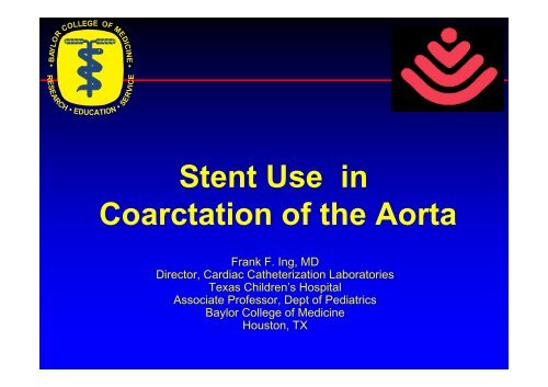
• BAYLOR COLLEGE OF MEDICINE •<br />
RESEARCH • EDUCATION • SERVICE<br />
<strong>Stent</strong> <strong>Use</strong> <strong>in</strong><br />
<strong>Coarctation</strong> <strong>of</strong> <strong>the</strong> <strong>Aorta</strong><br />
Frank F. Ing, MD<br />
Director, Cardiac Ca<strong>the</strong>terization Laboratories<br />
Texas Children’s Hospital<br />
Associate Pr<strong>of</strong>essor, Dept <strong>of</strong> Pediatrics<br />
Baylor College <strong>of</strong> Medic<strong>in</strong>e<br />
Houston, TX
<strong>Stent</strong> use <strong>in</strong> coarctation <strong>of</strong> <strong>the</strong> aorta<br />
• Various morphologies <strong>of</strong> COA<br />
• <strong>Stent</strong>s available for COA<br />
• <strong>Stent</strong> implantation techniques<br />
– Special considerations<br />
• <strong>Stent</strong> <strong>com</strong>plications
• Ridge like medial<br />
thicken<strong>in</strong>g, usually<br />
at <strong>the</strong> juxtaductal<br />
region<br />
• Extension <strong>of</strong> ductal<br />
tissue <strong>in</strong>to aorta<br />
• Decreased aortic<br />
flow <strong>in</strong> utero<br />
COA-anatomy
Variations <strong>of</strong> COA<br />
Discrete Long segment Comb<strong>in</strong>ation<br />
Discrete/long segment<br />
Fold/k<strong>in</strong>k<br />
Ao arch Thoracic Ao Abdom<strong>in</strong>al Ao Multiple levels
<strong>Coarctation</strong> / recoarctation<br />
<strong>of</strong> <strong>the</strong> aorta<br />
• Indications for <strong>in</strong>tervention:<br />
– ≥20mm Hg gradient<br />
– Upper extremity hypertension<br />
– Decreased left ventricular function<br />
regardless <strong>of</strong> gradient<br />
• Late repair (> 9 years):<br />
– accelerated a<strong>the</strong>rosclerosis<br />
– persistent systemic hypertension<br />
– berry aneurysm
Interventional options:<br />
Based on anatomy<br />
• Discrete<br />
– Angioplasty first option<br />
– <strong>Stent</strong> if residual gradient<br />
• Long segment<br />
– <strong>Stent</strong><br />
– If severe, test dilation with low pressure balloon<br />
• K<strong>in</strong>k/fold<br />
– Need stent to straighten out aorta<br />
• Arch, thoracic, abdom<strong>in</strong>al COA<br />
– Usually long segment; need stent<br />
– Adjacent to major arterial side branches
Interventional options:<br />
Based on patient size & aorta diameter<br />
• Patient size ≤ 25 kg<br />
– Femoral arteries may be too small to ac<strong>com</strong>modate<br />
large sheath (m<strong>in</strong>imal 9 Fr sheath)<br />
– Femoral artery angiogram to evaluate size<br />
– Angioplasty first option<br />
• Normal diameter adjacent to COA ≤10mm<br />
– Angioplasty first<br />
– Insufficient long term data to prove that stents dilated<br />
< 10 mm can to serially dilated to adult size (at least<br />
18 mm diam)<br />
• In extreme cases (ie, poor surgical candidates),<br />
will stent if angioplasty <strong>in</strong>adequate<br />
– <strong>Use</strong> medium size stents on smaller balloons<br />
– Will require future surgical removal
COA-<strong>Stent</strong> implantation techniques<br />
• Careful measurement <strong>of</strong> COA and adjacent normal<br />
anatomy (may need multiple angled views)<br />
• Retrograde course: stiff exchange guidewire across<br />
COA to right subclavian artery or ascend<strong>in</strong>g aorta<br />
• Balloon size 80-110% <strong>of</strong> adjacent normal anatomy<br />
• If severe COA:<br />
– Evaluate aortic wall <strong>com</strong>pliance with test balloon dilation<br />
us<strong>in</strong>g low pressure (2-4 atmospheric pressure)<br />
– If high <strong>com</strong>pliance, consider implant stent with smaller<br />
balloon and serially dilated over time.<br />
• Post-stent angiography to evaluate for dissection or<br />
aneurysm<br />
• Have surgical backup available
COA- <strong>Stent</strong> implantation:<br />
Complications<br />
• <strong>Stent</strong> malposition / embolization<br />
• Thromboembolic events / CNS<br />
– Fully hepar<strong>in</strong>ize (keep ACT >200)<br />
• Vascular (femoral artery) trauma<br />
• Aneurysm formation<br />
• Dissection (Surgical emergency)
<strong>Stent</strong> selection for COA<br />
Velocity Genesis<br />
medium Genesis<br />
large<br />
Small<br />
(max 5 mm<br />
diam.)<br />
Medium<br />
(max 8-10 mm<br />
diam.)<br />
Genesis XD Maxi-LD Palmaz XL CP 8-zig<br />
Large<br />
(max 18 mm<br />
Extra large stents<br />
diam.) (max 24 mm diam.)<br />
<strong>Use</strong> only <strong>the</strong>se for COA
Palmaz XL<br />
Dilated w/ 24 mm BIB<br />
Undilated<br />
40mm length<br />
24mm diam<br />
27.6mm length<br />
(31% FS)
CP 8-zig<br />
Dilated w/ 24 mm BIB<br />
Undilated<br />
45mm length<br />
24mm diam<br />
23.4mm length<br />
(48% FS)
EV3 MaxLD- serial dilation w/ 18<br />
mm BIB, <strong>the</strong>n 24 mm BIB<br />
Undilated<br />
34.4 mm<br />
9 mm<br />
34.3 mm<br />
18 mm<br />
34.1-34.7 mm<br />
24 mm<br />
32.7 mm<br />
4.9% shorten<strong>in</strong>g
EV3 MaxLD-direct dilation w/ 24 mm BIB<br />
Undilated<br />
34.8 mm<br />
12 mm<br />
34.8 mm<br />
24 mm<br />
25.2-28.2 mm<br />
(23.3% FS)
MaxLD 24 mm expansion<br />
Variable length based on expansion <strong>of</strong> open-cells<br />
Serial dilation<br />
S<strong>in</strong>gle dilation
Comparison <strong>of</strong> extra large stents<br />
(24 mm diameter)<br />
Max LD Palmaz XL CP 8-zig
<strong>Coarctation</strong> <strong>of</strong> <strong>the</strong> <strong>Aorta</strong><br />
• Balloon dilation
Angioplasty <strong>of</strong> COA
Pre & post dilation angiogram<br />
Lat<br />
pre<br />
post
Pre & post dilation angiogram<br />
AP<br />
pre<br />
post
Aortic diameter changes<br />
with a pulsatile aorta<br />
10.2 mm 11.9 mm<br />
12 mm balloon used<br />
•Measure <strong>the</strong> largest vessel<br />
diameter <strong>of</strong> <strong>the</strong> cardiac cycle
Pre and post dilation gradients<br />
Asc Ao 100/57<br />
Desc Ao 68/53<br />
Asc Ao 92-100/61<br />
Desc Ao 80/58<br />
Now what would you do?<br />
Stop and wait or proceed to stent?<br />
Pre-dilation<br />
Post-dilation
Comparison <strong>of</strong> aortograms<br />
Pre-dilation Post-dilation Post-stent
Comparison <strong>of</strong> gradients<br />
Pre-dilation<br />
Asc Ao 100/57<br />
Desc Ao 68/53<br />
Post-dilation<br />
Asc Ao 100/61<br />
Desc Ao 80/58<br />
Post-stent<br />
Asc Ao 90/53<br />
Desc Ao 84/54
25 yr. F w/ severe pre-eclampsia dur<strong>in</strong>g<br />
second trimester pregnancy<br />
• Severe discrete COA<br />
• Surgery vs. stent?<br />
• Radiation vs. bypass<br />
surgery?
Cath data<br />
• 54 mmHg gradient<br />
• M<strong>in</strong> diam: 4.6-5.7 mm<br />
• Isthmus diam: 13-15 mm
Dynamic posterior bend
Pre & post AP angiogram<br />
Pre<br />
Post
Pre & post lat angiogram<br />
QuickTime?and a<br />
Video de<strong>com</strong>pressor<br />
are needed to see this picture.<br />
Pre<br />
Post<br />
<strong>Stent</strong> prevents<br />
dynamic bend
S-bend COA<br />
QuickTime?and a<br />
Motion JPEG A de<strong>com</strong>pressor<br />
are needed to see this picture.<br />
Vessel diameter change<br />
dur<strong>in</strong>g cardiac cycle
<strong>Stent</strong> straighten out <strong>the</strong> S-bend<br />
<strong>Stent</strong> implant<br />
Pre<br />
Post<br />
QuickTime?and a<br />
Motion JPEG A de<strong>com</strong>pressor<br />
are needed to see this picture.
Severe coarctation <strong>of</strong> <strong>the</strong><br />
aorta-(virtual <strong>in</strong>terruption)<br />
AP<br />
Lat
Severe coarctation <strong>of</strong> <strong>the</strong><br />
aorta-(virtual <strong>in</strong>terruption)<br />
AP<br />
Lat
Cross<strong>in</strong>g coarctation w/ wire &<br />
ca<strong>the</strong>ter<br />
AP<br />
Lat
Test dilation w/ 10 mm low pressure<br />
balloon (2-4 atmospheric pressure)<br />
Compliant vessel wall<br />
Residual coarctation
<strong>Stent</strong> implantation<br />
MaxLD 36mm on 12 mm balloon<br />
Flared distal end<br />
w/ 14 mm balloon
Pre and post stent angiograms<br />
AP<br />
Pre<br />
Post
Pre and post stent angiograms<br />
Lat<br />
Pre<br />
Post
Multiple views may be needed<br />
to view entire aorta<br />
21 year old M with post-surgical re-COA<br />
AP<br />
Lat<br />
? Second COA
Multiple views may be needed<br />
to view entire aorta<br />
LAO 69<br />
QuickTime?and a<br />
de<strong>com</strong>pressor<br />
are needed to see this picture.<br />
RAO20<br />
CAU14<br />
QuickTime?and a<br />
de<strong>com</strong>pressor<br />
are needed to see this picture.<br />
Significant<br />
COA
<strong>Stent</strong> implantation<br />
<strong>Stent</strong> #1<br />
MaxLD16<br />
<strong>Stent</strong> embolized <strong>Stent</strong> #2<br />
MaxLD26
15 yo M w/ severe mid-thoracic COA<br />
AP<br />
Lat
Post implantation <strong>of</strong> 2 stents (P308)<br />
AP<br />
Lat
1 year follow-up:<br />
Re-COA at distal stent edge<br />
Intimal proliferation
Implantation <strong>of</strong> 2 additional stents<br />
<strong>Stent</strong> #3<br />
<strong>Stent</strong>s<br />
separated<br />
<strong>Stent</strong>s #4<br />
AP<br />
Lat
Post implantation angiograms<br />
AP<br />
Lat
Neur<strong>of</strong>ibromatosis<br />
w/ mid-aortic syndrome<br />
Pre-op angiogram<br />
Post-op: residual stenosis
5 stents<br />
implanted<br />
S/P aortic & bilateral<br />
renal artery stents
Pre and post angios<br />
Pre<br />
Post
William’s syndrome w/ long<br />
abdom<strong>in</strong>al COA across renal arteries
Implant stent<br />
jailed left renal artery<br />
Balloon SMA
Pre and post angios<br />
Pre<br />
Post
6 month F/U cath:<br />
recoarctation<br />
Good flow through<br />
jailed renal artery
S/P additional stents <strong>in</strong> aorta
Pre and post angiograms<br />
Bilateral jailed<br />
renal artery<br />
Pre F/U cath Post-additional stent
Angiogram <strong>of</strong> 8.2 kg <strong>in</strong>fant<br />
with HLHS s/p Glenn<br />
• HLHS s/p Glenn<br />
• LPA stenosis s/p stent<br />
• Glenn stenosis s/p stent<br />
• Severe neo-aortic<br />
coarctation<br />
• Only 5mm Hg grad due to<br />
poor RV function<br />
• Considered for heart<br />
transplant<br />
Glenn stent<br />
LPA stent
<strong>Coarctation</strong>-pre stent<br />
14 yoF s/p COA repair<br />
as <strong>in</strong>fant with arch<br />
hypoplasia<br />
Arch gradient: 19mmHg
Echocardiogram-good flow<br />
through subclavian artery<br />
<strong>Stent</strong><br />
LSCA<br />
QuickTime?and a<br />
Sorenson Video de<strong>com</strong>pressor<br />
are needed to see this picture.
CT Angiogram
10 mm balloon<br />
MaxLD stent - cell dilation<br />
w/ 10, <strong>the</strong>n 12 mm balloon<br />
12 mm balloon
Palmaz XL stent - cell dilation<br />
w/ 6, <strong>the</strong>n 8 mm balloon
Comparison on cells after max<br />
dilation<br />
12 mm balloon 8 mm balloon
25 yo w/ systemic hypertension:<br />
Long segment coarctation <strong>of</strong> <strong>the</strong> <strong>Aorta</strong><br />
AP(3.8mm)<br />
Lat
Poorly-centered stent slipped<br />
forward dur<strong>in</strong>g <strong>in</strong>flation<br />
<strong>Stent</strong> slipped<br />
forward on<br />
balloon<br />
dur<strong>in</strong>g<br />
adjustment<br />
<strong>Stent</strong> should<br />
be centered<br />
over balloon<br />
QuickTime?and a<br />
Video de<strong>com</strong>pressor<br />
are needed to see this picture.
Maneuvers to reposition stent<br />
With grabber<br />
With snare
<strong>Stent</strong> implanted <strong>in</strong> safe position;<br />
2nd stent implanted at COA site
COA stent<strong>in</strong>g can be<br />
technically difficult<br />
Pre<br />
3.8 mm<br />
Post<br />
16mm
Discrete COA<br />
AP<br />
Lat
Balloon <strong>in</strong>flation only after stent<br />
re-centered on balloon
Pre and post stent angiograms<br />
AP<br />
Lat
<strong>Coarctation</strong> <strong>of</strong> <strong>the</strong> aorta
Angioplasty <strong>of</strong> <strong>Coarctation</strong>
Post-dilation <strong>of</strong> <strong>Coarctation</strong>
2 year F/U angiogram:<br />
Aneurysm
5 year F/U CT angio<br />
Aneurysm at side <strong>of</strong> stent
Second stent implanted
Covered stents
Summary<br />
• COA presents with variable morphologies<br />
• Discrete stenosis can be treated with angioplasty<br />
• <strong>Stent</strong> implantation is more effective <strong>in</strong> reliev<strong>in</strong>g<br />
gradients <strong>in</strong> COA but technically more challeng<strong>in</strong>g<br />
• Complications are few, but can be catastrophic<br />
• The <strong>in</strong>terventionalist need to be<strong>com</strong>e fully familiar<br />
with how to deal with stent <strong>com</strong>plications<br />
• Surgical backup should be available for all cases