Basic CXR Interpretation - EthnoMed
Basic CXR Interpretation - EthnoMed
Basic CXR Interpretation - EthnoMed

Create successful ePaper yourself
Turn your PDF publications into a flip-book with our unique Google optimized e-Paper software.
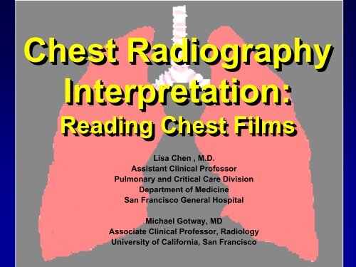
Chest Radiography<br />
<strong>Interpretation</strong>:<br />
Reading Chest Films<br />
Lisa Chen , M.D.<br />
Assistant Clinical Professor<br />
Pulmonary and Critical Care Division<br />
Department of Medicine<br />
San Francisco General Hospital<br />
Michael Gotway, MD<br />
Associate Clinical Professor, Radiology<br />
University of California, San Francisco
Approach to the <strong>CXR</strong>:<br />
Technical Aspects<br />
• Inspiratory effort<br />
• 9-10 posterior ribs<br />
• Penetration<br />
• thoracic intervertebral disc space just visible<br />
• Positioning/rotation<br />
• medial clavicle heads equidistant to spinous<br />
process
Low Lung Volumes
Over Exposure<br />
Proper Exposure
What to Evaluate<br />
• Lungs<br />
• Pleural surfaces<br />
• Cardiomediastinal contours<br />
• Bones and soft tissues<br />
• Abdomen
Where to Look<br />
• Apices<br />
• Retrocardiac areas (left and right)<br />
• Below diaphragm
Apical TB
Left Retrocardiac Opacity
Normal Anatomy: Frontal <strong>CXR</strong><br />
• Heart<br />
• Aorta<br />
• Pulmonary arteries<br />
• Airways<br />
• Diaphragm/costophrenic sulci<br />
• Junction lines
Normal Anatomy: Lateral<br />
• Heart<br />
• Aorta<br />
• Pulmonary arteries<br />
• Airways<br />
• Spine
AA<br />
RV<br />
LV
Chest Radiography:<br />
<strong>Basic</strong> Principles<br />
• X-ray photon fates:<br />
• completely absorbed in patient<br />
• transmitted through patient; strike film<br />
• scattered within patient; strike film<br />
• X-ray absorption depends on:<br />
• beam energy (constant)<br />
• tissue density
Maximum x-rayx<br />
Transmission<br />
(least dense tissue)<br />
Blackest<br />
air<br />
fat<br />
soft tissue<br />
calcium<br />
bone<br />
Maximum x–ray x<br />
Absorption<br />
(densest tissue)<br />
x-ray contrast<br />
metal<br />
Whitest
Chest Radiography:<br />
<strong>Basic</strong> Principles<br />
• All cardiothoracic pathology and<br />
normal anatomy is visualized (or not)<br />
by 7 different densities<br />
• How is this accomplished?<br />
• differential x-ray x<br />
absorption
Differential X-Ray X<br />
Absorption<br />
• A structure is rendered visible on a<br />
radiograph by the juxtaposition of two<br />
different densities
Silhouette Sign<br />
• Loss of the expected interface<br />
normally created by juxtaposition of<br />
two structures of different density<br />
• No boundary can be seen between<br />
two structures of similar density
Right Lower Lobe Pneumonia
Differential X-Ray X<br />
Absorption<br />
• The absence of a normal interface<br />
may indicate disease;<br />
• The presence of an unexpected<br />
interface may also indicate disease<br />
• The presence of interfaces can be<br />
used to localize abnormalities
Chest Radiographic<br />
Patterns of Disease<br />
• Air space opacity<br />
• Interstitial opacity<br />
• Nodules and masses<br />
• Lymphadenopathy<br />
• Cysts and cavities<br />
• Lung volumes<br />
• Pleural diseases
Chest Radiographic<br />
Patterns of Disease<br />
• Cardiomediastinal contour abnormalities<br />
• Bone and soft tissue abnormalities<br />
• Below the diaphragm: abdominal and<br />
retroperitoneal disease
Air Space Opacity<br />
• Components:<br />
• air bronchogram: : air-filled bronchus<br />
surrounded by airless lung<br />
• confluent opacity extending to pleural<br />
surfaces<br />
• segmental distribution
Air Space Opacity: DDX<br />
• Blood (hemorrhage)<br />
• Pus (pneumonia)<br />
• Water (edema)<br />
• hydrostatic or non-cardiogenic<br />
• Cells (tumor)<br />
• Protein/fat: alveolar proteinosis and<br />
lipoid pneumonia
LUL Pneumonia
Interstitial Opacity<br />
• Hallmarks:<br />
• small, well-defined nodules<br />
• lines<br />
‣ interlobular septal thickening<br />
‣ fibrosis<br />
• reticulation
Interstitial Opacity: Small Nodules
Interstitial Opacity:<br />
Lines
Interstitial Opacity: Lines & Reticulation
Interstitial Opacity: DDX<br />
• Idiopathic interstitial pneumonias<br />
• Infections (TB, viruses)<br />
••Edema<br />
• Hemorrhage<br />
• Non–infectious inflammatory lesions<br />
• sarcoidosis<br />
• Tumor
Nodules and Masses<br />
• Nodule: any pulmonary lesion<br />
represented in a radiograph by a sharply<br />
defined, discrete, nearly circular opacity<br />
2-30 mm in diameter<br />
• Mass: larger than 3 cm
Nodules and Masses<br />
• Qualifiers:<br />
• single or multiple<br />
• size<br />
• border definition<br />
• presence or absence of calcification<br />
• location
Well-Defined<br />
Ill-Defined<br />
Mass<br />
Calcification
Lymphadenopathy<br />
• Non-specific presentations:<br />
• mediastinal widening<br />
• hilar prominence<br />
• Specific patterns:<br />
• particular station enlargement
Right Paratracheal<br />
Lymphadenopathy
Right Hilar LAN
Right Hilar LAN
Left Hilar LAN
Subcarinal LAN
Subcarinal LAN<br />
*
AP Window LAN
Cysts & Cavities<br />
• Cyst: abnormal pulmonary<br />
parenchymal space, not containing<br />
lung but filled with air and/or fluid,<br />
congenital or acquired, with a wall<br />
thickness greater than 1 mm<br />
• epithelial lining often present
Cysts & Cavities<br />
• Cavity: abnormal pulmonary<br />
parenchymal space, not containing<br />
lung but filled with air and/or fluid,<br />
caused by tissue necrosis, with a<br />
definitive wall greater than 1 mm in<br />
thickness and comprised of<br />
inflammatory and/or neoplastic<br />
elements
Cysts & Cavities<br />
• Characterize:<br />
• wall thickness at thickest portion<br />
• inner lining<br />
• presence/absence of air/fluid level<br />
• number and location
Benign Lung Cyst : PCP Pneumatocele<br />
• Uniform wall thickness<br />
• 1 mm<br />
• Smooth inner lining
Benign Cavities :<br />
Cryptococcus<br />
• max wall thickness ≤4 4 mm<br />
• minimally irregular inner lining
Indeterminate Cavities<br />
• max wall thickness 5-15 mm<br />
• mildly irregular inner lining
Malignant Cavities: Squamous Cell Ca<br />
• max wall thickness ≥16 mm<br />
• Irregular inner lining
Pleural Disease: <strong>Basic</strong> Patterns<br />
• Effusion<br />
• angle blunting to massive<br />
• mobility<br />
• Thickening<br />
• distortion, no mobility<br />
••Mass<br />
••Air<br />
• Calcification
Pleural Effusion
Pleural Effusion
Subpulmonic Effusion
Pleural Calcification