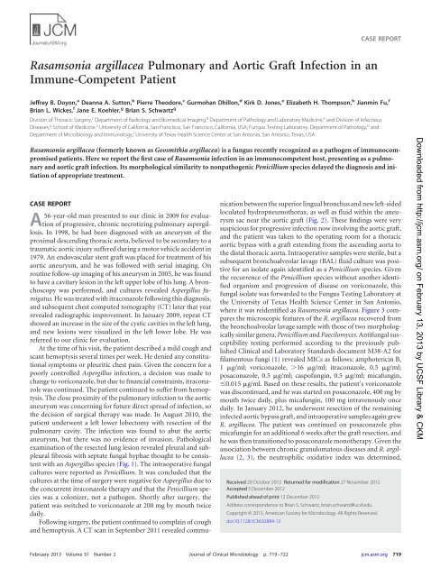
Rasamsonia argillacea Pulmonary and Aortic Graft Infection in an ...
Rasamsonia argillacea Pulmonary and Aortic Graft Infection in an ...
Rasamsonia argillacea Pulmonary and Aortic Graft Infection in an ...

Create successful ePaper yourself
Turn your PDF publications into a flip-book with our unique Google optimized e-Paper software.
CASE REPORT<br />
<strong>Rasamsonia</strong> <strong>argillacea</strong> <strong>Pulmonary</strong> <strong><strong>an</strong>d</strong> <strong>Aortic</strong> <strong>Graft</strong> <strong>Infection</strong> <strong>in</strong> <strong>an</strong><br />
Immune-Competent Patient<br />
Jeffrey B. Doyon, a De<strong>an</strong>na A. Sutton, b Pierre Theodore, c Gurmoh<strong>an</strong> Dhillon, d Kirk D. Jones, e Elizabeth H. Thompson, b Ji<strong>an</strong>m<strong>in</strong> Fu, f<br />
Bri<strong>an</strong> L. Wickes, f J<strong>an</strong>e E. Koehler, g Bri<strong>an</strong> S. Schwartz g<br />
Division of Thoracic Surgery, c Department of Radiology <strong><strong>an</strong>d</strong> Biomedical Imag<strong>in</strong>g, d Department of Pathology <strong><strong>an</strong>d</strong> Laboratory Medic<strong>in</strong>e, e <strong><strong>an</strong>d</strong> Division of Infectious<br />
Diseases, g School of Medic<strong>in</strong>e, a University of California, S<strong>an</strong> Fr<strong>an</strong>cisco, S<strong>an</strong> Fr<strong>an</strong>cisco, California, USA; Fungus Test<strong>in</strong>g Laboratory, Department of Pathology, b <strong><strong>an</strong>d</strong><br />
Department of Microbiology <strong><strong>an</strong>d</strong> Immunology, f University of Texas Health Science Center at S<strong>an</strong> Antonio, S<strong>an</strong> Antonio, Texas, USA<br />
<strong>Rasamsonia</strong> <strong>argillacea</strong> (formerly known as Geosmithia <strong>argillacea</strong>) is a fungus recently recognized as a pathogen of immunocompromised<br />
patients. Here we report the first case of <strong>Rasamsonia</strong> <strong>in</strong>fection <strong>in</strong> <strong>an</strong> immunocompetent host, present<strong>in</strong>g as a pulmonary<br />
<strong><strong>an</strong>d</strong> aortic graft <strong>in</strong>fection. Its morphological similarity to nonpathogenic Penicillium species delayed the diagnosis <strong><strong>an</strong>d</strong> <strong>in</strong>itiation<br />
of appropriate treatment.<br />
CASE REPORT<br />
A56-year-old m<strong>an</strong> presented to our cl<strong>in</strong>ic <strong>in</strong> 2009 for evaluation<br />
of progressive, chronic necrotiz<strong>in</strong>g pulmonary aspergillosis.<br />
In 1998, he had been diagnosed with <strong>an</strong> <strong>an</strong>eurysm of the<br />
proximal descend<strong>in</strong>g thoracic aorta, believed to be secondary to a<br />
traumatic aortic <strong>in</strong>jury suffered dur<strong>in</strong>g a motor vehicle accident <strong>in</strong><br />
1979. An endovascular stent graft was placed for treatment of his<br />
aortic <strong>an</strong>eurysm, <strong><strong>an</strong>d</strong> he was followed with serial imag<strong>in</strong>g. On<br />
rout<strong>in</strong>e follow-up imag<strong>in</strong>g of his <strong>an</strong>eurysm <strong>in</strong> 2005, he was found<br />
to have a cavitary lesion <strong>in</strong> the left upper lobe of his lung. A bronchoscopy<br />
was performed, <strong><strong>an</strong>d</strong> cultures revealed Aspergillus fumigatus.<br />
He was treated with itraconazole follow<strong>in</strong>g this diagnosis,<br />
<strong><strong>an</strong>d</strong> subsequent chest computed tomography (CT) later that year<br />
revealed radiographic improvement. In J<strong>an</strong>uary 2009, repeat CT<br />
showed <strong>an</strong> <strong>in</strong>crease <strong>in</strong> the size of the cystic cavities <strong>in</strong> the left lung,<br />
<strong><strong>an</strong>d</strong> new lesions were visualized <strong>in</strong> the left lower lobe. He was<br />
referred to our cl<strong>in</strong>ic for evaluation.<br />
At the time of his visit, the patient described a mild cough <strong><strong>an</strong>d</strong><br />
sc<strong>an</strong>t hemoptysis several times per week. He denied <strong>an</strong>y constitutional<br />
symptoms or pleuritic chest pa<strong>in</strong>. Given the concern for a<br />
poorly controlled Aspergillus <strong>in</strong>fection, a decision was made to<br />
ch<strong>an</strong>ge to voriconazole, but due to f<strong>in</strong><strong>an</strong>cial constra<strong>in</strong>ts, itraconazole<br />
was cont<strong>in</strong>ued. The patient cont<strong>in</strong>ued to suffer from hemoptysis.<br />
The close proximity of the pulmonary <strong>in</strong>fection to the aortic<br />
<strong>an</strong>eurysm was concern<strong>in</strong>g for future direct spread of <strong>in</strong>fection, so<br />
the decision of surgical therapy was made. In August 2010, the<br />
patient underwent a left lower lobectomy with resection of the<br />
pulmonary cavity. The <strong>in</strong>fection was found to abut the aortic<br />
<strong>an</strong>eurysm, but there was no evidence of <strong>in</strong>vasion. Pathological<br />
exam<strong>in</strong>ation of the resected lung lesion revealed pleural <strong><strong>an</strong>d</strong> subpleural<br />
fibrosis with septate fungal hyphae thought to be consistent<br />
with <strong>an</strong> Aspergillus species (Fig. 1). The <strong>in</strong>traoperative fungal<br />
cultures were reported as Penicillium. It was concluded that the<br />
cultures at the time of surgery were negative for Aspergillus due to<br />
the concurrent itraconazole therapy <strong><strong>an</strong>d</strong> that the Penicillium species<br />
was a colonizer, not a pathogen. Shortly after surgery, the<br />
patient was switched to voriconazole at 200 mg by mouth twice<br />
daily.<br />
Follow<strong>in</strong>g surgery, the patient cont<strong>in</strong>ued to compla<strong>in</strong> of cough<br />
<strong><strong>an</strong>d</strong> hemoptysis. A CT sc<strong>an</strong> <strong>in</strong> September 2011 revealed communication<br />
between the superior l<strong>in</strong>gual bronchus <strong><strong>an</strong>d</strong> new left-sided<br />
loculated hydropneumothorax, as well as fluid with<strong>in</strong> the <strong>an</strong>eurysm<br />
sac near the aortic graft (Fig. 2). These f<strong>in</strong>d<strong>in</strong>gs were very<br />
suspicious for progressive <strong>in</strong>fection now <strong>in</strong>volv<strong>in</strong>g the aortic graft,<br />
<strong><strong>an</strong>d</strong> the patient was taken to the operat<strong>in</strong>g room for a thoracic<br />
aortic bypass with a graft extend<strong>in</strong>g from the ascend<strong>in</strong>g aorta to<br />
the distal thoracic aorta. Intraoperative samples were sterile, but a<br />
subsequent bronchoalveolar lavage (BAL) fluid culture was positive<br />
for <strong>an</strong> isolate aga<strong>in</strong> identified as a Penicillium species. Given<br />
the recurrence of the Penicillium species without <strong>an</strong>other identified<br />
org<strong>an</strong>ism <strong><strong>an</strong>d</strong> progression of disease on voriconazole, this<br />
fungal isolate was forwarded to the Fungus Test<strong>in</strong>g Laboratory at<br />
the University of Texas Health Science Center <strong>in</strong> S<strong>an</strong> Antonio,<br />
where it was reidentified as <strong>Rasamsonia</strong> <strong>argillacea</strong>. Figure 3 compares<br />
the microscopic features of the R. <strong>argillacea</strong> recovered from<br />
the bronchoalveolar lavage sample with those of two morphologically<br />
similar genera, Penicillium <strong><strong>an</strong>d</strong> Paecilomyces. Antifungal susceptibility<br />
test<strong>in</strong>g performed accord<strong>in</strong>g to the previously published<br />
Cl<strong>in</strong>ical <strong><strong>an</strong>d</strong> Laboratory St<strong><strong>an</strong>d</strong>ards document M38-A2 for<br />
filamentous fungi (1) revealed MICs as follows: amphoteric<strong>in</strong> B,<br />
1 g/ml; voriconazole, 16 g/ml; itraconazole, 0.5 g/ml;<br />
posaconazole, 0.5 g/ml; caspofung<strong>in</strong>, 0.5 g/ml; micafung<strong>in</strong>,<br />
0.015 g/ml. Based on these results, the patient’s voriconazole<br />
was discont<strong>in</strong>ued, <strong><strong>an</strong>d</strong> he was started on posaconazole, 400 mg by<br />
mouth twice daily, plus micafung<strong>in</strong>, 100 mg <strong>in</strong>travenously once<br />
daily. In J<strong>an</strong>uary 2012, he underwent resection of the rema<strong>in</strong><strong>in</strong>g<br />
<strong>in</strong>fected aortic bypass graft, <strong><strong>an</strong>d</strong> <strong>in</strong>traoperative samples aga<strong>in</strong> grew<br />
R. <strong>argillacea</strong>. The patient was cont<strong>in</strong>ued on posaconazole plus<br />
micafung<strong>in</strong> for <strong>an</strong> additional 6 weeks after the graft resection, <strong><strong>an</strong>d</strong><br />
he was then tr<strong>an</strong>sitioned to posaconazole monotherapy. Given the<br />
association between chronic gr<strong>an</strong>ulomatous diseases <strong><strong>an</strong>d</strong> R. <strong>argillacea</strong><br />
(2, 3), the neutrophilic oxidative <strong>in</strong>dex was determ<strong>in</strong>ed,<br />
Received 29 October 2012 Returned for modification 27 November 2012<br />
Accepted 5 December 2012<br />
Published ahead of pr<strong>in</strong>t 12 December 2012<br />
Address correspondence to Bri<strong>an</strong> S. Schwartz, bri<strong>an</strong>.schwartz@ucsf.edu.<br />
Copyright © 2013, Americ<strong>an</strong> Society for Microbiology. All Rights Reserved.<br />
doi:10.1128/JCM.02884-12<br />
Downloaded from http://jcm.asm.org/ on February 13, 2013 by UCSF Library & CKM<br />
February 2013 Volume 51 Number 2 Journal of Cl<strong>in</strong>ical Microbiology p. 719–722 jcm.asm.org 719
Case Report<br />
FIG 1 Grocott’s methenam<strong>in</strong>e silver (A) or hematoxyl<strong>in</strong>-<strong><strong>an</strong>d</strong>-eos<strong>in</strong> (B) sta<strong>in</strong><strong>in</strong>g of sections from a bronchiectatic cystic structure, show<strong>in</strong>g a mass of <strong>in</strong>terwoven<br />
septate hyphae. The hyphae are th<strong>in</strong> (3 to 5 m) with parallel walls.<br />
which was with<strong>in</strong> normal limits. Test<strong>in</strong>g for HIV was also negative.<br />
The patient’s postoperative course was complicated by a<br />
bronchopleural fistula, but he subsequently was discharged<br />
<strong><strong>an</strong>d</strong> has improved cl<strong>in</strong>ically 9 months after his last surgery. A<br />
posaconazole trough was 0.76 g/ml, <strong><strong>an</strong>d</strong> a repeat CT sc<strong>an</strong> of the<br />
chest revealed no residual <strong>in</strong>fection of the graft site, but the exam<br />
did reveal persistent, slowly resolv<strong>in</strong>g hydropneumothorax.<br />
Fungal identification. The BAL fluid <strong><strong>an</strong>d</strong> aortic tissue isolates<br />
were forwarded to the Fungus Test<strong>in</strong>g Laboratory at the University<br />
of Texas Health Science Center <strong>in</strong> S<strong>an</strong> Antonio for species<br />
identification <strong><strong>an</strong>d</strong> <strong>an</strong>tifungal susceptibility test<strong>in</strong>g <strong><strong>an</strong>d</strong> were accessioned<br />
<strong>in</strong>to their culture collection as UTHSC 11-3152 <strong><strong>an</strong>d</strong><br />
UTHSC 12-238, respectively. Salient phenotypic features, previously<br />
described <strong>in</strong> detail elsewhere (2–5), <strong>in</strong>cluded cream to buff<br />
to pale brown colonies on potato dextrose agar prepared <strong>in</strong>-house,<br />
growth at elevated temperatures (37, 40, <strong><strong>an</strong>d</strong> 45°C), roughened<br />
conidiophores <strong><strong>an</strong>d</strong> conidiogenous cells (phialides), <strong><strong>an</strong>d</strong> smooth,<br />
hyal<strong>in</strong>e, cyl<strong>in</strong>drical to cuneiform (wedge-shaped) conidia borne<br />
<strong>in</strong> long columnar cha<strong>in</strong>s (Fig. 3A). Based on these features, both<br />
isolates were identified morphologically as R. <strong>argillacea</strong>. Molecular<br />
characterization of the BAL fluid <strong><strong>an</strong>d</strong> aortic tissue isolates as<br />
conducted by PCR amplification <strong><strong>an</strong>d</strong> sequenc<strong>in</strong>g of the ribosomal<br />
<strong>in</strong>ternal tr<strong>an</strong>scribed spacer (ITS) region <strong><strong>an</strong>d</strong> 5= end of the nuclear<br />
large subunit ribosomal DNA (rDNA) at the UTHSCSA Adv<strong>an</strong>ced<br />
Nucleic Acids Core facility, as previously described (6).<br />
Sequences were then used to perform a BLASTn search of the<br />
GenB<strong>an</strong>k database at the NCBI (http://www.ncbi.nlm.nih.gov).<br />
Outputs were sorted based on percent identity <strong><strong>an</strong>d</strong> were considered<br />
signific<strong>an</strong>t at 99% identity <strong><strong>an</strong>d</strong> 90% query coverage. The top<br />
three matches for the UTHSC 12-238 ITS rDNA search were all<br />
<strong>Rasamsonia</strong> <strong>argillacea</strong> (EU862337.1, 99% identity; EU862335.1, 99%<br />
identity; <strong><strong>an</strong>d</strong> GU165730.1, 99% identity), <strong><strong>an</strong>d</strong> the top three matches<br />
for the LSU D1/D2 sequence were also all <strong>Rasamsonia</strong> <strong>argillacea</strong><br />
(EU862338.1, 99% identity; EU862336.1, 99% identity; <strong><strong>an</strong>d</strong><br />
AB047236.1, 99% identity). The search results with the UTHSC 11-<br />
3152 ITS sequence were all <strong>Rasamsonia</strong> <strong>argillacea</strong> (GU165733.1, 99%<br />
identity; EU862337.1, 99% identity; <strong><strong>an</strong>d</strong> GU165730.1, 99% identity),<br />
as were the search results for the LSU D1/D2 rDNA sequence<br />
(EU862338.1, 100% identity; EU862336.1, 100% identity; <strong><strong>an</strong>d</strong><br />
AB047236.1, 100% identity). Based on the phenotypic <strong><strong>an</strong>d</strong> genotypic<br />
results, both isolates were identified as <strong>Rasamsonia</strong> <strong>argillacea</strong>. The<br />
case isolates were deposited <strong>in</strong> the University of Alberta Microfungus<br />
Collection under accession numbers UAMH 11662 <strong><strong>an</strong>d</strong> UAMH<br />
11663.<br />
Downloaded from http://jcm.asm.org/ on February 13, 2013 by UCSF Library & CKM<br />
FIG 2 Selected axial image from a contrast-enh<strong>an</strong>ced study of the thorax, with<br />
arterial phase tim<strong>in</strong>g at the proximal descend<strong>in</strong>g thoracic aorta. The arrow<br />
identifies <strong>an</strong> endovascular stent graft for treatment of <strong>an</strong> aortic <strong>an</strong>eurysm. The<br />
arrowhead <strong>in</strong>dicates gas with<strong>in</strong> the residual <strong>an</strong>eurysm sac.<br />
Discussion. <strong>Rasamsonia</strong> <strong>argillacea</strong> is a rare fungal pathogen<br />
first described by Stolk et al. <strong>in</strong> 1969 as a thermotoler<strong>an</strong>t fungus<br />
under the name Penicillium argillaceum (7). In 1979, Pitt (8)<br />
erected a new genus, Geosmithia, to accommodate species whose<br />
colonies were t<strong>an</strong> to buff colored <strong><strong>an</strong>d</strong> never green/blue-green like<br />
those <strong>in</strong> most other Penicillium species <strong><strong>an</strong>d</strong> whose conidia were<br />
rect<strong>an</strong>gular rather th<strong>an</strong> globose or ellipsoidal, as <strong>in</strong> Penicillium or<br />
Paecilomyces, respectively (Fig. 3). Geosmithia was also dist<strong>in</strong>guished<br />
by rough-walled metulae (cells support<strong>in</strong>g the phialides)<br />
<strong><strong>an</strong>d</strong> phialides. More recently, molecular characterization of thermotoler<strong>an</strong>t<br />
members of the family Trichocomaceae, <strong>in</strong>clud<strong>in</strong>g<br />
720 jcm.asm.org Journal of Cl<strong>in</strong>ical Microbiology
Case Report<br />
FIG 3 Photomicrographs show differences <strong>in</strong> the microscopic morphology of <strong>Rasamsonia</strong> <strong>argillacea</strong> (A) from those of a Penicillium species (B) or Paecilomyces<br />
variotii (C). All have phialidic conidiogenous cells (P), m<strong>an</strong>y of which are supported by metulae (M, cells directly beneath the phialide); however, those <strong>in</strong> R.<br />
<strong>argillacea</strong> are noticeably roughened. Conidial shape (C) is also dist<strong>in</strong>ctive for various species <strong>in</strong> each of the genera, as shown <strong>in</strong> the examples above, usually be<strong>in</strong>g<br />
globose (round) to oval or ellipsoidal <strong>in</strong> Penicillium or Paecilomyces, respectively, <strong><strong>an</strong>d</strong> rect<strong>an</strong>gular to cuneiform (wedge shaped) <strong>in</strong> R. <strong>argillacea</strong>. Slide cultures<br />
from which photomicrographs were taken were mounted <strong>in</strong> lactophenol cotton blue.<br />
Downloaded from http://jcm.asm.org/<br />
Geosmithia, us<strong>in</strong>g several gene sequences (RPB2, RNA polymerase<br />
II gene encod<strong>in</strong>g the second-largest prote<strong>in</strong> submit; TSR1, encod<strong>in</strong>g<br />
a putative ribosome biogenesis prote<strong>in</strong>; CCT8, encod<strong>in</strong>g the<br />
putative chaperon<strong>in</strong> complex component TCP-1) have shown<br />
that these species form a clade dist<strong>in</strong>ct from other genera <strong>in</strong> the<br />
Trichocomaceae family (9). Thus, the genus <strong>Rasamsonia</strong> was proposed,<br />
with the name honor<strong>in</strong>g Robert A. Samson at the CBS-<br />
KNAW Fungal Biodiversity Center on the occasion of his 65th<br />
birthday, his 40 years of service at the CBS, <strong><strong>an</strong>d</strong> for his m<strong>an</strong>y<br />
contributions to fungal taxonomy. The genus currently conta<strong>in</strong>s<br />
six species (9).<br />
<strong>Rasamsonia</strong> <strong>argillacea</strong> was only recently identified as <strong>an</strong> etiologic<br />
agent <strong>in</strong> hum<strong>an</strong>s <strong><strong>an</strong>d</strong> other <strong>an</strong>imals, reported under the<br />
name Geosmithia <strong>argillacea</strong> (2–5, 10, 11). In 2009, a fatal case of<br />
dissem<strong>in</strong>ated <strong>in</strong>fection <strong>in</strong> a Germ<strong>an</strong> shepherd dog was reported<br />
(5). Subsequently, eight cases of airway colonization <strong>in</strong> patients<br />
with cystic fibrosis, without evidence of cl<strong>in</strong>ical <strong>in</strong>fection, were<br />
reported (4, 10). N<strong>in</strong>e cases of pulmonary <strong>in</strong>fections <strong>in</strong> patients<br />
with chronic gr<strong>an</strong>ulomatous disease have been reported, two of<br />
which <strong>in</strong>volved the chest wall <strong><strong>an</strong>d</strong> ribs <strong><strong>an</strong>d</strong> one of which dissem<strong>in</strong>ated<br />
to the bra<strong>in</strong> (2, 3). Four of these patients died from complications<br />
of their <strong>in</strong>fections. F<strong>in</strong>ally, fatal G. <strong>argillacea</strong> <strong>in</strong>fection was<br />
recently found <strong>in</strong> a stem cell tr<strong>an</strong>spl<strong>an</strong>t recipient receiv<strong>in</strong>g immune-suppressive<br />
therapy for graft-versus-host disease (11).<br />
Other Geosmithia species have not been reported as etiologic<br />
agents of hum<strong>an</strong> <strong>in</strong>fection.<br />
Several common themes arise from the <strong>Rasamsonia</strong> cases<br />
(reported as Geosmithia) described above. This pathogen was<br />
often misidentified microscopically as a Penicillium or Paecilomyces<br />
species. All isolates of this species tested were resist<strong>an</strong>t to<br />
voriconazole but were variably resist<strong>an</strong>t to itraconazole, amphoteric<strong>in</strong><br />
B, <strong><strong>an</strong>d</strong> posaconazole. Ech<strong>in</strong>oc<strong><strong>an</strong>d</strong><strong>in</strong> resist<strong>an</strong>ce was<br />
<strong>in</strong>frequent. Until the present case, R. <strong>argillacea</strong> had been associated<br />
with signific<strong>an</strong>t morbidity <strong><strong>an</strong>d</strong> mortality only <strong>in</strong> immunocompromised<br />
patients.<br />
Here we report the first case of <strong>in</strong>vasive <strong>Rasamsonia</strong> <strong>argillacea</strong><br />
<strong>in</strong>fection <strong>in</strong> <strong>an</strong> immunocompetent patient. This patient developed<br />
a pulmonary <strong>in</strong>fection <strong>in</strong> the context of airway compression<br />
precipitated by a large posttraumatic aortic <strong>an</strong>eurysm. The orig<strong>in</strong>al<br />
Aspergillus <strong>in</strong>fection was likely cleared with itraconazole. In the<br />
sett<strong>in</strong>g of this treatment regimen, the patient developed a secondary<br />
<strong>Rasamsonia</strong> <strong>argillacea</strong> <strong>in</strong>fection that was characterized by septate<br />
hyphae <strong>in</strong> tissue similar to those seen <strong>in</strong> aspergillosis (Fig. 1)<br />
<strong><strong>an</strong>d</strong> a microscopic morphology that was similar to that of Penicillium<br />
<strong><strong>an</strong>d</strong> Paecilomyces (Fig. 3). Similar to the reported cases, this<br />
patient’s <strong>Rasamsonia</strong> isolate was <strong>in</strong>itially misidentified microscopically<br />
as a Penicillium species, <strong><strong>an</strong>d</strong> <strong>in</strong> vitro <strong>an</strong>tifungal susceptibility<br />
test<strong>in</strong>g suggested resist<strong>an</strong>ce to voriconazole. This case<br />
highlights that cl<strong>in</strong>ical suspicion for a <strong>Rasamsonia</strong> <strong>argillacea</strong> <strong>in</strong>fection<br />
needs to be raised for patients whose fungal <strong>in</strong>fections worsen<br />
<strong><strong>an</strong>d</strong> whose cultures are reported as a Penicillium species, regardless<br />
of the patient’s immune status, especially if these patients are receiv<strong>in</strong>g<br />
voriconazole.<br />
Nucleotide sequence accession numbers. The ITS <strong><strong>an</strong>d</strong> LSU<br />
D1/D2 rDNA sequence data were deposited <strong>in</strong> GenB<strong>an</strong>k under<br />
accession numbers JX514398 to JX514401.<br />
ACKNOWLEDGMENTS<br />
J.E.K. received fund<strong>in</strong>g support from a Burroughs Wellcome Fund Cl<strong>in</strong>ical<br />
Scientist Award <strong>in</strong> Tr<strong>an</strong>slational Research.<br />
We acknowledge Dora McCarthy for perform<strong>in</strong>g <strong>an</strong>tifungal susceptibility<br />
test<strong>in</strong>g on the case isolates.<br />
on February 13, 2013 by UCSF Library & CKM<br />
February 2013 Volume 51 Number 2 jcm.asm.org 721
Case Report<br />
REFERENCES<br />
1. CLSI. 2008. Reference method for broth dilution <strong>an</strong>tifungal susceptibility<br />
test<strong>in</strong>g of filamentous fungi, 2nd ed. Approved st<strong><strong>an</strong>d</strong>ard M38-A2. CLSI,<br />
Wayne, PA.<br />
2. De Rav<strong>in</strong> SS, Challipalli M, Anderson V, Shea YR, Marci<strong>an</strong>o B, Hilligoss<br />
D, Marquesen M, Decastro R, Liu YC, Sutton DA, Wickes BL, Kammeyer<br />
PL, Sigler L, Sulliv<strong>an</strong> K, K<strong>an</strong>g EM, Malech HL, Holl<strong><strong>an</strong>d</strong> SM, Zelazny AM.<br />
2011. Geosmithia <strong>argillacea</strong>: <strong>an</strong> emerg<strong>in</strong>g cause of <strong>in</strong>vasive mycosis <strong>in</strong> hum<strong>an</strong><br />
chronic gr<strong>an</strong>ulomatous disease. Cl<strong>in</strong>. Infect. Dis. 52:e136–e143.<br />
3. Machouart M, Garcia-Hermoso D, Rivier A, Hassouni N, Cather<strong>in</strong>ot E,<br />
Salmon A, Debourgogne A, Coignard H, Lecuit M, Bougnoux ME,<br />
Bl<strong>an</strong>che S, Lortholary O. 2011. Emergence of dissem<strong>in</strong>ated <strong>in</strong>fections<br />
due to Geosmithia <strong>argillacea</strong> <strong>in</strong> patients with chronic gr<strong>an</strong>ulomatous disease<br />
receiv<strong>in</strong>g long-term azole <strong>an</strong>tifungal prophylaxis. J. Cl<strong>in</strong>. Microbiol.<br />
49:1681–1683.<br />
4. Giraud S, Pihet M, Razafim<strong><strong>an</strong>d</strong>imby B, Carrere J, Deg<strong><strong>an</strong>d</strong> N, Mely L,<br />
Favennec L, D<strong>an</strong>naoui E, Bouchara JP, Calenda A. 2010. Geosmithia<br />
<strong>argillacea</strong>: <strong>an</strong> emerg<strong>in</strong>g pathogen <strong>in</strong> patients with cystic fibrosis. J. Cl<strong>in</strong>.<br />
Microbiol. 48:2381–2386.<br />
5. Gr<strong>an</strong>t DC, Sutton DA, S<strong><strong>an</strong>d</strong>berg CA, Tyler RD, Jr, Thompson EH,<br />
Rom<strong>an</strong>elli AM, Wickes BL. 2009. Dissem<strong>in</strong>ated Geosmithia <strong>argillacea</strong><br />
<strong>in</strong>fection <strong>in</strong> a Germ<strong>an</strong> shepherd dog. Med. Mycol. 47:221–226.<br />
6. Rom<strong>an</strong>elli AM, Sutton DA, Thompson EH, R<strong>in</strong>aldi MG, Wickes BL.<br />
2010. Sequence-based identification of filamentous basidiomycetous<br />
fungi from cl<strong>in</strong>ical specimens: a cautionary note. J. Cl<strong>in</strong>. Microbiol. 48:<br />
741–752.<br />
7. Stolk AC, Ev<strong>an</strong>s HC, Nilsson T. 1969. Penicillium argillaceum sp. nov., a<br />
thermotoler<strong>an</strong>t Penicillium. Tr<strong>an</strong>s. Br. Mycol. Soc. 53:307–311.<br />
8. Pitt JI. 1979. Geosmithia gen. -nov. for Penicillium lavendulum <strong><strong>an</strong>d</strong> related<br />
species.C<strong>an</strong>. J. Bot. 57:2021–2030.<br />
9. Houbraken J, Spierenburg H, Frisvad JC. 2012. <strong>Rasamsonia</strong>, a new genus<br />
compris<strong>in</strong>g thermotoler<strong>an</strong>t <strong><strong>an</strong>d</strong> thermophilic Talaromyces <strong><strong>an</strong>d</strong> Geosmithia<br />
species. Antonie V<strong>an</strong> Leeuwenhoek 101:403–421.<br />
10. Barton RC, Borm<strong>an</strong> AM, Johnson EM, Houbraken J, Hobson RP,<br />
Denton M, Conway SP, Brownlee KG, Peckham D, Lee TW. 2010.<br />
Isolation of the fungus Geosmithia <strong>argillacea</strong> <strong>in</strong> sputum of people with<br />
cystic fibrosis. J. Cl<strong>in</strong>. Microbiol. 48:2615–2617.<br />
11. Valent<strong>in</strong> T, Neumeister P, Pichler M, Rohn A, Koidl C, Haas D, Heil<strong>in</strong>g<br />
B, Asslaber M, Zollner-Schwetz I, Hoenigl M, Salzer HJ, Krause R,<br />
Buz<strong>in</strong>a W. 2012. Dissem<strong>in</strong>ated Geosmithia <strong>argillacea</strong> <strong>in</strong>fection <strong>in</strong> a patient<br />
with gastro<strong>in</strong>test<strong>in</strong>al GvHD. Bone Marrow Tr<strong>an</strong>spl<strong>an</strong>t. 47:734–736.<br />
Downloaded from http://jcm.asm.org/<br />
on February 13, 2013 by UCSF Library & CKM<br />
722 jcm.asm.org Journal of Cl<strong>in</strong>ical Microbiology