Principles of Nucleic Acid Separation by Agarose Gel Electrophoresis
Principles of Nucleic Acid Separation by Agarose Gel Electrophoresis
Principles of Nucleic Acid Separation by Agarose Gel Electrophoresis

Create successful ePaper yourself
Turn your PDF publications into a flip-book with our unique Google optimized e-Paper software.
<strong>Principles</strong> <strong>of</strong> <strong>Nucleic</strong> <strong>Acid</strong> <strong>Separation</strong><br />
<strong>by</strong> <strong>Agarose</strong> <strong>Gel</strong> <strong>Electrophoresis</strong><br />
Muhittin Yılmaz * , Cem Ozic and İlhami Gok<br />
University <strong>of</strong> Kafkas, Department <strong>of</strong> Biology, Faculty <strong>of</strong> Sciences, Kars,<br />
Turkey<br />
3<br />
1. Introduction<br />
1.1 <strong>Principles</strong> <strong>of</strong> nucleic acid separation <strong>by</strong> agarose gel electrophoresis<br />
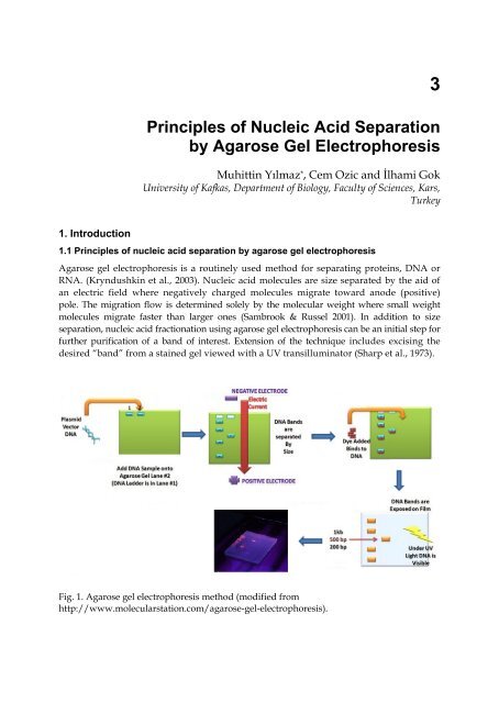
<strong>Agarose</strong> gel electrophoresis is a routinely used method for separating proteins, DNA or<br />
RNA. (Kryndushkin et al., 2003). <strong>Nucleic</strong> acid molecules are size separated <strong>by</strong> the aid <strong>of</strong><br />
an electric field where negatively charged molecules migrate toward anode (positive)<br />
pole. The migration flow is determined solely <strong>by</strong> the molecular weight where small weight<br />
molecules migrate faster than larger ones (Sambrook & Russel 2001). In addition to size<br />
separation, nucleic acid fractionation using agarose gel electrophoresis can be an initial step for<br />
further purification <strong>of</strong> a band <strong>of</strong> interest. Extension <strong>of</strong> the technique includes excising the<br />
desired “band” from a stained gel viewed with a UV transilluminator (Sharp et al., 1973).<br />
Fig. 1. <strong>Agarose</strong> gel electrophoresis method (modified from<br />
http://www.molecularstation.com/agarose-gel-electrophoresis).
34<br />
<strong>Gel</strong> <strong>Electrophoresis</strong> – <strong>Principles</strong> and Basics<br />
In order to visualize nucleic acid molecules in agarose gels, ethidium bromide or SYBR<br />
Green are commonly used dyes. Illumination <strong>of</strong> the agarose gels with 300-nm UV light is<br />
subsequently used for visualizing the stained nucleic acids. Throughout this chapter, the<br />
common methods for staining and visualization <strong>of</strong> DNA are described in details.<br />
<strong>Agarose</strong> gel electrophoresis provides multiple advantages that make it widely popular. For<br />
example, nucleic acids are not chemically altered during the size separation process and<br />
agarose gels can easily be viewed and handled. Furthermore, samples can be recovered and<br />
extracted from the gels easily for further studies. Still another advantage is that the resulting<br />
gel could be stored in a plastic bag and refrigerated after the experiment, there may be<br />
limits. Depending on buffer during electrophoresis in order to generate a suitable electric<br />
current and to reduce the heat generated <strong>by</strong> electric current can be considered as limitations<br />
<strong>of</strong> electrophoretic techniques (Sharp et al., 1973; B<strong>of</strong>fey, 1984; Lodge et al. 2007).<br />
1.2 Application<br />
The agarose gel electrophoresis is widely employed to estimate the size <strong>of</strong> DNA fragments<br />
after digesting with restriction enzymes, e.g. in restriction mapping <strong>of</strong> cloned DNA. It has<br />
also been a routine tool in molecular genetics diagnosis or genetic fingerprinting via<br />
analyses <strong>of</strong> PCR products. <strong>Separation</strong> <strong>of</strong> restricted genomic DNA prior to Southern blot and<br />
separation <strong>of</strong> RNA prior to Northern blot are also dependent on agarose gel electrophoresis.<br />
<strong>Agarose</strong> gel electrophoresis is commonly used to resolve circular DNA with different<br />
supercoiling topology, and to resolve fragments that differ due to DNA synthesis. DNA<br />
damage due to increased cross-linking proportionally reduces electrophoretic DNA<br />
migration (Blasiak et al., 2000; Lu & Morimoto, 2009).<br />
In addition to providing an excellent medium for fragment size analyses, agarose gels allow<br />
purification <strong>of</strong> DNA fragments. Since purification <strong>of</strong> DNA fragments size separated in an<br />
agarose gel is necessary for a number molecular techniques such as cloning, it is vital to be<br />
able to purify fragments <strong>of</strong> interest from the gel (Sharp et al. 1973).<br />
Increasing the agarose concentration <strong>of</strong> a gel decreases the migration speed and thus<br />
separates the smaller DNA molecules makes more easily. Increasing the voltage, however,<br />
accelerates the movement <strong>of</strong> DNA molecules. Nonetheless, elevating the currency voltage is<br />
associated with the lower resolution <strong>of</strong> the bands and the elevated possibility <strong>of</strong> melting the<br />
gel (above about 5 to 8 V/cm).<br />
1.3 Visualization<br />
Ethidium bromide (EtBr -Figure2.) is the common dye for nucleic acid visualization. The<br />
early protocol that describes the usage <strong>of</strong> Ethidium bromide (2,7-diamino-10-ethyl-9-<br />
phenylphenanthridiniumbromide-) for staining DNA and RNA in agarose gels dates as far<br />
back as 1970s (Sharp et al., 1973). Although the with a lower efficiency compare to the<br />
double- stranded DNA, EtBr is also used to stain single- stranded DNA or RNA. Under UV<br />
illumination, the maximum excitation and fluorescence emission <strong>of</strong> EtBr can be obtained<br />
from 500- 590 nm. Exposing DNA to UV fluorescence should be performed rapidly because<br />
nucleic acids degrade <strong>by</strong> long exposures and thus, the sharpness <strong>of</strong> the bands would be<br />
negatively affected.
<strong>Principles</strong> <strong>of</strong> <strong>Nucleic</strong> <strong>Acid</strong> <strong>Separation</strong> <strong>by</strong> <strong>Agarose</strong> <strong>Gel</strong> <strong>Electrophoresis</strong> 35<br />
Fig. 2. Chemical formula <strong>of</strong> ethidium bromide.<br />
An alternative dsDNA stain is SYBR Green I, produced <strong>by</strong> Invitrogen. Despite the fact that<br />
SYBR Green is more expensive, it is 25 times more sensitive than ethidium bromide (Jin et<br />
al., 1994). SYBR Safe, a variant <strong>of</strong> SYBR Green, has been shown to have low levels <strong>of</strong><br />
mutagenicity and toxicity compared with ethidium bromide (Madruga et al., 1997) while<br />
providing similar sensitivity levels EtBr (Madruga et al., 1997). Nevertheless, similar to the<br />
SYBR Green, SYBR Safe is also more expensive when compared to EtBr.<br />
Since EtBr stained DNA is not visible in natural light, negatively charged loading buffers are<br />
commonly added to DNA prior to loading to the gel. Loading buffers are particularly useful<br />
because they are visible in natural light and they co-sediment with DNA. Xylene<br />
cyanol and Bromophenol blue are the two common dyes used as loading buffers and they<br />
run about the same speed as DNA fragments that are 5000 bp and 300 bp respectively. The<br />
other less frequently used progress markers are Cresol Red and Orange G which run at<br />
about 125 bp and 50 bp, respectively.<br />
If some <strong>of</strong> the bands after size separation in an agarose gel are intended to be purified for<br />
further analyses, it is advisable to avoid the exposure <strong>of</strong> gel with UV light. As an alternative,<br />
a blue light excitation source could be used. A blue excitable stain is therefore required for<br />
such cases. SYBR Green or <strong>Gel</strong> Green stains could serve for the purpose. Blue light is also<br />
convenient for visualization, because it is safe and also it passes through transparent plastic<br />
and glass.<br />
1.4 Preparing and running standard agarose DNA gels<br />
Several electrophoresis buffers can be used for fractionating nucleic acid such as, Trisacetate-EDTA<br />
(TAE) or Tris-borate-EDTA (TBE) (Sharp et al., 1973; B<strong>of</strong>fey, 1984; Lodge et<br />
al., 2007). TAE gel buffer systems are more convenient than TBE systems, if post-separation<br />
methods are the ultimate goal <strong>of</strong> running a gel (Rapley, 2000). For gel preparation, agarose<br />
powder electrophoresis grade is mixed with electrophoresis buffer to the desired<br />
concentrations (usually with a range <strong>of</strong> 0,5-2%) then heated in a microwave oven until<br />
completely dissolved. Ethidium bromide is usually added to the gel at concentration <strong>of</strong> 0.5<br />
ug/ml for nucleic acid visualization. The mixture is cooled to 60 0 C and poured into the<br />
casting tray for solidification. Immediately after the gel solidification, the comb is removed.<br />
The gel is kept in its plastic during electrophoresis and PCR product mixed with loading<br />
dye is placed in the wells. As nucleic acids are negatively charged, wells should be placed<br />
towards the negative electrode. At the same time, ethidium bromide migrates in the reverse<br />
direction, meets and couples with DNA fragments. DNA fragments are visualized <strong>by</strong><br />
staining with ethidium bromide when adequate migration has occurred. Then, this<br />
fluorescent dye intercalates between bases <strong>of</strong> DNA and RNA (Corley, 2005).
36<br />
<strong>Gel</strong> <strong>Electrophoresis</strong> – <strong>Principles</strong> and Basics<br />
Linear DNA fragments migrate through agarose gels with a velocity that is inversely<br />
proportional to the log10 <strong>of</strong> their molecular weight (Sambrook & Russel, 2001). Circular<br />
forms <strong>of</strong> plasmids migrate in agarose gels differently compared to linear DNA <strong>of</strong> the same<br />
size. Typically, uncut plasmids will migrate faster than the same plasmid when linearized<br />
(Sambrook & Russel, 2001).<br />
The several factors listed below are effecting the mobility <strong>of</strong> DNA fragments in agarose gels.<br />
1.4.1 <strong>Agarose</strong> concentration<br />
<strong>Agarose</strong> gel electrophoresis can be used for the separation <strong>of</strong> DNA fragments ranging from<br />
50 base pair to several mega bases (Mb) using specialized apparatus. In the gel, the distance<br />
between DNA bands <strong>of</strong> a given length is determined <strong>by</strong> the percent agarose. Higher<br />
concentrations have the disadvantage <strong>of</strong> long run times. PFGE is used to separate higher<br />
Mw <strong>by</strong> applying different voltage.<br />
Most agarose gels are prepared with the agarose concentrations ranging 0.7% (good<br />
separation or resolution <strong>of</strong> large 5–10kb DNA fragments) to 2% (good resolution for small<br />
0.2–1kb fragments) (Table 1- Lewis, 2011).<br />
<strong>Agarose</strong> Concentration in <strong>Gel</strong> (% [w/v]) Range <strong>of</strong> <strong>Separation</strong> <strong>of</strong> Linear DNA Molecules<br />
(kb)<br />
0.3 5-60<br />
0.6 1-20<br />
0.7 0.8-10<br />
0.9 0.5-7<br />
1.2 0.4-6<br />
1.5 0.2-3<br />
2.0 0.1-2<br />
Table 1. The suggested agarose concentrations for separation <strong>of</strong> different ranges <strong>of</strong> Linear<br />
DNA molecules (Lewis, 2011).<br />
1.4.2 Voltage<br />
Migration <strong>of</strong> fragments in an agarose gel depends on the difference in electric current.<br />
Different optimal voltages are required for different fragment sizes. For instance, the best<br />
resolution for fragments larger than 2 kb could be obtained <strong>by</strong> applying no more than 5<br />
volts per cm to the gel (Sharp et al., 1973; B<strong>of</strong>fey, 1984; Lodge et al., 2007;Harrington 1993;<br />
Lane et al., 1992).<br />
1.4.3 <strong>Electrophoresis</strong> buffer<br />
Various buffers are used for agarose electrophoresis. The two most common buffers for<br />
nucleic acids are Tris/Acetate/EDTA (TAE) and Tris/Borate/EDTA (TBE). DNA fragments<br />
migrate with different rates in these two buffers due to differences in ionic strength. Buffers<br />
not only establish an ideal pH, but provide ions to support conductivity. In general, the<br />
ideal buffer should produce less heat, have a long life and a good conductivity. For example,<br />
deviations from the optimal concentration <strong>of</strong> the buffer (over concentrated) could produce<br />
enough heat to melt the gel (Sharp et al., 1973; B<strong>of</strong>fey, 1984; Lodge et al., 2007; Harrington<br />
1993; Lane et al. 1992).
<strong>Principles</strong> <strong>of</strong> <strong>Nucleic</strong> <strong>Acid</strong> <strong>Separation</strong> <strong>by</strong> <strong>Agarose</strong> <strong>Gel</strong> <strong>Electrophoresis</strong> 37<br />
Fig. 3. Schematic illustration <strong>of</strong> a typical horizontal gel electrophoresis setup for the<br />
separation <strong>of</strong> nucleic acids.<br />
The two buffers vary according to the advantages and disadvantages. For instance, Borate<br />
has disadvantages as it polymerizes and/or interacts with cis diols found in RNA. TAE on<br />
the other hand has the lowest buffering capacity but provides the best resolution for larger<br />
DNA which implies the need for lower voltage and more time with a better product.<br />
Lithium Borate (LB) - relatively new buffer and is ineffective in resolving fragments larger<br />
than 5 kbp. However, with its low conductivity, a much higher voltage could be used (up to<br />
35 V/cm) and this high voltage leads a shorter analysis time for routine electrophoresis.<br />
1.4.4 Effect <strong>of</strong> ethidium bromide<br />
Ethidium bromide is a fluorescent dye and it intercalates between nucleic acids bases and<br />
provides opportunity to easily detect nucleic acid fragments in gels (Sharp et al. 1973;<br />
B<strong>of</strong>fey, 1984; Lodge et al. 2007; Harrington, 1993; Lane et al., 1992).The gel subsequently is<br />
being illuminated with an ultraviolet lamp usually <strong>by</strong> placing it on a light box. An<br />
apparatus integrated with the illumination system is used to take images <strong>of</strong> the gel with the<br />
presence <strong>of</strong> UV illumination. The gel can be subsequently photographed usually with a<br />
digital camera and images are usually shown in black and white, despite the fact that the<br />
stained nucleic acid fluoresces reddish-orange.<br />
A<br />
B<br />
Fig. 4. <strong>Gel</strong> electrophoresis based image analysis. <strong>Agarose</strong> gels, stained <strong>by</strong> Ethidium bromide<br />
(A) and UV light (B).
38<br />
<strong>Gel</strong> <strong>Electrophoresis</strong> – <strong>Principles</strong> and Basics<br />
For more Imaged agarose gels can be analyzed using image analysis tools after high<br />
resolution scan. An example for an open access image analysis tool is Image J provided <strong>by</strong><br />
NIH (http://rsbweb.nih.gov/ij/docs/user-guide.pdf). Ethidium bromide fluoresces orange<br />
when intercalating DNA and when exposed to UV light (Figure 4).<br />
Protocol 1: <strong>Agarose</strong> <strong>Gel</strong> <strong>Electrophoresis</strong> (Modified from Sambrook &Russel 2001)<br />
2. Materials<br />
<strong>Nucleic</strong> <strong>Acid</strong>s and Oligonucleotides; DNA samples, DNA size standards and PCR product<br />
Buffers and Solutions; <strong>Agarose</strong> solutions, <strong>Electrophoresis</strong> buffer, DNA staining solution<br />
and 6x <strong>Gel</strong>-loading buffer<br />
DNA Staining Solution; Ethidium bromide (10 mg/ml) or SYBR Green.<br />
Ethidium Bromide: Add 1 g <strong>of</strong> ethidium bromide to 100 ml <strong>of</strong> H 2 O. Stir on a magnetic<br />
stirrer for several hours to ensure that the dye has dissolved. Wrap the container in<br />
aluminum foil or transfer the %1 (10 mg/ml) solution to a dark bottle and store at room<br />
temperature. Ethidium bromide is a powerful mutagen and toxic.<br />
SYBR Green: SYBR Green (Molecular Probes) is supplied as a stock solution <strong>of</strong> unknown<br />
concentration in dimethylsulfoxide. <strong>Agarose</strong> gels are stained in a working solution <strong>of</strong> SYBR<br />
Green, which is a 1:10,000 dilution <strong>of</strong> SYBR Green nucleic acid stain in electrophoresis<br />
buffer. Prepare working stocks <strong>of</strong> SYBR Green daily and store in the dark at regulated room<br />
temperature.<br />
<strong>Electrophoresis</strong> Buffer; TAE, TPE and TBE<br />
TAE; Prepare a 10x stock solution in 1 liter <strong>of</strong> H 2 O:<br />
48.4 g Tris base [tris(hydroxymethyl)aminomethane]<br />
11.4 ml glacial acetic acid (17.4 M)<br />
20 ml <strong>of</strong> 0.5 M EDTA or 3.7 g EDTA, disodium salt.<br />
Dissolve all in 800 ml deionized water and mass up to 1 liter, store in room temperature and<br />
the solution should be diluted to 1X prior to use [100 ml (10 x stock) up to 1 liter deionized<br />
water].<br />
TBE; Prepare a 10x stock solution in 1 liter <strong>of</strong> H 2 O:<br />
48.4 g Tris base [tris(hydroxymethyl)aminomethane]<br />
55 g <strong>of</strong> boric acid<br />
40 ml <strong>of</strong> 0.5 M EDTA (pH 8.0)<br />
TPE; Prepare a 10x stock solution in 1 liter <strong>of</strong> H 2 O:<br />
108 g Tris base<br />
15.5 ml <strong>of</strong> 85% (1.679 g/ml) phosphoric acid<br />
40 ml <strong>of</strong> 0.5 M EDTA (pH 8.0)<br />
The 1x working solution is 90 mM Tris-phosphate/2 mM EDTA.<br />
6x <strong>Gel</strong>-loading Buffer I<br />
0.25% (w/v) bromophenol blue<br />
0.25% (w/v) xylene cyanol FF<br />
40% (w/v) sucrose in H 2 O
<strong>Principles</strong> <strong>of</strong> <strong>Nucleic</strong> <strong>Acid</strong> <strong>Separation</strong> <strong>by</strong> <strong>Agarose</strong> <strong>Gel</strong> <strong>Electrophoresis</strong> 39<br />
2.1 Method<br />
1. Prepare a solution <strong>of</strong> agarose in electrophoresis buffer at a concentration appropriate<br />
for separating the particular size fragments expected in the DNA sample(s).<br />
2. If using a glass bottle, loose the cap. Heat the mixture in a boiling-water bath or a<br />
microwave oven until the agarose dissolves.<br />
3. Use insulated gloves to transfer the flask into a water bath at 55°C. When the melted gel<br />
has cooled, add ethidium bromide to a final concentration <strong>of</strong> 0.5 μg/ml. Mix the gel<br />
solution thoroughly <strong>by</strong> gentle swirling.<br />
4. While the agarose solution is cooling, choose an appropriate comb for forming the<br />
sample slots in the gel. Position the comb 0.5-1.0 mm above the plate so that a complete<br />
well is formed when the agarose is added to the mold.<br />
5. Pour the warm agarose solution into the mold.<br />
6. Allow the gel to polymerize completely (20-45 minutes at room temperature), then pour<br />
a small amount <strong>of</strong> electrophoresis buffer on the top <strong>of</strong> the gel, and carefully remove the<br />
comb. Pour <strong>of</strong>f the electrophoresis buffer and carefully remove the tape. Mount the gel<br />
in the electrophoresis tank.<br />
7. Place the gel into the electrophoresis device and enough electrophoresis buffers to cover<br />
the gel to a depth <strong>of</strong> approx. 1 mm.<br />
8. Mix the sample <strong>by</strong> loading dye with a ration 1:5 or 1:10.<br />
9. Slowly load the sample mixture into the slots <strong>of</strong> the submerged gel using a disposable<br />
micropipette, an automatic micropipettor, or a drawn-out Pasteur pipette or glass<br />
capillary tube. Load size standards into slots on both the right and left sides <strong>of</strong> the gel.<br />
10. Close the lid <strong>of</strong> the gel tank and attach the electrical leads so that the DNA will migrate<br />
toward the positive anode (red lead). Apply a voltage <strong>of</strong> 1-5 V/cm. If the leads have<br />
been attached correctly, bubbles should be generated at the anode and cathode, and<br />
within a few minutes, the bromophenol blue should migrate from the wells into the<br />
body <strong>of</strong> the gel. Run the gel until the bromophenol blue and xylene cyanol FF have<br />
migrated for distance through the <strong>of</strong>ten to the last third og the gel.<br />
11. When the DNA samples or dyes have migrated for a sufficient distance through the gel,<br />
turn <strong>of</strong>f the electric current and remove the leads and lid from the gel tank. Otherwise,<br />
stain the gel <strong>by</strong> immersing it in electrophoresis buffer or H 2 O containing ethidium<br />
bromide (0.5 μg/ml) for 20-45 minutes at room temperature or <strong>by</strong> soaking in a 1:10,000-<br />
fold dilution <strong>of</strong> SYBR Green stock solution in electrophoresis buffer.<br />
3. Detection <strong>of</strong> DNA in agarose gels<br />
<strong>Nucleic</strong> acids running on an electrophoresis can be detected <strong>by</strong> staining with a dye and<br />
visualized under 300-nm UV light. Staining and visualization <strong>of</strong> DNA are conducted <strong>by</strong><br />
using either ethidium bromide or SYBR Green. The most convenient and commonly used<br />
method to visualize DNA in agarose a gel is ethidium bromide. Ethidium bromide can be<br />
used to detect both single- and double-stranded nucleic acids (both DNA and RNA).<br />
However, the resolution <strong>of</strong> single-stranded nucleic acid is relatively low and the fluorescent<br />
yield is poor compared to the SYBR Green. In fact, most fluorescence associated with<br />
staining single-stranded DNA or RNA is attributable to binding <strong>of</strong> the dye to short<br />
intrastrand duplexes in the molecules (Sambrook &Russel 2001).<br />
The banding pattern <strong>of</strong> DNA resolved through the gel <strong>by</strong> recorded images. Images <strong>of</strong><br />
ethidium bromide stained gels may be captured <strong>by</strong> using transmitted or incident UV light.
40<br />
<strong>Gel</strong> <strong>Electrophoresis</strong> – <strong>Principles</strong> and Basics<br />
However, the amount <strong>of</strong> nicking <strong>of</strong> the DNA is much lower at 302 nm compared to 254 nm.<br />
If SYBR Green used instead <strong>of</strong> ethidium bromide another 10-20-fold increase in the<br />
sensitivity using conventional image taking techniques is in the range <strong>of</strong> possibility.<br />
Detection <strong>of</strong> DNAs stained with this dye requires the use <strong>of</strong> a yellow or green gelatin or<br />
cellophane filter with the camera along with the illumination with 300-nm UV light.<br />
4. References<br />
Blasiak J, Trzeciak A, Malecka-Panas E, Drzewoski J & Wojewódzka M (2000). In vitro<br />
genotoxicity <strong>of</strong> ethanol and acetaldehyde in human lymphocytes and the<br />
gastrointestinal tract mucosa cells.Toxicology in Vitro 14(4): 287–295.<br />
B<strong>of</strong>fey, S. A. (1984). Isolation <strong>of</strong> high molecular weight DNA, in Methods in Molecular<br />
Biology, vol. 2: <strong>Nucleic</strong> <strong>Acid</strong>s (Walker, J. M., ed.), Humana, Totowa, NJ, 333-341.<br />
Brody, J.R. & Kern, S.E. (2004). History and principles <strong>of</strong> conductive media for standard<br />
DNA electrophoresis. Anal Biochem. 333(1):1-13.<br />
Corley, R.B.(2005). A guide to methods in the biomedical sciences. ISBN: 0-387-22845-4<br />
Harrington, R.E. (1993). Studies <strong>of</strong> DNA bending and flexibility using gelelectrophoresis.<strong>Electrophoresis</strong>.<br />
14,732-746.<br />
Jin X., Yue S., Wells K.S. & Singer V.L. (1994). SYBR Green: I. A new fluorescent dye optomized<br />
for detection <strong>of</strong> picogram amounts <strong>of</strong> DNA in gels. Biophys. J.,66, p. A159.<br />
Lodge J, Lund P. & Minchin S. (2007). Gene cloning: principles and applications. ISBN 0-<br />
7487-6534-4.<br />
Kryndushkin DS, Alexandrov IM, Ter-Avanesyan MD & Kushnirov VV (2003). Yeast [PSI+]<br />
prion aggregates are formed <strong>by</strong> small Sup35 polymers fragmented <strong>by</strong><br />
Hsp10. Journal <strong>of</strong> Biological Chemistry.278 (49): 49636.<br />
Lane, D., Prentki, P. & Chandler, M. (1992). Use <strong>of</strong> gel retardation to analyse protein nucleic<br />
acid interactions. Microbiological Reviews. 56,509-528.<br />
Lewis M. <strong>Agarose</strong> gel electrophoresis (basic method). Biological Protocols. Retrieved 2011.<br />
Lu Y & Morimoto K. (2009). Is habitual alcohol drinking associated with reduced<br />
electrophoretic DNA migration in peripheral blood leukocytes from ALDH2-<br />
deficient male Japanes. Mutagenesis. 24 (4): 303–308.<br />
Madruga M.H, Moscatello D.K, Emlet D.R, Dıeterıch R & Wong A.J. (1997). Grb2 associated<br />
binder mediates phosphatidylinositol 3-kinase activation and the promotion <strong>of</strong> cell<br />
survival <strong>by</strong> nevre growth factor. Proc. Natl. Acad. Sci.Vol. 94, pp. 12419–12424<br />
Rapley R., (2000). The nucleic acid protocols handbook. ISBN 0-89603-459-3.<br />
Sahoo L. (2007). Plant biotechnology lab. manual.<br />
Sambrook J&Russel DW(2001). Molecular Cloning: A Laboratory Manual 3rd Ed. Cold<br />
Spring Harbor Laboratory Press. Cold Spring Harbor, NY.<br />
Sharp P.A., Sugden B. & Sambrook J. (1973). Detection <strong>of</strong> two restriction endonuclease<br />
activities in Haemophilus parainfluenzae using analytical agarose-ethidium bromide<br />
electrophoresis. Biochemistry. 12:3055-3063.<br />
http://www.molecularstation.com/agarose-gel-electrophoresis/<br />
http://ocw.mit.edu/courses/biological-engineering/20-109-laboratory-fundamentals-inbiological-engineering-fall-2007/labs/mod1_2/<br />
http://parts.mit.edu/igem07/index.php/<strong>Agarose</strong>_<strong>Gel</strong>_<strong>Electrophoresis</strong><br />
http://rsbweb.nih.gov/ij/docs/user-guide.pdf