

wound-healing-reading-chapters
wound-healing-reading-chapters
wound-healing-reading-chapters

You also want an ePaper? Increase the reach of your titles
YUMPU automatically turns print PDFs into web optimized ePapers that Google loves.
SRPS Volume 10, Number 7, Part 1<br />
Rudolph 35,36 found a direct relationship between<br />
the rate of <strong>wound</strong> contraction and the number of<br />
myofibroblasts within a <strong>wound</strong>. 35 Rudolph 36 also<br />
demonstrated the presence of myofibroblasts<br />
throughout the <strong>wound</strong>, not just adjacent to the<br />
<strong>wound</strong> margins. McGrath and Hundahl 37 confirmed<br />
the parallel paths of <strong>wound</strong> contraction and number<br />
of myofibroblasts in the <strong>wound</strong> and the relatively<br />
even distribution of myofibroblasts in granulation<br />
tissue except at the <strong>wound</strong> bed (fewer) and<br />
adjacent to foci of inflammation (more). Their findings<br />
support the “pull theory” of <strong>wound</strong> contraction,<br />
which holds that the entire granulating surface<br />
of the <strong>wound</strong> acts as a contractile organ. This concept<br />
implies contraction of individual myofibroblasts<br />
to shorten the <strong>wound</strong>, followed by collagen deposition<br />
and crosslinking to maintain the shortening, in<br />
a lock-step mechanism.<br />
Prostaglandin inhibitors do not inhibit myofibroblast<br />
production, therefore <strong>wound</strong> contraction is<br />
not altered. 38 Although present in a number of contracture<br />
disorders like Dupuytren’s disease, 39<br />
Peyronie’s, and lederhosen disease, 32 myofibroblasts<br />
have not been implicated in their etiology.<br />
TENSILE STRENGTH<br />
The tensile strength of a <strong>wound</strong> is a measurement<br />
of its load capacity per unit area. A <strong>wound</strong>’s<br />
breaking strength is defined as the force required<br />
to break it regardless of its dimensions. Depending<br />
solely on different skin thicknesses, breaking strength<br />
can vary severalfold; tensile strength, on the other<br />
hand, is constant for <strong>wound</strong>s of similar size.<br />
Experimental studies give evidence that collagen<br />
fibers are largely responsible for the tensile strength<br />
of <strong>wound</strong>s. 13,40 The rate at which a <strong>healing</strong> <strong>wound</strong><br />
regains strength varies not only among species, but<br />
also among individuals and even among different<br />
tissues in the same individual. 29 The <strong>healing</strong> pattern<br />
of the various tissues, however, is remarkably similar<br />
within a philogenetic family.<br />
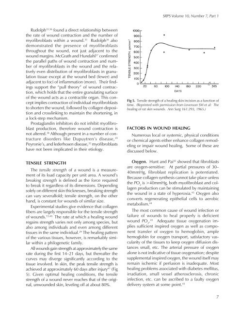
All <strong>wound</strong>s gain strength at approximately the same<br />
rate during the first 14–21 days, but thereafter the<br />
curves may diverge significantly according to the<br />
tissue involved. In skin, the peak tensile strength is<br />
achieved at approximately 60 days after injury 41 (Fig<br />
5). Given optimal <strong>healing</strong> conditions, the tensile<br />
strength of a <strong>wound</strong> never reaches that of the original,<br />
un<strong>wound</strong>ed skin, leveling off at about 80%.<br />
Fig 5. Tensile strength of a <strong>healing</strong> skin incision as a function of<br />
time. (Reprinted with permission from Levenson SM et al: The<br />
<strong>healing</strong> of rat skin <strong>wound</strong>s. Ann Surg 161:293, 1965.)<br />
FACTORS IN WOUND HEALING<br />
Numerous local or systemic, physical conditions<br />
or chemical agents either enhance collagen remodeling<br />
or impair <strong>wound</strong> <strong>healing</strong>. Some of these are<br />
discussed below.<br />
Oxygen. Hunt and Pai 42 showed that fibroblasts<br />
are oxygen-sensitive: At partial pressures of 30–<br />
40mmHg, fibroblast replication is potentiated.<br />
Because collagen synthesis cannot take place unless<br />
the PO 2<br />
is >40mmHg, both myofibroblast and collagen<br />
production can be stimulated by maintaining<br />
the <strong>wound</strong> in a state of hyperoxia. 43 Oxygen also<br />
converts regenerating epithelial cells to aerobic<br />
metabolism. 44<br />
The most common cause of <strong>wound</strong> infection or<br />
failure of <strong>wound</strong>s to heal properly is deficient<br />
<strong>wound</strong> PO 2<br />
. 45 Adequate tissue oxygenation implies<br />
sufficient inspired oxygen as well as component<br />
transfer of oxygen to hemoglobin, ample<br />
hemoglobin for oxygen transport, satisfactory vascularity<br />
of the tissues to keep oxygen diffusion distances<br />
small, etc. The arterial pressure of oxygen<br />
alone is not indicative of tissue oxygenation; despite<br />
supplemental inspired oxygen, the <strong>wound</strong> itself may<br />
remain ischemic if perfusion is inadequate. Most<br />
<strong>healing</strong> problems associated with diabetes mellitus,<br />
irradiation, small vessel atherosclerosis, chronic<br />
infection, etc. can be ascribed to a faulty oxygen<br />
delivery system at some point. 45<br />
7