Retroperitoneal Benign Mature Teratoma - InaActaMedica.org
Retroperitoneal Benign Mature Teratoma - InaActaMedica.org
Retroperitoneal Benign Mature Teratoma - InaActaMedica.org

Create successful ePaper yourself
Turn your PDF publications into a flip-book with our unique Google optimized e-Paper software.
MEDICAL ILLUSTRATION<br />
<strong>Retroperitoneal</strong> <strong>Benign</strong> <strong>Mature</strong> <strong>Teratoma</strong><br />
Pankaj Kumar Garg, Vivek Jaswal, Bhupendra Kumar Jain<br />
Department of Surgery, University College of Medical Sciences and Guru Teg Bahadur Hospital, University of Delhi,<br />
Dilshad Garden Delhi, India 110095<br />
Correspondence mail to: dr.pankajgarg@gmail.com<br />
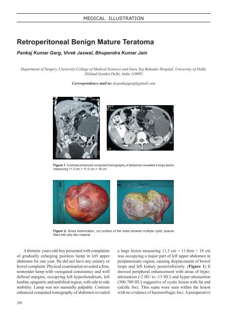
Figure 1. Contrast enhanced computed tomography of abdomen revealed a large lesion<br />
measuring 11.3 cm × 11.0 cm × 18 cm<br />
Figure 2. Gross examination, cut surface of the mass showed multiple cystic spaces<br />
filled with jelly like material<br />
A thirteen- years-old-boy presented with complaints<br />
of gradually enlarging painless lump in left upper<br />
abdomen for one year. He did not have any urinary or<br />
bowel complaint. Physical examination revealed a firm,<br />
nontender lump with variegated consistency and well<br />
defined margins, occupying left hypochondrium, left<br />
lumbar, epigastric and umbilical region, with side to side<br />
mobility. Lump was not manually palpable. Contrast<br />
enhanced computed tomography of abdomen revealed<br />
a large lesion measuring 11.3 cm × 11.0cm × 18 cm<br />
was occupying a major part of left upper abdomen in<br />
peripancreatic region, causing displacement of bowel<br />
loops and left kidney posterinferiorly. (Figure 1) It<br />
showed peripheral enhancement with areas of hypoattenuation<br />
(-2 HU to -13 HU) and hyper-attenuation<br />
(300-700 HU) suggestive of cystic lesion with fat and<br />
calcific foci. Thin septa were seen within the lesion<br />
with no evidence of haemorrhagic foci. A preoperative<br />
206
Vol 43 • Number 3 • July 2011<br />
diagnosis of mesenteric dermoid cyst was kept based<br />
on physical and radiological findings. The patient<br />
underwent exploratory laparotomy that showed a<br />
18cm x 25 cm mass in retroperitoneum pushing<br />
transverse colon and descending colon forward. The<br />
mass was extending behind the pancreas and inferiorly<br />
extending down to the bifurcation of aorta. The mass<br />
was completely excised. On gross examination, cut<br />
surface of the mass showed multiple cystic spaces filled<br />
with jelly like material. (Figure 2) Histopathological<br />
examination confirmed the diagnosis of retroperitoneal<br />
benign mature teratoma. The patient is asymptomatic<br />
at 9 months follow up.<br />
<strong>Retroperitoneal</strong> teratomas comprise 3.5 – 4% of all<br />
germ cell tumours in children. <strong>Teratoma</strong>s arise from<br />
germ cells that fail to mature normally in the gonadal<br />
locations. These totipotent cells can differentiate<br />
into tissue components representing derivatives of<br />
mesoderm, ectoderm and endoderm. 1 The distribution<br />
of teratomas are described in order of decreasing<br />
frequency, in the ovaries, the testes, the anterior<br />
mediastinum, the retroperitoneal space, the presacral<br />
and coccygeal areas, pineal and other intracranial<br />
sites, the neck and abdominal viscera other than the<br />
gonads. 2 <strong>Retroperitoneal</strong> teratomas are often located<br />
near the upper pole of the kidney, with a preponderance<br />
on the left side. <strong>Retroperitoneal</strong> teratomas are<br />
usually asymptomatic. The differential diagnosis of<br />
retroperitoneal teratomas include ovarian tumors,<br />
renal cysts, adrenal tumors, retroperitoneal fibromas,<br />
sarcomas, hemangiomas, xantogranuloma, enlarged<br />
lymph nodes and perirenal abscess. 3 Plain abdominal<br />
film shows a tissue mass and calcification. Sonography<br />
Retriperitoneal <strong>Mature</strong> <strong>Benign</strong> <strong>Teratoma</strong><br />
can identify the cystic, solid or complex components<br />
of the tumor. CT is better than sonography in defining<br />
the teratoma extent to the surrounding <strong>org</strong>ans and in<br />
evaluating the cyst wall. 4 Magnetic resonance imaging<br />
is superior to sonography and CT to demonstrate the<br />
anatomical relationship with surrounding structures<br />
like abdominal aorta. 5 Macroscopically, teratomas<br />
can be divided into either cystic or solid. Cystic<br />
teratomas are mostly benign, containing sebaceous<br />
materials and mature tissue types. On the other hand,<br />
solid teratomas are usually malignant and composed<br />
of immature embryonic tissues in addition to adipose,<br />
cartilaginous, fibrosis and bony components. 6 The<br />
prognosis is excellent for benign retroperitoneal<br />
teratoma if complete resection can be accomplished.<br />
REFERENCES<br />
1. Gschwend J, Burke TW, Woodward JE, Heller PB.<br />
<strong>Retroperitoneal</strong> teratomas presenting as an abdominal-pelvic<br />
mass. Obstet Gynecol. 1997;70:500-2.<br />
2. Engel RM, Elkins RC, Fletcher BD. <strong>Retroperitoneal</strong> teratoma.<br />
Review of the literature and presentation of an unusual case.<br />
Cancer. 1998;22:1068-73.<br />
3. Pandya JS, Pai MV, Muchhala S. <strong>Retroperitoneal</strong> teratoma<br />
presenting as acute abdomen in an elderly person. Indian J<br />
Gastroenterol. 2000;19:89-90.<br />
4. Davidson AJ, Hartman DS, Goldman SM. <strong>Mature</strong> teratoma<br />
of the retroperitoneum: radiologic, pathologic, and clinical<br />
correlation. Radiology. 1999;172:421-5.<br />
5. Bellin MF, Duron JJ, Curet P, Dion-Voirin E, Grellet J.<br />
Primary retroperitoneal teratoma in the adult: correlation of<br />
MRI features with CT and pathology. Magn Reson Imaging.<br />
1991;9:263-6.<br />
6. Bruneton JN, Diard F, Drouillard JP, Sabatier JC, Tavernier JF.<br />
Primary retroperitoneal teratoma in adults: presentation of two<br />
cases and review of the literature. Radiology. 1990;134:613-6.<br />
207