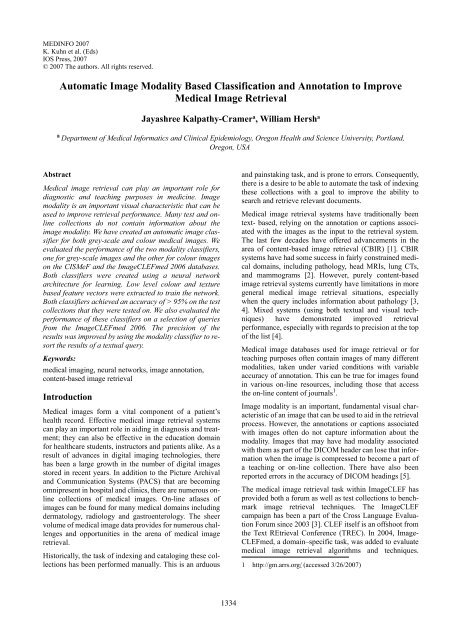
Automatic Image Modality Based Classification and Annotation to ...
Automatic Image Modality Based Classification and Annotation to ...
Automatic Image Modality Based Classification and Annotation to ...

Create successful ePaper yourself
Turn your PDF publications into a flip-book with our unique Google optimized e-Paper software.
MEDINFO 2007<br />
K. Kuhn et al. (Eds)<br />
IOS Press, 2007<br />
© 2007 The authors. All rights reserved.<br />
<strong>Au<strong>to</strong>matic</strong> <strong>Image</strong> <strong>Modality</strong> <strong>Based</strong> <strong>Classification</strong> <strong>and</strong> <strong>Annotation</strong> <strong>to</strong> Improve<br />
Medical <strong>Image</strong> Retrieval<br />
Jayashree Kalpathy-Cramer a , William Hersh a<br />
a Department of Medical Informatics <strong>and</strong> Clinical Epidemiology, Oregon Health <strong>and</strong> Science University, Portl<strong>and</strong>,<br />
Oregon, USA<br />
Abstract<br />
Medical image retrieval can play an important role for<br />
diagnostic <strong>and</strong> teaching purposes in medicine. <strong>Image</strong><br />
modality is an important visual characteristic that can be<br />
used <strong>to</strong> improve retrieval performance. Many test <strong>and</strong> online<br />
collections do not contain information about the<br />
image modality. We have created an au<strong>to</strong>matic image classifier<br />
for both grey-scale <strong>and</strong> colour medical images. We<br />
evaluated the performance of the two modality classifiers,<br />
one for grey-scale images <strong>and</strong> the other for colour images<br />
on the CISMeF <strong>and</strong> the <strong>Image</strong>CLEFmed 2006 databases.<br />
Both classifiers were created using a neural network<br />
architecture for learning. Low level colour <strong>and</strong> texture<br />
based feature vec<strong>to</strong>rs were extracted <strong>to</strong> train the network.<br />
Both classifiers achieved an accuracy of > 95% on the test<br />
collections that they were tested on. We also evaluated the<br />
performance of these classifiers on a selection of queries<br />
from the <strong>Image</strong>CLEFmed 2006. The precision of the<br />
results was improved by using the modality classifier <strong>to</strong> resort<br />
the results of a textual query.<br />
Keywords:<br />
medical imaging, neural networks, image annotation,<br />
content-based image retrieval<br />
Introduction<br />
Medical images form a vital component of a patient’s<br />
health record. Effective medical image retrieval systems<br />
can play an important role in aiding in diagnosis <strong>and</strong> treatment;<br />
they can also be effective in the education domain<br />
for healthcare students, instruc<strong>to</strong>rs <strong>and</strong> patients alike. As a<br />
result of advances in digital imaging technologies, there<br />
has been a large growth in the number of digital images<br />
s<strong>to</strong>red in recent years. In addition <strong>to</strong> the Picture Archival<br />
<strong>and</strong> Communication Systems (PACS) that are becoming<br />
omnipresent in hospital <strong>and</strong> clinics, there are numerous online<br />
collections of medical images. On-line atlases of<br />
images can be found for many medical domains including<br />
derma<strong>to</strong>logy, radiology <strong>and</strong> gastroenterology. The sheer<br />
volume of medical image data provides for numerous challenges<br />
<strong>and</strong> opportunities in the arena of medical image<br />
retrieval.<br />
His<strong>to</strong>rically, the task of indexing <strong>and</strong> cataloging these collections<br />
has been performed manually. This is an arduous<br />
<strong>and</strong> painstaking task, <strong>and</strong> is prone <strong>to</strong> errors. Consequently,<br />
there is a desire <strong>to</strong> be able <strong>to</strong> au<strong>to</strong>mate the task of indexing<br />
these collections with a goal <strong>to</strong> improve the ability <strong>to</strong><br />
search <strong>and</strong> retrieve relevant documents.<br />
Medical image retrieval systems have traditionally been<br />
text- based, relying on the annotation or captions associated<br />
with the images as the input <strong>to</strong> the retrieval system.<br />
The last few decades have offered advancements in the<br />
area of content-based image retrieval (CBIR) [1]. CBIR<br />
systems have had some success in fairly constrained medical<br />
domains, including pathology, head MRIs, lung CTs,<br />
<strong>and</strong> mammograms [2]. However, purely content-based<br />
image retrieval systems currently have limitations in more<br />
general medical image retrieval situations, especially<br />
when the query includes information about pathology [3,<br />
4]. Mixed systems (using both textual <strong>and</strong> visual techniques)<br />
have demonstrated improved retrieval<br />
performance, especially with regards <strong>to</strong> precision at the <strong>to</strong>p<br />
of the list [4].<br />
Medical image databases used for image retrieval or for<br />
teaching purposes often contain images of many different<br />
modalities, taken under varied conditions with variable<br />
accuracy of annotation. This can be true for images found<br />
in various on-line resources, including those that access<br />
the on-line content of journals 1 .<br />
<strong>Image</strong> modality is an important, fundamental visual characteristic<br />
of an image that can be used <strong>to</strong> aid in the retrieval<br />
process. However, the annotations or captions associated<br />
with images often do not capture information about the<br />
modality. <strong>Image</strong>s that may have had modality associated<br />
with them as part of the DICOM header can lose that information<br />
when the image is compressed <strong>to</strong> become a part of<br />
a teaching or on-line collection. There have also been<br />
reported errors in the accuracy of DICOM headings [5].<br />
The medical image retrieval task within <strong>Image</strong>CLEF has<br />
provided both a forum as well as test collections <strong>to</strong> benchmark<br />
image retrieval techniques. The <strong>Image</strong>CLEF<br />
campaign has been a part of the Cross Language Evaluation<br />
Forum since 2003 [3]. CLEF itself is an offshoot from<br />
the Text REtrieval Conference (TREC). In 2004, <strong>Image</strong>-<br />
CLEFmed, a domain–specific task, was added <strong>to</strong> evaluate<br />
medical image retrieval algorithms <strong>and</strong> techniques.<br />
1 http://gm.arrs.org/ (accessed 3/26/2007)<br />
1334
J. Kalpathy-Cramer et al. / <strong>Au<strong>to</strong>matic</strong> <strong>Image</strong> <strong>Modality</strong> <strong>Based</strong> <strong>Classification</strong> <strong>and</strong> <strong>Annotation</strong> <strong>to</strong> Improve Medical <strong>Image</strong> Retrieval<br />
Approaches combining both visual <strong>and</strong> textual techniques<br />
for retrieval have shown some promise at medical image<br />
retrieval tasks [3]. In 2005, a medical image annotation<br />
task was added <strong>to</strong> <strong>Image</strong>CLEF. The goal of this task was <strong>to</strong><br />
correctly classify 1000 test images in<strong>to</strong> 116 classes given a<br />
set of 10,000 training images. The classes differed primarily<br />
in ana<strong>to</strong>my <strong>and</strong> view of the image. It should be noted,<br />
however, that these images were primarily of a single<br />
modality (X-rays).The goal of the <strong>Image</strong>CLEF medical<br />
image retrieval task of 2006 was <strong>to</strong> retrieve relevant<br />
images for thirty <strong>to</strong>pics from a test collection of about<br />
50,000 annotated images of different modalities. These<br />
tasks were divided by the organizers in<strong>to</strong> those expected <strong>to</strong><br />
be amenable <strong>to</strong> textual, visual, or mixed retrieval<br />
techniques.<br />
We participated in both the medical image retrieval <strong>and</strong> the<br />
au<strong>to</strong>matic medical image annotation tasks at <strong>Image</strong>CLEF<br />
2006 [6, 7]. The techniques developed for those tasks have<br />
been extended for the more general task of medical image<br />
modality classification <strong>and</strong> annotation.<br />
Using medical image modality for image annotation <strong>and</strong><br />
retrieval has recently been studied. Florea et al [8] have<br />
compared the efficacy of two different systems (MedIC<br />
<strong>and</strong> MedGIFT) in classifying the modality of a database<br />
with six st<strong>and</strong>ard modalities for radiology <strong>and</strong> nuclear<br />
medicine images.<br />
In this paper, we compare the results obtained on our system<br />
with those described in previous publications [8] for<br />
the six modalities of the CISMeF database. We will also<br />
extend this technique <strong>to</strong> classify colour images from the<br />
<strong>Image</strong>CLEF medical retrieval task collection [6] in<strong>to</strong> six<br />
categories. We will finally report on the improvement in<br />
precision that we observed for a selected number of tasks<br />
of the <strong>Image</strong>CLEF medical retrieval task for 2006 by<br />
incorporating the modality classifier in series with a textbased<br />
retrieval system.<br />
Methods<br />
We employed a supervised machine learning approach <strong>to</strong><br />
problem of medical image modality classification using a<br />
hierarchical classification scheme as seen in figure 1.<br />
There were two primary databases that were used <strong>to</strong> create<br />
<strong>and</strong> test the classifiers. We worked with a small subset of<br />
the CISMeF database as the primary target for our greyscale<br />
(radiographic <strong>and</strong> nuclear medicine) image classifier<br />
[9]. This database had a set of 1332 images classified in<strong>to</strong><br />
one of six classes based on modality. These include<br />
angiography, computerized <strong>to</strong>mography scans (CT), X-<br />
ray, Magnetic resonance (MRI), ultrasound, <strong>and</strong> scintigraphy.<br />
The images in this database had been acquired under<br />
differing conditions over a long period of time. Consequently,<br />
there was considerable intra-class variation in<br />
quality, size, contrast, illumination <strong>and</strong> background.<br />
The imageCLEFmed database contains 50,000 images of<br />
differing modalities, including radiography <strong>and</strong> nuclear<br />
medicine, as well as microscopic <strong>and</strong> his<strong>to</strong>pathological<br />
images, pho<strong>to</strong>graphs <strong>and</strong> gross pathology images, power<br />
point slides, electroencephalograhical images (EEGs) <strong>and</strong><br />
electrocardiograms (ECGs), as well as a few miscellaneous<br />
images.<br />
Figure 1 - Hierarchical classification scheme for images<br />
A neural network-based scheme using a variety of low<br />
level, primarily global image features was used <strong>to</strong> create a<br />
six-class classification system for the grayscale images.<br />
The multilayer perceptron architecture used a hidden layer<br />
of approximately 50-150 nodes. The classification system<br />
was created in MATLAB 2 , in part using several routines<br />
modified from the Netlab <strong>to</strong>olbox 3 .<br />
We experimented with a variety of feature vec<strong>to</strong>rs as<br />
inputs <strong>to</strong> the network. A combination of texture <strong>and</strong> intensity<br />
his<strong>to</strong>gram features provided the best classification [10,<br />
11]. All images were first resized while maintaining the<br />
aspect ratio such that the smaller dimension was 256 pixels.<br />
The image was divided in<strong>to</strong> five overlapping blocks.<br />
Grey level correlation matrices were computed for each<br />
block using four angles <strong>and</strong> an offset of 1 pixel. Contrast,<br />
correlation, energy, homogeneity <strong>and</strong> entropy were calculated<br />
for each matrix. A quantized grey scale his<strong>to</strong>gram<br />
was then appended resulting in a 132-dimension feature<br />
vec<strong>to</strong>r for each image for the texture. All inputs <strong>to</strong> the neural<br />
network (the image feature vec<strong>to</strong>rs) were normalized<br />
using the training set <strong>to</strong> have a mean of zero <strong>and</strong> variance<br />
of 1.<br />
The 1332 images in the database were r<strong>and</strong>omly split in<strong>to</strong><br />
a training set of 1000 images <strong>and</strong> a test set of 332 images.<br />
A small r<strong>and</strong>om subset of the training images was initially<br />
used <strong>to</strong> create the classifier (200 images). The classifier<br />
2 www.mathworks.com (accessed 3/26/2007)<br />
3 http://www.ncrg.as<strong>to</strong>n.ac.uk/netlab/index.php (accessed<br />
3/26/2007)<br />
1335
J. Kalpathy-Cramer et al. / <strong>Au<strong>to</strong>matic</strong> <strong>Image</strong> <strong>Modality</strong> <strong>Based</strong> <strong>Classification</strong> <strong>and</strong> <strong>Annotation</strong> <strong>to</strong> Improve Medical <strong>Image</strong> Retrieval<br />
was then applied <strong>to</strong> the entire training set <strong>and</strong> images that<br />
were misclassified were then added <strong>to</strong> the images used <strong>to</strong><br />
refine the classifier. The classifier was finally tested on the<br />
test images.<br />
A similar scheme was used <strong>to</strong> create the classifier for<br />
colour images. We believe this novel idea can improve the<br />
retrieval performance of purely textual systems or for<br />
images for which the associated modalities are not known.<br />
Although modality detec<strong>to</strong>rs for grey-scale medical<br />
images have been reported [9], we are unaware of a similar<br />
effort for classification of other categories of medical<br />
images like those produced by his<strong>to</strong>-pathology <strong>and</strong> endoscopy.<br />
The images used for this classification task were<br />
taken from the test collection of images used in the <strong>Image</strong>-<br />
CLEFmed retrieval task. 2250 colour images in this<br />
collection were broadly categorized in<strong>to</strong> six categories as<br />
microscopic, gross pathology, EEG/ECG or other charts,<br />
powerpoint slides, endoscopic images <strong>and</strong> other. There<br />
was considerable intra-class variability in this dataset.<br />
These 2250 images were again split r<strong>and</strong>omly in<strong>to</strong> training<br />
(1750) <strong>and</strong> test images (500). A similar training methodology<br />
<strong>to</strong> that described above was used <strong>to</strong> incrementally<br />
improve the classifier, starting with a smaller subset of the<br />
training database.<br />
A two-layer architecture with 25-150 hidden nodes was<br />
used for the neural network. The feature vec<strong>to</strong>r in this case<br />
consisted of colour his<strong>to</strong>gram features, as well as texture<br />
features obtained using the grey level correlation matrices.<br />
The image was split in<strong>to</strong> 9 uneven blocks. Colour his<strong>to</strong>gram<br />
properties of image after conversion in<strong>to</strong> the<br />
L*A*B* colour space were calculated, while texture features<br />
were calculated after converting the image <strong>to</strong> greyscale<br />
These neural network classifiers can be created <strong>to</strong> further<br />
classify images within a given modality. For instance, x-<br />
ray images could now be classified <strong>to</strong> account for ana<strong>to</strong>my.<br />
Ana<strong>to</strong>mical classifiers were used in the au<strong>to</strong>matic annotation<br />
task at <strong>Image</strong>CLEFmed.<br />
The tasks had been stated in English, German <strong>and</strong> French,<br />
<strong>and</strong> had <strong>and</strong> provided example images. All but three of the<br />
tasks stated the desired modality of the image <strong>to</strong> be<br />
retrieved. Two examples of the tasks are shown in figure 2.<br />
Show me images of a h<strong>and</strong> x-ray.<br />
Zeige mir Röntgenbilder einer H<strong>and</strong>.<br />
Montre-moi des radiographies de la main.<br />
Show me blood smears that include polymorphonuclear<br />
neutrophils. Zeige mir Blutabstriche mit polymophonuklearer<br />
Neutrophils. Montre-moi des échantillons de<br />
sang incluant des neutrophiles polymorphonucléaires.<br />
Figure 2 - Sample textual <strong>and</strong> visual queries at<br />
<strong>Image</strong>CLEFmed 2006<br />
Once our classifiers had been trained <strong>to</strong> achieve >95%<br />
classification accuracy, they were tested on a r<strong>and</strong>om subset<br />
of the <strong>Image</strong>CLEFmed <strong>to</strong>pics.<br />
The schematic of our modified retrieval system is shown<br />
below. The query was initially fed <strong>to</strong> our Lucene 4 based<br />
text retrieval system. The queries were manually edited by<br />
one of the authors. The resulting images were subsequently<br />
classified by the hierarchical classifier for<br />
modality. <strong>Image</strong>s of the desired modality (as stated in the<br />
query or as discerned by the au<strong>to</strong>matic classifier based on<br />
the sample images) were moved <strong>to</strong> the <strong>to</strong>p of the list while<br />
maintaining the ranking of the textual system within a<br />
class.<br />
Figure 3 - <strong>Image</strong> retrieval system used for the<br />
<strong>Image</strong>CLEFmed 2006 test collection<br />
We compared the results of our purely textual system with<br />
that including the addition of the modality classifier.<br />
Results<br />
A classification accuracy of 96.4% was achieved on the<br />
CISMeF database. The confusion matrix suggests that the<br />
primary misclassification occur between the MRI <strong>and</strong> CT<br />
scan classes. This is not surprising as these classes are<br />
visually quite similar. Florea et al [8] have reported similar<br />
results both in terms of accuracy <strong>and</strong> inter-class misclassification<br />
patterns. The classification of grey-scale medical<br />
images in<strong>to</strong> commonly occurring modalities using low<br />
level image features <strong>and</strong> machine learning techniques<br />
appears <strong>to</strong> be a tractable task. We expect <strong>to</strong> achieve over<br />
98% accuracy with further refinement of our machine<br />
4 http://lucene.apache.org/ (accessed 3/26/2007)<br />
1336
J. Kalpathy-Cramer et al. / <strong>Au<strong>to</strong>matic</strong> <strong>Image</strong> <strong>Modality</strong> <strong>Based</strong> <strong>Classification</strong> <strong>and</strong> <strong>Annotation</strong> <strong>to</strong> Improve Medical <strong>Image</strong> Retrieval<br />
learning approach by the use of more advanced cross-validation,<br />
bootstrapping, boosting or bagging techniques.<br />
Preliminary testing of the classifiers on 2250 colour<br />
images of the imageCLEFmed test collection resulted in a<br />
modality classification accuracy of 98.6%. Most of the<br />
misclassifications involved the “other” class with contained<br />
a set of miscellaneous images not belonging <strong>to</strong> the<br />
other five specific categories<br />
The colour modality classifier was tested on a small r<strong>and</strong>om<br />
subset of the <strong>Image</strong>CLEFmed 2006 <strong>to</strong>pics. The <strong>to</strong>pics<br />
for imageCLEFmed 2006 fell in<strong>to</strong> three categories (visual,<br />
mixed, semantic) consisting of 10 tasks each. Although<br />
visual techniques had, in general, performed extremely<br />
poorly at the semantic tasks, use of some visual information<br />
(primarily modality) was shown <strong>to</strong> increase the<br />
precision [4].<br />
Analysis of our textual results indicated that in many queries,<br />
especially those of a visual or mixed nature, up <strong>to</strong><br />
75% of the <strong>to</strong>p 1000 results were not of the correct modality.<br />
A compelling example is given in figure 4 <strong>and</strong> table 1.<br />
Only 90 of the <strong>to</strong>p 2000 images returned by the textual<br />
query were of the desired modality.<br />
Task 1 - Show me images of the oral cavity<br />
including teeth <strong>and</strong> gum tissue<br />
<strong>Image</strong> type<br />
Number of images<br />
Total returned by textual query 2000<br />
Grey-scale 1831<br />
1<br />
0.9<br />
0.8<br />
0.7<br />
0.6<br />
0.5<br />
0.4<br />
0.3<br />
0.2<br />
0.1<br />
0<br />
Task 1 Precision<br />
P5 P10 P15 P20 P30 P100 P200 P500 P1000<br />
OHSU textual + modality classifier OSHU textual Best system overall<br />
Figure 5 - Improvement in precision resulting from<br />
modality classification.<br />
Figure 5 plots the precision for varying number of documents<br />
retrieved for the purely textual system, the<br />
improvement with the use of the modality classifier <strong>and</strong><br />
the overall best system (mixed visual <strong>and</strong> textual based)<br />
that participated in <strong>Image</strong>CLEFmed 2006. This increased<br />
the precision of the query as seen in figure 5. The improvement<br />
in precision at the <strong>to</strong>p of the ranked list (P5 – P200) is<br />
better with the use of the modality detec<strong>to</strong>r compared <strong>to</strong> a<br />
purely textual search. We should note that a perfect modality<br />
classifier will only improve the precision of the search<br />
<strong>and</strong> not the recall if it is applied in the serial manner<br />
described above. The mean average precision (MAP)<br />
would still be limited by the number of relevant images<br />
that are retrieved by the textual search (recall of the textual<br />
search).<br />
Even in searches that are expected <strong>to</strong> be semantic, we see<br />
an improvement in precision by using the modality classifier<br />
as seen in figure 6 <strong>and</strong> 7.<br />
Task 2 - Show me microscopic images of tissue from the<br />
cerebellum (semantic query)<br />
Pho<strong>to</strong>graph/gross pathology 90<br />
Microscope 71<br />
Other 8<br />
Figure 4- Sample query suitable for visual retrieval at<br />
<strong>Image</strong>CLEFmed 2006<br />
These images were then classified using our modality classifier.<br />
The ranked list of retrieved images was resorted<br />
taking in<strong>to</strong> account the desired modality based on the<br />
query.<br />
<strong>Image</strong> type<br />
Total returned by<br />
textual query<br />
Number of images<br />
2000<br />
Greyscale 1476<br />
Pho<strong>to</strong>graph/gross<br />
pathology<br />
408<br />
Microscope 116<br />
Figure 6 - Sample query suitable for visual retrieval at<br />
<strong>Image</strong>CLEFmed 2006<br />
1337
J. Kalpathy-Cramer et al. / <strong>Au<strong>to</strong>matic</strong> <strong>Image</strong> <strong>Modality</strong> <strong>Based</strong> <strong>Classification</strong> <strong>and</strong> <strong>Annotation</strong> <strong>to</strong> Improve Medical <strong>Image</strong> Retrieval<br />
The precision of this search was similarly improved by the<br />
use of the modality detec<strong>to</strong>r as seen in figure 7.<br />
1<br />
0.9<br />
0.8<br />
0.7<br />
0.6<br />
0.5<br />
0.4<br />
0.3<br />
0.2<br />
0.1<br />
0<br />
Figure 7 - Improvement in precision resulting from<br />
modality classification<br />
Four of the six tasks tested showed improvement in precision<br />
by the use of the modality detec<strong>to</strong>r for colour images.<br />
There were two tasks amenable <strong>to</strong> textual methods for<br />
which there very little change in precision with the addition<br />
of the modality information.<br />
We plan on testing the performance of the modality detec<strong>to</strong>r<br />
on the complete set of tasks for <strong>Image</strong>CLEFmed 2005<br />
<strong>and</strong> 2006. We also intend <strong>to</strong> index the entire collection of<br />
50,000 images used in the <strong>Image</strong>CLEFmed test collection<br />
using the modality classifier. Information about the class<br />
membership of an image will be added <strong>to</strong> the metadata.<br />
This should improve the performance of the retrieval in<br />
two ways. Clustering of the data by modality <strong>and</strong> perhaps<br />
ana<strong>to</strong>my will speed up the search process as fewer documents<br />
will have <strong>to</strong> be compared <strong>to</strong> the query image/text.<br />
Secondly, we expect that the overall precision of the search<br />
will improve by considering the modality of the image that<br />
is desired by the user. However, we can expect a small<br />
degradation in the recall due <strong>to</strong> potentially misclassified<br />
images not being searched.<br />
Conclusion<br />
Task 25 Precision<br />
P5<br />
P10<br />
P15<br />
P20<br />
P30<br />
P100<br />
P200<br />
P500<br />
P1000<br />
OHSU textual + modality classifier OSHU t ext ual Best system overall<br />
We have developed a neural network based, hierarchical<br />
classifier for the modality classification of medical<br />
images. This system can classify colour images including<br />
his<strong>to</strong>-pathological <strong>and</strong> endoscopic images, <strong>and</strong> pho<strong>to</strong>graphs<br />
as well as grey-scale (radiological <strong>and</strong> nuclear<br />
medicine). The classifier uses a his<strong>to</strong>gram <strong>and</strong> texture<br />
properties as inputs <strong>to</strong> the two level neural network. This<br />
classifier results in a classification accuracy of greater than<br />
95% for the grey-scale images of the CISMeF database as<br />
well as a selection of colour <strong>and</strong> grey-scale images from<br />
the <strong>Image</strong>CLEFmed database. The use of this classifier<br />
increases the precision of retrieval of our primarily text<br />
based retrieval system by moving images of the desired<br />
modality <strong>to</strong> the <strong>to</strong>p of the ranked list.<br />
Acknowledgments<br />
The authors would like <strong>to</strong> thank Henning Müller for his valuable<br />
input in discussions as well as providing access <strong>to</strong> the CISMeF<br />
images. We would also like <strong>to</strong> thank Jean-Nicholas Dacher at the<br />
Rouen Hospital, France, for proving the CISMeF images used in<br />
this paper. We would like <strong>to</strong> thank Dr. T. Lehmann at RWTH<br />
Aachen University, Germany, for access <strong>to</strong> the IRMA image<br />
database. We acknowledge the support of NLM Training grant<br />
5T15 LM07088-15 <strong>and</strong> NSF Grant ITR-0325160.<br />
References<br />
[1] Smeulders AWM, Worring M, Santini S, Gupta A, Jain R.<br />
Content-based image retrieval at the end of the early years.<br />
IEEE Transactions on Pattern Analysis <strong>and</strong> Machine<br />
Intelligence 2000;22(12), 1349-80.<br />
[2] Müller H, Michoux N, B<strong>and</strong>on D, Geissbuhler A. A review<br />
of content-based image retrieval systems in medicine –<br />
clinical benefits <strong>and</strong> future directions. International Journal<br />
of Medical Informatics 2004; 73, 1-23.<br />
[3] Müller H, Deselaers T, Lehmann T, Clough P, Hersh W.<br />
Overview of the <strong>Image</strong>CLEFmed 2006 medical retrieval<br />
annotation tasks, Evaluation of Multilingual <strong>and</strong> Multimodal<br />
Information Retrieval, Seventh Workshop of the<br />
Cross-Language Evaluation Forum, CLEF 2006, edi<strong>to</strong>rs:<br />
Peters C, Clough P, Gey F, Karlgren J, Magnini B,<br />
Oard DW, de Rijke M, Stempfhuber M, LNCS<br />
2006,Alicante, Spain, <strong>to</strong> appear.<br />
[4] Hersh W, Kalpathy-Cramer J, Jensen, J. Medical <strong>Image</strong><br />
Retrieval <strong>and</strong> Au<strong>to</strong>mated <strong>Annotation</strong>: OHSU at<br />
<strong>Image</strong>CLEF 2006. Working Notes for the CLEF 2006<br />
Workshop, Alicante, Spain. http://www.clef-campaign.org/<br />
2006/<br />
[5] Güld MO, Kohnen M, Keysers D, Schubert H, Wein BB,<br />
Bredno J, Lehmann TM, Quality of DICOM header<br />
information for image categorization, Proceedings SPIE,<br />
4685, 280-287, 2002.<br />
[6] Hersh W, Müller H, Jensen J, Yang J, Gorman P, Ruch P.<br />
Advancing biomedical image retrieval: development <strong>and</strong><br />
analysis of a test collection. J Amer Med Inform Assoc<br />
2006; 13(5).<br />
[7] Lehmann TM, Güld MO, Thies C, Fischer B, Keysers D,<br />
Kohnen M, Schubert H, Wein BB. Content-based image<br />
retrieval in medical applications for picture archiving <strong>and</strong><br />
communication systems, in Medical Imaging, SPIE<br />
Proceedings, 5033, 440-451, San Diego, California, 2003.<br />
[8] Florea F, Müller H, Rogozan A, Geissbühler A, Darmoni S,<br />
Medical image categorization with MedIC <strong>and</strong>MedGIFT.<br />
Medical Informatics Europe (MIE 2006).<br />
[9] Douyere M, Soualmia LF, Neveol A, Rogozan A, Dahamna<br />
B, Leroy JP, Thirion B, Darmoni SJ. Enhancing the MeSH<br />
thesaurus <strong>to</strong> retrieve French online health resources in a<br />
quality-controlled gateway. Health Info Libr J 2004;<br />
21(4):253-61.<br />
[10] Haralick R. Statistical <strong>and</strong> structural approaches <strong>to</strong> texture.<br />
Proceedings of the IEEE 67, 786-804, 1979.<br />
[11] Howarth P, Ruger S. Evaluation of texture features for<br />
content-based imageretrieval. In: Proceedings of the<br />
International Conference on <strong>Image</strong> <strong>and</strong> Video Retrieval,<br />
Springer-Verlag, pp. 326-324, 2004.<br />
Address for correspondence<br />
Jayashree Kalpathy-Cramer<br />
5th Floor, Biomedical Information Communication Center<br />
3181 S.W. Sam Jackson Park Rd.<br />
Portl<strong>and</strong>, Oregon 97239-3098<br />
Email: kalpathy@ohsu.edu<br />
1338