Chediak-Higashi Syndrome Presenting in Accelerated Phase
Chediak-Higashi Syndrome Presenting in Accelerated Phase
Chediak-Higashi Syndrome Presenting in Accelerated Phase

Create successful ePaper yourself
Turn your PDF publications into a flip-book with our unique Google optimized e-Paper software.
CASE REPORT<br />
<strong>Chediak</strong>-<strong>Higashi</strong> <strong>Syndrome</strong> <strong>Present<strong>in</strong>g</strong> <strong>in</strong> <strong>Accelerated</strong> <strong>Phase</strong><br />
Tanzeel Imran 1 , Lubna Zafar 2 , Madeeha Rehan 1 , Aqsa Nasir 4 , Parveen Akhtar Tariq 3 and Iffat Batool 3<br />
ABSTRACT<br />
<strong>Chediak</strong>-<strong>Higashi</strong> <strong>Syndrome</strong> (CHS) is a rare autosomal recessive disorder, characterized by silver hair, recurrent <strong>in</strong>fections,<br />
partial oculo-cutaneous alb<strong>in</strong>ism, mild coagulation defect and progressive neuropathy. The characteristic feature of CHS<br />
is the presence of huge lysosomes and cytoplasmic <strong>in</strong>clusions with<strong>in</strong> different body cells like the white blood cells. The<br />
disease has an early onset but usually presents <strong>in</strong> an accelerated phase. We present a case of a 2 years old boy with high<br />
grade fever, bilateral cervical lymphadenopathy, hepatosplenomegaly, abdom<strong>in</strong>al distention of 28 days duration. He was<br />
diagnosed with <strong>Chediak</strong>-<strong>Higashi</strong> syndrome <strong>in</strong> accelerated phase on the basis of cl<strong>in</strong>ical presentation, morphological<br />
f<strong>in</strong>d<strong>in</strong>gs on peripheral blood film and bone marrow aspirate.<br />
Key words:<br />
<strong>Chediak</strong>-<strong>Higashi</strong> syndrome. Oculo-cutaneous. Alb<strong>in</strong>ism. <strong>Accelerated</strong> phase. Giant lysosomal <strong>in</strong>clusions.<br />
INTRODUCTION<br />
<strong>Chediak</strong>- <strong>Higashi</strong> <strong>Syndrome</strong> (CHS) is a rare autosomal<br />
recessive disorder; this means that both parents must<br />
contribute a defective gene to the child to show<br />
symptoms of the disease. It is usually a disease of<br />
children but it can also affect adults. 1 CHS was<br />
described first by Beguez Cesar <strong>in</strong> 1943. Later, Chédiak<br />
<strong>in</strong> 1952 described the full cl<strong>in</strong>ical and haematological<br />
features, and <strong>Higashi</strong> <strong>in</strong> 1954 discovered that the<br />
<strong>in</strong>clusions are peroxidase (Sudan Black B) positive. The<br />
gene responsible for this defect was characterized <strong>in</strong><br />
1996 as the lysosomal traffick<strong>in</strong>g regulator (LYST or<br />
CHS1 gene). It is located on chromosome-1 (1q42-43). 1<br />
There are two types of mutations responsible for the<br />
disease i.e. frameshift and nonsense. Both are<br />
associated with cl<strong>in</strong>ically mild and severe form of<br />
disease. 2<br />
CHS prote<strong>in</strong> is expressed <strong>in</strong> the cytoplasm of the cells of<br />
different tissues like blood (neutrophils, lymphocytes,<br />
histiocytes), melanocytes, neurons etc. and may<br />
represent an abnormality of organeller prote<strong>in</strong>. It is<br />
characterized by recurrent pyogenic <strong>in</strong>fection, partial<br />
oculo-cutaneous alb<strong>in</strong>ism, mild coagulation defect and<br />
progressive peripheral neuropathy. 3,4 Neurological<br />
symptoms manifest <strong>in</strong> approximately half of the affected<br />
<strong>in</strong>dividuals. These patients exhibit alteration <strong>in</strong><br />
neutrophils which <strong>in</strong>clude neutropenia, decreased<br />
deformability, result<strong>in</strong>g <strong>in</strong> impaired chemotaxis; and<br />
delay <strong>in</strong> the fusion of phagolysosomes lead<strong>in</strong>g to<br />
defective bactericidal activity. 2<br />
1 Department of Haematology / Pathology 2 / Paediatrics 3 ,<br />
Fauji Foundation Hospital, Rawalp<strong>in</strong>di.<br />
4 Department of Pathology, Foundation University Medical<br />
College, Rawalp<strong>in</strong>di.<br />
Correspondence: Dr. Tanzeel Imran, Department of Haematology,<br />
Fauji Foundation Hospital, Jehlum Road, Rawalp<strong>in</strong>di.<br />
E-mail: dr_tanzeel@yahoo.co.uk<br />
Received November 15, 2010; accepted March 10, 2012.<br />
Although it is an <strong>in</strong>herited disorder present s<strong>in</strong>ce birth,<br />
mostly the patients present <strong>in</strong> an accelerated phase<br />
(50-85%) with fever, lymphadenopathy, anaemia,<br />
hepatosplenomegaly, jaundice, squ<strong>in</strong>t and wide spread<br />
lymphohistiocytic <strong>in</strong>filtration. 2,5 Mortality is high <strong>in</strong><br />
accelerated phase. Early diagnosis of the syndrome and<br />
bone marrow transplantation before the onset of<br />
accelerated phase is the only curable treatment. 6<br />
The present patient presented with <strong>Chediak</strong>-<strong>Higashi</strong><br />
syndrome <strong>in</strong> accelerated phase which has poor<br />
prognosis.<br />
CASE REPORT<br />
A 2 years old boy presented to Paediatric Department of<br />
Fauji Foundation Hospital, Rawalp<strong>in</strong>di, with high grade<br />
fever, cough, swell<strong>in</strong>g of neck and abdom<strong>in</strong>al distention<br />
for the last 15 days. He was treated for these symptoms<br />
<strong>in</strong> a local hospital and afterwards <strong>in</strong> Fauji Foundation<br />
Hospital, Kalar Kahar, from where he was referred to<br />
Fauji Foundation Hospital (FFH), Rawalp<strong>in</strong>di, because<br />
of his deteriorat<strong>in</strong>g condition.<br />
The patient had previous history of similar episodes but<br />
with lesser <strong>in</strong>tensity. On physical exam<strong>in</strong>ation, he had<br />
pallor, hepatosplenomegaly significant cervical lymphadenopathy,<br />
swollen tonsils covered with whitish<br />
membrane and enlarged axillary and <strong>in</strong>gu<strong>in</strong>al lymph<br />
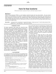
nodes. He also had silver hair and partial cutaneous<br />
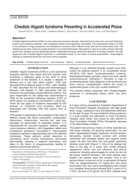
alb<strong>in</strong>ism s<strong>in</strong>ce birth (Figure 1). One of his sibl<strong>in</strong>gs had<br />
died at the age of 3 years with similar physical<br />
appearance (silver hair and alb<strong>in</strong>ism) and cl<strong>in</strong>ical<br />
symptoms <strong>in</strong>clud<strong>in</strong>g splenomegaly for which he did not<br />
get proper medical treatment. Two other sibl<strong>in</strong>gs were<br />
healthy. His parents were first cous<strong>in</strong>s. Due to his<br />
serious illness, he was put on <strong>in</strong>j. co-amoxiclav 50 mg/kg<br />
TDS, <strong>in</strong>j. dexamethasome 0.5 mg/kg B.D and syrup<br />
fluconazole TDS. His rout<strong>in</strong>e <strong>in</strong>vestigations were carried<br />
out, complete blood picture showed W.B.C count of 5.3<br />
x 10 9 /L, Hb level of 9.2 gm/dl, Hct of 30%, platelet count<br />
Journal of the College of Physicians and Surgeons Pakistan 2012, Vol. 22 (8): 539-541 539
Tanzeel Imran, Lubna Zafar, Madeeha Rehan, Aqsa Nasir, Parveen Akhtar Tariq and Iffat Batool<br />
Figure 1: Features of <strong>Chediak</strong>-<strong>Higashi</strong> syndrome<br />
with silver hair, partial alb<strong>in</strong>ism and bilateral<br />
cervical lymphadenopathy.<br />
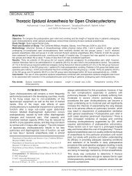
Figure 2: Peripheral blood film show<strong>in</strong>g large<br />
<strong>in</strong>clusions with<strong>in</strong> neutrophils.<br />
Figure 3: Bone marrow aspirate show<strong>in</strong>g large<br />
<strong>in</strong>clusions with<strong>in</strong> myeloid precursors.<br />
of 113 x 10 9 /L, MCV of 70 fl, differential leucocyte count<br />
showed neutrophils 47%, lymphocytes 41% and<br />
monocytes at 11%. Coagulation studies revealed PT of<br />
16/second and APTT 34/second. His serum total<br />
bilirub<strong>in</strong> was 70 umol/L, ALT was 79U/L, ALP 1910 U/L<br />
and serology was negative for HBV/HCV.<br />
In view of his cl<strong>in</strong>ical presentation and bicytopenia, he<br />
was referred for bone marrow aspiration to rule out<br />
leukemia/lymphoma. Exam<strong>in</strong>ation of his peripheral<br />
blood film showed large gray green <strong>in</strong>clusions <strong>in</strong> almost<br />
all granulocytes and lymphocytes (Figure 2). These<br />
<strong>in</strong>clusions were also appreciated on bone marrow<br />
aspirate (Figure 3) and were Sudan Black B positive.<br />
Bone marrow aspirate also revealed prom<strong>in</strong>ent<br />
histiocytes and haemophagocytosis. On the basis of his<br />
cl<strong>in</strong>ical presentation, peripheral blood film and bone<br />
marrow aspirate f<strong>in</strong>d<strong>in</strong>gs, the diagnosis of <strong>Chediak</strong>-<br />
<strong>Higashi</strong> syndrome was established. Bone marrow<br />
treph<strong>in</strong>e was consistent with aspiration f<strong>in</strong>d<strong>in</strong>gs.<br />
As he had enlarged cervical lymph node, f<strong>in</strong>e needle<br />
aspiration cytology was done that showed reactive<br />
lymphadenitis. Exam<strong>in</strong>ation of the eyes revealed<br />
decreased iris pigmentation. Molecular studies were not<br />
carried out because of unavailability. The patient's<br />
condition kept on deteriorat<strong>in</strong>g with the development of<br />
diarrhea and fits although his cerebrosp<strong>in</strong>al fluid (CSF)<br />
rout<strong>in</strong>e exam<strong>in</strong>ation and culture sensitivity, metabolic<br />
profile and renal functional test were normal. His<br />
treatment was changed to Inj. imipenam 60 mg/kg TDS,<br />
Inj. amoxacill<strong>in</strong> 20 mg/kg/day <strong>in</strong> 4 divided doses and<br />
syrup cotrimoxazole 50 ml B.D. The patient received<br />
multiple transfusions <strong>in</strong>clud<strong>in</strong>g platelets, packed red<br />
blood cells and fresh frozen plasma because of severe<br />
bleed<strong>in</strong>g. Unfortunately patient expired after 24 days of<br />
hospital stay.<br />
DISCUSSION<br />
<strong>Chediak</strong>-<strong>Higashi</strong> syndrome is a rare autosomal<br />
recessive disorder that affects multiple systems of the<br />
body. 1 Two cases have been reported from Pakistan.<br />
Ahmed and his colleagues reported a 13 months female<br />
baby with fever, pallor, abdom<strong>in</strong>al distention, recurrent<br />
<strong>in</strong>fections and cutaneous alb<strong>in</strong>ism. 3 Her brother died<br />
with similar compla<strong>in</strong>ts at the age of 11 months and her<br />
parents had consangu<strong>in</strong>eous marriage.<br />
Children are more commonly affected though it can also<br />
occur <strong>in</strong> adults. 1 The average life span of children<br />
affected by this disease is 6 years. The disease has two<br />
phases, chronic (stable) and accelerated (progressive).<br />
The chronic phase is characterized by repeated<br />
<strong>in</strong>fections. The first accelerated phase of this disease<br />
may occur shortly after birth or may occur <strong>in</strong> affected<br />
<strong>in</strong>dividual many years later. 7 Long surviv<strong>in</strong>g patients<br />
may develop rare complications of olivo-cerebellar<br />
degeneration and amyloid deposits. 6,8<br />
Recurrent bacterial sk<strong>in</strong> <strong>in</strong>fections with S. aureus and<br />
Streptococcus spp. are seen <strong>in</strong> the chronic stable phase<br />
of this disease. Therefore, adequate hygiene should be<br />
ma<strong>in</strong>ta<strong>in</strong>ed to avoid recurrent bacterial <strong>in</strong>fections. 7 The<br />
sk<strong>in</strong> should be washed with dis<strong>in</strong>fectant soap to prevent<br />
sk<strong>in</strong> <strong>in</strong>fections. F<strong>in</strong>gernails should be kept at short length<br />
to reduce auto<strong>in</strong>oculation.<br />
Genetic defects of platelets give rise to bleed<strong>in</strong>g<br />
disorders of vary<strong>in</strong>g severity. Spontaneous bleed<strong>in</strong>g is<br />
mostly muco-cutaneous and of mild to moderate<br />
severity. In accelerated phase transfusion of platelets<br />
rema<strong>in</strong>s the most common treatment while <strong>in</strong> mild<br />
bleed<strong>in</strong>g, management with desmopress<strong>in</strong> is recommended.<br />
Drugs that <strong>in</strong>terfere with platelet function (e.g.<br />
acetylsalicylic acid conta<strong>in</strong><strong>in</strong>g products) should not be<br />
used. 4 This patient also had bleed<strong>in</strong>g from nose, gums<br />
and f<strong>in</strong>ally <strong>in</strong>tracranial bleed lead<strong>in</strong>g to death.<br />
Epste<strong>in</strong>-Barr virus (EBV) is implicated <strong>in</strong> the accelerated<br />
phase. It is thought that the <strong>in</strong>ability to clear the EBV<br />
<strong>in</strong>fection leads to a state of constant lymphoproliferation<br />
result<strong>in</strong>g <strong>in</strong> leukemia/lymphoma like picture as seen <strong>in</strong><br />
the accelerated phase of disease. 9 Nargund and<br />
colleagues have reported a case that was referred to<br />
them with bilateral neck swell<strong>in</strong>gs and the cl<strong>in</strong>ical<br />
diagnosis of leukemia/lymphoma. 7 This patient had a<br />
similar presentation and was referred to us for suspicion<br />
of leukemia/lymphoma.<br />
<strong>Chediak</strong>-<strong>Higashi</strong> syndrome (CHS) is one of the causes<br />
of Haemophagocytic <strong>Syndrome</strong> (HPS). 10 Epste<strong>in</strong>-Barr<br />
540 Journal of the College of Physicians and Surgeons Pakistan 2012, Vol. 22 (8): 539-541
<strong>Chediak</strong>-higashi syndrome present<strong>in</strong>g <strong>in</strong> accelerated phase<br />
virus may be responsible for the haemophagocytic<br />
syndrome caused by CHS. Phagocytosis of blood cells<br />
and their precursors is a hallmark of haemophagocytic<br />
syndrome. 9,10 Activated macrophages may engulf<br />
erythrocytes, leukocytes, and platelets, their precursors,<br />
and cellular fragments caus<strong>in</strong>g cells to appear filled-up<br />
with other blood cells. Prom<strong>in</strong>ent haemophagocytosis is<br />
seen <strong>in</strong> bone marrow, spleen, lymph nodes, sk<strong>in</strong> and<br />
central nervous system. Haemophagocytosis may also<br />
be present <strong>in</strong> the liver. 10 These features were also seen<br />
<strong>in</strong> our patient <strong>in</strong> bone marrow aspirate and treph<strong>in</strong>e<br />
biopsy.<br />
Prognosis of CHS is generally not favourable especially<br />
once the accelerated phase has started; the syndrome is<br />
usually fatal with<strong>in</strong> 30 months of onset of accelerated<br />
phase. Usual cause of death is <strong>in</strong>fection and bleed<strong>in</strong>g.<br />
Allogeneic bone marrow transplant is the treatment of<br />
choice. 6 <strong>Accelerated</strong> phase should be treated with<br />
appropriate antibiotics, antiviral, ascorbic acid, blood<br />
component transfusion and GM-CSF <strong>in</strong>jections to<br />
achieve remission. 5,7 The favourable outcome <strong>in</strong> this<br />
phase after transplant is achieved only if it is performed<br />
when patient is <strong>in</strong> remission. 6 This patient presented late<br />
and <strong>in</strong> accelerated phase, so ma<strong>in</strong>stay of treatment was<br />
supportive care. Tardieu et al. studied the cl<strong>in</strong>ical course<br />
of 3 patients who underwent successful allogeneic bone<br />
marrow transplant with no recurrence of <strong>in</strong>fection or<br />
haemophagocytic syndrome. 8 However, these patients<br />
developed neurologic deficit manifested by difficulty <strong>in</strong><br />
walk<strong>in</strong>g, loss of balance, and tremor later on dur<strong>in</strong>g their<br />
adult life. 8<br />
The differential diagnosis of CHS <strong>in</strong>cludes Griscelli and<br />
Elejalde syndromes. 11 These are all autosomal recessive<br />
syndromes. Elejalde syndrome is differentiated from the<br />
other two syndromes by the facts that it does not enter<br />
<strong>in</strong>to the accelerated phase and recurrent <strong>in</strong>fections and<br />
immune defects are not manifested <strong>in</strong> it. Giant leukocytic<br />
granules are the only major differentiat<strong>in</strong>g po<strong>in</strong>t between<br />
Griscelli and <strong>Chediak</strong>-<strong>Higashi</strong> syndrome as seen only <strong>in</strong><br />
<strong>Chediak</strong>-<strong>Higashi</strong> syndrome.<br />
REFERENCES<br />
1. Kaplan J, De Domenico I, Ward DM. <strong>Chediak</strong>-<strong>Higashi</strong> syndrome.<br />
Curr Op<strong>in</strong> Hematol 2008; 15:22-9.<br />
2. Morrone K, Wang Y, Huiz<strong>in</strong>g M, Sutton E, White JG, Gahl WA,<br />
et al. Two novel mutations identified <strong>in</strong> an African-American child<br />
with <strong>Chediak</strong>-<strong>Higashi</strong> syndrome [Internet]. 2010. Available from:<br />
http://www.h<strong>in</strong>dawi.com/journals/crim/2010/967535/<br />
3. Ahmed N, Maqbool S, Zaman Q, Mahmood K. <strong>Chediak</strong>-<strong>Higashi</strong><br />
syndrome (CHS). Pak Paed J 2003; 27:137-8.<br />
4. Nurden P, Nurden AT. Congenital disorders associated with<br />
platelet dysfunctions. Thromb Haemost 2008; 99:253-63.<br />
5. Farhoudi A, Chavoshzadeh Z, Pourpak Z, Izadyar M,<br />
Gharagozlou M, Movahedi M, et al. Report of six cases of<br />
<strong>Chediak</strong>-<strong>Higashi</strong> syndrome with regard to cl<strong>in</strong>ical and laboratory<br />
f<strong>in</strong>d<strong>in</strong>gs. Iran J Allergy Asthma Immunol 2003; 2:189-92.<br />
6. Eapen M, DeLaat CA, Baker KS, Cairo MS, Cowan MJ,<br />
Kurtzberg J, et al. Haematopoietic cell transplantation for <strong>Chediak</strong>-<br />
<strong>Higashi</strong> syndrome. Bone Marrow Transplant 2007; 39:411-5.<br />
7. Nargund AR, Madhumathi DS, Premalatha CS, Rao CR, Appaji<br />
L, Lakshmidevi V. <strong>Accelerated</strong> phase of <strong>Chediak</strong>-<strong>Higashi</strong><br />
syndrome mimick<strong>in</strong>g lymphoma: a case report. J Pediatr Hematol<br />
Oncol 2010; 32:e223-6.<br />
8. Tardieu M, Lacroix C, Neven B, Bordigoni P, de Sa<strong>in</strong>t Basile G,<br />
Blanche S, et al. Progressive neurologic dysfunctions 20 years<br />
after allogeneic bone marrow transplantation for <strong>Chediak</strong>-<br />
<strong>Higashi</strong> syndrome. Blood 2005; 106:40-2.<br />
9. Mer<strong>in</strong>o F, Henle W, Ramirez-Duque P. Chronic active Epste<strong>in</strong>-<br />
Barr virus <strong>in</strong>fection <strong>in</strong> patients with <strong>Chediak</strong>-<strong>Higashi</strong> syndrome.<br />
J Cl<strong>in</strong> Immunol 1986; 6:299-305.<br />
10. Rubib CM, Burke BA, McKenna RW, McCla<strong>in</strong> KL, White JG,<br />
Nesbit ME Jr, et al. The accelerated phase of <strong>Chediak</strong>-<strong>Higashi</strong><br />
syndrome. An expression of the virus-associated haemophagocytic<br />
syndrome? Cancer 1985; 56:524-30.<br />
11. Cahali JB, Fernandez SA, Oliveira ZN, Machado MC. Valente<br />
NS, Sotto MN. Elejalde syndrome: report of a case and review<br />
of the literature. Pediatr Dermatol 2004; 21:479-82.<br />
l l l l lOl l l l l<br />
Journal of the College of Physicians and Surgeons Pakistan 2012, Vol. 22 (8): 539-541 541