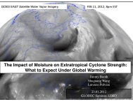
download a .pdf of this paper (4.9 MB). - Lamont-Doherty Earth ...
download a .pdf of this paper (4.9 MB). - Lamont-Doherty Earth ...
download a .pdf of this paper (4.9 MB). - Lamont-Doherty Earth ...

You also want an ePaper? Increase the reach of your titles
YUMPU automatically turns print PDFs into web optimized ePapers that Google loves.
336 JOURNAL OF VERTEBRATE PALEONTOLOGY, VOL. 23, NO. 2, 2003<br />
sumably tail (most <strong>of</strong> which is not preserved in UNC 15574)<br />
(Figs. 1, 2, 4, 5). Unlike in Hesperosuchus (Clark et al., 2001),<br />
there is no unpaired first osteoderm immediately behind the<br />
median occipital margin <strong>of</strong> the skull ro<strong>of</strong>. At least 28 pairs <strong>of</strong><br />
osteoderms are preserved in the region from the craniocervical<br />
junction to the base <strong>of</strong> the tail. Each osteoderm overlaps the<br />
anterior end <strong>of</strong> its successor and forms an unsculptured anterolateral<br />
process, which projects anteriorly below the preceding<br />
osteoderm; <strong>this</strong> process is short on the first cervical osteoderm.<br />
A low longitudinal ridge extends obliquely posterolaterally on<br />
the dorsal surface from the medial end <strong>of</strong> the anterolateral process<br />
to the posterolateral corner <strong>of</strong> each osteoderm. It marks<br />
the division <strong>of</strong> the osteoderm into a horizontal medial and a<br />
slightly ventrolaterally deflected lateral portion. The dorsal surface<br />
<strong>of</strong> the osteoderm bears a distinct sculpturing <strong>of</strong> irregular<br />
pits separated by ridges originating from the longitudinal ridge,<br />
whereas the ventral surface is smooth. The anterior margin <strong>of</strong><br />
each plate is concave, and its lateral edge is convex. A shallow<br />
notch is developed in the posterolateral corner <strong>of</strong> the more posterior<br />
osteoderms. The straight medial border forms the longest<br />
side <strong>of</strong> each plate. With the exception <strong>of</strong> the first pair <strong>of</strong> more<br />
or less pentagonal plates, the osteoderms are longer than wide<br />
and become more rectangular further posteriorly; the disparity<br />
between osteoderm length and width appears to be most pronounced<br />
in the mid-dorsal region and decreases again in the<br />
pelvic region.<br />
A small, irregularly shaped piece <strong>of</strong> bone is preserved attached<br />
to the medial aspect <strong>of</strong> the distal portion <strong>of</strong> the left femur<br />
and possibly represents an appendicular osteoderm.<br />
Pectoral Girdle and Forelimb<br />
The left scapulocoracoid and forelimb (Figs. 2, 3) are nearly<br />
completely preserved. The right scapulocoracoid and forelimb<br />
are apparently represented only by a partial scapular blade and<br />
two fragments <strong>of</strong> the humerus.<br />
Scapulocoracoid The proximal portion <strong>of</strong> the strongly<br />
curved scapula broadly contacts the coracoid. The two bones<br />
are almost completely fused to each other, with sutural separation<br />
persisting only for a short distance anterior to the glenoid.<br />
The anterior end <strong>of</strong> the scapula bears a distinct acromial ridge<br />
laterally. The distal portion <strong>of</strong> the scapular blade is flattened<br />
and greatly expanded, especially anteriorly so that the anterior<br />
margin <strong>of</strong> the blade is distinctly concave in lateral view. The<br />
posterior margin <strong>of</strong> the bone is less concave and becomes thicker<br />
proximally where it supports the posterolaterally and ventrally<br />
facing scapular glenoid facet. Just above the buttress for<br />
the glenoid facet, the scapula bears a rugose thickening, which<br />
probably marks the origin <strong>of</strong> the caput scapulare <strong>of</strong> M. triceps<br />
brachii as in extant crocodylians (Fürbringer, 1876).<br />
The anterior portion <strong>of</strong> the coracoid is thin, plate-like, and<br />
perforated by a large foramen. The posterolaterally and dorsally<br />
facing glenoid facet <strong>of</strong> the coracoid is gently convex and has a<br />
posterolateral ‘lip.’ The coracoid forms a prominent, posteromedially<br />
directed process behind the glenoid region. This process<br />
is shorter than the body <strong>of</strong> the coracoid, unlike the greatly<br />
elongated process in Dibothrosuchus (Wu and Chatterjee, 1993)<br />
and Sphenosuchus (Walker, 1990), and tapers posteriorly. Its<br />
ventral surface bears a deep groove, which is delimited dorsally<br />
by a more or less horizontal ridge and probably contacted the<br />
interclavicle medially (Walker, 1990).<br />
Humerus The complete left humerus is 89 mm long. Its<br />
slender, hollow shaft is round in transverse section between the<br />
expanded proximal and distal articular ends. The proximal portion<br />
lacks the round depression on the anterior surface <strong>of</strong> the<br />
proximal end reported in Dibothrosuchus (Wu and Chatterjee,<br />
1993). Its medial margin is strongly arched. The head <strong>of</strong> the<br />
humerus is reflected and forms a distinct, rounded articular surface.<br />
The well-developed deltopectoral crest projects anteromedially,<br />
rising just distal to the head to a median apex and<br />
terminating in the proximal third <strong>of</strong> the bone. A distinct ridge<br />
extends anterolaterally where the deltopectoral crest turns anteromedially.<br />
The distal end <strong>of</strong> the humerus bears two condyles,<br />
which are separated by a groove and together form a slightly<br />
saddle-shaped articular surface for the radius and ulna.<br />
Radius and Ulna The left ulna is 102 mm long and thus<br />
distinctly longer than the humerus (89 mm; 114.6%). The radius<br />
appears to be shorter than the ulna (although its proximal end<br />
could not be fully exposed during preparation), and its shaft is<br />
slightly more slender than that <strong>of</strong> the latter. Both bones are<br />
slender and have only slightly expanded distal ends. The proximal<br />
end <strong>of</strong> the ulna bears a well-developed olecranon process.<br />
Carpus and Manus The carpus and manus are largely disarticulated,<br />
resulting in the loss <strong>of</strong> a number <strong>of</strong> smaller bones.<br />
Both the radiale and ulnare are columnar and elongated in typically<br />
crocodylomorph fashion. Although its distal end is damaged,<br />
the radiale is more robust than the ulnare; its estimated<br />
length is 18 mm. The preserved metacarpals are long and slender.<br />
Based on the preserved, mostly scattered phalanges, the<br />
manus was small.<br />
Pelvic Girdle and Hindlimb<br />
The left ilium is still preserved in articulation with the two<br />
sacral vertebrae. A fragment <strong>of</strong> bone just anterior to the left<br />
ilium is possibly the proximal portion <strong>of</strong> the left pubis. The<br />
hindlimbs are represented by the complete left femur and the<br />
distal end <strong>of</strong> the right femur, both tibiae, the proximal and ?distal<br />
portions <strong>of</strong> the left fibula, an incomplete left calcaneum and<br />
(associated with the latter) three metatarsals from the left pes.<br />
The hindlimb is much longer than the forelimb; the ratio <strong>of</strong><br />
the combined length <strong>of</strong> femur and tibia (274 mm) to that <strong>of</strong><br />
humerus and ulna (191 mm) is 1.43.<br />
Ilium The slightly ventrolaterally inclined blade <strong>of</strong> the ilium<br />
(Fig. 5) is clearly set <strong>of</strong>f from the acetabular region. It is<br />
long anteroposteriorly but low dorsoventrally. The more or less<br />
horizontal dorsal margin <strong>of</strong> the blade is thickened, especially<br />
more anteriorly. The preacetabular process <strong>of</strong> the ilium is more<br />
slender than the postacetabular one and tapers anteriorly in lateral<br />
view. (Its anterior tip is not preserved.) Medially, the postacetabular<br />
process bears a prominent ridge along its ventral<br />
margin, which is in contact with the broadly flaring second<br />
sacral rib. The deeply concave acetabulum is partially overhung<br />
by a broad supra-acetabular crest, the central portion <strong>of</strong> which<br />
continues dorsally as a thick vertical ridge. The crest is widest<br />
anteriorly but does not extend to the posterior end <strong>of</strong> the acetabulum.<br />
The ventral margin <strong>of</strong> the acetabular wall between the<br />
anterior and posterior peduncles is gently convex rather than<br />
concave. The articular surfaces for contact with the pubis and<br />
ischium are broad. Just anterior to the lateral margin <strong>of</strong> the facet<br />
for contact with the ischium, there is a distinct, slightly rugose<br />
area, which probably represents an antitrochanter.<br />
?Pubis A fragment <strong>of</strong> bone preserved adjacent to the last<br />
three dorsal vertebrae possibly represents the proximal portion<br />
<strong>of</strong> the left pubis (Fig. 5B). Its identification is based on its<br />
resemblance to the pubes <strong>of</strong> Saltoposuchus (Huene, 1921:fig.<br />
19) and Terrestrisuchus (Crush, 1984:fig. 8).<br />
Femur The complete left femur (Fig. 6) is 144 mm long.<br />
Its proximal portion is flattened transversely and twisted relative<br />
to the long axis <strong>of</strong> the bone so that the distinct head projects<br />
anteromedially. The femoral head is set at a right angle to the<br />
shaft, suggesting a fully erect posture <strong>of</strong> the hindlimb in life.<br />
Its terminal articular surface is gently convex medially and extends<br />
posterolaterally across the proximal end <strong>of</strong> the femur. The<br />
posteromedial margin <strong>of</strong> the facet forms a distinct tubercle. A<br />
low, thick, and rugose ridge just distal to the lateral end <strong>of</strong> the