download a .pdf of this paper (4.9 MB). - Lamont-Doherty Earth ...
download a .pdf of this paper (4.9 MB). - Lamont-Doherty Earth ...
download a .pdf of this paper (4.9 MB). - Lamont-Doherty Earth ...

Create successful ePaper yourself
Turn your PDF publications into a flip-book with our unique Google optimized e-Paper software.
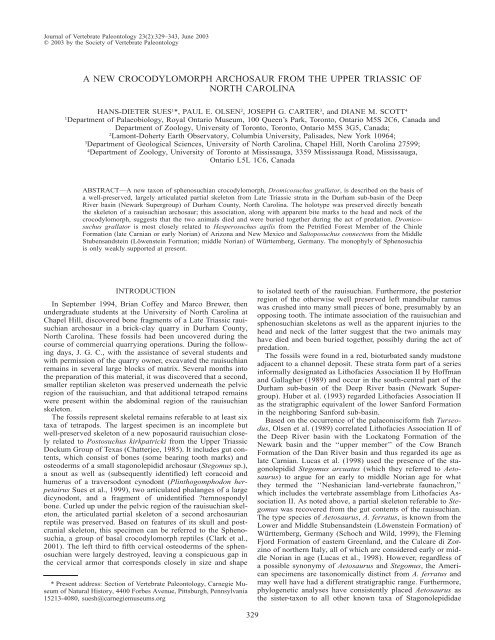
Journal <strong>of</strong> Vertebrate Paleontology 23(2):329–343, June 2003<br />
2003 by the Society <strong>of</strong> Vertebrate Paleontology<br />
A NEW CROCODYLOMORPH ARCHOSAUR FROM THE UPPER TRIASSIC OF<br />
NORTH CAROLINA<br />
HANS-DIETER SUES 1 *, PAUL E. OLSEN 2 , JOSEPH G. CARTER 3 , and DIANE M. SCOTT 4<br />
1<br />
Department <strong>of</strong> Palaeobiology, Royal Ontario Museum, 100 Queen’s Park, Toronto, Ontario M5S 2C6, Canada and<br />
Department <strong>of</strong> Zoology, University <strong>of</strong> Toronto, Toronto, Ontario M5S 3G5, Canada;<br />
2<br />
<strong>Lamont</strong>-<strong>Doherty</strong> <strong>Earth</strong> Observatory, Columbia University, Palisades, New York 10964;<br />
3<br />
Department <strong>of</strong> Geological Sciences, University <strong>of</strong> North Carolina, Chapel Hill, North Carolina 27599;<br />
4<br />
Department <strong>of</strong> Zoology, University <strong>of</strong> Toronto at Mississauga, 3359 Mississauga Road, Mississauga,<br />
Ontario L5L 1C6, Canada<br />
ABSTRACT—A new taxon <strong>of</strong> sphenosuchian crocodylomorph, Dromicosuchus grallator, is described on the basis <strong>of</strong><br />
a well-preserved, largely articulated partial skeleton from Late Triassic strata in the Durham sub-basin <strong>of</strong> the Deep<br />
River basin (Newark Supergroup) <strong>of</strong> Durham County, North Carolina. The holotype was preserved directly beneath<br />
the skeleton <strong>of</strong> a rauisuchian archosaur; <strong>this</strong> association, along with apparent bite marks to the head and neck <strong>of</strong> the<br />
crocodylomorph, suggests that the two animals died and were buried together during the act <strong>of</strong> predation. Dromicosuchus<br />
grallator is most closely related to Hesperosuchus agilis from the Petrified Forest Member <strong>of</strong> the Chinle<br />
Formation (late Carnian or early Norian) <strong>of</strong> Arizona and New Mexico and Saltoposuchus connectens from the Middle<br />
Stubensandstein (Löwenstein Formation; middle Norian) <strong>of</strong> Württemberg, Germany. The monophyly <strong>of</strong> Sphenosuchia<br />
is only weakly supported at present.<br />
INTRODUCTION<br />
In September 1994, Brian C<strong>of</strong>fey and Marco Brewer, then<br />
undergraduate students at the University <strong>of</strong> North Carolina at<br />
Chapel Hill, discovered bone fragments <strong>of</strong> a Late Triassic rauisuchian<br />
archosaur in a brick-clay quarry in Durham County,<br />
North Carolina. These fossils had been uncovered during the<br />
course <strong>of</strong> commercial quarrying operations. During the following<br />
days, J. G. C., with the assistance <strong>of</strong> several students and<br />
with permission <strong>of</strong> the quarry owner, excavated the rauisuchian<br />
remains in several large blocks <strong>of</strong> matrix. Several months into<br />
the preparation <strong>of</strong> <strong>this</strong> material, it was discovered that a second,<br />
smaller reptilian skeleton was preserved underneath the pelvic<br />
region <strong>of</strong> the rauisuchian, and that additional tetrapod remains<br />
were present within the abdominal region <strong>of</strong> the rauisuchian<br />
skeleton.<br />
The fossils represent skeletal remains referable to at least six<br />
taxa <strong>of</strong> tetrapods. The largest specimen is an incomplete but<br />
well-preserved skeleton <strong>of</strong> a new poposaurid rauisuchian closely<br />
related to Postosuchus kirkpatricki from the Upper Triassic<br />
Dockum Group <strong>of</strong> Texas (Chatterjee, 1985). It includes gut contents,<br />
which consist <strong>of</strong> bones (some bearing tooth marks) and<br />
osteoderms <strong>of</strong> a small stagonolepidid archosaur (Stegomus sp.),<br />
a snout as well as (subsequently identified) left coracoid and<br />
humerus <strong>of</strong> a traversodont cynodont (Plinthogomphodon herpetairus<br />
Sues et al., 1999), two articulated phalanges <strong>of</strong> a large<br />
dicynodont, and a fragment <strong>of</strong> unidentified ?temnospondyl<br />
bone. Curled up under the pelvic region <strong>of</strong> the rauisuchian skeleton,<br />
the articulated partial skeleton <strong>of</strong> a second archosaurian<br />
reptile was preserved. Based on features <strong>of</strong> its skull and postcranial<br />
skeleton, <strong>this</strong> specimen can be referred to the Sphenosuchia,<br />
a group <strong>of</strong> basal crocodylomorph reptiles (Clark et al.,<br />
2001). The left third to fifth cervical osteoderms <strong>of</strong> the sphenosuchian<br />
were largely destroyed, leaving a conspicuous gap in<br />
the cervical armor that corresponds closely in size and shape<br />
* Present address: Section <strong>of</strong> Vertebrate Paleontology, Carnegie Museum<br />
<strong>of</strong> Natural History, 4400 Forbes Avenue, Pittsburgh, Pennsylvania<br />
15213-4080, suesh@carnegiemuseums.org<br />
to isolated teeth <strong>of</strong> the rauisuchian. Furthermore, the posterior<br />
region <strong>of</strong> the otherwise well preserved left mandibular ramus<br />
was crushed into many small pieces <strong>of</strong> bone, presumably by an<br />
opposing tooth. The intimate association <strong>of</strong> the rauisuchian and<br />
sphenosuchian skeletons as well as the apparent injuries to the<br />
head and neck <strong>of</strong> the latter suggest that the two animals may<br />
have died and been buried together, possibly during the act <strong>of</strong><br />
predation.<br />
The fossils were found in a red, bioturbated sandy mudstone<br />
adjacent to a channel deposit. These strata form part <strong>of</strong> a series<br />
informally designated as Lith<strong>of</strong>acies Association II by H<strong>of</strong>fman<br />
and Gallagher (1989) and occur in the south-central part <strong>of</strong> the<br />
Durham sub-basin <strong>of</strong> the Deep River basin (Newark Supergroup).<br />
Huber et al. (1993) regarded Lith<strong>of</strong>acies Association II<br />
as the stratigraphic equivalent <strong>of</strong> the lower Sanford Formation<br />
in the neighboring Sanford sub-basin.<br />
Based on the occurrence <strong>of</strong> the palaeonisciform fish Turseodus,<br />
Olsen et al. (1989) correlated Lith<strong>of</strong>acies Association II <strong>of</strong><br />
the Deep River basin with the Lockatong Formation <strong>of</strong> the<br />
Newark basin and the ‘‘upper member’’ <strong>of</strong> the Cow Branch<br />
Formation <strong>of</strong> the Dan River basin and thus regarded its age as<br />
late Carnian. Lucas et al. (1998) used the presence <strong>of</strong> the stagonolepidid<br />
Stegomus arcuatus (which they referred to Aetosaurus)<br />
to argue for an early to middle Norian age for what<br />
they termed the ‘‘Neshanician land-vertebrate faunachron,’’<br />
which includes the vertebrate assemblage from Lith<strong>of</strong>acies Association<br />
II. As noted above, a partial skeleton referable to Stegomus<br />
was recovered from the gut contents <strong>of</strong> the rauisuchian.<br />
The type species <strong>of</strong> Aetosaurus, A. ferratus, is known from the<br />
Lower and Middle Stubensandstein (Löwenstein Formation) <strong>of</strong><br />
Württemberg, Germany (Schoch and Wild, 1999), the Fleming<br />
Fjord Formation <strong>of</strong> eastern Greenland, and the Calcare di Zorzino<br />
<strong>of</strong> northern Italy, all <strong>of</strong> which are considered early or middle<br />
Norian in age (Lucas et al., 1998). However, regardless <strong>of</strong><br />
a possible synonymy <strong>of</strong> Aetosaurus and Stegomus, the American<br />
specimens are taxonomically distinct from A. ferratus and<br />
may well have had a different stratigraphic range. Furthermore,<br />
phylogenetic analyses have consistently placed Aetosaurus as<br />
the sister-taxon to all other known taxa <strong>of</strong> Stagonolepididae<br />
329
330 JOURNAL OF VERTEBRATE PALEONTOLOGY, VOL. 23, NO. 2, 2003<br />
(Parrish, 1994; Heckert and Lucas, 1999). Thus the Aetosaurus<br />
lineage must predate more derived, Carnian-age taxa such as<br />
Stagonolepis, and its occurrence in pre-Norian strata is to be<br />
expected.<br />
The new poposaurid rauisuchian is closely related to Postosuchus<br />
kirkpatricki from the Cooper Canyon Formation (Dockum<br />
Group) <strong>of</strong> Texas (Chatterjee, 1985), which is considered<br />
early Norian in age. However, Long and Murry (1995) also<br />
referred (without further discussion) various late Carnian specimens<br />
to P. kirkpatricki. The traversodont cynodont Plinthogomphodon<br />
herpetairus is not useful for stratigraphic correlation<br />
because its phylogenetic relationships are as yet unresolved<br />
(Sues et al., 1999). The sphenosuchian described in <strong>this</strong> <strong>paper</strong><br />
is most closely related to Hesperosuchus agilis from the Petrified<br />
Forest Member <strong>of</strong> the Chinle Formation (late Carnian or<br />
early Norian) <strong>of</strong> Arizona and New Mexico (Clark et al., 2001)<br />
and Saltoposuchus connectens from the Middle Stubensandstein<br />
(Löwenstein Formation; middle Norian) <strong>of</strong> Württemberg, Germany<br />
(Huene, 1921; Sereno and Wild, 1992; Schoch and Wild,<br />
1999). The biostratigraphic evidence cannot definitely resolve<br />
the question whether the tetrapod assemblage from Lith<strong>of</strong>acies<br />
Association II is late Carnian or early Norian in age. Paleomagnetic<br />
sampling currently in progress by D. V. Kent and P.<br />
E. O. suggests an early Norian date for the fossil-bearing strata.<br />
The new sphenosuchian skeleton from North Carolina is<br />
nearly complete and well preserved, and thus <strong>of</strong> considerable<br />
interest for discussions regarding the anatomy and interrelationships<br />
<strong>of</strong> basal crocodylomorph archosaurs. We present here a<br />
description <strong>of</strong> <strong>this</strong> specimen and assess its phylogenetic relationships.<br />
Institutional Abbreviations AMNH, American Museum<br />
<strong>of</strong> Natural History, New York; BP, Bernard Price Institute for<br />
Palaeontological Research, University <strong>of</strong> the Witwatersrand, Johannesburg,<br />
South Africa; UNC, Department <strong>of</strong> Geological Sciences,<br />
University <strong>of</strong> North Carolina at Chapel Hill.<br />
SYSTEMATIC PALEONTOLOGY<br />
ARCHOSAURIA Cope, 1869<br />
CROCODYLOMORPHA Hay, 1930 sensu Walker, 1970<br />
SPHENOSUCHIDAE Haughton, 1924<br />
Comment The interrelationships <strong>of</strong> basal crocodylomorph<br />
archosaurs are still poorly resolved (see below), and thus it is<br />
inadvisable to <strong>of</strong>fer a phylogenetic definition <strong>of</strong> Sphenosuchidae<br />
at <strong>this</strong> point.<br />
DROMICOSUCHUS, gen. nov.<br />
Etymology From Greek dromikos, fleet, quickly walking,<br />
and soukhos, Greek rendering <strong>of</strong> the ancient Egyptian crocodile-headed<br />
deity Sebek or Sobk and traditional suffix for generic<br />
nomina <strong>of</strong> crocodylomorph reptiles, in reference to the<br />
inferred cursorial habits <strong>of</strong> <strong>this</strong> crocodylomorph.<br />
Type Species Dromicosuchus grallator, sp. nov. (by monotypy).<br />
Diagnosis As for the type and only known species, given<br />
below.<br />
DROMICOSUCHUS GRALLATOR, sp. nov.<br />
Etymology Latin grallator, one who walks on stilts, in reference<br />
to the very long and slender limbs.<br />
Holotype UNC 15574, nearly complete skull with mandible<br />
in tight occlusion and partial, largely articulated postcranial<br />
skeleton (Fig. 1), comprising the vertebral column from the<br />
atlas-axis complex back to the second caudal vertebra, dorsal<br />
dermal armor, ribs and gastralia elements, left scapulocoracoid<br />
and almost complete left forelimb, partial right scapula and<br />
proximal portion <strong>of</strong> the right humerus, left ilium, left femur,<br />
distal end <strong>of</strong> the right femur, both tibiae, proximal and ?distal<br />
portions <strong>of</strong> the left fibula, incomplete left calcaneum, three left<br />
metatarsals, and fragments <strong>of</strong> several currently unidentifiable<br />
limb-bones.<br />
Type Horizon and Locality Mudstone facies <strong>of</strong> Lith<strong>of</strong>acies<br />
Association II sensu H<strong>of</strong>fman and Gallagher (1989), southcentral<br />
region <strong>of</strong> the Durham sub-basin <strong>of</strong> the Deep River basin,<br />
Newark Supergroup. GPS coordinates (recorded by P. E. O.):<br />
latitude 35 52 28 N; longitude 78 53 81 W. Genlee, Durham<br />
County, North Carolina, U.S.A. Age: Late Triassic (late<br />
Carnian or early Norian).<br />
Diagnosis Distinguished from Saltoposuchus connectens<br />
by the presence <strong>of</strong> paired crests separated by a median groove<br />
on the dorsal surface <strong>of</strong> the parietals and the more prominent<br />
development <strong>of</strong> the dorsolateral crest on the squamosal. Differs<br />
from Hesperosuchus agilis in the absence <strong>of</strong> the dorsoventral<br />
expansion <strong>of</strong> the anterior end <strong>of</strong> the dentary and the presence<br />
<strong>of</strong> a conical recess at the anterior end <strong>of</strong> the antorbital fossa.<br />
Distinguished from Sphenosuchus acutus and Dibothrosuchus<br />
elaphros by the presence <strong>of</strong> a V-shaped, rather than straight,<br />
transverse occipital crest, the presence <strong>of</strong> paired crests separated<br />
by a median groove, rather than a single median crest, on the<br />
dorsal surface <strong>of</strong> the parietals, the presence <strong>of</strong> a conical recess<br />
at the anterior end <strong>of</strong> the antorbital fossa, and the less elongated<br />
posteromedial process <strong>of</strong> the coracoid. Differs from Kayentasuchus<br />
walkeri in the absence <strong>of</strong> a lateral groove on the squamosal<br />
and the presence <strong>of</strong> an anterior caniniform tooth in the<br />
dentary.<br />
DESCRIPTION<br />
UNC 15574 comprises much <strong>of</strong> an articulated skeleton,<br />
which was recovered as a series <strong>of</strong> individual blocks as well as<br />
a number <strong>of</strong> isolated bones and bone fragments (Fig. 1). The<br />
animal was preserved with its ventral side facing up and its<br />
head folded against the left side <strong>of</strong> its neck. The left forelimb<br />
was tucked under the head and neck. The largest block (Figs.<br />
2, 3) contains the skull, left scapulocoracoid and forelimb, and<br />
a mostly articulated series <strong>of</strong> 10 complete as well as two partial<br />
cervical and anterior dorsal vertebrae with associated ribs and<br />
13 pairs <strong>of</strong> osteoderms. Its upper surface also preserves several<br />
segments <strong>of</strong> gastralia elements and the proximal end <strong>of</strong> the left<br />
tibia <strong>of</strong> the overlying rauisuchian skeleton. A small piece <strong>of</strong><br />
matrix containing a partial and two complete vertebrae, together<br />
with jumbled osteoderms, indeterminate bone fragments, and a<br />
large chunk <strong>of</strong> a rauisuchian limb-bone, appears to join <strong>this</strong><br />
block to another block, which represents the mid-dorsal region<br />
(Fig. 4). The latter contains an articulated set <strong>of</strong> three complete<br />
and three partial vertebrae with associated osteoderms (five<br />
complete and two partial pairs), ribs, and gastralia. It can be<br />
fitted to an articulated segment <strong>of</strong> vertebral column comprising<br />
the last four dorsal (the first <strong>of</strong> which is incomplete), the two<br />
sacral and the first two caudal vertebrae, which are associated<br />
with pairs <strong>of</strong> osteoderms, the left ilium, and what may be the<br />
proximal portion <strong>of</strong> the left pubis (Fig. 5). The bones <strong>of</strong> the<br />
hindlimbs and right forelimb are now completely separated<br />
from the rest <strong>of</strong> the skeleton and from each other. When the<br />
material was collected, the proximal head <strong>of</strong> the left femur still<br />
adhered to the wall <strong>of</strong> the acetabulum on the left ilium, but it<br />
was subsequently removed and reattached to the femoral shaft.<br />
UNC 15574 is closely similar in all comparable linear dimensions<br />
to the holotype <strong>of</strong> Hesperosuchus agilis (AMNH<br />
6758; Colbert, 1952:table 1). Assuming body proportions similar<br />
to those reconstructed for Dibothrosuchus elaphros (Wu<br />
and Chatterjee, 1993), it represents an individual with an estimated<br />
total length between 1.2 and 1.3 m. It is difficult to assess<br />
the ontogenetic stage <strong>of</strong> UNC 15574 using the standard crite-
SUES ET AL.—TRIASSIC CROCODYLOMORPH<br />
331<br />
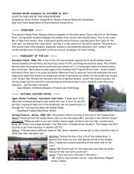
FIGURE 1. Dromicosuchus grallator, UNC 15574 (holotype), digital photograph <strong>of</strong> the skeleton (mostly in dorsal view) as reassembled after<br />
completed preparation. Scale bar equals 5 cm.<br />
FIGURE 2. Dromicosuchus grallator, UNC 15574 (holotype), block with skull and anterior portion <strong>of</strong> the skeleton in dorsal view. Scale bar<br />
equals 1 cm. Areas in white represent matrix. Abbreviations: an, angular; ao.f, antorbital fossa; ar, articular; d, dentary; e.n, external naris; f,<br />
frontal; h, humerus; j, jugal; l, lacrimal; m, maxilla; n, nasal; or, orbit; p, parietal; pm, premaxilla; po, postorbital; po.p, paroccipital process;<br />
prf, prefrontal; q, quadrate; sc, scapula; sq, squamosal; st.f, supratemporal fenestra. X denotes apparent bite damage to cervical armor. ? denotes<br />
possible quadratojugal.
332 JOURNAL OF VERTEBRATE PALEONTOLOGY, VOL. 23, NO. 2, 2003<br />
FIGURE 3. Dromicosuchus grallator, UNC 15574 (holotype), block with skull and anterior portion <strong>of</strong> the skeleton in ventral view. Gastralia<br />
and fragment <strong>of</strong> limb-bone in lower right corner <strong>of</strong> drawing belong to the overlying rauisuchian skeleton. Scale bar equals 1 cm. Abbreviations<br />
as in Figure 1 plus: c, coronoid; c6, c9, cervical 6, 9; cb, ceratobranchial I; co, coracoid; mc, metacarpal; pa, prearticular; r, radius; ra, radiale;<br />
sp, splenial; u, ulna. X denotes apparent bite damage to posterior region <strong>of</strong> left mandibular ramus.<br />
rion <strong>of</strong> closure <strong>of</strong> the neurocentral sutures (Brochu, 1996). The<br />
neural arches <strong>of</strong> several dorsal vertebrae show separation from<br />
as well as some displacement relative to the centra, but it is<br />
uncertain whether <strong>this</strong> condition reflects the original presence<br />
<strong>of</strong> open neurocentral sutures. The exposed right side <strong>of</strong> the sixth<br />
cervical vertebra apparently shows a faint neurocentral suture.<br />
However, the almost complete fusion <strong>of</strong> the scapula and coracoid<br />
suggests maturity <strong>of</strong> the animal (Brochu, 1992).<br />
Skull<br />
The nearly complete skull <strong>of</strong> UNC 15574 (Figs. 2, 3) was<br />
obliquely crushed in a dorsolateral direction during fossilization<br />
so that its right side is now preserved in almost the same horizontal<br />
plane as the anterior portion <strong>of</strong> the skull ro<strong>of</strong>. The wellpreserved<br />
right side <strong>of</strong> the skull shows considerable detail,<br />
whereas compression has severely distorted the left side. Many<br />
bones are traversed by fractures. Several elements, especially<br />
along the perimeter <strong>of</strong> the orbit, were separated along their sutural<br />
contacts. The mandibular rami are tightly appressed to the<br />
skull so that the dentary teeth are largely concealed from view.<br />
The anterior end <strong>of</strong> the snout was broken <strong>of</strong>f during collecting,<br />
but was recovered and readily reattached to the remainder <strong>of</strong><br />
the skull.<br />
The lightly built skull has a long and narrow snout and a<br />
transversely broad temporal region. It is about 150 mm long<br />
(measured along the midline <strong>of</strong> the skull ro<strong>of</strong> from the anterior<br />
tip <strong>of</strong> the snout to the anterior end <strong>of</strong> the V-shaped occipital<br />
embayment). The length <strong>of</strong> the antorbital region <strong>of</strong> the skull<br />
(measured from the anterior terminus <strong>of</strong> the orbit to the tip <strong>of</strong><br />
the snout) is more than twice that <strong>of</strong> the postorbital region<br />
(measured from the posterior end <strong>of</strong> the orbit to the level <strong>of</strong><br />
the posterolateral termini <strong>of</strong> the occipital crests). The external<br />
nares face laterally and are separated from each other by a bony<br />
bar formed by the nasals and premaxillae. The antorbital fossa<br />
is more or less triangular in lateral view and large, with an<br />
anteroposterior length <strong>of</strong> 46 mm and a maximum height <strong>of</strong> 19<br />
mm. The antorbital fenestra is long (31 mm) but low (4 mm).<br />
The orbit is nearly circular in outline, with an anteroposterior<br />
diameter <strong>of</strong> about 30 mm. The supratemporal fenestra is longer<br />
anteroposteriorly than wide transversely. The external surfaces<br />
<strong>of</strong> most cranial bones are devoid <strong>of</strong> sculpturing.<br />
Premaxilla The recurved posterolateral process <strong>of</strong> the premaxilla<br />
overlaps the nasal and maxilla on the side <strong>of</strong> the snout,<br />
excluding the latter from participation in the posterior margin<br />
<strong>of</strong> the external naris. Although damage to both sides <strong>of</strong> the<br />
snout has obscured some details <strong>of</strong> <strong>this</strong> feature, a laterally open<br />
notch is present between the posterior edge <strong>of</strong> the premaxilla<br />
and the anterior edge <strong>of</strong> the maxilla and receives an anterior<br />
caniniform tooth <strong>of</strong> the dentary. Anteriorly, the premaxilla<br />
forms the short anterodorsal portion <strong>of</strong> the slender internarial<br />
bar and the nasals make up the more posterior part. The premaxilla<br />
holds five teeth, the first <strong>of</strong> which is smaller and more<br />
slender than the others.
SUES ET AL.—TRIASSIC CROCODYLOMORPH<br />
333<br />
FIGURE 4. Dromicosuchus grallator, UNC 15574 (holotype), segment<br />
<strong>of</strong> mid-dorsal region in dorsal view. Anterior is toward the top <strong>of</strong><br />
the figure. Scale bar equals 1 cm.<br />
Maxilla The long but rather low maxilla forms most <strong>of</strong> the<br />
rostral portion <strong>of</strong> the skull. Its alveolar margin is distinctly sinuous<br />
in lateral view, reaching its greatest depth at about the<br />
level <strong>of</strong> the sixth maxillary tooth. The facial portion <strong>of</strong> the<br />
maxilla extends vertically. Its ascending process projects posteriorly<br />
and slightly dorsally. It contacts the anterior ramus <strong>of</strong><br />
the lacrimal half way along the dorsal rim <strong>of</strong> the antorbital<br />
fenestra, excluding the nasal from participation in the dorsal<br />
margin <strong>of</strong> the antorbital fossa. The anterior and ventral margins<br />
<strong>of</strong> the large, subtriangular antorbital fossa are formed by a thin<br />
medial lamina <strong>of</strong> the maxilla, which is inset relative to the remainder<br />
<strong>of</strong> the lateral surface <strong>of</strong> <strong>this</strong> bone. Anteriorly, the fossa<br />
terminates in a deep conical pit, which is largely concealed in<br />
lateral view by the lateral portion <strong>of</strong> the ascending process <strong>of</strong><br />
the maxilla. A similar pit is present in both Saltoposuchus<br />
(Clark et al., 2001) and Terrestrisuchus (Crush, 1984). The antorbital<br />
fenestra is restricted to the more ventral portion <strong>of</strong> the<br />
antorbital fossa. The lateral surface <strong>of</strong> the maxilla bears scattered<br />
small neurovascular openings. A row <strong>of</strong> large supralabial<br />
foramina, presumably for passage <strong>of</strong> cutaneous branches <strong>of</strong> N.<br />
alveolaris superior and associated blood vessels, extends just<br />
dorsal and parallel to the alveolar margin. The more completely<br />
preserved left maxillary tooth row comprises 20 teeth (some <strong>of</strong><br />
which have partially dropped out <strong>of</strong> their alveoli) and ends posteriorly<br />
just behind the anterior margin <strong>of</strong> the orbit.<br />
Nasal The nasal is narrow, thick, and long, extending from<br />
the region <strong>of</strong> the external naris back to the level <strong>of</strong> the anterior<br />
margin <strong>of</strong> the orbit. Its anterior portion forms most <strong>of</strong> the dorsal<br />
margin <strong>of</strong> the narial fenestra. In the region between the margin<br />
<strong>of</strong> the external naris and the anterior end <strong>of</strong> the antorbital fossa,<br />
the lateral portion <strong>of</strong> the nasal is somewhat deflected ventrolaterally<br />
and thus faces dorsolaterally. Although the dorsal surfaces<br />
<strong>of</strong> both nasals are slightly eroded, they appear to bear a<br />
weakly developed, irregular sculpturing <strong>of</strong> pits and longitudinal<br />
grooves, especially more anteriorly. Posteromedially, the nasals<br />
form a shallow depression along the midline <strong>of</strong> the skull ro<strong>of</strong>,<br />
as in Sphenosuchus (Walker, 1990). The nasal forms straight<br />
lateral sutural contacts with the lacrimal and maxilla.<br />
Lacrimal In lateral view, the lacrimal has an inverted L-<br />
shape and is inclined forward. It forms the preorbital bar and<br />
contributes a broad, thin medial lamina to the medial wall <strong>of</strong><br />
the antorbital fossa. Its vertical portion bears a narrow but distinct<br />
lateral crest. Anteriorly, the lacrimal is overlapped by the<br />
maxilla along the dorsal margin <strong>of</strong> the antorbital fossa. Posteriorly,<br />
it forms an extensive lateral contact with the prefrontal<br />
along the preorbital bar. The dorsal exposure <strong>of</strong> the lacrimal on<br />
the skull ro<strong>of</strong> is narrow. The posterior opening <strong>of</strong> the lacrimal<br />
canal is situated about halfway up the anterior margin <strong>of</strong> the<br />
orbit on the suture between the lacrimal and prefrontal. Ventrally,<br />
the lacrimal is expanded anteroposteriorly at its contact<br />
with the jugal and maxilla.<br />
Prefrontal The prefrontal extends posteriorly to about midway<br />
along the dorsal rim <strong>of</strong> the orbit. Due to crushing, it is<br />
uncertain whether it extended under the frontal more posteriorly<br />
as in other sphenosuchians (Sereno and Wild, 1992). The narrow<br />
dorsal surface <strong>of</strong> the prefrontal is more or less triangular<br />
in dorsal view and set <strong>of</strong>f from the ventrolateral portion <strong>of</strong> the<br />
bone by a ridge extending along the lateral edge <strong>of</strong> the skull<br />
ro<strong>of</strong>. Within the anterodorsal part <strong>of</strong> the orbit, the prefrontal is<br />
considerably expanded and, as in Sphenosuchus (Walker, 1990),<br />
appears to form three processes: one extending ventrally along<br />
the medial aspect <strong>of</strong> the lacrimal, another projecting toward the<br />
midline <strong>of</strong> the skull, and a third extending dorsomedially toward<br />
the ventral surface <strong>of</strong> the frontal.<br />
Jugal The jugal is slender and triradiate. Its lateral surface<br />
bears a low ridge that extends back from below the orbit and<br />
fades into the posterior or infratemporal process posteriorly.<br />
The infratemporal process tapers toward the jaw joint. The dorsal<br />
process <strong>of</strong> the jugal is very slender. The anterior process<br />
forms the entire ventral margin <strong>of</strong> the orbit but does not extend<br />
to the posteroventral corner <strong>of</strong> the antorbital fenestra anteriorly.<br />
It overlaps the ventral part <strong>of</strong> the lacrimal.<br />
Frontal The frontal is much longer than wide and forms<br />
much <strong>of</strong> the ro<strong>of</strong> as well as the slightly raised rim <strong>of</strong> the orbit.<br />
Its dorsal surface is concave transversely. A low ridge extends<br />
along the median suture between the frontals, fading into the<br />
bones more anteriorly. Posterolaterally, the supratemporal fossa<br />
continues forward onto the dorsal surface <strong>of</strong> the frontal, forming<br />
a distinct depression on the latter. Anteriorly, the frontals<br />
extend far forward between the posterior ends <strong>of</strong> the nasals<br />
along the midline <strong>of</strong> the skull ro<strong>of</strong>, resulting in a V-shaped<br />
suture, and, posteriorly, they contact the parietals along a transverse<br />
suture.<br />
Parietal Dorsally, the parietals bear paired crests, which<br />
are separated by a median longitudinal groove. Each crest forms<br />
the posteromedial edge <strong>of</strong> the adjacent supratemporal fossa. The<br />
interparietal suture extends in the median groove between the<br />
two crests. This condition differs from the single median crest<br />
on the parietals in Dibothrosuchus, Saltoposuchus, and Sphenosuchus,<br />
but closely resembles the paired parietal crests in Hesperosuchus<br />
(Clark et al., 2001). In a partial skull referred to<br />
Saltoposuchus connectens, the dorsal surface <strong>of</strong> the parietals is<br />
flat between the medial margins <strong>of</strong> the supratemporal fenestrae<br />
(Sereno and Wild, 1992:fig. 5A). The posterolateral wings <strong>of</strong><br />
the parietals in UNC 15574 diverge posteriorly, forming a V-<br />
shaped embayment in the occipital margin <strong>of</strong> the skull ro<strong>of</strong>, as<br />
in Hesperosuchus and Saltoposuchus but unlike the transverse<br />
occipital crest in Dibothrosuchus and Sphenosuchus.<br />
Postorbital The postorbital is triradiate in lateral view. It<br />
forms the anterior half <strong>of</strong> the supratemporal bar and overlaps<br />
the squamosal posteriorly. A prominent anterolateral ridge overhangs<br />
the ventral process for contact with the jugal. Anteromedially,<br />
the postorbital contacts the posterolateral end <strong>of</strong> the<br />
frontal.
334 JOURNAL OF VERTEBRATE PALEONTOLOGY, VOL. 23, NO. 2, 2003<br />
FIGURE 5. Dromicosuchus grallator, UNC 15574 (holotype), articulated segment <strong>of</strong> vertebral column consisting <strong>of</strong> posterior four dorsal, two<br />
sacral, and first two caudal vertebrae, with articulated left ilium and disarticulated proximal portion <strong>of</strong> left ?pubis in A, right lateral, B, ventral<br />
and C, dorsal views. Scale bar equals 1 cm. Abbreviations: ac, acetabulum; pu?, possible pubis; s1, s2, sacral vertebra 1, 2.<br />
Squamosal The squamosal forms the posterolateral corner<br />
<strong>of</strong> the skull table and overhangs the infratemporal region and<br />
suspensorium laterally. It is rather thin and ventrally concave.<br />
Laterally, the squamosal is deflected so that <strong>this</strong> portion <strong>of</strong> the<br />
bone probably assumed a nearly vertical oriention. Anteriorly,<br />
it extends ventral to the postorbital to participate in the formation<br />
<strong>of</strong> the postorbital bar. A prominent crest extends along<br />
the posterolateral margin <strong>of</strong> the supratemporal fossa on the dorsal<br />
surface <strong>of</strong> the squamosal, continuing the anterolateral ridge<br />
on the postorbital and the parasagittal ridge on the parietal.<br />
Together, these ridges surround most <strong>of</strong> the supratemporal fossa<br />
and sharply demarcate the skull table from the occiput and the<br />
sides <strong>of</strong> the skull. In Saltoposuchus, the squamosal appears to<br />
be wider transversely and has a less prominent posterolateral<br />
crest (Sereno and Wild, 1992:fig. 5A).<br />
Quadrate The quadrate is steeply inclined anterodorsally<br />
so that its proximal end is situated well forward <strong>of</strong> its distal<br />
mandibular condyles. A lateral ridge extends along the anterior<br />
margin <strong>of</strong> the bone. Medial to <strong>this</strong> ridge, the posterior surface<br />
<strong>of</strong> the quadrate bears a distinct, oval depression. On the left<br />
side <strong>of</strong> the skull, postmortem crushing has pushed the quadrate<br />
in a dorsolateral direction through the supratemporal fenestra,<br />
clearly exposing the single, anteroposteriorly elongate proximal<br />
head <strong>of</strong> the quadrate. The right quadrate has been similarly<br />
displaced.<br />
?Quadratojugal On the right side <strong>of</strong> the skull, bone is visible<br />
in the infratemporal fenestra anterior to the quadrate and<br />
medial to the somewhat displaced jugal. This bone probably<br />
represents the quadratojugal, but details are obscured by fracturing<br />
and displacement.<br />
Braincase The crushing <strong>of</strong> the skull during fossilization<br />
has destroyed and/or obscured most <strong>of</strong> the palate and braincase.<br />
The distal end <strong>of</strong> the paroccipital process formed by the otoccipital<br />
(fused exoccipital and opisthotic) is distinctly expanded<br />
vertically. The posterior surface <strong>of</strong> the process is gently convex<br />
dorsoventrally.<br />
Hyoid A curved fragment <strong>of</strong> rod-like bone, which is appressed<br />
to the medial surface <strong>of</strong> the right articular, probably<br />
represents a ceratobranchial I (cornu branchiale I).<br />
Mandible<br />
The mandibular rami are long and slender (Figs. 2, 3). Few<br />
structural details can be discerned due to crushing on both rami.<br />
The large external mandibular fenestra was bounded by the dentary<br />
anteriorly and dorsally, the surangular posterodorsally, and<br />
the angular ventrally.<br />
Dentary The dentary is long and low. Its anterior (symphyseal)<br />
end lacks the distinct dorsoventral expansion diagnostic<br />
for Hesperosuchus (Clark et al., 2001). Although details are<br />
obscured by crushing and displacement <strong>of</strong> the mandibular rami,<br />
the symphysis apparently did not extend much posterior to the<br />
region <strong>of</strong> the third or fourth dentary tooth. The lateral surface<br />
<strong>of</strong> the anterior portion <strong>of</strong> the dentary, especially in the symphyseal<br />
region, bears numerous scattered neurovascular foramina.<br />
The number <strong>of</strong> teeth in each dentary cannot be determined
SUES ET AL.—TRIASSIC CROCODYLOMORPH<br />
335<br />
due to the tight contact between the mandibular rami and the<br />
skull.<br />
Coronoid On the lingual surface <strong>of</strong> the right mandibular<br />
ramus, a long and very slender coronoid bone extends just ventral<br />
and parallel to the alveolar margin <strong>of</strong> the dentary and ventrally<br />
contacts the splenial, much as in Sphenosuchus (Walker,<br />
1990:fig. 31). Wu and Chatterjee (1993) reported the presence<br />
<strong>of</strong> a crescentic coronoid bone in Dibothrosuchus, but that interpretation<br />
requires confirmation.<br />
Splenial The flat splenial covers most <strong>of</strong> the medial surface<br />
<strong>of</strong> the dentary including the Meckelian groove. Due to breakage<br />
along its anterior edge, it is not clear whether the splenial contributed<br />
to the formation <strong>of</strong> the mandibular symphysis.<br />
Surangular The surangular overlaps the articular laterally<br />
and extends back to the posterior end <strong>of</strong> the mandibular ramus.<br />
It makes a small contribution to the lateral portion <strong>of</strong> the articular<br />
facet for the mandibular condyles <strong>of</strong> the quadrate.<br />
Angular The angular forms the ventral margin <strong>of</strong> the external<br />
mandibular fenestra and overlaps the prearticular and surangular<br />
more posteriorly.<br />
Prearticular The expanded posterior portion <strong>of</strong> the prearticular<br />
contacts the articular posteromedially. The curved dorsal<br />
edge <strong>of</strong> <strong>this</strong> bone delimits the adductor fossa ventromedially.<br />
Articular The articular is inserted between the surangular<br />
laterally and the prearticular medially. It is wide transversely<br />
and bears a prominent, dorsomedially directed process just posteromedial<br />
to the articular facet for the mandibular condyles <strong>of</strong><br />
the quadrate, as in Dibothrosuchus (Wu and Chatterjee, 1993:<br />
fig. 8). Unlike in extant crocodylians (Iordansky, 1973), there<br />
is no long, dorsally curving retroarticular process.<br />
Dentition<br />
All teeth have labiolingually flattened crowns with finely serrated<br />
anterior (mesial) and posterior (distal) carinae. The premaxillary<br />
and more anterior maxillary teeth have slender, only<br />
slightly flattened and recurved crowns. Behind the small first<br />
maxillary tooth, the tooth crowns rapidly increase in size back<br />
to the sixth tooth, which is the largest <strong>of</strong> the tooth row. Immediately<br />
behind the latter, the tooth crowns decrease again in<br />
size and the posterior maxillary teeth are the smallest ones. The<br />
crowns <strong>of</strong> the more posterior teeth are somewhat recurved and<br />
have convex anterior and gently concave or straight posterior<br />
carinae. One <strong>of</strong> the anterior dentary teeth, probably either the<br />
third or fourth, is enlarged and fits into the notch between the<br />
premaxilla and maxilla visible on the left side <strong>of</strong> the snout.<br />
Vertebrae and Ribs<br />
Much <strong>of</strong> the vertebral column, from the atlas-axis complex<br />
back to the second caudal vertebra, was found articulated (Figs.<br />
1–5). It consists <strong>of</strong> at least 23 presacral, two sacral, and the<br />
first two caudal vertebrae. One or perhaps two presacral vertebrae<br />
may now be missing as there are small gaps between<br />
consecutive blocks. The complete presacral series <strong>of</strong> the partial<br />
skeleton referred to Dibothrosuchus by Wu and Chatterjee<br />
(1993) comprises 24 vertebrae, and Crush (1984) inferred the<br />
same number for Terrestrisuchus. Bonaparte (1972) reported 23<br />
presacral vertebrae in Pseudhesperosuchus, but noted that the<br />
total count could be as high as 26.<br />
Due to local displacement within and damage <strong>of</strong> the vertebral<br />
column as well as concealment by adjacent bones or overlying<br />
osteoderms, few vertebrae are sufficiently exposed for detailed<br />
examination. All centra are amphicoelous and laterally constricted<br />
at mid-length.<br />
Cervical Vertebrae Of the atlas, the neural arch and atlantal<br />
intercentrum are partially exposed on the right side. The axis<br />
has short prezygapophyses, and its parapophyseal facet is situated<br />
low on the centrum. The post-axial cervical centra are<br />
almost twice as long as high and are transversely compressed.<br />
The ventral surface <strong>of</strong> each centrum is distinctly concave and<br />
bears a well-developed median keel; the former feature is<br />
shared by Hesperosuchus but the latter is not (Long and Murry,<br />
1995). The ventrolaterally facing diapophyseal facets are welldeveloped<br />
and separated from the parapophyseal facets by a<br />
groove along the lateral surface <strong>of</strong> the centrum. The prezygapophyses<br />
are not as elongated as on the cervical vertebrae <strong>of</strong><br />
Hesperosuchus (Colbert, 1952:fig. 15). The neural spines in the<br />
mid-cervical region are laterally compressed and lack apical<br />
expansion.<br />
The number <strong>of</strong> cervical vertebrae is uncertain. Walker (1990)<br />
inferred the presence <strong>of</strong> nine cervicals in Sphenosuchus, based<br />
on the condition in extant crocodylians, and Wu and Chatterjee<br />
(1993) identified the same number in Dibothrosuchus. In UNC<br />
15574, the ninth and tenth presacral vertebrae are incompletely<br />
preserved; thus the position <strong>of</strong> the cervicodorsal junction is uncertain.<br />
Dorsal Vertebrae The dorsal centra are longer than high.<br />
Their ventral surfaces are not keeled and become rather flat<br />
more posteriorly along the column. The bases <strong>of</strong> the neural<br />
spines are laterally constricted. On what appears to be the 13th<br />
presacral vertebra, the dia- and parapophysis have become confluent.<br />
Where visible between the overlying osteoderms, the<br />
neural spines appear to be <strong>of</strong> moderate height.<br />
Sacral Vertebrae There are two sacral vertebrae. The centrum<br />
<strong>of</strong> the first is more laterally constricted than that <strong>of</strong> the<br />
second, and the first sacral rib is restricted to the anterior half<br />
<strong>of</strong> the centrum. The second sacral rib is shaped like the head<br />
<strong>of</strong> a hatchet and much wider anteroposteriorly than the first,<br />
occupying most <strong>of</strong> the length <strong>of</strong> the centrum. Proximally, the<br />
ribs were inserted into sockets on the sides <strong>of</strong> the centra and<br />
the pedicles <strong>of</strong> the neural arches; these sockets are visible on<br />
the right side <strong>of</strong> the two vertebrae where the ribs were not<br />
preserved.<br />
Caudal Vertebrae The centra <strong>of</strong> the first and second caudal<br />
vertebrae are only slightly longer than high. They have flattened<br />
ventral surfaces and bevelled rims anteriorly and posteriorly.<br />
The transverse processes are dorsoventrally flattened and<br />
slightly deflected. The bases <strong>of</strong> the (broken) neural spines are<br />
laterally constricted. The first chevron facet is located on the<br />
second caudal.<br />
Based on the elongation <strong>of</strong> the centrum, a nearly complete<br />
isolated vertebra and an isolated centrum represent distal caudals.<br />
Ribs The first and second cervical ribs each have a single<br />
proximal head and a very long, rod-like shaft. The second rib<br />
extends along the dorsal edge <strong>of</strong> the first. Starting at the third<br />
cervical vertebra, the ribs become double-headed and ‘‘ploughshaped,’’<br />
with a more or less horizontal shaft, which extends<br />
more or less parallel to the vertebral centrum and is continued<br />
anteriorly as a short, tapering process. The posterior end <strong>of</strong> the<br />
rib shaft contacts the anterior tip <strong>of</strong> the anterior process <strong>of</strong> the<br />
succeeding rib.<br />
The capitulum and tuberculum on the dorsal rib-heads are<br />
widely separated. The proximal portion <strong>of</strong> the slender shaft on<br />
the more completely preserved mid-dorsal ribs bears a small<br />
anterolateral process similar to that in Hesperosuchus (Colbert,<br />
1952). Two disarticulated but well-preserved anterior dorsal<br />
ribs each have a posterior flange extending along the proximal<br />
region <strong>of</strong> the shaft.<br />
Gastralia Gastralia are represented by a number <strong>of</strong> disarticulated<br />
pieces in the mid-dorsal region, including one V-<br />
shaped median segment.<br />
Dermal Armor<br />
As in other basal crocodylomorph archosaurs, two rows <strong>of</strong><br />
paramedian dorsal osteoderms cover the neck, trunk, and pre-
336 JOURNAL OF VERTEBRATE PALEONTOLOGY, VOL. 23, NO. 2, 2003<br />
sumably tail (most <strong>of</strong> which is not preserved in UNC 15574)<br />
(Figs. 1, 2, 4, 5). Unlike in Hesperosuchus (Clark et al., 2001),<br />
there is no unpaired first osteoderm immediately behind the<br />
median occipital margin <strong>of</strong> the skull ro<strong>of</strong>. At least 28 pairs <strong>of</strong><br />
osteoderms are preserved in the region from the craniocervical<br />
junction to the base <strong>of</strong> the tail. Each osteoderm overlaps the<br />
anterior end <strong>of</strong> its successor and forms an unsculptured anterolateral<br />
process, which projects anteriorly below the preceding<br />
osteoderm; <strong>this</strong> process is short on the first cervical osteoderm.<br />
A low longitudinal ridge extends obliquely posterolaterally on<br />
the dorsal surface from the medial end <strong>of</strong> the anterolateral process<br />
to the posterolateral corner <strong>of</strong> each osteoderm. It marks<br />
the division <strong>of</strong> the osteoderm into a horizontal medial and a<br />
slightly ventrolaterally deflected lateral portion. The dorsal surface<br />
<strong>of</strong> the osteoderm bears a distinct sculpturing <strong>of</strong> irregular<br />
pits separated by ridges originating from the longitudinal ridge,<br />
whereas the ventral surface is smooth. The anterior margin <strong>of</strong><br />
each plate is concave, and its lateral edge is convex. A shallow<br />
notch is developed in the posterolateral corner <strong>of</strong> the more posterior<br />
osteoderms. The straight medial border forms the longest<br />
side <strong>of</strong> each plate. With the exception <strong>of</strong> the first pair <strong>of</strong> more<br />
or less pentagonal plates, the osteoderms are longer than wide<br />
and become more rectangular further posteriorly; the disparity<br />
between osteoderm length and width appears to be most pronounced<br />
in the mid-dorsal region and decreases again in the<br />
pelvic region.<br />
A small, irregularly shaped piece <strong>of</strong> bone is preserved attached<br />
to the medial aspect <strong>of</strong> the distal portion <strong>of</strong> the left femur<br />
and possibly represents an appendicular osteoderm.<br />
Pectoral Girdle and Forelimb<br />
The left scapulocoracoid and forelimb (Figs. 2, 3) are nearly<br />
completely preserved. The right scapulocoracoid and forelimb<br />
are apparently represented only by a partial scapular blade and<br />
two fragments <strong>of</strong> the humerus.<br />
Scapulocoracoid The proximal portion <strong>of</strong> the strongly<br />
curved scapula broadly contacts the coracoid. The two bones<br />
are almost completely fused to each other, with sutural separation<br />
persisting only for a short distance anterior to the glenoid.<br />
The anterior end <strong>of</strong> the scapula bears a distinct acromial ridge<br />
laterally. The distal portion <strong>of</strong> the scapular blade is flattened<br />
and greatly expanded, especially anteriorly so that the anterior<br />
margin <strong>of</strong> the blade is distinctly concave in lateral view. The<br />
posterior margin <strong>of</strong> the bone is less concave and becomes thicker<br />
proximally where it supports the posterolaterally and ventrally<br />
facing scapular glenoid facet. Just above the buttress for<br />
the glenoid facet, the scapula bears a rugose thickening, which<br />
probably marks the origin <strong>of</strong> the caput scapulare <strong>of</strong> M. triceps<br />
brachii as in extant crocodylians (Fürbringer, 1876).<br />
The anterior portion <strong>of</strong> the coracoid is thin, plate-like, and<br />
perforated by a large foramen. The posterolaterally and dorsally<br />
facing glenoid facet <strong>of</strong> the coracoid is gently convex and has a<br />
posterolateral ‘lip.’ The coracoid forms a prominent, posteromedially<br />
directed process behind the glenoid region. This process<br />
is shorter than the body <strong>of</strong> the coracoid, unlike the greatly<br />
elongated process in Dibothrosuchus (Wu and Chatterjee, 1993)<br />
and Sphenosuchus (Walker, 1990), and tapers posteriorly. Its<br />
ventral surface bears a deep groove, which is delimited dorsally<br />
by a more or less horizontal ridge and probably contacted the<br />
interclavicle medially (Walker, 1990).<br />
Humerus The complete left humerus is 89 mm long. Its<br />
slender, hollow shaft is round in transverse section between the<br />
expanded proximal and distal articular ends. The proximal portion<br />
lacks the round depression on the anterior surface <strong>of</strong> the<br />
proximal end reported in Dibothrosuchus (Wu and Chatterjee,<br />
1993). Its medial margin is strongly arched. The head <strong>of</strong> the<br />
humerus is reflected and forms a distinct, rounded articular surface.<br />
The well-developed deltopectoral crest projects anteromedially,<br />
rising just distal to the head to a median apex and<br />
terminating in the proximal third <strong>of</strong> the bone. A distinct ridge<br />
extends anterolaterally where the deltopectoral crest turns anteromedially.<br />
The distal end <strong>of</strong> the humerus bears two condyles,<br />
which are separated by a groove and together form a slightly<br />
saddle-shaped articular surface for the radius and ulna.<br />
Radius and Ulna The left ulna is 102 mm long and thus<br />
distinctly longer than the humerus (89 mm; 114.6%). The radius<br />
appears to be shorter than the ulna (although its proximal end<br />
could not be fully exposed during preparation), and its shaft is<br />
slightly more slender than that <strong>of</strong> the latter. Both bones are<br />
slender and have only slightly expanded distal ends. The proximal<br />
end <strong>of</strong> the ulna bears a well-developed olecranon process.<br />
Carpus and Manus The carpus and manus are largely disarticulated,<br />
resulting in the loss <strong>of</strong> a number <strong>of</strong> smaller bones.<br />
Both the radiale and ulnare are columnar and elongated in typically<br />
crocodylomorph fashion. Although its distal end is damaged,<br />
the radiale is more robust than the ulnare; its estimated<br />
length is 18 mm. The preserved metacarpals are long and slender.<br />
Based on the preserved, mostly scattered phalanges, the<br />
manus was small.<br />
Pelvic Girdle and Hindlimb<br />
The left ilium is still preserved in articulation with the two<br />
sacral vertebrae. A fragment <strong>of</strong> bone just anterior to the left<br />
ilium is possibly the proximal portion <strong>of</strong> the left pubis. The<br />
hindlimbs are represented by the complete left femur and the<br />
distal end <strong>of</strong> the right femur, both tibiae, the proximal and ?distal<br />
portions <strong>of</strong> the left fibula, an incomplete left calcaneum and<br />
(associated with the latter) three metatarsals from the left pes.<br />
The hindlimb is much longer than the forelimb; the ratio <strong>of</strong><br />
the combined length <strong>of</strong> femur and tibia (274 mm) to that <strong>of</strong><br />
humerus and ulna (191 mm) is 1.43.<br />
Ilium The slightly ventrolaterally inclined blade <strong>of</strong> the ilium<br />
(Fig. 5) is clearly set <strong>of</strong>f from the acetabular region. It is<br />
long anteroposteriorly but low dorsoventrally. The more or less<br />
horizontal dorsal margin <strong>of</strong> the blade is thickened, especially<br />
more anteriorly. The preacetabular process <strong>of</strong> the ilium is more<br />
slender than the postacetabular one and tapers anteriorly in lateral<br />
view. (Its anterior tip is not preserved.) Medially, the postacetabular<br />
process bears a prominent ridge along its ventral<br />
margin, which is in contact with the broadly flaring second<br />
sacral rib. The deeply concave acetabulum is partially overhung<br />
by a broad supra-acetabular crest, the central portion <strong>of</strong> which<br />
continues dorsally as a thick vertical ridge. The crest is widest<br />
anteriorly but does not extend to the posterior end <strong>of</strong> the acetabulum.<br />
The ventral margin <strong>of</strong> the acetabular wall between the<br />
anterior and posterior peduncles is gently convex rather than<br />
concave. The articular surfaces for contact with the pubis and<br />
ischium are broad. Just anterior to the lateral margin <strong>of</strong> the facet<br />
for contact with the ischium, there is a distinct, slightly rugose<br />
area, which probably represents an antitrochanter.<br />
?Pubis A fragment <strong>of</strong> bone preserved adjacent to the last<br />
three dorsal vertebrae possibly represents the proximal portion<br />
<strong>of</strong> the left pubis (Fig. 5B). Its identification is based on its<br />
resemblance to the pubes <strong>of</strong> Saltoposuchus (Huene, 1921:fig.<br />
19) and Terrestrisuchus (Crush, 1984:fig. 8).<br />
Femur The complete left femur (Fig. 6) is 144 mm long.<br />
Its proximal portion is flattened transversely and twisted relative<br />
to the long axis <strong>of</strong> the bone so that the distinct head projects<br />
anteromedially. The femoral head is set at a right angle to the<br />
shaft, suggesting a fully erect posture <strong>of</strong> the hindlimb in life.<br />
Its terminal articular surface is gently convex medially and extends<br />
posterolaterally across the proximal end <strong>of</strong> the femur. The<br />
posteromedial margin <strong>of</strong> the facet forms a distinct tubercle. A<br />
low, thick, and rugose ridge just distal to the lateral end <strong>of</strong> the
SUES ET AL.—TRIASSIC CROCODYLOMORPH<br />
337<br />
FIGURE 6. Dromicosuchus grallator, UNC 15574 (holotype), left femur in A, posterior; B, anterior; C, medial; and D, lateral views. Scale bar<br />
equals 1 cm. Abbreviations: fi.c, fibular condyle; l.c, lateral condyle; m.c, medial condyle; o?, possible appendicular osteoderm; p.i.f.e, insertion<br />
for M. puboischi<strong>of</strong>emoralis externus; p.i.f.i2, insertion for M. puboischi<strong>of</strong>emoralis internus 2; t.q, fourth trochanter (insertion for M. caudifemoralis<br />
longus).<br />
proximal articular surface probably represents the site <strong>of</strong> insertion<br />
for M. puboischi<strong>of</strong>emoralis externus, as in extant crocodylians<br />
(Romer, 1923; ‘‘pseudointernal trochanter’’ sensu Walker<br />
[1970]). A rugose area on the anterolateral surface <strong>of</strong> the<br />
proximal end presumably served as the point <strong>of</strong> insertion for<br />
M. puboischi<strong>of</strong>emoralis internus 2 (Romer, 1923; Hutchinson,<br />
2001); it appears to be homologous to the ‘‘lesser trochanter’’<br />
identified on the femur <strong>of</strong> Hallopus by Walker (1970:fig. 6). A<br />
prominent ridge on the posteromedial surface <strong>of</strong> the shaft, situated<br />
about one-fourth <strong>of</strong> the length <strong>of</strong> the femur distal from<br />
the head, represents the fourth trochanter for the insertion <strong>of</strong><br />
M. caudifemoralis longus. In lateral view, the slender, hollow<br />
shaft <strong>of</strong> the femur is distinctly bowed forward. Because its<br />
proximal quarter is nearly straight, the shaft lacks the sigmoid<br />
flexure characteristic <strong>of</strong> crocodyliform femora. It is flattened<br />
transversally proximally but becomes more robust toward the<br />
distal end <strong>of</strong> the bone. The well-developed distal condyles <strong>of</strong><br />
the femur, especially the lateral one, project posteriorly. They<br />
are separated by a deep intercondylar sulcus posteriorly and a<br />
slightly more shallow patellar groove anteriorly. As in Hesperosuchus<br />
(Parrish, 1991), a distinct fibular condyle is developed<br />
just anterior and lateral to the lateral condyle and is separated<br />
from the latter by a mediolaterally extending sulcus.<br />
Tibia The tibia (Fig. 7) is shorter than the femur (130 mm<br />
vs. 144 mm; 90.27%). Its slender, hollow shaft is bowed anteriorly<br />
as well as slightly medially toward the distal end <strong>of</strong> the<br />
bone. The transversely broad, robust proximal end bears a<br />
prominent, medially directed process. The proximal surface for<br />
contact with the femoral condyles slopes slightly laterally. It is<br />
divided into two articular surfaces by a low ridge. In articular<br />
view, the transversely expanded distal end for contact with the<br />
astragalus has a convex anterior and a deeply concave posterior<br />
margin. Its articular surface is divided into two distinct facets,<br />
which are separated posteriorly by a deep sulcus. The flat lateral<br />
facet faces posterodistally and forms a slight lateral rim. The<br />
medial facet descends farther distally than the lateral one and<br />
has a convex, anteroposteriorly longer surface. The posterior<br />
surface <strong>of</strong> the distal end is concave above the articular surface.<br />
The shaft <strong>of</strong> the right tibia bears a distinct swelling along its<br />
lateral margin at about mid-length; <strong>this</strong> feature appears to be<br />
pathological in origin.<br />
Fibula Only the proximal end <strong>of</strong> the left fibula (Fig. 8) can
338 JOURNAL OF VERTEBRATE PALEONTOLOGY, VOL. 23, NO. 2, 2003<br />
FIGURE 7.<br />
equals 1 cm.<br />
Dromicosuchus grallator, UNC 15574 (holotype), left tibia in A, medial; B, lateral; C, anterior; and D, posterior views. Scale bar<br />
be identified with certainty. A fragment <strong>of</strong> a mediolaterally flattened<br />
limb-bone may represent the distal end <strong>of</strong> the same bone,<br />
but it cannot be joined to the proximal segment. The proximal<br />
portion is expanded, mediolaterally flattened, and curves posteriorly<br />
in the sagittal plane. Its lateral surface bears a ridge<br />
anteriorly, probably for the insertion <strong>of</strong> M. ili<strong>of</strong>ibularis (Huene,<br />
1921).<br />
Calcaneum The calcaneum (Fig. 9) bears a robust tuber.<br />
The tuber was broken <strong>of</strong>f during recovery and can no longer<br />
be precisely fitted onto the body <strong>of</strong> the calcaneum. Its base is<br />
flattened dorsoventrally and appears to be relatively wider<br />
transversely than in extant crocodylians. The medial surface <strong>of</strong><br />
the calcaneum bears a deep, round pit for the reception <strong>of</strong> the<br />
lateral ‘‘peg’’ <strong>of</strong> the astragalus (which is not preserved) and<br />
was delimited posteriorly by a distinct, medially projecting process.<br />
The lateral surface <strong>of</strong> the calcaneum is concave, especially<br />
in the region <strong>of</strong> the calcaneal condyle.<br />
Pes Three bones found in association with the left calcaneum<br />
are metatarsals that probably belong to the left pes (Fig.<br />
10). They are long and have straight shafts. The longest probably<br />
represents metatarsal III.<br />
PHYLOGENETIC RELATIONSHIPS<br />
OF DROMICOSUCHUS<br />
Walker (1968, 1970) demonstrated conclusively that certain<br />
Late Triassic and Jurassic crocodile-like archosaurs, which had<br />
traditionally been referred to the grade ‘‘Thecodontia,’’ were<br />
closely related to ‘‘true crocodilians’’ (Crocodyliformes sensu<br />
Clark, 1986). Thus he proposed Crocodylomorpha for the reception<br />
<strong>of</strong> both groups. Ever since Haughton’s (1915) original<br />
report on Sphenosuchus acutus from the Lower Jurassic Elliot<br />
Formation <strong>of</strong> South Africa, various ‘‘crocodile-like thecodontians’’<br />
<strong>of</strong> Late Triassic and Jurassic age have been explicitly<br />
compared to that form. Bonaparte (1972, 1982) established a<br />
suborder Sphenosuchia for these taxa, which he interpreted as<br />
broadly ancestral to crocodylians. The first phylogenetic analyses<br />
(Clark, 1986; Parrish, 1991) considered Sphenosuchia a<br />
paraphyletic assemblage <strong>of</strong> basal Crocodylomorpha, with some<br />
sphenosuchians being more closely related to Crocodyliformes<br />
than others. However, Sereno and Wild (1992) and Wu and<br />
Chatterjee (1993) independently argued for the monophyly <strong>of</strong><br />
Sphenosuchia. Most recently, Clark et al. (2001) discussed the<br />
interrelationships <strong>of</strong> basal crocodylomorph archosaurs in detail.
SUES ET AL.—TRIASSIC CROCODYLOMORPH<br />
339<br />
FIGURE 8. Dromicosuchus grallator, UNC 15574 (holotype), proximal<br />
portion <strong>of</strong> left fibula in A, medial; B, lateral; C, posterior; and D,<br />
anterior views. Scale bar equals 1 cm.<br />
They critically reviewed the previously published character evidence<br />
and found only weak support for the monophyly <strong>of</strong><br />
Sphenosuchia in their own analysis (see also Clark and Sues,<br />
2002).<br />
We assessed the phylogenetic position <strong>of</strong> UNC 15574 using<br />
a modified version <strong>of</strong> the character-taxon data matrix compiled<br />
by Clark et al. (2001) for selected taxa <strong>of</strong> basal crocodylomorph<br />
archosaurs (see Appendix and Table 1). First, several characterstates<br />
for BP/1/5237 (Gow and Kitching, 1988) were rescored<br />
after further cleaning and re-examination <strong>of</strong> the skull <strong>of</strong> <strong>this</strong><br />
specimen (Clark and Sues, 2002). Second, we added characterstates<br />
for one additional taxon, Kayentasuchus from the Lower<br />
Jurassic Kayenta Formation <strong>of</strong> Arizona (Clark, 1986; Clark and<br />
Sues, 2002). Third, character 13 was added. Fourth, character<br />
26 <strong>of</strong> Clark et al. (2001) was deleted because the derived character-state<br />
represents an autapomorphy for Hesperosuchus and<br />
thus is uninformative for the purpose <strong>of</strong> the present analysis.<br />
The matrix was analyzed invoking the branch-and-bound search<br />
option <strong>of</strong> PAUP 3.1.1 (Sw<strong>of</strong>ford, 1993) and with characters 16,<br />
20, 22 and 23 treated as ordered. The analysis yielded 20 equally<br />
most parsimonious trees (MPTs), each with a length <strong>of</strong> 63<br />
steps, a Consistency Index (CI) <strong>of</strong> 0.619, and a Retention Index<br />
(RI) <strong>of</strong> 0.680. Both strict consensus and Adams consensus trees<br />
were calculated for <strong>this</strong> set <strong>of</strong> trees (Fig. 11). The former contains<br />
only those monophyletic groupings shared by all trees,<br />
whereas the latter provides the highest possible ‘‘resolution’’<br />
among multiple trees (Adams, 1972).<br />
In the strict consensus, UNC 15574 is placed with Hesperosuchus,<br />
Saltoposuchus, Dibothrosuchus, Sphenosuchus and<br />
Kayentasuchus (Fig. 11). The Adams consensus shows a node<br />
comprising UNC 15574, Hesperosuchus, and Kayentasuchus;<br />
in turn, that grouping forms a trichotomy with Saltoposuchus<br />
and Dibothrosuchus Sphenosuchus (Fig. 11). However, tree<br />
support is very weak, and if tree length is increased by only a<br />
single step to 64, the strict consensus no longer provides any<br />
resolution among Crocodylomorpha.<br />
UNC 15574 differs from Saltoposuchus connectens Huene,<br />
1921 from the Middle Stubensandstein (Löwenstein Formation;<br />
middle Norian) <strong>of</strong> Württemberg, Germany primarily in the presence<br />
<strong>of</strong> paired crests separated by a median groove on the dorsal<br />
surface <strong>of</strong> the parietals and the more prominent development<br />
<strong>of</strong> the posterolateral crest on the squamosal (Sereno and Wild,<br />
1992). It shares these character-states with Hesperosuchus agilis<br />
Colbert, 1952 from the Petrified Forest Member <strong>of</strong> the Chinle<br />
Formation (late Carnian or early Norian) in Arizona and New<br />
Mexico (Clark et al., 2001). However, like Saltoposuchus, UNC<br />
15574 is distinguished from Hesperosuchus by the absence <strong>of</strong><br />
the dorsoventral expansion <strong>of</strong> the symphyseal portion <strong>of</strong> the<br />
dentary and the presence <strong>of</strong> a conical recess at the anterior end<br />
<strong>of</strong> the antorbital fossa. Based on the account by Long and Murry<br />
(1995), there are also differences in the structure <strong>of</strong> the cervical<br />
vertebrae. Furthermore, Hesperosuchus has a large palpebral<br />
bone (Clark et al., 2001), but the absence <strong>of</strong> <strong>this</strong> element<br />
in other known sphenosuchian specimens may simply be a taphonomic<br />
artifact. UNC 15574 differs from Sphenosuchus acutus<br />
Haughton, 1915, from the Lower Jurassic Elliot Formation <strong>of</strong><br />
South Africa (Walker, 1990), and Dibothrosuchus elaphros<br />
Simmons, 1965, from the Lower Jurassic Lower Lufeng Formation<br />
<strong>of</strong> Yunnan, China (Wu and Chatterjee, 1993), in the<br />
presence <strong>of</strong> a V-shaped, rather than straight, transverse occipital<br />
FIGURE 9. Dromicosuchus grallator, UNC 15574 (holotype), incomplete left calcaneum in A, ventral; B, dorsal; C, lateral; and D, medial<br />
views. The tuber was accidentally broken <strong>of</strong>f during preparation and can no longer be precisely fitted onto the body <strong>of</strong> the bone. Abbreviations:<br />
as, pit for lateral ‘‘peg’’ <strong>of</strong> astragalus; co.c, calcaneal condyle; t.c, calcaneal tuber. Scale bar equals 1 cm.
340 JOURNAL OF VERTEBRATE PALEONTOLOGY, VOL. 23, NO. 2, 2003<br />
FIGURE 10. Dromicosuchus grallator, UNC 15574 (holotype), three metatarsals (each illustrated in two views) found in association with left<br />
calcaneum. White areas represent reconstructed portions <strong>of</strong> the bones based on impressions in the matrix. Scale bar equals 1 cm.<br />
FIGURE 11. Strict consensus and Adams consensus <strong>of</strong> 20 most parsimonious trees (MPTs) generated by PAUP analysis <strong>of</strong> the character-taxon<br />
matrix in Table 1. Each MPT has a length <strong>of</strong> 63 steps, a Consistency Index (CI) <strong>of</strong> 0.619, and a Retention Index (RI) <strong>of</strong> 0.680.
SUES ET AL.—TRIASSIC CROCODYLOMORPH<br />
341<br />
TABLE 1. List <strong>of</strong> character-states for three outgroup taxa and 11 taxa <strong>of</strong> Crocodylomorpha, modified from Clark et al. (2001). ‘‘0’’ denotes<br />
primitive, ‘‘1’’ and ‘‘2’’ denote derived character-states. ‘‘X’’ and ‘‘N’’ represent transformational changes <strong>of</strong> uncertain polarity, both <strong>of</strong> which<br />
were treated as ‘‘?’’ in the PAUP analysis.<br />
Taxon<br />
Outgroup taxa<br />
Stagonolepis<br />
Gracilisuchus<br />
Postosuchus<br />
Ingroup taxa<br />
BP/1/5237<br />
Pseudhesperosuchus<br />
Dromicosuchus<br />
Saltoposuchus<br />
Terrestrisuchus<br />
Hesperosuchus<br />
Dibothrosuchus<br />
Sphenosuchus<br />
Kayentasuchus<br />
Protosuchus<br />
Alligator<br />
Character<br />
00000 00001 11111 11112 22222 22223 3333<br />
12345 67890 12345 67890 12345 67890 1234<br />
0000X XX000 00010 00010 0XX00 00?X0 0000<br />
?1?10 N?000 01010 1000? ???0? ?0?0? 00?1<br />
01000 N?000 1?000 01100 00000 00000 00??<br />
00?1? ?1101 ?00?? 1100? ???0? ?101? ????<br />
01??1 1?101 110?? 0?10? ????0 0??1? ?0??<br />
00?1? ??111 110?? 0?10? ???0? ?111? 1011<br />
00?1? ?1101 110?? 0?10? ????? ??1?1 1011<br />
?111? ?1101 100?0 0?001 1??0? ?101? 11??<br />
00?1? ?1111 1101? 0?10? ???00 11111 1?11<br />
00111 10111 1?012 21111 12000 11?11 ??00<br />
00111 01111 11011 21111 11100 11?1? ????<br />
0010? ???11 111?1 ?1001 11??? 1?1?? 1?11<br />
11111 0?101 10101 21012 12211 N112? 1011<br />
1N1N1 10101 10101 21012 12011 N0120 1000<br />
margin <strong>of</strong> the skull ro<strong>of</strong> and the presence <strong>of</strong> paired sagittal<br />
crests on the parietals. Furthermore, the posteromedial process<br />
<strong>of</strong> the coracoid is less elongate than that in the latter two taxa.<br />
Finally, UNC 15574 differs from Kayentasuchus from the Lower<br />
Jurassic Kayenta Formation <strong>of</strong> Arizona (Clark and Sues,<br />
2002) in the absence <strong>of</strong> a lateral groove on the squamosal and<br />
the presence <strong>of</strong> an anterior caniniform tooth in the dentary.<br />
Although the analysis indicates that UNC 15574 is most<br />
closely related to Hesperosuchus agilis it differs from the latter<br />
in several features, especially the absence <strong>of</strong> the dorsoventral<br />
expansion <strong>of</strong> the symphyseal end <strong>of</strong> the dentary. In view <strong>of</strong> the<br />
distinctive character combination exhibited by <strong>this</strong> specimen,<br />
we propose a new Linnean binomen, Dromicosuchus grallator,<br />
for its reception. However, we have not yet identified autapomorphies<br />
to support the strict monophyly <strong>of</strong> <strong>this</strong> taxon.<br />
ACKNOWLEDGMENTS<br />
We are grateful to Brian C<strong>of</strong>fey for informing J. G. C. <strong>of</strong> the<br />
fossil locality. We thank the owner <strong>of</strong> the quarry, Röben Tonbaust<strong>of</strong>fe<br />
GmbH, especially Messrs. Wilhelm Röben and Pat<br />
Brown, for granting access to their property and donating the<br />
specimen to the University <strong>of</strong> North Carolina. H.-D. S. acknowledges<br />
many discussions concerning the anatomy and phylogeny<br />
<strong>of</strong> crocodylomorph archosaurs with Jim Clark (George<br />
Washington University). We thank Nick Fraser (Virginia Museum<br />
<strong>of</strong> Natural History, Martinsville) and Xiao-chun Wu (Canadian<br />
Museum <strong>of</strong> Nature, Ottawa) for their constructive reviews<br />
<strong>of</strong> the manuscript. This research was supported by operating<br />
grants from the Natural Sciences and Engineering Research<br />
Council <strong>of</strong> Canada (to H.-D. S.), NSF grant EAR<br />
98-14475 (to P. E. O. and H.-D. S.), and a University <strong>of</strong> North<br />
Carolina Research Council Grant (to J. G. C.).<br />
LITERATURE CITED<br />
Adams, E. N. 1972. Consensus techniques and the comparison <strong>of</strong> taxonomic<br />
trees. Systematic Zoology 21:390–397.<br />
Bonaparte, J. F. 1972. Los tetrápodos del sector superior de la Formación<br />
Los Colorados, La Rioja, Argentina (Triásico Superior). 1 Parte.<br />
Opera Lilloana 22:1–183. [The date <strong>of</strong> publication is given as<br />
‘‘1971’’ on the cover, but the actual date <strong>of</strong> publication is stated as<br />
April 28, 1972 on p. 183.]<br />
——— 1982. Classification <strong>of</strong> the Thecodontia. Géobios, Mémoire<br />
Spécial 6:99–112.<br />
Brochu, C. A. 1992. Late-stage ontogenetic changes in the postcranium<br />
<strong>of</strong> crocodylians. Journal <strong>of</strong> Vertebrate Paleontology 12(3, suppl.):<br />
19A.<br />
——— 1996. Closure <strong>of</strong> neurocentral sutures during crocodilian ontogeny:<br />
implications for maturity assessment in fossil archosaurs. Journal<br />
<strong>of</strong> Vertebrate Paleontology 16:49–62.<br />
Chatterjee, S. 1985. Postosuchus, a new thecodontian reptile from the<br />
Triassic <strong>of</strong> Texas and the origin <strong>of</strong> tyrannosaurs. Philosophical<br />
Transactions <strong>of</strong> the Royal Society <strong>of</strong> London, B, 309:395–460.<br />
Clark, J. M. 1986. Phylogenetic relationships <strong>of</strong> the crocodylomorph<br />
archosaurs. Ph.D. dissertation, The University <strong>of</strong> Chicago, Chicago,<br />
556 pp.<br />
———, H.-D. Sues, and D. S. Berman. 2001. A new specimen <strong>of</strong> Hesperosuchus<br />
agilis from the Upper Triassic <strong>of</strong> New Mexico and the<br />
interrelationships <strong>of</strong> basal crocodylomorph archosaurs. Journal <strong>of</strong><br />
Vertebrate Paleontology 20:683–704.<br />
———, and ——— 2002. Two new basal crocodylomorph archosaurs<br />
from the Lower Jurassic and the monophyly <strong>of</strong> Sphenosuchia. Zoological<br />
Journal <strong>of</strong> the Linnean Society 136:77–95.<br />
Colbert, E. H. 1952. A pseudosuchian reptile from Arizona. Bulletin <strong>of</strong><br />
the American Museum <strong>of</strong> Natural History 99:561–592.<br />
Cope, E. D. 1869. Synopsis <strong>of</strong> the extinct Batrachia, Reptilia, and Aves<br />
<strong>of</strong> North America. Transactions <strong>of</strong> the American Philosophical Society<br />
14:1–252. [Only pp. 1–104 were published in 1869; the remainder<br />
<strong>of</strong> the text was issued in two installments in 1870.]<br />
Crush, P. J. 1984. A late upper Triassic sphenosuchid crocodilian from<br />
Wales. Palaeontology 27:131–157.<br />
Fürbringer, M. 1876. Zur vergleichenden Anatomie der Schultermuskeln.<br />
III. Theil. Morphologisches Jahrbuch 1:636–816.<br />
Gow, C. E., and J. W. Kitching. 1988. Early Jurassic crocodilomorphs<br />
[sic] from the Stormberg <strong>of</strong> South Africa. Neues Jahrbuch für Geologie<br />
und Paläontologie, Monatshefte 1988:517–536.<br />
Haughton, S. H. 1915. Investigations in South African fossil reptiles<br />
and Amphibia. 9. A new thecodont from the Stormberg Beds<br />
(Sphenosuchus acutus g. et sp. nov.). Annals <strong>of</strong> the South African<br />
Museum 12:98–105.<br />
——— 1924. The fauna and stratigraphy <strong>of</strong> the Stormberg Series. Annals<br />
<strong>of</strong> the South African Museum 12:323–497.<br />
Hay, O. P. 1930. Second Bibliography and Catalogue <strong>of</strong> the Fossil Vertebrata<br />
<strong>of</strong> North America, Vol. II. Carnegie Institution <strong>of</strong> Washington<br />
Publication No. 390. Carnegie Institution <strong>of</strong> Washington, Washington,<br />
D.C., XIV 1,074 pp.<br />
Heckert, A. B., and S. G. Lucas. 1999. A new aetosaur (Reptilia: Archosauria)<br />
from the Upper Triassic <strong>of</strong> Texas and the phylogeny <strong>of</strong><br />
aetosaurs. Journal <strong>of</strong> Vertebrate Paleontology 19:50–68.<br />
H<strong>of</strong>fman, C. W., and P. E. Gallagher. 1989. Geology <strong>of</strong> the Southeast<br />
Durham and Southwest Durham 7.5-minute quadrangles, North<br />
Carolina. North Carolina Department <strong>of</strong> Natural Resources and
342 JOURNAL OF VERTEBRATE PALEONTOLOGY, VOL. 23, NO. 2, 2003<br />
Community Development, Geological Survey Section, Raleigh,<br />
Bulletin 92:1–34.<br />
Huber, P., S. G. Lucas, and A. P. Hunt. 1993. Revised age and correlation<br />
<strong>of</strong> the Upper Triassic Chatham Group (Deep River Basin,<br />
Newark Supergroup), North Carolina. Southeastern Geology 33:<br />
171–193.<br />
Huene, F. von. 1921. Neue Pseudosuchier und Coelurosaurier aus dem<br />
württembergischen Keuper. Acta Zoologica 2:329–403.<br />
Hutchinson, J. R. 2001. The evolution <strong>of</strong> femoral osteology and s<strong>of</strong>t<br />
tissues on the line to extant birds (Neornithes). Zoological Journal<br />
<strong>of</strong> the Linnean Society 131:169–197.<br />
Iordansky, N. N. 1973. The skull <strong>of</strong> the Crocodilia; pp. 201–262 in C.<br />
Gans (ed.), The Biology <strong>of</strong> the Reptilia, Vol. 4: Morphology D.<br />
Academic Press, London and New York.<br />
Long, R. A., and P. A. Murry. 1995. Late Triassic (Carnian and Norian)<br />
tetrapods from the southwestern United States. New Mexico Museum<br />
<strong>of</strong> Natural History and Science, Bulletin 4:1–254.<br />
Lucas, S. G., A. B. Heckert, and P. Huber. 1998. Aetosaurus (Archosauromorpha)<br />
from the Upper Triassic <strong>of</strong> the Newark Supergroup,<br />
eastern United States, and its biochronological significance. Palaeontology<br />
41:1215–1230.<br />
Olsen, P. E., R. W. Schlische, and P. J. W. Gore (eds.). 1989. Tectonic,<br />
Depositional, and Paleoecological History <strong>of</strong> Early Mesozoic Rift<br />
Basins, Eastern North America. Guidebook for Field Trip T351,<br />
28th International Geological Congress. American Geophysical<br />
Union, Washington, D.C., X 174 pp.<br />
Parrish, J. M. 1991. A new specimen <strong>of</strong> an early crocodylomorph (cf.<br />
Sphenosuchus sp.) from the Upper Triassic Chinle Formation <strong>of</strong><br />
Petrified Forest National Park. Journal <strong>of</strong> Vertebrate Paleontology<br />
11:198–212.<br />
——— 1994. Cranial osteology <strong>of</strong> Longosuchus meadei and the phylogeny<br />
and distribution <strong>of</strong> the Aetosauria. Journal <strong>of</strong> Vertebrate<br />
Paleontology 14:196–209.<br />
Romer, A. S. 1923. Crocodilian pelvic muscles and their avian and<br />
reptilian homologues. Bulletin <strong>of</strong> the American Museum <strong>of</strong> Natural<br />
History 48:533–552.<br />
Schoch, R., and R. Wild. 1999. Die Wirbeltier-Fauna im Keuper von<br />
Süddeutschland; pp. 395–408 in N. Hauschke and V. Wilde (eds.),<br />
Trias—Eine ganz andere Welt. Verlag Dr. Friedrich Pfeil, Munich.<br />
Sereno, P. C., and R. Wild. 1992. Procompsognathus: theropod, ‘‘thecodont’’<br />
or both? Journal <strong>of</strong> Vertebrate Paleontology 12:435–458.<br />
Simmons, D. J. 1965. The non-therapsid reptiles <strong>of</strong> the Lufeng Basin,<br />
Yunnan, China. Fieldiana, Geology 15:1–93.<br />
Sues, H.-D., P. E. Olsen, and J. G. Carter. 1999. A Late Triassic traversodont<br />
cynodont from the Newark Supergroup <strong>of</strong> North Carolina.<br />
Journal <strong>of</strong> Vertebrate Paleontology 19:351–354.<br />
Sw<strong>of</strong>ford, D. L. 1993. Phylogenetic Analysis Using Parsimony (PAUP),<br />
Version 3.1.1. Smithsonian Institution, Washington, D. C.<br />
Walker, A. D. 1968. Protosuchus, Proterochampsa, and the origin <strong>of</strong><br />
phytosaurs and crocodiles. Geological Magazine 105:1–14.<br />
——— 1970. A revision <strong>of</strong> the Jurassic reptile Hallopus victor (Marsh),<br />
with remarks on the classification <strong>of</strong> crocodiles. Philosophical<br />
Transactions <strong>of</strong> the Royal Society <strong>of</strong> London, B, 257:323–372.<br />
——— 1990. A revision <strong>of</strong> Sphenosuchus acutus Haughton, a crocodylomorph<br />
reptile from the Elliot Formation (late Triassic or early<br />
Jurassic) <strong>of</strong> South Africa. Philosophical Transactions <strong>of</strong> the Royal<br />
Society <strong>of</strong> London, B, 330:1–120.<br />
Wu, X.-c., and S. Chatterjee. 1993. Dibothrosuchus elaphros, a crocodylomorph<br />
from the Lower Jurassic <strong>of</strong> China and the phylogeny<br />
<strong>of</strong> the Sphenosuchia. Journal <strong>of</strong> Vertebrate Paleontology 13:58–89.<br />
Received 19 November 2001; accepted 9 May 2002.
SUES ET AL.—TRIASSIC CROCODYLOMORPH<br />
343<br />
APPENDIX<br />
List <strong>of</strong> characters and their character-states for the phylogenetic analysis <strong>of</strong> crocodylomorph archosaurs (modified from Clark et al. [2001]).<br />
1. Posterodorsal process <strong>of</strong> the premaxilla overlaps anterodorsal surface <strong>of</strong> the maxilla (0) or dorsal process <strong>of</strong> premaxilla vertical, strongly<br />
sutured to maxilla (1).<br />
2. Facial portion <strong>of</strong> maxilla anterior to anterior edge <strong>of</strong> antorbital fenestra equal in length or longer than portion posterior to anterior edge <strong>of</strong><br />
fenestra (0) or shorter than posterior portion (1).<br />
3. Maxillae do not meet on palate (0) or meet on palate to form secondary bony palate anterior to choana (1).<br />
4. Jugal participates in posterior margin <strong>of</strong> antorbital fenestra (0) or is excluded by lacrimal or maxilla (1).<br />
5. Descending process <strong>of</strong> prefrontal absent (0) or present (1).<br />
6. Descending process <strong>of</strong> prefrontal does not contact palate (0) or contacts palate (1).<br />
7. Prefrontal not underlying anterolateral edge <strong>of</strong> frontal to a significant degree (0) or with distinct posterior process underlying frontal dorsal<br />
to orbit (1).<br />
8. Postfrontal present (0) or absent (1).<br />
9. Dorsal surface <strong>of</strong> frontal flat (0) or with longitudinal median ridge (1).<br />
10. Squamosal not significantly overhanging lateral temporal region (0) or with broad lateral expansion overhanging lateral temporal region (1).<br />
11. Descending process <strong>of</strong> squamosal anterior to quadrate present (0) or absent (1).<br />
12. Squamosal without ridge on dorsal surface along edge <strong>of</strong> supratemporal fossa (0) or with ridge (1).<br />
13. Lateral edge <strong>of</strong> squamosal without (0) or with longitudinal groove (1).<br />
14. Quadratojugal extending anterodorsally to contact postorbital (0) or not contacting postorbital (1).<br />
15. Quadrate not in contact with prootic (0) or contacting prootic (1).<br />
16. In presumed adults, parietals separate (0), interparietal suture partially obliterated (1), or interparietal suture absent (2). [Ordered.]<br />
17. Posteroventral edge <strong>of</strong> parietals extending more than half the width <strong>of</strong> the occiput (0) or less than half the width <strong>of</strong> the occiput (1).<br />
18. Medial margins <strong>of</strong> supratemporal fossae on lateral surfaces <strong>of</strong> parietals separated on midline by broad, flat area (0) or by sagittal crest (which<br />
may be divided by median sulcus) (1).<br />
19. Occipital margin <strong>of</strong> parietals V-shaped in dorsal view (0) or straight (1).<br />
20. Exoccipitals broadly separated dorsal to foramen magnum (0), approaching midline without contacting (1), or contacting below supraoccipital<br />
(2). [Ordered.]<br />
21. Prootic broadly contacting anterior surface <strong>of</strong> paroccipital process (0) or not in broad contact (1).<br />
22. Depression for mastoid antrum: absent (0), present on lateral surface <strong>of</strong> prootic dorsal to otic capsule (1), or entering into prootic and<br />
connecting with each other through supraoccipital (2). [Ordered.]<br />
23. Depression for posterior tympanic recess: absent (0), depression posterior to fenestra ovalis on anterior surface <strong>of</strong> paroccipital process (1), or<br />
penetrating prootic and paroccipital process (2). [Ordered.]<br />
24. Paroccipital process dorsoventrally tall and distinctly expanded distally (0) or process narrower dorsoventrally, distal end only slightly expanded<br />
(1).<br />
25. Basipterygoid processes <strong>of</strong> basisphenoid present (0) or absent (1).<br />
26. Basipterygoid processes simple, without large cavity (0) or greatly expanded, with large cavity (1).<br />
27. Articular without dorsomedial projection posterior to glenoid fossa (0) or with dorsomedial projection (1).<br />
28. Posterior edge <strong>of</strong> maxillary and more posterior dentary teeth concave or straight (0) or distinctly convex (1).<br />
29. Coracoid subcircular in lateral view (0), with elongate post-glenoidal process posteromedially (1), or with elongate ventromedial process<br />
expanded ventrally (2).<br />
30. Proximal ends <strong>of</strong> metacarpals overlap (0) or abut one another without overlapping (1).<br />
31. Proximal head <strong>of</strong> femur confluent with shaft (0) or with distinct, medially directed head set <strong>of</strong>f from shaft (1).<br />
32. Tibia/femur length ratio: less than 1 (0), greater than 1 (1).<br />
33. Anterior edge <strong>of</strong> paramedian dorsal osteoderms straight (0) or with anterior process (1).<br />
34. Paramedian dorsal osteoderms flat (0) or with distinct longitudinal bend near lateral edge (1).