Bell's Palsy: The Spontaneous Course of 2,500 Peripheral ... - Admin
Bell's Palsy: The Spontaneous Course of 2,500 Peripheral ... - Admin
Bell's Palsy: The Spontaneous Course of 2,500 Peripheral ... - Admin

Create successful ePaper yourself
Turn your PDF publications into a flip-book with our unique Google optimized e-Paper software.
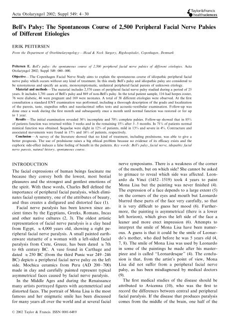
Acta Otolaryngol 2002; Suppl 549: 4–30<br />
Bell’s <strong>Palsy</strong>: <strong>The</strong> <strong>Spontaneous</strong> <strong>Course</strong> <strong>of</strong> 2,<strong>500</strong> <strong>Peripheral</strong> Facial Nerve Palsies<br />
<strong>of</strong> Different Etiologies<br />
ERIK PEITERSEN<br />
From the Department <strong>of</strong> Otorhinolaryngolog y—Head & Neck Surgery, Rigshospitalet, Copenhagen, Denmark<br />
Peitersen E. Bell’s palsy: the spontaneous course <strong>of</strong> 2,<strong>500</strong> peripheral facial nerve palsies <strong>of</strong> different etiologies. Acta<br />
Otolaryngol 2002; Suppl 549: 000–000.<br />
Objective—<strong>The</strong> Copenhagen Facial Nerve Study aims to explain the spontaneous course <strong>of</strong> idiopathic peripheral facial<br />
nerve palsy which occurs without any kind <strong>of</strong> treatment. In this study Bell’s palsy and idiopathic palsy are considered to<br />
be synonymous and specify an acute, monosymptomatic, unilateral peripheral facial paresis <strong>of</strong> unknown etiology.<br />
Material and methods —<strong>The</strong> material includes 2,570 cases <strong>of</strong> peripheral facial nerve palsy studied during a period <strong>of</strong> 25<br />
years. It includes 1,701 cases <strong>of</strong> Bell’s palsy and 869 <strong>of</strong> non-Bell’s palsy. In the total patient sample, 116 had herpes zoster,<br />
76 were diabetic, 46 were pregnant and 169 were neonates. A total <strong>of</strong> 38 different etiologies were observed. At the rst<br />
consultation a standard ENT examination was performed, including a thorough description <strong>of</strong> the grade and localization<br />
<strong>of</strong> the paresis, taste, stapedius re ex and nasolacrimal re ex tests and acoustic-vestibular examination. Follow-up was<br />
done once a week during the rst month and subsequently once a month until normal function was restored or for up<br />
to 1 year.<br />
Results —<strong>The</strong> initial examination revealed 30% incomplete and 70% complete palsies. Follow-up showed that in 85%<br />
<strong>of</strong> patients function was returned within 3 weeks and in the remaining 15% after 3–5 months. In 71% <strong>of</strong> patients normal<br />
mimical function was obtained. Sequelae were slight in 12% <strong>of</strong> patients, mild in 13% and severe in 4%. Contracture and<br />
associated movements were found in 17% and 16% <strong>of</strong> patients, respectively.<br />
Conclusion —A survey <strong>of</strong> the literature showed that no kind <strong>of</strong> treatment, including prednisone, was able to give a<br />
better prognosis. <strong>The</strong> use <strong>of</strong> prednisone raises a big ethical problem because no evidence <strong>of</strong> its ef cacy exists and the<br />
euphoric side-effect induces a false feeling <strong>of</strong> bene t in the patients. Key words : Bell’s palsy, facial nerve, idiopathic facial<br />
nerve paresis, natural history, spontaneous course.<br />
INTRODUCTION<br />
<strong>The</strong> facial expressions <strong>of</strong> human beings fascinate me<br />
because they convey both the lowest, most bestial<br />
pleasures and the strongest and gentlest emotions <strong>of</strong><br />
the spirit. With these words, Charles Bell de ned the<br />
importance <strong>of</strong> peripheral facial paralysis, which eliminates<br />
facial symmetry, one <strong>of</strong> the attributes <strong>of</strong> beauty,<br />
and thus creates a dis gured and distorted face (1).<br />
Facial nerve paralysis has been known since ancient<br />
times by the Egyptians, Greeks, Romans, Incas<br />
and other native cultures (2, 3). <strong>The</strong> oldest artistic<br />
representation <strong>of</strong> facial nerve paralysis is a clay head<br />
from Egypt, :4,000 years old, showing a right peripheral<br />
facial nerve paralysis. A small painted earthenware<br />
statuette <strong>of</strong> a woman with a left-sided facial<br />
paralysis from Crete, Greece, has been dated :7th<br />
to 6th century BC. A vase found in Carthage and<br />
dated :250 BC (from the third Punic war 249–246<br />
BC) depicts a peripheral facial nerve palsy on the left<br />
side. Mochica ceramics from Peru (AD 200–700)<br />
made in clay and carefully painted represent typical<br />
asymmetrical faces caused by facial nerve paralysis.<br />
In the Middle Ages and during the Renaissance<br />
many artists portrayed gures with asymmetrical and<br />
distorted faces. <strong>The</strong> portrait <strong>of</strong> Mona Lisa is the most<br />
famous and her enigmatic smile has been discussed<br />
for many years all over the world and at several facial<br />
nerve symposiums. <strong>The</strong>re is a weakness <strong>of</strong> the corner<br />
<strong>of</strong> the mouth, but on which side? She cannot be asked<br />
to grimace to reveal which side was affected. Leonardo<br />
da Vinci (1452–1519) took 4 years to paint<br />
Mona Lisa but the painting was never nished (4).<br />
<strong>The</strong> expression <strong>of</strong> a face depends to a large extent (5)<br />
on the corners <strong>of</strong> the eyes and mouth but Leonardo<br />
blurred these parts <strong>of</strong> the face very carefully, so that<br />
it is very dif cult to guess her mood (6). Furthermore,<br />
the painting is asymmetrical (there is a lower<br />
left horizon), which gives the left side <strong>of</strong> the face a<br />
leaner and more erect impression (6). Attempts to<br />
interpret the smile <strong>of</strong> Mona Lisa have been numerous.<br />
A guess is that it could be the smile <strong>of</strong> Leonardo’s<br />
mother, who died before he was 5 years old (4,<br />
7, 8). <strong>The</strong> smile <strong>of</strong> Mona Lisa was used by Leonardo<br />
in some <strong>of</strong> the paintings he made after his masterpiece<br />
and is called ‘‘Leonardesque’’ (4). <strong>The</strong> conclusion<br />
is that, from the artist’s point <strong>of</strong> view, Mona<br />
Lisa did not suffer from a peripheral facial nerve<br />
palsy, as has been misdiagnosed by medical doctors<br />
(9).<br />
<strong>The</strong> rst medical studies <strong>of</strong> the disease should be<br />
attributed to Avicenna (10), who was the rst to<br />
record the differences between central and peripheral<br />
facial paralysis. If the disease that produces paralysis<br />
comes from the middle <strong>of</strong> the brain, one half <strong>of</strong> the<br />
© 2002 Taylor & Francis. ISSN 0001-6489
Bell’s palsy 5<br />
body is paralyzed. If the disease is not in the brain<br />
but instead in the nerve, then only what depends on<br />
this nerve is paralyzed.<br />
<strong>The</strong> representation <strong>of</strong> facial nerve palsy in medical<br />
publications began in the 18th century. In 1797,<br />
Pr<strong>of</strong>essor Niclaus A. Friedreich from Würzburg, Germany<br />
treated three patients with idiopathic facial<br />
nerve paresis and documented recovery <strong>of</strong> normal<br />
function (11). <strong>The</strong> observation was published in the<br />
German medical literature in 1798 under the title<br />
‘‘Paralysis Musculorum Faciei Rheumatica’’. <strong>The</strong><br />
rst English review appeared in 1800 in the Annals <strong>of</strong><br />
Medicine published in Edinburgh and it is possible<br />
that Charles Bell, who was studying medicine in<br />
Edinburgh at the time, read this paper. Bell (later Sir<br />
Charles Bell) described the innervation <strong>of</strong> the facial<br />
muscles and the skin <strong>of</strong> the face and consequently the<br />
trigeminal nerve is called Bell’s nerve. Eventually the<br />
eponym ‘‘Bell’s palsy’’ became synonymous with idiopathic<br />
peripheral facial nerve paralysis. However,<br />
Friedreich described the syndrome 23 years before<br />
Bell.<br />
During the 19th century the treatment <strong>of</strong> peripheral<br />
facial nerve palsies was anti-rheumatic. At the<br />
beginning <strong>of</strong> the 20th century electrical stimulation<br />
was used and some very ingenious apparatuses were<br />
constructed. In 1932 Balance and Duel (12) published<br />
the results <strong>of</strong> inserting a free nerve graft between the<br />
cut ends <strong>of</strong> the facial nerve and also advocated<br />
decompression <strong>of</strong> the mastoid segment <strong>of</strong> the facial<br />
nerve for Bell’s palsy when its degeneration delayed<br />
recovery. This was the beginning <strong>of</strong> the modern era<br />
<strong>of</strong> facial nerve surgery.<br />
During the following three decades, the number <strong>of</strong><br />
facial nerve decompression operations increased<br />
rapidly. <strong>The</strong> leading experts were Cawthorne from<br />
London (13), Miehlke from Germany (14), Jongkees<br />
from <strong>The</strong> Netherlands (15), Fisch from Switzerland<br />
(16) and Kettel from Denmark (17), who was the<br />
organizer <strong>of</strong> the First International Symposium on<br />
Facial Nerve Surgery held in Copenhagen in 1964<br />
(18). At that time, the surgeon’s view was that peripheral<br />
facial paralysis should be treated with decompression<br />
after 2 months in cases <strong>of</strong> continued<br />
paralysis. <strong>The</strong> discussions at the symposium revealed<br />
a great deal <strong>of</strong> skepticism, especially from neurologists<br />
and neurophysiologists, regarding the ef cacy <strong>of</strong><br />
the decompression operations. Sunderland (19, 20)<br />
described nerve injuries based on the pathology <strong>of</strong> the<br />
nerve trunk and classi ed ve degrees <strong>of</strong> damage<br />
(Table I). Furthermore, he combined the severity <strong>of</strong><br />
the injuries with the time <strong>of</strong> recovery and was the rst<br />
to create a pro le <strong>of</strong> the recovery <strong>of</strong> a motoric nerve<br />
palsy.<br />
AIM OF THE INVESTIGATION<br />
In Copenhagen it was decided to study the natural<br />
history <strong>of</strong> Bell’s palsy. <strong>The</strong> aim <strong>of</strong> this ‘‘Copenhagen<br />
Facial Nerve Study’’ was to provide a description <strong>of</strong><br />
the spontaneous course <strong>of</strong> idiopathic peripheral facial<br />
palsy, i.e. that which occurs without any kind <strong>of</strong><br />
treatment. <strong>The</strong> prospective study was designed to<br />
include a large number <strong>of</strong> patients, with no exclusion<br />
<strong>of</strong> special groups <strong>of</strong> patients, and to involve adequate<br />
follow-up, exact descriptions <strong>of</strong> sequelae and a statistical<br />
analysis with signi cant conclusions. It was decided<br />
not to publish the results <strong>of</strong> the investigation<br />
until the conclusions were statistically signi cant.<br />
Preliminary observations were presented in Zürich<br />
in 1976, Pittsburgh in 1977, Los Angeles in 1981,<br />
Paris and Rio de Janeiro in 1988, Cologne in 1992,<br />
Matsuyama in 1997 and Berlin in 2000.<br />
ETIOLOGY<br />
<strong>The</strong>ories and hypotheses<br />
<strong>The</strong> aim <strong>of</strong> this investigation is not to list or discuss<br />
all possible hypotheses or theories but to describe the<br />
spontaneous course <strong>of</strong> Bell’s palsy. Nevertheless, a<br />
short summary <strong>of</strong> these theories is included. For<br />
reviews, see Kettel (17), Miehlke (14) and May (21).<br />
Friedreich (11) hypothesized that the cause <strong>of</strong> facial<br />
paralysis in his three patients was ‘‘rheumatic’’ because<br />
<strong>of</strong> exposure to cold <strong>of</strong>ten followed by fever,<br />
chills and local pain and swelling in and around the<br />
neck. Brunninghausen speculated that the paralysis<br />
arose from the nerve sheath becoming thickened and<br />
compressed in the stylomastoid foramen. Berard in<br />
1836 repeated this hypothesis. <strong>The</strong> cold hypothesis,<br />
or paralysis e frigore, maintained that exposure to<br />
draughts produced the palsy. <strong>The</strong> ischemic hypothesis<br />
maintained that ischemia resulting from disturbed<br />
circulation in the vasa nervorum led to nerve injury.<br />
Later on a combination with secondary ischemia was<br />
advocated by a number <strong>of</strong> surgeons (13–17). <strong>The</strong><br />
immunological hypothesis was introduced by<br />
McGovern and co-workers (22, 23).<br />
During the last 25 years or more, viral infections<br />
have been proposed as causes <strong>of</strong> Bell’s palsy. In 1972,<br />
Table I. Classi cation <strong>of</strong> nerve injuries after Sunderland<br />
(19, 20)<br />
Degree<br />
Pathology<br />
Recovery<br />
1 Neuropraxia<br />
Complete<br />
2 Axonotmesis<br />
Complete<br />
3 Neurotmesis Incomplete<br />
4 Perineurium disruption Non-functional<br />
5 Complete disruption None
6<br />
E. Peitersen<br />
Table II. Timetable for examinations<br />
First examination as soon as possible<br />
Once a week until function returned<br />
<strong>The</strong>reafter every second week<br />
After 6 months once a month<br />
Follow-up discontinued after function restored or after<br />
1 year<br />
McCormick (24) published his hypothesis suggesting<br />
that reactivation <strong>of</strong> herpes simplex virus type 1 (HSV-<br />
1) causes in ammation and edema in the bony<br />
fallopian canal and results in peripheral facial palsy.<br />
Adour et al. (25) accepted this theory. In contrast,<br />
Mulkens et al. (26) were unable to demonstrate a<br />
direct link with HSV-1. However, new molecular<br />
biological techniques, such as polymerase chain reaction<br />
(PCR), have proved to be more sensitive. Murakami<br />
et al. (27) studied 14 Bell’s palsy patients in<br />
vivo using HSV DNA PCR. Endoneurinal uid from<br />
the facial nerve and biopsies from the posterior auricular<br />
muscle were tested using PCR for the presence <strong>of</strong><br />
HSV DNA. HSV DNA was detected in samples from<br />
11:14 patients. Further experiments in the future will<br />
hopefully resolve this issue.<br />
De nition<br />
<strong>The</strong> term Bell’s palsy is accepted in many Anglo-<br />
American countries to describe a peripheral paresis <strong>of</strong><br />
the facial nerve, independent <strong>of</strong> etiology. Idiopathic<br />
peripheral facial nerve paresis is an acute,<br />
monosymptomatic, peripheral facial nerve paresis <strong>of</strong><br />
unknown etiology. Formerly the disease was described<br />
as rheumatic or ischemic, as mentioned<br />
above. Patients suffering from an underlying disease<br />
or condition, for example collagenosis, pregnancy or<br />
diabetes mellitus, sometimes develop peripheral facial<br />
nerve palsies, but these pareses are never classi ed as<br />
Bell’s palsy. However, a reduction in future in the<br />
group <strong>of</strong> idiopathic pareses is anticipated on account<br />
<strong>of</strong> better knowledge <strong>of</strong> many diseases and <strong>of</strong> the<br />
pathological conditions governing peripheral paresis.<br />
Bell’s palsy and idiopathic peripheral facial nerve<br />
paresis are considered as synonymous in this study.<br />
MATERIAL AND METHODS<br />
Before the start <strong>of</strong> this investigation a lot <strong>of</strong> time was<br />
taken to work out a very speci c plan for the study.<br />
As a result <strong>of</strong> 6 months <strong>of</strong> pilot studies it was possible<br />
to construct a timetable for the following years. <strong>The</strong><br />
study design was as follows.<br />
<strong>The</strong> patient was rst examined as soon as possible<br />
after the onset <strong>of</strong> palsy, i.e. within 1–5 days after the<br />
onset <strong>of</strong> paresis (Table II). Follow-up examinations<br />
Table III. <strong>The</strong> questionnaire administered to the<br />
patients concerning history <strong>of</strong> Bell’s palsy<br />
Beginning—palsy—date<br />
Remission date—unchanged—regression<br />
Facial palsy before—relatives with facial palsy<br />
Head trauma— systemic disease—diabetes mellitus<br />
Pregnancy—infections—skin eruptions<br />
Colds —exposure to draught<br />
Other cranial nerve symptoms<br />
Headache—paresthesia—paresis<br />
Otitis—hearing loss —tinnitus—vertigo<br />
Postauricular pains<br />
Before—simultaneous —after<br />
Taste—phonophobia—tear ow—dry eye<br />
were performed once a week until the returning <strong>of</strong><br />
function was observed. After 6 months, examinations<br />
were conducted once a month. Follow-up was discontinued<br />
after restoration <strong>of</strong> function or after 1 year. If<br />
a patient did not attend for examination a new<br />
appointment was mailed to them. If the patient still<br />
failed to attend, a telephone interview was conducted<br />
or the patient was asked to complete a questionnaire.<br />
All the patients lived within a radius <strong>of</strong> 25 km <strong>of</strong> the<br />
hospital and using the Danish Central Personal Register<br />
it was easy to trace them. <strong>The</strong> majority <strong>of</strong><br />
patients failed to attend the follow-up examinations<br />
because normal function had been restored. Followup<br />
included 98% <strong>of</strong> all patients. Great importance<br />
was attached to the date <strong>of</strong> onset <strong>of</strong> palsy as well as<br />
the date <strong>of</strong> the rst sign <strong>of</strong> returning <strong>of</strong> function. <strong>The</strong><br />
questionnaire administered to the patients is shown in<br />
Table III. It should be stressed that patients suffering<br />
from acute peripheral facial palsy were always able to<br />
give the exact date and hour <strong>of</strong> the rst sign <strong>of</strong><br />
paresis.<br />
<strong>The</strong> examination <strong>of</strong> the patient is very important<br />
and an exact description <strong>of</strong> the paresis must be made<br />
at the rst examination (Table IV). Is the paresis<br />
complete or incomplete? Does it affect all branches or<br />
is it localized to only one or maybe two branches?<br />
Bell’s palsy usually involves all branches, although<br />
not to the same degree. Patients with involvement <strong>of</strong><br />
only one or perhaps two branches should arouse<br />
Table IV. Description <strong>of</strong> the standard examination <strong>of</strong><br />
patients with Bell’s palsy<br />
Routine ENT examination<br />
Description <strong>of</strong> facial nerve palsy<br />
Grade and localization<br />
Cranial nerve check<br />
Taste, stapedius re ex and nasolacrimal re ex tests<br />
Acoustic and vestibular function tests<br />
Laboratory tests
Bell’s palsy 7<br />
suspicion <strong>of</strong> another etiology, for example a parotid<br />
gland tumor.<br />
All topographical tests were performed during the<br />
patient’s rst visit. <strong>The</strong>se involved examination <strong>of</strong><br />
taste, stapedius re ex and nasolacrimal re ex. <strong>The</strong>se<br />
tests were repeated at each <strong>of</strong> the two subsequent<br />
visits, if the result <strong>of</strong> the preceding investigation was<br />
not normal. Patients also underwent acoustic and<br />
vestibular tests including tympanometry, audiometry,<br />
electronystagmograph y for spontaneous and positional<br />
nystagmus and a caloric test according to<br />
Hallpike.<br />
If the anamnesis and objective ndings indicated<br />
Bell’s palsy, only a few laboratory tests were carried<br />
out; these always included measurement <strong>of</strong> blood<br />
pressure and a urine test for glucose. All patients<br />
were investigated for serum antibodies against HSV,<br />
herpes zoster and borreliosis. In patients with a second<br />
episode <strong>of</strong> palsy or those in whom function was<br />
not restored within 4 months an extensive battery <strong>of</strong><br />
tests was performed, including cerebrospinal uid<br />
(CSF) cell counting, differential cell counting, determination<br />
<strong>of</strong> Borrelia burghdorferi antibodies, CT and<br />
MRI scans and a variety <strong>of</strong> other tests.<br />
Electrodiagnosis <strong>of</strong> the facial nerve<br />
Duchenne in 1855 (28) was the rst to use contraction<br />
<strong>of</strong> the facial muscles evoked by electrical stimuli<br />
to the nerve as a way <strong>of</strong> predicting recovery. In<br />
contrast to many lesions <strong>of</strong> peripheral nerves, electrodiagnosis<br />
<strong>of</strong> cranial nerves is hampered by one major<br />
dif culty, namely that in the majority <strong>of</strong> cases it is<br />
not possible to stimulate proximally to the site <strong>of</strong> the<br />
lesion. Thus, it is not possible to compare the response<br />
to proximal stimulation with the response to<br />
stimulation distally from the site <strong>of</strong> the lesion.<br />
A very simple and widely-used electrical test is the<br />
so-called nerve excitability test (NET), in which the<br />
threshold <strong>of</strong> the electrical stimulus producing visible<br />
muscle twitch is determined. Although the NET is a<br />
very simple test, it has limitations and its accuracy is<br />
dubious. When using a stronger stimulation to obtain<br />
maximal contraction <strong>of</strong> the facial muscles the test is<br />
known as the maximal stimulation test (MST). According<br />
to Blumenthal and May (29), the NET was<br />
not reliable when the response was normal, as 42% <strong>of</strong><br />
their patients with a normal response had residual<br />
facial function de cits 6 months later. Furthermore,<br />
Groves and Gibson (30) and Laumans (31) noted<br />
that some <strong>of</strong> their patients with an abnormal response<br />
to the NET recovered full function. To the<br />
best <strong>of</strong> my knowledge, the NET is not reliable or<br />
useful for predicting recovery from peripheral facial<br />
nerve palsies. Langworth and Taverner (32) recommended<br />
conduction velocity as the best parameter for<br />
predicting prognosis.<br />
Electromyography is not very reliable at revealing<br />
recent facial nerve palsies and has no prognostic<br />
signi cance in Bell’s palsy according to Buchthal (33).<br />
However, after :10–12 days the so-called denervation<br />
potentials can be recorded and the test is also<br />
able to demonstrate regeneration.<br />
Electrical tests are not routinely used in Bell’s palsy<br />
patients in my department. <strong>The</strong> Department <strong>of</strong> Clinical<br />
Neurophysiology performed all the electrodiagnostic<br />
tests <strong>of</strong> the facial nerve in this study. <strong>The</strong><br />
majority <strong>of</strong> cases were examined by Olsen (34), who<br />
in 1975 described the technique <strong>of</strong> electroneurography<br />
(ENoG). <strong>The</strong> method is identical to that used by<br />
Esslen in 1977 (35). ENoG is very useful for demonstrating<br />
degeneration. <strong>The</strong> principle is to compare the<br />
evoked potentials on the paretic side with those on<br />
the healthy side. For further information, see Olsen<br />
(34) and Esslen (35).<br />
Statistical analyses<br />
<strong>The</strong> statistical data analyses were performed by Arne<br />
Nørby Rasmussen, BScEE and Poul Aabo Osterhammel,<br />
EE, EDA. <strong>The</strong> x 2 test was used to compare data<br />
between different groups <strong>of</strong> patients. p¾0.05 was<br />
considered signi cant. In addition, the Shapiro–Wilk<br />
test for normality was used with a signi cance level <strong>of</strong><br />
p¾0.05.<br />
Patients<br />
<strong>The</strong> patients in this study came from the Copenhagen<br />
area during a 25-year period. <strong>The</strong> investigation included<br />
2570 patients suffering from peripheral facial<br />
nerve paresis (Table V). <strong>The</strong>re were 1,701 patients<br />
with Bell’s palsy (66%) and 869 with non-Bell’s palsy<br />
(34%).<br />
RESULTS<br />
Incidence <strong>of</strong> Bell’s palsy<br />
In this study ‘‘incidence’’ is de ned as the number <strong>of</strong><br />
cases per year in a population <strong>of</strong> 100,000 inhabitants.<br />
Studies in the literature have shown great variations<br />
in incidence. However, many <strong>of</strong> these studies cannot<br />
be considered as representative, because basic criteria<br />
have not been ful lled. <strong>The</strong>re must be a well-de ned<br />
area and all patients should be included. <strong>The</strong> number<br />
<strong>of</strong> all incoming and outgoing inhabitants should be<br />
registered, as well as the age and sex <strong>of</strong> the population;<br />
however, only a few investigations meet these<br />
requirements. In this study the incidence <strong>of</strong> Bell’s<br />
palsy was 32.<br />
Recurring palsies and familial Bell’s palsy<br />
A total <strong>of</strong> 6.8% <strong>of</strong> Bell’s palsy patients in the sample<br />
had previously suffered from facial palsy on either
8<br />
E. Peitersen<br />
Table V. Etiologies <strong>of</strong> the 2,570 study patients<br />
Etiology<br />
n<br />
Idiopathic palsy<br />
1,701<br />
Neonatal age<br />
169<br />
Herpes zoster 116<br />
Trauma<br />
95<br />
Diabetes mellitus<br />
76<br />
Pregnancy<br />
46<br />
Polyneuritis 44<br />
Parotid tumor<br />
43<br />
Vascular (brainstem)<br />
34<br />
Hemifacial spasm<br />
27<br />
Sarcoidosis<br />
21<br />
Multiple sclerosis<br />
20<br />
Melkersson–Rosenthal syndrome<br />
19<br />
Collagenosis<br />
18<br />
Cholesteatoma <strong>of</strong> the middle ear<br />
18<br />
Children with bilateral palsy 18<br />
Breast cancer with metastasis<br />
16<br />
Chronic otitis<br />
15<br />
Infectious mononucleosis 9<br />
Leukemia<br />
6<br />
Malignant lymphoma<br />
6<br />
Cholesteatoma <strong>of</strong> the inner ear 5<br />
Cancer <strong>of</strong> the middle ear 5<br />
Borreliosis<br />
4<br />
AIDS 4<br />
Neurinoma <strong>of</strong> the VIIth extracranial nerve 3<br />
Bronchial cancer with metastasis<br />
3<br />
Nephropathia<br />
2<br />
Non-de nite paresis<br />
2<br />
Hypothyreosis 2<br />
Pemphigus<br />
1<br />
Granuloma eosinophilia<br />
1<br />
Tuberculosis<br />
1<br />
Poliomyelitis<br />
1<br />
Smallpox vaccine sequelae<br />
1<br />
Herxheimer reaction 1<br />
Paget’s disease<br />
1<br />
Hysteria<br />
1<br />
the same or the opposite site <strong>of</strong> the face. Some<br />
authors use the term ‘‘recurrent’’ palsy exclusively to<br />
describe paresis occurring on the same side <strong>of</strong> the<br />
face; paresis on the opposite side is termed ‘‘alternating’’.<br />
Although this classi cation is logical, the term<br />
‘‘recurrent’’ is in general use and is therefore used in<br />
this study to describe both ipsi- and contralateral<br />
palsies. <strong>The</strong> occurrence <strong>of</strong> familial Bell’s palsy is well<br />
known. In this study, 4.1% <strong>of</strong> all cases <strong>of</strong> Bell’s palsy<br />
observed represented familial Bell’s palsy.<br />
Seasonal variation and clustering<br />
<strong>The</strong> seasonal incidence <strong>of</strong> Bell’s palsy has been discussed<br />
for many years. <strong>The</strong> explanation <strong>of</strong> ndings <strong>of</strong><br />
seasonal variation or clustering in some samples<br />
could be that the patient sample was too small. In<br />
this study the mean number <strong>of</strong> patients per month<br />
with Bell’s palsy was 142 (range 126–156) (Fig. 1).<br />
Fig. 1. Number <strong>of</strong> patients with Bell’s palsy per month<br />
over the 25-year period <strong>of</strong> the study.<br />
<strong>The</strong> Shapiro–Wilk test for normality revealed no<br />
signi cant difference from month to month (p¾0.1)<br />
and thus no seasonal variation or clustering was<br />
found.<br />
Decade variation<br />
Variations from year to year or from decade to<br />
decade could not be demonstrated because the number<br />
<strong>of</strong> patients with Bell’s palsy has largely remained<br />
constant over the study period: mean 68 (range 48–<br />
89) (Fig. 2). <strong>The</strong> Shapiro –Wilk test showed no signi<br />
cant difference from year to year (p¾0.4).<br />
Sex distribution<br />
<strong>The</strong> question <strong>of</strong> a gender predominance among patients<br />
af icted with idiopathic facial palsy has been<br />
discussed in the literature. One <strong>of</strong> the main reasons<br />
for the discussion has undoubtedly been the small<br />
amount <strong>of</strong> material available. This study included<br />
1,701 patients with Bell’s palsy, 818 (48.1%) <strong>of</strong> whom<br />
were male and 883 (51.9%) female. <strong>The</strong> underlying<br />
population comprises 47.8% males and 52.2% females,<br />
clearly indicating that there is no difference in<br />
sex distribution among patients with Bell’s palsy<br />
(0.8BpB0.9).<br />
Fig. 2. Number <strong>of</strong> patients with Bell’s palsy per year over<br />
the 25-year period <strong>of</strong> the study.
Bell’s palsy 9<br />
Fig. 3. Age distribution <strong>of</strong> patients with Bell’s palsy in<br />
comparison with that <strong>of</strong> the underlying population.<br />
Side <strong>of</strong> the face<br />
<strong>The</strong>re was no difference in localization, with 828<br />
(48.7%) right- and 873 (51.3%) left-sided palsies<br />
(0.1 BpB0.2).<br />
Age distribution<br />
This subject has also been debated, without de nitive<br />
conclusions having been reached. Figure 3 demonstrates<br />
the age distribution <strong>of</strong> patients with Bell’s<br />
palsy in comparison to the age distribution <strong>of</strong> the<br />
underlying population. <strong>The</strong> incidence <strong>of</strong> Bell’s palsy<br />
reaches a maximum between the ages <strong>of</strong> 15 and 45<br />
years and this differs highly signi cantly from the age<br />
distribution <strong>of</strong> the underlying population (pB0.001).<br />
<strong>The</strong> disease is signi cantly less common below the<br />
age <strong>of</strong> 15 years and above the age <strong>of</strong> 60 years (pB<br />
0.001). For the group aged 45–59 years the incidence<br />
<strong>of</strong> Bell’s palsy did not differ signi cantly from the age<br />
distribution <strong>of</strong> the underlying population (p¾0.4).<br />
<strong>The</strong> in uence <strong>of</strong> age on the incidence <strong>of</strong> Bell’s palsy<br />
would therefore seem to be convincingly demonstrated.<br />
Symptoms<br />
<strong>The</strong> most alarming symptom <strong>of</strong> Bell’s palsy is <strong>of</strong><br />
course the paresis itself. Approximately 50% <strong>of</strong> patients<br />
believe that they have suffered a stroke, 25%<br />
fear an intracranial tumor and the remaining 25%<br />
have no clear conception <strong>of</strong> what is wrong, but are<br />
Table VI. Distribution <strong>of</strong> symptoms <strong>of</strong> Bell’s palsy<br />
among the patient sample<br />
Symptom<br />
n %<br />
Taste disorders 580 34<br />
Phonophobia 234 14<br />
Tear ow 1137 67<br />
Dry eye 69<br />
4<br />
Postauricular pains 881 52<br />
Fig. 4. Distribution <strong>of</strong> time <strong>of</strong> beginning recovery after the<br />
onset <strong>of</strong> paresis.<br />
extremely anxious. <strong>The</strong> distribution <strong>of</strong> symptoms<br />
among the patient sample is shown in Table VI.<br />
Postauricular pains, which were experienced by almost<br />
half the patients, are <strong>of</strong> the utmost interest.<br />
<strong>The</strong>se pains occurred simultaneously with the palsy in<br />
:50% <strong>of</strong> patients, whilst in 25% they occurred 2 or<br />
3 days before the onset <strong>of</strong> palsy. <strong>The</strong> remaining 25%<br />
<strong>of</strong> patients experienced pains after the onset <strong>of</strong> palsy.<br />
<strong>The</strong> pains are located deep in the mastoid region,<br />
usually persist for one to several weeks and require<br />
analgesia.<br />
Every third patient will complain about taste disorders<br />
but when examined objectively four out <strong>of</strong> ve<br />
show a reduced sense <strong>of</strong> taste. This difference can be<br />
explained by the fact that patients can still use the<br />
normal side <strong>of</strong> the tongue to taste.<br />
Only a few patients are able to perceive restricted<br />
stapedius re ex paresis, which may lead to phonophobia<br />
or diplaucusis. In comparison, two out <strong>of</strong><br />
three patients complain about tear ow. However,<br />
this is not caused by hypersecretion but by the diminished<br />
function <strong>of</strong> the musculus orbicularis oculi,<br />
which prevents tears from being transported medially<br />
to the lacrimal sac.<br />
Time <strong>of</strong> beginning recovery<br />
Very little interest has been paid in the literature to<br />
the question <strong>of</strong> recovery time and therefore it was felt<br />
worthwhile to record the pattern <strong>of</strong> remission for the<br />
idiopathic palsies. <strong>The</strong> time <strong>of</strong> the rst sign <strong>of</strong> muscular<br />
movement in relation to the onset <strong>of</strong> palsy was<br />
recorded.<br />
Of a total <strong>of</strong> 1,701 patients, 1,189 suffered from<br />
complete paralysis (70%) and 512 from incomplete<br />
paralysis (30%). Recovery occurred within 3 weeks<br />
for 1,448 patients (85%) and within 3–5 months for<br />
the remaining 253 patients (15%).<br />
<strong>The</strong> results are shown in Fig. 4. By de nition, all<br />
patients with incomplete paresis have function back<br />
at time zero, i.e. at the onset <strong>of</strong> paresis. In the rst
10<br />
E. Peitersen<br />
week 6% <strong>of</strong> patients achieved remission, in the second<br />
week 33% and in the third week 16%. No patients<br />
achieved remission between 3 weeks and 3–5 months<br />
after the onset <strong>of</strong> paresis. This is because patients<br />
who showed improvement in the rst 3 weeks had<br />
only partial degeneration and blocking <strong>of</strong> nerve conduction<br />
whilst patients who showed improvement<br />
after 3–5 months had total degeneration. After 3–5<br />
months 10% <strong>of</strong> patients experienced remission and<br />
after 5–6 months an additional 5% experienced remission.<br />
This investigation shows that all patients<br />
diagnosed with Bell’s palsy achieve some degree <strong>of</strong><br />
muscular function. However, this does not imply that<br />
all patients achieve normal function. <strong>The</strong> period between<br />
the rst 3 weeks without remission and after<br />
the third month without remission is one characterized<br />
by ‘‘hibernation <strong>of</strong> the facial nerve’’. Although<br />
the nerve seems to be dead it is in fact still alive and<br />
in the process <strong>of</strong> repairing the damage.<br />
Complete recovery<br />
<strong>The</strong> next question is naturally how many patients will<br />
achieve complete remission? In this study, <strong>of</strong> a total<br />
<strong>of</strong> 1,701 patients, 1,202 (71%) achieved normal facial<br />
nerve function. <strong>The</strong> second question is how long does<br />
it take before patients achieve normal function?<br />
Prospects are decidedly better for the group with<br />
some remission within the rst 3 weeks (Table VII).<br />
Patients with the poorest prognosis are <strong>of</strong> course<br />
those with total degeneration and late return <strong>of</strong> function.<br />
This group does not regain normal mimical<br />
function. Normal function is regained as early as<br />
within 2 months for the majority (58%) <strong>of</strong> all patients<br />
(Fig. 5). <strong>The</strong> possibility <strong>of</strong> normalization is very small<br />
after 3 months, at which time 64% <strong>of</strong> patients have<br />
regained normal function, and beyond 6 months no<br />
patients regained normal mimical function (Fig. 6).<br />
As noted before (Table VII), the incomplete Bell’s<br />
palsy patients have a very good prognosis for full<br />
recovery (481:512; 94%). Of the patients with complete<br />
Bell’s palsy, 721:1,189 (61%) regained normal<br />
facial muscle function. <strong>The</strong> difference between the<br />
two groups in terms <strong>of</strong> the number <strong>of</strong> patients who<br />
Fig. 5. Distribution <strong>of</strong> time <strong>of</strong> complete recovery after the<br />
onset <strong>of</strong> paresis.<br />
achieved full recovery was highly signi cant (pB<br />
0.001).<br />
Gender and recovery<br />
<strong>The</strong> numbers <strong>of</strong> males and females who experienced<br />
full recovery were 564 (69%) and 638 (72%), respectively.<br />
<strong>The</strong>re was no statistically signi cant difference<br />
between the two groups (p\2).<br />
Factors in uencing the nal resuls<br />
Time <strong>of</strong> beginning remission. <strong>The</strong> number <strong>of</strong> days<br />
between the onset <strong>of</strong> paresis and the beginning <strong>of</strong><br />
remission is a very decisive factor in the degree <strong>of</strong><br />
recovery (Fig. 7). A total <strong>of</strong> 94% <strong>of</strong> patients with<br />
incomplete paresis regained normal function. Of patients<br />
who showed remission in the rst week, 88%<br />
regained normal function, as opposed to 83% <strong>of</strong><br />
those who showed remission in the second week and<br />
61% <strong>of</strong> those who showed remission in the third<br />
week. <strong>The</strong> prognosis for patients with incomplete<br />
paresis was signi cantly better than that for the<br />
group who recovered in the rst week (p¾0.03).<br />
<strong>The</strong>re was no signi cant difference in prognosis between<br />
patients who recovered in the rst and second<br />
weeks (p¾0.2) but patients who recovered in the<br />
Table VII. Distribution <strong>of</strong> patients with initial incomplete<br />
and complete paresis who make a full recovery<br />
from Bell’s palsy<br />
Paresis with<br />
full recovery<br />
Initial<br />
n<br />
Final<br />
% n %<br />
Incomplete 512 30 481 94<br />
Complete 1189 70 721 61<br />
Fig. 6. Distribution <strong>of</strong> time <strong>of</strong> complete recovery (cumulative)<br />
after the onset <strong>of</strong> paresis.
Bell’s palsy 11<br />
Fig. 7. Proportion <strong>of</strong> patients who achieve complete recovery<br />
as a function <strong>of</strong> time <strong>of</strong> beginning recovery after the<br />
onset <strong>of</strong> paresis.<br />
third week had a signi cantly worse outcome (pB<br />
0.001). It is clear that the time <strong>of</strong> beginning remission<br />
is highly signi cant to the prognosis.<br />
Age <strong>of</strong> patients. Age is another parameter that<br />
in uences the nal result (Fig. 8). Children aged 514<br />
years had the most favorable prognosis, with 90%<br />
achieving full recovery. Patients aged 15–29 years<br />
had a fairly good chance <strong>of</strong> recovery (84%). <strong>The</strong><br />
chance <strong>of</strong> a full recovery was reduced for patients<br />
aged between 30 and 44 years (75%). Above the age<br />
<strong>of</strong> 45 years, the chances <strong>of</strong> recovery diminished signi<br />
cantly (64%). Above the age <strong>of</strong> 60 years, only<br />
about one-third <strong>of</strong> patients will experience the return<br />
<strong>of</strong> normal function. <strong>The</strong> in uence <strong>of</strong> age on the nal<br />
outcome is therefore highly signi cant (pB0.001).<br />
Postauricular pains. As noted above, postauricular<br />
pains were registered in 52% <strong>of</strong> all cases <strong>of</strong> Bell’s<br />
palsy. A total <strong>of</strong> 78% <strong>of</strong> patients with no pain regained<br />
normal function, as opposed to only 64% <strong>of</strong><br />
patients with pain (pB0.001).<br />
<strong>The</strong> prognostic value <strong>of</strong> topographical tests. It must<br />
be stressed that the examination <strong>of</strong> taste, stapedius<br />
re ex and tear ow or nasolacrimal re ex (Table<br />
Fig. 8. Age distribution <strong>of</strong> patients who achieve complete<br />
recovery.<br />
VIII) should be performed very carefully and always<br />
in exactly the same way or else the comparison <strong>of</strong><br />
results is meaningless. <strong>The</strong> results should not depend<br />
on the person performing the tests.<br />
Taste. <strong>The</strong> taste test according to Boernstein (36) is<br />
semiquantitative and based on recognition <strong>of</strong> four<br />
basic tastes—sweet, salt, sour and bitter—at three<br />
different concentrations. Initial taste examination<br />
showed that 83% <strong>of</strong> patients had partially reduced or<br />
abolished taste while 12% had normal taste. Final<br />
taste examination showed that 80% <strong>of</strong> patients had<br />
regained normal taste function. Taste function and<br />
the muscular function <strong>of</strong> the face normalized at approximately<br />
the same time.<br />
Stapedius re ex. <strong>The</strong> stapedius re ex is an acoustic<br />
facial re ex provoked on both sides by one-sided<br />
sound stimulation (37). Initially, 72% <strong>of</strong> patients had<br />
a reduced or abolished re ex and only 22% had a<br />
normal re ex. When remission occurs, stapedius<br />
re ex will usually return 1–2 weeks before visual<br />
function <strong>of</strong> the facial muscles can be con rmed.<br />
Normal function was restored in 86% <strong>of</strong> patients.<br />
Tearing or nasolacrimal re ex. <strong>The</strong> nasolacrimal<br />
re ex passes from the nasal mucosa to the superior<br />
salivary nucleus and thence to the secretory bers,<br />
with the facial nerve ending in the lacrimal gland. In<br />
this study a modi cation <strong>of</strong> Schirmer’s test II (38)<br />
was used, which is based on measurement <strong>of</strong> tear ow<br />
for 1 min. Stimulation with benzene is carried out for<br />
30 s and measurement is performed by using lter<br />
paper placed in the lower fornix. At the end <strong>of</strong> the<br />
test the length <strong>of</strong> the soaked strip <strong>of</strong> lter paper is<br />
measured on both sides. <strong>The</strong> difference between the 2<br />
sides is B20% in 95% <strong>of</strong> normal persons. Initially,<br />
11% <strong>of</strong> patients had partially reduced or abolished<br />
tearing. <strong>The</strong> nal result showed that 97% <strong>of</strong> patients<br />
achieved normal tearing. In comparison with taste<br />
and stapedius re ex testing, tearing became normal in<br />
a surprisingly high proportion <strong>of</strong> patients.<br />
Figure 9 shows the prognostic value <strong>of</strong> the three<br />
topographical tests. <strong>The</strong> patients were divided into<br />
two groups: one group with normal facial muscle<br />
function and the other with muscular sequelae. Comparison<br />
<strong>of</strong> the results <strong>of</strong> the initial taste tests for the<br />
groups with and without sequelae showed that 91%<br />
and 80% <strong>of</strong> patients, respectively had partially reduced<br />
or abolished taste (pB0.001). Concerning the<br />
stapedius re ex, it was found that 91% and 63% <strong>of</strong><br />
patients, respectively had a reduced or abolished<br />
re ex (pB0.001). <strong>The</strong> nasolacrimal re ex is also a<br />
reliable prognostic indicator because 27% and 5% <strong>of</strong><br />
patients, respectively had abolished or reduced<br />
lacrimal function (pB0.001). In conclusion, all three<br />
<strong>of</strong> the topographical tests provide reliable prognostic<br />
information.
12<br />
E. Peitersen<br />
Table VIII. Results <strong>of</strong> the initial and nal examinations <strong>of</strong> taste, stapedius re ex and nasolacrimal re ex<br />
Test<br />
Reduced or abolished function<br />
None assessible<br />
Normal function<br />
n<br />
% n % n %<br />
Taste<br />
Initial 1,419<br />
83 111<br />
7 171 10<br />
Final<br />
Stapedius re ex<br />
Initial<br />
236<br />
1,221<br />
14<br />
72<br />
111<br />
108<br />
7<br />
6<br />
1,354<br />
372<br />
80<br />
22<br />
Final<br />
Nasolacrimal re ex<br />
Initial<br />
122<br />
192<br />
7<br />
11<br />
122<br />
27<br />
7<br />
2<br />
1,471<br />
1,482<br />
86<br />
87<br />
Final<br />
29 2 27 1<br />
1,645 97<br />
Sequelae. As mentioned before, 71% <strong>of</strong> patients<br />
regain normal function <strong>of</strong> their facial muscles after an<br />
idiopathic paresis. <strong>The</strong> remaining 29% <strong>of</strong> patients<br />
suffer from varying degrees <strong>of</strong> sequelae. It is extremely<br />
dif cult to describe the sequelae accurately and to<br />
group the degree <strong>of</strong> sequelae; however, it should be<br />
borne in mind that the daily discomfort <strong>of</strong> sequelae is<br />
a signi cant problem for the patients. Today, there<br />
are at least 10 systems available for facial nerve<br />
grading but none <strong>of</strong> these systems is ideal (39, 40).<br />
<strong>The</strong> existing systems include gross scales, regional<br />
systems and speci c scales. One <strong>of</strong> the biggest problems<br />
with grading systems is nding a balance between<br />
an exact description <strong>of</strong> the sequelae and<br />
minimizing the number <strong>of</strong> groups into which the<br />
patients are classi ed. <strong>The</strong> scale should combine the<br />
main parameters (paresis, contracture and associated<br />
movements) based on speci c de nitions. An ideal<br />
system is one with high speci city and an acceptable<br />
sensitivity. Before proposing my gross scale the rst<br />
200 <strong>of</strong> the patients in this study were examined 3<br />
times. <strong>The</strong> scale emerged from an examination <strong>of</strong><br />
these 200 patients who suffered from peripheral facial<br />
nerve palsy with different grades <strong>of</strong> reduced function<br />
and other sequelae. It should be stressed that the scale<br />
includes all types <strong>of</strong> motoric dysfunction. Other secondary<br />
defects, such as crocodile tears, decreased<br />
tearing, taste disturbances and stapedius muscle problems,<br />
are registered and should be added to the other<br />
sequelae to nd the total number <strong>of</strong> sequelae. My<br />
scale is a modi cation <strong>of</strong> that <strong>of</strong> Botman and Jongkees<br />
(41).<br />
<strong>The</strong> factors determining the degree <strong>of</strong> sequelae are<br />
paresis, contracture and associated movements (also<br />
known as mass movements or synkinesis). <strong>The</strong> de nition<br />
<strong>of</strong> synkinesis is an involuntary movement accompanying<br />
a voluntary one. <strong>The</strong> nal cosmetic result<br />
depends on a combination <strong>of</strong> these three elements.<br />
Table IX shows the grade <strong>of</strong> palsy. De nitions <strong>of</strong><br />
grades 0–IV are given in Table X. Contracture causes<br />
a narrowed palpebral ssure on the involved side<br />
when the face is in repose. <strong>The</strong> corner <strong>of</strong> the mouth<br />
occurs higher on the affected side and the folds <strong>of</strong> the<br />
face are abnormally deep; in particular the nasal labial<br />
fold is very marked. Associated movements are found<br />
in the muscles <strong>of</strong> the eye, cheek and mouth and more<br />
seldom in the forehead. Associated movements are<br />
visible when the patient tries to close his:her eye and<br />
smiles involuntarily or vice versa. Associated movements<br />
<strong>of</strong>ten cause more cosmetic inconvenience to a<br />
patient than a slight paresis or contracture. <strong>The</strong> three<br />
parameters should be combined to give an accurate<br />
description <strong>of</strong> sequelae.<br />
<strong>The</strong> degree <strong>of</strong> recovery for all patients is demonstrated<br />
in Fig. 10, which shows that 71% recovered<br />
completely, 12% had slight sequelae, 13% had moderate<br />
sequelae and only 4% had severe sequelae. It<br />
should be stressed that no patients became paralyzed.<br />
In summary, 83% <strong>of</strong> patients had a good recovery and<br />
17% a bad recovery without any kind <strong>of</strong> treatment.<br />
Fig. 9. Proportions <strong>of</strong> patients with and without muscular<br />
sequelae who had reduced or abolished function in the<br />
three topographical tests.
Bell’s palsy 13<br />
Table IX. Grades <strong>of</strong> palsy in the Peitersen grading<br />
system<br />
Grade<br />
Degree <strong>of</strong><br />
palsy<br />
Description <strong>of</strong> palsy<br />
0 None<br />
I Slight<br />
II Moderate<br />
III Severe<br />
IV Complete<br />
Normal function<br />
Only visible when patient grimaces<br />
Visible with small facial movements<br />
Function just visible<br />
No function<br />
Table X. Description <strong>of</strong> sequelae associated with<br />
grades 0–IV <strong>of</strong> palsy in the Peitersen grading system<br />
Fig. 10. Distribution <strong>of</strong> degree <strong>of</strong> recovery for the patients<br />
with Bell’s palsy.<br />
Figure 11 shows the types <strong>of</strong> initial damage <strong>of</strong> the<br />
facial nerve. A total <strong>of</strong> 85% <strong>of</strong> patients experienced<br />
different types <strong>of</strong> damage, such as neuropraxia, axonotmesis,<br />
neurotmesis and partial degeneration. For<br />
all these patients recovery began within 3 weeks after<br />
the onset <strong>of</strong> palsy. Only 15% <strong>of</strong> patients suffered total<br />
degeneration <strong>of</strong> the nerve, with recovery beginning<br />
]3 months after the onset <strong>of</strong> palsy.<br />
Figure 12 demonstrates the nal outcomes after<br />
recovery. As mentioned above, 83% <strong>of</strong> patients had a<br />
good outcome and 17% a bad outcome.<br />
Comparison <strong>of</strong> the results shown in Figs 11 and 12<br />
shows that there is a very clear connection between<br />
early pathology in the facial nerve and the nal<br />
outcome. A total <strong>of</strong> 85% <strong>of</strong> patients had a ‘‘mild<br />
pathology’’ and 83% achieved a fair nal result. A<br />
total <strong>of</strong> 15% <strong>of</strong> patients had a ‘‘severe pathology’’,<br />
namely total degeneration <strong>of</strong> the nerve. All <strong>of</strong> these<br />
patients (and 17% in total) were in the group with<br />
severe sequelae.<br />
A comparison <strong>of</strong> my scale <strong>of</strong> sequelae and that<br />
designed by House and Brackmann (40) is possible to<br />
some extent. If their grades IV and V are combined<br />
then the new group is almost identical to my grade<br />
III (Table XI). <strong>The</strong> number <strong>of</strong> Bell’s palsy patients in<br />
this group is very small and to the best <strong>of</strong> my<br />
knowledge it is very dif cult to distinguish between<br />
House and Brackmann’s grades IV and V.<br />
<strong>The</strong> distribution <strong>of</strong> sequelae is listed in Table XII.<br />
Paresis is the commonest sequela but not the most<br />
uncomfortable for the patients. Associated movements<br />
and contracture were found in 16% and 17% <strong>of</strong><br />
the sample, respectively. Both sequelae cause more<br />
inconvenience for the patient than a slight paresis.<br />
<strong>The</strong> most troublesome sequelae for patients are the<br />
associated movements. Contracture gives the patient<br />
a feeling <strong>of</strong> stiffness in the muscles <strong>of</strong> the face. In ve<br />
patients with dis guring contractures biopsies were<br />
taken from the musculus orbicularis oris on both the<br />
paretic and normal sides. Microscopy showed a reduction<br />
in the number and size <strong>of</strong> the muscle cells<br />
and an increased amount <strong>of</strong> connective tissue and fat<br />
on the paretic side. As can be seen from Table XII,<br />
crocodile tears and dry eyes are very rare, with both<br />
Fig. 11. Distribution <strong>of</strong> types <strong>of</strong> initial damage <strong>of</strong> the facial<br />
nerve.<br />
Grade<br />
<strong>Palsy</strong><br />
Contracture<br />
Associated<br />
movements<br />
0 None<br />
I Slight<br />
II Moderate<br />
III Severe<br />
IV Complete<br />
None<br />
Just visible<br />
(B1 mm)<br />
Clearly visible<br />
Dis guring<br />
None<br />
None<br />
None<br />
Visible<br />
Marked<br />
None<br />
Fig. 12. Distribution <strong>of</strong> nal outcomes after recovery.
14<br />
E. Peitersen<br />
Table XI. Comparison <strong>of</strong> the Peitersen and House and Brackmann grading systems<br />
Peitersen<br />
House and Brackmann<br />
Patients with Bell’s palsy<br />
Grade<br />
Degree <strong>of</strong> palsy Grade Degree <strong>of</strong> palsy n<br />
%<br />
0<br />
None I None 1,202 71<br />
I<br />
Slight<br />
II Mild dysfunction 211 12<br />
II<br />
Moderate III Moderate 220<br />
13<br />
III Severe<br />
IV &V Moderate:severe and severe 68 4<br />
IV<br />
Complete<br />
VI Total paralysis 0 0<br />
Table XII. Distribution <strong>of</strong> sequelae <strong>of</strong> Bell’s palsy in<br />
the total patient sample<br />
Sequela<br />
Paresis<br />
29<br />
Associated movements<br />
16<br />
Contracture<br />
17<br />
Crocodile tears 2<br />
Dry eye 2<br />
sequelae occurring in only 2% <strong>of</strong> all Bell’s palsy<br />
patients.<br />
<strong>The</strong> prognosis for Bell’s palsy<br />
<strong>The</strong> prognosis depends to a great extent on the time<br />
at which recovery begins. Early recovery gives a good<br />
prognosis and late recovery a bad prognosis. Age is<br />
another parameter that in uences the nal result.<br />
Young people have a good prognosis and old people<br />
a worse prognosis. Normal taste, stapedius re ex and<br />
tearing give a signi cantly better prognosis than if<br />
these functions are impaired. <strong>The</strong> prognosis for Bell’s<br />
palsy is signi cantly negatively affected if postauricular<br />
pains occur. Finally, the recovery pro le is signi -<br />
cantly better for patients with incomplete paresis<br />
compared to those with complete paralysis.<br />
PERIPHERAL FACIAL NERVE PALSIES<br />
CAUSED BY HERPES ZOSTER<br />
<strong>The</strong> Copenhagen Facial Nerve Study includes 116<br />
cases <strong>of</strong> untreated peripheral facial nerve palsy<br />
caused by herpes zoster. <strong>The</strong> combination <strong>of</strong> this<br />
viral infection with facial nerve paralysis is not unusual:<br />
in this sample the ratio <strong>of</strong> idiopathic facial<br />
nerve palsy to herpes zoster palsy was 15:1.<br />
Herpes zoster oticus generally has a poor prognosis<br />
and many patients are left with permanent facial<br />
nerve sequelae. <strong>The</strong> syndrome was described by<br />
Miehlke in 1904 (14), but is better known as Ramsay<br />
Hunt syndrome. Hunt (42) described the pathological<br />
ndings in the geniculate ganglion in 1907. However,<br />
%<br />
the infection is not only localized to the ganglion but<br />
is, according to Miehlke (43), a generalized mucodermato-polyneuro-encephalo-myelo-meningitis<br />
disease.<br />
In this study the 116 patients with peripheral facial<br />
nerve palsy caused by herpes zoster comprised 51<br />
males and 65 females. <strong>The</strong> age <strong>of</strong> the patients ranged<br />
from 11 to 89 years. Fig. 13 demonstrates the age<br />
distribution <strong>of</strong> these patients in comparison with that<br />
<strong>of</strong> the underlying population. <strong>The</strong>re is a maximum<br />
incidence <strong>of</strong> peripheral facial nerve palsy caused by<br />
herpes zoster above the age <strong>of</strong> 45 years; only a few<br />
children and young people suffer from it.<br />
Table XIII shows that 102 (88%) <strong>of</strong> the herpes<br />
zoster patients had paralysis and only 14 (12%) suffered<br />
from incomplete paresis. Compared with Bell’s<br />
palsy patients, herpes zoster patients have more<br />
severe lesions. Table XIII also shows that there was a<br />
high incidence <strong>of</strong> associated symptoms such as hearing<br />
loss (73%) and vestibular disturbances (64%). In<br />
55% <strong>of</strong> the patients combined cochleovestibular lesions<br />
were found. <strong>The</strong> very typical hearing loss is a<br />
sensory neural high-tone loss and as a rule is non-reversible.<br />
More severe hearing losses can be seen but<br />
anacusis has not been observed.<br />
<strong>The</strong> results <strong>of</strong> topographical testing are shown in<br />
Table XIV. It is evident that the number <strong>of</strong> sequelae<br />
is much higher in the herpes zoster patients than in<br />
the Bell’s palsy patients.<br />
<strong>The</strong> diagnosis <strong>of</strong> herpes zoster oticus is to a large<br />
extent based on clinical observations. Table XV<br />
shows the localization <strong>of</strong> the herpes zoster vesicles in<br />
all 116 patients. Only two-thirds <strong>of</strong> the patients had<br />
blisters localized in the ear, proving that it is necessary<br />
to inspect the head, neck, oral cavity, pharynx<br />
and thorax. Another problem concerns when the<br />
vesicles appear. <strong>The</strong>re are no diagnostic problems if<br />
the vesicles occur before the facial nerve palsy. However,<br />
if the vesicles do not occur rst then the paresis<br />
could be diagnosed as Bell’s palsy. In this sample,<br />
paresis occurred after the vesicles in 60% <strong>of</strong> cases, in<br />
25% they occurred simultaneously and in 15% the<br />
vesicles occurred after the facial nerve palsy.
Bell’s palsy 15<br />
Table XV. Localization <strong>of</strong> herpes zoster vesicles in the<br />
116 patients with herpes zoster<br />
Nerve:skin innervation %<br />
Concha and external canal<br />
66<br />
Trigeminal 15<br />
Glossopharyngeal 5<br />
Vagus 4<br />
Cervical nerves 2 and 3 7<br />
Thorax<br />
Total<br />
3<br />
100<br />
Fig. 13. Age distribution <strong>of</strong> patients with herpes zoster in<br />
comparison with that <strong>of</strong> the underlying population.<br />
<strong>The</strong> diagnosis <strong>of</strong> herpes zoster was based on clinical<br />
observations and determination <strong>of</strong> speci c antibodies<br />
in serum and CSF. A lumbar puncture was<br />
performed in 20 consecutive patients. CSF showed a<br />
raised protein level <strong>of</strong> 0.8–1.8 g:l and pleocytosis <strong>of</strong><br />
40–300 ½10 6 leukocytes:l. (Normal protein levels are<br />
de ned as B0.5 g:l for young and middle-aged patients<br />
and B0.7 g:l for elderly patients. Pleocytosis is<br />
de ned as \3½10 6 leukocytes:l.) Positive antibodies<br />
were also found in all patients. Re-examination was<br />
performed in six patients 3–6 months after the initial<br />
examination. All patients still had raised protein levels<br />
and pleocytosis but the degree <strong>of</strong> abnormality had<br />
decreased.<br />
<strong>The</strong> prognosis for restoration <strong>of</strong> facial nerve function<br />
in herpes zoster patients is poor (Fig. 14). Only<br />
Table XIII. Distribution <strong>of</strong> herpes zoster palsies<br />
Paresis<br />
Complete 102 88<br />
Incomplete 14 12<br />
Hearing loss 85 73<br />
Vestibular lesions 74 64<br />
Combined cochleovestibular disturbances 64 55<br />
Table XIV. Final results <strong>of</strong> topographical tests for<br />
Bell’s palsy patients and herpes zoster patients. Values<br />
shown represent percentages <strong>of</strong> patients with normal<br />
function in the tests<br />
Test<br />
Patients with<br />
herpes zoster<br />
Taste 43 79<br />
Stapedius re ex 46 87<br />
Nasolacrimal re ex 87<br />
96<br />
n<br />
Bell’s palsy<br />
patients<br />
%<br />
Fig. 14. Distribution <strong>of</strong> degree <strong>of</strong> recovery for herpes zoster<br />
patients.<br />
21% achieve normal function, 25% have mild sequelae<br />
only, 26% have moderate sequelae, 24% severe<br />
sequelae and 4% no function at all. <strong>The</strong> recovery<br />
pro le is fair for 46% <strong>of</strong> patients and bad for 54%.<br />
When this study was nished, treatment <strong>of</strong> herpes<br />
zoster palsies was started. Only 28 patients were<br />
included in this program. A 6-day treatment with<br />
acyclovir, 10 mg:kg i.v. every 8 h, was used (44). <strong>The</strong><br />
results were not encouraging. <strong>The</strong> recovery for the<br />
treated group did not differ signi cantly from that for<br />
the untreated group. No effect could be demonstrated<br />
on hearing loss, but it was obvious that tinnitus and<br />
vertigo decreased within a few days. However, the<br />
patient sample was too small for de nitive conclusions<br />
to be drawn.<br />
PERIPHERAL FACIAL NERVE PALSIES AND<br />
DIABETES MELLITUS<br />
Diabetes mellitus is a generalized metabolic disorder<br />
characterized by elevation <strong>of</strong> the blood glucose level.<br />
<strong>The</strong> disease causes damage to the vascular system and<br />
this insuf ciency produces very common central and<br />
peripheral nervous system disorders. <strong>The</strong> nerves innervating<br />
the eye muscles are the most frequently<br />
affected, followed by the facial nerve. <strong>The</strong> paresis is<br />
unilateral and recurrent paresis is common; however,<br />
bilateral peripheral facial nerve palsies have also been
16<br />
E. Peitersen<br />
described. This study included 76 diabetics (42%<br />
male; 58% female) with peripheral facial nerve paresis.<br />
<strong>The</strong> patients were aged between 21 and 82 years<br />
and all were treated with insulin. <strong>The</strong> ratio <strong>of</strong> idiopathic<br />
palsy to palsy in diabetics was 22:1.<br />
Of the 76 patients, 47 (62%) suffered from incomplete<br />
paresis and 29 (38%) from complete paralysis. It<br />
was a surprise that the majority <strong>of</strong> these patients<br />
(almost two-thirds <strong>of</strong> them) had incomplete paresis<br />
and an even greater surprise that the recovery for<br />
these patients was very poor, with only 25% achieving<br />
normal facial muscle function. <strong>The</strong> explanation for<br />
the poorer degree <strong>of</strong> recovery <strong>of</strong> facial nerve function<br />
in these patients is undoubtedly the underlying disease<br />
<strong>of</strong> diabetic polyneuropathia. In Denmark diabetes<br />
is estimated to affect 3–4% <strong>of</strong> the population.<br />
PERIPHERAL FACIAL NERVE PALSIES IN<br />
CHILDREN<br />
Of a total <strong>of</strong> 2570 patients with peripheral facial palsy,<br />
349 were aged B15 years. <strong>The</strong> etiology <strong>of</strong> these<br />
patients is shown in Table XVI; for a review see May<br />
(21). It can be seen that Bell’s palsy, which includes<br />
idiopathic palsy, comprises about one-third <strong>of</strong> the<br />
cases and that the largest group <strong>of</strong> patients comprises<br />
neonates. <strong>The</strong> group <strong>of</strong> multiple malformations includes<br />
the real congenital palsies (neonatal age n¾<br />
169). <strong>The</strong> subject <strong>of</strong> congenital versus birth traumatic<br />
palsies will be discussed later. <strong>The</strong>re are very few<br />
patients in the other groups, with the exception <strong>of</strong><br />
bilateral palsies, which are probably caused by viral<br />
infections. Eight <strong>of</strong> these cases occurred during the<br />
same month. A slight fever, headache and, in ve <strong>of</strong><br />
the cases, vomiting were reported before the facial<br />
paresis occurred. Three <strong>of</strong> the patients showed bilateral<br />
paresis within 24 h. Lumbar puncture was performed<br />
in ve cases but CSF was normal and Echo and<br />
Coxsackie virus investigations were negative. In the<br />
other three cases there were intervals <strong>of</strong> 3–8 days<br />
Table XVI. Etiology <strong>of</strong> peripheral facial nerve palsies<br />
in children (B15 years)<br />
Etiology<br />
Bell’s palsy 138<br />
Neonatal age<br />
169<br />
Bilateral palsy<br />
16<br />
Acute otitis 7<br />
Temporal bone fracture 6<br />
Herpes zoster<br />
2<br />
Chronic otitis<br />
2<br />
Infectious mononucleosis 2<br />
Leukemia<br />
2<br />
Cholesteatoma <strong>of</strong> the middle ear<br />
1<br />
Melkersson–Rosenthal syndrome 1<br />
Malignant lymphoma 1<br />
Pemphigus 1<br />
Smallpox vaccine sequelae<br />
Total<br />
1<br />
349<br />
between the occurrence <strong>of</strong> paresis on both sides. All the<br />
children regained normal facial function.<br />
Table XVII illustrates that neonates with presumed<br />
birth trauma are the largest group. In 33 patients all<br />
branches were affected and in 68 only the marginal<br />
mandibular branch <strong>of</strong> the facial nerve was affected.<br />
<strong>The</strong> marginal mandibular branch is the most vulnerable<br />
<strong>of</strong> all branches. This branch innervates the depressor<br />
muscle group <strong>of</strong> the lower lip and in cases <strong>of</strong> paresis<br />
this will result in a straight lip on the paretic side.<br />
This particular group <strong>of</strong> patients is continually<br />
under discussion with regard to whether paresis is<br />
congenital, due presumably to aplasia <strong>of</strong> the facial<br />
nucleus, or is perhaps due to birth trauma. For<br />
several reasons many supporters <strong>of</strong> the rst theory<br />
have now abandoned it. <strong>The</strong> primary reason being<br />
that, if the theory <strong>of</strong> congenital paresis holds, then<br />
function would not be likely to improve. Improvement<br />
has, however, been seen in a certain number <strong>of</strong><br />
cases. Second, from surgical experience and from<br />
knowledge <strong>of</strong> Bell’s palsy it is known that the mar-<br />
n<br />
Table XVII. Localization <strong>of</strong> facial nerve paresis in neonates with presumed birth injuries and distribution <strong>of</strong> degree<br />
<strong>of</strong> recovery as a function <strong>of</strong> localization <strong>of</strong> paresis<br />
n<br />
Branches<br />
Paresis<br />
000 Recovery<br />
n<br />
No<br />
Yes<br />
Normal<br />
33 All Complete 7 5 2<br />
0<br />
Incomplete 26<br />
1<br />
25<br />
9<br />
44 \1 Complete 12<br />
9 3 1<br />
Incomplete 32<br />
1<br />
31<br />
14<br />
68 M.m.br. Complete 41<br />
39 2 0<br />
Incomplete 27<br />
4<br />
23<br />
11<br />
Total 145<br />
59 86 35<br />
M.m.br.¾marginal mandibular branch.
Bell’s palsy 17<br />
Table XVIII. Congenital abnormalities and facial<br />
nerve palsies in neonates<br />
Abnormality<br />
n<br />
Treacher Collins syndrome 16<br />
Moebius syndrome<br />
2<br />
13-trisomy (Patau’s syndrome)<br />
1<br />
18-trisomy (Edward’s syndrome) 1<br />
Multiple defects<br />
4<br />
Total<br />
24<br />
ginal mandibular branch is the most vulnerable and<br />
that its regeneration is the poorest in Bell’s palsy<br />
patients, with :10% never recovering function <strong>of</strong><br />
this branch. Third, the number <strong>of</strong> birth traumatic<br />
palsies has decreased to :15% during the last 25<br />
years as a result <strong>of</strong> improved obstetric techniques.<br />
Finally, electrical tests, such as electromyography and<br />
EnoG, allow one to distinguish between peripheral<br />
and central lesions. However, babies untouched by<br />
hands and forceps in utero can still have paralysis <strong>of</strong><br />
the marginal mandibular branch. Experience shows<br />
that if the marginal mandibular branch is paralyzed<br />
then function will never be restored. Details are<br />
shown in Table XVII. Table XVIII shows some<br />
congenital abnormalities and facial nerve palsies in<br />
neonates. <strong>The</strong> majority <strong>of</strong> cases suffer from Treacher<br />
Collins syndrome. <strong>The</strong> other children had multiple<br />
defects or chromosomal abnormalities with one-sided<br />
or bilateral palsies.<br />
Figure 3 demonstrates the age distribution <strong>of</strong> patients<br />
with Bell’s palsy in comparison with that <strong>of</strong> the<br />
underlying population. <strong>The</strong> disease is signi cantly<br />
less common below the age <strong>of</strong> 15 years (pB0.001).<br />
Furthermore, children have the most favorable prognosis,<br />
with 90% achieving full recovery (Fig. 8).<br />
PERIPHERAL FACIAL NERVE PALSIES<br />
IN PREGNANCY<br />
<strong>Peripheral</strong> facial nerve palsy is uncommon in pregnant<br />
women. <strong>The</strong> ratio <strong>of</strong> idiopathic facial nerve<br />
palsy in women to palsy in pregnancy was 19:1 in this<br />
study. A comparison <strong>of</strong> recovery between non-pregnant<br />
females aged 15–44 years and pregnant women<br />
showed that normal function was obtained in 80%<br />
and 61%, respectively. <strong>The</strong> prognosis <strong>of</strong> peripheral<br />
facial nerve palsy for pregnant women is signi cantly<br />
worse than that for non-pregnant women <strong>of</strong> the same<br />
age (pB0.001).<br />
PERIPHERAL FACIAL NERVE PALSIES OF<br />
DIFFERENT ETIOLOGIES<br />
Figure 15 shows a comparison <strong>of</strong> the nal results <strong>of</strong><br />
facial nerve palsies <strong>of</strong> different etiologies. It can be<br />
Fig. 15. Proportions <strong>of</strong> patients who achieved complete<br />
recovery from facial nerve palsies <strong>of</strong> different etiologies.<br />
seen that 71% <strong>of</strong> the idiopathic group, 61% <strong>of</strong> pregnant<br />
women, 21% <strong>of</strong> herpes zoster patients, 25% <strong>of</strong><br />
diabetes mellitus patients and 24% <strong>of</strong> polyneuritis<br />
patients recovered completely. <strong>The</strong> majority <strong>of</strong> patients<br />
suffering from B. burghdorferi infection were<br />
included in the polyneuritis group. <strong>The</strong>se gures lead<br />
to the conclusion that idiopathic palsy has the best<br />
prognosis <strong>of</strong> all the types <strong>of</strong> peripheral facial nerve<br />
paralysis.<br />
AN EVALUATION OF TREATMENT OF<br />
BELL’S PALSY<br />
Can Bell’s palsy be treated?<br />
As mentioned in the Introduction, Friedreich was the<br />
rst doctor to attempt to treat patients suffering from<br />
idiopathic peripheral facial nerve palsies. During the<br />
last two centuries an unknown, but large, number <strong>of</strong><br />
these patients have been treated with almost every<br />
kind <strong>of</strong> medicine, physiotherapy, electrical stimulation<br />
and surgery. More than 1,000 papers have been<br />
published and the conclusions, with very few exceptions,<br />
have been that the patients bene ted from the<br />
treatment and that the authors were convinced <strong>of</strong> its<br />
ef cacy, even though there was no real pro<strong>of</strong>.<br />
Fortunately, reviews <strong>of</strong> the literature on the treatment<br />
<strong>of</strong> facial paralysis exist. Cleveland gave a review<br />
covering the period 1932–38 (45) and Ghiora and<br />
Winter (45) reviewed the literature on conservative<br />
treatment <strong>of</strong> Bell’s palsy between 1939 and 1960.<br />
<strong>The</strong>se reviews make very interesting reading and it is<br />
stressed that the evaluation <strong>of</strong> therapy is made<br />
dif cult by the usually high percentage <strong>of</strong> spontaneous<br />
and complete recovery. Many patients show<br />
signs <strong>of</strong> returning function as early as 10 days after<br />
onset, even without treatment. Conservative treatment<br />
is <strong>of</strong> course designed to reduce edema, ischemia,<br />
congestion and compression, and thus to prevent<br />
total degeneration. In the following sections a short<br />
overview <strong>of</strong> treatment methods will be presented.
18<br />
E. Peitersen<br />
<strong>The</strong>rmal methods<br />
In the older literature the majority <strong>of</strong> authors advocate<br />
conductive, radiative and convective heat transfer in<br />
order to achieve vasodilatation. Vasodilatation may be<br />
both local and re ex in nature and it is logical to<br />
attempt vasodilatation in view <strong>of</strong> the acceptance <strong>of</strong><br />
vasoconstriction as the major pathogenic factor. Furthermore,<br />
experience has shown the soothing effect <strong>of</strong><br />
heat treatment in patients with Bell’s palsy. However,<br />
some authors assert that heat treatment may increase<br />
edema and thus is inadvisable. Instead <strong>of</strong> heat treatment,<br />
some patients were treated by applying ice over<br />
the mastoid region with the aim <strong>of</strong> relieving edema.<br />
However, according to the ischemia theory, such<br />
treatment would increase vasoconstriction and work<br />
against the intention <strong>of</strong> vasodilatation (45). No controlled<br />
clinical trials have been published in this area<br />
and heat therapy may be considered to be a form <strong>of</strong><br />
psychotherapy.<br />
Electrotherapy<br />
Electrotherapy is one <strong>of</strong> the most controversial subjects<br />
in the treatment <strong>of</strong> peripheral facial nerve paralysis.<br />
Some authors have advocated electrotherapy but<br />
others have criticized it. According to Ghiora and<br />
Winter it is useless and possibly even dangerous,<br />
because it may cause contractures (45). Mosforth and<br />
Taverner (46) reported a controlled trial <strong>of</strong> the value<br />
<strong>of</strong> galvanic stimulation in the management <strong>of</strong> 86 Bell’s<br />
palsy patients. <strong>The</strong> authors concluded that although<br />
no signi cant advantage could be demonstrated by the<br />
use <strong>of</strong> galvanic stimulation the presence <strong>of</strong> contracture<br />
was not related to the mild electrical treatment. However,<br />
Williams (47) cautioned against exaggerated<br />
forms <strong>of</strong> physiotherapy, especially electrical stimulation,<br />
as this kind <strong>of</strong> therapy may lead to permanent<br />
contractures. As electromyography has demonstrated<br />
that even a denervated muscle will preserve its function<br />
for at least a year, it is dif cult to see what<br />
physiotherapy will contribute to recovery. If reinnervation<br />
takes place it will occur within a year and the<br />
innervated muscles will recover their function regardless<br />
<strong>of</strong> physiotherapy. On the other hand, if denervation<br />
is permanent, no amount <strong>of</strong> physiotherapy will<br />
prevent muscular degeneration.<br />
Different electrical stimulation apparatuses, some <strong>of</strong><br />
them very sophisticated, were constructed during the<br />
last century. Galvanic and faradic stimulation were<br />
used.<br />
This study included 28 patients with recurrent paresis<br />
on the opposite side. <strong>The</strong> rst paresis in these<br />
patients had been treated with intensive electrical<br />
stimulation. <strong>The</strong> second paresis was not treated but<br />
was followed until normal function was obtained or<br />
for up to 1 year. A comparison <strong>of</strong> the 2 sides <strong>of</strong> the<br />
face showed that 23 patients had more marked contractures<br />
on the treated side. <strong>The</strong> grade <strong>of</strong> sequelae<br />
was II–III on the treated side and I–II on the<br />
untreated side but, as stressed by Langworth and<br />
Taverner (32), the development <strong>of</strong> contractures requires<br />
degeneration.<br />
Massage<br />
<strong>The</strong> value <strong>of</strong> massage is to produce hyperemia and<br />
maintain tonus <strong>of</strong> the facial muscles. Different opinions<br />
exist concerning the ef cacy <strong>of</strong> massage, but again<br />
no signi cant studies have been published. Massage<br />
may be considered to be a form <strong>of</strong> psychotherapy (45).<br />
Facial exercise<br />
For many years facial exercises have been recommended<br />
for peripheral facial nerve palsy patients with<br />
both complete and incomplete paresis. <strong>The</strong> patient<br />
should stand in front <strong>of</strong> a mirror and watch the face<br />
while raising the eyebrows, gently closing the eyes,<br />
wrinkling the nose, whistling, blowing out the cheeks<br />
and grinning. <strong>The</strong>se facial exercises should be performed<br />
twice a day (21, 45, 48). Although the effect <strong>of</strong><br />
facial exercises has not been statistically evaluated,<br />
patients appreciate the exercises to a very great extent.<br />
However, they should be considered a form <strong>of</strong> psychotherapy<br />
according to Wolferman (49).<br />
Cervical sympathetic block<br />
Blocking <strong>of</strong> sympathetic pathways may relieve vasodilatation<br />
<strong>of</strong> the vasa nervorum to the facial nerve. This<br />
may be applied to the stellate ganglion using procaine.<br />
Korkis (50) claimed satisfactory results, but no controlled<br />
trials have been reported. Fearnley et al. (51)<br />
found no signi cant bene t from the use <strong>of</strong> this<br />
method.<br />
Surgery<br />
In 1932 Balance and Duel (12) advocated a transmastoid<br />
decompression operation in patients with Bell’s<br />
palsy. In the years that followed the number <strong>of</strong><br />
operations increased dramatically, but the well-known<br />
decompression surgeons Cawthorne (13), Jongkees<br />
(15), Miehlke (14) and Kettel (17) did not try to<br />
explain their treatment results. According to Jongkees<br />
(52), Kettel (53) and Miehlke (14) the indication for<br />
surgery was paralysis lasting 2 months. In their experience<br />
patients achieved function :1 month after the<br />
operation. However, this study has documented that<br />
the operation is useless and that spontaneous regeneration<br />
results in regained function. As mentioned before,<br />
indication for surgery was based on a mechanical<br />
way <strong>of</strong> thinking and furthermore there is not suf cient<br />
material to prove the ef cacy <strong>of</strong> decompression<br />
surgery for Bell’s palsy.
Bell’s palsy 19<br />
Fisch (16, 54) recommended total decompression<br />
<strong>of</strong> the facial nerve from the styloid foramen to the<br />
internal ear canal if the electroneurographic degeneration<br />
exceeded 90% within 6 days after the onset <strong>of</strong><br />
palsy. This indication is based on his so-called ‘‘bottleneck<br />
theory’’. <strong>The</strong> present patient sample is too<br />
small to ful ll the requirements for randomization<br />
(55, 56).<br />
Yanagihara et al. (57), who believe in transmastoid<br />
decompression, have sought to prove the ef cacy <strong>of</strong><br />
the operation. <strong>The</strong>ir patient sample included 101<br />
Bell’s palsy patients initially treated with steroid but<br />
with denervation exceeding 95% and, at early examination,<br />
a function equivalent to House and Brackmann’s<br />
grade V or VI. Surgery was performed during<br />
three different periods from the onset <strong>of</strong> palsy: (i)<br />
within 1 month; (ii) during the second month; or (iii)<br />
after 2–4 months. <strong>The</strong> best results were obtained in<br />
the rst group, i.e. those with the earliest surgery.<br />
Unfortunately, information about the time from the<br />
onset <strong>of</strong> palsy to the rst sign <strong>of</strong> recovery is not<br />
available. This study shows that patients with no<br />
function after 3 weeks undergo total degeneration<br />
and begin recovery after 3–5 months. <strong>The</strong> time <strong>of</strong><br />
operation and the time <strong>of</strong> spontaneous recovery coincide.<br />
Information about the period between the operation<br />
and the beginning <strong>of</strong> recovery would have been<br />
valuable but is also not available. <strong>The</strong>re was an age<br />
difference between the surgery and control groups<br />
and furthermore the number <strong>of</strong> patients was too<br />
small to permit signi cant statistical evaluation. It is<br />
impossible to explain how decompression can help a<br />
patient 2–4 months after the onset <strong>of</strong> palsy, when the<br />
facial nerve is undergoing regeneration. <strong>The</strong> trial <strong>of</strong><br />
Yanagihara et al. does not provide evidence <strong>of</strong> the<br />
ef cacy <strong>of</strong> the decompression operation in Bell’s<br />
palsy patients who were initially treated unsuccessfully<br />
with steroids.<br />
Drug therapy<br />
Despite the appearance <strong>of</strong> many publications on drug<br />
therapy during the period 1930–60, there were few<br />
adequate therapeutic trials. Initially, vasodilators<br />
were mainly used, based on the etiologic factor <strong>of</strong><br />
ischemia <strong>of</strong> the vasa nervorum <strong>of</strong> the facial nerve.<br />
Later on steroids were introduced in an attempt to<br />
in uence a possible non-speci c acute in ammatory<br />
reaction.<br />
Vasodilators<br />
Different authors have used a variety <strong>of</strong> vasodilator<br />
drugs. Despite pharmacological observations in normal<br />
persons and animals, the actual value <strong>of</strong> many<br />
drugs in peripheral vascular disorders is uncertain.<br />
<strong>The</strong> drugs used were histamine, procaine, nicotinic<br />
acid, nitrites and papaverine (45). Alarming side-effects<br />
associated with histamine and i.v. procaine were<br />
not unusual and ushing resulted from the administration<br />
<strong>of</strong> large doses <strong>of</strong> nicotinic acid. Korkis (50)<br />
concluded that it was possible that the vasodilated<br />
vessels leading to the affected nerve caused further<br />
swelling <strong>of</strong> the nerve within the bony canal, thereby<br />
aggravating compression and secondary ischemia. In<br />
view <strong>of</strong> the danger and doubtful bene t <strong>of</strong> many <strong>of</strong><br />
these drugs in many peripheral vascular disorders,<br />
there seems little basis for recommending<br />
vasodilators.<br />
Prednisone<br />
Taverner (58) in 1954 was the rst to design a controlled<br />
treatment trial <strong>of</strong> steroids but unfortunately<br />
the number <strong>of</strong> patients was too small to permit a<br />
signi cant statistical evaluation. Attempts to treat<br />
Bell’s palsy with steroids changed in the 1970s. <strong>The</strong><br />
publication <strong>of</strong> Adour et al. (59) in 1972 concerning<br />
prednisone treatment for idiopathic facial paralysis<br />
was a milestone in the treatment <strong>of</strong> Bell’s palsy but<br />
unfortunately the double-blind protocol was abandoned<br />
in 1970, because the placebo-treated patients<br />
demanded steroids. What these patients were really<br />
seeking was the euphoric side-effect <strong>of</strong> prednisone.<br />
<strong>The</strong> trial included 194 treated and 110 untreated<br />
Bell’s palsy patients. <strong>The</strong> controls were retrospective<br />
and the study was not blind or randomized. <strong>The</strong><br />
NET was used to demonstrate the absence <strong>of</strong> complete<br />
nerve denervation in all treated patients but, as<br />
stressed by Blumenthal (29), Groves and Gibson (30),<br />
Laumans (31) and Wolferman (49), this test is not<br />
reliable. <strong>The</strong> description <strong>of</strong> sequelae was not exact,<br />
because patients with only 76% function <strong>of</strong> the facial<br />
muscles obtained a maximum points score <strong>of</strong> 10, so<br />
that it is impossible to nd out how many patients<br />
regained normal function. However, the conclusion<br />
was that the treated group experienced fuller recovery<br />
and less severe complications.<br />
After the initial publication <strong>of</strong> Adour et al. (59),<br />
several series <strong>of</strong> treatments with prednisone for Bell’s<br />
palsy were designed, but almost all <strong>of</strong> them were <strong>of</strong><br />
unsatisfactory quality. Nevertheless, the majority <strong>of</strong><br />
authors claimed that they had shown steroids to be<br />
bene cial to a statistically signi cant degree. One<br />
exception was the report <strong>of</strong> May et al. (60) in 1976.<br />
<strong>The</strong>y concluded that there was no proven ef cacious<br />
treatment for Bell’s palsy. Unfortunately the material<br />
was too small to permit a valid statistical analysis.<br />
Burgess et al. (61) described the problems in 1984,<br />
but more recently designed trials do not ful ll the<br />
requirements for a clinical study (Table XIX). In a<br />
comprehensive review <strong>of</strong> the literature in 1987,<br />
Stankiewicz (62) concluded that ‘‘A de nitive statisti-
20<br />
E. Peitersen<br />
cally valid study considering the bene t <strong>of</strong> steroids in<br />
the treatment <strong>of</strong> idiopathic facial nerve palsy has yet<br />
to be performed’’. Prescott (63) in 1988 could not<br />
demonstrate any effect <strong>of</strong> prednisone treatment in<br />
879 patients.<br />
Based on the conclusion <strong>of</strong> Stankiewicz, Austin et<br />
al. (64) in 1993 concluded that a randomized controlled<br />
study to evaluate the ef cacy <strong>of</strong> oral steroids<br />
in the treatment <strong>of</strong> idiopathic facial nerve palsy was<br />
necessary. <strong>The</strong>y published a well-designed study comparing<br />
the use <strong>of</strong> oral prednisone versus placebo in<br />
the treatment <strong>of</strong> idiopathic facial nerve palsies. It was<br />
obvious that only one objection could be raised<br />
against the trial, albeit a very serious one. <strong>The</strong>re were<br />
too few patients in the study (n¾76) to enable<br />
statistically signi cant conclusions to be drawn (61).<br />
Nevertheless the conclusion was that patients treated<br />
with prednisone had less denervation and a signi -<br />
cant improvement in the facial grade <strong>of</strong> recovery than<br />
the placebo-treated patients. No differences were<br />
found between the two groups in terms <strong>of</strong> the time<br />
period for recovery, the percentage <strong>of</strong> patients with<br />
synkinesis and the percentage <strong>of</strong> patients with<br />
crocodile tears. Both synkinesis and crocodile tears<br />
are caused by degeneration and so it is not easy to see<br />
how the treatment could help when the time <strong>of</strong><br />
recovery and the percentage <strong>of</strong> patients with synkinesis<br />
were the same in the prednisone- and placebotreated<br />
groups.<br />
Shafshak et al. (65) performed a prospective study<br />
on 160 unilateral non-recurrent Bell’s palsy patients<br />
treated with prednisone at suggested suf cient doses.<br />
<strong>The</strong> study was randomized, but there was a male<br />
predominance (129 males, 31 females) and placebo<br />
was not used. <strong>The</strong> number <strong>of</strong> patients was less than<br />
half that required to draw statistically signi cant<br />
conclusions (61). Statistical comparison between the<br />
control group and the steroid subgroups revealed that<br />
patients who started prednisone intake within 24 h <strong>of</strong><br />
onset had a signi cantly better recovery than those in<br />
the control group, but only 23 patients bene ted in<br />
Table XIX. Requirements for documentation <strong>of</strong> therapy<br />
for peripheral facial nerve palsies<br />
Prospective study<br />
Suf cient number <strong>of</strong> patients<br />
No selection <strong>of</strong> patients<br />
Clear inclusion and exclusion criteria<br />
Randomized study<br />
Double-blind placebo study<br />
Follow-up until restoration <strong>of</strong> normal function or for<br />
1 year<br />
Exact description <strong>of</strong> sequelae<br />
Adequate statistical analysis<br />
Conclusions based on the results<br />
this way. Furthermore, there was no signi cant difference<br />
in outcome between patients in the control<br />
group and those who started prednisone intake \24<br />
h after onset. <strong>The</strong> study showed that only 23:93<br />
patients treated bene ted from the medication.<br />
Ramsey et al. (66) in 2000 conducted a meta-analysis<br />
to evaluate facial recovery in patients with complete<br />
idiopathic facial nerve paralysis by comparing<br />
outcomes <strong>of</strong> those treated with prednisone therapy<br />
with outcomes <strong>of</strong> those treated with placebo or with<br />
no treatment. A total <strong>of</strong> 47 trials were identi ed; 20<br />
<strong>of</strong> these trials were excluded because they were retrospective<br />
and 24 prospective studies were excluded for<br />
a variety <strong>of</strong> reasons such as lack <strong>of</strong> outcome, multiple<br />
medical or surgical treatments or a steroid dose that<br />
did not meet the inclusion criteria. <strong>The</strong>refore only<br />
three trials were assessed to meet the inclusion criteria,<br />
namely those by May et al. (60), Austin et al. (64)<br />
and Shafshak et al. (65). <strong>The</strong> meta-analysis <strong>of</strong> Ramsey<br />
et al. (66) is a considerable piece <strong>of</strong> work but<br />
seems to contain some illogical statements. Although<br />
the statistical analysis is extremely thorough, the data<br />
were manipulated: initially three trials (60, 64, 65)<br />
were included but later the trial <strong>of</strong> May et al. (60)<br />
was excluded because <strong>of</strong> the small sample size that<br />
showed a worse outcome with the use <strong>of</strong> steroid<br />
treatment. Consequently, a pooled analysis from the<br />
other two trials was presented. A minor problem with<br />
the meta-analysis is the incorrect number <strong>of</strong> subjects<br />
mentioned by May et al. (60) and by Austin et al.<br />
(64). A more serious problem is that the numbers <strong>of</strong><br />
patients in all three trials are too small and even a<br />
combination <strong>of</strong> the three samples does not give a<br />
suf cient number <strong>of</strong> patients, according to Burgess et<br />
al. (61).<br />
<strong>The</strong> meta-analysis <strong>of</strong> Ramsey et al. (66) concludes<br />
that ‘‘Corticosteroid treatment provides a clinically<br />
and statistically signi cant improvement in recovery<br />
<strong>of</strong> function in complete idiopathic facial nerve paralysis’’.<br />
This conclusion is based on what is called ‘‘marginal<br />
signi cance’’ in the trial <strong>of</strong> Austin et al. (64) and<br />
‘‘signi cance’’ in the study <strong>of</strong> Shafshak et al. (65).<br />
However, Shafshak et al. showed only a better recovery<br />
for the subgroup <strong>of</strong> 23 patients who began steroid<br />
intake within 24 h after the onset <strong>of</strong> palsy. <strong>The</strong>refore<br />
only 23:93 patients treated bene ted from the medication.<br />
To the best <strong>of</strong> my knowledge I cannot see that<br />
the conclusion <strong>of</strong> the meta-analysis is correct because<br />
there is a marked discrepancy between the data analyzed<br />
and the conclusion in the abstract.<br />
Antiviral drugs<br />
In 1996 Adour et al. (67) reported that combined<br />
treatment with prednisoneacyclovir restored 92%<br />
<strong>of</strong> patients to normal function but, as in these au-
Bell’s palsy 21<br />
thors’ prednisone study from 1972 (59), a special<br />
grading system was used. Furthermore, 13% <strong>of</strong> patients<br />
developed contracture and synkinesis, so that<br />
the proportion <strong>of</strong> patients who achieved a fair result<br />
should be reduced to 87%; again it is impossible to<br />
see how many patients obtained full recovery. Furthermore,<br />
the patients in the study <strong>of</strong> Adour et al.<br />
were highly selected, with 80% having incomplete<br />
paresis. It should be stressed that :95% <strong>of</strong> initially<br />
incomplete paresis patients obtain normal function<br />
without treatment. <strong>The</strong> follow-up used in this study<br />
(4 months) was too short. After 4 months it is not<br />
possible to assess sequelae because contracture and<br />
synkinesis have not developed to their full extent. As<br />
mentioned above it is necessary to follow these patients<br />
for 12 months until a steady state is attained.<br />
<strong>The</strong> number <strong>of</strong> patients in the study was also too<br />
small to permit statistical signi cance (61). Adour et<br />
al. (67) found contracture and synkinesis in 13% <strong>of</strong><br />
the acyclovir prednisone-treated group and in 28%<br />
<strong>of</strong> the placebo (prednisone-treated) group. In 1972<br />
Adour et al. (59) had found contracture and synkinesis<br />
in 15% <strong>of</strong> the prednisone-treated group and 21%<br />
<strong>of</strong> the untreated group. It is dif cult to explain the<br />
difference between the prednisone-treated groups in<br />
1972 and 1996. <strong>The</strong> placebo (prednisone-treated)<br />
group in the 1996 study had a worse outcome (28%)<br />
than the untreated group in 1972 (21%) and this<br />
discrepancy is inexplicable. Despite this Adour et al.<br />
emphasized that the NET did not show degeneration<br />
in the steroid-treated group; they described contracture<br />
and synkinesis in 15% <strong>of</strong> patients and as a<br />
matter <strong>of</strong> fact development <strong>of</strong> contracture as well as<br />
synkinesis requires severe degeneration (68).<br />
A comparison between the present study, in which<br />
patients were not treated, and that <strong>of</strong> Adour et al.<br />
(67) shows no difference in the proportion <strong>of</strong> patients<br />
achieving a fair outcome: 83% and 87%, respectively.<br />
As stressed above, the proportion <strong>of</strong> patients with<br />
incomplete paresis in the 2 studies was 30% and 80%,<br />
respectively.<br />
Combined therapy<br />
Some authors regard Bell’s palsy as an emergency<br />
that should be treated using all available measures<br />
against possible multiple factors. <strong>The</strong>refore a combination<br />
<strong>of</strong> medicine, electrical stimulation, physical<br />
therapy and sometimes even decompression operation<br />
has been used. Sittel et al. (69) published a<br />
retrospective trial in 2000 that included 334 patients<br />
suffering from sudden facial paralysis <strong>of</strong> unknown<br />
cause. Only 239 patients were recorded as having<br />
been treated with a drug cocktail consisted <strong>of</strong> prednisone,<br />
dextran and pentoxifylline. <strong>The</strong> term antiphlogistic–rheologic<br />
infusion therapy (ARIT) has<br />
been coined to describe this regimen. <strong>The</strong> majority <strong>of</strong><br />
patients 173 (72%) had incomplete palsy and only 66<br />
(28%) had complete paralysis. Patients with incomplete<br />
palsy obtained full recovery in 98% <strong>of</strong> cases, a<br />
result which does not differ from the result (94%)<br />
obtained without treatment in this study. Patients<br />
with complete paralysis regained normal function in<br />
77% <strong>of</strong> cases, as opposed to 61% <strong>of</strong> patients in this<br />
study. It is surprising that in the study <strong>of</strong> Sittel et al.<br />
(69) diabetics obtained the same nal outcome as<br />
normal persons. In the present study it was found<br />
that only 25% <strong>of</strong> diabetics regained normal function<br />
and, furthermore, they had recurrences more <strong>of</strong>ten.<br />
When comparing trials <strong>of</strong> treatment <strong>of</strong> facial<br />
palsies it is generally considered that treatment<br />
should start within 5 days after the onset <strong>of</strong> palsy.<br />
Most authors consider that treatment should last for<br />
a maximum <strong>of</strong> 10 days (18 days for ARIT). This<br />
means that patients can be treated for up to 4 weeks<br />
after the onset <strong>of</strong> palsy. <strong>The</strong> majority <strong>of</strong> trials (64, 65,<br />
67) require that treatment should start as early as<br />
possible to prevent damage <strong>of</strong> the nerve; however,<br />
Sittel et al. (69) instead used a later start but a longer<br />
treatment period. It should be stressed that the trial<br />
<strong>of</strong> Sittel et al. did not use double blinding with<br />
placebo and that it was a retrospective study with a<br />
drop-out rate <strong>of</strong> almost 30% and a high proportion<br />
(72%) <strong>of</strong> patients with incomplete paresis.<br />
Conclusions<br />
A review <strong>of</strong> the literature <strong>of</strong> the last century regarding<br />
the treatment <strong>of</strong> peripheral facial nerve paresis<br />
reveals three major problems:<br />
1. <strong>The</strong> etiology <strong>of</strong> Bell’s palsy is unclear and, consequently,<br />
treatment options vary widely.<br />
2. <strong>The</strong> spontaneous course <strong>of</strong> Bell’s palsy has not<br />
been systematically examined, so that it has been<br />
impossible to compare the effect <strong>of</strong> treatment with<br />
the outcome <strong>of</strong> the natural history <strong>of</strong> the palsy.<br />
3. Trials designed to prove the ef cacy <strong>of</strong> a given<br />
treatment are inadequate and do not ful ll the<br />
requirements for a rigorous clinical study.<br />
Although thermal methods, massage and facial exercises<br />
are appreciated to a very large extent by the<br />
patients, no controlled clinical trials have been published.<br />
<strong>The</strong>se therapies may be considered to be forms<br />
<strong>of</strong> psychotherapy. Electrotherapy is not useful; intensive<br />
electrical stimulation may increase the risk <strong>of</strong><br />
contractures and should be abandoned. Vasodilators<br />
are <strong>of</strong> doubtful value and no documentation <strong>of</strong> their<br />
effect exists. In terms <strong>of</strong> surgery, there is no documentation<br />
that any kind <strong>of</strong> surgery proves a better<br />
outcome for patients with idiopathic peripheral facial<br />
nerve palsies. No signi cant ef cacy could be demon-
22<br />
E. Peitersen<br />
strated with either prednisone or combined prednisone<br />
acyclovir treatment. Combined therapy with<br />
ARIT does not improve the nal outcome.<br />
DISCUSSION<br />
Etiology<br />
<strong>The</strong> aim <strong>of</strong> this study, as mentioned above, is not to<br />
discuss all possible theories but instead to describe the<br />
natural history <strong>of</strong> Bell’s palsy. For reviews, see Miehlke<br />
(14) and May (21). <strong>The</strong> original cold hypothesis<br />
has been improved to an edema theory, which indicated<br />
the necessity <strong>of</strong> surgery. However, is the edema<br />
essential? It is in no way speci c and can, in humans,<br />
be caused by many insults. Trauma, burns, infections,<br />
bacteria, viruses and irradiation can also cause edema<br />
in tissue. <strong>The</strong> edema is a result <strong>of</strong> damage to the<br />
normal tissue structure and is not a disease. <strong>The</strong><br />
temporal bone contains a narrow canal with the facial<br />
nerve inside and pressure from the edema will damage<br />
the nerve. However, the edema is a secondary effect<br />
caused by several known and unknown factors, which<br />
should be considered as the primary effect. To date no<br />
pro<strong>of</strong> has been given <strong>of</strong> the importance or consequence<br />
<strong>of</strong> the edema. During parotidectomia the facial<br />
nerve swelled to twice its normal diameter as a result<br />
<strong>of</strong> manipulation in dif cult cases; however, no paresis<br />
developed. Steroids have been given to counteract the<br />
in ammatory edema, but their effect has not yet been<br />
proven.<br />
<strong>Peripheral</strong> facial nerve paresis occurs in patients<br />
with many different diseases and can also be caused by<br />
infections, such as herpes zoster (21). Some idiopathic<br />
palsies may resemble an infectious disease (70) or a<br />
reactivation <strong>of</strong> HSV-1 (27) or other viruses, such as<br />
Epstein–Barr virus or cytomegalovirus (71). Recently,<br />
Vrabec and Payne (72), using a PCR assay, detected<br />
HSV and varicella-zoster virus in 42% and 44%,<br />
respectively <strong>of</strong> cases <strong>of</strong> cranial nerve ganglia. <strong>The</strong><br />
trigeminal, geniculate, vestibular, spinal and vagal<br />
ganglia were examined. Vrabec and Payne concluded<br />
that ‘‘In order to con rm a viral etiology for various<br />
cranial nerve disorders, demonstration <strong>of</strong> a signi cant<br />
difference in prevalence <strong>of</strong> the viruses in specimens<br />
from af icted individuals will be necessary’’. This is a<br />
logical and thought-provokin g conclusion; however, a<br />
nal conclusion has yet to be drawn.<br />
Natural history<br />
<strong>The</strong> aim <strong>of</strong> this study was to describe the spontaneous<br />
course <strong>of</strong> peripheral facial nerve palsies <strong>of</strong> unknown<br />
etiology. In addition to exposing the recovery pro le<br />
<strong>of</strong> Bell’s palsy, insight was also gained into many other<br />
aspects <strong>of</strong> the disease. One <strong>of</strong> the most important was<br />
that in order to draw statistically signi cant conclusions<br />
one requires a large number <strong>of</strong> patients; as a<br />
result the number <strong>of</strong> sophisticated statistical calculations<br />
required can automatically be reduced. When<br />
<strong>500</strong> patients were collected no signi cant conclusions<br />
could be drawn for the subgroups. When the number<br />
<strong>of</strong> patients reached 750 the situation looked better, but<br />
in some subgroups there was still no signi cance.<br />
Having collecting 1,000 patients the conclusions were<br />
signi cant and did not change after reaching 1,700<br />
patients. <strong>The</strong> foolpro<strong>of</strong> conclusion that can be drawn<br />
is that studies involving only a small number <strong>of</strong><br />
patients are useless.<br />
Incidence<br />
Investigators from the Mayo Clinic, Rochester, MN<br />
have performed very careful analyses regarding the<br />
incidence <strong>of</strong> Bell’s palsy: Hauser et al. (73), in 1971,<br />
found the incidence to be 22 and Katusic et al. (74),<br />
in 1986, found an incidence <strong>of</strong> 25; however, the patient<br />
samples in these two studies were small. Adour et al.<br />
(75), in 1978, found an incidence <strong>of</strong> 17–19 based on<br />
a sample <strong>of</strong> 1,048 patients. Devriese et al. (76), in 1990,<br />
based on a survey <strong>of</strong> :1,000 patients, found the<br />
incidence to be 20. In the present study the incidence<br />
was estimated to be 32. I do not have de nitive<br />
knowledge <strong>of</strong> geographical and demographic differences<br />
in the incidence <strong>of</strong> Bell’s palsy and therefore<br />
further investigations must be considered necessary in<br />
order to solve this problem.<br />
Recurring and familial Bell’s palsy<br />
This sample includes 6.8% <strong>of</strong> Bell’s palsy patients who<br />
had previously suffered from facial palsy on the same<br />
or opposite side <strong>of</strong> the face. Ghiora and Winter (45),<br />
in a review <strong>of</strong> the literature between 1939 and 1960,<br />
reported recurrence rates <strong>of</strong> 4.5–15%. Park and<br />
Watkins (48) found a 7% recurrence rate in a sample<br />
<strong>of</strong> 440 patients. Adour et al. (75) described previous<br />
paralysis in 9.3% <strong>of</strong> a group <strong>of</strong> 1,000 patients. Devriese<br />
et al. (76) found an 8.6% recurrence rate in a sample<br />
<strong>of</strong> 1,235 patients.<br />
Several case reports <strong>of</strong> familial Bell’s palsy have<br />
appeared in the literature, e.g. Knudstrup (77). De<br />
Santo and Schubert (78) observed 10 cases in a family<br />
and Willbrand et al. (79) reported 29 cases in a single<br />
family. <strong>The</strong> familial incidence <strong>of</strong> Bell’s palsy in this<br />
study was 4.1% and in 1 family 18 cases were observed<br />
over 3 generations during a 58-year period.<br />
Both the recurrence rate and familial incidence <strong>of</strong><br />
Bell’s palsy are surprisingly high. Genetics may play a<br />
role, but knowledge <strong>of</strong> which factors are actually<br />
inherited remains unknown. <strong>The</strong> association between<br />
human leukocyte antigen and Bell’s palsy is still<br />
unclear (80). Further genetic studies are needed to<br />
resolve these problems.
Bell’s palsy 23<br />
Seasonal variation and clustering<br />
No seasonal variation or clustering was evident in<br />
this sample and furthermore no variation from year<br />
to year or from one decade to another could be<br />
demonstrated (Fig. 1). Adour et al. (75) did not<br />
observe signi cant differences in the number <strong>of</strong> cases<br />
occurring during the cold and warm seasons and<br />
neither did they report any epidemic incidence. Devriese<br />
et al. (76) reported that far more cases <strong>of</strong> Bell’s<br />
palsy were seen during the last 3 months <strong>of</strong> the year.<br />
Parry and King (81), in a 25-year study including 516<br />
patients, found a slight, but non-signi cant, tendency<br />
for the condition to be more common during the<br />
winter months. Park and Watkins (48) found no<br />
statistically signi cant seasonal variation in a sample<br />
<strong>of</strong> <strong>500</strong> cases. Leibowitz (82) analyzed the time distribution<br />
<strong>of</strong> 499 consecutive cases <strong>of</strong> Bell’s palsy and<br />
found it to be statistically different from the distribution<br />
<strong>of</strong> independent cases, because the cases appeared<br />
in clusters. <strong>The</strong> explanation for the nding <strong>of</strong> seasonal<br />
variation or clustering in some samples could<br />
be that the number <strong>of</strong> patients was too small. <strong>The</strong><br />
lack <strong>of</strong> seasonal variation must be noted but exerts<br />
no in uence on the etiology, even if it de nitely<br />
eliminates any possibility <strong>of</strong> epidemic viral infections.<br />
Sex distribution<br />
<strong>The</strong> question <strong>of</strong> a gender predominance among patients<br />
af icted with idiopathic facial palsy has been<br />
discussed in the literature, without any conclusions<br />
being drawn. One <strong>of</strong> the main reasons for the discussion<br />
has again undoubtedly been the small amount <strong>of</strong><br />
material available. This investigation included 1,701<br />
patients with Bell’s palsy, 818 <strong>of</strong> whom were male<br />
and 883 female, a gender ratio equal to that in the<br />
general population. In almost all trials no comparison<br />
has been made between the sex distributions in<br />
the patients and in the general population. Adour et<br />
al. (75) found the proportions <strong>of</strong> males and females<br />
with Bell’s palsy to be about equal. Devriese et al.<br />
(76) reported more men than women in their sample<br />
but the difference was not signi cant and no comparison<br />
was made with the general population. In<br />
contrast, Park and Watkins (48) found palsy to be<br />
more frequent in women than in men; however, again<br />
the sex distribution was not compared to that in the<br />
general population and the study included only 450<br />
patients.<br />
Side <strong>of</strong> the face<br />
<strong>The</strong>re is no disagreement in the literature about this<br />
aspect <strong>of</strong> Bell’s palsy. <strong>The</strong> largest patient samples (75,<br />
76, this study) show no signi cant difference in the<br />
number <strong>of</strong> palsies <strong>of</strong> the right and left sides <strong>of</strong> the<br />
face.<br />
Age distribution<br />
This study showed that the incidence <strong>of</strong> Bell’s palsy<br />
reaches a maximum between the ages <strong>of</strong> 15 and 45<br />
years and that the disease is signi cantly less common<br />
below the age <strong>of</strong> 15 years and above 60 years <strong>of</strong> age.<br />
It should be stressed that it is very important to<br />
compare the age distribution <strong>of</strong> the patients with that<br />
<strong>of</strong> the underlying population, because the different<br />
age groups are not <strong>of</strong> equal size. In spite <strong>of</strong> this, the<br />
largest series (48, 63, 75, 76) showed the highest<br />
frequencies <strong>of</strong> Bell’s palsy in the second, third and<br />
fourth decades.<br />
Time <strong>of</strong> beginning recovery and nal outcome<br />
<strong>The</strong> number <strong>of</strong> days between the onset <strong>of</strong> paresis and<br />
the beginning <strong>of</strong> remission <strong>of</strong> function is a very<br />
crucial time, during which the fate <strong>of</strong> the nerve is<br />
determined (Fig. 7). In this study, 94% <strong>of</strong> patients<br />
with incomplete paresis experienced full recovery. Of<br />
those who showed signs <strong>of</strong> remission in the rst and<br />
second weeks, 88% and 83%, respectively experienced<br />
full recovery. <strong>The</strong>re was no signi cant difference in<br />
outcome between patients who showed signs <strong>of</strong> remission<br />
in the rst and second weeks, but remission<br />
in the third week was associated with a signi cantly<br />
worse outcome (61%). After 3 weeks a period without<br />
remission occurred which lasted until :3 months<br />
after the onset <strong>of</strong> the palsy and this is referred to as<br />
‘‘the hibernation <strong>of</strong> the facial nerve’’. <strong>The</strong>se patients<br />
underwent total degeneration and did not regain<br />
normal function, as previously stressed by Taverner<br />
(68) in 1965.<br />
Ramsey et al. (66) concluded that the literature<br />
clearly shows that virtually all patients with clinically<br />
incomplete paresis have excellent recovery <strong>of</strong> facial<br />
function, independent <strong>of</strong> treatment: in this study a<br />
rate <strong>of</strong> 94% was found, Parry and King (81) found a<br />
rate <strong>of</strong> 96% and Sittel et al. (69) a rate <strong>of</strong> 98%. In the<br />
group with initially complete Bell’s palsy in this<br />
study, 61% regained normal facial muscle function.<br />
In contrast, Ramsey et al. (66) did not ‘‘expect’’ to<br />
observe full recovery in \40% <strong>of</strong> the patients with<br />
paralysis. Steroid treatment was ‘‘expected’’ to improve<br />
the recovery rate to :57%. <strong>The</strong>refore there<br />
seems to be no difference between the percentage <strong>of</strong><br />
recovery observed in the present study and in the<br />
steroid-treated patients.<br />
Age-dependent recovery<br />
Age is another parameter that in uences the nal<br />
result (Fig. 8). Children have the most favorable<br />
prognosis, with 90% experiencing full recovery.<br />
Above the age <strong>of</strong> 60 years, only about one-third <strong>of</strong><br />
patients will experience the return <strong>of</strong> normal function.<br />
Adour and Wingerd (83) described a worse
24<br />
E. Peitersen<br />
outcome for patients aged \60 years. Prescott (63)<br />
and Devriese et al. (76) emphasized that the age <strong>of</strong><br />
the patients was an important factor determining<br />
recovery. Katusic et al. (74) reported that those aged<br />
]55 years had a signi cantly higher rate <strong>of</strong> incomplete<br />
recovery. <strong>The</strong> in uence <strong>of</strong> age on the nal<br />
outcome is therefore highly signi cant.<br />
Postauricular pains<br />
In this study, postauricular pains were registered in<br />
52% <strong>of</strong> all Bell’s palsy patients. Patients with pains<br />
have a signi cantly worse prognosis than those without<br />
pains. Katusic et al. (74) evaluated prognostic<br />
factors and reported that pain other than in the ear<br />
had a signi cant relationship with incomplete<br />
recovery.<br />
<strong>The</strong> prognostic value <strong>of</strong> topographical tests<br />
Figure 9 shows the prognostic value <strong>of</strong> the three<br />
topographical tests: taste, stapedius re ex and nasolacrimal<br />
re ex. In the literature a dry eye has always<br />
been associated with a bad nal outcome. Adour and<br />
Wingerd (83), May et al. (60) and Katusic et al. (74)<br />
found a worse outcome in patients with dry eyes.<br />
Hypertension<br />
<strong>The</strong> in uence <strong>of</strong> hypertension on the outcome <strong>of</strong><br />
Bell’s palsy is controversial. Adour and Wingerd (83),<br />
in a sample <strong>of</strong> 446 patients with Bell’s palsy, found 55<br />
(12%) with hypertension and demonstrated a signi -<br />
cantly higher risk <strong>of</strong> a worse outcome in these patients.<br />
Devriese et al. (76), in a sample <strong>of</strong> 1,235<br />
patients with Bell’s palsy, diagnosed 145 patients<br />
(13%) with hypertension. <strong>The</strong>y attributed the worse<br />
outcome for the patients with hypertension to the<br />
higher age <strong>of</strong> those patients.<br />
In this study, 134:1,701 patients (8%) suffered from<br />
hypertension. A total <strong>of</strong> 264 patients were aged \60<br />
years and 37 <strong>of</strong> these patients (14%) had hypertension.<br />
Bell’s palsy did not occur at a higher frequency<br />
than expected in patients with hypertension. <strong>The</strong><br />
frequency <strong>of</strong> sequelae in patients with hypertension<br />
did not differ from that in the group with normal<br />
blood pressure. Katusic et al. (74) con rmed the<br />
hypothesis that hypertension is a risk factor for Bell’s<br />
palsy and also concluded that increasing age resulted<br />
in a greater frequency <strong>of</strong> incomplete recovery. <strong>The</strong><br />
importance <strong>of</strong> comparing hypertensive patients with<br />
non-hypertensive patients <strong>of</strong> equal age should be<br />
emphasized.<br />
This study (Fig. 8) shows that recovery depends to<br />
a very large extent on age, as also stressed by Devriese<br />
et al. (76), so the connection between advanced<br />
age and hypertension would seem to be a coincidence.<br />
Another explanation for this nding may be<br />
misdiagnosis <strong>of</strong> an incomplete central paresis as a<br />
peripheral paresis.<br />
Sequelae<br />
<strong>The</strong> factors determining the degree <strong>of</strong> sequelae are<br />
paresis, contracture and associated movements (Tables<br />
IX and X). <strong>The</strong> mechanism that causes contracture<br />
is not known in detail, but some degree <strong>of</strong><br />
regeneration is necessary. Contracture is almost always<br />
combined with associated movements or synkinesis<br />
caused by misdirection <strong>of</strong> the nerve bers<br />
during regeneration. It is necessary to follow these<br />
patients for 1 year, until a steady state is attained.<br />
In this study, associated movements and contracture<br />
were found in 16% and 17% <strong>of</strong> patients, respectively.<br />
Adour and co-workers (59, 67) found<br />
contracture and synkinesis in 15% <strong>of</strong> prednisonetreated<br />
patients and 21% <strong>of</strong> untreated patients, respectively<br />
in 1972 and in 28% <strong>of</strong> prednisone<br />
(placebo)-treated patients and 13% <strong>of</strong> acyclovir <br />
prednisone-treated patients, respectively in 1996.<br />
Austin et al. (64) described synkinesis in 13% <strong>of</strong> both<br />
steroid-treated and untreated patients. <strong>The</strong>refore,<br />
prednisone does not seem to prevent sequelae.<br />
How to avoid misdiagnosis <strong>of</strong> Bell’s palsy?<br />
Although Bell’s palsy is the commonest form <strong>of</strong><br />
peripheral nerve palsy, care should be taken not to<br />
misdiagnose any palsy as idiopathic. Although some<br />
workers try to determine the etiology by using a wide<br />
range <strong>of</strong> tests, this approach is not always successful.<br />
<strong>The</strong> symptomatology <strong>of</strong> Bell’s palsy is not speci c<br />
and if the acute phase <strong>of</strong> the palsy does not affect all<br />
branches it is important to consider another etiology<br />
because a partial palsy <strong>of</strong> one or two branches<br />
strongly suggests disease localized distally from the<br />
stylomastoid foramen, and may indicate a cancer in<br />
the parotid gland.<br />
If symptoms from other cranial nerves are observed<br />
an obvious supposition would be viral infection<br />
or neoplastic disease. In 75% <strong>of</strong> cases a<br />
peripheral facial palsy caused by herpes zoster shows<br />
acoustic and:or vestibular disturbances. <strong>The</strong> diagnosis<br />
can be veri ed by CSF or serological tests if<br />
typical vesicles are not seen. If endocrinopathic diseases<br />
are suspected, diabetes mellitus is a possibility<br />
and can be veri ed by a glucose tolerance test. If the<br />
parotid gland is enlarged and eye symptoms are<br />
observed, sarcoidosis is a possibility and can be veri<br />
ed by lung X-ray and possibly by mediastinoscopy<br />
with gland biopsy. A recurrent palsy should be examined<br />
using standard investigations and, in addition,<br />
eye and neurological examinations should be performed.<br />
CT scans <strong>of</strong> the facial nerve canal should be<br />
made even if pathology seldom occurs. Of 69 patients
Bell’s palsy 25<br />
with recurrent Bell’s palsy in this study, only one had<br />
a pathology <strong>of</strong> the facial nerve canal, a bi d canal<br />
answering to the third part which shows that this type<br />
<strong>of</strong> pathology is very rare.<br />
Among 13 other patients with recurrent palsy <strong>of</strong><br />
different etiologies 1 patient with ear cholesteatoma<br />
was found. Another had pronounced destruction <strong>of</strong><br />
the second part <strong>of</strong> the bony canal involving the<br />
geniculate ganglion region. Upon surgery a tumor was<br />
found and histology revealed the same picture as that<br />
<strong>of</strong> a breast cancer for which the patient had been<br />
treated 27 years earlier. Another patient with radiologically<br />
demonstrable destruction <strong>of</strong> the temporal bone<br />
suffered from Paget’s disease.<br />
As mentioned before, Bell’s palsy is an acute,<br />
monosymptomatic, peripheral facial palsy <strong>of</strong> unknown<br />
etiology. A monosymptomati c diagnosis is<br />
required in order to distinguish the paresis from<br />
tick-borne B. burghdorferi infection, collagenosis and<br />
polyneuritis, for which the symptoms are polysymptomatic.<br />
Speci c laboratory tests are required in order<br />
to differentiate between many <strong>of</strong> these diseases.<br />
Table II shows the timetable for examinations <strong>of</strong><br />
peripheral facial nerve palsies. Using a follow-up<br />
timetable with short intervals between visits provides<br />
many chances for assessing and reassessing the diagnosis.<br />
Furthermore, supplementary tests can also be<br />
performed. In this study the preliminary diagnosis was<br />
changed during follow-up in :2% <strong>of</strong> all patients.<br />
Side-effects<br />
Surprisingly, very few <strong>of</strong> the publications that have<br />
tried to prove the effect <strong>of</strong> steroids have revealed the<br />
frequency and severity <strong>of</strong> side-effects. Elderly patients<br />
are excluded from many trials because the treatment<br />
could cause severe side-effects. It is obvious that the<br />
results <strong>of</strong> such studies would be over-exaggerated,<br />
because age is a very important factor determining<br />
recovery (63, 74, 76, 83; Fig. 8).<br />
A drug as potent as prednisone, given in high doses,<br />
is estimated to produce side-effects in :10% <strong>of</strong><br />
patients. Adour et al. (59) used one <strong>of</strong> these side-effects<br />
as a indication for treatment: the treated patients were<br />
euphoric and the untreated patients asked for treatment.<br />
Psychotherapy<br />
All patients with acute peripheral facial nerve palsy<br />
complain <strong>of</strong> anxiety. <strong>The</strong> best way to help these<br />
patients is to explain that the symptoms are caused not<br />
by a stroke but by a lesion <strong>of</strong> the peripheral facial<br />
nerve, maybe <strong>of</strong> unknown etiology. <strong>The</strong> patient should<br />
be informed that 85% <strong>of</strong> patients begin recovery within<br />
2–3 weeks and that in the other 15% beginning<br />
function is obtained within 3–5 months. All patients<br />
are tested according to the scheme shown in Table IV.<br />
Patients should be told that 3:4 patients achieve full<br />
recovery with normal mimical facial function within<br />
3–4 months.<br />
Follow-up <strong>of</strong> patients should occur once a week<br />
initially. It is essential that patients feel better, are less<br />
depressed after the consultation and are looking forward<br />
to beginning recovery. Many patients feel comfortable<br />
with moderate heat applied to the paretic side<br />
<strong>of</strong> face. Facial exercises have a great psychological<br />
effect: when function starts to return the patient can<br />
see the improvement. In my experience giving the<br />
patient information and repeating follow-up is the best<br />
way to help them; however, it should be stressed that<br />
there is no documentation <strong>of</strong> the ef cacy <strong>of</strong> any kind<br />
<strong>of</strong> treatment.<br />
Ethics<br />
<strong>The</strong> Third International Nerve Symposium was held in<br />
Zürich in 1976. <strong>The</strong> Swedish pr<strong>of</strong>essor Hamberger (84)<br />
participated in a panel discussion and stressed the<br />
importance <strong>of</strong> thorough examinations, frequent follow-up<br />
and the provision <strong>of</strong> detailed information for<br />
patients with peripheral facial nerve palsies. No treatment<br />
was given by Hamberger with the exception <strong>of</strong><br />
psychotherapy, which included mild physiotherapy.<br />
This seems to me to be a honest way to manage<br />
patients with peripheral facial palsies. Nevertheless,<br />
Katusic et al. (74) from the Mayo Clinic expressed in<br />
a publication from 1986 that ‘‘<strong>The</strong> extreme attitude,<br />
according to Hamberger, is that <strong>of</strong> the Scandinavian<br />
physicians who only treat patients with Bell’s palsy<br />
with physiotherapy or massage as a ‘psychological<br />
treatment’ ’’ and continued: ‘‘Our results suggest that<br />
individual differences in patients may in uence the<br />
type <strong>of</strong> therapy chosen’’. A close reading <strong>of</strong> the<br />
publication revealed a retrospective study <strong>of</strong> a small<br />
sample without randomization. <strong>The</strong> trial was not<br />
double-blind or placebo-controlled and no indication<br />
was given as to how individual differences in patients<br />
may in uence the type <strong>of</strong> therapy. Nevertheless, the<br />
authors recommended treatment with prednisone. Using<br />
high-dose prednisone is risky and, without documentation<br />
<strong>of</strong> the nal outcome, may be unethical.<br />
It is time, after two to three decades, to require<br />
valid documentation before using prednisone. This<br />
raises an ethical problem: how can such a high-potency<br />
drug with marked side-effects have been used at<br />
high doses for such a long time without documentation?<br />
It is unbelievable and must raise a huge ethical<br />
dilemma for those doctors who prescribe prednisone<br />
for Bell’s palsy patients. Now we are back to the<br />
accusation <strong>of</strong> Hamberger (84) by Katusic et al. (74).<br />
Hamberger does not have an ethical problem because<br />
he was honest. Katusic et al. do not have any basis
26<br />
E. Peitersen<br />
for their accusation and treatment; this is an ethical<br />
problem <strong>of</strong> large dimensions and they are still using<br />
the drug without pro<strong>of</strong> <strong>of</strong> its ef cacy and whilst only<br />
informing the patients <strong>of</strong> the bene ts. Even worse is<br />
the fact that the euphoric side-effect is used as an<br />
indication for the use <strong>of</strong> the drug (59), which is very<br />
unethical.<br />
Final remarks<br />
Evaluation <strong>of</strong> therapy for Bell’s palsy is made<br />
dif cult by the spontaneous and usually complete<br />
recovery observed in the majority <strong>of</strong> patients. In this<br />
study, 71% <strong>of</strong> all patients regained normal facial<br />
muscle function.<br />
In a trial <strong>of</strong> treatment only :20% <strong>of</strong> patients need<br />
the treatment, meaning that a large number <strong>of</strong> patients<br />
are required in order for the trial to be valid.<br />
According to Burgess et al. (61) at least 200 patients<br />
are required in both the treated and placebo groups.<br />
<strong>The</strong> most striking aw <strong>of</strong> many trials is the insuf -<br />
cient number <strong>of</strong> patients enrolled. In many trials the<br />
aim seems to be to show the effect <strong>of</strong> a given treatment<br />
rather than to make an objective evaluation <strong>of</strong><br />
the data and a statistical analysis. It seems obvious<br />
that bias is unavoidable (85). Furthermore, discrepancies<br />
are <strong>of</strong>ten found between the results <strong>of</strong> the<br />
data analyzed and the conclusions. Careful reading is<br />
crucial.<br />
As mentioned above, no documentation <strong>of</strong> the<br />
ef cacy <strong>of</strong> physiotherapy, electrotherapy or surgery<br />
has been given. In a review <strong>of</strong> the literature in 1987,<br />
Stankievicz (62) concluded that a de nitive and statistically<br />
valid study considering the bene t <strong>of</strong> steroids<br />
in the treatment <strong>of</strong> Bell’s palsy had yet to be performed.<br />
Even today the situation remains unchanged,<br />
because no valid study to prove the effectiveness <strong>of</strong><br />
predisone has been published.<br />
Maybe it is time to investigate new directions in the<br />
treatment <strong>of</strong> Bell’s palsy. A parallel can be drawn<br />
with Guillain–Barré syndrome (86), an acute immune-mediated<br />
polyradiculoneuropath y preceded by<br />
an infection with Campylobacter jejuni, mycoplasma,<br />
cytomegalovirus or Epstein–Barr virus. Today’s<br />
treatment is i.v. immunoglobulin in high doses (87).<br />
Steroids do not help these patients, but were used for<br />
many years.<br />
It would be most logical to rst prove the etiology<br />
<strong>of</strong> Bell’s palsy, as it is obvious that the best treatment<br />
for a disease can be given when its etiology is known.<br />
Bell’s palsy is not a disease sui generis but a paresis<br />
caused by several known and unknown generating<br />
factors. When is the optimal time to start treatment?<br />
Generally, all kinds <strong>of</strong> treatment should be started as<br />
soon as possible, as long as the diagnosis is con -<br />
rmed. <strong>The</strong> electrophysiological examinations performed<br />
by Langworth and Taverner (32), Olsen (34)<br />
and Esslen (35) showed that degeneration occurred<br />
within 7–10 days. Based on these results, the tendency<br />
was to start treatment, either surgery or prednisone<br />
treatment, as early as possible. Pulec (88, 89)<br />
advocated early decompression. Brown (90), Austin<br />
et al. (64) and Shafshak et al. (65) recommended<br />
early prednisone treatment. As stressed by Adour et<br />
al. (67), no electrical test is able to predict the outcome<br />
in all patients but ENoG seems to be the most<br />
valuable (91). When Wallerian degeneration has<br />
started, it takes up to 3 days before the nerve is<br />
inexcitable; this means that one is always about 3<br />
days behind the progression <strong>of</strong> degeneration. <strong>The</strong> rst<br />
week seems to be the crucial time for the nerve to<br />
survive and if any treatment could help it is obvious<br />
that it should be started as early as possible.<br />
It is recommended that prospective investigators<br />
should read the paper by Schulz et al. (85). <strong>The</strong><br />
conclusion is ‘‘This study provides empirical evidence<br />
that inadequate methodological approaches in controlled<br />
trials, particularly those representing poor allocation<br />
concealment, are associated with bias.<br />
Readers <strong>of</strong> trial reports should be wary <strong>of</strong> these<br />
pitfalls, and investigators must improve their design,<br />
execution, and reporting <strong>of</strong> trials’’. <strong>The</strong> tutorials by<br />
Neely and co-workers (92–95) are also highly<br />
recommended.<br />
CONCLUSIONS<br />
<strong>The</strong> Copenhagen Facial Nerve Study collected a large<br />
amount <strong>of</strong> data and, based on an analysis <strong>of</strong> these<br />
data, the following statistically signi cant conclusions<br />
can be drawn.<br />
Time <strong>of</strong> beginning recovery <strong>of</strong> function: early recovery<br />
leads to excellent function and late recovery<br />
implies sequelae.<br />
Age <strong>of</strong> the patient: young patients have a good<br />
prognosis and elderly patients a worse outcome.<br />
Topographical tests: a dry eye and abolished taste<br />
and stapedius re ex affect the prognosis<br />
negatively.<br />
Postauricular pains: patients without pains fare<br />
signi cantly better than those with pains.<br />
<strong>The</strong> incidence <strong>of</strong> Bell’s palsy is estimated to be 32<br />
per 100,000 population per year.<br />
Recurrent palsies were found in 7% <strong>of</strong> Bell’s palsy<br />
patients and 4% <strong>of</strong> all cases <strong>of</strong> Bell’s palsy represented<br />
familial Bell’s palsy.<br />
Seasonal variation and clustering were not found<br />
and variations from year to year or from decade to<br />
decade could not be demonstrated because the<br />
in ux <strong>of</strong> Bell’s palsy patients remained constant<br />
over the 25-year period <strong>of</strong> the study.
Bell’s palsy 27<br />
<strong>The</strong> investigation clearly indicates that there is no<br />
difference in the sex distribution <strong>of</strong> Bell’s palsy<br />
patients.<br />
<strong>The</strong>re is no difference regarding left or right localization<br />
<strong>of</strong> Bell’s palsy.<br />
<strong>The</strong> age distribution <strong>of</strong> Bell’s palsy patients should<br />
be compared with that <strong>of</strong> the underlying population.<br />
<strong>The</strong> incidence reaches a maximum between<br />
the ages <strong>of</strong> 15 and 45 years. It is signi cantly less<br />
common below the age <strong>of</strong> 15 years and above the<br />
age <strong>of</strong> 60 years.<br />
Complete recovery was observed in 71% <strong>of</strong> all<br />
patients. Males and females recovered in the same<br />
proportions.<br />
Patients with incomplete paresis regained normal<br />
function in 94% <strong>of</strong> cases and those with complete<br />
paralysis regained normal function in 61% <strong>of</strong><br />
cases.<br />
All Bell’s palsy patients recovered to some extent:<br />
83% <strong>of</strong> all patients recovered with a fair result; 5%<br />
<strong>of</strong> patients were left with an unacceptably high<br />
degree <strong>of</strong> sequelae.<br />
Normal function was regained within 3 months in<br />
about two-thirds <strong>of</strong> all patients; after 6 months no<br />
additional patients regained normal mimical<br />
function.<br />
Sequelae were found as follows: residual paresis,<br />
29%; contracture, 17%; associated movements or<br />
synkinesis, 16%; dry eye and crocodile tears, 2%.<br />
Bell’s palsy is not more frequent in hypertensive<br />
patients and the prognosis in these patients is not<br />
worse than that in non-hypertensive patients; the<br />
connection between advanced age and hypertension<br />
would seem to be a coincidence.<br />
Pregnant women with peripheral facial nerve palsy<br />
regained normal function in 61% <strong>of</strong> cases.<br />
Paresis caused by herpes zoster was complete in<br />
88% <strong>of</strong> cases and incomplete in 12%. <strong>The</strong> prognosis<br />
for these patients is bad, with normal recovery<br />
in 21% and no recovery in 4%.<br />
Facial nerve palsy in diabetics was incomplete in<br />
62% <strong>of</strong> cases and complete in 38%. In spite <strong>of</strong> the<br />
high proportion <strong>of</strong> mild pareses the nal outcome,<br />
as a result <strong>of</strong> polyneuropathy, was poor, with only<br />
25% regaining normal function.<br />
Bell’s palsy in children is uncommon and the<br />
prognosis is excellent, with 90% regaining normal<br />
function. <strong>The</strong> non-Bell’s palsy group had different<br />
etiologies determining the prognosis. <strong>The</strong> incidence<br />
<strong>of</strong> birth trauma facial nerve paresis has decreased<br />
dramatically during the last 25 years, as a result <strong>of</strong><br />
improvements in obstetrical techniques.<br />
Bell’s palsy has the best prognosis <strong>of</strong> all peripheral<br />
facial nerve palsies.<br />
Based on the experience <strong>of</strong> this investigation it<br />
should be emphasized that clinical trials require a<br />
large number <strong>of</strong> patients in order to be valid.<br />
<strong>The</strong> facial nerve surgeon from London Sir Terence<br />
Cawthorne advised his assistants to examine facial<br />
nerve patients properly. It would be a catastrophe to<br />
diagnose facial palsy as idiopathic if the patient suffered<br />
from a severe disease:<br />
‘‘All that glitters is not gold’’ (William Shakespeare)<br />
‘‘All that palsies is not Bell’’ (Cawthorne)<br />
For more than 35 years I have studied facial nerve<br />
paralysis and have learned that close reading <strong>of</strong> the<br />
literature is crucial:<br />
‘‘To be or not to be’’ (William Shakespeare)<br />
‘‘To be skeptical’’ (Peitersen)<br />
FUTURE DIRECTIONS<br />
Regarding idiopathic peripheral facial nerve paralysis:<br />
where are we and where are we going? <strong>The</strong> focus<br />
should be on the treatment. Today’s problems are<br />
similar to those encountered in the 1950s and 1960s,<br />
when facial nerve surgeons believed in decompression<br />
operations but never provided signi cant documentation<br />
<strong>of</strong> its ef cacy. Prednisone treatment began in the<br />
1970s and increased dramatically in the 1980s and<br />
1990s, especially in the USA but less so Europe and<br />
has never really been accepted in Scandinavia. A<br />
survey <strong>of</strong> the literature today reveals at least 100<br />
trials that have reported the effectiveness <strong>of</strong> prednisone<br />
treatment. Nevertheless no trial has ful lled<br />
the requirement for a clinical, randomized, doubleblind<br />
study with a suf cient number <strong>of</strong> patients and<br />
with exact descriptions <strong>of</strong> the time <strong>of</strong> recovery and<br />
sequelae, including residual paresis, associated movements<br />
and contracture, graded after 1 year. This is a<br />
very depressing statement to make but nevertheless it<br />
is the truth.<br />
Simple electrical tests have been used to predict the<br />
degree <strong>of</strong> denervation. Based on these results the nal<br />
outcome was predicted, but this was not based on a<br />
clinical examination after 1 year. <strong>The</strong> value <strong>of</strong> electrical<br />
tests has been overestimated. If the nerve is<br />
inexcitable then Wallerian degeneration must have<br />
begun 3 days previously and the denervation has to<br />
be accepted. <strong>The</strong>re is no test available that can tell us<br />
early enough what has happened to the nerve. From<br />
this point <strong>of</strong> view any viable treatment should be<br />
begun as early as possible. But is there a drug <strong>of</strong><br />
choice? Not yet is the correct answer. Early treatment<br />
raises another problem: :80% <strong>of</strong> Bell’s palsy patients<br />
do not need treatment and it may be unethical<br />
to treat this group, given the chance <strong>of</strong> side-effects.
28<br />
E. Peitersen<br />
<strong>The</strong> enormous use <strong>of</strong> prednisone over the last two<br />
decades is a huge problem. To use a potent drug with<br />
a high risk <strong>of</strong> side-effects, but without documented<br />
ef cacy, is unethical. Although patients bene t from<br />
the euphoric side-effect they do not have the chance<br />
to differentiate between the effect and the side-effect.<br />
In my opinion this seems to represent a real ethical<br />
problem for doctors. <strong>The</strong> time has come to stop the<br />
use or misuse <strong>of</strong> prednisone, to be honest with the<br />
patients and to accept that there is no effective treatment<br />
for Bell’s palsy.<br />
Future etiologic studies should be expanded to<br />
reveal the pathogenesis <strong>of</strong> Bell’s palsy. Obtaining<br />
additional knowledge may open the door for a speci<br />
c treatment. Last but not least, how can the effect<br />
<strong>of</strong> all kinds <strong>of</strong> treatment be explained? Only by<br />
following the spontaneous course <strong>of</strong> idiopathic peripheral<br />
facial nerve palsy.<br />
ACKNOWLEDGMENTS<br />
I wish to express my sincere gratitude to the following: my<br />
colleagues, for their support and for supplying me with the<br />
patients for the study; Mr. Arne Nørby Rasmussen and<br />
Mr. Poul Aabo Osterhammel, for performing the statistical<br />
data analyses; my secretary, Bente Rasmussen, for typing<br />
the manuscript with great accuracy; my daughter, Anette,<br />
for helping me with the translation and for correcting the<br />
language; and the board <strong>of</strong> the Danish Society <strong>of</strong> Otolaryngology—Head<br />
& Neck Surgery for providing nancial<br />
support from Oda Pedersen’s Research Fund.<br />
REFERENCES<br />
1. Bell C. On the nerves: giving an account <strong>of</strong> some<br />
experiments on their structure and functions which<br />
leads to a new arrangement <strong>of</strong> the systems. Philos<br />
Trans R Soc London 1821; 111: 398–424.<br />
2. Resende LAL. <strong>Peripheral</strong> facial paralysis in history. In:<br />
Castro D, ed. <strong>The</strong> facial nerve. Amsterdam: Kugler &<br />
Ghedini, 1988: 65 –75.<br />
3. Kindler W. Die Fazialislähmungen in der Darstellen<br />
der Kunst seit mehr als vier Jahntausenden. Z Laryngol<br />
Rhinol Otol 1969; 48: 135–9.<br />
4. Freud S. <strong>The</strong> standard edition <strong>of</strong> the complete psychological<br />
works. London: Hogarth Press, 1973.<br />
5. Hjortsö CH. Människans ansikte ock mimiska spraÊ ket.<br />
Malmö, Sweden: Studenterlitteratur Forlagshuset Norden<br />
AB, 1969.<br />
6. Gombrich EH. Kunstens historie. Copenhagen: Steen<br />
Hasselbalch’s Forlag, 1960.<br />
7. Lange-Eichbaum W, Wolfram K. Irrsinn und Ruhm.<br />
Munich: Ernst Reinhardt Verlag, 1967.<br />
8. Marcus Aa. Leonardo da Vinci. Copenhagen: Gyldendals<br />
Uglebøger, 1964.<br />
9. Nielsen SJ. Mona Lisa’s gaÊ defulde smil. Ugeskr Laeger<br />
1975; 137: 1668–9.<br />
10. Kataye S. La paralysic faciale selon Avicenne. Ann<br />
Otolaryngol Chir Cervic<strong>of</strong>ac 1975; 92: 79–82.<br />
11. Bird TD. Nicolaus A. Friedreich’s description <strong>of</strong> peripheral<br />
facial nerve paralysis in 1798. J Neurol Neurosurg<br />
Psychiatry 1979; 42: 56–8.<br />
12. Balance C, Duel AB. <strong>The</strong> operative treatment <strong>of</strong> facial<br />
palsy. Arch Otolaryngol 1932; 15: 1–70.<br />
13. Cawthorne T. Pathology and surgical treatment <strong>of</strong><br />
Bell’s palsy. J Laryngol Otol 1951; 65: 792–804.<br />
14. Miehlke A. Surgery <strong>of</strong> the facial nerve. Munich: Urban<br />
& Schwarzenberg, 1973.<br />
15. Jongkees LBW. Treatment <strong>of</strong> Bell’s palsy. Neurology<br />
1957; 7: 697–702.<br />
16. Fisch U. Total intratemporal exposure <strong>of</strong> the facial<br />
nerve. Arch Otolaryngol 1972; 95: 335–41.<br />
17. Kettel K. <strong>Peripheral</strong> facial palsy, pathology and<br />
surgery. Copenhagen: Munksgaard, 1959.<br />
18. Danish Otolaryngological Society. Management <strong>of</strong> peripheral<br />
facial palsies. Arch Otolaryngol 1965; 81: 441–<br />
546.<br />
19. Sunderland S. A classi cation <strong>of</strong> peripheral nerve injuries<br />
producing loss <strong>of</strong> function. Brain 1951; 74: 491–<br />
516.<br />
20. Sunderland S. Some anatomical and pathophysiological<br />
data relevant to facial nerve injury and repair. In:<br />
Fisch U, ed. Facial nerve surgery. Amstelveen:Birmingham:<br />
Kugler:Aesculapius, 1977: 47–61.<br />
21. May M. <strong>The</strong> facial nerve. New York: Thieme Inc,<br />
1986.<br />
22. McGovern FH, Konigsmark BW, Sydnon JB. An immunological<br />
concept for Bell’s palsy. Experimental<br />
study. Laryngoscope 1972; 82: 1594–601.<br />
23. McGovern FH, Estevez J, Jackson R. Immunological<br />
concept for Bell’s palsy. Further experimental study.<br />
Ann Otol Rhinol Laryngol 1977; 86: 300–5.<br />
24. McCormick DP. Herpes simplex virus as cause <strong>of</strong> Bell’s<br />
palsy. Lancet 1972; 1: 937–9.<br />
25. Adour KK, Bell DN, Hilsinger RL Jr. Herpes simplex<br />
virus in idiopathic facial paralysis (Bell’s palsy). JAMA<br />
1975; 233: 527–30.<br />
26. Mulkens PSJZ, Bleeker JD, Schröder FP. Acute facial<br />
paralysis: a virological study. Clin Otolaryngol 1980; 5:<br />
303 –10.<br />
27. Murakami S, Mizobuchi M, Nakashiro Y, Doi T,<br />
Hato N, Yanagihara N. Bell’s palsy and herpes simplex<br />
virus: identi cation <strong>of</strong> viral DNA in endoneurinal uid<br />
and muscle. Ann Intern Med 1996; 124: 27–30.<br />
28. Duchenne GB. De L’electrisation Localisée. Paris: Baillière,<br />
1855: 372–401.<br />
29. Blumenthal F, May M. Electrodiagnosis. In: May M,<br />
ed. <strong>The</strong> facial nerve. New York: Thieme Inc, 1986:<br />
241 –63.<br />
30. Groves J, Gibson WPR. Bell’s (idiopathic facial) palsy.<br />
<strong>The</strong> nerve excitability test in selection <strong>of</strong> cases for early<br />
treatment. J Laryngol Otol 1974; 88: 851–4.<br />
31. Laumans EPJ. Nerve excitability tests in facial paralysis.<br />
Arch Otolaryngol 1965; 81: 478–85.<br />
32. Langworth EP, Taverner D. <strong>The</strong> prognosis in facial<br />
palsy. Brain 1963; 86: 465–80.<br />
33. Buchthal F. Electromyography in paralysis <strong>of</strong> the facial<br />
nerve. Arch Otolaryngol 1965; 81: 463–9.<br />
34. Olsen PZ. Prediction <strong>of</strong> recovery in Bell’s palsy.<br />
Copenhagen: Munksgaard, 1975.<br />
35. Esslen E. <strong>The</strong> acute facial palsies. Berlin: Springer<br />
Verlag, 1977.<br />
36. Boernstein WS. Cortical representation <strong>of</strong> taste in man<br />
and monkey. II. <strong>The</strong> localization <strong>of</strong> the cortical taste in<br />
man and a method <strong>of</strong> measuring impairment <strong>of</strong> taste in<br />
man. Yale J Biol Med 1949; 13: 133–48.<br />
37. Jepsen O. Studies on the acoustic impedance <strong>of</strong> the<br />
tympanic membrane in normal individuals and in pa-
Bell’s palsy 29<br />
tients with peripheral facial palsy. Aarhus, Denmark:<br />
Universitetsforlaget, 1955.<br />
38. Zilstorff K. Modern oto-neurology. <strong>The</strong> progress from<br />
acoustic-vestibular studies to cranial nerve diagnosis. J<br />
Laryngol Otol 1962; 76: 45 –54.<br />
39. House JW. Facial nerve grading systems. Laryngoscope<br />
1983; 93: 1056–69.<br />
40. House JW, Brackmann DE. Facial nerve grading system.<br />
Otolaryngol Head Neck Surg 1985; 93: 146–7.<br />
41. Botman JWM, Jonkees LBW. <strong>The</strong> result <strong>of</strong> intratemporal<br />
treatment <strong>of</strong> facial palsy. Pract Otorhinolaryngol<br />
1955; 17: 80–100.<br />
42. Hunt JR. A further contribution to herpetic in ammations<br />
<strong>of</strong> the geniculate ganglion. Am J Med Sci 1908;<br />
136: 226–32.<br />
43. Miehlke A. Die Chirurgie des Nervus facialis. Munich:<br />
Urban & Schwarzenberg, 1960.<br />
44. Dickins JR, Smith JT, Graham SS. Herpes zoster<br />
oticus: treatment with intravenous acyclovir. Laryngoscope<br />
1988; 98: 776–9.<br />
45. Ghiora A, Winter ST. <strong>The</strong> conservative treatment <strong>of</strong><br />
Bell’s palsy. A review <strong>of</strong> the literature, 1939–1960. Am<br />
J Phys Med 1962; 41: 213–27.<br />
46. Mosforth J, Taverner D. Physiotherapy for Bell’s<br />
palsy. BMJ 1958; ii: 675–7.<br />
47. Williams HL. Bell’s palsy. Arch Otolaryngol 1959; 70:<br />
436–43.<br />
48. Park HW, Watkins AL. Facial paralysis. Analysis <strong>of</strong><br />
<strong>500</strong> cases. Arch Phys Med 1949; 30: 749–61.<br />
49. Wolferman A. <strong>The</strong> present status <strong>of</strong> therapy <strong>of</strong> Bell’s<br />
paralysis: a critical evaluation. Ann Otol Rhinol Laryngol<br />
Suppl 1974; 12: 1–15.<br />
50. Korkis FB. A treatment <strong>of</strong> recent Bell’s palsy on a<br />
rational etiological basis. Arch Otolaryngol 1959; 70:<br />
562–9.<br />
51. Fearnley ME, Rainer EH, Taverner D, Boyle T McM,<br />
Miles DW. Cervical sympathetic block in treatment <strong>of</strong><br />
Bell’s palsy. Lancet 1964; i: 725–7.<br />
52. Jongkees LBW. On the histology <strong>of</strong> Bell’s palsy. Acta<br />
Otolaryngol (Stockh) 1954; 44: 336–43.<br />
53. Kettel K. Danish Otolaryngological Society Symposium.<br />
Management <strong>of</strong> peripheral facial palsies. Arch<br />
Otolaryngol 1964; 81: 462.<br />
54. Fisch U. Surgery for Bell’s palsy. Arch Otolaryngol<br />
1981; 107: 1–11.<br />
55. Linder T. Facial nerve decompression: still justi ed?<br />
Laryngorhinootologie 2000; 79 (Suppl 1): 174.<br />
56. Gantz BJ, Rubinstein JT, Gidley P, Woodworth GG.<br />
Surgical management <strong>of</strong> Bell’s palsy. Laryngoscope<br />
1999; 109: 1177–88.<br />
57. Yanagihara N, Hato N, Murakami S, Honda N.<br />
Transmastoid decompression as a treatment <strong>of</strong> Bell<br />
palsy. Otolaryngol Head Neck Surg 2001; 124: 182–6.<br />
58. Taverner D. Cortisone treatment <strong>of</strong> Bell’s palsy. Lancet<br />
1954; ii: 1052–4.<br />
59. Adour KK, Wingerd J, Bell DN, Manhing JJ, Hurley<br />
JP. Prednisone treatment for idiopathic facial paralysis<br />
(Bell’s palsy). N Engl J Med 1972; 287: 1268–72.<br />
60. May M, Wette R, Hardin WB Jr, Sullivan J. <strong>The</strong> use <strong>of</strong><br />
steroids in Bell’s palsy: a prospective controlled study.<br />
Laryngoscope 1976; 86: 1111–22.<br />
61. Burgess LPA, Yim DWS, Lepore ML. Bell’s palsy: the<br />
steroid controversy revisited. Laryngoscope 1984; 94:<br />
1472 –6.<br />
62. Stankiewicz JA. A review <strong>of</strong> the published data on<br />
steroids and idiopathic facial paralysis. Otolaryngol<br />
Head Neck Surg 1987; 97: 481–6.<br />
63. Prescott CAJ. Idiopathic facial nerve palsy (the effect<br />
<strong>of</strong> treatment with steroids). J Laryngol Otol 1988; 102:<br />
403 –7.<br />
64. Austin JR, Peskind SP, Austin SG, Rice DH. Idiopathic<br />
facial nerve paralysis: a randomized double<br />
blind controlled study <strong>of</strong> placebo versus prednisone.<br />
Laryngoscope 1993; 103: 1326–33.<br />
65. Shafshak TS, Essa AY, Bakey FA. <strong>The</strong> possible contributing<br />
factors for the success <strong>of</strong> steroid therapy in<br />
Bell’s palsy: a clinical and electrophysiological study. J<br />
Laryngol Otol 1994; 108: 940–3.<br />
66. Ramsey MJ, Der Simonian R, Holtel MR, Burgess<br />
LPA. Corticosteroid treatment for idiopathic facial<br />
nerve paralysis: a meta-analysis. Laryngoscope 2000;<br />
110: 335–41.<br />
67. Adour KK, Ruboyianes JM, von Dorrsten PG, et al.<br />
Bell’s palsy treatment with acyclovir and prednisone<br />
compared with prednisone alone: a double-blind, randomized,<br />
controlled trial. Ann Otol Rhinol Laryngol<br />
1996; 105: 371–8.<br />
68. Taverner D. Treatment <strong>of</strong> facial palsy. Arch Otolaryngol<br />
1965; 81: 489–93.<br />
69. Sittel C, Sittel A, Guntinas-Lichius O, Eckel HE, Stenners<br />
E. Bell’s palsy: a 10-year experience with antiphlogistic-rheologic<br />
infusion therapy. Am J Otol 2000; 21:<br />
425 –32.<br />
70. Jonsson L. On the etiology <strong>of</strong> Bell’s palsy. Immunological<br />
and magnetic resonance imaging abnormalities.<br />
Uppsala, Sweden: Acta Universitatis Upsaliensis, 1987.<br />
71. Hydén D, Roberg M, Forsberg P, et al. Acute ‘‘idiopathic’’<br />
peripheral facial palsy. Clinical, serological and<br />
cerebrospinal uid ndings and effects <strong>of</strong> corticosteroids.<br />
Am J Otol 1993; 14: 179–86.<br />
72. Vrabec JT, Payne DA. Prevalence <strong>of</strong> herpes viruses in<br />
cranial nerve ganglia. Acta Otolaryngol 2001; 121:<br />
831 –5.<br />
73. Hauser WA, Karnes WE, Annis J, Kurland LT. Incidence<br />
and prognosis <strong>of</strong> Bell’s palsy in the population <strong>of</strong><br />
Rochester, Minnesota. Mayo Clin Proc 1971; 46: 258–<br />
64.<br />
74. Katusic SK, Beard CM, Wiederholt WC, Bergstralh<br />
EJ, Kurland LT. Incidence, clinical features, and prognosis<br />
in Bell’s palsy, Rochester, Minnesota 1968–1982.<br />
Ann Neurol 1986; 20: 622–7.<br />
75. Adour KK, Byl FM, Hilsinger RL Jr, Kahn ZM,<br />
Sheldon MI. <strong>The</strong> true nature <strong>of</strong> Bell’s palsy: analysis <strong>of</strong><br />
1000 consecutive patients. Laryngoscope 1978; 88:<br />
787 –801.<br />
76. Devriese PP, Schumacher T, Scheide A, De jonge RH,<br />
Houtkooper JM. Incidence, prognosis and recovery <strong>of</strong><br />
Bell’s palsy. A survey <strong>of</strong> about 1000 patients (1974–<br />
1983). Clin Otolaryngol 1990; 15: 15 –27.<br />
77. Knudstrup P. Familial peripheral facial palsy. Dan<br />
Med Bull 1964; 11: 224–6.<br />
78. De Santo LW, Schubert HA. Bell’s palsy: ten cases in<br />
a family. Arch Otolaryngol 1969; 89: 700–2.<br />
79. Willbrand JW, Blumhagen JD, May M. Inherited Bell’s<br />
palsy. Ann Otol Rhinol Laryngol 1974; 83: 343–6.<br />
80. Shibahara T, Okamura H, Yanagihara N. A possible<br />
association <strong>of</strong> HLA with Bell’s palsy as an etiological<br />
genetic factor. In: Castro D, ed. <strong>The</strong> facial nerve.<br />
Amsterdam: Kugler & Ghedini, 1988: 323–5.<br />
81. Parry CBW, King PF. Results <strong>of</strong> treatment in peripheral<br />
facial paralysis. J Laryngol Otol 1977; 91: 551–64.<br />
82. Leibowitz U. Epidemic incidence <strong>of</strong> Bell’s palsy. Brain<br />
1969; 92: 109–14.
30<br />
E. Peitersen<br />
83. Adour KK, Wingerd MA. Idiopathic facial paralysis<br />
(Bell’s palsy): factors affecting severity and outcome in<br />
446 patients. Neurology 1974; 24: 1112–6.<br />
84. Hamberger C-A. Panel discussion no. 9: incidence and<br />
management <strong>of</strong> Bell’s palsy according to geographic<br />
distribution. In: Fisch U, ed. Facial nerve surgery.<br />
Amstelveen:Birmingham: Kugler:Aesculapius, 1977:<br />
334.<br />
85. Schulz KF, Chalmers I, Hayes RJ, Altman DG. Empirical<br />
evidence <strong>of</strong> bias. Dimensions <strong>of</strong> methodological<br />
quality associated with estimates <strong>of</strong> treatment effects in<br />
controlled trials. JAMA 1995; 273: 408–12.<br />
86. Ropper AH. <strong>The</strong> Guillain-Barré syndrome. N Engl J<br />
Med 1992; 17: 1130–6.<br />
87. Van der Meche FGA. Intravenous immuneglobulin in<br />
the Guillain-Barré syndrome. Clin Exp Immunol 1994;<br />
97 (Suppl 1): 43 –7.<br />
88. Pulec JL. Bell’s palsy: diagnosis, management and results<br />
<strong>of</strong> treatment. Laryngoscope 1974; 39: 185–94.<br />
89. Pulec JL. Early decompression <strong>of</strong> the facial nerve in<br />
Bell’s palsy. Ann Otol Rhinol Laryngol 1981; 90: 570–<br />
7.<br />
90. Brown JS. Bell’s palsy: a 5 year review <strong>of</strong> 174 consecutive<br />
cases: an attempted double blind study. Laryngoscope<br />
1982; 92: 1369–73.<br />
91. Engström M. Magnetic resonance imaging, electroneurographic,<br />
and clinical ndings in Bell’s palsy. Uppsala,<br />
Sweden: Acta Universitatis Upsaliensis, 1998.<br />
92. Neely JG, Hartman JM, Wallace MS. Building the<br />
powerful 10-minute <strong>of</strong> ce visit. Part I: introduction<br />
to the new section. Laryngoscope 2000; 110: 1595–<br />
601.<br />
93. Neely JG, Hartman JM, Wallace MS. Building the<br />
powerful 10-minute <strong>of</strong> ce visit. Part II: beginning a<br />
critical literature review. Laryngoscope 2001; 111: 70–<br />
6.<br />
94. Neely JG, Hartman JM, Wallace MS, Forsen JW Jr.<br />
Tutorials in clinical research: Part III: selecting a research<br />
approach to best answer a clinical question.<br />
Laryngoscope 2001; 111: 821–31.<br />
95. Hartman JM, Forsen JW Jr, Wallace MS, Neely JG.<br />
Tutorials in clinical research: Part IV: recognizing and<br />
controlling bias. Laryngoscope 2002; 112: 23–31.<br />
Address for correspondence:<br />
Erik Peitersen, MD, DMSc<br />
Department <strong>of</strong> Otorhinolaryngology<br />
Head & Neck Surgery<br />
Rigshospitalet<br />
Blegdamsvej 9<br />
DK-2100 Copenhagen Ø<br />
Denmark<br />
Tel.: 45 35 45 23 77<br />
Fax.: 45 35 45 26 90<br />
E-mail: RH01836@rh.dk