Targeted deletion of miR-182, an abundant ... - Molecular Vision
Targeted deletion of miR-182, an abundant ... - Molecular Vision
Targeted deletion of miR-182, an abundant ... - Molecular Vision

Create successful ePaper yourself
Turn your PDF publications into a flip-book with our unique Google optimized e-Paper software.
<strong>Molecular</strong> <strong>Vision</strong> 2009; 15:523-533 <br />
Received 15 December 2008 | Accepted 2 March 2009 | Published 9 March 2009<br />
© 2009 <strong>Molecular</strong> <strong>Vision</strong><br />
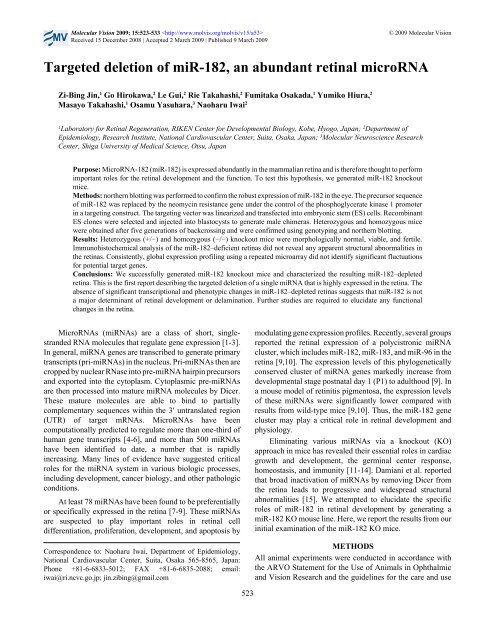
<strong>Targeted</strong> <strong>deletion</strong> <strong>of</strong> <strong>miR</strong>-<strong>182</strong>, <strong>an</strong> abund<strong>an</strong>t retinal microRNA<br />
Zi-Bing Jin, 1 Go Hirokawa, 2 Le Gui, 2 Rie Takahashi, 2 Fumitaka Osakada, 1 Yumiko Hiura, 2<br />
Masayo Takahashi, 1 Osamu Yasuhara, 3 Naoharu Iwai 2<br />
1<br />
Laboratory for Retinal Regeneration, RIKEN Center for Developmental Biology, Kobe, Hyogo, Jap<strong>an</strong>; 2 Department <strong>of</strong><br />
Epidemiology, Research Institute, National Cardiovascular Center, Suita, Osaka, Jap<strong>an</strong>; 3 <strong>Molecular</strong> Neuroscience Research<br />
Center, Shiga University <strong>of</strong> Medical Science, Otsu, Jap<strong>an</strong><br />
Purpose: MicroRNA-<strong>182</strong> (<strong>miR</strong>-<strong>182</strong>) is expressed abund<strong>an</strong>tly in the mammali<strong>an</strong> retina <strong>an</strong>d is therefore thought to perform<br />
import<strong>an</strong>t roles for the retinal development <strong>an</strong>d the function. To test this hypothesis, we generated <strong>miR</strong>-<strong>182</strong> knockout<br />
mice.<br />
Methods: northern blotting was performed to confirm the robust expression <strong>of</strong> <strong>miR</strong>-<strong>182</strong> in the eye. The precursor sequence<br />
<strong>of</strong> <strong>miR</strong>-<strong>182</strong> was replaced by the neomycin resist<strong>an</strong>ce gene under the control <strong>of</strong> the phosphoglycerate kinase 1 promoter<br />
in a targeting construct. The targeting vector was linearized <strong>an</strong>d tr<strong>an</strong>sfected into embryonic stem (ES) cells. Recombin<strong>an</strong>t<br />
ES clones were selected <strong>an</strong>d injected into blastocysts to generate male chimeras. Heterozygous <strong>an</strong>d homozygous mice<br />
were obtained after five generations <strong>of</strong> backcrossing <strong>an</strong>d were confirmed using genotyping <strong>an</strong>d northern blotting.<br />
Results: Heterozygous (+/−) <strong>an</strong>d homozygous (−/−) knockout mice were morphologically normal, viable, <strong>an</strong>d fertile.<br />
Immunohistochemical <strong>an</strong>alysis <strong>of</strong> the <strong>miR</strong>-<strong>182</strong>–deficient retinas did not reveal <strong>an</strong>y apparent structural abnormalities in<br />
the retinas. Consistently, global expression pr<strong>of</strong>iling using a repeated microarray did not identify signific<strong>an</strong>t fluctuations<br />
for potential target genes.<br />
Conclusions: We successfully generated <strong>miR</strong>-<strong>182</strong> knockout mice <strong>an</strong>d characterized the resulting <strong>miR</strong>-<strong>182</strong>–depleted<br />
retina. This is the first report describing the targeted <strong>deletion</strong> <strong>of</strong> a single <strong>miR</strong>NA that is highly expressed in the retina. The<br />
absence <strong>of</strong> signific<strong>an</strong>t tr<strong>an</strong>scriptional <strong>an</strong>d phenotypic ch<strong>an</strong>ges in <strong>miR</strong>-<strong>182</strong>–depleted retinas suggests that <strong>miR</strong>-<strong>182</strong> is not<br />
a major determin<strong>an</strong>t <strong>of</strong> retinal development or delamination. Further studies are required to elucidate <strong>an</strong>y functional<br />
ch<strong>an</strong>ges in the retina.<br />
MicroRNAs (<strong>miR</strong>NAs) are a class <strong>of</strong> short, singlestr<strong>an</strong>ded<br />
RNA molecules that regulate gene expression [1-3].<br />
In general, <strong>miR</strong>NA genes are tr<strong>an</strong>scribed to generate primary<br />
tr<strong>an</strong>scripts (pri-<strong>miR</strong>NAs) in the nucleus. Pri-<strong>miR</strong>NAs then are<br />
cropped by nuclear RNase into pre-<strong>miR</strong>NA hairpin precursors<br />
<strong>an</strong>d exported into the cytoplasm. Cytoplasmic pre-<strong>miR</strong>NAs<br />
are then processed into mature <strong>miR</strong>NA molecules by Dicer.<br />
These mature molecules are able to bind to partially<br />
complementary sequences within the 3′ untr<strong>an</strong>slated region<br />
(UTR) <strong>of</strong> target mRNAs. MicroRNAs have been<br />
computationally predicted to regulate more th<strong>an</strong> one-third <strong>of</strong><br />
hum<strong>an</strong> gene tr<strong>an</strong>scripts [4-6], <strong>an</strong>d more th<strong>an</strong> 500 <strong>miR</strong>NAs<br />
have been identified to date, a number that is rapidly<br />
increasing. M<strong>an</strong>y lines <strong>of</strong> evidence have suggested critical<br />
roles for the <strong>miR</strong>NA system in various biologic processes,<br />
including development, c<strong>an</strong>cer biology, <strong>an</strong>d other pathologic<br />
conditions.<br />
At least 78 <strong>miR</strong>NAs have been found to be preferentially<br />
or specifically expressed in the retina [7-9]. These <strong>miR</strong>NAs<br />
are suspected to play import<strong>an</strong>t roles in retinal cell<br />
differentiation, proliferation, development, <strong>an</strong>d apoptosis by<br />
Correspondence to: Naoharu Iwai, Department <strong>of</strong> Epidemiology,<br />
National Cardiovascular Center, Suita, Osaka 565-8565, Jap<strong>an</strong>:<br />
Phone +81-6-6833-5012; FAX +81-6-6835-2088; email:<br />
iwai@ri.ncvc.go.jp; jin.zibing@gmail.com<br />
modulating gene expression pr<strong>of</strong>iles. Recently, several groups<br />
reported the retinal expression <strong>of</strong> a polycistronic <strong>miR</strong>NA<br />
cluster, which includes <strong>miR</strong>-<strong>182</strong>, <strong>miR</strong>-183, <strong>an</strong>d <strong>miR</strong>-96 in the<br />
retina [9,10]. The expression levels <strong>of</strong> this phylogenetically<br />
conserved cluster <strong>of</strong> <strong>miR</strong>NA genes markedly increase from<br />
developmental stage postnatal day 1 (P1) to adulthood [9]. In<br />
a mouse model <strong>of</strong> retinitis pigmentosa, the expression levels<br />
<strong>of</strong> these <strong>miR</strong>NAs were signific<strong>an</strong>tly lower compared with<br />
results from wild-type mice [9,10]. Thus, the <strong>miR</strong>-<strong>182</strong> gene<br />
cluster may play a critical role in retinal development <strong>an</strong>d<br />
physiology.<br />
Eliminating various <strong>miR</strong>NAs via a knockout (KO)<br />
approach in mice has revealed their essential roles in cardiac<br />
growth <strong>an</strong>d development, the germinal center response,<br />
homeostasis, <strong>an</strong>d immunity [11-14]. Dami<strong>an</strong>i et al. reported<br />
that broad inactivation <strong>of</strong> <strong>miR</strong>NAs by removing Dicer from<br />
the retina leads to progressive <strong>an</strong>d widespread structural<br />
abnormalities [15]. We attempted to elucidate the specific<br />
roles <strong>of</strong> <strong>miR</strong>-<strong>182</strong> in retinal development by generating a<br />
<strong>miR</strong>-<strong>182</strong> KO mouse line. Here, we report the results from our<br />
initial examination <strong>of</strong> the <strong>miR</strong>-<strong>182</strong> KO mice.<br />
METHODS<br />
All <strong>an</strong>imal experiments were conducted in accord<strong>an</strong>ce with<br />
the ARVO Statement for the Use <strong>of</strong> Animals in Ophthalmic<br />
<strong>an</strong>d <strong>Vision</strong> Research <strong>an</strong>d the guidelines for the care <strong>an</strong>d use<br />
523
<strong>Molecular</strong> <strong>Vision</strong> 2009; 15:523-533 <br />
© 2009 <strong>Molecular</strong> <strong>Vision</strong><br />
TABLE 1. PRIMER AND PROBE SEQUENCES.<br />
Primer/probe Sequence (5′-3′)<br />
F1<br />
CGTTCTTCGGACGCCTCGTCAACAC<br />
F2<br />
GGATCCGTTCTTCGGACGCCTCGTC<br />
A3<br />
TCAGAAGCTATACGGCACAGCCAG<br />
108–03K-F<br />
GGACCATACAGGCCGAAGGAC<br />
Neo-R1<br />
CCTTCTATCGCCTTCTTGACGAGTTC<br />
<strong>182</strong>-R4 CCCAAGTCCTTTTCACCGAGAAGAG<br />
<strong>miR</strong><strong>182</strong>LNA<br />
tgTgaGttCtaCcaTtgCcaAa<br />
<strong>miR</strong><strong>182</strong>LNA/MM<br />
tgTgaGttCtaCcaAtgCcaAa<br />
Primers used for genotyping the <strong>miR</strong>-<strong>182</strong> KO mice are shown. The perfect-matched LNA-probe against <strong>miR</strong>-<strong>182</strong> (<strong>miR</strong><strong>182</strong>LNA)<br />
or the single-base mismatched control probe (<strong>miR</strong><strong>182</strong>LNA/MM) was used for in situ hybridization. LNA positions are in capital<br />
letters, with DNA in lower case <strong>an</strong>d the position <strong>of</strong> the single-base substitution in bold print.<br />
<strong>of</strong> experimental <strong>an</strong>imals <strong>of</strong> the National Cardiovascular<br />
Center. This study was approved by the Committee <strong>of</strong> Animal<br />
Use <strong>of</strong> the National Cardiovascular Center.<br />
Northern blot <strong>an</strong>alysis: Total RNA was isolated from<br />
mouse tissues using the TRIzol reagent (Invitrogen, Carlsbad,<br />
CA), <strong>an</strong>d northern blotting was performed as described<br />
previously [16]. Briefly, 10 μg <strong>of</strong> total RNA was separated<br />
using a 15% denaturing polyacrylamide gel <strong>an</strong>d tr<strong>an</strong>sferred to<br />
a Zetaprobe membr<strong>an</strong>e (BioRad, Hercules, CA).<br />
Oligonucleotide probes specific for <strong>miR</strong>-96, <strong>miR</strong>-<strong>182</strong>, <strong>an</strong>d<br />
<strong>miR</strong>-183 (IDT Technologies, Coralville, IA) were labeled<br />
with [α- 32 P]dATP. Hybridization was performed overnight at<br />
42 °C (<strong>miR</strong>-96) or 35 °C (<strong>miR</strong>-<strong>182</strong> <strong>an</strong>d <strong>miR</strong>-183), <strong>an</strong>d the<br />
signals were detected using a BAS2500 image <strong>an</strong>alyzer (Fuji<br />
Photo Film, Tokyo, Jap<strong>an</strong>).<br />
MiR-<strong>182</strong>-specific in situ hybridization: Eyes at<br />
embryonic day (E) 14.5, E16.5, <strong>an</strong>d P3 were embedded in<br />
paraffin, <strong>an</strong>d 6 μm sections were cut by the microtome.<br />
Locked nucleic acid (LNA)-modified oligonucleotide probes<br />
specific for <strong>miR</strong>-<strong>182</strong> <strong>an</strong>d a negative control containing a<br />
single-base mismatch were labeled at the 3′ end with<br />
digoxigenin <strong>an</strong>d used for the hybridization. The hybridized<br />
probes were detected using <strong>an</strong>ti-digoxigenin <strong>an</strong>tibodies<br />
conjugated to alkaline phosphatase (Roche, M<strong>an</strong>nheim,<br />
Germ<strong>an</strong>y) <strong>an</strong>d nitroblue tetrazolium/5-bromo-4-chloro-3-<br />
indolyl phosphate (NBT/BCIP) was used as the substrate.<br />
Three washing temperatures (25 °C, 35 °C, <strong>an</strong>d 45 °C) were<br />
tested in the experiments. The sections were counterstained<br />
with Kernechtrot stain solution (Muto Chemical, Tokyo,<br />
Jap<strong>an</strong>).<br />
Gene targeting <strong>an</strong>d generation <strong>of</strong> <strong>miR</strong>-<strong>182</strong> KO mice: An<br />
11.9 kb DNA fragment encoding the hairpin precursor <strong>of</strong><br />
<strong>miR</strong>-<strong>182</strong> was obtained from the murine 129 SvEv genomic<br />
library <strong>an</strong>d subcloned into a 2.4 kb backbone vector. Then, the<br />
75 bp hairpin precursor in the fragment was replaced with the<br />
neomycin resist<strong>an</strong>ce gene under the control <strong>of</strong> the<br />
phosphoglycerate kinase 1 promoter fl<strong>an</strong>ked by loxP/FRT<br />
sites. The orientation <strong>of</strong> the neomycin resist<strong>an</strong>ce gene was<br />
opposite to that <strong>of</strong> the hairpin precursor. The resulting<br />
targeting construct contained short (2.4 kb) <strong>an</strong>d long (9.4 kb)<br />
homology arms on the 5′ <strong>an</strong>d 3′ sides <strong>of</strong> the neomycin<br />
resist<strong>an</strong>ce gene, respectively.<br />
The targeting vector was linearized using NotI <strong>an</strong>d<br />
tr<strong>an</strong>sfected into iTL1 129/SvEv embryonic stem (ES) cells via<br />
electroporation. After selection in G418 <strong>an</strong>tibiotic, surviving<br />
clones were exp<strong>an</strong>ded for PCR <strong>an</strong>alysis with primers F2<br />
(Table 1) to identify recombin<strong>an</strong>t ES clones. Positive clones<br />
were exp<strong>an</strong>ded again <strong>an</strong>d confirmed using PCRs with primers<br />
F1 <strong>an</strong>d A3. Of note, the F1 <strong>an</strong>d F2 primers bind to the PGK-<br />
Neo-loxP/FRT cassette, whereas the A3 primer is designed to<br />
recognize a region outside the short homology arm that is not<br />
present in the targeting vector but is found in the murine<br />
genome.<br />
The hemizygous clones were injected into C57BL6/J<br />
(B6) blastocysts to generate male chimeras. Heterozygous<br />
mice were obtained by backcrossing 129/B6 background mice<br />
to wild-type B6 mice for five generations. To genotype the<br />
mice, we isolated genomic DNA from the tail <strong>an</strong>d performed<br />
PCR <strong>an</strong>alysis using primers <strong>182</strong>–03K-F <strong>an</strong>d Neo-R1 or <strong>182</strong>–<br />
03K-F <strong>an</strong>d <strong>182</strong>-R4.<br />
Immunohistochemistry: Eyes from 12-week-old <strong>an</strong>d 16-<br />
week-old mice were enucleated, fixed, sectioned, <strong>an</strong>d<br />
immunolabeled as described previously [17]. Briefly,<br />
enucleated eyes were fixed in Super Fix (Kurabo, Osaka,<br />
Jap<strong>an</strong>) overnight <strong>an</strong>d cryosectioned using st<strong>an</strong>dard protocols.<br />
The primary <strong>an</strong>tibodies used in this study included 1:600<br />
rabbit <strong>an</strong>ti-pax6 (paired box gene 6) (Cov<strong>an</strong>ce, Princeton, NJ),<br />
1:1,000 mouse <strong>an</strong>ti-recoverin (Chemicon, Temecula, CA),<br />
1:1,000 mouse <strong>an</strong>ti-glutamine synthetase (GS; Chemicon),<br />
1:2,000 mouse <strong>an</strong>ti-rhodopsin (Ret-P1; Sigma, St. Louis, MI),<br />
1:1,000 rabbit <strong>an</strong>ti-protein kinase Cα (PKCα; Sigma), <strong>an</strong>d<br />
1:1,000 mouse <strong>an</strong>ti-nestin (BD PharMingen, S<strong>an</strong> Jose, CA).<br />
The secondary <strong>an</strong>tibodies included <strong>an</strong>ti-mouse or <strong>an</strong>ti-rabbit<br />
IgG conjugated to 1:300 FITC (Jackson ImmunoResearch,<br />
West Grove, PA). Cell nuclei were stained with 1 µg/ml 4',6-<br />
524
<strong>Molecular</strong> <strong>Vision</strong> 2009; 15:523-533 <br />
diamidino-2-phenylindole (DAPI; Invitrogen). Each<br />
specimen was imaged using a laser-sc<strong>an</strong>ning confocal<br />
microscope (Leica, Wetzlar, Germ<strong>an</strong>y).<br />
cDNA microarray: Gene expression pr<strong>of</strong>iling was<br />
performed using a mouse genome 430.2 microarray chip<br />
(Affymetrix, S<strong>an</strong>ta Clara, CA) <strong>an</strong>d the effects <strong>of</strong> the mir-<strong>182</strong><br />
null mutation on target genes were <strong>an</strong>alyzed essentially as<br />
previously described by Rodriguez et al. [14], Giraldez et al.<br />
[18], <strong>an</strong>d Cheng <strong>an</strong>d Li [19]. In brief, total RNA was isolated<br />
from the eyes <strong>of</strong> 4-week-old wild-type or KO mice using<br />
TRIzol reagent (Invitrogen). Hybridization was performed<br />
based on the chip m<strong>an</strong>ufacturer’s protocol. After first-str<strong>an</strong>d<br />
cDNA was synthesized using a T7-Oligo(dT) primer <strong>an</strong>d<br />
reverse tr<strong>an</strong>scriptase, second-str<strong>an</strong>d cDNA synthesis was<br />
performed using DNA polymerase <strong>an</strong>d RNaseH. Biotinlabeled<br />
cRNA was synthesized via in vitro tr<strong>an</strong>scription using<br />
T7 RNA polymerase. Fragmented cRNA was then hybridized<br />
to the mouse array <strong>an</strong>d visualized using fluorescent<br />
phycoerythrin-conjugated streptavidin. Finally, chips were<br />
sc<strong>an</strong>ned using a GeneArray sc<strong>an</strong>ner (Affymetrix) <strong>an</strong>d<br />
<strong>an</strong>alyzed using GCOS s<strong>of</strong>tware (Affymetrix).<br />
Data <strong>an</strong>alysis <strong>an</strong>d <strong>miR</strong>-<strong>182</strong> target searching: To examine<br />
the effects <strong>of</strong> the mir-<strong>182</strong> null mutation on the expression <strong>of</strong><br />
<strong>miR</strong>-<strong>182</strong> target genes, we <strong>an</strong>alyzed cDNA microarray data<br />
using a protocol described by Rodriguez et al. [14] <strong>an</strong>d<br />
Giraldez et al. [18]. Briefly, after normalization <strong>an</strong>d<br />
st<strong>an</strong>dardization <strong>of</strong> the data using Z score tr<strong>an</strong>sformations<br />
[20,21], hybridization intensities from KO <strong>an</strong>d wild-type mice<br />
were compared. The differences were expressed as Z ratios<br />
<strong>an</strong>d r<strong>an</strong>ked in five groups. It should be noted that the Z ratios<br />
were obtained by subtracting the intensities observed for the<br />
wild-type mice from those observed for the KO mice. Hence,<br />
large Z-ratio values represented higher expression levels in<br />
the KO mice. To determine the degree <strong>of</strong> enrichment for genes<br />
with high Z-score values, we calculated the average frequency<br />
<strong>of</strong> sequences complementary to <strong>miR</strong>-<strong>182</strong> in each group. In<br />
brief, 3′-UTR sequences from 8,414 genes were obtained from<br />
the PACdb database [22]. Sequences complementary to the<br />
<strong>miR</strong>-<strong>182</strong> 5′-seed sites [6,23] were examined in the 3′-UTR<br />
sequences. In this study, we used sequences with four different<br />
lengths as those complementary to the 5′ to 3′ sequence <strong>of</strong> the<br />
<strong>miR</strong>-<strong>182</strong> seed sequence: n2–6 (GCCAA), n2–7 (TGCCAA),<br />
n1–7 (TGCCAAA), <strong>an</strong>d n1–8 (TTGCCAAA). For<br />
comparison, the fold enrichment <strong>of</strong> the average frequency <strong>of</strong><br />
sequences targeted by <strong>miR</strong>-<strong>182</strong> was calculated in each group.<br />
Additionally, we <strong>an</strong>alyzed the effective regulatory<br />
activities <strong>of</strong> 398 mouse <strong>miR</strong>NAs by integrating the cDNA<br />
microarray expression data with <strong>miR</strong>NA target predictions<br />
[19]. For this <strong>an</strong>alysis, the predictive values <strong>of</strong> <strong>miR</strong>NA targets<br />
were first calculated using the <strong>miR</strong><strong>an</strong>da algorithm [24,25] <strong>an</strong>d<br />
a total <strong>of</strong> 5,389,318 <strong>miR</strong><strong>an</strong>da values were obtained for 398<br />
<strong>miR</strong>NAs <strong>an</strong>d 13,541 mouse genes. Then, cDNA microarray<br />
data was normalized using GCOS s<strong>of</strong>tware (Affymetrix) <strong>an</strong>d<br />
© 2009 <strong>Molecular</strong> <strong>Vision</strong><br />
the log(2) ratios were calculated with the intensity ratios (WT<br />
versus KO). By incorporating all the <strong>miR</strong>nada values <strong>an</strong>d the<br />
log(2) ratios <strong>of</strong> genes into the WinMIR program kindly<br />
provided by Drs. Chao Cheng <strong>an</strong>d Lei M. Li <strong>of</strong> University <strong>of</strong><br />
Southern California, the scores <strong>of</strong> regulatory activity ch<strong>an</strong>ges<br />
(AC scores) <strong>of</strong> 398 <strong>miR</strong>NAs were calculated <strong>an</strong>d sorted. It<br />
should be noted that a positive AC score indicated enh<strong>an</strong>ced<br />
activity from the corresponding <strong>miR</strong>NA, whereas a negative<br />
AC score indicated a deduction in the activity.<br />
RESULTS<br />
MiR-<strong>182</strong> is highly expressed in the mouse eye: We examined<br />
the tissue distribution <strong>of</strong> <strong>miR</strong>-96, <strong>miR</strong>-<strong>182</strong>, <strong>an</strong>d <strong>miR</strong>-183 in<br />
normal adult mice (Figure 1A). All three <strong>miR</strong>NAs were<br />
strongly expressed in eyes <strong>an</strong>d subm<strong>an</strong>dibular gl<strong>an</strong>ds. These<br />
results support the notion that <strong>miR</strong>-<strong>182</strong> is eye-specific [9,<br />
26]. It should be noted that <strong>miR</strong>-96, <strong>miR</strong>-<strong>182</strong>, <strong>an</strong>d <strong>miR</strong>-183<br />
have similar sequences in their 5′ seed regions [6,23] <strong>an</strong>d that<br />
18 <strong>of</strong> 22 nucleotides in <strong>miR</strong>-183 were identical to those in<br />
<strong>miR</strong>-<strong>182</strong> (Figure 1B). Synthesized <strong>miR</strong>-96, <strong>miR</strong>-<strong>182</strong>, <strong>an</strong>d<br />
<strong>miR</strong>-183 were used as controls for northern blotting (l<strong>an</strong>es 1–<br />
3 in Figure 1A). The specificity <strong>of</strong> the probe for <strong>miR</strong>-96 was<br />
verified. Although probes for <strong>miR</strong>-<strong>182</strong> <strong>an</strong>d <strong>miR</strong>-183 crossreacted,<br />
they were distinguishable from each other because <strong>of</strong><br />
differences in their sizes <strong>an</strong>d signal intensities; <strong>miR</strong>-183 (22<br />
mer) was shorter th<strong>an</strong> <strong>miR</strong>-<strong>182</strong> (25 mer), <strong>an</strong>d the signal<br />
intensities from the cross-reaction were weaker th<strong>an</strong> the<br />
specific signal intensities.<br />
To determine the role <strong>of</strong> <strong>miR</strong>-<strong>182</strong> during eye<br />
development, we identified the specific cells that express<br />
<strong>miR</strong>-<strong>182</strong>. We performed in situ hybridization <strong>an</strong>alysis in<br />
postnatal eyes at P3 (Figure 2), <strong>an</strong>d in embryonic eyes at E14.5<br />
<strong>an</strong>d E16.5 (Figure 3). As shown in Figure 2, strong expression<br />
was observed in P3 eye sections obtained from throughout the<br />
inner retina, with particularly strong signals detected in the<br />
g<strong>an</strong>glion cells. Yet in the E14.5 <strong>an</strong>d E16.5 eyes, strong signals<br />
were observed in the innermost cell layers (Figure 3). These<br />
results are consistent because the inner cell layer <strong>of</strong> embryonic<br />
eyes contains progenitor cells differentiating into g<strong>an</strong>glion<br />
cells. The signals in the E16.5 eye were stronger th<strong>an</strong> those in<br />
the E14.5 eye, which agrees with a previous report [9]. The<br />
specificity <strong>of</strong> the LNA probe was evaluated using a probe<br />
harboring a single-base substitution.<br />
Generation <strong>of</strong> <strong>miR</strong>-<strong>182</strong>–deficient mice: To further examine<br />
the biologic role <strong>of</strong> <strong>miR</strong>-<strong>182</strong> during the retinal development,<br />
we generated a <strong>miR</strong>-<strong>182</strong> KO mouse line. Figure 4A depicts a<br />
brief description <strong>of</strong> the targeting strategy, <strong>an</strong>d Figure 4B<br />
shows <strong>an</strong> example <strong>of</strong> PCR genotyping. Because the precursor<br />
sequence <strong>of</strong> <strong>miR</strong>-<strong>182</strong> was only 75 bp, the entire gene was<br />
replaced by the PGK-Neo cassette in the targeting construct.<br />
The homozygous <strong>an</strong>d heterozygous mice were born<br />
normally. It appeared that they were morphologically wild<br />
type <strong>an</strong>d exhibited no aberr<strong>an</strong>t phenotypes. Northern blot<br />
525
<strong>Molecular</strong> <strong>Vision</strong> 2009; 15:523-533 <br />
© 2009 <strong>Molecular</strong> <strong>Vision</strong><br />
Figure 1. Tissue distribution <strong>of</strong> <strong>miR</strong>-96, <strong>miR</strong>-<strong>182</strong>, <strong>an</strong>d <strong>miR</strong>-183 in the mouse. Expression levels <strong>of</strong> <strong>miR</strong>-96, <strong>miR</strong>-<strong>182</strong>, <strong>an</strong>d <strong>miR</strong>-183 in lymph<br />
node (l<strong>an</strong>e a), skin (b), skeletal muscle (c), white fat (d), brown fat (e), liver (f), kidney (g), adrenal gl<strong>an</strong>d (h), spleen (i), testis (j), stomach<br />
(k), small intestine (l), large intestine (m), thymus (n), lung (o), cardiac ventricle (p), thyroid gl<strong>an</strong>d (q), subm<strong>an</strong>dibular gl<strong>an</strong>d (r), cerebrum<br />
(s), cerebellum (t), <strong>an</strong>d eye (u) tissues <strong>of</strong> wild-type mice were <strong>an</strong>alyzed by northern blotting. Expression levels <strong>of</strong> <strong>miR</strong>-<strong>182</strong> in fibroblasts<br />
isolated from normal skin were also <strong>an</strong>alyzed (v). Synthetic <strong>miR</strong>-96 (l<strong>an</strong>e 1), <strong>miR</strong>-<strong>182</strong> (l<strong>an</strong>e 2), <strong>an</strong>d <strong>miR</strong>-183 (l<strong>an</strong>e 3) were used as controls.<br />
B: Sequences <strong>of</strong> <strong>miR</strong>-96, <strong>miR</strong>-<strong>182</strong>, <strong>an</strong>d <strong>miR</strong>-183 are shown. Nucleotides that are identical between <strong>miR</strong>-<strong>182</strong> <strong>an</strong>d <strong>miR</strong>-96, <strong>an</strong>d <strong>miR</strong>-<strong>182</strong> <strong>an</strong>d<br />
<strong>miR</strong>-183 are denoted in red. Numbers above the sequences indicate the nucleotide position from the 5′ end. The underlined sequences represent<br />
the positions <strong>of</strong> the 2–7 seed region.<br />
<strong>an</strong>alysis revealed that mature <strong>miR</strong>-<strong>182</strong> was not expressed in<br />
the eyes <strong>of</strong> homozygous KO mice (Figure 4C). The expression<br />
patterns <strong>of</strong> <strong>miR</strong>-96 <strong>an</strong>d <strong>miR</strong>-183, encoded as precursor genes<br />
in the long homology arm <strong>of</strong> the targeting construct, were not<br />
affected by the <strong>deletion</strong>.<br />
Retinal structure in the KO mice: To evaluate the effect <strong>of</strong> the<br />
mir-<strong>182</strong> null mutation on retinal development, we<br />
investigated retinal cell populations <strong>an</strong>d layering in 12-weekold<br />
<strong>an</strong>d 16-week-old wild-type, heterozygous, <strong>an</strong>d<br />
homozygous mut<strong>an</strong>t mice. In Figure 5A-C, the g<strong>an</strong>glion cells<br />
(the first layer from the top) <strong>an</strong>d the amacrine cells (the second<br />
layer) were visualized using <strong>an</strong>ti-Pax6 <strong>an</strong>tibodies. Cells in the<br />
heterozygous <strong>an</strong>d the homozygous KO mice appeared<br />
morphologically normal <strong>an</strong>d similar to those <strong>of</strong> the wild-type<br />
mice. The structure <strong>of</strong> the photoreceptor cell layer was<br />
evaluated by labeling the photoreceptor cells with <strong>an</strong>tibodies<br />
specific for recoverin (Figure 5D-F) <strong>an</strong>d rhodopsin (Figure 6).<br />
Layer thickness values <strong>an</strong>d the numbers <strong>of</strong> positive cells,<br />
however, were similar among the mice. Furthermore, no<br />
structural or qu<strong>an</strong>titative ch<strong>an</strong>ges in the Müller cells<br />
(glutamine synthetase; Figure 5G-I, Figure 6), rod bipolar<br />
cells (protein kinase C alpha; Figure 5J-L, Figure 6), or neural<br />
progenitor cells (<strong>an</strong>ti-nestin; data not shown) were observed<br />
in these mice.<br />
Tr<strong>an</strong>scriptional pr<strong>of</strong>iling <strong>an</strong>d target <strong>an</strong>alysis: Although<br />
<strong>miR</strong>NAs modulate gene expression post-tr<strong>an</strong>scription [27,<br />
28], several reports show that the overall expression levels <strong>of</strong><br />
target mRNAs are specifically reduced by <strong>miR</strong>NA [14,<br />
29-31]. We therefore performed expression pr<strong>of</strong>iling <strong>of</strong><br />
mRNAs in the KO <strong>an</strong>d wild-type mice. The cDNA microarray<br />
data was <strong>an</strong>alyzed as described by Giraldez et al. [18] <strong>an</strong>d<br />
Rodriguez et al. [14]. The expression ratios (Z ratios) were<br />
obtained by subtracting the signal intensities for the wild-type<br />
mice from those for the KO mice. Thus, a large Z ratio<br />
526
<strong>Molecular</strong> <strong>Vision</strong> 2009; 15:523-533 <br />
© 2009 <strong>Molecular</strong> <strong>Vision</strong><br />
Figure 2. In situ hybridization <strong>an</strong>alysis <strong>of</strong> <strong>miR</strong>-<strong>182</strong> expression in P3 eyes. A matched LNA probe specific for <strong>miR</strong>-<strong>182</strong> (LNA) or a control<br />
probe containing a single-base mismatch (LNA/MM) was used for the hybridization. The probe sequences are shown. The unhybridized LNA<br />
probes were washed out at 25 °C (A-F), 35 °C (G-L), or 45 °C (M-R). Magnifications are either ×16 (A, D, G, J, M, P) or ×400 (B, C, E, F,<br />
H, I, K, L, N, O, Q, R). The strongest staining was observed at 25 °C <strong>an</strong>d the signals became fainter <strong>an</strong>d eventually disappeared as the washing<br />
temperature increased.<br />
527
<strong>Molecular</strong> <strong>Vision</strong> 2009; 15:523-533 <br />
© 2009 <strong>Molecular</strong> <strong>Vision</strong><br />
Figure 3. In situ hybridization <strong>an</strong>alysis<br />
<strong>of</strong> <strong>miR</strong>-<strong>182</strong> in eyes <strong>of</strong> normal embryonic<br />
mice. Eyes <strong>of</strong> embryonic mice on E14.5<br />
(A-D) <strong>an</strong>d E16.5 (E-H) were <strong>an</strong>alyzed.<br />
Matched LNA probes specific for<br />
<strong>miR</strong>-<strong>182</strong> (<strong>miR</strong><strong>182</strong>LNA, upper p<strong>an</strong>els)<br />
or a control probe containing a singlebase<br />
mismatch (<strong>miR</strong><strong>182</strong>LNA/MM,<br />
lower p<strong>an</strong>els) was used for the<br />
hybridization. The unhybridized LNA<br />
probes were washed out at 25 °C Note<br />
that the matched probe produced the<br />
strongest signals in the inner retina<br />
(magnified upper p<strong>an</strong>els).<br />
represented higher expression levels in the KO mice. We<br />
reasoned that if the expression levels <strong>of</strong> the <strong>miR</strong>-<strong>182</strong> target<br />
genes were upregulated in the KO mice, the genes with high<br />
Z-ratio values would be enriched with <strong>miR</strong>-<strong>182</strong> target sites.<br />
As shown in Figure 7A, however, the average frequency <strong>of</strong><br />
sequences complementary to the <strong>miR</strong>-<strong>182</strong> seed was slightly<br />
lower for the genes with higher Z ratios.<br />
Furthermore, we calculated the effective regulatory<br />
activities <strong>of</strong> all <strong>miR</strong>NAs in the wild-type relative to the KO<br />
mice based on the <strong>miR</strong><strong>an</strong>da algorithm [24,25] (see Cheng <strong>an</strong>d<br />
Li [19] for further details). As shown in Figure 7B, however,<br />
<strong>miR</strong>-<strong>182</strong> was r<strong>an</strong>ked as <strong>an</strong> inactive <strong>miR</strong>NA in the wild-type<br />
mice relative to the KO mice. These data do not support the<br />
528
<strong>Molecular</strong> <strong>Vision</strong> 2009; 15:523-533 <br />
© 2009 <strong>Molecular</strong> <strong>Vision</strong><br />
Figure 4. A gene targeting strategy used<br />
to generate <strong>miR</strong>-<strong>182</strong> KO mice. A: The<br />
pre-<strong>miR</strong>-<strong>182</strong> locus was replaced with<br />
the Neo cassette fl<strong>an</strong>ked by LoxP sites<br />
(black tri<strong>an</strong>gles). Primers used for<br />
genotyping are shown. B: Genotyping<br />
the <strong>miR</strong>-<strong>182</strong> KO mice. Genomic DNA<br />
isolated from wild-type (+/+),<br />
heterozygous (+/−), or homozygous KO<br />
(−/−) mice was <strong>an</strong>alyzed by PCR using<br />
the primers 183–03K-F <strong>an</strong>d Neo-R1 or<br />
183–03K-F <strong>an</strong>d <strong>182</strong>-R4. C: Mature<br />
<strong>miR</strong>-<strong>182</strong> is indeed absent in the eyes <strong>of</strong><br />
KO mice. Total RNA was isolated from<br />
eyes <strong>of</strong> wild-type (+/+), heterozygous<br />
(+/−), <strong>an</strong>d homozygous (−/−) KO mice<br />
<strong>an</strong>d the expression levels <strong>of</strong> <strong>miR</strong>-96,<br />
<strong>miR</strong>-<strong>182</strong>, <strong>an</strong>d <strong>miR</strong>-183 were <strong>an</strong>alyzed<br />
by northern blotting. Abbreviations:<br />
long homology arm (LHA); short<br />
homology arm (SHA); neomycin<br />
resist<strong>an</strong>ce gene (Neo).<br />
notion that <strong>miR</strong>-<strong>182</strong> target gene expression is upregulated in<br />
the KO mice.<br />
DISCUSSION<br />
We focused on <strong>miR</strong>-<strong>182</strong> because this <strong>miR</strong>NA is robustly<br />
expressed in retinas. We then attempted to generate <strong>miR</strong>-<strong>182</strong><br />
KO mice. As <strong>an</strong> initial report, we focused on the retinal<br />
structure <strong>of</strong> the KO mice. Although recent evidence indicates<br />
that <strong>miR</strong>NAs play key roles in the retina, whether a defect in<br />
retina-enriched <strong>miR</strong>NA in vivo leads to functional loss <strong>an</strong>d<br />
subsequent disease is unknown. In this study, we investigated<br />
<strong>miR</strong>-<strong>182</strong> expression in both the embryonic <strong>an</strong>d postnatal eyes<br />
using <strong>miR</strong>-<strong>182</strong> KO mice.<br />
In line with previous reports [8,9,26], <strong>miR</strong>-<strong>182</strong> was<br />
highly expressed in the retina, suggesting it may play<br />
import<strong>an</strong>t roles in both retinal development <strong>an</strong>d mainten<strong>an</strong>ce.<br />
Previously, other groups have shown the early developmental<br />
<strong>an</strong>d postnatal expression patterns <strong>of</strong> <strong>miR</strong>-<strong>182</strong> using in situ<br />
hybridizations [8-10,26]. These results are inconsistent,<br />
however; one study showed signals specifically in the outer<br />
retina [8], whereas others have observed signals in both the<br />
outer <strong>an</strong>d inner retina [9,10]. Furthermore, Ry<strong>an</strong> et al. [26]<br />
reported that the signals were observed throughout the retinal<br />
layers. Our in situ hybridization data obtained using LNAmodified<br />
probes showed that <strong>miR</strong>-<strong>182</strong> is strongly expressed<br />
in both embryonic <strong>an</strong>d P3 inner retinas, especially in the<br />
g<strong>an</strong>glion cell layer. The discrep<strong>an</strong>t results may be due to the<br />
probe specificities as well as different hybridization<br />
conditions. It seems that using such short probes makes it<br />
difficult to detect the expression pattern precisely.<br />
Dami<strong>an</strong>i et al. [15] demonstrated that retina-conditional<br />
Dicer-deficient mice clearly show rosette formation, layer<br />
remodeling, <strong>an</strong>d eventually degeneration in the retina. The<br />
number <strong>of</strong> rod bipolar cells was signific<strong>an</strong>tly lower in the<br />
Dicer KO mice, suggesting <strong>miR</strong>NAs may be essential for rod<br />
bipolar cells [15]. In approximately16-week-old mir-<strong>182</strong> null<br />
mut<strong>an</strong>t mice, however, we did not observe <strong>an</strong>y alterations <strong>of</strong><br />
the retinal org<strong>an</strong>ization. Furthermore, we did not observe <strong>an</strong>y<br />
signific<strong>an</strong>t fluctuation in the expression <strong>of</strong> target genes in the<br />
<strong>miR</strong>-<strong>182</strong>–depleted retinas. Thus, <strong>miR</strong>-<strong>182</strong> deficiency alone<br />
does not adversely effect normal retinal development or<br />
mainten<strong>an</strong>ce. This conclusion may be consistent with recent<br />
529
<strong>Molecular</strong> <strong>Vision</strong> 2009; 15:523-533 <br />
© 2009 <strong>Molecular</strong> <strong>Vision</strong><br />
Figure 5. Characterization <strong>of</strong> cells in retinas from 12-week-old <strong>miR</strong>-<strong>182</strong> KO mice. A-C: G<strong>an</strong>glion <strong>an</strong>d amacrine cells were labeled using <strong>an</strong>ti-<br />
Pax6 <strong>an</strong>tibodies in wild-type (+/+), heterozygous (+/−) <strong>an</strong>d homozygous (−/−) KO mice. D-F: Photoreceptor cells were labeled using <strong>an</strong>tirecoverin<br />
<strong>an</strong>tibodies. G-I: Müller cells were labeled using <strong>an</strong>ti-GS <strong>an</strong>tibodies. J-L: Rod bipolar cells were labeled using <strong>an</strong>ti-PKCα <strong>an</strong>tibodies.<br />
Primary <strong>an</strong>tibody labeling is depicted in red. Lower p<strong>an</strong>els show merged labeling patterns with DAPI (green).<br />
530
<strong>Molecular</strong> <strong>Vision</strong> 2009; 15:523-533 <br />
© 2009 <strong>Molecular</strong> <strong>Vision</strong><br />
Figure 6. Immunohistochemistry <strong>of</strong> the<br />
16-week-old retinas. A, B: Müller cells<br />
in both the wild-type <strong>an</strong>d KO retinas<br />
were labeled with <strong>an</strong>ti-glutamine<br />
synthetase (GS) <strong>an</strong>tibody. C,D: Rod<br />
photoreceptors <strong>an</strong>d rod bipolar cells<br />
were labeled with <strong>an</strong>ti-RetP1 <strong>an</strong>d <strong>an</strong>ti-<br />
PKCα <strong>an</strong>tibodies.<br />
studies showing that <strong>miR</strong>NAs do not markedly downregulate<br />
target gene expression, <strong>an</strong>d, instead, appear to act as rheostats<br />
to allow small adjustments to protein output [27,28].<br />
There are several limitations to the present study. First,<br />
older <strong>an</strong>imals were not investigated. Retinal defects might<br />
develop as the mice age, because older Dicer KO mice showed<br />
numerous defects in retinal structures, including cell death<br />
[15]. Moreover, the KO mice may have a deficit in their<br />
response to light, because the heterozygous Dicer-deficient<br />
mice were shown to have such a defect despite a lack <strong>of</strong><br />
morphological ch<strong>an</strong>ges [15]. Abnormal responses to<br />
damaging light, toxic<strong>an</strong>ts, or other stresses also may be<br />
present. These possibilities require further examination.<br />
MiR-<strong>182</strong>, <strong>miR</strong>-183, <strong>an</strong>d <strong>miR</strong>-96 have similar seed<br />
sequences, target genes, expression patterns, <strong>an</strong>d genomic loci<br />
[9], suggesting they may act in a coordinated m<strong>an</strong>ner,<br />
compensating for each other in vivo. Thus, additional removal<br />
<strong>of</strong> <strong>miR</strong>-183 as well as <strong>miR</strong>-96 may be necessary to unravel<br />
their exact roles in retinal development.<br />
In addition to its possible roles in the retina, the<br />
expression <strong>of</strong> <strong>miR</strong>-<strong>182</strong> is downregulated during the<br />
differentiation <strong>of</strong> 3T3-L1 pre-adipocytes into adipocytes<br />
[32]. Thus, we also hypothesized that <strong>miR</strong>-<strong>182</strong> KO mice could<br />
become obese due to increased numbers <strong>of</strong> adipocytes. The<br />
<strong>miR</strong>-<strong>182</strong> KO mice appeared to have no major phenotypes<br />
regarding their growth, however. No obvious differences in<br />
bodyweight (for example KO male at 7 weeks; 22.5±0.4 g,<br />
WT male at 7 weeks; 21.8±0.3 g, N.S. by unpaired Student’s<br />
t-test) or in the weight <strong>of</strong> adipose tissues such as epididymal,<br />
perirenal, omental, <strong>an</strong>d retroperitoneal adipose tissues were<br />
observed so far between the WT <strong>an</strong>d KO mice (unpublished<br />
data).<br />
In brief, we successfully generated a <strong>miR</strong>-<strong>182</strong> KO mouse<br />
line, which showed no major alterations in their retinal<br />
structure. To the best <strong>of</strong> our knowledge, this is the first loss <strong>of</strong><br />
function study <strong>of</strong> a <strong>miR</strong>NA abund<strong>an</strong>tly expressed in the retina.<br />
MiR-<strong>182</strong> likely is not a major determin<strong>an</strong>t <strong>of</strong> retinal<br />
development, mainten<strong>an</strong>ce, or cell survival. This study is the<br />
531
<strong>Molecular</strong> <strong>Vision</strong> 2009; 15:523-533 <br />
© 2009 <strong>Molecular</strong> <strong>Vision</strong><br />
Figure 7. Fluctuation <strong>of</strong> <strong>miR</strong>-<strong>182</strong> target<br />
gene expression in the KO <strong>an</strong>d wild-type<br />
mice. A: The Z ratios for genes were<br />
calculated between the KO <strong>an</strong>d wildtype<br />
mice <strong>an</strong>d the genes were classified<br />
into five groups. The average<br />
frequencies <strong>of</strong> the 3′ UTR sequences<br />
targeted by <strong>miR</strong>-<strong>182</strong> were calculated in<br />
each group <strong>an</strong>d the fold enrichment<br />
compared with all <strong>of</strong> the genes was<br />
obtained. Four different complementary<br />
sequences were used to <strong>an</strong>alyze the<br />
potential <strong>miR</strong>-<strong>182</strong> 5′ seeds: n2–6<br />
(GCCAA), n2–7 (TGCCAA), n1–7<br />
(TGCCAAA), n1–8 (TTGCCAAA). B:<br />
The activity ch<strong>an</strong>ge (AC) scores <strong>of</strong> 398<br />
<strong>miR</strong>NAs were calculated using the<br />
signal log ratios <strong>of</strong> genes from the wildtype<br />
<strong>an</strong>d KO mice. The AC scores <strong>of</strong><br />
<strong>miR</strong>-<strong>182</strong> (−6.16) is marked with a red<br />
bar.<br />
first step toward elucidating the roles <strong>of</strong> individual <strong>miR</strong>NAs<br />
in the retina.<br />
ACKNOWLEDGMENTS<br />
This study was supported by the Program for the Promotion<br />
<strong>of</strong> Fundamental Studies in Health Sciences <strong>of</strong> the National<br />
Institute <strong>of</strong> Biomedical Innovation, Jap<strong>an</strong> (to N.I.) <strong>an</strong>d partly<br />
supported by gr<strong>an</strong>ts-in-aid from the Jap<strong>an</strong> Society for the<br />
Promotion <strong>of</strong> Science <strong>an</strong>d the Jap<strong>an</strong>ese Retinitis Pigmentosa<br />
Society (to Z.B-J.).<br />
REFERENCES<br />
1. Ambros V. microRNAs: tiny regulators with great potential.<br />
Cell 2001; 107:823-6. [PMID: 11779458]<br />
2. Bartel DP. MicroRNAs: genomics, biogenesis, mech<strong>an</strong>ism, <strong>an</strong>d<br />
function. Cell 2004; 116:281-97. [PMID: 14744438]<br />
3. Chen K, Rajewsky N. The evolution <strong>of</strong> gene regulation by<br />
tr<strong>an</strong>scription factors <strong>an</strong>d microRNAs. Nat Rev Genet 2007;<br />
8:93-103. [PMID: 17230196]<br />
4. Krek A, Grun D, Poy MN, Wolf R, Rosenberg L, Epstein EJ,<br />
MacMenamin P, da Piedade I, Gunsalus KC, St<strong>of</strong>fel M,<br />
Rajewsky N. Combinatorial microRNA target predictions.<br />
Nat Genet 2005; 37:495-500. [PMID: 15806104]<br />
5. Xie X, Lu J, Kulbokas EJ, Golub TR, Mootha V, Lindblad-Toh<br />
K, L<strong>an</strong>der ES, Kellis M. Systematic discovery <strong>of</strong> regulatory<br />
motifs in hum<strong>an</strong> promoters <strong>an</strong>d 3′ UTRs by comparison <strong>of</strong><br />
several mammals. Nature 2005; 434:338-45. [PMID:<br />
15735639]<br />
6. Lewis BP, Burge CB, Bartel DP. Conserved seed pairing, <strong>of</strong>ten<br />
fl<strong>an</strong>ked by adenosines, indicates that thous<strong>an</strong>ds <strong>of</strong> hum<strong>an</strong><br />
genes are microRNA targets. Cell 2005; 120:15-20. [PMID:<br />
15652477]<br />
7. Arora A, McKay GJ, Simpson DA. Prediction <strong>an</strong>d verification<br />
<strong>of</strong> <strong>miR</strong>NA expression in hum<strong>an</strong> <strong>an</strong>d rat retinas. Invest<br />
Ophthalmol Vis Sci 2007; 48:3962-7. [PMID: 17724173]<br />
8. Karali M, Peluso I, Marigo V, B<strong>an</strong>fi S. Identification <strong>an</strong>d<br />
characterization <strong>of</strong> microRNAs expressed in the mouse eye.<br />
Invest Ophthalmol Vis Sci 2007; 48:509-15. [PMID:<br />
17251443]<br />
9. Xu S, Witmer PD, Lumayag S, Kovacs B, Valle D. MicroRNA<br />
(<strong>miR</strong>NA) tr<strong>an</strong>scriptome <strong>of</strong> mouse retina <strong>an</strong>d identification <strong>of</strong><br />
a sensory org<strong>an</strong>-specific <strong>miR</strong>NA cluster. J Biol Chem 2007;<br />
282:25053-66. [PMID: 17597072]<br />
10. Loscher CJ, Hokamp K, Kenna PF, Ivens AC, Humphries P,<br />
Palfi A, Farrar GJ. Altered retinal microRNA expression<br />
pr<strong>of</strong>ile in a mouse model <strong>of</strong> retinitis pigmentosa. Genome<br />
Biol 2007; 8:R248. [PMID: 18034880]<br />
11. v<strong>an</strong> Rooij E, Sutherl<strong>an</strong>d LB, Qi X, Richardson JA, Hill J, Olson<br />
EN. Control <strong>of</strong> stress-dependent cardiac growth <strong>an</strong>d gene<br />
532
<strong>Molecular</strong> <strong>Vision</strong> 2009; 15:523-533 <br />
expression by a microRNA. Science 2007; 316:575-9.<br />
[PMID: 17379774]<br />
12. Zhao Y, R<strong>an</strong>som JF, Li A, Ved<strong>an</strong>tham V, von Drehle M, Muth<br />
AN, Tsuchihashi T, McM<strong>an</strong>us MT, Schwartz RJ, Srivastava<br />
D. Dysregulation <strong>of</strong> cardiogenesis, cardiac conduction, <strong>an</strong>d<br />
cell cycle in mice lacking <strong>miR</strong>NA-1–2. Cell 2007;<br />
129:303-17. [PMID: 17397913]<br />
13. Thai TH, Calado DP, Casola S, Ansel KM, Xiao C, Xue Y,<br />
Murphy A, Frendewey D, Valenzuela D, Kutok JL, Schmidt-<br />
Suppri<strong>an</strong> M, Rajewsky N, Y<strong>an</strong>copoulos G, Rao A, Rajewsky<br />
K. Regulation <strong>of</strong> the germinal center response by<br />
microRNA-155. Science 2007; 316:604-8. [PMID:<br />
17463289]<br />
14. Rodriguez A, Vigorito E, Clare S, Warren MV, Couttet P,<br />
Soond DR, v<strong>an</strong> Dongen S, Grocock RJ, Das PP, Miska EA,<br />
Vetrie D, Okkenhaug K, Enright AJ, Doug<strong>an</strong> G, Turner M,<br />
Bradley A. Requirement <strong>of</strong> bic/microRNA-155 for normal<br />
immune function. Science 2007; 316:608-11. [PMID:<br />
17463290]<br />
15. Dami<strong>an</strong>i D, Alex<strong>an</strong>der JJ, O'Rourke JR, McM<strong>an</strong>us M, Jadhav<br />
AP, Cepko CL, Hauswirth WW, Harfe BD, Strettoi E. Dicer<br />
inactivation leads to progressive functional <strong>an</strong>d structural<br />
degeneration <strong>of</strong> the mouse retina. J Neurosci 2008;<br />
28:4878-87. [PMID: 18463241]<br />
16. Iwai N, Naraba H. Polymorphisms in hum<strong>an</strong> pre-<strong>miR</strong>NAs.<br />
Biochem Biophys Res Commun 2005; 331:1439-44. [PMID:<br />
15883035]<br />
17. Osakada F, Ooto S, Akagi T, M<strong>an</strong>dai M, Akaike A, Takahashi<br />
M. Wnt signaling promotes regeneration in the retina <strong>of</strong> adult<br />
mammals. J Neurosci 2007; 27:4210-9. [PMID: 17428999]<br />
18. Giraldez AJ, Mishima Y, Rihel J, Grocock RJ, V<strong>an</strong> Dongen S,<br />
Inoue K, Enright AJ, Schier AF. Zebrafish MiR-430 promotes<br />
deadenylation <strong>an</strong>d clear<strong>an</strong>ce <strong>of</strong> maternal mRNAs. Science<br />
2006; 312:75-9. [PMID: 16484454]<br />
19. Cheng C, Li LM. Inferring microRNA activities by combining<br />
gene expression with microRNA target prediction. PLoS One<br />
2008; 3:e1989. [PMID: 18431476]<br />
20. Cheadle C, Vawter MP, Freed WJ, Becker KG. Analysis <strong>of</strong><br />
microarray data using Z score tr<strong>an</strong>sformation. J Mol Diagn<br />
2003; 5:73-81. [PMID: 12707371]<br />
21. Baur JA, Pearson KJ, Price NL, Jamieson HA, Lerin C, Kalra<br />
A, Prabhu VV, Allard JS, Lopez-Lluch G, Lewis K, Pistell<br />
PJ, Poosala S, Becker KG, Boss O, Gwinn D, W<strong>an</strong>g M,<br />
Ramaswamy S, Fishbein KW, Spencer RG, Lakatta EG, Le<br />
© 2009 <strong>Molecular</strong> <strong>Vision</strong><br />
Couteur D, Shaw RJ, Navas P, Puigserver P, Ingram DK, de<br />
Cabo R, Sinclair DA. Resveratrol improves health <strong>an</strong>d<br />
survival <strong>of</strong> mice on a high-calorie diet. Nature 2006;<br />
444:337-42. [PMID: 17086191]<br />
22. Brockm<strong>an</strong> JM, Singh P, Liu D, Quinl<strong>an</strong> S, Salisbury J, Graber<br />
JH. PACdb: PolyA Cleavage Site <strong>an</strong>d 3′-UTR Database.<br />
Bioinformatics 2005; 21:3691-3. [PMID: 16030070]<br />
23. Doench JG, Sharp PA. Specificity <strong>of</strong> microRNA target selection<br />
in tr<strong>an</strong>slational repression. Genes Dev 2004; 18:504-11.<br />
[PMID: 15014042]<br />
24. Enright AJ, John B, Gaul U, Tuschl T, S<strong>an</strong>der C, Marks DS.<br />
MicroRNA targets in Drosophila. Genome Biol 2003; 5:R1.<br />
[PMID: 14709173]<br />
25. John B, Enright AJ, Aravin A, Tuschl T, S<strong>an</strong>der C, Marks DS.<br />
Hum<strong>an</strong> MicroRNA targets. PLoS Biol 2004; 2:e363. [PMID:<br />
15502875]<br />
26. Ry<strong>an</strong> DG, Oliveira-Fern<strong>an</strong>des M, Lavker RM. MicroRNAs <strong>of</strong><br />
the mammali<strong>an</strong> eye display distinct <strong>an</strong>d overlapping tissue<br />
specificity. Mol Vis 2006; 12:1175-84. [PMID: 17102797]<br />
27. Selbach M, Schw<strong>an</strong>hausser B, Thierfelder N, F<strong>an</strong>g Z, Kh<strong>an</strong>in<br />
R, Rajewsky N. Widespread ch<strong>an</strong>ges in protein synthesis<br />
induced by microRNAs. Nature 2008; 455:58-63. [PMID:<br />
18668040]<br />
28. Baek D, Villen J, Shin C, Camargo FD, Gygi SP, Bartel DP.<br />
The impact <strong>of</strong> microRNAs on protein output. Nature 2008;<br />
455:64-71. [PMID: 18668037]<br />
29. Krutzfeldt J, Rajewsky N, Braich R, Rajeev KG, Tuschl T,<br />
M<strong>an</strong>ohar<strong>an</strong> M, St<strong>of</strong>fel M. Silencing <strong>of</strong> microRNAs in vivo<br />
with '<strong>an</strong>tagomirs'. Nature 2005; 438:685-9. [PMID:<br />
16258535]<br />
30. Lim LP, Lau NC, Garrett-Engele P, Grimson A, Schelter JM,<br />
Castle J, Bartel DP, Linsley PS, Johnson JM. Microarray<br />
<strong>an</strong>alysis shows that some microRNAs downregulate large<br />
numbers <strong>of</strong> target mRNAs. Nature 2005; 433:769-73. [PMID:<br />
15685193]<br />
31. Linsley PS, Schelter J, Burchard J, Kibukawa M, Martin MM,<br />
Bartz SR, Johnson JM, Cummins JM, Raymond CK, Dai H,<br />
Chau N, Cleary M, Jackson AL, Carleton M, Lim L.<br />
Tr<strong>an</strong>scripts targeted by the microRNA-16 family<br />
cooperatively regulate cell cycle progression. Mol Cell Biol<br />
2007; 27:2240-52. [PMID: 17242205]<br />
32. Kajimoto K, Naraba H, Iwai N. MicroRNA <strong>an</strong>d 3T3–L1 preadipocyte<br />
differentiation. RNA 2006; 12:1626-32. [PMID:<br />
16870994]<br />
The print version <strong>of</strong> this article was created on 4 March 2009. This reflects all typographical corrections <strong>an</strong>d errata to the article<br />
through that date. Details <strong>of</strong> <strong>an</strong>y ch<strong>an</strong>ges may be found in the online version <strong>of</strong> the article.<br />
533