

ORA waveform in normal and keratoconic eyes Fine-needle ...
ORA waveform in normal and keratoconic eyes Fine-needle ...
ORA waveform in normal and keratoconic eyes Fine-needle ...

Create successful ePaper yourself
Turn your PDF publications into a flip-book with our unique Google optimized e-Paper software.
Augsburger JJ, et al.<br />
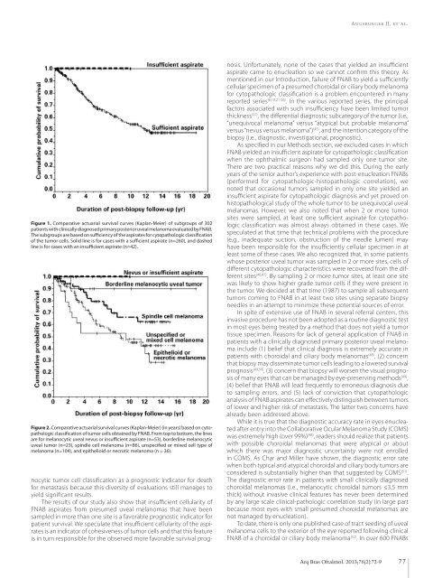
Figure 1. Comparative actuarial survival curves (Kaplan-Meier) of subgroups of 302<br />
patients with cl<strong>in</strong>ically diagnosed primary posterior uveal melanoma evaluated by FNAB.<br />
The subgroups are based on sufficiency of the aspirates for cytopathologic classification<br />
of the tumor cells. Solid l<strong>in</strong>e is for cases with a sufficient aspirate (n=260), <strong>and</strong> dashed<br />
l<strong>in</strong>e is for cases with an <strong>in</strong>sufficient aspirate (n=42).<br />
Figure 2. Comparative actuarial survival curves (Kaplan-Meier) (<strong>in</strong> years) based on cytopathologic<br />
classification of tumor cells obta<strong>in</strong>ed by FNAB. From top to bottom, the l<strong>in</strong>es<br />
are for melanocytic uveal nevus or <strong>in</strong>sufficient aspirate (n=53), borderl<strong>in</strong>e melanocytic<br />
uveal tumor (n=23), sp<strong>in</strong>dle cell melanoma (n=86), unspecified or mixed cell type of<br />
melanoma (n=104), <strong>and</strong> epithelioid or necrotic melanoma (n = 36).<br />
nocytic tumor cell classification as a prognostic <strong>in</strong>dicator for death<br />
for metastasis because this diversity of evaluations still manages to<br />
yield significant results.<br />
The results of our study also show that <strong>in</strong>sufficient cellularity of<br />
FNAB aspirates from presumed uveal melanomas that have been<br />
sampled <strong>in</strong> more than one site is a favorable prognostic <strong>in</strong>dicator for<br />
patient survival. We speculate that <strong>in</strong>sufficient cellularity of the aspirates<br />
is an <strong>in</strong>dicator of cohesiveness of tumor cells <strong>and</strong> that this feature<br />
is <strong>in</strong> turn responsible for the observed more favorable survival prognosis.<br />
Unfortunately, none of the cases that yielded an <strong>in</strong>sufficient<br />
aspirate came to enucleation so we cannot confirm this theory. As<br />
mentioned <strong>in</strong> our Introduction, failure of FNAB to yield a sufficiently<br />
cellular specimen of a presumed choroidal or ciliary body melanoma<br />
for cytopathologic classification is a problem encountered <strong>in</strong> many<br />
reported series (8,10,21-26) . In the various reported series, the pr<strong>in</strong>cipal<br />
factors associated with such <strong>in</strong>sufficiency have been limited tumor<br />
thickness (22) , the differential diagnostic subcategory of the tumor (i.e.,<br />
“unequivocal melanoma” versus “atypical but probable melanoma”<br />
versus “nevus versus melanoma”) (45) , <strong>and</strong> the <strong>in</strong>tention category of the<br />
biopsy (i.e., diagnostic, <strong>in</strong>vestigational, prognostic).<br />
As specified <strong>in</strong> our Methods section, we excluded cases <strong>in</strong> which<br />
FNAB yielded an <strong>in</strong>sufficient aspirate for cytopathologic classification<br />
when the ophthalmic surgeon had sampled only one tumor site.<br />
There are two practical reasons why we did this. Dur<strong>in</strong>g the early<br />
years of the senior author’s experience with post-enucleation FNABs<br />
(performed for cytopathologic-histopathologic correlation), we<br />
noted that occasional tumors sampled <strong>in</strong> only one site yielded an<br />
<strong>in</strong>sufficient aspirate for cytopathologic diagnosis <strong>and</strong> yet proved on<br />
histopathological study of the whole tumor to be unequivocal uveal<br />
melanomas. However, we also noted that when 2 or more tumor<br />
sites were sampled, at least one sufficient aspirate for cytopathologic<br />
classification was almost always obta<strong>in</strong>ed <strong>in</strong> these cases. We<br />
speculated at that time that technical problems with the procedure<br />
(e.g., <strong>in</strong>adequate suction, obstruction of the <strong>needle</strong> lumen) may<br />
have been responsible for the <strong>in</strong>sufficiently cellular specimen <strong>in</strong> at<br />
least some of these cases. We also recognized that, <strong>in</strong> some patients<br />
whose posterior uveal tumor was sampled <strong>in</strong> 2 or more sites, cells of<br />
different cytopathologic characteristics were recovered from the different<br />
sites (46,47) . By sampl<strong>in</strong>g 2 or more tumor sites, at least one site<br />
was likely to show higher grade tumor cells if they were present <strong>in</strong><br />
the tumor. We decided at that time (1987) to sample all subsequent<br />
tumors com<strong>in</strong>g to FNAB <strong>in</strong> at least two sites us<strong>in</strong>g separate biopsy<br />
<strong>needle</strong>s <strong>in</strong> an attempt to m<strong>in</strong>imize these potential sources of error.<br />
In spite of extensive use of FNAB <strong>in</strong> several referral centers, this<br />
<strong>in</strong> vasive procedure has not been adopted as a rout<strong>in</strong>e diagnostic test<br />
<strong>in</strong> most <strong>eyes</strong> be<strong>in</strong>g treated by a method that does not yield a tumor<br />
tissue specimen. Reasons for lack of general application of FNAB <strong>in</strong><br />
patients with a cl<strong>in</strong>ically diagnosed primary posterior uveal melanoma<br />
<strong>in</strong>clude (1) belief that cl<strong>in</strong>ical diagnosis is extremely accurate <strong>in</strong><br />
patients with choroidal <strong>and</strong> ciliary body melanomas (48) , (2) concern<br />
that biopsy may dissem<strong>in</strong>ate tumor cells lead<strong>in</strong>g to a lowered survival<br />
prognosis (49,50) , (3) concern that biopsy will worsen the visual prognosis<br />
of many <strong>eyes</strong> that can be managed by eye-preserv<strong>in</strong>g methods (49) ,<br />
(4) belief that FNAB will lead frequently to erroneous diagnosis due<br />
to sampl<strong>in</strong>g errors, <strong>and</strong> (5) lack of conviction that cytopathologic<br />
ana lysis of FNAB aspirates can effectively dist<strong>in</strong>guish between tumors<br />
of lower <strong>and</strong> higher risk of metastasis. The latter two concerns have<br />
already been addressed above.<br />
While it is true that the diagnostic accuracy rate <strong>in</strong> <strong>eyes</strong> enucleated<br />
after entry <strong>in</strong>to the Collaborative Ocular Melanoma Study (COMS)<br />
was extremely high (over 99%) (48) , readers should realize that pa tients<br />
with possible choroidal melanomas that were atypical or about<br />
which there was major diagnostic uncerta<strong>in</strong>ty were not enrolled<br />
<strong>in</strong> COMS. As Char <strong>and</strong> Miller have shown, the diagnostic error rate<br />
when both typical <strong>and</strong> atypical choroidal <strong>and</strong> ciliary body tumors are<br />
considered is substantially higher than that suggested by COMS (51) .<br />
The diagnostic error rate <strong>in</strong> patients with small cl<strong>in</strong>ically diagnosed<br />
choroidal melanomas (i.e., melanocytic choroidal tumors ≤3.5 mm<br />
thick) without <strong>in</strong>vasive cl<strong>in</strong>ical features has never been determ<strong>in</strong>ed<br />
by any large scale cl<strong>in</strong>ical-pathologic correlation study (<strong>in</strong> large part<br />
because most <strong>eyes</strong> with small presumed choroidal melanomas are<br />
not managed by enucleation).<br />
To date, there is only one published case of tract seed<strong>in</strong>g of uveal<br />
melanoma cells to the exterior of the eye reported follow<strong>in</strong>g cl<strong>in</strong>ical<br />
FNAB of a choroidal or ciliary body melanoma (52) . In over 600 FNABs<br />
Arq Bras Oftalmol. 2013;76(2):72-9<br />
77


