

article in PDF - Our Dermatology Online Journal
article in PDF - Our Dermatology Online Journal
article in PDF - Our Dermatology Online Journal

You also want an ePaper? Increase the reach of your titles
YUMPU automatically turns print PDFs into web optimized ePapers that Google loves.
Case Reports<br />
DOI: 10.7241/ourd.20124.72<br />
SOLITARY POROKERATOSIS OF MIBELLI AT AN<br />
UNUSUAL SITE<br />
Nithya Raghunath, Metikurke Vijayashankar<br />
Source of Support:<br />
Nil<br />
Compet<strong>in</strong>g Interests:<br />
None declared<br />
MVJ Medical College and Research Hospital, Bangalore, India<br />
Correspond<strong>in</strong>g author: Dr. Nithya Raghunath<br />
nithyareddy85@gmail.com<br />
<strong>Our</strong> Dermatol Onl<strong>in</strong>e. 2012; 3(4): 329-331 Date of submission: 28.03.2012 / acceptance: 30.04.2012<br />
Abstract<br />
Porokeratosis is an assorted group of five genetic disorders. These <strong>in</strong>clude porokeratosis of Mibelli, DSAP, punctate porokeratosis, porokeratosis<br />
palmaris et plantaris dissem<strong>in</strong>ata, and l<strong>in</strong>ear porokeratosis. They are disorders of abnormal kerat<strong>in</strong>ization characterized by the appearance of<br />
atrophic patches. Porokeratosis of Mibelli is referred to the more localized form of this disorder usually manifest<strong>in</strong>g as solitary or a small<br />
group of lesions. It was first described by Mibelli <strong>in</strong> 1893 who described atrophic patches surrounded by a cl<strong>in</strong>ically and histologically unique<br />
ridge like border termed the cornoid lamella. The cornoid lamella is formed by rapidly proliferat<strong>in</strong>g atypical kerat<strong>in</strong>ocytes that expands<br />
peripherally to form a raised boundary at the junction of abnormal and normal cells. These lesions are most commonly found on the extremities,<br />
but can also be found on genitalia, face, oral mucosa and cornea. Though the patches are generally asymptomatic they can often lead to<br />
ulcerative, verrucous, giant, and malignant lesions. We describe a case of Porokeratosis of Mibelli at an unusual site <strong>in</strong> a 22 yr old male. The<br />
present<strong>in</strong>g history, cl<strong>in</strong>ical f<strong>in</strong>d<strong>in</strong>gs, biopsy results and available literature are reviewed.<br />
Key words: porokeratosis of mibelli; ala nasi; solitary<br />
Cite this <strong>article</strong>:<br />
Nithya Raghunath, Metikurke Vijayashankar: Solitary Porokeratosis of Mibelli at an unusual site: A hospital based study. <strong>Our</strong> Dermatol Onl<strong>in</strong>e. 2012; 3(4):<br />
329-331<br />
Introduction<br />
Porokeratosis is an assorted group of five genetic<br />
disorders. These <strong>in</strong>clude porokeratosis of Mibelli, DSAP,<br />
punctate porokeratosis, porokeratosis palmaris et plantaris<br />
dissem<strong>in</strong>ata, and l<strong>in</strong>ear porokeratosis [1]. They are disorders<br />
of abnormal kerat<strong>in</strong>ization characterized by the appearance<br />
of atrophic patches. Porokeratosis of Mibelli is referred to the<br />
more localized form of this disorder usually manifest<strong>in</strong>g as<br />
solitary or a small group of lesions. It was first described by<br />
Mibelli <strong>in</strong> 1893 who described atrophic patches surrounded<br />
by a cl<strong>in</strong>ically and histologically unique ridge like border<br />
termed the cornoid lamella. The cornoid lamella is formed<br />
by rapidly proliferat<strong>in</strong>g atypical kerat<strong>in</strong>ocytes that expands<br />
peripherally to form a raised boundary at the junction of<br />
abnormal and normal cells. These lesions are most commonly<br />
found on the extremities, but can also be found on genitalia,<br />
face, oral mucosa and cornea [2]. Though the patches are<br />
generally asymptomatic they can often lead to ulcerative,<br />
verrucous, giant, and malignant lesions [3].<br />
We describe a case of Porokeratosis of Mibelli at an<br />
unusual site <strong>in</strong> a 22 yr old male. The present<strong>in</strong>g history,<br />
cl<strong>in</strong>ical f<strong>in</strong>d<strong>in</strong>gs, biopsy results and available literature are<br />
reviewed.<br />
Case Report<br />
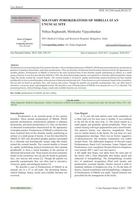
A 22 year old male patient came with compla<strong>in</strong>ts of<br />
a white rash over his nose s<strong>in</strong>ce 6 months. It was conf<strong>in</strong>ed<br />
to the left ala of the nose (Fig. 1). The lesion started as a<br />
small papule and gradually spread peripherally. There was<br />
central clear<strong>in</strong>g with hypo pigmentation and a raised border.<br />
The patient’s history was otherwise <strong>in</strong>significant. There<br />
was no similar history <strong>in</strong> the family. He was born of a non<br />
consangu<strong>in</strong>eous marriage. There was no history suggestive<br />
of photosensitivity. His systemic exam<strong>in</strong>ation was with<strong>in</strong><br />
normal limits. The differential diagnosis of Discoid Lupus<br />
Erythematoses, Basal Cell Carc<strong>in</strong>oma, Lupus Vulgaris and<br />
Porokeratosis were considered. Rout<strong>in</strong>e blood <strong>in</strong>vestigations<br />
were normal. HIV 1 and 2 was non reactive.<br />
A full depth sk<strong>in</strong> biopsy was taken from the outer part of<br />
the lesion with a 2mm disposable sk<strong>in</strong> biopsy punch and<br />
subjected to histopathology. The histopathology revealed<br />
foci of epidermal <strong>in</strong>vag<strong>in</strong>ation filled with kerat<strong>in</strong> and<br />
parakeratotic coronoid lamella. Dermis showed a mild to<br />
moderate <strong>in</strong>filtrate of lymphocytes (Fig. 2). On the basis of<br />
history, cl<strong>in</strong>ical exam<strong>in</strong>ation and histopathology, a diagnosis<br />
of Porokeratosis of Mibelli was made. Topical tret<strong>in</strong>o<strong>in</strong> was<br />
prescribed for the management of the lesion.<br />
www.odermatol.com<br />
© <strong>Our</strong> Dermatol Onl<strong>in</strong>e 4.2012 329
Figure 1. A s<strong>in</strong>gle plaque with central clear<strong>in</strong>g and a raised<br />
border over left ala of nose<br />
Figure 2. Histopathology (H&E 45x) shows foci of epidermal<br />
<strong>in</strong>vag<strong>in</strong>ation filled with kerat<strong>in</strong> and parakeratotic coronoid<br />
lamella. Dermis shows a mild to moderate <strong>in</strong>filtrate of<br />
lymphocytes<br />
Discussion<br />
Porokeratosis of Mibelli may be familial, <strong>in</strong>herited<br />
as an autosomal dom<strong>in</strong>ant disorder with the onset <strong>in</strong><br />
childhood or sporadic with later onset. Risk factors<br />
<strong>in</strong>clude immunosuppression, genetic <strong>in</strong>heritance, and UV<br />
radiation. The common underly<strong>in</strong>g pathway <strong>in</strong> all forms<br />
of porokeratosis is a clonal hyperproliferation of atypical<br />
kerat<strong>in</strong>ocytes result<strong>in</strong>g <strong>in</strong> the characteristic cornoid lamella<br />
[4]. Histopathologic exam<strong>in</strong>ation of a sk<strong>in</strong> biopsy specimen<br />
from the area of suspicion is essential for diagnosis.<br />
Porokeratosis of Mibelli is the most characteristic<br />
and dist<strong>in</strong>ctive variant of the five described forms of<br />
porokeratosis. The classical form of Mibelli consists of<br />
a s<strong>in</strong>gle plaque, or a small number of plaques, of variable<br />
size and can affect any part of the body. Common areas<br />
<strong>in</strong>volved are palms, soles and mucous membranes. Other<br />
areas of <strong>in</strong>volvement reported <strong>in</strong>clude labial commisures,<br />
lips [5]. Histologically, the <strong>in</strong>vag<strong>in</strong>ations of the epidermis <strong>in</strong><br />
Porokeratosis of Mibelli are wider and deeper, and there is<br />
prom<strong>in</strong>ent adjacent papillomatosis when compared with the<br />
other variants. All variants show a dim<strong>in</strong>ution of the granular<br />
layer, dilated superficial plexus capillaries, and a nonspecific<br />
superficial chronic <strong>in</strong>filtrate. Dermoscopy is also used <strong>in</strong> the<br />
diagnosis of porokeratosis [6].<br />
The management of such lesions <strong>in</strong>cludes many options.<br />
The approach to treatment is <strong>in</strong>dividualized and based<br />
on many factors, such as lesion size and location, risk of<br />
malignant transformation, and functional and aesthetical<br />
considerations. Sun protection, emollients, and observation<br />
for signs of malignant degeneration are mandatory. Medical<br />
modalities focus on <strong>in</strong>hibit<strong>in</strong>g cell growth and proliferation<br />
of the rapidly proliferat<strong>in</strong>g kerat<strong>in</strong>ocytes. Various modalities<br />
used <strong>in</strong>clude oral and topical ret<strong>in</strong>oids, 5-fluorouracil cream,<br />
vitam<strong>in</strong> D3 analogues, diclofenac gel, imiquimod cream<br />
cryotherapy, dermabrasion and surgical excision. Recently<br />
photodynamic therapy has been shown to be an effective and<br />
safe alternative [7].<br />
Conclusion<br />
A solitary Porokeratosis of Mibelli over the nose is<br />
uncommon. Such lesions may not always be cl<strong>in</strong>ically<br />
diagnosed, hence it requires histopathological exam<strong>in</strong>ation.<br />
Hence Porokeratosis of Mibelli should be a differential<br />
diagnosis for any such plaques over nose.<br />
<strong>Our</strong> case presented with a s<strong>in</strong>gle lesion over the nose. This<br />
is a rare presentation. There are only two such cases which<br />
have been reported [8,9].<br />
Acknowledgements<br />
Dr. Sujatha C, Prof. and Head Dept. of <strong>Dermatology</strong>, MVJ<br />
Medical College and Research Hospital.<br />
Dr. Padm<strong>in</strong>i Jeyachandran, Prof. and Head Dept. of<br />
Pathology, MVJ Medical College and Research Hospital.<br />
Dr. Vasantha Kumar S, Pr<strong>in</strong>cipal, MVJ Medical College and<br />
Research Hospital.<br />
REFERENCES<br />
1. Ruiz Villaverde R, Alonso Corral MJ, Sa´nchez Cano D, Pacheco<br />
Sa´nchez-Lafuente FJ: [L<strong>in</strong>ear porokeratosis of Mibelli]. An Pediatr<br />
(Barc). 2005;63:376-7.<br />
2. Bacharach-Buhles M, We<strong>in</strong>dorf N, Altmeyer P: Porokeratosis<br />
Mibelli gigantea. Hautarzt. 1990;41:633–5.<br />
3. L<strong>in</strong> JH, Hsu MM, Sheu HM, Lee JY: Coexistence of three<br />
variants of porokeratosis with multiple squamous cell carc<strong>in</strong>omas<br />
aris<strong>in</strong>g from lesions of giant hyperkeratotic porokeratosis. J Eur<br />
Acad Dermatol Venereol. 2006;20:621-3.<br />
4. Jurecka W, Neumann RA, Knobler RM: Porokeratoses:<br />
immunohistochemical, light and electron microscopic evaluation.<br />
J Am Acad Dermatol. 1991;24:96-101.<br />
5. Vergara G, Bañuls J, Botella R, Silvestre JF, Bel<strong>in</strong>chón I, Betlloch<br />
I: Porokeratosis of the lower lip. Eur J Dermatol. 2002;12:500-2.<br />
6. Delf<strong>in</strong>o M, Argenziano G, N<strong>in</strong>o M: Dermoscopy for the diagnosis<br />
of porokeratosis. J Eur Acad Dermatol Venereol. 2004;18:194-5.<br />
330 © <strong>Our</strong> Dermatol Onl<strong>in</strong>e 4.2012
7. Levitt J, Emer JJ, Emanuel PO: Treatment of porokeratosis<br />
of Mibelli with comb<strong>in</strong>ed use of photodynamic therapy and<br />
fluorouracil cream. Arch Dermatol. 2010;146:371-3.<br />
8. Chaudhary RG, Bilimore F, Katare SK: Large annular plaque<br />
with central atrophy over nose. Indian J Dermatol Venerol Leperol.<br />
2009;75:552.<br />
9. Ghorpade A: Localized act<strong>in</strong>ic nasal porokeratosis. Cl<strong>in</strong> Exp<br />
Dermatol. 2010;35:211-2.<br />
Copyright by Nithya Raghunath, et al. This is an open access <strong>article</strong> distributed under the terms of the Creative Commons Attribution<br />
License, which permits unrestricted use, distribution, and reproduction <strong>in</strong> any medium, provided the orig<strong>in</strong>al author and source are credited.<br />
© <strong>Our</strong> Dermatol Onl<strong>in</strong>e 4.2012 331


