An Unusual Cause of Hemoptysis in a Young Female - OMJ
An Unusual Cause of Hemoptysis in a Young Female - OMJ
An Unusual Cause of Hemoptysis in a Young Female - OMJ

You also want an ePaper? Increase the reach of your titles
YUMPU automatically turns print PDFs into web optimized ePapers that Google loves.
Oman Medical Journal (2011) Vol. 26, No. 6:457-458<br />
DOI 10. 5001/omj.2011.117<br />
Cl<strong>in</strong>ical Quiz<br />
<strong>An</strong> <strong>Unusual</strong> <strong>Cause</strong> <strong>of</strong> <strong>Hemoptysis</strong> <strong>in</strong> a <strong>Young</strong> <strong>Female</strong><br />
Pankaj Gupta, Raju Sharma, Surendra K. Sharma<br />
Received: 07 Jun 2011/ Accepted: 12 Aug 2011<br />
© OMSB, 2011<br />
Introduction<br />
A 28-year-old female presented with recurrent epistaxis<br />
and mild hemoptysis for two years. There was no fever, dyspnea,<br />
palpitations, obstructed breath<strong>in</strong>g or hematuria. There was no<br />
history <strong>of</strong> past tuberculosis or drug <strong>in</strong>take. Both her parents died<br />
when she was 5-years <strong>of</strong> age from suspected tuberculosis. On<br />
exam<strong>in</strong>ation, she had pallor and nasal telangiectasia.<br />
Blood pressure and pulse rate was normal. There was no<br />
clubb<strong>in</strong>g, cyanosis or pedal edema. Cardiovascular system<br />
exam<strong>in</strong>ation was normal. Laboratory <strong>in</strong>vestigations revealed<br />
normal hematological pr<strong>of</strong>ile with a hemoglob<strong>in</strong> <strong>of</strong> 12.5 g, platelet<br />
count <strong>of</strong> 250,000/µl, and prothromb<strong>in</strong> time <strong>of</strong> 12 seconds. Chest<br />
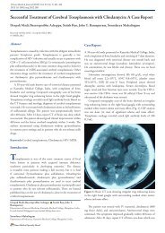
radiograph showed ill-def<strong>in</strong>ed nodules <strong>in</strong> the right middle and left<br />
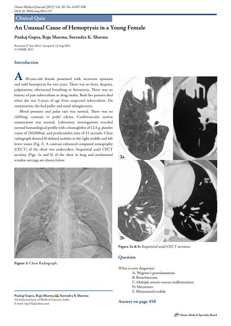
lower zones (Fig. 1). A contrast enhanced computed tomography<br />
(CECT) <strong>of</strong> the chest was undertaken. Sequential axial CECT<br />
sections (Figs. 2a and b) <strong>of</strong> the chest <strong>in</strong> lung and mediast<strong>in</strong>al<br />
w<strong>in</strong>dow sett<strong>in</strong>gs are shown below.<br />
2a<br />
2b<br />
Figure 2a & b: Sequential axial CECT sections.<br />
Figure 1: Chest Radiograph.<br />
Pankaj Gupta, Raju Sharma , Surendra K Sharma<br />
All India Institute <strong>of</strong> Medical Sciences, India<br />
E-mail: raju152@yahoo.com<br />
Question<br />
What is your diagnosis?<br />
A. Wegener’s granulomatosis<br />
B. Bronchiectasis<br />
C. Multiple arterio-venous malformations<br />
D. Metastases<br />
E. Rheumatoid nodule<br />
<strong>An</strong>swer on page 458<br />
Oman Medical Specialty Board
Oman Medical Journal (2011) Vol. 26, No. 6:457-458<br />
<strong>An</strong>swer<br />
C. Multiple arterio-venous malformations<br />
Discussion<br />
Pulmonary arteriovenous malformations (PAVMs) are rare.<br />
Most are congenital associated with hereditary haemorrhagic<br />
telangiectasia (HHT). Acquired causes <strong>in</strong>clude: chest trauma,<br />
cardiovascular <strong>in</strong>terventional procedures, chronic cirrhosis,<br />
mitral stenosis, and schistosomiasis. HHT is responsible for 80%<br />
<strong>of</strong> PAVMs and PAVMs are present <strong>in</strong> 15-45% <strong>of</strong> patients with<br />
HHT. 1 HHT, or Rendu-Osler-Weber disease, is an autosomal<br />
dom<strong>in</strong>ant multiorgan fibrovascular dysplasia with a reported<br />
prevalence <strong>of</strong> 1 <strong>in</strong> 10,000 to 1 <strong>in</strong> 5000. 2 The anatomic structure<br />
<strong>of</strong> the PAVMs <strong>in</strong> HHT is classified <strong>in</strong>to two types, 3 simple, with a<br />
s<strong>in</strong>gle feed<strong>in</strong>g artery with a s<strong>in</strong>gle dra<strong>in</strong><strong>in</strong>g ve<strong>in</strong> and complex, with<br />
two or more arterial branches, and two or more dra<strong>in</strong><strong>in</strong>g ve<strong>in</strong>s.<br />
Mutations associated with HHT <strong>in</strong>clude: endogl<strong>in</strong> mutation,<br />
activ<strong>in</strong> receptor-like k<strong>in</strong>ase (ALK)-1 mutation and SMAD4<br />
mutation. 4<br />
Epistaxis secondary to nasal mucosa telangiectasia is the<br />
most common manifestation. Cl<strong>in</strong>ical manifestations related to<br />
pulmonary <strong>in</strong>volvement <strong>in</strong>clude dyspnea, cyanosis, clubb<strong>in</strong>g and<br />
hemoptysis. Complications associated with untreated PAVMs<br />
are life threaten<strong>in</strong>g hemorrhage and neurological complications.<br />
Neurological complications <strong>in</strong>clude transient ischaemic attack<br />
and stroke; results from the paradoxical embolization <strong>of</strong> leg<br />
ve<strong>in</strong> thrombus to the left-sided arterial circulation through the<br />
PAVMs. Rarely, the thrombus can orig<strong>in</strong>ate <strong>in</strong> situ <strong>in</strong> PAVMs.<br />
<strong>An</strong>other common neurological complication is cerebral abscess<br />
secondary to paradoxical embolization <strong>of</strong> bacterial bi<strong>of</strong>ilms.<br />
Diagnosis is based on the Curaçao Criteria established<br />
by the Scientific Division <strong>of</strong> the Hereditary Hemorrhagic<br />
Telangiectasia International Foundation. 5 Diagnosis is made<br />
<strong>in</strong> the presence <strong>of</strong> three <strong>of</strong> the follow<strong>in</strong>g: recurrent spontaneous<br />
epistaxis, mucocutaneous telangiectasis, an autosomal dom<strong>in</strong>ant<br />
familial distribution, and visceral AVMs. Screen<strong>in</strong>g for PAVMs<br />
is recommended <strong>in</strong> all HHT patients due to the serious<br />
complications associated with them and the availability <strong>of</strong> safe<br />
and effective <strong>in</strong>terventions. 6 The screen<strong>in</strong>g method <strong>of</strong> choice is<br />
Contrast echocardiography with a sensitivity <strong>of</strong> 90%. 1 Unenhanced<br />
Computed tomography (CT) thorax is used to confirm diagnosis<br />
and measure the size <strong>of</strong> PAVMs <strong>in</strong> patients with positive screen<strong>in</strong>g<br />
echocardiography. With the availability <strong>of</strong> multidetector CT<br />
(MDCT), contrast enhancement is not required for detection<br />
<strong>of</strong> PAVMs. 7 On CT, the f<strong>in</strong>d<strong>in</strong>gs <strong>in</strong>clude a serpig<strong>in</strong>ous mass or<br />
nodule with one or more enlarged feed<strong>in</strong>g arteries and one or more<br />
dra<strong>in</strong><strong>in</strong>g ve<strong>in</strong>s (depend<strong>in</strong>g on whether the malformation is simple<br />
or complex). Embolization <strong>of</strong> PAVM with coils is the treatment <strong>of</strong><br />
choice. 2 A feed<strong>in</strong>g artery calibre <strong>of</strong> at least 3 mm is considered the<br />
threshold for treatment <strong>of</strong> PAVMs. 5 Recurrence <strong>of</strong> PAVMs occurs<br />
<strong>in</strong> around 19% <strong>of</strong> treated patients. Therefore, follow-up CTs are<br />
recommended at 1 year and every 3-5 years thereafter.<br />
Other visceral manifestations <strong>in</strong>clude central nervous<br />
system manifestations: cerebral vascular malformations, sp<strong>in</strong>al<br />
cord vascular malformation, encephalopathy, bra<strong>in</strong> abscess and<br />
stroke. 8 Liver is <strong>in</strong>volved <strong>in</strong> 30% to 73% <strong>of</strong> patients with HHT.<br />
Hepatic fibrovascular dysplasia ranges from small telangiectases<br />
to large vascular masses. 9 Recurrent gastro<strong>in</strong>test<strong>in</strong>al bleed<strong>in</strong>g from<br />
mucosal fibrovascular dysplasia occurs <strong>in</strong> about 20% <strong>of</strong> patients<br />
with HHT.<br />
Based on the patient’s presentation with epistaxis and<br />
hemoptysis; cl<strong>in</strong>ical differential diagnoses <strong>of</strong> pulmonary<br />
tuberculosis, Wegener’s granulomatosis, bleed<strong>in</strong>g disorders,<br />
drug <strong>in</strong>take (drugs affect<strong>in</strong>g the coagulation pr<strong>of</strong>ile), and collagen<br />
vascular disorders were considered. Radiological differential<br />
diagnoses <strong>of</strong> multiple lung nodules on chest radiograph <strong>in</strong>clude<br />
neoplastic lesions (metastases, synchronous lung cancers), multiple<br />
pulmonary arterio-venous malformations, <strong>in</strong>fections (multiple<br />
abscesses, fungal <strong>in</strong>fections, hydatid disease) and immunological<br />
conditions (wegener’s granulomatosis, rheumatoid nodules,<br />
sarcoidosis, amyloidosis, organis<strong>in</strong>g pneumonia). 10<br />
Acknowledgements<br />
The authors reported no conflict <strong>of</strong> <strong>in</strong>terest and no fund<strong>in</strong>g was<br />
receive on this work.<br />
References<br />
1. Cott<strong>in</strong> V, Plauchu H, Bayle JY, Barthelet M, Revel D, Cordier JF. Pulmonary<br />
arteriovenous malformations <strong>in</strong> patients with hereditary hemorrhagic<br />
telangiectasia. Am J Respir Crit Care Med 2004 May;169(9):994-1000.<br />
2. Cott<strong>in</strong> V, Ch<strong>in</strong>et T, Lavolé A, Corre R, Marchand E, Reynaud-Gaubert<br />
M, et al; Groupe d’Etudes et de Recherche sur les Maladies “Orphel<strong>in</strong>es”<br />
Pulmonaires (GERM”O”P). Pulmonary arteriovenous malformations <strong>in</strong><br />
hereditary hemorrhagic telangiectasia: a series <strong>of</strong> 126 patients. Medic<strong>in</strong>e<br />
(Baltimore) 2007 Jan;86(1):1-17.<br />
3. White RI Jr, Lynch-Nyhan A, Terry P, Buescher PC, Farmlett EJ, Charnas<br />
L, et al. Pulmonary arteriovenous malformations: techniques and long-term<br />
outcome <strong>of</strong> embolotherapy. Radiology 1988 Dec;169(3):663-669.<br />
4. Letteboer TG, Mager JJ, Snijder RJ, Koeleman BP, L<strong>in</strong>dhout D, Ploos<br />
van Amstel JK, et al. Genotype-phenotype relationship <strong>in</strong> hereditary<br />
haemorrhagic telangiectasia. J Med Genet 2006 Apr;43(4):371-377.<br />
5. Shovl<strong>in</strong> CL, Guttmacher AE, Buscar<strong>in</strong>i E, Faughnan ME, Hyland RH,<br />
Westermann CJ, et al. Diagnostic criteria for hereditary hemorrhagic<br />
telangiectasia (Rendu-Osler-Weber syndrome). Am J Med Genet 2000<br />
Mar;91(1):66-67.<br />
6. Gossage JR. Primary pulmonary hypertension or portopulmonary<br />
hypertension? Chest 1998 Oct;114(4):1224-1225.<br />
7. Pollak JS, Saluja S, Thabet A, Henderson KJ, Denbow N, White RI<br />
Jr. Cl<strong>in</strong>ical and anatomic outcomes after embolotherapy <strong>of</strong> pulmonary<br />
arteriovenous malformations. J Vasc Interv Radiol 2006 Jan;17(1):35-44,<br />
quiz 45.<br />
8. Haitjema T, Westermann CJ, Overtoom TT, Timmer R, Disch F, Mauser H,<br />
et al. Hereditary hemorrhagic telangiectasia (Osler-Weber-Rendu disease):<br />
new <strong>in</strong>sights <strong>in</strong> pathogenesis, complications, and treatment. Arch Intern Med<br />
1996 Apr;156(7):714-719.<br />
9. Siddiki H, Doherty MG, Fletcher JG, Stanson AW, Vrtiska TJ, Hough DM,<br />
et al. Abdom<strong>in</strong>al f<strong>in</strong>d<strong>in</strong>gs <strong>in</strong> hereditary hemorrhagic telangiectasia: pictorial<br />
essay on 2D and 3D f<strong>in</strong>d<strong>in</strong>gs with isotropic multiphase CT. Radiographics<br />
2008 Jan-Feb;28(1):171-184.<br />
Oman Medical Specialty Board