Solid pseudopapillary tumor of the pancreas: Report of a ... - doiSerbia
Solid pseudopapillary tumor of the pancreas: Report of a ... - doiSerbia
Solid pseudopapillary tumor of the pancreas: Report of a ... - doiSerbia

You also want an ePaper? Increase the reach of your titles
YUMPU automatically turns print PDFs into web optimized ePapers that Google loves.
Imaging in oncology<br />
The usual clinical signs are vague gastrointestinal symptoms like upper<br />
abdominal discomfort or pain caused by enlarging and <strong>of</strong>ten palpable<br />
abdominal mass. STP <strong>of</strong>ten remains asymptomatic until <strong>the</strong> <strong>tumor</strong> has<br />
enlarged considerably and <strong>the</strong>refore it is sometimes detected incidentally<br />
on US imaging for unrelated diseases or after a blunt abdominal trauma.<br />
Laboratory parameters including <strong>tumor</strong> markers are normal.<br />
The pathologic diagnosis <strong>of</strong> SPT is based on <strong>the</strong> presence <strong>of</strong> characteristic<br />
light microscopic features. <strong>Solid</strong> areas alternating with <strong>pseudopapillary</strong> formations;<br />
evidence <strong>of</strong> cellular degeneration, including cholesterol clefts and<br />
aggregates <strong>of</strong> foamy histiocytes; nuclear grooves and aggregates <strong>of</strong> hyaline<br />
cytoplasmic globules are found.<br />
CASE REPORT<br />
A 10-year old girl came for CT examination because <strong>of</strong> co0lic pain in abdomen<br />
appearing after meals. The results <strong>of</strong> ultrasound examination showed<br />
cystic lesion in <strong>pancreas</strong> <strong>of</strong> unknown origin. CT findings reported <strong>the</strong> focal<br />
capsulated hypodense lesion in <strong>pancreas</strong>, discreetly calcified (Post-traumatic<br />
cyst? Echinococcus cyst?; Neuroendocrine <strong>tumor</strong>?).<br />
The patient underwent surgery on July 25, 2003; <strong>tumor</strong> <strong>of</strong> 2.5 cm in diameter was<br />
enucleated. Surgeon reported <strong>the</strong> capsulated cystic <strong>tumor</strong> with partially solid content.<br />
Five years after <strong>the</strong> operation, MR findings (Figures 2a, 2b) were normal and<br />
<strong>the</strong> patient was in good condition.<br />
Tumor consists <strong>of</strong> oval eosinophilic cells, vascularized cytoplasm, with oval<br />
nucleus <strong>of</strong> dispersed chromatin and usually with one visible nucleus. Cells are<br />
organized in solid clusters and numerous abortive papillary formations with<br />
discrete fibrovascular stroma. Cystic formations are visible focally with new<br />
hemorrhage. Necroses are rare and small.<br />
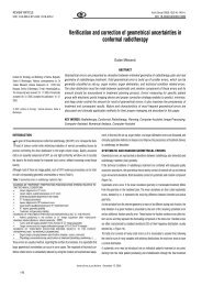
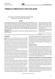
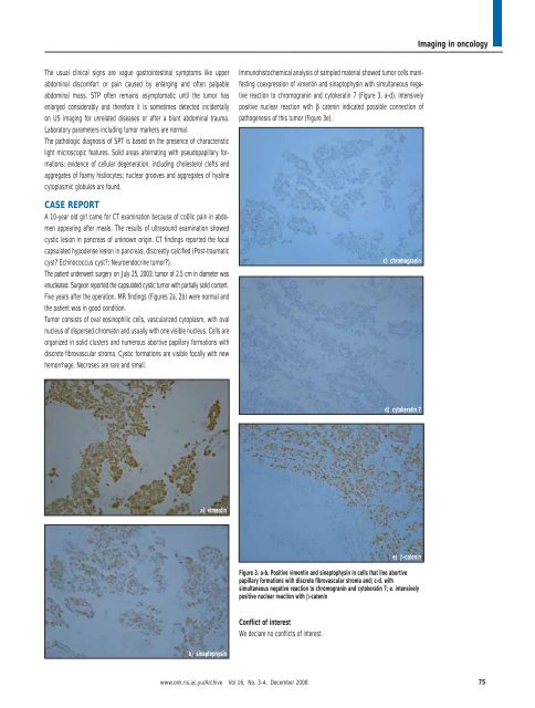
Immunohistochemical analysis <strong>of</strong> sampled material showed <strong>tumor</strong> cells manifesting<br />
coexpression <strong>of</strong> vimentin and sinaptophysin with simultaneous negative<br />
reaction to chromogranin and cytokeratin 7 (Figure 3, a-d). Intensively<br />
positive nuclear reaction with catenin indicated possible connection <strong>of</strong><br />
pathogenesis <strong>of</strong> this <strong>tumor</strong> (Figure 3e).<br />
c) chromogranin<br />
d) cytokeratin 7<br />
a) vimentin<br />
e) -catenin<br />
Figure 3. a-b. Positive vimentin and sinaptophysin in cells that line abortive<br />
papillary formations with discrete fibrovascular stroma and; c-d. with<br />
simultaneous negative reaction to chromogranin and cytokeratin 7; e. intensively<br />
positive nuclear reaction with -catenin<br />
Conflict <strong>of</strong> interest<br />
We declare no conflicts <strong>of</strong> interest.<br />
b) sinaptophysin<br />
www.onk.ns.ac.yu/Archive Vol 16, No. 3-4, December 2008<br />
75