Solid pseudopapillary tumor of the pancreas: Report of a ... - doiSerbia
Solid pseudopapillary tumor of the pancreas: Report of a ... - doiSerbia
Solid pseudopapillary tumor of the pancreas: Report of a ... - doiSerbia

You also want an ePaper? Increase the reach of your titles
YUMPU automatically turns print PDFs into web optimized ePapers that Google loves.
®<br />
Imaging in oncology<br />
<strong>Solid</strong> <strong>pseudopapillary</strong> <strong>tumor</strong> <strong>of</strong> <strong>the</strong> <strong>pancreas</strong>:<br />
<strong>Report</strong> <strong>of</strong> a case after 5-year follow-up<br />
<br />
Key words: Pancreatic Neoplasms; Pancreatic Pseudocyst; Child; Female<br />
Arch Oncol 2007;16(3-4):74-6.<br />
UDC: 616.37-006:616-079.1<br />
DOI: 10.2298/AOO0804074P<br />
Oncology Institute <strong>of</strong> Vojvodina,<br />
Sremska Kamenica, Serbia<br />
Correspondence to:<br />
<br />
<strong>of</strong> Vojvodina, Institutski put 4, 21204<br />
Sremska Kamenica, Serbia<br />
prvulov@hotmail.com<br />
Received: 25.11.2008<br />
Provisionally accepted: 01.12.2008<br />
Accepted: 08.12.2008<br />
© 2008, Oncology Institute <strong>of</strong><br />
Vojvodina, Sremska Kamenica<br />
<strong>Solid</strong> <strong>pseudopapillary</strong> <strong>tumor</strong> (STP) <strong>of</strong> <strong>the</strong> <strong>pancreas</strong> is an exceptionally rare neoplasm<br />
in children accounting from 1% to 2% <strong>of</strong> exocrine pancreatic <strong>tumor</strong>s. Frantz ZE first<br />
described STP in 1959 as a papillary <strong>tumor</strong> <strong>of</strong> <strong>the</strong> <strong>pancreas</strong> benign or malignant.<br />
This <strong>tumor</strong> from 1996 has only recently <strong>of</strong>ficially been called solid <strong>pseudopapillary</strong><br />
<strong>tumor</strong> <strong>of</strong> <strong>the</strong> exocrine <strong>pancreas</strong>, but <strong>the</strong> paste has carried names like solid and papillary<br />
cystic <strong>tumor</strong>, solid and papillary epi<strong>the</strong>lial neoplasm in a child and adenocarcinoma<br />
<strong>of</strong> <strong>the</strong> <strong>pancreas</strong> in childhood, as well as Frantz <strong>tumor</strong>. Various names reflect<br />
<br />
are uniform around fibrovascular septa, whereas in <strong>pseudopapillary</strong> areas show<br />
degenerative changes in <strong>the</strong> cells away from <strong>the</strong> vasculature.<br />
STP is a <strong>tumor</strong> <strong>of</strong> <strong>the</strong> primitive epi<strong>the</strong>lial cells, which having capacity for<br />
exocrine and endocrine differentiation.<br />
The pathogenesis <strong>of</strong> STP is unknown and nowadays, molecular studies<br />
are intensive. It is believed that mutations <strong>of</strong> -catenin (S33C; <strong>the</strong> serine is<br />
changed to cystine) can cause disorganization <strong>of</strong> <strong>the</strong> E-cadherin causing <strong>the</strong><br />
loss <strong>of</strong> adhesion junction with cytoskeleton <strong>of</strong> <strong>the</strong> cells and/or overexpression <strong>of</strong><br />
transcription factors, which to lead to development <strong>of</strong> STP. STP typically affects<br />
young women in <strong>the</strong>ir second and third decade <strong>of</strong> life, especially adolescent<br />
girls. Its clinical behavior and its pathologic appearance are consistent with<br />
low-grade malignancy with an excellent prognosis if complete surgical excision<br />
is performed. Metastases have been described, however, reportedly after long<br />
disease- free periods; <strong>the</strong> lesion’s size is not a predictor <strong>of</strong> unresectability.<br />
a<br />
a<br />
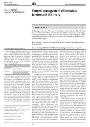
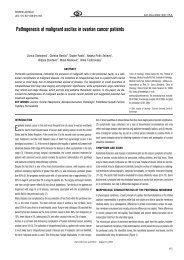
Figure 2. MRI scan performed five years after surgery (November 19, 2008):<br />
a) before contrast medium, b) with contrast medium. Both images show normal<br />
morphological appearance <strong>of</strong> <strong>the</strong> <strong>pancreas</strong> with no signs <strong>of</strong> local relapse<br />
b<br />
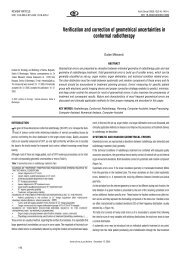
Figure 1. CT performed before surgery in December 2002: a) native, b) with<br />
contrast medium<br />
Native CT scan shows discreet focal lesion at <strong>the</strong> transition <strong>of</strong> <strong>the</strong> corpus into <strong>the</strong><br />
tail <strong>of</strong> <strong>pancreas</strong>; calcification deposits are minimal. Postcontrast image shows<br />
hypodense circular lesion, relatively homogenous, 2 cm in diameter and with<br />
clearly marginated from surrounding<br />
b<br />
The characteristic ultrasound (US) findings <strong>of</strong> STP are those <strong>of</strong> well-encapsulated,<br />
cystic and solid mass, but sometimes a purely solid mass or internal<br />
separation or calcification are seen. By imaging studies, STP is circumscribed<br />
by precise margins with a well-vascularized and defined capsule. Calcification<br />
and septa are characteristic features <strong>of</strong> SPTs. The alteration <strong>of</strong> solid and cystic<br />
areas, with a necrotic or hemorrhagic component may also be present.<br />
74<br />
www.onk.ns.ac.yu/Archive Vol 16, No. 3-4, December 2008
Imaging in oncology<br />
The usual clinical signs are vague gastrointestinal symptoms like upper<br />
abdominal discomfort or pain caused by enlarging and <strong>of</strong>ten palpable<br />
abdominal mass. STP <strong>of</strong>ten remains asymptomatic until <strong>the</strong> <strong>tumor</strong> has<br />
enlarged considerably and <strong>the</strong>refore it is sometimes detected incidentally<br />
on US imaging for unrelated diseases or after a blunt abdominal trauma.<br />
Laboratory parameters including <strong>tumor</strong> markers are normal.<br />
The pathologic diagnosis <strong>of</strong> SPT is based on <strong>the</strong> presence <strong>of</strong> characteristic<br />
light microscopic features. <strong>Solid</strong> areas alternating with <strong>pseudopapillary</strong> formations;<br />
evidence <strong>of</strong> cellular degeneration, including cholesterol clefts and<br />
aggregates <strong>of</strong> foamy histiocytes; nuclear grooves and aggregates <strong>of</strong> hyaline<br />
cytoplasmic globules are found.<br />
CASE REPORT<br />
A 10-year old girl came for CT examination because <strong>of</strong> co0lic pain in abdomen<br />
appearing after meals. The results <strong>of</strong> ultrasound examination showed<br />
cystic lesion in <strong>pancreas</strong> <strong>of</strong> unknown origin. CT findings reported <strong>the</strong> focal<br />
capsulated hypodense lesion in <strong>pancreas</strong>, discreetly calcified (Post-traumatic<br />
cyst? Echinococcus cyst?; Neuroendocrine <strong>tumor</strong>?).<br />
The patient underwent surgery on July 25, 2003; <strong>tumor</strong> <strong>of</strong> 2.5 cm in diameter was<br />
enucleated. Surgeon reported <strong>the</strong> capsulated cystic <strong>tumor</strong> with partially solid content.<br />
Five years after <strong>the</strong> operation, MR findings (Figures 2a, 2b) were normal and<br />
<strong>the</strong> patient was in good condition.<br />
Tumor consists <strong>of</strong> oval eosinophilic cells, vascularized cytoplasm, with oval<br />
nucleus <strong>of</strong> dispersed chromatin and usually with one visible nucleus. Cells are<br />
organized in solid clusters and numerous abortive papillary formations with<br />
discrete fibrovascular stroma. Cystic formations are visible focally with new<br />
hemorrhage. Necroses are rare and small.<br />
Immunohistochemical analysis <strong>of</strong> sampled material showed <strong>tumor</strong> cells manifesting<br />
coexpression <strong>of</strong> vimentin and sinaptophysin with simultaneous negative<br />
reaction to chromogranin and cytokeratin 7 (Figure 3, a-d). Intensively<br />
positive nuclear reaction with catenin indicated possible connection <strong>of</strong><br />
pathogenesis <strong>of</strong> this <strong>tumor</strong> (Figure 3e).<br />
c) chromogranin<br />
d) cytokeratin 7<br />
a) vimentin<br />
e) -catenin<br />
Figure 3. a-b. Positive vimentin and sinaptophysin in cells that line abortive<br />
papillary formations with discrete fibrovascular stroma and; c-d. with<br />
simultaneous negative reaction to chromogranin and cytokeratin 7; e. intensively<br />
positive nuclear reaction with -catenin<br />
Conflict <strong>of</strong> interest<br />
We declare no conflicts <strong>of</strong> interest.<br />
b) sinaptophysin<br />
www.onk.ns.ac.yu/Archive Vol 16, No. 3-4, December 2008<br />
75
Imaging in oncology<br />
REFERENCES<br />
1 Martin RC, Klimstra DS, Brennan MF, Conlon KC. <strong>Solid</strong>–<strong>pseudopapillary</strong> <strong>tumor</strong> <strong>of</strong><br />
<strong>the</strong> <strong>pancreas</strong>: a surgical enigma. Ann Surg Oncol. 2002;9(1):35-40.<br />
2 Casanova M, Collini P, Ferrari A, Cecchetto G, Dall'igna P, Mazzaferro V. <strong>Solid</strong><strong>pseudopapillary</strong><br />
<strong>tumor</strong> <strong>of</strong> <strong>the</strong> <strong>pancreas</strong> (Frantz <strong>tumor</strong>) in children. Medical and<br />
Pediatric oncology. 2003; 41(1):74-6.<br />
3 Huang HL, Shih SC, Chang WH, Wang TE, Chen MJ, Chan YJ. <strong>Solid</strong>-<strong>pseudopapillary</strong><br />
<strong>tumor</strong> <strong>of</strong> <strong>the</strong> <strong>pancreas</strong>: clinical experience and literature review. World J<br />
Gastroenterol. 2005;11(9):1403-9.<br />
4 Tang WW, Stelter AA, French S, Shen S, Qiu S, Venegas R, et al. Loss <strong>of</strong> celladhesion<br />
molecule complexes in solid <strong>pseudopapillary</strong> <strong>tumor</strong> <strong>of</strong> <strong>pancreas</strong>. Mod<br />
Pathol. 2007;20(5):509-13.<br />
5 Kandpal H, Sharma R, Das CJ, Sahni P, Das AK, Neyaz Z. <strong>Solid</strong> <strong>pseudopapillary</strong><br />
<strong>tumor</strong> <strong>of</strong> <strong>the</strong> <strong>pancreas</strong> with portal vein compression presenting as portal hypertension.<br />
Australas Radiol. 2007;51 Suppl:B287-91.<br />
6 Coleman KM, Doherty MC, Bigler SA. <strong>Solid</strong>-Pseudopapillary Tumor <strong>of</strong> <strong>the</strong> Pancreas.<br />
Radiographics. 2003;23:1644-8.<br />
76<br />
www.onk.ns.ac.yu/Archive Vol 16, No. 3-4, December 2008