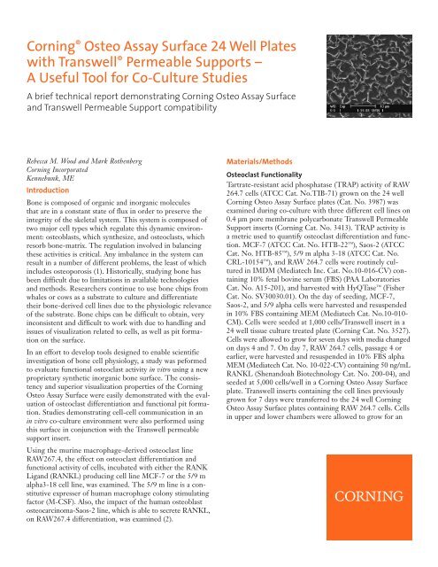
Corning® Osteo Assay Surface 24 Well Plates with Transwell ...
Corning® Osteo Assay Surface 24 Well Plates with Transwell ...
Corning® Osteo Assay Surface 24 Well Plates with Transwell ...

Create successful ePaper yourself
Turn your PDF publications into a flip-book with our unique Google optimized e-Paper software.
Corning ® <strong>Osteo</strong> <strong>Assay</strong> <strong>Surface</strong> <strong>24</strong> <strong>Well</strong> <strong>Plates</strong><br />
<strong>with</strong> <strong>Transwell</strong> ® Permeable Supports –<br />
A Useful Tool for Co-Culture Studies<br />
A brief technical report demonstrating Corning <strong>Osteo</strong> <strong>Assay</strong> <strong>Surface</strong><br />
and <strong>Transwell</strong> Permeable Support compatibility<br />
Rebecca M. Wood and Mark Rothenberg<br />
Corning Incorporated<br />
Kennebunk, ME<br />
Introduction<br />
Bone is composed of organic and inorganic molecules<br />
that are in a constant state of flux in order to preserve the<br />
integrity of the skeletal system. This system is composed of<br />
two major cell types which regulate this dynamic environment:<br />
osteoblasts, which synthesize, and osteoclasts, which<br />
resorb bone-matrix. The regulation involved in balancing<br />
these activities is critical. Any imbalance in the system can<br />
result in a number of different problems, the least of which<br />
includes osteoporosis (1). Historically, studying bone has<br />
been difficult due to limitations in available technologies<br />
and methods. Researchers continue to use bone chips from<br />
whales or cows as a substrate to culture and differentiate<br />
their bone-derived cell lines due to the physiologic relevance<br />
of the substrate. Bone chips can be difficult to obtain, very<br />
inconsistent and difficult to work <strong>with</strong> due to handling and<br />
issues of visualization related to cells, as well as pit formation<br />
on the surface.<br />
In an effort to develop tools designed to enable scientific<br />
investigation of bone cell physiology, a study was peformed<br />
to evaluate functional osteoclast activity in vitro using a new<br />
proprietary synthetic inorganic bone surface. The consistency<br />
and superior visualization properties of the Corning<br />
<strong>Osteo</strong> <strong>Assay</strong> <strong>Surface</strong> were easily demonstrated <strong>with</strong> the evaluation<br />
of osteoclast differentiation and functional pit formation.<br />
Studies demonstrating cell-cell communication in an<br />
in vitro co-culture environment were also performed using<br />
this surface in conjunction <strong>with</strong> the <strong>Transwell</strong> permeable<br />
support insert.<br />
Using the murine macrophage-derived osteoclast line<br />
RAW267.4, the effect on osteoclast differentiation and<br />
functional activity of cells, incubated <strong>with</strong> either the RANK<br />
Ligand (RANKL) producing cell line MCF-7 or the 5/9 m<br />
alpha3-18 cell line, was examined. The 5/9 m line is a constitutive<br />
expresser of human macrophage colony stimulating<br />
factor (M-CSF). Also, the impact of the human osteoblast<br />
osteocarcinoma-Saos-2 line, which is able to secrete RANKL,<br />
on RAW267.4 differentiation, was examined (2).<br />
Materials/Methods<br />
<strong>Osteo</strong>clast Functionality<br />
Tartrate-resistant acid phosphatase (TRAP) activity of RAW<br />
264.7 cells (ATCC Cat. No.TIB-71) grown on the <strong>24</strong> well<br />
Corning <strong>Osteo</strong> <strong>Assay</strong> <strong>Surface</strong> plates (Cat. No. 3987) was<br />
examined during co-culture <strong>with</strong> three different cell lines on<br />
0.4 µm pore membrane polycarbonate <strong>Transwell</strong> Permeable<br />
Support inserts (Corning Cat. No. 3413). TRAP activity is<br />
a metric used to quantify osteoclast differentiation and function.<br />
MCF-7 (ATCC Cat. No. HTB-22 ), Saos-2 (ATCC<br />
Cat. No. HTB-85 ), 5/9 m alpha 3-18 (ATCC Cat. No.<br />
CRL-10154 ), and RAW 264.7 cells were routinely cultured<br />
in IMDM (Mediatech Inc. Cat. No.10-016-CV) containing<br />
10% fetal bovine serum (FBS) (PAA Laboratories<br />
Cat. No. A15-201), and harvested <strong>with</strong> HyQTase (Fisher<br />
Cat. No. SV30030.01). On the day of seeding, MCF-7,<br />
Saos-2, and 5/9 alpha cells were harvested and resuspended<br />
in 10% FBS containing MEM (Mediatech Cat. No.10-010-<br />
CM). Cells were seeded at 1,000 cells/<strong>Transwell</strong> insert in a<br />
<strong>24</strong> well tissue culture treated plate (Corning Cat. No. 3527).<br />
Cells were allowed to grow for seven days <strong>with</strong> media changed<br />
on days 4 and 7. On day 7, RAW 264.7 cells, passage 4 or<br />
earlier, were harvested and resuspended in 10% FBS alpha<br />
MEM (Mediatech Cat. No. 10-022-CV) containing 50 ng/mL<br />
RANKL (Shenandoah Biotechnology Cat. No. 200-04), and<br />
seeded at 5,000 cells/well in a Corning <strong>Osteo</strong> <strong>Assay</strong> <strong>Surface</strong><br />
plate. <strong>Transwell</strong> inserts containing the cell lines previously<br />
grown for 7 days were transferred to the <strong>24</strong> well Corning<br />
<strong>Osteo</strong> <strong>Assay</strong> <strong>Surface</strong> plates containing RAW 264.7 cells. Cells<br />
in upper and lower chambers were allowed to grow for an
additional 7 days <strong>with</strong> a media change on day 4 (10% alpha<br />
MEM containing 50 ng/mL RANKL). The culture period<br />
chosen was based on previously published research in order<br />
to achieve optimal cytokine production (3). On day 7, inserts<br />
were removed and discarded, and medium from the Corning ®<br />
<strong>Osteo</strong> <strong>Assay</strong> <strong>Surface</strong> plate was sampled for TRAP activity<br />
following the manufacturers protocol (B-Bridge Cat. No.<br />
AK04). As an internal control, RAW267.4 cells were also differentiated<br />
<strong>with</strong>out additional cell lines but in the presence<br />
of RANKL. Differentiated osteoclasts were stained using<br />
the Millipore ® Actin Cytoskeleton and Focal Adhesion<br />
Staining Kit (Millipore Cat. No. FAK100) for both actin<br />
and nuclear localization. Cells were visualized using the<br />
EVOS ® fl microscope (AMG Advanced Microscopy<br />
Group).<br />
Results and Discussion<br />
To establish the feasibility of using a co-culturing technique<br />
to study bone cell physiology, two unique platforms from<br />
Corning Life Sciences were utilized. The study used the<br />
Corning <strong>Osteo</strong> <strong>Assay</strong> <strong>Surface</strong> in conjunction <strong>with</strong> <strong>Transwell</strong> ®<br />
permeable supports. The impact of three cell lines (MCF-7,<br />
5/9 m alpha 3-18 and the Saos-1) on osteoclast differentiation<br />
was examined. The three lines are known to express<br />
important regulators of osteoclast physiology. Figure 1 is a<br />
diagram of how the co-culture system was set up. Figure 2<br />
depicts the result of a 7-day incubation of RAW267.4<br />
cultured on the Corning <strong>Osteo</strong> <strong>Assay</strong> <strong>Surface</strong> <strong>with</strong> MCCF-7,<br />
5/9 m alpha 3-18 and Saos-2 cell lines cultured on the Apical<br />
0.4 µm pore size membrane. At the end of the osteoclast differentiation<br />
period, the inserts containing cells were removed<br />
and TRAP activity of the multiple well plates was analyzed<br />
using the protocol provided by B-bridge. The results (Table<br />
1) show that the 5/9 m alpha 3-18 cell line, a commercially<br />
available CHO line that constitutively expresses M-CSF,<br />
inhibits TRAP activity (Figure 2, 49 ±1%). The data, expressed<br />
as Percent TRAP activity <strong>with</strong> differentiated RAW267.4 as<br />
the control, showed no change in RAW267.4 activity when<br />
co-cultured <strong>with</strong> the MCF-7 cell line (100 ±1%). The human<br />
osteosarcoma line, Saos-2, which under the appropriate conditions<br />
can differentiate into osteoblasts and express RANKL<br />
(2), showed enhanced RAW264.7 differentiation and TRAP<br />
activity (147 ±5%). These data indicate that osteoclast physiology<br />
can be studied and modified using this co-culture<br />
system. Figure 3A and B show differentiated RAW267.4<br />
Table 1. Percent TRAP Activity of Differentiated RAW264.7 Cells<br />
After Co-culture <strong>with</strong> Three Different Cell Lines<br />
Overall (%) Standard Deviation<br />
MCF-7 + RankL 102 0.1<br />
5/9 alpha + RankL 45 0.1<br />
Saos-2 + RankL 141 0.5<br />
200<br />
Percent TRAP Activity of Positive Control<br />
<strong>Transwell</strong> Insert<br />
Cell line of interest<br />
RAW267.4 derived<br />
osteoclasts<br />
Percent TRAP Activity<br />
150<br />
100<br />
50<br />
Synthetic <strong>Osteo</strong><br />
<strong>Assay</strong> <strong>Surface</strong><br />
Pits<br />
0<br />
MCF-7 +<br />
RANKL<br />
5/9 alpha +<br />
RANKL<br />
Saos-2 +<br />
RANKL<br />
Figure 1. Schematic of a <strong>Transwell</strong> Permeable Support – Corning <strong>Osteo</strong><br />
<strong>Assay</strong> <strong>Surface</strong> Co-Culture System<br />
Figure 2. Percent TRAP activity of co-cultures expressed as compared to<br />
control wells containing RAW 264.7 cells in the presence of RANKL. (n=3)
A<br />
F-Actin<br />
B<br />
Viniculin<br />
F-Actin<br />
Nuclei<br />
Nuclei<br />
Viniculin<br />
<strong>Osteo</strong> pits<br />
Figure 3. RAW264.7 cells were grown for 7 days in the presence of RANKL on Corning <strong>Osteo</strong> <strong>Assay</strong> <strong>Surface</strong> <strong>24</strong> well plates. Cells were fixed and stained using<br />
an Actin/Cytoskeleton and Focal Adhesion Staining Kit (Millipore) and visualized using an EVOS® fl microscope. Figures 3A and B are representative<br />
osetoclasts stained using the same protocol.<br />
cells stained <strong>with</strong> Rhodamine-Phalloidin, DAPI, and Vinculin<br />
antibodies to reveal the osteoclast-like structure of the cells<br />
after 7 days of differentiation. The observed staining and<br />
characteristic multinucleated structures present are indicative<br />
of an osteoclast cell. Pit formation can also be observed in<br />
Figure 3B which correlated <strong>with</strong> the TRAP data (data not<br />
shown).<br />
This type of co-culture experimental design can also be used<br />
to examine cellular migration. Figure 4 is an example of how<br />
such an experiment can be set up to examine how osteoclasts<br />
or osteoblasts modulate cancer cell (e.g., breast or prostate)<br />
migration ex vivo.<br />
Conclusions<br />
◗ Utilizing Corning ® 0.4 µm pore size membrane <strong>Transwell</strong> ®<br />
Permeable Supports and the new Corning <strong>Osteo</strong> <strong>Assay</strong><br />
<strong>Surface</strong> technology, co-culture studies can be conducted to<br />
better understand the effects on osteoclast differentiation<br />
and activity.<br />
◗ The Corning <strong>Osteo</strong> <strong>Assay</strong> <strong>Surface</strong> can be used <strong>with</strong><br />
fluorescent techniques to examine cellular markers of<br />
differentiation.<br />
◗ The <strong>Transwell</strong> Permeable Support-Corning <strong>Osteo</strong> <strong>Assay</strong><br />
<strong>Surface</strong> system can be used for examination of a number of<br />
interesting physiologic events, including, but not limited<br />
to, migration of various cell lines where bone or bonerelated<br />
cells may play a role.<br />
<strong>Transwell</strong> Insert<br />
Cell line of interest<br />
RAW267.4 derived<br />
osteoclasts<br />
Synthetic <strong>Osteo</strong><br />
<strong>Assay</strong> <strong>Surface</strong><br />
Pits<br />
Figure 4. Schematic showing the use of the <strong>Transwell</strong> Permeable Support<br />
system <strong>with</strong> the Corning <strong>Osteo</strong> <strong>Assay</strong> <strong>Surface</strong> for migration studies.<br />
References<br />
1. Zhao, W. et al. 2009. The Role of T Cells in <strong>Osteo</strong>porosis, an<br />
Update. Int. J. Clin. Exp. Pathol. 2:544-552.<br />
2. Hofbauer, L.C. et al. 2008. Fatal attraction: why breast cancer<br />
cells home to bone. Breast Cancer Research 10:101.<br />
3. Nicolin, V. et al. 2008. Breast adenocarcinoma MCF-7 cell line<br />
induces spontaneous osteoclastogenesis via a RANK-liganddependent<br />
pathway. Acta Histochemica Vol. 110(5):388-396.
For additional product or technical information, please visit<br />
www.corning.com/lifesciences or call 800.492.1110. Outside<br />
the United States, please call +1.978.442.2200.<br />
Corning Incorporated<br />
Life Sciences<br />
Tower 2, 4th Floor<br />
900 Chelmsford St.<br />
Lowell, MA 01851<br />
t 800.492.1110<br />
t 978.442.2200<br />
f 978.442.<strong>24</strong>76<br />
www.corning.com/lifesciences<br />
Worldwide<br />
Support Offices<br />
A S I A / P A C I F I C<br />
Australia/New Zealand<br />
t 0402-794-347<br />
China<br />
t 86 21 2215 2888<br />
f 86 21 6215 2988<br />
India<br />
t 91 1<strong>24</strong> 4604000<br />
f 91 1<strong>24</strong> 4604099<br />
Japan<br />
t 81 3-3586 1996<br />
f 81 3-3586 1291<br />
Korea<br />
t 82 2-796-9500<br />
f 82 2-796-9300<br />
Singapore<br />
t 65 6733-6511<br />
f 65 6861-2913<br />
Taiwan<br />
t 886 2-2716-0338<br />
f 886 2-2516-7500<br />
Corning and <strong>Transwell</strong> are registered trademarks of Corning Incorporated, Corning, NY.<br />
All other trademarks in this document are the property of their respective owners.<br />
Corning Incorporated, One Riverfront Plaza, Corning, NY 14831-0001<br />
E U R O P E<br />
France<br />
t 0800 916 882<br />
f 0800 918 636<br />
Germany<br />
t 0800 101 1153<br />
f 0800 101 <strong>24</strong>27<br />
The Netherlands<br />
t 31 20 655 79 28<br />
f 31 20 659 76 73<br />
United Kingdom<br />
t 0800 376 8660<br />
f 0800 279 1117<br />
All Other European<br />
Countries<br />
t 31 (0) 20 659 60 51<br />
f 31 (0) 20 659 76 73<br />
L AT I N A M E R I C A<br />
Brasil<br />
t (55-11) 3089-7419<br />
f (55-11) 3167-0700<br />
Mexico<br />
t (52-81) 8158-8400<br />
f (52-81) 8313-8589<br />
© 2011 Corning Incorporated Printed in U.S.A. 5/11 POD CLS-AN-173