3B Scientific - Microscopy Catalog
3bscientific.com
3bscientific.com
You also want an ePaper? Increase the reach of your titles
YUMPU automatically turns print PDFs into web optimized ePapers that Google loves.
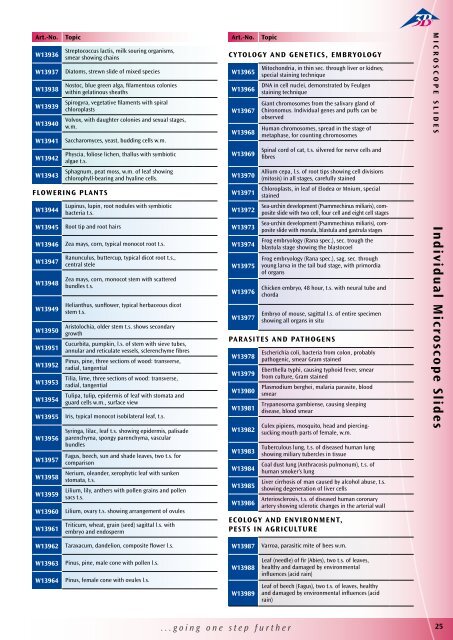
Art.-No.<br />
W13936<br />
W13937<br />
W13938<br />
W13939<br />
W13940<br />
W13941<br />
W13942<br />
W13943<br />
W13944<br />
W13945<br />
W13946<br />
W13947<br />
W13948<br />
W13949<br />
W13950<br />
W13951<br />
W13952<br />
W13953<br />
W13954<br />
W13955<br />
W13956<br />
W13957<br />
W13958<br />
W13959<br />
W13960<br />
W13961<br />
Topic<br />
Streptococcus lactis, milk souring organisms,<br />
smear showing chains<br />
Diatoms, strewn slide of mixed species<br />
Nostoc, blue green alga, filamentous colonies<br />
within gelatinous sheaths<br />
Spirogyra, vegetative filaments with spiral<br />
chloroplasts<br />
Volvox, with daughter colonies and sexual stages,<br />
w.m.<br />
Saccharomyces, yeast, budding cells w.m.<br />
Physcia, foliose lichen, thallus with symbiotic<br />
algae t.s.<br />
Sphagnum, peat moss, w.m. of leaf showing<br />
chlorophyll-bearing and hyaline cells.<br />
Flowering Plants<br />
Lupinus, lupin, root nodules with symbiotic<br />
bacteria t.s.<br />
Root tip and root hairs<br />
Zea mays, corn, typical monocot root t.s.<br />
Ranunculus, buttercup, typical dicot root t.s.,<br />
central stele<br />
Zea mays, corn, monocot stem with scattered<br />
bundles t.s.<br />
Helianthus, sunflower, typical herbaceous dicot<br />
stem t.s.<br />
Aristolochia, older stem t.s. shows secondary<br />
growth<br />
Cucurbita, pumpkin, l.s. of stem with sieve tubes,<br />
annular and reticulate vessels, sclerenchyme fibres<br />
Pinus, pine, three sections of wood: transverse,<br />
radial, tangential<br />
Tilia, lime, three sections of wood: transverse,<br />
radial, tangential<br />
Tulipa, tulip, epidermis of leaf with stomata and<br />
guard cells w.m., surface view<br />
Iris, typical monocot isobilateral leaf, t.s.<br />
Syringa, lilac, leaf t.s. showing epidermis, palisade<br />
parenchyma, spongy parenchyma, vascular<br />
bundles<br />
Fagus, beech, sun and shade leaves, two t.s. for<br />
comparison<br />
Nerium, oleander, xerophytic leaf with sunken<br />
stomata, t.s.<br />
Lilium, lily, anthers with pollen grains and pollen<br />
sacs t.s.<br />
Lilium, ovary t.s. showing arrangement of ovules<br />
Triticum, wheat, grain (seed) sagittal l.s. with<br />
embryo and endosperm<br />
Art.-No.<br />
W13969<br />
W13970<br />
W13971<br />
W13972<br />
W13973<br />
W13974<br />
W13975<br />
W13976<br />
W13977<br />
W13978<br />
W13979<br />
W13980<br />
W13981<br />
W13982<br />
W13983<br />
W13984<br />
W13985<br />
W13986<br />
Topic<br />
Cytology and Genetics, Embryology<br />
Mitochondria, in thin sec. through liver or kidney,<br />
W13965 special staining technique<br />
DNA in cell nuclei, demonstrated by Feulgen<br />
W13966 staining technique<br />
Giant chromosomes from the salivary gland of<br />
W13967 Chironomus. Individual genes and puffs can be<br />
observed<br />
Human chromosomes, spread in the stage of<br />
W13968 metaphase, for counting chromosomes<br />
Spinal cord of cat, t.s. silvered for nerve cells and<br />
fibres<br />
Allium cepa, l.s. of root tips showing cell divisions<br />
(mitosis) in all stages, carefully stained<br />
Chloroplasts, in leaf of Elodea or Mnium, special<br />
stained<br />
Sea-urchin development (Psammechinus miliaris), composite<br />
slide with two cell, four cell and eight cell stages<br />
Sea-urchin development (Psammechinus miliaris), composite<br />
slide with morula, blastula and gastrula stages<br />
Frog embryology (Rana spec.), sec. trough the<br />
blastula stage showing the blastocoel<br />
Frog embryology (Rana spec.), sag. sec. through<br />
young larva in the tail bud stage, with primordia<br />
of organs<br />
Chicken embryo, 48 hour, t.s. with neural tube and<br />
chorda<br />
Embryo of mouse, sagittal l.s. of entire specimen<br />
showing all organs in situ<br />
Parasites and pathogens<br />
Escherichia coli, bacteria from colon, probably<br />
pathogenic, smear Gram stained<br />
Eberthella typhi, causing typhoid fever, smear<br />
from culture, Gram stained<br />
Plasmodium berghei, malaria parasite, blood<br />
smear<br />
Trypanosoma gambiense, causing sleeping<br />
disease, blood smear<br />
Culex pipiens, mosquito, head and piercingsucking<br />
mouth parts of female, w.m.<br />
Tuberculous lung, t.s. of diseased human lung<br />
showing miliary tubercles in tissue<br />
Coal dust lung (Anthracosis pulmonum), t.s. of<br />
human smoker’s lung<br />
Liver cirrhosis of man caused by alcohol abuse, t.s.<br />
showing degeneration of liver cells<br />
Arteriosclerosis, t.s. of diseased human coronary<br />
artery showing sclerotic changes in the arterial wall<br />
Ecology and environment,<br />
pests in agriculture<br />
Microscope Slides<br />
Individual Microscope Slides<br />
W13962<br />
Taraxacum, dandelion, composite flower l.s.<br />
W13987<br />
Varroa, parasitic mite of bees w.m.<br />
W13963<br />
W13964<br />
Pinus, pine, male cone with pollen l.s.<br />
Pinus, female cone with ovules l.s.<br />
W13988<br />
W13989<br />
Leaf (needle) of fir (Abies), two t.s. of leaves,<br />
healthy and damaged by environmental<br />
influences (acid rain)<br />
Leaf of beech (Fagus), two t.s. of leaves, healthy<br />
and damaged by environmental influences (acid<br />
rain)<br />
...going one step further 25


