Hallux Rigidus
Hallux Rigidus
Hallux Rigidus

Create successful ePaper yourself
Turn your PDF publications into a flip-book with our unique Google optimized e-Paper software.
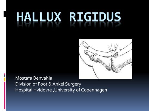
HALLUX RIGIDUS<br />
Mostafa Benyahia<br />
Division of Foot & Ankel Surgery<br />
Hospital Hvidovre ,University of Copenhagen
Normal function first MTP<br />
joint<br />
• Cam-shaped condylar hinged joint<br />
• Alignment varies 5 degrees varus to 15<br />
degrees valgus<br />
• Normal range of motion<br />
40-100 degrees dorsiflexion<br />
3-45 degrees plantarflexion<br />
70-90 degrees<br />
45 degrees
<strong>Hallux</strong> <strong>Rigidus</strong> Definition<br />
• Arthritic condition characterized by stiff painful<br />
1 st MTP joint<br />
• Early stages – involvement dorsal aspect<br />
articular cartilage with prominent dorsal<br />
osteophyte<br />
• Later stages – central and plantar aspects of<br />
articular cartilage involved<br />
X-rays – Almost always underestimate the extent of<br />
disease
Pathogenesis:<strong>Hallux</strong> <strong>Rigidus</strong><br />
• Traumatic: intra-articular fracture, crush,<br />
direct jamming to MTP joint<br />
• Idiopathic: predisposing factors may cause<br />
increased joint stresses with subsequent<br />
swelling, synovitis, joint degeneration<br />
Metatarsus elevatus<br />
OCD<br />
Long first ray<br />
Pes planus
Clinical Features H.R.<br />
• Painful, stiff MTP joint<br />
• Shoewear difficult due to<br />
dorsal osteophytes<br />
• Difficulty with pushoff during gait<br />
• Dorsiflexion impingement occurs at<br />
spur with pain and stiffness<br />
• Painful plantarflexion<br />
• Stress transfer (transfer metatarsalgia)<br />
with first ray unloading<br />
• Dorsal skin irritation due to spur
<strong>Hallux</strong> <strong>Rigidus</strong><br />
• Localized first MTP arthritis<br />
• Decreased range of motion (DF), deformity,<br />
pain
Grading (Hattrup & Johnson)<br />
• Grade I<br />
Joint space maintained<br />
Minimal osteophytes<br />
• Grade II<br />
Larger osteophytes<br />
Subchondral sclerosis<br />
• Grade III<br />
Complete loss of visible joint space<br />
Subchondral cysts<br />
Osteophytes<br />
Hypertrophy of sesamoids
Grading (Coughlin & Shurnas)<br />
• Grade 0<br />
<br />
<br />
<br />
40-60 degrees dorsiflexion or 10% to 20% less DF than<br />
opposite side<br />
Normal xrays<br />
No pain, only stiffness<br />
• Grade 1<br />
<br />
<br />
<br />
<br />
<br />
30-40 degrees dorsiflexion or 20% to 50% less DF than<br />
opposite side<br />
Dorsal osteophytes<br />
Minimal narrowing<br />
Periarticular sclerosis or head<br />
flattening<br />
Mild or occasional pain at extremes<br />
of motion
(Coughlin & Shurnas)<br />
• Grade 2<br />
10-30 degrees dorsiflexion or 50%<br />
to 70% less than opposite side<br />
Dorsal, lateral, +/- medial<br />
osteophytes<br />
Less than 25% of dorsal joint<br />
space involved<br />
Mild-to-moderate joint space<br />
narrowing<br />
Moderate-to-severe pain at<br />
extremes of motion
• Grade 3<br />
25% of joint involved<br />
Constant pain and substantial<br />
stiffness at extremes but not<br />
midrange of motion<br />
• Grade 4<br />
Same criteria as grade 3 but with pain<br />
at mid-range of motion
Grading - Regnauld<br />
• Grade I – functional limitation of MTP joint, no<br />
radiographic degenerative changes<br />
• Grade II – flattening of MT head, osteochondral<br />
defect, pain on end ROM, mild dorsal<br />
prominence<br />
• Grade III – severe flattening of MT head,<br />
osteophyte formation, narrowing of joint space,<br />
articular degeneration, pain on full ROM<br />
• Grade IV – obliteration of joint space,<br />
osteophytes + loose bodies, less than 10 degrees<br />
ROM, deformity
Radiographic Features of HR<br />
• Early – xrays may be normal,<br />
soft-tissue swelling and dorsal<br />
osteophytes on oblique view<br />
• Moderate – squaring of MT<br />
head, dorsal osteophytes,<br />
narrowed dorsal joint space<br />
• Advanced – minimal joint<br />
space, osteophytes,<br />
asymmetric joint narrowing,<br />
subchondral cysts
Treatment<br />
• Nonoperative<br />
• Operative: four basic types<br />
Debridement/cheilectomy<br />
Osteotomy<br />
Arthroplasty (soft-tissue,<br />
implant)<br />
Arthrodesis
Evidence for decisionmaking:Problematic<br />
• Lack of high quality studies<br />
• Different grading scales employed for both<br />
preoperative severity and postoperative function<br />
Preoperative Scales<br />
• Hattrup & Johnson<br />
• Coughlin & Shurnas<br />
• Regnauld<br />
Postoperative Function<br />
• Patient satisfaction<br />
• VAS<br />
• AOFAS score
AOFAS MTP Scale<br />
Pain<br />
Function<br />
Alignment
Non-Operative Treatment<br />
• Rocker bottom/shoe mods<br />
• Orthotics with medial forefoot stiffness<br />
• Activity mods :Avoid kneeling or extremes of<br />
DF at 1 st MTP joint
Surgical Options
Cheilectomy<br />
• Involves excision of spurs that limit motion<br />
(DF)<br />
• Goal is to achieve at least 90 degrees DF<br />
intra-operatively<br />
• Free up any Contracted scar/adhesions<br />
• Indicated for mild/moderate xray changes
MacKay et al (1997)<br />
Cheilectomy results<br />
• 3.8 year F/U of grade 1, 2 and 3 treated with<br />
cheilectomy<br />
• Footwear selection greatly improved in<br />
patients with grade 1 and 2, but not 3<br />
• Significant improvements in pain, ROM,<br />
tiptoe and activity level- all patients
Cheilectomy:Indications<br />
• Most authors recommend:<br />
Mild/moderate xray changes<br />
Young or active patients<br />
Less than 50% articular cartilage loss (dorsal)
Débridement/Cheilectomy<br />
• Grade I or II involvement<br />
• Simple procedure<br />
• Quick recovery<br />
• Usually not a permanent solution*<br />
• 2010 systematic literature review<br />
Cheilectomy revision rates<br />
• Grade I – 20%<br />
• Grade II – 15%<br />
• Grade III – 9%<br />
• Grade IV – 56%
Cheilectomy:Contradindications<br />
(Coughlin & Shurnas)<br />
• Extensive degenerative arthritis of first MTP<br />
joint<br />
• Articular cartilage degeneration >50% of<br />
metatarsal head
Osteotomy: phalangeal/metatarsal<br />
• Reshaping orientation creates more joint<br />
space (joint “decompression”)<br />
• Longer recovery<br />
• Complications – residual pain, reduced<br />
push-off strength, transfer of forces<br />
laterally<br />
• 2010 systematic review of the literature<br />
<br />
<br />
<br />
<br />
73% of patients satisfied<br />
23% required revision<br />
31% developed metatarsalgia or stress<br />
fracture<br />
Authors recommendations “…should be used<br />
with caution or not at all.”<br />
• 2005 study – phalangeal and MT<br />
osteotomies<br />
54% satisfaction with MT osteotomies, 65%<br />
with phalangeal osteotomies
Moberg (proximal phalanx)<br />
Osteotomy<br />
• Closing wedge dorsal osteotomy of proximal<br />
phalanx<br />
• Increased DF by translating the arc of motion<br />
from PF to DF<br />
• Increases functional ROM of MTP to more<br />
dorsal position<br />
• Requires adequate preoperative PF
Moberg Indications?<br />
• Running athletes? (Inc DF Needs)<br />
• Intra-op cheilectomy that has not achieved<br />
adequate DF on O.R. table
Moberg Osteotomy:Complications<br />
• Decreased push-off power<br />
• Accelerated progression of DJD<br />
• Nonunion, malunion<br />
• Elevated resting position of 1 st toe
Metatarsal Osteotomies for<br />
<strong>Hallux</strong> <strong>Rigidus</strong><br />
Not indicated<br />
•High rates of joint contracture<br />
•Stress fractures<br />
• Transfer lesions<br />
•Acceleration of DJD
Arthrodesis of 1 st MTP Joint<br />
• Eliminates pain<br />
• Allows weight bearing on first<br />
MT<br />
• Long recovery/Non-union rate<br />
• Permanent loss of motion<br />
• Limited shoe wear selection<br />
(no high heels)
Arthrodesis: Contraindications<br />
(Coughlin & Shurnas)<br />
• Patient in whom absence of MTP joint<br />
motion is unacceptable!!!<br />
• Severe osteopenia that prevents adequate<br />
internal fixation<br />
• Less severe hallux rigidus with >50% of MT<br />
head articular cartilage remaining
Arthrodesis Complications<br />
• Nonunion (5-10%)<br />
• Malposition (“Malunion”)<br />
Excess DF at fusion site causes IP joint flexion<br />
Excess PF at fusion site causes IP joint<br />
hyperextension<br />
• Accelerated arthritis at IP and TMT joints<br />
• Shorter step length<br />
• Less ankle power/torque
Arthroplasty - Interpositional<br />
• 2011 study –satisfactory<br />
results in 75% of 25 feet<br />
with grade III/IV hallux<br />
rigidus<br />
• Weakness, transfer of<br />
forces laterally<br />
• Osteophyte recurrence ?
Excisional/Interpositional<br />
Arthroplasty<br />
Literature review Numerous complications<br />
• Floppy big toe<br />
• <strong>Hallux</strong> weakness/push-off weakness<br />
• Transfer lesions under 2 nd MTP<br />
• Elevation/clawing/shortened 1st toe
Silicone Interpositional<br />
Arthroplasty<br />
• Silicone does not possess adequate structural<br />
durability to withstand severe shear and<br />
tension stresses/ambulation<br />
• Severe synovitis and osteolysis at 1 st MTP<br />
• Silicone granulomatous disease
Arthroplasty – Implant<br />
Evolution<br />
• 1 st generation: material – silicone, design –<br />
hemi and total<br />
• 2 nd generation: material – improved silicone,<br />
design – hemi and total with grommets<br />
• 3 rd generation: material – metallic, design –<br />
hemi and total press fit<br />
• 4 th generation: material – metallic, design –<br />
hemi and total with threaded stem
1 ST MTP Total Joint<br />
Arthroplasty - Implants<br />
• Biomet total toe system - 83% excellent<br />
results subjectively – but no postop ROM or<br />
length of follow-up reported (Koenig &<br />
Horwitz)<br />
• Bio-Action great toe implant – areas of bone<br />
resorption indicative of early loosening (Olms<br />
& Dietz)<br />
• ReFlexion – 60% satisfactory results –<br />
malalignment, stiffness, revision (Ess et al.)
Hemiarthroplasty - Implants<br />
• Few studies with sufficient numbers, followup<br />
• Significant lack of comparative studies<br />
• Criteria for grading results mixed: objective vs<br />
subjective
Hemiarthroplasty - Implant<br />
• Biopro – longest follow-up, largest numbers<br />
• Implant loosening and plantar cutout<br />
commonly reported<br />
• Wrong side of joint?<br />
• Osteophyte recurrence?
Biopro Implant
HemiCAP DF(Arthrosurface) –new<br />
form 1 st MTP hemiarthroplasty<br />
Addresses side of joint most affected<br />
• “Patient matching” of mtp anatomy<br />
with cobalt chrome articular<br />
implants -<br />
• Central fixation component<br />
• Instrumentation used to map the<br />
native joint surface, prepare the<br />
joint/bone and implant the<br />
prosthesis.<br />
• Precise alignment of surface of<br />
implant to the contour of the<br />
patient’s articular surface
Screw Fixation Design<br />
Titanium screw coated with plasma spray for bony ingrowth<br />
Bone ingrowth to prosthetic interface – (basic science pics)<br />
UHZ Sports Medicine Institute
Contraindications: HemiCap<br />
• Hx of septic joint/Active infection/Osteomyelitis<br />
• Neuropathic Joint<br />
• Systemic Arthritis<br />
• Severe Sesamoid - Metatarsal arthritic disease<br />
• Nickel Allergy<br />
• Unrealistic Patient Expectations
Design Rationale<br />
• Based on anatomy and unique<br />
kinematics of the 1 st MTP joint<br />
• Dorsal flange with receding<br />
dorsal slope<br />
• “Great toe dorsal roll-back”<br />
UHZ Sports Medicine Institute
DF Implant: Advantages<br />
• For the patient<br />
Designed for patients who live longer, are more<br />
active<br />
Outstanding pain relief, rapid recovery<br />
Outpatient procedure<br />
Preserves joint and surrounding bone<br />
Maintains joint biomechanics
Toe DF Design<br />
1. Created dorsal flange<br />
geometry on articular<br />
component that covers<br />
dorsal aspect of met<br />
head preventing<br />
osteophyte formation<br />
2. Create compound<br />
curve articular surface<br />
that is continuous and<br />
increases joint space<br />
with increasing dorsiflexion<br />
UHZ Sports Medicine Institute
Toe DF Design<br />
• Compound articular<br />
curvature based on<br />
clinical literature<br />
• Changing center of<br />
curvature along<br />
articular surface<br />
• Dorsal curve<br />
segment is tangent to<br />
neutral curve segment<br />
at approximately 12<br />
degrees of dorsal<br />
flexion<br />
UHZ Sports Medicine Institute
Technical Pearls/Musts for<br />
Successful DF Surgery<br />
• ACHIEVE 90 DEGREES OF DF ON OPERATING ROOM TABLE<br />
• ADEQUATE SOFT TISSUE RELEASE**<br />
this is a contracted tight scarred ankylosed joint<br />
Dr Hasselman TKA –TIGHT SOFT TISSUE POST CAPSULE<br />
FLEXION CONTRACTURE ANALOGY<br />
TAKE DOWN JT ADHESIONS SUBPERIOSTEALLY<br />
COLLATERALS,SCARRING AT SESAMOIDS ETC<br />
IF STILL NOT 90 DEGREES SUBPERIOSTEAL FLEXOR HALLICUS<br />
BREVIS ETC
FLEXOR HALLICUS BREVIS
• CENTER YOUR ALIGNMENT OF THE<br />
IMPLANT /ARTICULAR SURFACE<br />
INTERFACE ON THE INFERIOR<br />
PORTION OF THE MET HEAD SO IN<br />
DORSIFLEXION THE SESAMOIDS<br />
WONT CLICK OR RUB AT INTERFACE<br />
OF METAL/CARTILAGE<br />
UHZ Sports Medicine Institute
FAVOR VIGOROUS AND ADEQUATE SOFT<br />
TISSUE RELEASE OVER DECOMPRESSION<br />
Iatrogenic shortening<br />
transfer metatarsalgia<br />
Employ soft tissue interposition on proximal<br />
phalanx articular surface if required (use of<br />
redundant dorsal capsule /ecrb)<br />
Aggressive rom and joint mobilization of MTP<br />
joint in 1 st postoperative week<br />
UHZ Sports Medicine Institute
• Perform adequate resection of dorsal spur of<br />
1 st metatarsal head<br />
New DF has a bit of “idiot-proofing “<br />
appreciated in Austin Texas<br />
UHZ Sports Medicine Institute
UHZ Sports Medicine Institute<br />
Classic vs DF
HemiCAP DF: Advantages<br />
• For the surgeon<br />
Short learning curve<br />
Procedure intuitive, reproducible<br />
Outpatient procedure, approx. 1 hour<br />
Maintains soft-tissue envelope, joint mechanics –<br />
preserves future surgical/options<br />
Bail out (fusion)would be good<br />
success<br />
Precision instrumentation
CLINICAL RESULTS<br />
Arthrosurface<br />
HemiCap Classic
• Giovanni 86 patients/97 implants<br />
• 8-month follow-up<br />
• 94% excellent to good results<br />
• 64% improvement in AOFAS scores<br />
• No revisions
2010 Carpenter et al 32 patients with avg 27<br />
months f/u<br />
No failures and all pts satisfied<br />
AOFAS scores avg 89.31<br />
Grade 3 pts :AOFAS score 91.43 vs Grade 2 at 83.89
Hasselman/Shields 2008 25 patients at avg of<br />
20 months f/u<br />
All patients satisfied and avg AOFAS score of 82.1<br />
Range of motion increase an avg of 42 degrees<br />
No loosening or subsidence<br />
88% survivorship at 5 yrs, rest same/more satisfied<br />
Converted pts would still go thru resurfacing again
Summary HemiCap<br />
• Addresses side of joint most affected<br />
• Only implant with Morse taper screw interlock<br />
design does not show loosening like typical<br />
stemmed/pegged implant<br />
• Only implant that factors in the changing radii<br />
curvature of dorsal articular surface, allows<br />
prox. phalanx to glide with “dorsal roll-back”<br />
• High patient satisfaction – pain relief,<br />
improvement in motion
Thank you
• 32 implants/30 patients<br />
• Grade II and III hallux rigidus<br />
• Average follow-up 23 months<br />
• All patients happy with outcome<br />
• Mean AOFAS score improved by 58.5 points<br />
• No implants revised or removed
Giovanni 36 pts at an avg of 45 months f/u<br />
80% pt satisfaction w/ avg 26 degrees ROM increase<br />
No loosening, superior results vs other hemi’s