DOS BULLETIN - Dansk Ortopædisk Selskab
DOS BULLETIN - Dansk Ortopædisk Selskab
DOS BULLETIN - Dansk Ortopædisk Selskab
You also want an ePaper? Increase the reach of your titles
YUMPU automatically turns print PDFs into web optimized ePapers that Google loves.
2010-378_<strong>DOS</strong> nr. 3 2010 29/09/10 10:08 Side 108<br />
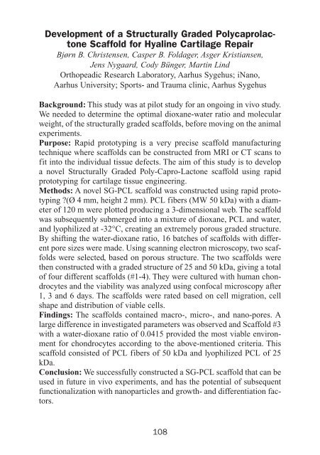
Development of a Structurally Graded Polycaprolactone<br />
Scaffold for Hyaline Cartilage Repair<br />
Bjørn B. Christensen, Casper B. Foldager, Asger Kristiansen,<br />
Jens Nygaard, Cody Bünger, Martin Lind<br />
Orthopeadic Research Laboratory, Aarhus Sygehus; iNano,<br />
Aarhus University; Sports- and Trauma clinic, Aarhus Sygehus<br />
Background: This study was at pilot study for an ongoing in vivo study.<br />
We needed to determine the optimal dioxane-water ratio and molecular<br />
weight, of the structurally graded scaffolds, before moving on the animal<br />
experiments.<br />
Purpose: Rapid prototyping is a very precise scaffold manufacturing<br />
technique where scaffolds can be constructed from MRI or CT scans to<br />
fit into the individual tissue defects. The aim of this study is to develop<br />
a novel Structurally Graded Poly-Capro-Lactone scaffold using rapid<br />
prototyping for cartilage tissue engineering.<br />
Methods: A novel SG-PCL scaffold was constructed using rapid prototyping<br />
?(Ø 4 mm, height 2 mm). PCL fibers (MW 50 kDa) with a diameter<br />
of 120 m were plotted producing a 3-dimensional web. The scaffold<br />
was subsequently submerged into a mixture of dioxane, PCL and water,<br />
and lyophilized at -32°C, creating an extremely porous graded structure.<br />
By shifting the water-dioxane ratio, 16 batches of scaffolds with different<br />
pore sizes were made. Using scanning electron microscopy, two scaffolds<br />
were selected, based on porous structure. The two scaffolds were<br />
then constructed with a graded structure of 25 and 50 kDa, giving a total<br />
of four different scaffolds (#1-4). They were cultured with human chondrocytes<br />
and the viability was analyzed using confocal microscopy after<br />
1, 3 and 6 days. The scaffolds were rated based on cell migration, cell<br />
shape and distribution of viable cells.<br />
Findings: The scaffolds contained macro-, micro-, and nano-pores. A<br />
large difference in investigated parameters was observed and Scaffold #3<br />
with a water-dioxane ratio of 0.0415 provided the most viable environment<br />
for chondrocytes according to the above-mentioned criteria. This<br />
scaffold consisted of PCL fibers of 50 kDa and lyophilized PCL of 25<br />
kDa.<br />
Conclusion: We successfully constructed a SG-PCL scaffold that can be<br />
used in future in vivo experiments, and has the potential of subsequent<br />
functionalization with nanoparticles and growth- and differentiation factors.<br />
108


