DOS BULLETIN - Dansk Ortopædisk Selskab
DOS BULLETIN - Dansk Ortopædisk Selskab
DOS BULLETIN - Dansk Ortopædisk Selskab
Create successful ePaper yourself
Turn your PDF publications into a flip-book with our unique Google optimized e-Paper software.
2010-378_<strong>DOS</strong> nr. 3 2010 29/09/10 10:08 Side 96<br />
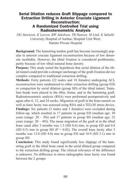
Serial Dilation reduces Graft Slippage compared to<br />
Extraction Drilling in Anterior Cruciate Ligament<br />
Reconstruction:<br />
A Randomized Controlled Trial using<br />
Radiostereometric Analysis<br />
OG Sørensen, K Larsen, BW Jakobsen, TB Hansen, M Lind, K Søballe<br />
University Hospital of Aarhus; Hospital Unit West;<br />
Hamlet Private Hospital<br />
Background: The hamstring tendon graft has become increasingly popular<br />
in anterior cruciate ligament reconstruction because of low donorsite<br />
morbidity. However, the tibial fixation is considered problematic,<br />
partly because of low tibial mineral bone density.<br />
Purpose: This study tested the hypothesis that serial dilation of the tibial<br />
tunnel could provide a stronger anchorage of the graft-fixation-device<br />
complex compared to traditional extraction drilling.<br />
Methods: Forty patients (22 males and 18 females) undergoing ACL<br />
reconstruction were randomized to either extraction drilling (group ED)<br />
or compaction by serial dilation (group SD) of the tibial tunnel. Tantalum<br />
beads were placed in the tibia, femur, and in the hamstring graft.<br />
Radiostereometric analysis (RSA) were performed postoperatively and<br />
again after 6, 12, and 24 weeks. Migration of graft in the bone tunnels as<br />
well as knee laxity was assessed using RSA and a TELOS stress device.<br />
Findings: Six patients (3 males and 3 females) were excluded during<br />
follow-up, which resulted in 17 patients in group ED (median age: 30<br />
years (range: 20 – 50)) and 17 patients in group SD (median age: 32<br />
years (range: 20 – 49)). The mean migration of the graft at in the tibial<br />
bone canal after 3 months was 1.3 (SD 0.6) mm, in group ED and 0.8<br />
(SD 0.5) mm in group SD (P = 0.02). The overall knee laxity after 3<br />
months was 13.0 (SD 4.0) mm in group ED and 10.9 (SD 3.1) mm in<br />
group SD.<br />
Conclusion: This study found significantly less slippage of the hamstring<br />
graft in the tibial bone canal in the serial dilated group compared<br />
to the extraction drilling group. The clinical relevance of the difference<br />
is unknown. No difference in stress radiographic knee laxity was found<br />
between the 2 groups.<br />
96


