
Create successful ePaper yourself
Turn your PDF publications into a flip-book with our unique Google optimized e-Paper software.
FF<br />
prima LOK<br />
facet fixation system<br />
primaLOK FF FACET FIXATION SURGICAL TECHNIQUE GUIDE<br />
page 1
FF<br />
prima LOK<br />
Description<br />
The OSTEOMED SPINE PrimaLOK FF Facet Fixation System is designed to stabilize the posterior elements<br />
of a spinal level as an adjunct to fusion in the lumbar spine. These facet screws are cannulated to assist in<br />
placement and have a cancellous thread form for fixation into the pedicle. The 4.5mm diameter PrimaLOK<br />
FF screw implant assembly is available in 25mm, 30mm, 35mm, 40mm and 45mm lengths to accommodate<br />
variations in anatomy. The PrimaLOK FF Facet Fixation System provides instruments to facilitate<br />
proper placement of the screws. These include an access needle, guide wire, wire stiffener, cannula inserter<br />
assembly, standard cannula, EMG cannula, cannulated tap, cannulated drill, ratcheting driver handle, implant<br />
driver, threaded implant driver shaft - cannulated, threaded implant driver shaft - solid, and implant removal<br />
driver.<br />
Indications for Use<br />
The OSTEOMED SPINE PrimaLOK FF Facet Fixation System is intended to stabilize the spine as an aid to<br />
fusion through bilateral immobilization of the facet joints. It is intended for use with or without bone graft, at<br />
a single or multiple levels from L1 to S1 inclusive.<br />
The OSTEOMED SPINE PrimaLOK FF Facet Fixation System is indicated for the posterior surgical treatment<br />
of any or all of the following: degenerative disc disease (DDD) as defined by back pain of discogenic<br />
origin with degeneration of the disc confirmed by history and radiographic studies, degenerative disease of<br />
the facets with instability, trauma (i.e., fracture or dislocation), spondylolisthesis, spondylolysis, and pseudarthrosis<br />
and failed fusions which are symptomatic or which may cause secondary instability or deformity.<br />
For transfacet fixation, the screws are inserted through the inferior articular process across the facet joint and<br />
into the pedicle.<br />
Warnings<br />
Use of an undersized device in an area of high functional stresses may lead to implant fracture and failure.<br />
Plates, rods and screws, wires, or other appliances of dissimilar metals should not be used together in or near<br />
the implant site. The PrimaLOK FF Facet Fixation System has not been evaluated for safety and compatibility<br />
in the MR environment. The PrimaLOK FF Facet Fixation System has not been tested for heating<br />
or migration in the MR environment. This device is not intended for fixation to the posterior elements of the<br />
cervical or thoracic spine. Based on fatigue testing results, the physician/surgeon should consider the levels<br />
of implantation, patient weight, patient activity level, other patient conditions, etc. which may impact the performance<br />
of the system. <strong>OsteoMed</strong> single use devices cannot be reused and/or reprocessed. The device has<br />
been labeled as single use only for patient safety. The design of the device and the intricacies of the surfaces<br />
may not facilitate cleaning and sterilization after contact with body tissues or fluids, so there is an increased<br />
risk of contamination if reused. This may lead to potential risks of cross infection/contamination associated<br />
with using inadequately cleaned and sterilized devices. Because this device has not been validated for multiple<br />
use, <strong>OsteoMed</strong> cannot guarantee the safety and effectiveness of the device if it is used on more than<br />
one patient. It is recommended to remove any fractured implants from patients during surgery. If unable to<br />
remove, notify patient. Pre-Plan to avoid damage to vital soft tissue and ensure guide wires are not implanted<br />
too deep per the preplanned location. Ensure during procedure that the guide wire does not advance further<br />
than the planned location. If guide wire is inadvertently removed during procedure, reinsert by repeating<br />
surgical steps in surgical technique guide.<br />
page 2
FF<br />
Table of Contents<br />
prima LOK<br />
Implant Anatomy ............................................................ Page 3<br />
Instrumentation .............................................................. Page 4<br />
Patient Positioning .......................................................... Page 5<br />
Targeting Anatomy .......................................................... Page 6<br />
<strong>Surgical</strong> Access & <strong>Guide</strong> Wire Placement ............................ Page 8<br />
Dilation & Cannula Insertion ............................................. Page 9<br />
Drilling ...................................................................... Page 13<br />
Tapping ....................................................................... Page 14<br />
Implant Placement ......................................................... Page 15<br />
Removal ...................................................................... Page 18<br />
Contraindications and Complications .................................. Page 20<br />
Product Insert ............................................................... Page 21<br />
The following surgical technique is for illustrative purposes only. As with all orthopedic surgical procedures, the technique used<br />
in each case will depend on the surgeon’s medical judgment as to the best treatment for each patient. Detailed pre-operative<br />
planning is essential. Preoperative diagnostic evaluation followed by a carefully executed surgical technique is required. Only<br />
those individuals with specialized training and experience in spinal surgery should attempt use of the Osteomed PrimaLok<br />
FF Facet Fixation System. Refer to the instructions for use for the complete list of indications, contraindications, warnings,<br />
cautions and other information about the system.<br />
page 3
FF<br />
Implant Anatomy<br />
prima LOK<br />
Cannulated for percutaneous delivery<br />
Low profile polyaxial washer<br />
with counter-rotation lock-out<br />
Self tapping screw thread profile<br />
Full range of screw lengths<br />
PrimaLOK FF 4.5mm Facet<br />
Fixation screws are available in 25,<br />
30, 35, 40, and 45mm lengths to<br />
accommodate variations in vertebral<br />
body sizes, and pedicle<br />
page 4
FF<br />
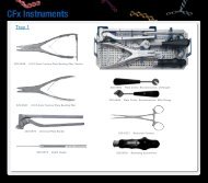
Instrumentation<br />
prima LOK<br />
800-1211 PrimaLOK FF Implant Driver<br />
800-1212 PrimaLOK FF Threaded Implant Driver Shaft - Cannulated<br />
800-1213 PrimaLOK FF Threaded Implant Driver Shaft - Solid<br />
800-1210 PrimaLOK FF Cannula Inserter<br />
800-1205 PrimaLOK FF Standard Cannula<br />
800-1206 PrimaLOK FF EMG Cannula<br />
800-1215 PrimaLOK FF Ratcheting Driver Handle<br />
800-1201 PrimaLOK FF 3.0mm Cannulated Drill<br />
800-1202 PrimaLOK FF 4.5mm Cannulated Tap<br />
800-1203 PrimaLOK FF Removal Driver<br />
800-1204 PrimaLOK FF <strong>Guide</strong> Wire Stiffener<br />
800-1207 PrimaLOK FF Access Needle<br />
800-1200 PrimaLOK FF <strong>Guide</strong> Wire 20” x .053”<br />
page 5
FF<br />
Patient Positioning<br />
prima LOK<br />
Patient Positioning<br />
Position the patient prone on a radiolucent<br />
table. Ensure adequate clearance around the<br />
surgical table for movement of the fluoroscopic<br />
C-arm.<br />
If possible, use bi-planar fluoroscopy to make the surgery quicker and smoother. If the room set up<br />
allows, bring the lateral c-arm from the foot of the bed, and the AP c-arm should come in obliquely from<br />
the head of the bed.<br />
page 6
FF<br />
Targeting Anatomy<br />
prima LOK<br />
Use AP and Lateral fluoroscopy to target the appropriate spinal level, the start point on the facet, the<br />
trajectory of the implant, and the entry point on the skin.<br />
The AP view should be adjusted to the lordosis of the level being treated. Place the c arm into a Ferguson<br />
angle so the inferior end plate of the superior level being treated appears as a single line.<br />
To determine the entry point on the facets indentify and mark the following. Using a guide wire or similar<br />
tool locate the midline of the spine, the inferior endplate of the superior vertebral body being treated, and<br />
a line connecting the medial borders of the pedicles of the levels above and below the level being treated.<br />
The intersection of these two lines represents the docking point for the PrimaLOKFF access needle.<br />
page 7
FF<br />
Targeting Anatomy<br />
prima LOK<br />
Switching to a lateral view, determine the trajectory of the access needle. This will help determine the<br />
entry point thru the skin. The entry point is usually a level or two above the level being treated. The<br />
trajectory needs to go thru the superior and inferior facet, and continue thru the center of the pedicle<br />
into the vertebral body.<br />
Once you have determined your docking point on the facet and your lateral trajectory, a mid line entry<br />
point can be established. In the AP view, mark the point on the mid line where a line from the medial<br />
half of the pedicle through your docking point on the facet intersects midline with the same line from<br />
the opposite side.<br />
page 8
FF<br />
<strong>Surgical</strong> Access & <strong>Guide</strong> Wire Placement<br />
prima LOK<br />
Once the entry point is determined, make a<br />
small midline stab incision to introduce the<br />
access needle. The trajectory of the implant<br />
will be medial to lateral. The medial to lateral<br />
trajectory will be roughly 10°.<br />
Insert the single bevel access needle with the bevel facing up. This will help prevent the needle from<br />
skiving off the facet. Advancing of the needle through the skin incision is accompanied by repeated AP<br />
and lateral fluoroscopy to confirm precise placement of the access needle.<br />
Once precise placement is confirmed, tap the access needle into the cortex of the superior facet.<br />
page 9
FF<br />
Dilation & Cannula Insertion<br />
prima LOK<br />
At this point the access needle can be carefully advanced through the superior facet and into the inferior<br />
facet, or the inner obturator of the access needle can be removed and guide wire inserted. This will be<br />
dependent upon quality of bone and surgeon preference. The guide wire can be driven manually or under<br />
the assistance of a power wire driver. Drive the guide wire across the facet joint and through the center<br />
of the pedicle into the posterior third of the inferior vertebral body. The wire stiffener placed over the wire<br />
at the proximal end of the access needle will help prevent wire bending.<br />
Advancement of the guide wire should be accompanied by a series of fluoroscopy images to insure the<br />
wire does not change trajectory as it passed across the facet joint and into the pedicle. Once the guide<br />
wire is placed remove the access needle.<br />
page 10
FF<br />
Dilation & Cannula Insertion<br />
prima LOK<br />
With the guide wire placed, extend the skin incision and facial incisions to 15 mm for placement of the<br />
dilator and working cannula. The facial incision needs to be incised inferiorly to the skin incision to allow<br />
proper placement of the cannula introducer.<br />
Prepare the cannula introducer by sliding the gold cannula over the dilator shaft while the knob is in<br />
position #2. Lock the cannula into place by turning the knob to position #1.<br />
The cannula introducer is placed over the guide wire and advanced until the dilator is docked on the<br />
facet.<br />
page 11
FF<br />
Dilation & Cannula Insertion<br />
prima LOK<br />
1. With the dilator docked on the facet turn the knob to position #2. This unlocks the cannula.<br />
2. Press the handle down, sliding the cannula into place.<br />
page 12
FF<br />
Dilation & Cannula Insertion<br />
prima LOK<br />
3. With downward pressure, hold the gold cannula in place and carefully pull up on the handle,<br />
removing the dilator. Make sure that as the Cannula Introducer is removed that the guide wire stays<br />
in place.<br />
The working cannula is now placed and is a clear working channel to drill, tap the facet, and place the<br />
implant.<br />
page 13
FF<br />
Drilling<br />
prima LOK<br />
Next the drill shaft is placed on the handle and passed over the guide wire. Confirm with fluoroscopy the<br />
trajectory and depth as drilling.<br />
Note: This can also be done under power. Make sure the drill doesn’t advance the guide wire as the<br />
drill advances.<br />
Advance the drill roughly 30mm, to the junction of the pedicle and the vertebral body.<br />
page 14
FF<br />
Tapping (Optional)<br />
prima LOK<br />
NOTE: Tapping is optional. The PrimaLOK FF Facet Screw is self tapping.<br />
Seat the tap into the handle, pass over the guide wire, and tap to the desired depth. Tapping needs to<br />
be done by hand. Confirm with fluoroscopy the trajectory and depth as tapping. Make sure the tap<br />
doesn’t advance the guide wire as the tap is advanced. As a visual reference, the threaded portion of<br />
the tap is 20 mm. This can help the surgeon confirm the desired screw length.<br />
page 15
FF<br />
Implant Placement<br />
prima LOK<br />
Attach the desire implant to the screw driver. The screw driver inner shaft is designed with a threaded<br />
distal tip to retain the screw to the driver. This insures the screw will not disengage the driver while being<br />
passed over the guide wire. Seat the tip of the driver into the head of the screw and rotate the knob on<br />
the proximal end of the driver clockwise to capture the threads in the head of the screw<br />
Pass the screw and driver over the guide wire. The guide wire can be removed once the screw is into<br />
the vertebral body.<br />
NOTE: If the screw is being placed under direct visualization and a guide wire is not utilized, the optional<br />
solid non-cannulated threaded implant driver shaft should be used to retain the screw to the driver. Since<br />
this shaft is solid and not cannulated, it may not be used over a guide wire.<br />
page 16
FF<br />
Implant Placement<br />
prima LOK<br />
Confirm with fluoroscopy the trajectory and depth as the screw is advanced over the guide wire. Careful<br />
not to advance the guide wire as the screw is advanced.<br />
Placement of the implant should be confirmed using a series of fluoroscopy images.<br />
page 17
FF<br />
Implant Placement<br />
prima LOK<br />
Once screw placement has confirmed, the screw driver can be disengaged from the<br />
implant. Turn the knob on the proximal end of the screw driver counter clockwise to unthread the inner<br />
retention shaft from the screw.<br />
1. 2.<br />
page 18
FF<br />
Removal<br />
prima LOK<br />
To remove the PrimaLOK FF Facet Screw use the Removal Driver seated into the Ratcheting Driver<br />
Handle. Gain access to the head of the screw through a small incision.<br />
800-1215 PrimaLOK FF Ratcheting Driver Handle<br />
800-1203 PrimaLOK FF Removal Driver<br />
Using the solid Removal Driver, engage the Driver tip with the screw head and rotate counterclockwise.<br />
Due to the initial lagging of the screw into the bone, and the anti rotation mechanism in the washer, a<br />
fair amount of resistance may be encountered to break the screw, washer, and bone interfaces.<br />
page 19
FF<br />
Removal<br />
prima LOK<br />
Note: The Removal Driver is a non-cannulated instrument. Do not use the PrimaLOK FF Implant<br />
Driver and Threaded Implant Driver Shaft to remove a screw. Do not try and re-engage the threads of<br />
the Threaded Implant Driver Shaft with the threads in the head of the screw, as cross threading may<br />
occur, damaging the Threaded Implant Driver Shaft. If the threaded tip of the implant driver shaft<br />
breaks in the screw, the screw can be still be removed using the implant driver without the implant<br />
driver shaft.<br />
page 20
FF<br />
prima LOK<br />
Contraindications<br />
Contraindications may be relative or absolute. The choice to implant the PrimaLOK FF Facet Fixation<br />
device must be carefully weighed against the patient’s overall evaluation. Circumstances listed below<br />
may reduce the chance of a successful outcome. Contraindications include, but are not limited to:<br />
• Allergy or sensitivity to Titanium or Titanium Alloy<br />
• Active or suspected infection<br />
• Patients who are immune-compromised<br />
• Any condition that may affect the process of normal bone remodeling, including, but not limited to<br />
osteoporosis, bone absorption, osteopenia, or certain metabolic disorders affecting osteogenesis.<br />
• Morbid obesity<br />
• Signs of local infection or inflammation.<br />
• The PrimaLOK FF Facet Fixation System is also contraindicated where an anatomical deficit exists<br />
leaving an absence or destruction of any portion of the facet joint, pars defect, or in conjunction with<br />
procedures which require removal of any portion of the facet joint.<br />
• Spondylolisthesis > grade 1<br />
• Alcoholism or heavy smoking<br />
• Pregnancy<br />
• Any case requiring the mixing of metals from two different systems.<br />
• Any patients exhibiting disorders which would cause the patient to ignore the limitations of<br />
rigid fixation screw implants.<br />
Possible Complications<br />
Possible complications specific to the device may include:<br />
• Implant breakage, failure, loosening, or migration<br />
• Bone fracture or fracture to the spinous process<br />
• Allergic reaction to the implant material<br />
Other general complications associated with any spinal surgery may include:<br />
• Pseudoarthrosis<br />
• Pain<br />
• Revision surgery<br />
• Bleeding<br />
• Infection, early or late<br />
• Tissue or nerve damage<br />
• Spinal fluid leakage<br />
• Scar formation<br />
• Complications due to the use of bone grafting, including donor site complications<br />
page 21
FF<br />
Product Insert<br />
prima LOK<br />
OSTEOMED SPINE<br />
Facet Fixation System<br />
PrimaLOK TM FF<br />
Product Information and Instructions for Use<br />
Description<br />
The OSTEOMED SPINE PrimaLOK TM FF Facet Fixation System is designed to stabilize the<br />
posterior elements of a spinal level as an adjunct to fusion in the lumbar spine. These facet<br />
screws are cannulated to assist in placement and have a cancellous thread form for fixation into<br />
the pedicle. The 4.5mm diameter PrimaLOK TM FF screw implant assembly is available in 25mm,<br />
30mm, 35mm, 40mm and 45mm lengths to accommodate variations in anatomy. The<br />
PrimaLOK TM FF Facet Fixation System provides instruments to facilitate proper placement of the<br />
screws. These include an access needle, guide wire, wire stiffener, cannula inserter assembly,<br />
standard cannula, EMG cannula, cannulated tap, cannulated drill, ratcheting driver handle,<br />
implant driver, threaded implant driver shaft-cannulated, threaded implant driver shaft- solid and<br />
implant removal driver.<br />
Material<br />
The facet fixation implant assembly device is made from Titanium Alloy (ASTM F-136) and<br />
Titanium (ASTM F-67). The instrumentation is made from various grades of stainless steel, TiN<br />
coated stainless steel, titanium alloy (ASTM F-136), anodized aluminum, and/or medical grade<br />
plastic.<br />
Clinical Indications<br />
The OSTEOMED SPINE PrimaLOK TM FF Facet Fixation System is intended to stabilize the spine<br />
as an aid to fusion through bilateral immobilization of the facet joints. It is intended for use with<br />
or without bone graft, at a single or multiple levels from L1 to S1 inclusive,<br />
The OSTEOMED SPINE PrimaLOK TM FF Facet Fixation System is indicated for the posterior<br />
surgical treatment of any or all of the following: degenerative disc disease (DDD) as defined by<br />
back pain of discogenic origin with degeneration of the disc confirmed by history and<br />
radiographic studies, degenerative disease of the facets with instability, trauma (i.e., fracture or<br />
dislocation), spondylolisthesis, spondylolysis, and pseudarthrosis and failed fusions which are<br />
symptomatic or which may cause secondary instability or deformity.<br />
For transfacet fixation, the screws are inserted through the inferior articular process across the<br />
facet joint and into the pedicle.<br />
Contraindications<br />
Contraindications may be relative or absolute. The choice to implant the PrimaLOK TM FF Facet<br />
Fixation Device must be carefully weighed against the patients overall evaluation.<br />
Circumstances listed below may reduce the chance of a successful outcome. Contraindications<br />
include, but not limited to:<br />
1. Allergy or sensitivity to Titanium or Titanium Alloy<br />
2. Active or suspected infection<br />
3. Patients who are immune-compromised<br />
4. Any condition that may affect the process of normal bone remodeling, including, but not<br />
limited to osteoporosis, bone absorption, osteopenia, or certain metabolic disorders<br />
affecting osteogenesis.<br />
5. Morbid obesity<br />
6. Signs of local infection or inflammation.<br />
7. The PrimaLOK TM FF Facet Fixation System is also contraindicated where an anatomical<br />
deficit exists leaving an absence or destruction of any portion of the facet joint, pars defect,<br />
or in conjunction with procedures which require removal of any portion of the facet joint.<br />
8. Spondylolisthesis > grade 1<br />
9. Alcoholism or heavy smoking<br />
10. Pregnancy<br />
11. Any case requiring the mixing of metals from two different systems.<br />
12. Any patients exhibiting disorders which would cause the patient to ignore the limitations of<br />
rigid fixation screw implants.<br />
Possible Complications<br />
Possible complications specific to the device may include:<br />
1. Implant breakage, failure, loosening, or migration<br />
2. Bone fracture or fracture to the spinous process<br />
3. Allergic reaction to the implant material<br />
Other general complications associated with any spinal surgery may include:<br />
1. Pseudoarthrosis<br />
2. Pain<br />
3. Revision surgery<br />
4. Bleeding<br />
5. Infection, early or late<br />
6. Tissue or nerve damage<br />
7. Spinal fluid leakage<br />
8. Scar formation<br />
9. Complications due to the use of bone grafting, including donor site complications.<br />
Warnings<br />
1. Use of an undersized device in an area of high functional stresses may lead to implant<br />
fracture and failure.<br />
2. Plates, rods and screws, wires, or other appliances of dissimilar metals should not be used<br />
together in or near the implant site.<br />
3. The PrimaLOK TM FF Facet Fixation System has not been evaluated for safety and<br />
compatibility in the MR environment. The PrimaLOK TM FF Facet Fixation System has not<br />
been tested for heating or migration in the MR environment.<br />
4. This device is not intended for fixation to the posterior elements of the cervical or thoracic<br />
spine.<br />
5. Based on fatigue testing results, the physician/surgeon should consider the levels of<br />
implantation, patient weight, patient activity level, other patient conditions, etc. which may<br />
impact the performance of the system.<br />
6. <strong>OsteoMed</strong> single use devices cannot be reused and/or reprocessed. The device has been<br />
labeled as single use only for patient safety. The design of the device and the intricacies of<br />
the surfaces may not facilitate cleaning and sterilization after contact with body tissues or<br />
fluids, so there is an increased risk of contamination if reused. This may lead to potential<br />
risks of cross infection/contamination associated with using inadequately cleaned and<br />
sterilized devices. Because this device has not been validated for multiple use, <strong>OsteoMed</strong><br />
cannot guarantee the safety and effectiveness of the device if it is used on more than one<br />
patient.<br />
7. It is recommended to remove any fractured implants from patients during surgery. If unable<br />
to remove, notify patient.<br />
8. Pre-Plan to avoid damage to vital soft tissue and ensure guide wires are not implanted too<br />
deep per the preplanned location.<br />
9. Ensure during procedure that the guide wire does not advance further than the planned<br />
location.<br />
10. If guide wire is inadvertently removed during procedure, reinsert by repeating surgical steps<br />
in surgical technique guide (030-0806).<br />
Maintaining Device Effectiveness<br />
1. The surgeon should have specific training, experience, and thorough familiarity with the use<br />
of rigid fixation products and techniques.<br />
2. The surgeon must exercise reasonable judgment when deciding which implant sizes to use<br />
for specific indications.<br />
3. The OSTEOMED SPINE PrimaLOK TM FF Facet Fixation System is not intended to endure<br />
excessive abnormal functional stresses.<br />
4. Failure to use dedicated, unique OSTEOMED SPINE Instruments for every step of the<br />
implantation technique may compromise the integrity of the implanted device, leading to<br />
premature device failure and subsequent patient injury. Failed devices may require reoperation<br />
and removal.<br />
5. Carefully inspect the OSTEOMED SPINE instruments before and after each procedure to<br />
assure they are in proper operation condition. Instruments which are faulty, damaged, or<br />
suspect should not be used. They should be replaced or sent to OSTEOMED SPINE for<br />
disposition and repair.<br />
6. All the instruments are reusable with the exception of the access needle and guide wires.<br />
7. OSTEOMED SPINE recommends the use of OSTEOMED SPINE products in a sterile<br />
environment.<br />
Instructions for Use, PrimaLOK TM FF Facet Fixation System<br />
Attention, See Instructions For Use located in the <strong>Surgical</strong><br />
Technique guide, P/N 030-0806, available at no charge or<br />
Full IFU 030-1668<br />
Removal of PrimaLOK TM FF Implant Assembly<br />
1. To remove the PrimaLOK TM FF Screw use the Removal Driver seated into the Ratcheting<br />
Driver Handel. Gain Access to the head of the screw through a small incision.<br />
2. Using the solid Removal Driver, engage the Driver tip with the screw head and rotate<br />
counterclockwise. Due to the initial lagging of the screw into the bone, and the anti rotation<br />
mechanism in the washer, a fair amount of resistance may be encountered to break the<br />
screw, washer, and bone interfaces.<br />
Note: The Removal Driver is a non-cannulated instrument. Do not use the PrimaLOK TM FF<br />
Implant Driver and Threaded shaft to remove the screw. Do not try to re-engage the<br />
threads of the Threaded Implant Driver Shaft with the threads in the head of the screw, as<br />
cross threading may occur, damaging the Threaded Implant Driver Shaft.<br />
Note: If the tip of the internal implant driver shaft breaks in the screw, the screw can be<br />
removed by using the Implant driver without the Threaded Implant Driver Shaft.<br />
Cleaning<br />
Instruments must be carefully cleaned prior to sterilization. Trained personnel must<br />
perform cleaning and mechanical inspection prior to sterilization.<br />
Compliance is required with the equipment manufacturer’s user instructions (manual and/or<br />
machine cleaning, ultrasound treatment, etc.) and recommendations for chemical<br />
detergents.<br />
OSTEOMED recommends the following cleaning and sterilization instructions for<br />
Instrumentation:<br />
1. Prepare an enzymatic detergent as Enzol® according to the manufacturer’s<br />
recommendations at 1oz/gal of lukewarm tap water.<br />
2. Using a clean soft cloth that has been soak in the detergent, wipe the entire article.<br />
3. Immerse the article in the detergent. Flush with 50mL all hard to reach areas using a<br />
syringe with the prepared detergent. Allow the article to soak for 15 minutes.<br />
4. Using a soft bristled brush, brush the entire surface of the article paying close<br />
attention to all hard to clean areas such as cracks, crevices and matted surfaces.<br />
Actuate the article while brushing to ensure the reach of all areas. Use a syringe to<br />
flush any lumen or matted surface with at least 50mL of prepared detergent.<br />
5. Brush any lumen a minimum of 5 times from both ends using a lumen brush or similar.<br />
6. Rinse the articles in RO/DI (reverse-osmosis/deionized) water until all visible evidence<br />
of detergent is removed. Flush any lumens or matted surfaces with the RO/DI water.<br />
Once all evidence of detergent is removed continue to rinse for an additional 30<br />
seconds.<br />
7. Drain excess water from the article and dry using a clean soft cloth or filtered<br />
pressurized air.<br />
8. Prepare a non-enzymatic detergent such as Renu-Klenz TM per manufacturer’s<br />
recommendations at ¼ oz/gal of lukewarm tap water.<br />
9. Fully immerse the articles in the detergent and flush all lumens with at least 50mL of<br />
the detergent.<br />
10. Allow the articles to soak for a minimum of 15 minutes.<br />
11. Thoroughly brush the exterior of the articles using a soft bristled brush. Flush all<br />
lumens and matted surfaces a minimum of 5 times.<br />
12. Prepare a fresh cleaning detergent such as Renu-Klenz TM in an ultra sonic cleaner.<br />
Fully immerse the articles in the solution and allow the articles to sonicate for a<br />
minimum of 10 minutes.<br />
13. Rinse the articles in RO/DI water until all visible evidence of detergent is removed.<br />
Flush any lumens or matted surfaces with the RO/DI water. Once all evidence of<br />
detergent is removed continue to rinse for an additional 30 seconds.<br />
14. Drain excess water from the article and dry using a clean soft cloth or filtered<br />
pressurized air.<br />
15. Visually examine each instrument for cleanliness. If visible soil is noted on the<br />
instruments, repeat cleaning procedure.<br />
16. Steam Autoclave per the following Sterilization Instructions.<br />
Sterilization Instructions<br />
Sterility<br />
1. Facet Fixation Screw Devices are supplied STERILE.<br />
2. Instrumentation is supplied NON-STERILE.<br />
3. Use of the sterilizer shall comply with the manufacturer’s user instructions for sterilizers.<br />
4. The user facility must clean and disinfect devices prior to sterilization per manual or<br />
mechanical cleaning options listed above or per standard hospital procedures.<br />
5. Non-sterile devices should be sterilized by steam sterilization (autoclaving). For<br />
sterilization of OSTEOMED PrimaLOK TM FF implant systems, the following parameters<br />
should be used.<br />
TRAY SYSTEM:<br />
Pre-Vacuum Steam Sterilization<br />
Preconditioning Pulses: 3<br />
Temperature: 270 º F (132 º C)<br />
Cycle Time: 10 minutes<br />
Dry Time: 30 minutes<br />
Configuration: Wrapped in two layers of 1-ply polypropylene wrap (Kimguard KC600-510(k)<br />
K082554) using sequential wrapping techniques.<br />
Do not exceed 275 º F (135 º C), to avoid compromising functions of polymeric instrumentation.<br />
Caution<br />
Federal (United States) law restricts this device for sale by or on the order of a<br />
medical practitioner licensed to do so.<br />
Do not attempt a surgical procedure with faulty, damaged, or suspect <strong>OsteoMed</strong><br />
instruments or implants. Inspect all components preoperatively to assure utility.<br />
Alternate fixation methods should be available intraoperatively.<br />
<strong>OsteoMed</strong> L.P.<br />
3885 Arapaho Road<br />
Addison, Texas 75001 USA<br />
Customer Service: 800/456-7779<br />
Outside USA: 972/677-4600<br />
Single Use<br />
Only<br />
Use By<br />
(Date)<br />
Symbols and Definitions<br />
Batch Code<br />
(Lot Number)<br />
Date of<br />
Manufacture<br />
Attention,<br />
See Instructions For Use<br />
Caution, Consult<br />
Accompanying<br />
Documents<br />
REF<br />
European Representative:<br />
Shotwell & Carr<br />
25 Baker Close<br />
Fishbourne, Chichester<br />
West Sussex, UK P018 8BJ<br />
Tel: +44 (0) 1243 779 550<br />
Catalogue<br />
Number<br />
Do Not use if Sterile<br />
package is damaged<br />
Consult Instructions<br />
For Use<br />
Authorized Representative in<br />
the European Community<br />
Manufacturer<br />
Sterile, Method of<br />
Sterilization Using<br />
Irradiation<br />
OSTEOMED SPINE, Inc. is a wholly owned subsidiary of <strong>OsteoMed</strong> L.P.<br />
Part No.030-1669<br />
Printed in U.S.A.<br />
Rev. B OSTEOMED July 2011<br />
page 22
FF<br />
prima LOK<br />
page 23<br />
3885 Arapaho Road<br />
Addison, TX 75001<br />
Customer Service|800.456.7779<br />
Fax|800.390.2620<br />
www.osteomed-spine.com<br />
030-0806 Rev.C