View Document - OSTI
View Document - OSTI
View Document - OSTI

You also want an ePaper? Increase the reach of your titles
YUMPU automatically turns print PDFs into web optimized ePapers that Google loves.
-_-<br />
Donner Laboratory<br />
Division of Biology and Medicine<br />
of the<br />
Lawrence Berkeley Laboratory<br />
University of California<br />
Berkeley, CA<br />
I<br />
COVER
DISCLAIMER<br />
This report was prepared as an account of work sponsored by an<br />
agency of the United States Government. Neither the United States<br />
Government nor any agency Thereof, nor any of their employees,<br />
makes any warranty, express or implied, or assumes any legal<br />
liability or responsibility for the accuracy, completeness, or<br />
usefulness of any information, apparatus, product, or process<br />
disclosed, or represents that its use would not infringe privately<br />
owned rights. Reference herein to any specific commercial product,<br />
process, or service by trade name, trademark, manufacturer, or<br />
otherwise does not necessarily constitute or imply its endorsement,<br />
recommendation, or favoring by the United States Government or any<br />
agency thereof. The views and opinions of authors expressed herein<br />
do not necessarily state or reflect those of the United States<br />
Government or any agency thereof.
DISCLAIMER<br />
Portions of this document may be illegible in<br />
electronic image products. Images are produced<br />
from the best available original document.
~ DE88<br />
I<br />
LBL-PUB--268<br />
001404 ,<br />
,<br />
I<br />
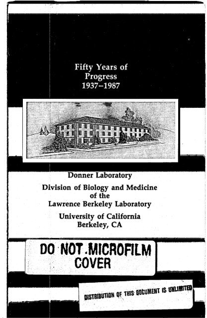
Fifty Years of Progress<br />
1937-1987<br />
Donner Laboratory<br />
Division of Biology and Medicine<br />
of the<br />
. Lawrence Berkeley Laboratory<br />
University of California<br />
Berkeley<br />
DISCLAIMER<br />
This report was prepared as an account of work sponsored by an agency of the United States<br />
Government. Neither the United States Government nor any agency thereof, nor any of their<br />
employees, makes any warranty, express or implied, or assumes any legal liability or respnsibility<br />
for the accuracy, completeness, or usefulness of any information, apparatus, product, or<br />
process discld, or represents that its use would not infringe privately owned rights. Reference<br />
herein to any specific commercial product, process, or service by trade name, trademark,<br />
manufacturer, or otherwise docs not necessarily .constitute or 'imply its -endorsement, recommendation,<br />
or favoring by the United States Government or any agency thereof. The vkws<br />
and opinions of authors expressed herein do not necessarily state or reflect those of the<br />
United States Government or any agency thereof.
Preface<br />
This booklet was prepared for the 50th anniversary of medical and<br />
biological research at the Donner Laboratory and the Lawrence Berkeley<br />
Laboratory of the University of California. The intent is to present historical<br />
facts and to highlight important facets of fifty years of accomplishments<br />
in medical and biological sciences. A list of selected scientific<br />
publications from 1937 to 1960 is included to demonstrate the character<br />
and lasting importance of early pioneering work.<br />
Dr. William Myers, Dr. Patricia Durbin and Ms. Jan DeMoor provided<br />
much of the historical material. Julie Twitchell, Ralph Dennis,<br />
and Loretta Lizama assisted in the technical pioduction. The organizational<br />
concept is tcz show the research themes. starting with thehistory,<br />
’<br />
then discoveries of medically important ractionudrdes, Itlien the use of<br />
accelerated charged particles in therapy, next human physiology studies<br />
then sequentially studies of biology from’ tissues to macromolecules; and<br />
finally studies of the genetic code.<br />
Unfortunately, space and time did not permit us to acknowledge<br />
all the great scientists and represent accurately their contributions in this<br />
booklet. We hope the information in this booklet will further inform<br />
and inspire our colleagues and students.<br />
Thomas F. Budinger<br />
Editor
John H. Lawrence<br />
4
Table of Contents<br />
Radiation Laboratory ............................................................................................................. 5<br />
Cmcker Laboratory ................................................................................................................ 7<br />
Donner Laboratory ................................................................................................................. 9<br />
Department of Biophysics and Medical Physics ............................................................... 10<br />
Historical Bibliography ....................................................................................................... 10<br />
Radionuclide Discoveries and Early Uses ......................................................................... 14<br />
"'I discovery ..................................................................................................................... 14<br />
"'I thyroid study ............................................................................................................... 15<br />
Discovery of radionuclides .................................................................................................. 16<br />
Discovery of Technetium-99m ............................................................................................ 17<br />
Radionuclide generators ..................................................................................................... 18<br />
Radiation h a d and safety .............................................................................................. 20<br />
Therapy by 9 and =I ........................................................................................................ 21<br />
Heavy Ion Biology & Medicine .......................................................................................... 21<br />
Treatment of disease by radionuclides ................................................................................. 21<br />
Heavy ion interaction physics ............................................................................................. 22<br />
Heavy ion radiation advantages .......................................................................................... 23<br />
Medical use of accelerated charged particles ......................................................................... 24<br />
Tissue effects and laminar lesion production ........................................................................ 25<br />
Patient treatments at the 184" ............................................................................................. 26<br />
Aaomegaly treatment ........................................................................................................ 27<br />
Heavy ions as injected tracers ............................................................................................. 28<br />
Bevalac ............................................................................................................................. 29<br />
Heavy ion therapy advantages ............................................................................................ 29<br />
Heavy ion treatment of tumors ........................................................................................... 30<br />
Heavy ion treatment of arteriovenous malformation ............................................................. 31<br />
Nuclear Medicine and Physiology with Tracers .............................................................. 31<br />
Radioactive tracer studies ............................................................................ . 32<br />
Whole body scanner ..........-.............................................................................................. 33<br />
Hematology ...................................................................................................................... 34<br />
schizophrenia metabolic studies .......................................................................................... 35<br />
Anger scintillation camera .................................................................................................. 36<br />
Hematology with the positron camera ................................................................................. 37<br />
Reconstruction tomography mathematics ............................................................................. 38<br />
Charged partide tomography .............................................................................................. 39<br />
Positron tomography ......................................................................................................... 40<br />
Heart muscle blood flow .................................................................................................... 41<br />
Alzhiemer's disease by PET ................................................................................................ 42<br />
Alzhiemer's disease by NMR .............................................................................................. 43<br />
Body composition studies ................................................................................................... 44<br />
Low Oxygen Physiology and Space Radiation ................................................................. 45<br />
High altitude physiology .................................................................................................... 45<br />
Space radiation and light flash experiments .......................................................................... 47<br />
Magnetic Fields and NMR .................................................................................................. 48<br />
Magnetic field biophysics ................................................................................................... 48<br />
NMR s-py in vivo ................................................................................................. 49<br />
Morphology and Biophysics ................................................................................................ 50<br />
Scanning electron microscope ............................................................................................. 50<br />
Staflow system .................................................................................................................. 52<br />
Macromolecular Biology ...................................................................................................... 52<br />
Radioimmune assay ........................................................................................................... 52<br />
Cell Biology .......................................................................................................................... 52<br />
Erythropoietin ................................................................................................................... 52<br />
Cell cultures and morphology ............................................................................................. 53<br />
Lipoproteins & Atherosclerosis .......................................................................................... 54<br />
Structural Membrane and Molecular Biology ................................................................... 56<br />
Genetics ................................................................................................................................. 58
Beginning<br />
The beginnings of what is now<br />
the Donner Laboratory and the<br />
Division of Biology and Medicine<br />
of the Lawrence Radiation<br />
Laboratory were firmly established<br />
in 1937 by the appointment<br />
of Dr. John H. Lawrence to<br />
the regular staff of the Radiation<br />
Laboratory and the faculty of<br />
University of California Medical<br />
School. The Radiation Laboratory<br />
was an entity under E.O.<br />
Lawrence in 1936.<br />
Prior to that time John Lawrence<br />
was at Berkeley on leave of<br />
absence from Yale University<br />
where, since 1934, he had been<br />
on the Yale Medical School<br />
faculty, but had begun collaborative<br />
work with his brother Ernest<br />
0. Lawrence and Paul A. Aebersold<br />
with work on the use of neutrons<br />
in the treatment of cancer<br />
which Dr. John Lawrence<br />
presented in 1936 before the<br />
American Society of Clinical<br />
Investigations. In 1936 and 1937<br />
members of the Radiation Laboratory<br />
regular staff and visiting fellows<br />
are shown below.<br />
(Left to right and top to bottom): A.S. Langsdorf, S.J. Simmons, J.G. Hamilton,<br />
D.H. Sloan, J.R. Oppenheimer, W.M. Brobeck, R. Comog, R.R. Wilson, E. Viez,<br />
J.J. Livingood, J. Backus, W.B. Mann, P.C. Aebersold, E.M. McMillan,. E.M.<br />
Lyman, M.D. Kamen, D.C. Kalbfell, W.W. Salisbury, J.H. Lawrence, R. Serber,<br />
F.N.D. Kurie, R.T. Birge, E.O. Lawrence, D. Cooksey, A.H. Snell, L.W. Alvarez,<br />
P.H. Abelson.<br />
5
With the completion of the<br />
Crocker cyclotron in 1936,<br />
research in biology and medicine<br />
had a strong emphasis along with<br />
chemistry and physics at the<br />
Radiation Laboratory directed by<br />
E.O. Lawrence. During the f it<br />
twenty years the major categories<br />
of truly pioneering work included<br />
exploration of the use of neutrons<br />
for therapy, establishment of radiation<br />
safety criteria, use of<br />
radioactive tracers for studies of<br />
human physiology and the treatment<br />
of disease, use of<br />
accelerated charged particle beams<br />
for therapy, development of<br />
radionuclide imaging<br />
instrumentation, establishment of<br />
the importance of cholesterol and<br />
lipoprotein abnormalities in heart<br />
disease, and exploration of the<br />
characteristics of yeast as a model<br />
for elucidation of genetic mechanisms.<br />
The early history is documented<br />
by the selected publications<br />
whose titles describe the work<br />
accomplished to 1960.<br />
Drs. Ernest and John Lawrence at the controls of the 60-Inch Cyclotron.<br />
6
Above is the Crocker Laboratory, which housed the Radiation Laboratory before<br />
the facilities at the Donner Laboratory and the Lawrence Berkeley Laboratory<br />
were built. Below is the beam from the 60-Inch Crocker Cyclotron.<br />
7
Donner Laboratory and<br />
Pavilion on the Berkeley<br />
Campus<br />
In 1941, Donner Laboratory was<br />
built at the cost of $650,000 with<br />
financing of $465,000 from the<br />
International Cancer Research<br />
Foundation. This new home for<br />
biologists and physicians was<br />
named in honor of William H.<br />
Donner, who was the president of<br />
the Internationa€ Cancer Research<br />
Foundation, which later was<br />
renamed the Donier Foundation.<br />
In fact the Donner Foundation<br />
had funds contributed entirely, or<br />
almost so, by Donner himself.<br />
Circumstances surrounding this<br />
donation were as follows.<br />
William H. Donner’s son, Robert,<br />
had died of cancer, and as a<br />
result, his father was particularly<br />
interested in medical work on<br />
cancer. He heard about the treatment<br />
of cancer by neutron beams<br />
by Dr. John Lawrence and in<br />
1940 Mr. Donner visited Dr.<br />
Ernest Lawrence and was greatly<br />
impressed with the work in progress.<br />
Near the end of his visit,<br />
Lawrence received $150,000 from<br />
Donner for a building to use for<br />
the work in Medical Physics. The<br />
actual construction of Donner<br />
Laboratory was started on July 21,<br />
1941.<br />
In the course of the decade after<br />
the establishment of Medical Physics,<br />
the increase in research work<br />
on human beings at Berkeley<br />
resulted in the establishment in<br />
1954 of a special ward called The<br />
Donner Pavilion as an addition to<br />
Cowell Hospital to care for such<br />
patients. This was a two-story<br />
addition to the east wing of<br />
Cowell Hospital, finished in 1954<br />
at a cost of $191,000, once again<br />
a gift from the Donner Foundation.<br />
The purpose of the Pavilion<br />
was “for research in<br />
radiobiology under supervision of<br />
the Donner Laboratory.”<br />
Academic Unit-Division<br />
of Medical Physics<br />
The actual beginning of Medical<br />
Physics at the Radiation Laboratory<br />
evolved in two stages. In<br />
August of 1944, initial approval<br />
was given to appoint four faculty<br />
members, Drs. John Lawrence,<br />
Joseph Hamilton, Cornelius<br />
Tobias and Hardin Jones, to the<br />
newly created Division of Medical<br />
Physics, within the administrative<br />
framework of the UC Berkeley<br />
Physics Department. The official<br />
approval was granted by the<br />
Regents effective July 1, 1945<br />
with specific recommendations as<br />
follows:<br />
“(a) There should be a nucleus<br />
of men including Drs.<br />
John Lawrence, Joseph<br />
Hamilton, Aebersold and<br />
Tobias who would hold<br />
joint appointments in the<br />
Medical School and in the<br />
Department of Physics, and<br />
then a second group of people<br />
who would play important<br />
parts in the development<br />
of medical physics,<br />
such as Doctors Miller,<br />
Chaikoff, Hamilton, Anderson,<br />
Robert Aird, Strait,<br />
9<br />
..
Low-Beer, Althausen, David<br />
Greenberg, C.L.A. Schmitt<br />
and Soley. This second<br />
group would be carrying<br />
out experimental studies<br />
and therapeutic studies<br />
through the Medical<br />
School."<br />
There were additional written<br />
expectations and guidelines for<br />
the Division of Medical Physics.<br />
Of great importance was the proposal<br />
by Dr. John Lawrence that a<br />
small clinic in Berkeley could be<br />
considered a medical physics<br />
branch of the outpatient department<br />
of the hospital but<br />
"there should be no limitation<br />
or 'ham-stringing' of<br />
the freedom of the members<br />
of the subdivision of Medical<br />
Physics or others in the<br />
medical school to carry on<br />
treatment or investigations<br />
at Berkeley if research were<br />
the prime interest."<br />
The Directors of Donner Laboratory<br />
in succession are: Dr. John<br />
Lawrence, Dr. James Born, Dr.<br />
Edward Alpen and Dr. Paul<br />
Silverman.<br />
Department of Biophysics<br />
and Medical Physics<br />
The Division of Medical Physics,<br />
though officially under the<br />
administrative aegis of the<br />
Department of Physics, operated<br />
as a self-administered group.<br />
Over a period of 42 years,<br />
academic programs became<br />
closely integrated with undergraduate<br />
and graduate units of the<br />
Berkeley campus under the<br />
leadership of Drs. Lawrence,<br />
Tobias and Jones. In 1979, that<br />
Division became the Department<br />
of Biophysics and Medical Physics<br />
at U.C. Berkeley and along with<br />
the Biophysics Group is responsible<br />
for 105 undergraduate students<br />
and approximately 100 graduate<br />
students.<br />
Achievements and examples of<br />
ongoing activities to 1987 are<br />
illustrated throughout the<br />
remainder of this booklet.<br />
Historical Publications<br />
Lawrence, E.O.<br />
Radioactive Sodium Produced by Deuton<br />
Bombardment. Phys. Rev. 46 746-747 (1934).<br />
Chievitz, 0. and Hevesy, G.<br />
Radioactive Indicators in the Study of Phosphorus<br />
Metabolism in Rats. Nature 136<br />
754-755 (1935).<br />
Lawrence, J.H. and Lawrence, E.O.<br />
The Biological Action of Neutron Rays.<br />
Proc. Natl. Acad. Sci. 22 124-133 (1936).<br />
Hamilton, J.G.<br />
Rates Of Absorption Of Radio-Sodium in<br />
Normal Human SuL+xts. hoc. Nat. Acad. sci.<br />
23: 521-527 (1937).<br />
10
I<br />
Joseph G. Hamilton<br />
I<br />
First medical physiology studies of the dynamics- of sodium transport in the<br />
body by Dr. Hamilton involved observing the amval of "Na in the hand vascular<br />
system after oral ingestion.<br />
1 11
Hamilton, J.G. and Stone, R.S.<br />
Exuetion of Radio-Sodium Following<br />
Intravenous Administration to Man. hac. Soc.<br />
Exptl. Biol. and Med. 35: 595-598 (1937). .<br />
Hamilton, J.G. and Stone, R.S.<br />
The Intravenous and Intraduodenal Administration<br />
of Radio-Sodium. Radiology 28<br />
178-188 (1937).<br />
Lawrence, J.H. and Tennant, R.<br />
Comparative Effects of Neutrons and X-rays<br />
on Whole Body. J. Exper. Med. 66 667-688<br />
(1937).<br />
Livingd, J.J. and Sea- G.T.<br />
Radioactive Antimony Isotopes.<br />
Review 52: 135-136 (1937):<br />
Physical<br />
Livingood, J.J., Seabow G.T., and Fairbrother, F.<br />
Radioactive Isotopes of Manganese, Iron and<br />
Cobalt. Phys. Rev. 5 2 135 (1937).<br />
Hamilton, J.G.<br />
The Rates of Absorption of Radio-Sodium in<br />
Normal Human Subjects. PIOr. Natl. Acad. Sd.<br />
23 521-527 (September 1937).<br />
Hertz, S., Roberts, A. and Evans, R.D.<br />
Radioiodine as Indicator in Study of Thyroid<br />
Physiology. Proc. Soc. Exper. Biol. and Med. 38<br />
510 (1938).<br />
!3e@, E. and Seaborg, G.T.<br />
Nuclear Isomerism in Element 43. Physical<br />
Review 54 772 (1938).<br />
Hamilton, J.G.<br />
The Rates of Absorption of the Radioactive<br />
Isotopes of Sodium, Potassium, Chlorine, Bromine,<br />
and Iodine in Normal Human Subjects.<br />
Am. J. of Physiol. 124 667-678 (1938).<br />
Hamilton, J.G. and Alles, G.A.<br />
The Physiological Action of Natural and<br />
Artificial Radioactivity. American Journal of<br />
Physiology 125: No. 2, 410-413 (1939).<br />
Hamilton, J.G. and Soley, M.H.<br />
Studies in Iodine Metabolism by Use of New<br />
Radioactive Isotope of Iodine. Am. J. Physiol.<br />
127 557 (1939).<br />
Lawrence, J.H., Scott, K.G. and Tuttle, LW.<br />
Shidies on Leukemia with the Aid of<br />
Radioactive Phosphorus. In: New Intl. Clinics,<br />
VoI. III, J.B. Lippincott Co., pp. 35-58 (1939).<br />
Ruben, S., Hassid, W.Z. and Kamen, M.D.<br />
Radioactive Carbon in Study of Photosynthesis.<br />
J. Am. Chem. Soc. 61: 661 (1939).<br />
Ruben, S., Hassid, W.Z. and Kamen, M.D.<br />
Radioactive Nitrogen in the Study of N, Fucation<br />
by Non-leguminous Plants. Science 91:<br />
578-579 (1940).<br />
Seaborg, G.T. and Se@, E.<br />
Nuclear Isomerism in Element 43. Physical<br />
Review 55: 808-814 (1939).<br />
Hamilton, J.G. and Soley, M.H.<br />
Studies in Iodine Metabolism by the Use of a<br />
New Radioactive Isotope of Iodine. Am. J. Physiol.<br />
127: 557-572 (1939).<br />
Hamilton, J.G. and %ley, M.H.<br />
A Comparison of the Metabolism of Iodine<br />
and of Element 85 (EKA-Iodine). Proc. Natl.<br />
Acad. Sd. 26: 483-489 (1940).<br />
Lawrence, J.H.<br />
Nuclear Physics and Therapy: Preliminary<br />
Report on a New Method for the Treatment of<br />
Leukemia and Polycythemia. Radiology 35<br />
51-59 (1940).<br />
Lawrence, J.H., Tuttle, LW., Scott, K.G. and<br />
Conner, C.L.<br />
Studies on Neoplasms with Aid of Radioactive<br />
Phosphorus: I. Total Phosphorus Metabolism<br />
of Normal and Leukemic Mice. J. Clii.<br />
Invest. 19: 267 (1940).<br />
Ruben, S. and Kamen, M.D.<br />
Radioactive Carbon in Study of Respiration<br />
in Heterotrophic Systems. Proc. Nat. Acad. Sci.<br />
26 418-422 (1940).<br />
Stone, R.S., Lawrence, J.H. and Aebersold, P.C.<br />
Preliminary Report on Use of Fast Neutrons<br />
in Treatment of Malignant Disease. Radiology<br />
35: 322-327 (1940).<br />
Hamilton, J.G.<br />
The Application of Radioactive Tracers to<br />
Biology and Medicine. J. Awl. Phys. 12<br />
440-460 (1941).<br />
Lawrence, J.H., Hamilton, J.G., Ed L.A. and<br />
Pecher, C.<br />
Recent Advances in Clinical Medicine with<br />
the Aid of Artificially Prepared Radioactive Isotopes<br />
(Abstract). J. Clin. Invest. 20: 436 (1941).<br />
Aebersold, P.C.<br />
The Cyclotron: A Nuclear Transformer.<br />
Radiology 39 513-540 (1942).<br />
Hamilton, J.G.<br />
The Use of Radioactive Tracers in Biology<br />
and Medicine. Radiology 39 541-572 (1942).<br />
12
Low-Beer, Bertram V.A., Lawrence, J.H., and<br />
Stone, R.S.<br />
The Therapeutic Use of Artificially Produced<br />
Radioactive Substances. Radiophosphorus,<br />
Radiostrontium, Radioiodine, with Special Reference<br />
to Leukemia and Allied Diseases. Radiology<br />
39 573-597 (1942).<br />
Stone, R.S. and Larkin, Jr., J.C.<br />
The Treatment of Cancer with Fast Neutrons.<br />
Radiology 39 608-620 (1942).<br />
Tobias, C.A., Lawrence, J.H., Roughton, F.J.W.,<br />
Root, W.S. and Gregersen, M.I.<br />
The Elimination of Carbon Monoxide from<br />
the Human Body with Reference to the Possible<br />
Conversion of CO to Cot. Am. J. Physiol. 145:<br />
253-263 (1945).<br />
Lawrence, J.H., Loomis, W.F., Tobias, C.A. and<br />
Turpin, F.H.<br />
Preliminary Observations on Nmtic Effect<br />
of Xenon, with Review of Values for Solubilities<br />
of Gases in Water and Oil. 1. Physiol. 195:<br />
197-204 (1946).<br />
Tobias, C.A., Weymouth, P.P., Wasserman, L.R.<br />
and Stapleton, G.E.<br />
Some Biological Effects Due to Nuclear Fission.<br />
Science 107 115-118 (1948).<br />
Huff, R.L., Hennessy, T.G., Austin, R.E., Gama,<br />
J.F., Roberts, B.M. and Lawrence, J.H.<br />
Plasma and,Red Cell Iron Turnover in Normal<br />
Subjects and in Patients Having Various<br />
Hematopoietic Disorders. J. Clin. invest. 29<br />
1041-1052 (1950).<br />
Lawrence, J.H. and Wasserman, L.R.<br />
Multiple Myeloma: Study of 24 Patients<br />
Treated with Radioactive Isotopes. Ann. Int.<br />
Med. 33: 41 (1950).<br />
Huff, R.L., Tobias, C.A. and Lawrence, J.H.<br />
Test for Red Cell Production. Acta Haemat.<br />
7 129-142 (1952).<br />
Lawrence, J.H., Low-Beer, B.V.A. and Carpender,<br />
J.W.J.<br />
Chronic Lymphatic Leukemia; A Study of<br />
100 Patients Treated with Radioactive Phosphorus.<br />
J.A.M.A. 140 585 (1949).<br />
Lawrence, J.H. and Berh, N.I.<br />
Relative Polycythemia-Polycythemia of<br />
Stress. Yale I. Biol. and Med. 24 498-505<br />
(1952).<br />
Anger, H.O.<br />
Multiple Sintillation Counter in vivo<br />
Scanner. Am. J. Roentgenol. 70 605 (1953).<br />
Farr, L.E., Sweet, W.H., Robertson, J.S.,Foster,<br />
C.G., Locksley, H.B., Sutherland, D.L., Mendelsohn,<br />
M.L. and Stickley, E.E.<br />
Neutron Capture Therapy with Boron in the<br />
Treatment of Glioblastoma Multiforme. Am. J.<br />
Roent. 71: 279-293 (1954).<br />
Tobias, C.A., Van Dyke, D.C., Simpson, M.E.,<br />
Anger, H.O., Huff, R.L. and Koneff, A.A.<br />
Irradiation of the Pituitary of the Rat with<br />
High Energy Deuterons. Am. J. Romt. 72 1-21<br />
(1954).<br />
Wasserman, L.<br />
Polycythemia Vera: Its Course and Treatment,<br />
Relation to Myeloid Metaplasia and<br />
Leukemia. Bull. New York Acad. Med. 30: 343<br />
(1954).<br />
Si, W.E.<br />
Gross Composition of the Body. In:<br />
Advances Biol. and Physics, Vol. 4, New York<br />
Academic Press, pp. 239-280 (1956).<br />
Tolbert, B.M., Kirk, M., Harmon, D. and<br />
Lawrence, J.H.<br />
Respiratory Carbon-14 Dioxide Patterns in<br />
Humans (Abstract). Clin. Res. Proc. 4: 68-69<br />
(1956).<br />
Tolbert, B.M., Lawrence, J.H. and Calvin, M.<br />
Respiratory Carbon-I4 Patterns and Physiological<br />
State. In: Proc. Intl. Conf. on Peaceful<br />
Uses of Atomic Energy, Geneva, 1955, Vol. 12,<br />
United Nations, New York, pp. 281-285 (1956).<br />
Anger, H.O.<br />
Scintillation camera. Rev. Scientific Inshuments<br />
29 27-33 (1958).<br />
Tobias, C.A., Roberts, J.E., Lawrence, J.H., Low-<br />
Beer, B.V.A., Anger, H.O., Born, J.L., McCombs,<br />
R.K. and Huggins, C.<br />
Pituitary Irradiation with High-Energy Proton<br />
Beams: A Preliminary Report. Cancer Res.<br />
18: 121-134 (1958).<br />
Anger, H.O. and Rosenthal, D.J.<br />
Scintillation camera and positron camera: in<br />
Medical Radioisotope Scanning, IAEA Vienna<br />
59-82 (1959).<br />
Johnston, J.R.<br />
Use of snail digestive juices in isolation of<br />
yeast spore tetrads. Journal of Bacteriology 78:<br />
202 (1959).<br />
Hawthorn, D.C. and Mortimer, R.K.<br />
Chromosome mapping in saccharomyces:<br />
centromere-linked genes. 45: 1085-1110 (1960).<br />
13
Drs. Glenn Seaborg and Jack Livingood at the U.C. Berkeley Campus Sather<br />
Gate in 1938, on their way to the branch post office to mail their’manuscript on<br />
iodine-131, “Radioactive Isotopes of Iodine,” to Physical Review.<br />
14
~ ton<br />
1311 Discovery<br />
In the spring of 1938, Dr. Hamil-<br />
~ _ asked _ Dr. Seaborg if he could<br />
find anisotope of iodine with a<br />
half-life -of “about-one week.” Dr.<br />
Jack Livingood prepared tellurium<br />
targets which were bombarded by<br />
deuterons and neutrons at the<br />
37-Inch Cyclotron. After chemical<br />
separation, Drs. Seaborg and<br />
Livingood discovered I3’I (8-day<br />
half-life).<br />
.....<br />
Right: The first kinetic study on the<br />
function of the human thyroid using<br />
1311 and a Geiger-Muller counter.<br />
Below: Disease diagnosis is made poss ile by patterns of the time variation of<br />
activity in the thyroid. The first studies were done by the Berkeley team in<br />
1940.<br />
15
,H'<br />
Discovery of Radionuclides<br />
RADIO SODIUM<br />
PRODUCED BY DEUTERON BOMBARDM€NT<br />
I H2 + ,,Na""+Na"<br />
HIGH SPEED<br />
DEUTERON<br />
+ II<br />
- €N€RCY<br />
+<br />
HEAVY HYDROGEN NUCLUJS<br />
1 PROTON 1 NEUTRON<br />
W<br />
P-PARTKLW €MIlT€D WITH AN AVERAG€ €N€RGY OF APPROXIMAJELYSMV<br />
y-RAY €MIll€D WITH €N€RGY OF iMV,2MV,3flV, IN TH€ RATIO of3:3:2<br />
E.O. Lawrence, Physical Review (1934)<br />
Radionuclides used around the world in medicine discovered at LBL.<br />
27/37-lnch Cyclotron 60-Inch 184-Inch<br />
Oxygen-15<br />
Fluorine-18<br />
Calcium45<br />
Chromium-51<br />
Manganese-52<br />
Manganese-%<br />
Iron-59<br />
Cobalt67<br />
Cobalt-58<br />
Cobalt-60<br />
Gallium47<br />
Rubidium46<br />
Molybdenum-99<br />
Technetium-99m<br />
Tin-1 13<br />
Mine-124<br />
Iodine130<br />
Iodine131<br />
Iodine132<br />
Xenon-133<br />
Hydrogen-3<br />
Carbon-14<br />
Magnesium-28<br />
Potassium4<br />
Iodine-123<br />
Mercury-197<br />
Magnesium-28<br />
Iron-52<br />
CopPer-67<br />
Zinc-62<br />
Germanium-68<br />
Rubidium82<br />
Strontium42<br />
Cesium-129<br />
Thallium-201<br />
16
Discovery of Technetium-99m<br />
(the most commonly used isotope in medicine)<br />
Nuclear Isomerism in Element 43<br />
We wish to report briefly an interesting case of isomerism<br />
which has appeared during an investigation of the shortlived<br />
radioactive isotopes of element 43. The irradiation of<br />
molybdenum with deuterons or slow neutrons produces a<br />
radioactive molybdenum isotope with a half-life of 65 hours<br />
which emits electrons with an upper energy limit of approximately<br />
1 MeV. (This molybdenum activity has also<br />
been reported recently by Sagane, Kojima, Miyamoto and<br />
Ikawa.)l This molybdenum decays into a second activity<br />
which has a half-life of 6 hours and which emits only a line<br />
apectrum of electrons. Since the molybdenum emits electrons,<br />
the daughter activity must be ascribed to element 43;<br />
chemical identification has been carried out and has confirmed<br />
this identification of the Chour activity. Absorption<br />
measurements in aluminum and measurements with a<br />
. magnetieqectrograph' indicate an energy for the electrons<br />
of about I10 kev. This line spectrum must be due to the<br />
conversion electrons of a gamma-ray of about 130 kev<br />
energy. The Chour activity also emits x-radiation and<br />
7-radiation. The absorption of the xirays in molybdenum,<br />
columbium and zirconium shows a discontinuity that is<br />
consistent with the Kcl line of element 43, which is to be<br />
expected on the basis of the interpretation given below.<br />
The simplest and most reasonable explanation for these<br />
facts is the existence of an excited state in this isotope of<br />
element 43 which reverts to the ground state by the<br />
emission of conversion electrons and gamma-rays with a<br />
half-life of 6 hours. A line of conversion electrons corresponding<br />
to a similar transition seems to have been<br />
detected by Pontemrvo' during a study of the nuclear<br />
isomerism in rhodium. A more complete discussion and a<br />
description of the experiments will be published later in<br />
the Physid Review.<br />
We wish to thank Professor E. 0. Lawrence for the<br />
privilege of working with the cyclotron and for his interest<br />
in this problem.<br />
We wish also to express our appreciation to Mr. D. C.<br />
Kalbfell for the photographing of the line spectrum of<br />
electrons. This research has been aided by grants from the<br />
Research Corporation.<br />
E. SEGRL<br />
G. T. SEMJORG<br />
Radiatlon Laboratory.<br />
De artment of Ph sics (E.S.).<br />
Wprtrnent of I!hemist!y (G.T.S.),<br />
University of California.<br />
Berkeley. California,<br />
October 14. 1938.<br />
1 Sagane. Koiima. Mijamoto and Ikawa. Phn. Rev. 54.542 (1938).<br />
ri.v.'w. 542 (i9is).<br />
*Kalbfell Phya Rev 54 543 (1938)<br />
8 Pontc-o. ~hyr.<br />
Physical Review 64,772,1938<br />
17
Radionuclide Generators<br />
Generators for single photon imaging<br />
Generators for positron emitters<br />
! I<br />
Gammas<br />
Parent Half-life Daughter Half-life MeV (%)<br />
RM1' 4.7 h Kr-81 m 13.0 s O.lso(67)<br />
Me99 2.8 d Tc-99 m 6.0 h 0.140(90)<br />
Sn-113 115.0d In-113 m 1.7 h 0.393(64)<br />
W-178 21.5 d Ta-178 9.4 m 0.129(25)<br />
Os-191 15.0d lr-191 m 4.9s 0.129(25)<br />
Hg-195 m 41.6 h Au-195 m 30.5 s 0.262(68)<br />
Parent Half-life Daughter Half-life<br />
Fe-52 8.3 h Mn-52 m 21.1 m<br />
Zn-62 9.1 h Cu-62 9.8 m<br />
Ge-68' 275.0d Ga-68 68.0 m<br />
Sr-82' 25.0d Rb-82 75.0 s<br />
Te-118 6.0d Sb118 3.5 m<br />
XelP 20.1 h 1-122 3.5 m<br />
Ba-128' 2.43d Cs-128 3.8 m<br />
'Developed by Yukio Yano shown above with one of the @Rb portable systems.<br />
18
1<br />
Strontium-82/Rubidium-82 generator first developed 20 years ago by<br />
Yukio Yano.<br />
Xenon-122/Iodine-122 generator perfected for practical<br />
human u y by Dr. Chet Mathis.<br />
19
Radiation Hazards and Safety<br />
The first studies on the biological distribution of isotopes discovered at<br />
Berkeley as well as other available radionuclides were conducted by Drs.<br />
Joseph Hamilton and Patricia Durbin. The radiation standards are based<br />
on their data. The joint involvement by these investigators in both the<br />
medical uses of tracers and their potential hazards resulted in a remarkable<br />
history on the safe use of a potentially dangerous new investigative<br />
tool. Drs. John Lawrence and Paul Aebersold took responsibility for<br />
studies of the hazards of neutrons in the mid30s, and Drs. Cornelias<br />
Tobias, John Lawrence, Stanley Curtis and Eleanor Blakely have since<br />
investigated the hazards and benefits of charged particles.<br />
Comparison of the excretion of plutonium in man and the rat following<br />
intravenous administration.<br />
Earliest studies of the blood clearance<br />
of "F by Dr. Patricia Durbin.<br />
Autoradiograph (right) studies of bone<br />
distribution of plutonium.<br />
20
Treatment of Disease by Radioactive Isotopes<br />
Phosphorus-32<br />
The first use of a radioactive isotope<br />
for the treatment of disease<br />
was by Dr. John Lawrence on<br />
Christmas Eve 1936. This work<br />
came after extensive studies of the<br />
biological distribution of<br />
Phosphorus-32. The first patient<br />
had chronic leukemia and the<br />
Phosphorus-32 inhibited the<br />
abnormally high production of<br />
white cells by the bone marrow.<br />
This treatment was a successful<br />
precursor to therapy of some ’<br />
leukemias prior to the present distribution<br />
of chemotherapy and<br />
bone marrow transplantation.<br />
The most remarkable early use of<br />
Phosphorus-32 in therapy was the<br />
treatment of polycythemia vera, a<br />
disease of an excessive number of<br />
red cells. The rationale was that<br />
the radioactive phosphorus would<br />
inhibit the progenitors of red<br />
cells. This form of treatment is<br />
still in use today. The treatment<br />
was so effective, in fact, that Dr.<br />
Lawrence was called upon to treat<br />
Cardinal Stepinac while under<br />
house arrest in Zagreb by Tito in<br />
1953.<br />
Iodine-131<br />
Drs. Joseph Hamilton and Mayo<br />
Soley of the UC San Francisco<br />
Medical School treated hyperthyroid<br />
patients with Iodine-131<br />
discovered and produced at<br />
Berkeley.<br />
Other Radionuclides<br />
Yttrium-90 was used by Dr. Saul<br />
Winchel and William Loughman<br />
in attempts to treat leukemia.<br />
Yttrium-90 seeds were used for<br />
the treatment of pituitary disease,<br />
and Iodine-125 and Cobalt-60 are<br />
used for treatment of tumors by<br />
implantation of radioactive seeds.<br />
The potential of charged particles<br />
for deposition of radiation energy<br />
in known regions of the body led<br />
to a major emphasis on the application<br />
of accelerators to the treatment<br />
of disease in the late 1950s<br />
with a shift in focus from the use<br />
of radionuclides for therapy to the<br />
use of radionuclides for diagnostic<br />
imaging studies by the Donner<br />
scientists.<br />
21:
i<br />
Heavy Ion Interaction Physics<br />
:.<br />
. . e<br />
.<br />
* L<br />
%<br />
Accelerated charged particles scatter, fragment, and can disintegrate another<br />
nucleus as shown in the case above. The high energy release along and particularly<br />
at the end of the ion path causes radiation damage including DNA strand<br />
breaks.<br />
6 .<br />
*<br />
. . .<br />
. . . .<br />
6200 4<br />
-<br />
0 lo00 2000 xxx) 4000 m<br />
Toto1 energy deposition (MeV)<br />
The variety of ion fragments and their velocities are analyzed by Dr. Schimmerling<br />
and coworkers.<br />
22
Heavy Ion Radiation Advantages<br />
106<br />
c<br />
NE 10'<br />
0<br />
\<br />
m<br />
\<br />
><br />
f 10'<br />
I<br />
B<br />
z<br />
m<br />
i n<br />
102<br />
iz 10<br />
10<br />
The physical advantages of charged<br />
particles are their potentials for focusing<br />
into the body for precise energy<br />
deposition and secondly the high<br />
transfer of ionization energy which<br />
increases with the atomic number of<br />
the nucleus and increases as the velocity<br />
of a given nucleus decreases.<br />
The effects of energy deposition on<br />
biological tissues are shown by Drs.<br />
Paul Todd and Cornelius Tobias.<br />
These studies are being continued by<br />
Drs. Eleanor Blakely, Tracy Yang,<br />
Ruth Roots, John Ainsworth, Edward<br />
Alpen, Stanley Curtis, and others.<br />
23
Medical Use of Accelerated Charged Particles<br />
First proof of the efficacy of accelerated protrons for ablation of the pituitary<br />
was in 1948 by Dr. Cornelius Tobias and other Donner scientists.<br />
A B C D<br />
I<br />
A and D are control, well-fed rodents. B is a surgically hypophysectomized rat,<br />
and C is a rat 5 months after pituitary irradiation with a narrow beam of deuterons,<br />
1954.<br />
I<br />
i<br />
I<br />
:- 1<br />
2mm APERTURE<br />
i<br />
Studies of the function of the anterior hypothalamus and the reponse of the<br />
CNS to charged particles by Drs. Kathleen Brennan, Kenneth Frankel, Peter<br />
Valk, and others.<br />
24
. ._.. .... .<br />
Laminar<br />
Lesion<br />
Upper: Charged particle stopping beam effects on living tissues is studied to<br />
determine safe doses for treatment of cancer. Lower: Thin Bragg peak of protons<br />
in early experiments by Drs. Cornelius Tobias, Donald Van Dyke, and John<br />
Lawrence.<br />
25
John Lyman in his Irradiation Stereotactic Apparatus, Human (ISAH). Subsequent<br />
patient and treatment planning methods were developed by Drs. George<br />
Chen, William Chu, Sam Pitluck, Todd Richards, Sheri Henderson, Paula Petti,<br />
Michael Collier and Marc Kessler.<br />
First Parkinson's patient treated (1965).<br />
26
Diagram of the stereotactic setup for administration of heavy-particle irradiation<br />
to the sella turcica.<br />
Years after treatment<br />
Survival data for patients treated for acromegaly. Since the majority of the<br />
patients are alive, the curves will continue to approach the survival curves of<br />
age and sex-matched general population.<br />
27
Positron emitting isotopes ("0, 'OC, "C, I3N, 19Ne) can be produced by the spallation<br />
of accelerated nuclei in tissues or by actually accelerating beams of the<br />
short half-life positron emitters to specific energies. These beams will stop in a<br />
small volume of tissue at a depth which depends on the energy. Photons are<br />
released when the positron encounters an electron which is usually at the Bragg<br />
peak position. These annihilation photons'are used to spatially locate the position<br />
of the beam using positron tomography. Top: Drs. Chatterjee and Tobias<br />
are developing the method of implanting positron emitters as tracers for physiological<br />
studies. Left: Bragg curve of carbon for a depth from the surface of tissue<br />
of 14 cm. Right: Image of the treatment plan for neon where the stopping<br />
beam is adjusted in range to avoid the spinal cord.<br />
28
Bevatron Joins the Hilac to Bevalac<br />
At the suggestion of Al Ghiorso the HILAC was joined to the Bevatron for<br />
accelerating heavy ions in the mid-1970s.<br />
Brain tumor revealed using NMR is<br />
treated by Dr. Joseph Castro's stopping<br />
neon beams. The contours<br />
reflect the dose distribution.<br />
Medical trials show the improvement in<br />
local control in neon ion treatment over<br />
conventional photon treatment. (Data<br />
from LBL and UCSF)<br />
Tumor Neon Photons<br />
Site Ions<br />
Glioblastoma<br />
Brain<br />
Nasopharynx<br />
Paranasal<br />
sinus<br />
Salivary<br />
gland<br />
Lung<br />
Prostate<br />
Sarcoma<br />
*median survival<br />
17 mo: 9-12 mo:<br />
13 pts<br />
63% 21%<br />
21 pts<br />
80% 28%<br />
10 pts<br />
39% 22-40%<br />
18 pts<br />
100% =60-70%<br />
9 pts<br />
45% 28%<br />
24 pts<br />
29
Contours on an X-ray tomograph<br />
show the radiation treatment<br />
plan.<br />
Bevalac provides charged particles such as neon, which is being used to treat a<br />
patient with clivus cordoma.<br />
14 I<br />
I I I I I I<br />
n<br />
E<br />
0<br />
12 -<br />
I<br />
0 IO 20 30 40 50 60 10<br />
Range, mg/cm2<br />
The ratio of energy between the stopping point and the entry point is greater for<br />
the greater Z of the ion. Also, the capability to focus into tumors is better with<br />
carbon and heavier ions than with protons and helium ions.<br />
30
Treatment of Arteriovenous Malformation<br />
by Charged Particles<br />
Before<br />
After<br />
Above is shown the arterial-venous<br />
network in a patient before and after<br />
treatment by helium ions by Dr. Jacob<br />
Fabrikant. Small lesions deep in the<br />
brain and large malformations, which<br />
are difficult to treat by neurosurgery,<br />
have been treated successfully by<br />
charged particles.<br />
Middle images show blood volume by<br />
PET.<br />
Right is the treatment plan (contours)<br />
of a large A-V malformation.<br />
31
Radioactive Tracer Studies<br />
Fundamental aspects of plant, animal, and human physiology were studied with<br />
the newly discovered radionuclides. The availability of these tracers and the<br />
quest for knowledge in human biology motivated the development of nuclear<br />
instrumentation shown below.<br />
SCINTILLATION CAMERA<br />
WHOLE BOOY SCANNER<br />
POSITRON TRANSVERSE SECTION SYSTEMS<br />
TRANSVERSE SECTION IMA6El--<br />
-.<br />
RAOlO IMMUNE ASSAY<br />
AUTORADIO6RAPHY<br />
SCINTILLATION 4<br />
GAMMA COUNTING<br />
First Carbon-11 Studies of Plants<br />
S. Ruben, Hassid WZ and Kamen<br />
tosynthesis. J. Am. Chem. SOC., March, 1939.<br />
First Nitrogen-13 Studies of Plants<br />
&<br />
-<br />
-___<br />
S. Ruben, Hassid WZ and Kamen MD Radioactive Nitrogen in the Study of N,<br />
Fixation by Non-Leguminous Plants. Science, June, 1940.<br />
First Carbon-11 Studies in Human Subjects<br />
-<br />
-<br />
in the Study -of Pho-<br />
C.A. Tobias, Lawrence JH, Roughton FHW, Root WS, Gr'egersen MI: Elimination<br />
of Carbon Monoxide from the Human Body with Refer,mce to the Possible<br />
-_<br />
Conversion of CO to CO,. Am.*j. Physiol., 1945. .<br />
32
Whole Body Scanner<br />
H.O. Anger’s whole-body scanner<br />
was designed to image the spatial distibution<br />
of radionuclides such as<br />
Fe-59, 1-131, and Tc-99m.<br />
WHOLE BODY SCAM (64x384)<br />
sgmk-EHDP<br />
Right: This marvelous system was<br />
interfaced to a digital system by Dr.<br />
Budinger 17 years ago, and the first<br />
studies of bone Tc-99m pyrophosphate<br />
were performed by Y, Yano, Dr.<br />
Donald Van Dyke, and Dr. James<br />
McRae.<br />
33
Hematology with Tracers<br />
. * . .<br />
The movement of radioactive tracers through the body was an early application<br />
of the Multiprobe Detector "Monster" for in vivo kinetic analysis. Iron-59 was<br />
used in pioneering studies by Drs. Rex Huff, T. Hennessy, Myron Pollycove,<br />
Saul Winchell, and others.<br />
Hematopoiesis ~<br />
From the use of "Fe for the study of bone marrow production and 32P for the<br />
treatment of polycythemia by Drs. Lawrence, Louis Wasserman and others,<br />
evolved intense studies for the cell biology of blood cell formation by Drs. Shirley<br />
Ebbe, Jack Schooley, Gisela Clemons, and George Brecher. An essential<br />
finding of these studies is the importance of the matrix environment.<br />
34
Abnormal methionine oxidation was observed in patients with schizophrenia in<br />
1967 by Drs. Thomton Sargent and David Israelstam.<br />
’<br />
Hypothesis being tested is that an enzyme defect leads to abnormal methylation.<br />
35
The Anger Camera<br />
Photons from radionuclides interact in<br />
a NaI(T1) crystal, and the light scintillations<br />
are detected by photoelectron<br />
multiplier tubes. The position of the<br />
light flash (scintillation) is computed<br />
electronically.<br />
Hal Anger and his first scintillation<br />
camera displayed in 1954 at the 5th<br />
Meeting of the Society of Nuclear<br />
Medicine.<br />
Scintillation positions were transferred<br />
to a cathode ray tube screen from<br />
which data were recorded on photographic<br />
film.<br />
36
DONNER ALGORITHMS FOR<br />
RECONSTRUCTION TOMOGRAPHY<br />
RH. Huesman, G.T. Gullberg, WL Greenberg, T.F. Budinger<br />
Three-dimensional reconstruction of radionuclide distributions was done in the<br />
late 1960s at Donner Laboratory by analogue back projection methods (below)<br />
with the Anger camera by Drs. David Price, Hirsh Hanmaker, James McRae and<br />
Hal Anger (Dr. Kuhl's work was 1963). In 1973 the Donner team (above) did<br />
the fust quantitative digital reconstructions and created the computer library of<br />
methods in use throughout the world.<br />
38
Accelerated Charged Particles<br />
Used for Tomography<br />
SCINTILLATOR PADDLES<br />
WIRE CHAMBER<br />
c-3<br />
ROTATING CHAIR<br />
Helium ions from the 184-inch synchrocyclotron were used to explore their<br />
imaging capabilities in comparison to x-ray computed tomography by DE. Kenneth<br />
Crowe, Ronald Huesman, John Cahoon and Thomas Budinger (whose brain<br />
image is shown for x-rays and a very low dose of helium ions). Neon ions have<br />
recently been used by Drs. Tobias and Fabrikant.<br />
EM1<br />
HELIUM IONS<br />
Thalamus<br />
Pineal<br />
Lateral ventricle<br />
Choroid plexus<br />
1600 mrad<br />
180 VIEWS<br />
30 mrad<br />
64 VIEWS<br />
39
Positron Tomography<br />
Positron annihilation photons<br />
180" f 0.25"<br />
The first positron camera was produced by H.O. Anger in the late 1950s. The<br />
first fully dynamic positron emission tomograph using a circular akay of crystal<br />
detectors was developed in 1978 by Drs. Stephen Derenzo, Ronald Huesman,<br />
Thomas Budinger, and Mr. John Cahoon and Tony Vuletich. PET studies focus<br />
mainly on understanding in vivo biochemistry in health and disease.<br />
By 1986 tomograph technical developments led to the highest resolution system<br />
able to resolve small nervous system nuclei associated with specific functions<br />
(2.3 mm).<br />
40
A new method of noninvasive evaluation of heart muscle perfusion using PET<br />
was developed by Dr. T. Budinger and Y. Yano. Using conservation of mass<br />
principles, flow is determined from the PET measurements (F a C(T)/JA(t)dt).<br />
i<br />
41
I<br />
Studies of aging commenced in the late 1940s when Dr. Hardin 8. Jones<br />
observed that the clearance of radioactive gas was decreased with increasing age.<br />
It was not until 1980 that we were able to relate specific patterns of brain metabolism<br />
(below) and hippocampal anatomy changes (right) to Alzheimer’s dementia<br />
using PET and NMR respectively.<br />
..<br />
0%<br />
The decrease in brightness in the<br />
: f‘<br />
.<br />
0.<br />
a.<br />
0.<br />
*.<br />
e:. .<br />
~<br />
parietal and temporal (TP) cortical<br />
regions of the Alzheimer’s brain<br />
reflects a decrease in glucose metabolism<br />
of 30% relative to the frontal lobe<br />
cortex in studies by Dr. Robert Friedland<br />
and others.<br />
42
Pattern of hippocampal atrophy: Solid area represents atrophic hippocampus.<br />
Dashed line is outline of normal hippocampus. H-hippocampus. WM=white<br />
matter. V=ventricle.<br />
Normal<br />
Alzheimer's<br />
NMR proton density images detect atrophy in the hippocampal areas of<br />
Alzheimer's patients being studied by Drs. William Jagust, Phillip Seab, Mark<br />
Roos and Sam Wong. These regions of the brain are associated with memory.<br />
Abnormal lesions in the white matter<br />
of the brain in 30% of the nondemented<br />
population over 60 years of<br />
age are under intensive study by Drs.<br />
Peter Valk and Craig Van Dyke.<br />
43
Body Composition Studies<br />
The "Helium Chamber," developed by William Si, provided a passive procedure<br />
for accurately determining body density. Combined with oral administration<br />
of a trace of tritium labeled water, accurate measures of total body water,<br />
fat, protein, and bone mineral were obtained for patients seen at Donner Laboratory<br />
and in extensive studies of healthy subjects by age, sex, and other parameters.<br />
44
High Altitude Physiology<br />
The physiology of the bends'from high altitude exposures during World War I1<br />
was studied by Drs. Lawrence, Hardin Jones, and Cornelius Tobias. Later the<br />
response of the hematopoeitic system to oxygen pressure was studied in the<br />
Donner high altitude chamber by Wil Sin (who was a principal leader in the<br />
1963 Mt. Everest Expedition and is shown here at a simulated altitude of 17,000<br />
ft for 4 days) and by Drs. Winchell and Donald Van Dyke.<br />
45
Aviation Medicine<br />
During WWII systems to ensure the<br />
integrity of 0, supplies in aircraft<br />
were invented by Dr. Tobias who also<br />
invented an automatic parachute<br />
release with Donner scientists. The<br />
Donner group, including Drs. Hardin<br />
Jones and John Lawrence, studied the<br />
effects of low 0, on physiology and<br />
discovered the anesthetic properties of<br />
xenon gas.<br />
0 LO 40 60<br />
~~<br />
IC 100<br />
IC0 140 . 160 180 zoo<br />
TIYE IYlNUlESl<br />
The effect of progressive degrees of oxygen-lack on visual thresholds. A rise in<br />
threshold denotes a decrease in visual acuity.<br />
46
Observations of light flashes similar to those shown under “visual responses”<br />
(above) by astronauts on Apollo missions to the Moon led to experiments by<br />
Drs. Tobias and Budinger which demonstrated cosmic particles (protons, helium,<br />
carbon, nitrogen, and oxygen ions) were responsible for the light flashes. In the<br />
fist experiment dark-adapted Dr. Tobias is positioned in a 640 MeV neutron<br />
beam (only a few particles) by Budinger, with Drs. Lyman and Born checking<br />
the dosimetry. Above: Dr. Edward McMillan is positioned to make the first<br />
observations of accelerated nitrogen ions.<br />
47
Magnetic Field Biophysics<br />
Above: A 9 Tesla (90,000 Gauss) superconducting magnet that was custommade<br />
by Oxford Instruments, Inc., for LBL research. Drs.Tom Tenforde, Con<br />
Gaffey, and Robert Liburdy study the effects of magnetic fields on membranes,<br />
nerve bioelectric properties, and cardiovascular dynamics. The conformation of<br />
macromolecules using magnetic circular dichroism can also be pursued using this<br />
system.<br />
!<br />
Pre-exposure baboon ECG<br />
F"<br />
Right: Electrocardiograms recorded<br />
from a baboon before and during<br />
exposure to a 1.5 Tesla static magnetic<br />
field. The electrical potentials<br />
studied by Drs. Con Gaffey and Tom<br />
Tenforde are generated during pulsatile<br />
blood flow in the presence of a<br />
magnetic field. Vertical bars denote<br />
the times of opening (subscript "0")<br />
and dosing (subscript "c") of the<br />
mitral (M), tricuspid (T), pulmonary<br />
(P), and aortic (A) valves.<br />
I I I IPc<br />
I I<br />
Baboon ECG in B=1.5 Tesla field<br />
, POAO<br />
Mc Tc To Mo<br />
I I I IPc<br />
a<br />
, I I I<br />
H<br />
40 ms<br />
48
Structural and Morphological Biology<br />
Early studies in transmission EM and scanning electron microscopy (secondary<br />
electrons) started by Donner scientists (Drs. Thomas Hayes and Robert Glaeser)<br />
in 1950 and 1965, respectively.<br />
First living fruit fly head (about 0.7<br />
mm) imaged by the reflection scanning<br />
electron microscope. Purpose<br />
was to determine phenotype in<br />
genetic studies.<br />
Hydrated (quick-frozen) sample of the<br />
surface cells of Pelargonium. This<br />
visualization allows selection by<br />
micromanipulator of subcellular parts<br />
for analysis of chemical composition.<br />
50
Macrophage from the lung ingesting<br />
pollutant particles.<br />
/<br />
Ciliated cell of the lung airway with a<br />
trapped pollutant particle.<br />
Analysis of the shape and elemental composition of pollutant particles is accomplished<br />
by characteristic x-ray emission induced by the electron beam of the<br />
scanning electron microscope.<br />
51
Separation of Biological Units Using<br />
Their Biophysical Properties<br />
Dr. Howard Me1 and his unique stable-flow (STAFLOW) free boundary migration<br />
and fractionation system.<br />
Radioimmune Assay and Erythropoietin Studies<br />
Drs. Joseph Garcia, Jack Schooley, and Donald Van Dyke developed the first<br />
antibody to erythropoietin and Drs. Garcia and Gisela Clemons developed the<br />
radioimmune assay.<br />
52
New Cell Biology<br />
Scanning electron micrographs<br />
show normal cells removed<br />
from the body walls of a 10-<br />
day-old chicken embryo and<br />
grown in culture (left). When<br />
infected with Rous sarcoma<br />
virus by Drs. Mina Bissel and<br />
David Dolberg, the cells become<br />
malignant (right). This transformation<br />
does not occur inside an<br />
embryo, clearly indicating an<br />
interaction between the stage of<br />
development and the induction<br />
of cancer.<br />
Drs. Martha Stampfer<br />
and Jack Bartley<br />
Carcinogenic transformation in human epithelial cells in culture was fvst demonstrated<br />
by Drs.Stampfer and Bartley by treating normal human breast cells<br />
(Panel A) with benzo(a)pyrene, an ubiquitous environmental carcinogen. This<br />
treatment fvst produced widely heterogenous cultures with extended life spans<br />
(Panels B ,and E). Two immortally transformed cell lines with distinctly different<br />
morphological and biochemical properties (Panels C and F) were iso?ated from<br />
the two types of extended life cells.<br />
53
Lipoproteins and Coronary Artery Disease<br />
In 1948 Drs. John Gofman, Frank Lindgren, and Harold Elliot started studies of<br />
lipoproteins using the ultracentrifuge invented by Svedberg in 1924. Studies of<br />
anomalous patterns in the migrating boundaries detected by optical systems<br />
resulted in the delineation of the major lipoprotein components in the human<br />
plasma.<br />
Dietary Prevention<br />
and Treatment<br />
of Heart Disease<br />
New York<br />
by JOHN W. GOFMAN, PILL, MD.<br />
--.w#c.Eyanh.<br />
Bahly<br />
ALEX V. NICHOLS, th.D.<br />
--.u-*c.Eyanh.<br />
E-+=h<br />
E. VIRGINIA DOBBIN,<br />
v. ca.*IInaawn+td,<br />
U V * W d h *<br />
0. P. PUTNAM'S SONS<br />
1958<br />
The first biomedical application of the<br />
lipoprotein quantitative methodology<br />
developed by Drs. Gofman and<br />
Lindgren was to the major health<br />
problem, atherosclerosis and coronary<br />
artery disease. They identified the<br />
actual macromolecules that increased<br />
in concentration with the development<br />
of experimental atherosclerosis<br />
in the rabbit and these studies<br />
focused the world's attention on the<br />
influence of cholesterol and lipoprotein<br />
patterns in coronary artery<br />
disease.<br />
54
ULTRACENTRIFUGAL ANALYSIS<br />
'f lOg3<br />
F120<br />
-5 ln4 u13 Am tw 3P ?<br />
ELECTRON MICROSCOPY<br />
Sf 400-10' Sf 20-400 Sf 6-8 HDL2+3<br />
The major lipoprotein categories discovered at Donner are the subject of continuing<br />
studies on the subclass patterns in relation to genetic diseases by Drs.<br />
Ronald Krauss and Melissa Austin.<br />
"Bad"<br />
"Good"<br />
Electron microscopy by Dr. Trudy Forte played the lead role in visualizing the<br />
structure of low density ("bad") and high density ("good") lipoproteins.<br />
Biochemistry studies led by Dr.<br />
Alex Nichols with Keith Freeman<br />
are characterizing the lipids and<br />
lipoprotein biochemistry.<br />
55
Structural Membrane and Molecular .Biology<br />
Electron diffraction pattern of crystallized protein catalase is the first demonstration<br />
by Drs. Robert Glaeser and Kenneth Taylor that a protein molecule can<br />
be preserved in the vacuum of an electron microscope by a new method which<br />
allows determination of the structure at high resolution.<br />
Electron density map of bacterial rhodopsin is revealed in detail by the analysis<br />
of high resolution electron microscopy images and diffraction patterns. (Work<br />
resulted from collaboration between Dr. Ken Downing of Dr. Glaeser’s group<br />
and Richard Henderson, MRC, Laboratory of Molecular Biology).<br />
56
A low resolution three-dimensional<br />
model of helix-stabilizing protein<br />
(gp32.I dimer) isolated and characterized<br />
by Dr. Junko Hosoda and associates.<br />
The protein itself is thought to<br />
be essential for DNA replication,<br />
recombination, and repair.<br />
J<br />
~<br />
Stereochemical analysis of DNA damage by hydroxyl radicals studied by Dr.<br />
Aloke Chatterjee using theoretical chemistry and computer graphic techniques.<br />
Hydroxl radical track produced by charged particle radiation is shown approaching<br />
a "sphere of certain damage." Spheres around DNA stick model represent<br />
size of the regions sensitive to chemical attack.<br />
57
A photograph of attendees at the first meeting on DNA repair held at the<br />
University of Chicago in the 1960s. The identified individuals have either been<br />
in Donner Laboratory (1,3,5,7,8,15) or have had important interactions with Drs.<br />
Robert Mortimer, Cornelius Tobias, and others at the Donner Laboratory.<br />
1. Bob Haynes 6. Howard Adler 11. Sheldon Wolff<br />
2. Henry Kaplan 7. Robert Mortimer 12. Evelyn Witkin<br />
3. Paul Howard-Flanders . 8. Raymond Zirkle 13. Ruth Hill<br />
4. Ernie Pollard 9. Donald Hawthorne 14. Richard Setlow<br />
5. Reginald Deering 10. Dick Kimball 15. David Freifelder<br />
58
Genetic Map of S. cerevisiae<br />
From the early work by Dr. Robert Mortimer using yeast to study genetic<br />
mechanisms in 1959 has evolved methods and knowledge responsible for major<br />
advances in yeast and human genetics.<br />
59
The Next Fifty Years<br />
I<br />
I<br />
I<br />
The current research areas of Donner Laboratory and the Division of<br />
Biology and Medicine of the Lawrence Berkeley Laboratory are expected<br />
to grow, with new emphases stemming from integration of mathematical,<br />
physical, chemical, and biological techniques discovered and implemented<br />
over the past fifty years. The research efforts range from<br />
the development of radiotracers and accelerated heavy ion beams for<br />
human physiology and disease treatment to cellular and molecular biology<br />
and genetic deaphering of the human genome.<br />
The objectives are to explore relationships between genetics, mammolecular<br />
composition, cellular patterns and organismal form and<br />
behavior.<br />
Right: Photo by Dr.<br />
Paul Bartlett of UC<br />
Berkeley<br />
60
i<br />
before addition in 1954 (pg. 60).<br />
DISCLAIMER<br />
This document was prepared as an account of work sponsored by the United States Government. Neither<br />
the United States Government nor any agency thereof, nor The Regents of the University of California,<br />
nor any of their employees, makes any warranty, express or implied, or assumes any legal liability<br />
or responsibility for the accuracy, completeness, or usefulness of any information, apparatus, product,<br />
or process disclosed, or represents that its use would not infringe privately owned rights. Reference<br />
herein to any specific commercial products process, or service by its trade name, trademark,<br />
manufacturer, or otherwise, does not necessarily constitute or imply its endorsement, recommendation,<br />
or favoring by the United States Government or any agency thereof, or The Regents of the University<br />
of California. The views and opinions of authors expressed herein do not necessarily state or reflect<br />
those of the United States Government or any agency thereof or The Regents of the University of California<br />
and shall not be used for advertising or product endorsement purposes.<br />
' ~<br />
'<br />
Lawrence Berkeley Laboratory is an equal opportunity employer.<br />
This work is supported by U.S. Department of Energy under Contract DE-<br />
AC03-76SF00098.<br />
PUB-628/10/87/2m<br />
1<br />
!