The Gel Box â a Testing Device for the ... - Pain Physician
The Gel Box â a Testing Device for the ... - Pain Physician
The Gel Box â a Testing Device for the ... - Pain Physician

Create successful ePaper yourself
Turn your PDF publications into a flip-book with our unique Google optimized e-Paper software.
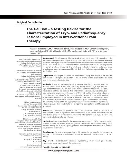
<strong>Pain</strong> <strong>Physician</strong> 2010; 13:263-271 • ISSN 1533-3159<br />
Original Contribution<br />
<strong>The</strong> <strong>Gel</strong> <strong>Box</strong> – a <strong>Testing</strong> <strong>Device</strong> <strong>for</strong> <strong>the</strong><br />
Characterization of Cryo- and Radiofrequency<br />
Lesions Employed in Interventional <strong>Pain</strong><br />
<strong>The</strong>rapy<br />
Christof Birkenmaier, MD 1 , Athanasios Terzis 1 , Bernd Wegener, MD 1 , Carolin Melcher, MD 1 ,<br />
Andreas Fottner, MD 1 , Jörg Hausdorf, MD 1 , Markus Schmidt-Sody, MD, PD 2 , and Volkmar<br />
Jansson, MD 1<br />
From: 1 Department of Orthopedic<br />
Surgery, Grosshadern Medical Center<br />
Ludwig-Maximilian-University<br />
Munich, Germany; 2 Medical Park<br />
Chiemse, Munich, Germany.<br />
Dr. Birkenmaier is with <strong>the</strong> Department<br />
of Orthopedic Surgery, Grosshadern<br />
Medical Center<br />
Ludwig-Maximilian-University<br />
Munich, Germany;<br />
Dr. Terzis and Prof. Jannson are with <strong>the</strong><br />
University of Munich Medical School,<br />
Munich, Bavaria, Germany<br />
Dr. Fottner, Dr. Hausdorf, and Dr.<br />
Melcher are with Department of<br />
Orthopedic Surgery, Grosshadern<br />
Medical Center, University of Munich,<br />
Bavaria, Germany.<br />
Dr. Schmidt-Sody is with <strong>the</strong> Medical<br />
Park Chiemsee, Bernau Felden, Bavaria,<br />
Germany<br />
Address correspondence:<br />
Christof Birkenmaier, MD<br />
Department of Orthopedic Surgery,<br />
Grosshadern Medical Center<br />
Ludwig-Maximilian-University<br />
Marchioninistr. 15<br />
81377 Munich, Germany<br />
Email:christof@doctor-b.de<br />
Disclaimer: Inomed GmbH,<br />
Teningen, Germany <strong>for</strong> supplying <strong>the</strong><br />
<strong>the</strong>rmoelements and micrometer drives.<br />
Parts of this work were per<strong>for</strong>med<br />
within <strong>the</strong> context of a doctoral (MD)<br />
<strong>the</strong>sis at <strong>the</strong> University of Munich.<br />
Conflict of interest: None.<br />
Manuscript received: 01/17/2010<br />
Revised manuscript received:<br />
04/05/2010<br />
Accepted <strong>for</strong> publication:<br />
04/07/2010<br />
Free full manuscript:<br />
www.painphysicianjournal.com<br />
Background: Radiofrequency (RF) and cryolesioning are established methods <strong>for</strong> <strong>the</strong><br />
<strong>the</strong>rapeutic interruption of sensory nerve supply to facet joints and o<strong>the</strong>r painful musculoskeletal<br />
structures. <strong>The</strong> varying clinical success rates of <strong>the</strong>se treatments have – among o<strong>the</strong>r technical<br />
issues – been attributed to <strong>the</strong> small size of <strong>the</strong>se lesions combined with <strong>the</strong> limited precision<br />
in placing <strong>the</strong>m. Since <strong>the</strong>re are 2 different physical methods <strong>for</strong> lesioning and a wide range<br />
of probes and lesion generators available, it is likely that <strong>the</strong> lesions generated by <strong>the</strong>m may<br />
be of different size.<br />
Objectives: We sought to devise an experimental setup that would allow <strong>for</strong> <strong>the</strong><br />
reproducible and comparable evaluation of <strong>the</strong> size of cryo and RF lesions as <strong>the</strong>y are being<br />
used in interventional pain <strong>the</strong>rapy.<br />
Methods: A wide range of potential media was evaluated <strong>for</strong> this purpose. Based on<br />
technical specifications, as well as on preliminary testing, a specific agar agar gel with<br />
a gel point of between 32 o C and 35 o C and a melting point of between 80 o C and 85 o C<br />
was selected <strong>for</strong> <strong>the</strong>se experiments. Two different testing containers were constructed<br />
from transparent acrylic: one with a volume of 1,500 mL and <strong>the</strong> o<strong>the</strong>r with a volume of<br />
12 mL. Each of <strong>the</strong>m allows <strong>for</strong> <strong>the</strong> introduction of a cryo or a RF probe and 2 bundles<br />
of <strong>the</strong>rmoelements into <strong>the</strong> gel volume. A water bath was used to maintain <strong>the</strong> gels at<br />
37 o C and bundled, ultrafine NiCr-Ni <strong>the</strong>rmoelements type K were used <strong>for</strong> measuring<br />
<strong>the</strong> iso<strong>the</strong>rms. A series of RF and cryolesions were per<strong>for</strong>med within <strong>the</strong>se experimental<br />
setups to evaluate <strong>the</strong>ir suitability <strong>for</strong> <strong>the</strong> comparative testing of cryo and RF probes and<br />
generators.<br />
Results: Both testing setups generated reproducible results and proved to be suitable <strong>for</strong><br />
measuring RF as well as cryolesions. Visual observation of <strong>the</strong> lesions was better with <strong>the</strong><br />
small testing container and rewarming / recooling after per<strong>for</strong>ming a cryo / RF lesion was<br />
more rapid with <strong>the</strong> smaller gel volume.<br />
Limitations: Our setup allows <strong>for</strong> <strong>the</strong> comparative measurement of RF and cryolesions, but<br />
it cannot simulate <strong>the</strong> realities within living tissue. While convection as a confounding factor<br />
was excluded by use of a gel, capillary perfusion and <strong>the</strong> specific characteristics of different<br />
tissues cannot be simulated.<br />
Conclusions: <strong>The</strong> testing setup described in this manuscript can serve <strong>for</strong> <strong>the</strong> comparative<br />
and reproducible study of RF and cryolesions that are commonly used in interventional pain<br />
<strong>the</strong>rapy.<br />
Key words: Radiofrequency lesioning, cryolesioning, interventional pain <strong>the</strong>rapy,<br />
experimental study, agar agar gel, <strong>the</strong>rmoelement.<br />
<strong>Pain</strong> <strong>Physician</strong> 2010; 13:263-271<br />
www.painphysicianjournal.com
<strong>Pain</strong> <strong>Physician</strong>: May/June 2010; 13:263-271<br />
<strong>The</strong>re is increasing evidence supporting <strong>the</strong><br />
efficacy of medial branch denervation in <strong>the</strong><br />
treatment of facet joint pain (1-8) and while<br />
<strong>the</strong> best randomized-controlled trials have been<br />
per<strong>for</strong>med using RF (9-14), trials of lesser quality using<br />
cryodenervation show similar <strong>the</strong>rapeutic effects<br />
(15-17). <strong>The</strong> scientific groundwork <strong>for</strong> <strong>the</strong>rapeutic<br />
cryolesioning was laid between 30 and 40 years ago<br />
and while <strong>the</strong> primary target of this technology was <strong>the</strong><br />
treatment of tumors, it has successfully been employed<br />
in <strong>the</strong> treatment of a wide range of peripheral pain<br />
syndromes (15-18).<br />
Radiofrequency lesioning also has a very long history<br />
in interventional pain <strong>the</strong>rapy and was first used<br />
<strong>for</strong> facet joint pain in <strong>the</strong> 1970s (19,20). Lesion size has<br />
been recognized as a factor in <strong>the</strong> efficacy of denervation<br />
procedures and <strong>the</strong>re have been attempts to improve<br />
<strong>the</strong> positioning of RF probes by using special radio-opaque,<br />
markers or to use more than one RF needle<br />
<strong>for</strong> RF lesioning (21-23). <strong>The</strong>re are a number of studies<br />
investigating <strong>the</strong> cryolesions used <strong>for</strong> <strong>the</strong> ablation of<br />
tumors in various tissues in vivo (24), ex vivo with life<br />
tissues (25), as well as in vitro in various media with or<br />
without cells (25-29). In addition, a number of ma<strong>the</strong>matical<br />
and finite element simulations have been done<br />
<strong>for</strong> <strong>the</strong> application of cryosurgery to tumor tissue ablation<br />
(30-34).<br />
O<strong>the</strong>r important factors influencing <strong>the</strong> effectiveness<br />
of cryolesioning as well as of radiofrequency<br />
lesioning are <strong>the</strong> shape and iso<strong>the</strong>rms of <strong>the</strong> lesions<br />
generated. For obvious reasons, such lesions cannot be<br />
studied in real patients and even real-time temperature<br />
measurements in <strong>the</strong> tissues of experimental animals<br />
have clear limitations. Such measurements would have<br />
to be obtained with open surgical access, which in turn<br />
distorts <strong>the</strong> conditions from what <strong>the</strong>y would be in a<br />
real <strong>the</strong>rapeutic situation where perfused, uninterrupted<br />
tissues surround <strong>the</strong> area of treatment.<br />
Surprisingly, <strong>the</strong>re are only few studies investigating<br />
<strong>the</strong> character and <strong>the</strong> size of <strong>the</strong> lesions generated<br />
by <strong>the</strong> typical cryoprobes / cryogenerators currently in<br />
use <strong>for</strong> interventional pain <strong>the</strong>rapy in an in vitro setup<br />
(35-37).<br />
Having extensive clinical experience with <strong>the</strong> cryodenervation<br />
of lumbar facet joints (16,38), we became<br />
interested in <strong>the</strong> underlying issues outlined above and<br />
sought an investigational method that would allow us<br />
to better understand <strong>the</strong> character of such lesions. It<br />
was our goal to devise a setup that would allow <strong>for</strong> <strong>the</strong><br />
testing of cryo and radiofrequency probes and <strong>for</strong> <strong>the</strong><br />
objective comparison of <strong>the</strong> lesions generated by such<br />
probes and generators in order to better understand<br />
<strong>the</strong> effects (or lack <strong>the</strong>reof) of such lesions in interventional<br />
pain <strong>the</strong>rapy.<br />
In consequence, we researched a wide range of<br />
potential media <strong>for</strong> simulating tissues, tested a range<br />
of <strong>the</strong>se media <strong>for</strong> <strong>the</strong>ir physical properties, and developed<br />
two alternative testing setups.<br />
Methods<br />
Preconditions<br />
While a laboratory testing setup can never simulate<br />
<strong>the</strong> realities of living tissue, we <strong>for</strong>mulated several preconditions,<br />
which we consider important. For one, <strong>the</strong><br />
setup should be transparent, so that <strong>the</strong> thawing and<br />
freezing cycles of cryolesions could be visually observed<br />
and documented. Second, <strong>the</strong> baseline temperature<br />
needs to be well controlled at 37 o C. Third, fluid convection<br />
on a macroscopic scale needs to be avoided. While<br />
convection occurs even within living tissues at a cellular<br />
level and / or in <strong>the</strong> interstitial space, <strong>the</strong>se effects are<br />
expected to be at a microscopic level. Were we to use a<br />
fluid medium <strong>for</strong> our experiments, however, convection<br />
would take place on a large scale around <strong>the</strong> cryo or RF<br />
lesion and would be expected to substantially alter <strong>the</strong><br />
temperatures around <strong>the</strong> probes and hence <strong>the</strong> size of<br />
<strong>the</strong> lesions created. In consequence, <strong>the</strong> medium surrounding<br />
<strong>the</strong> probe tip cannot be liquid.<br />
Fourth, this medium should have a melting point of<br />
above 37 o C in order to avoid convection in all situations<br />
during freezing from and thawing to normal body temperature.<br />
Ideally, <strong>the</strong> melting point should be at 80 o C<br />
or higher in order to allow <strong>for</strong> <strong>the</strong> identical setup to be<br />
used <strong>for</strong> <strong>the</strong> testing of radiofrequency probes. Fifth, <strong>the</strong><br />
medium should ideally contain ions at a concentration<br />
comparable to living tissues <strong>for</strong> <strong>the</strong> same reason.<br />
Test Medium<br />
Resulting from <strong>the</strong> above preconditions, <strong>the</strong> natural<br />
choice <strong>for</strong> a non-liquid transparent medium <strong>for</strong> our<br />
application would be a gel of some sort, <strong>the</strong> main constraints<br />
being transparency and melting point as outlined<br />
above.<br />
As secondary parameters, temperature storage<br />
capacity and temperature conductivity would be of<br />
importance, since <strong>the</strong> medium would have to be maintained<br />
at 37 o C by an external heat source and after <strong>the</strong><br />
application of cold or heat, <strong>the</strong> zone of <strong>the</strong> immediate<br />
cryolesion would need to be re-warmed to 37 o C prior<br />
264 www.painphysicianjournal.com
<strong>The</strong> <strong>Gel</strong> <strong>Box</strong> <strong>for</strong> <strong>Testing</strong> Cryo and RF Lesions<br />
to <strong>the</strong> next test cycle. <strong>The</strong> reverse would be required <strong>for</strong><br />
<strong>the</strong> measuring of radiofrequency lesions.<br />
<strong>The</strong>re are 3 major categories of gels available <strong>for</strong><br />
a host of scientific applications as well as <strong>for</strong> use in <strong>the</strong><br />
food industry: <strong>Gel</strong>atin-based substances, agar agar in its<br />
varieties, and alginate-based gels. While scientific-grade<br />
gelatins can possess high transparency, <strong>the</strong>y invariably<br />
have melting points of around or only slightly above<br />
37 o C. <strong>The</strong> agar agar group contains products whose<br />
properties are extremely widespread, but most are not<br />
highly transparent.<br />
<strong>The</strong> alginate-based gels, in contrast, do have higher<br />
melting points, good transparency, but are not widely<br />
available. <strong>The</strong>y are more expensive than ei<strong>the</strong>r of <strong>the</strong><br />
o<strong>the</strong>r products and <strong>the</strong> choices available are more<br />
limited.<br />
After casting a number of preliminary trial gels<br />
with various concentrations of <strong>the</strong> different base<br />
components, we found one agar agar gel which best<br />
matched our demands. <strong>The</strong> “05038” agar (manufactured<br />
by Fluka BioChemika, supplied by Sigma Aldrich<br />
Germany, product number 05038, CAS number 9002-18-<br />
0) is a (C12H18O9)n - polymer <strong>for</strong> special microbiology<br />
applications. It has a gel point between 32 o C and 35 o C<br />
and is highly transparent. Its melting point is between<br />
80 o C and 85 o C and <strong>the</strong> gel is well suited <strong>for</strong> immunoelectrophoresis,<br />
which also indicates its potential usefulness<br />
<strong>for</strong> <strong>the</strong> testing of radiofrequency probes. However,<br />
<strong>the</strong>re were no data available from <strong>the</strong> manufacturer as<br />
to <strong>the</strong> temperature storage capacity and <strong>the</strong> temperature<br />
conductivity of <strong>the</strong> gel. For <strong>the</strong> experiments, gels<br />
were prepared at <strong>the</strong> recommended concentration of<br />
1.5 g/100 mL saline 0.9%.<br />
Container<br />
Based on <strong>the</strong> preconditions outlined above, we<br />
chose transparent acrylic with a 3 mm wall strength. As<br />
mentioned above, <strong>the</strong>re were no pre-existing data on<br />
how <strong>the</strong> gel would per<strong>for</strong>m with regards to temperature<br />
storage capacity and temperature conductivity. So<br />
we could not decide from <strong>the</strong> outset whe<strong>the</strong>r a large<br />
gel mass would be of advantage in maintaining <strong>the</strong> core<br />
temperature steady or whe<strong>the</strong>r this would render <strong>the</strong><br />
temperature in <strong>the</strong> center of <strong>the</strong> gel uncontrollable.<br />
We <strong>the</strong>re<strong>for</strong>e built 2 different designs of testing<br />
containers: one with a large gel volume (1500 mL) and<br />
one with a very small gel volume (12 mL). <strong>The</strong> 2 designs,<br />
as well as <strong>the</strong>ir specifications, are displayed in Figs. 1<br />
and 2. A <strong>the</strong>rmostat-controlled water bath with an automatic<br />
circulator pump was chosen as a heat source in<br />
order to maintain <strong>the</strong> gel at 37 o C.<br />
<strong>The</strong> <strong>the</strong>rmo elements were positioned into <strong>the</strong><br />
center of <strong>the</strong> container through 2 canals opposite and<br />
Fig. 1. View of <strong>the</strong> large-volume testing container. Surrounding<br />
<strong>the</strong> container is <strong>the</strong> water bath. <strong>The</strong> cryoprobe<br />
enters from <strong>the</strong> left and <strong>the</strong> 2 bundles of <strong>the</strong>rmoelements<br />
from <strong>the</strong> top and from <strong>the</strong> right. <strong>The</strong> <strong>the</strong>rmoelement bundles<br />
are mounted on micrometer drives <strong>for</strong> fine adjustment. <strong>The</strong><br />
gel volume is approximately 1,500 mL and <strong>the</strong> visibility<br />
through <strong>the</strong> gel is quite good.<br />
Fig. 2. View of <strong>the</strong> small-volume testing container. Surrounding<br />
<strong>the</strong> container is <strong>the</strong> water bath. <strong>The</strong> cryoprobe enters from<br />
<strong>the</strong> left and <strong>the</strong> 2 bundles of <strong>the</strong>rmoelements from <strong>the</strong> top and<br />
from <strong>the</strong> right, each through waterproof acrylic tubes. <strong>The</strong><br />
<strong>the</strong>rmoelement bundles are mounted on micrometer drives <strong>for</strong><br />
fine adjustment. <strong>The</strong> lid of <strong>the</strong> testing container is held by<br />
rubber bands. <strong>The</strong> gel volume is approximately 12 mL and <strong>the</strong><br />
visibility through <strong>the</strong> gel is excellent. On <strong>the</strong> right-hand wall of<br />
<strong>the</strong> water bath, <strong>the</strong> water inlet and outlet can be seen.<br />
www.painphysicianjournal.com 265
<strong>Pain</strong> <strong>Physician</strong>: May/June 2010; 13:263-271<br />
perpendicular to <strong>the</strong> entry point of <strong>the</strong> cryoprobe / radiofrequency<br />
probe in <strong>the</strong> large volume container.<br />
<strong>The</strong> container was <strong>the</strong>n set into a second container<br />
that contained <strong>the</strong> water bath. Since <strong>the</strong> <strong>the</strong>rmoelements<br />
and <strong>the</strong> probes were completely embedded in<br />
gel, <strong>the</strong>re was no direct contact with <strong>the</strong> water bath.<br />
For <strong>the</strong> small volume container, <strong>the</strong> outside dimensions<br />
and materials remained identical, but a small internal<br />
box with internal dimensions of 20 x 20 x 30 mm<br />
made from 2 mm acrylic was added in <strong>the</strong> center of <strong>the</strong><br />
container. Only <strong>the</strong> small box was filled with gel while<br />
<strong>the</strong> surrounding container was filled with <strong>the</strong> water<br />
bath. Acrylic tubes were used to allow <strong>for</strong> passage of<br />
<strong>the</strong> <strong>the</strong>rmoelements as well as of <strong>the</strong> probes into <strong>the</strong><br />
gel container without having direct contact with <strong>the</strong><br />
water bath.<br />
<strong>The</strong>rmoelements<br />
NiCr-Ni <strong>the</strong>rmoelements of type K were used in this<br />
study, bundled in groups of 4 <strong>for</strong> measurements perpendicular<br />
to <strong>the</strong> probe and in groups of 3 <strong>for</strong> measurements<br />
longitudinally from <strong>the</strong> tip of <strong>the</strong> probe. <strong>The</strong> NiCr<br />
as well as <strong>the</strong> Ni wires were individually protected by an<br />
Inconel (Special Metals Corporation, New Hart<strong>for</strong>d, CN)<br />
coating and <strong>the</strong>n jointly protected by a stainless steel<br />
tubing (1N4301, external diameter 0.5 mm, internal diameter<br />
0.3 mm), closed at <strong>the</strong> end by highly accurate<br />
laser point welding. At <strong>the</strong> connector piece, <strong>the</strong> wires<br />
were soldered to a NiCr-Ni equivalence wire.<br />
In order to obtain exact one mm iso<strong>the</strong>rm readings,<br />
<strong>the</strong> <strong>the</strong>rmoelement bundles were precisely aligned and<br />
assembled under a laboratory binocular with a 0.1-millimeter<br />
scale. To reduce <strong>the</strong> chance of temperature<br />
transmission between neighboring <strong>the</strong>rmoelements,<br />
each individual <strong>the</strong>rmoelement was coated with special<br />
polyester tubing prior to being bundled. <strong>The</strong> assembled<br />
<strong>the</strong>rmoelement bundles were mounted on a micrometer<br />
drive and fitted onto docking rings on ei<strong>the</strong>r of<br />
<strong>the</strong> boxes. This allowed <strong>for</strong> <strong>the</strong> <strong>the</strong>rmoelement bundles<br />
being positioned exactly onto <strong>the</strong> surface of <strong>the</strong> cryoprobe<br />
until contact and <strong>the</strong>n to be withdrawn a fraction<br />
of a millimeter. <strong>The</strong> gel was cast after adjusting <strong>the</strong><br />
<strong>the</strong>rmoelements.<br />
By using this procedure, <strong>the</strong> most distally aligned<br />
<strong>the</strong>rmoelement was measured in <strong>the</strong> closest proximity<br />
of <strong>the</strong> cryoprobe without actually making surface<br />
contact, while <strong>the</strong> o<strong>the</strong>r <strong>the</strong>rmoelements were automatically<br />
set to a distance of one, 2 and 3 millimeters,<br />
respectively.<br />
Cryoprobe / Cryogenerator<br />
For our experiments we used a Lloyd Cryostat 2000<br />
generator (Spembly Medical Systems, supplied by inomed<br />
GmbH, Teningen, Germany) and <strong>the</strong> standard<br />
probe used <strong>for</strong> lumbar facet cryodenervation with a<br />
diameter of 2 mm. <strong>The</strong> experimental freezing was per<strong>for</strong>med<br />
with <strong>the</strong> probe fixed in <strong>the</strong> testing container as<br />
described above.<br />
<strong>The</strong> gas flow was adjusted according to <strong>the</strong> reading<br />
of <strong>the</strong> <strong>the</strong>rmoelements adjusted at 0 mm distance<br />
from <strong>the</strong> probe, which allowed <strong>for</strong> a more exact adjustment<br />
of <strong>the</strong> temperature minimum than <strong>the</strong> visual flow<br />
meter that is integrated in <strong>the</strong> cryogenerator.<br />
Radiofrequency Probe and Generator<br />
For <strong>the</strong> experimental radiofrequency lesions, we<br />
used an Electro<strong>The</strong>rmal 20S Spine System with <strong>the</strong><br />
matching unipolar needle electrodes (RF Canula/Needles<br />
10 cm, 5 mm Sharp Straight Tip) <strong>for</strong> facet joint denervation<br />
(Smith & Nephew GmbH, Marl, Germany).<br />
Results<br />
Both testing setups proved to be suited <strong>for</strong> <strong>the</strong> experiment<br />
and delivered reproducible measurements.<br />
While visibility was perfect in <strong>the</strong> small volume testing<br />
container, <strong>the</strong> gel itself proved sufficiently transparent<br />
to allow <strong>for</strong> visual control and documentation of <strong>the</strong><br />
experimental lesions even when cast in a large volume<br />
(Fig. 3).<br />
It was much easier to control <strong>the</strong> baseline temperature<br />
in <strong>the</strong> small-volume container than in <strong>the</strong><br />
large-volume container, already indicating that <strong>the</strong><br />
temperature conductivity of <strong>the</strong> 05038 agar agar gel is<br />
relatively poor. Due to its gel and melting points, <strong>the</strong><br />
gel effectively avoided convection at any temperature<br />
during <strong>the</strong> experiments with cryoprobes as well as with<br />
radiofrequency probes.<br />
One of our main findings was that <strong>the</strong> agar agar<br />
gel seems to be a relatively poor temperature conductor<br />
and a good temperature capacitator. Displayed in<br />
Fig. 4 is <strong>the</strong> cooling and re-warming graph after a typical<br />
freezing cycle with <strong>the</strong> large-volume testing container.<br />
It is obvious that while <strong>the</strong> return to 0 o C was<br />
reasonably rapid, it took beyond 14 minutes <strong>for</strong> <strong>the</strong> gel<br />
in <strong>the</strong> immediate surrounding of <strong>the</strong> cryoprobe tip to<br />
re-warm to 33 o C.<br />
<strong>The</strong> re-warming process, however, was only somewhat<br />
faster with <strong>the</strong> small-volume testing container<br />
(Fig. 5), taking around 10 minutes. <strong>The</strong> temperature<br />
266 www.painphysicianjournal.com
<strong>The</strong> <strong>Gel</strong> <strong>Box</strong> <strong>for</strong> <strong>Testing</strong> Cryo and RF Lesions<br />
curves obtained with <strong>the</strong> small-volume container were<br />
smoo<strong>the</strong>r than <strong>the</strong> ones obtained with <strong>the</strong> large-volume<br />
container.<br />
Discussion<br />
Fig. 3. Close-up view of <strong>the</strong> tips of <strong>the</strong> cryoprobe and <strong>the</strong><br />
<strong>the</strong>rmoelement bundles embedded in <strong>the</strong> agar agar gel in <strong>the</strong><br />
large-volume testing container<br />
We were able to devise a simple testing setup that<br />
allows <strong>for</strong> <strong>the</strong> reproducible and comparable characterization<br />
of cryo and RF lesions that are typically used in<br />
interventional pain medicine. <strong>The</strong> use of a gel permits<br />
<strong>for</strong> visual control of <strong>the</strong> correct setup and <strong>the</strong> development<br />
of <strong>the</strong> lesions while preventing convection at a<br />
macroscopic level.<br />
Our measurements show, that ei<strong>the</strong>r testing setup<br />
may prove useful in <strong>the</strong> characterization of small cryo<br />
or RF lesions. <strong>The</strong>y both allow <strong>for</strong> a ra<strong>the</strong>r precise determination<br />
of <strong>the</strong> iso<strong>the</strong>rms of <strong>the</strong> RF and cryoprobes<br />
Fig. 4. Temperature measurements obtained with <strong>the</strong> large-volume testing container. Freezing is begun at 0 seconds and stopped<br />
at 420 seconds. “Tip” indicates measurement in <strong>the</strong> axial extension from <strong>the</strong> tip of <strong>the</strong> cryoprobe, “side” indicates measurement<br />
perpendicular to <strong>the</strong> tip of <strong>the</strong> cryoprobe. 0, 1, 2 and 3 indicate <strong>the</strong> distance in millimeters.<br />
www.painphysicianjournal.com 267
<strong>Pain</strong> <strong>Physician</strong>: May/June 2010; 13:263-271<br />
Fig. 5. Temperature measurements obtained with <strong>the</strong> small-volume testing container. Freezing is begun at 0 seconds and stopped<br />
at 640 seconds. “Tip” indicates measurement in <strong>the</strong> axial extension from <strong>the</strong> tip of <strong>the</strong> cryoprobe, “side” indicates measurement<br />
perpendicular to <strong>the</strong> tip of <strong>the</strong> cryoprobe. 0, 1, 2 and 3 indicate <strong>the</strong> distance in millimeters.<br />
commonly used <strong>for</strong> interventional pain <strong>the</strong>rapy. Since<br />
<strong>the</strong>se setups are standardized, <strong>the</strong>y also allow <strong>for</strong> <strong>the</strong><br />
generation of comparable data and hence <strong>for</strong> <strong>the</strong><br />
comparison of <strong>the</strong> lesion sizes generated by different<br />
probes and different devices. As long as a gel with a<br />
sufficiently high melting point is used, <strong>the</strong>y also allow<br />
<strong>for</strong> <strong>the</strong> comparison of <strong>the</strong> lesion sizes of RF and<br />
cryolesions.<br />
Because of <strong>the</strong> better control of baseline temperature,<br />
<strong>the</strong> smoo<strong>the</strong>r temperature curves that we observed<br />
and <strong>the</strong> better visibility, we conclude that <strong>the</strong><br />
small-volume testing container is preferable over <strong>the</strong><br />
large-volume container. Using a gel ra<strong>the</strong>r than a fluid<br />
as medium effectively prevents convection on a macroscopic<br />
level and brings <strong>the</strong> testing setup one step closer<br />
to <strong>the</strong> simulation of tissue.<br />
Important limitations remain, however. Capillary<br />
perfusion cannot be simulated by using a gel. On a<br />
more complex level, <strong>the</strong> temperature-dependent interruption<br />
(during freezing or when capillary vessels are<br />
being destroyed by heat) and restoration of capillary<br />
perfusion (when thawing from cryolesioning) also can<br />
not be simulated.<br />
Our experimental setup cannot account <strong>for</strong> some<br />
specific characteristics of living tissues, namely <strong>the</strong> microconvection<br />
that might occur inside cells and within<br />
<strong>the</strong> interstitial space which may dynamically change<br />
during heating or freezing.<br />
A gel – like any homogenous medium – can also<br />
not account <strong>for</strong> <strong>the</strong> real life situation where an RF<br />
probe or a cryoprobe is positioned next to <strong>the</strong> target<br />
nerve with muscle and / or fatty connective tissue cover-<br />
268 www.painphysicianjournal.com
<strong>The</strong> <strong>Gel</strong> <strong>Box</strong> <strong>for</strong> <strong>Testing</strong> Cryo and RF Lesions<br />
Fig. 6. Temperature measurements obtained with <strong>the</strong> small-volume testing container. Radiofrequency heating is begun at 0<br />
seconds and first stopped at 930 seconds with repeat cycles. “Tip” indicates measurement in <strong>the</strong> axial extension from <strong>the</strong> tip of<br />
<strong>the</strong> cryoprobe, “side” indicates measurement perpendicular to <strong>the</strong> tip of <strong>the</strong> cryoprobe. 0, 1, 2 and 3 indicate <strong>the</strong> distance in millimeters.<br />
Impedance was within a range of between 259 and 316 Ohms.<br />
ing <strong>the</strong> probe from one side and with bone being <strong>the</strong><br />
neighboring tissue from <strong>the</strong> o<strong>the</strong>r side.<br />
Since any real <strong>the</strong>rapeutic situation is unique regarding<br />
its anatomical circumstances, a specific and<br />
precise simulation does not appear to make much sense<br />
and – more importantly – would not allow <strong>for</strong> a reliable<br />
comparison between different probes, devices and<br />
physical modalities. While <strong>the</strong> use of a gel always represents<br />
a compromise, <strong>the</strong> type of gel used determines<br />
what sort of compromise <strong>the</strong> investigator is willing to<br />
make. When visual observation is most important and<br />
when only cryolesions are to be studied, a gelatin-based<br />
or an alginate-based gel might be <strong>the</strong> best choice. But<br />
when RF lesions are to be compared with cryolesions,<br />
<strong>the</strong> melting point is <strong>the</strong> primary consideration, because<br />
convection needs to be excluded <strong>for</strong> <strong>the</strong> cryolesions as<br />
well as <strong>for</strong> <strong>the</strong> RF lesions.<br />
Most RF lesions are per<strong>for</strong>med using temperatures<br />
up to 80 o C, which is why we sought a gel with a melting<br />
point at or above that temperature. <strong>The</strong> gel we decided<br />
to use <strong>for</strong> <strong>the</strong> reasons outlined above proved to be a very<br />
good temperature capacitator and a poor temperature<br />
www.painphysicianjournal.com 269
<strong>Pain</strong> <strong>Physician</strong>: May/June 2010; 13:263-271<br />
1. Levin JH. Prospective, double-blind,<br />
randomized placebo-controlled trials<br />
in interventional spine: What <strong>the</strong> highest<br />
quality literature tells us. Spine J<br />
2009; 9:690-703.<br />
2. Falco FJ, Erhart S, Wargo BW, Bryce DA,<br />
Atluri S, Datta S, Hayek SM. Systematic<br />
review of diagnostic utility and <strong>the</strong>rapeutic<br />
effectiveness of cervical facet<br />
joint interventions. <strong>Pain</strong> <strong>Physician</strong><br />
2009; 12:323-344.<br />
3. Datta S, Lee M, Falco FJ, Bryce DA,<br />
Hayek SM. Systematic assessment of<br />
diagnostic accuracy and <strong>the</strong>rapeutic<br />
utility of lumbar facet joint interventions.<br />
<strong>Pain</strong> <strong>Physician</strong> 2009; 12:437-<br />
460.<br />
4. Atluri S, Datta S, Falco FJ, Lee M. Systematic<br />
review of diagnostic utility and<br />
<strong>the</strong>rapeutic effectiveness of thoracic<br />
facet joint interventions. <strong>Pain</strong> <strong>Physician</strong><br />
2008; 11:611-629.<br />
5. Sehgal N, Dunbar EE, Shah RV, Colson<br />
J. Systematic review of diagnostic utilconductor.<br />
While this does not change <strong>the</strong> overall results<br />
of our experiments, it delays <strong>the</strong> re-warming or re-cooling<br />
process within <strong>the</strong> test chamber and hence limits <strong>the</strong><br />
number of measurement cycles that can be per<strong>for</strong>med<br />
within a given amount of time. One conclusion could be<br />
to make <strong>the</strong> testing chamber even smaller than <strong>the</strong> 12<br />
mL that we tested.<br />
However, since <strong>the</strong> lesions and <strong>the</strong> <strong>the</strong>rmoelements<br />
need to be completely embedded in a homogenous<br />
medium, <strong>the</strong>re is a limit to making <strong>the</strong> testing chamber<br />
much smaller than <strong>the</strong> one we constructed.<br />
<strong>The</strong>re are fundamental limitations to <strong>the</strong> transfer<br />
of our study results into a clinical situation, since we did<br />
not realistically simulate a cryo or radiofrequency lesion<br />
being generated in living tissue.<br />
Results obtained by in vitro testing with our setup<br />
must <strong>the</strong>re<strong>for</strong>e be interpreted with <strong>the</strong> necessary degree<br />
of caution, since <strong>the</strong>se measurements were not<br />
per<strong>for</strong>med within a living body. On <strong>the</strong> o<strong>the</strong>r hand,<br />
placing <strong>the</strong> probes and <strong>the</strong> very fine and flexible <strong>the</strong>rmoelements<br />
inside a perfused tissue volume with <strong>the</strong><br />
same precision as in our experiment would require direct<br />
vision. This could not be achieved without tissue dissection<br />
and hence alteration of <strong>the</strong> tissue’s properties.<br />
Conclusion<br />
In conclusion, we were able to devise a simple testing<br />
setup that allows <strong>for</strong> <strong>the</strong> reproducible and comparable<br />
in vitro assessment of <strong>the</strong>rapeutic cryolesions and<br />
RF lesions.<br />
It will allow <strong>for</strong> fur<strong>the</strong>r study on <strong>the</strong>se lesions<br />
and may have an impact on <strong>the</strong> discussion of <strong>the</strong><br />
varying degrees of success that are being achieved<br />
with such techniques in clinical applications. While<br />
live tissue testing would be <strong>the</strong> desirable gold standard,<br />
we consider it to be unachievable with <strong>the</strong><br />
necessary degree of precision. We <strong>the</strong>re<strong>for</strong>e contend<br />
that a testing setup identical or similar to <strong>the</strong> one<br />
outlined in this paper will remain <strong>the</strong> next best thing<br />
to live tissue testing when <strong>the</strong> comparison of RF and<br />
cryolesions generated by different probes and devices<br />
is <strong>the</strong> goal.<br />
Acknowledgements<br />
We thank inomed GmbH, Teningen, Germany<br />
<strong>for</strong> supplying <strong>the</strong> <strong>the</strong>rmoelements and micrometer<br />
drives. Parts of this work were per<strong>for</strong>med within <strong>the</strong><br />
context of a doctoral (MD) <strong>the</strong>sis at <strong>the</strong> University of<br />
Munich.<br />
References<br />
ity of facet (zygapophysial) joint injections<br />
in chronic spinal pain: An update.<br />
<strong>Pain</strong> <strong>Physician</strong> 2007; 10:213-228.<br />
6. Geurts JW, van Wijk RM, Stolker RJ,<br />
Groen GJ. Efficacy of radiofrequency<br />
procedures <strong>for</strong> <strong>the</strong> treatment of spinal<br />
pain: A systematic review of randomized<br />
clinical trials. Reg Anesth <strong>Pain</strong> Med<br />
2001; 26:394-400.<br />
7. Niemisto L, Kalso E, Malmivaara A, Seitsalo<br />
S, Hurri H. Radiofrequency denervation<br />
<strong>for</strong> neck and back pain. A systematic<br />
review of randomized controlled trials.<br />
Cochrane Database Syst Rev 2003:<br />
CD004058.<br />
8. Hooten WM, Martin DP, Huntoon MA.<br />
Radiofrequency neurotomy <strong>for</strong> low back<br />
pain: Evidence-based procedural guidelines.<br />
<strong>Pain</strong> Med 2005; 6:129-138.<br />
9. Nath S, Nath CA, Pettersson K. Percutaneous<br />
lumbar zygapophysial (facet)<br />
joint neurotomy using radiofrequency<br />
current in <strong>the</strong> management of chronic<br />
low back pain: A randomized doubleblind<br />
trial. Spine 2008; 33:1291-1297;<br />
discussion 1298.<br />
10. Haspeslagh SR, van Suijlekom HA,<br />
Lame IE, Kessels AG, van Kleef M, Weber<br />
WE. Randomised controlled trial<br />
of cervical radiofrequency lesions as a<br />
treatment <strong>for</strong> cervicogenic headache<br />
[ISRCTN 07444684]. BMC Anes<strong>the</strong>siol<br />
2006; 6:1.<br />
11. van Wijk RM, Geurts JW, Wynne HJ,<br />
Hammink E, Buskens E, Lousberg R,<br />
Knape JT, Groen GJ. Radiofrequency denervation<br />
of lumbar facet joints in <strong>the</strong><br />
treatment of chronic low back pain: A<br />
randomized, double-blind, sham lesion-controlled<br />
trial. Clin J <strong>Pain</strong> 2005;<br />
21:335-344.<br />
12. Barnsley L. Percutaneous radiofrequency<br />
neurotomy <strong>for</strong> chronic neck pain:<br />
Outcomes in a series of consecutive patients.<br />
<strong>Pain</strong> Med 2005; 6:282-286.<br />
13. Leclaire R, Fortin L, Lambert R, Bergeron<br />
YM, Rossignol M. Radiofrequency facet<br />
joint denervation in <strong>the</strong> treatment of<br />
270 www.painphysicianjournal.com
<strong>The</strong> <strong>Gel</strong> <strong>Box</strong> <strong>for</strong> <strong>Testing</strong> Cryo and RF Lesions<br />
low back pain: A placebo-controlled<br />
clinical trial to assess efficacy. Spine<br />
2001; 26:1411-1416; discussion 1417.<br />
14. van Kleef M, Barendse GA, Kessels A,<br />
Voets HM, Weber WE, de Lange S. Randomized<br />
trial of radiofrequency lumbar<br />
facet denervation <strong>for</strong> chronic low back<br />
pain. Spine 1999; 24:1937-1942.<br />
15. Baerlocher CB, Krauss JK, Seiler RW.<br />
Kryorhizotomy: An alternative technique<br />
<strong>for</strong> lumbar medial branch rhizotomy<br />
in lumbar facet syndrome. J Neurosurg<br />
2003; 98:14-20.<br />
16. Birkenmaier C, Veihelmann A, Trouillier<br />
H, Hausdorf J, Devens C, Wegener<br />
B, Jansson V, von Schulze Pellengahr C.<br />
Percutaneous cryodenervation of lumbar<br />
facet joints: A prospective clinical<br />
trial. Int Orthop 2007; 31:525-530.<br />
17. Stander M, Marz U, Steude U, Tonn JC.<br />
[<strong>The</strong> facet syndrome: Frequent cause<br />
of chronic backaches]. MMW Fortschr<br />
Med 2006; 148:33-34.<br />
18. Arthur JM, Racz GB. Cryolysis. In Raj<br />
PP ed. <strong>Pain</strong> Medicine: A Comprehensive<br />
Review. 2nd Edition. Philadelphia,<br />
Mosby, 2003:297-303.<br />
19. Uematsu S, Udvarhelyi GB, Benson DW,<br />
Siebens AA. Percutaneous radiofrequency<br />
rhizotomy. Surg Neurol 1974;<br />
2:319-325.<br />
20. Shealy CN. Percutaneous radiofrequency<br />
denervation of spinal facets. Treatment<br />
<strong>for</strong> chronic back pain and sciatica.<br />
J Neurosurg 1975; 43:448-451.<br />
21. Jasper JF. Radiofrequency cannula with<br />
active tip radio-opaque marker: Image<br />
analysis <strong>for</strong> facet, gray ramus, and dorsal<br />
root ganglion techniques. <strong>Pain</strong> <strong>Physician</strong><br />
2008; 11:863-875.<br />
22. Derby R, Lee CH. <strong>The</strong> efficacy of a two<br />
needle electrode technique in percutaneous<br />
radiofrequency rhizotomy: An<br />
investigational laboratory study in an<br />
animal model. <strong>Pain</strong> <strong>Physician</strong> 2006;<br />
9:207-213.<br />
23. Pino CA, Hoeft MA, Hofsess C, Rathmell<br />
JP. Morphologic analysis of bipolar radiofrequency<br />
lesions: Implications <strong>for</strong><br />
treatment of <strong>the</strong> sacroiliac joint. Reg<br />
Anesth <strong>Pain</strong> Med 2005; 30:335-338.<br />
24. Popken F, Land M, Erberich H, Bosse M,<br />
Konig DP, Eysel P. <strong>The</strong> use of a new miniature<br />
cryoprobe <strong>for</strong> ablation of bone<br />
tissue: In vivo assessment of <strong>the</strong> probe<br />
and application of <strong>the</strong> method to bone<br />
in a sheep model. BMC Surg 2003; 3:3.<br />
25. Homasson JP, Thiery JP, Angebault M,<br />
Ovtracht L, Maiwand O. <strong>The</strong> operation<br />
and efficacy of cryosurgical, nitrous<br />
oxide-driven cryoprobe. I. Cryoprobe<br />
physical characteristics: <strong>The</strong>ir effects<br />
on cell cryodestruction. Cryobiology<br />
1994; 31:290-304.<br />
26. Popken F, Seifert JK, Engelmann R, Dutkowski<br />
P, Nassir F, Junginger T. Comparison<br />
of iceball diameter and temperature<br />
distribution achieved with 3-mm<br />
accuprobe cryoprobes in porcine and<br />
human liver tissue and human colorectal<br />
liver metastases in vitro. Cryobiology<br />
2000; 40:302-310.<br />
27. Budman HM, Dayan J, Shitzer A. Controlled<br />
freezing of nonideal solutions<br />
with application to cryosurgical processes.<br />
J Biomech Eng 1991; 113:430-<br />
437.<br />
28. Han B, Grassl ED, Barocas VH, Coad JE,<br />
Bischof JC. A cryoinjury model using engineered<br />
tissue equivalents <strong>for</strong> cryosurgical<br />
applications. Ann Biomed Eng<br />
2005; 33:972-982.<br />
29. Yang WH, Peng HH, Chang HC, Shen SY,<br />
Wu CL, Chang CH. An in vitro monitoring<br />
system <strong>for</strong> simulated <strong>the</strong>rmal process<br />
in cryosurgery. Cryobiology 2000;<br />
40:159-170.<br />
30. Budman H, Shitzer A, Del Giudice S.<br />
Investigation of temperature fields<br />
around embedded cryoprobes. J Biomech<br />
Eng 1986; 108:42-48.<br />
31. Budman H, Dayan J, Shitzer A. Control<br />
of <strong>the</strong> cryosurgical process in nonideal<br />
materials. IEEE Trans Biomed Eng 1991;<br />
38:1141-153.<br />
32. Poledna J, Berger W. A ma<strong>the</strong>matical<br />
model of temperature distribution<br />
in frozen tissue. Gen Physiol Biophys<br />
1996; 15:3-15.<br />
33. Rewcastle JC, Hahn LJ, Saliken JC,<br />
McKinnon JG. Use of a moratorium to<br />
achieve consistent liquid nitrogen cryoprobe<br />
per<strong>for</strong>mance. J Surg Oncol 1997;<br />
66:110-113.<br />
34. Rewcastle JC, Sandison GA, Muldrew<br />
K, Saliken JC, Donnelly BJ. A model <strong>for</strong><br />
<strong>the</strong> time dependent three-dimensional<br />
<strong>the</strong>rmal distribution within iceballs<br />
surrounding multiple cryoprobes. Med<br />
Phys 2001; 28:1125-1137.<br />
35. Mattmüller R. Radiofrequenzläsion<br />
und Kryoläsion. In Hankemeier U, Hildebrandt<br />
J eds. Neurodestruktive Verfahren<br />
in der Schmerz<strong>the</strong>rapie. 2 ed.<br />
Springer, Berlin: 2002:19-32.<br />
36. Schneider RK, Mayhew IG, Clarke GL.<br />
Effects of cryo<strong>the</strong>rapy on <strong>the</strong> palmar<br />
and plantar digital nerves in <strong>the</strong> horse.<br />
Am J Vet Res 1985; 46:7-12.<br />
37. Vinas FC, Zamorano L, Dujovny M, Zhao<br />
JZ, Hodgkinson D, Ho KL, Ausman JI. In<br />
vivo and in vitro study of <strong>the</strong> lesions<br />
produced with a computerized radiofrequency<br />
system. Stereotact Funct<br />
Neurosurg 1992; 58:121-313.<br />
38. Birkenmaier C, Veihelmann A, Trouillier<br />
HH, Hausdorf J, von Schulze Pellengahr<br />
C. Medial branch blocks versus pericapsular<br />
blocks in selecting patients<br />
<strong>for</strong> percutaneous cryodenervation of<br />
lumbar facet joints. Reg Anesth <strong>Pain</strong><br />
Med 2007; 32:27-33.<br />
www.painphysicianjournal.com 271