Pelviperineology - multidisciplinary pelvic floor journa
Pelviperineology - multidisciplinary pelvic floor journa
Pelviperineology - multidisciplinary pelvic floor journa

Create successful ePaper yourself
Turn your PDF publications into a flip-book with our unique Google optimized e-Paper software.
Perhimpunan Disfungsi Dasar Panggul Wanita Indonesia<br />
ISSN 1973-4905<br />
<br />
<br />
<br />
<br />
<br />
<br />
<br />
<br />
<br />
<br />
<br />
<br />
<br />
CONTENTS<br />
99 Editorial. Politics in the pelvis<br />
101 Intravaginal posterior sling procedure (PIVS) for the treatment of uterine descensus<br />
and vaginal vault prolapse: retrospective analysis of efficacy, safety, complications and<br />
patient satisfaction in 150 cases KIM M. HAEST, TOM H. HASAART, ILKNUR SANLI, ED T. GONDRIE, MARTIN G. BERGMANS<br />
104 Urinary retention in women with isolated Stage 3 rectocoele ELENA ANDRETTA, LISA POLA, MAURO PASTORELLO<br />
106 Pelvic Floor Digest<br />
108 A pilot study: The anal sphincter support procedure for the treatment of anal incontinence<br />
MAXWELL E. HAVERFIELD<br />
113 Colonic manometry and sacral nerve stimulation in patients with severe constipation<br />
PHILIP G. DINNING<br />
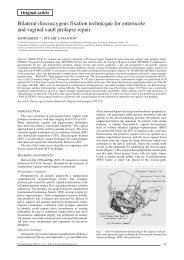
117 Reducing mesh exposure in Posterior Intra-Vaginal Slingplasty (PIVS) for vaginal apex suspension<br />
MENAHEM NEUMAN, YUVAL LAVY<br />
121 TVT-SECUR: 100 teaching operations with a novel anti-incontinence procedure MENAHEM NEUMAN<br />
124 Personal evolution of surgical technique using mesh for severe prolapse - a prospective audit<br />
of outcomes in 172 cases PHILIP PARIS-BROWNE<br />
127 Stapler-assisted trans-anal surgery for the treatment of outlet obstruction syndrome<br />
GIULIANO REBOA, MARCO GIPPONI, MATTEO LIGORIO, PAOLO MARINO, MASSIMO COSTANTINI<br />
132 Persistent dyschezia after double stapled transanal rectal resection for outlet obstruction: four case reports<br />
FILIPPO PUCCIANI, MARIA NOVELLA RINGRESSI, JACOPO GIANI<br />
136 Letter to the Editor<br />
2005
LigaSure Impact instrument, working exclusively with the ForceTriad energy<br />
platform, utilises the TissueFect sensing technology that precisely manages<br />
energy delivery for consistent, controlled tissue effects with faster fusion times<br />
than the original LigaSure generator.<br />
LF4200 Specifications<br />
<br />
<br />
<br />
<br />
<br />
<br />
<br />
<br />
<br />
<br />
ü<br />
ü<br />
ü<br />
ü<br />
ü<br />
60% larger fusion zone than the LigaSure Atlas 20 cm instrument<br />
Curved jaw design<br />
One instrument for tissue fusion and cutting; fewer instrument exchanges<br />
Hand activation provides sterile field control<br />
Utilises TissueFect sensing technology<br />
<br />
LigaSure Impact instrument when used with<br />
ForceTriad energy platform<br />
ü<br />
ü<br />
ü<br />
Average fusion cycle time of approximately 2-4 seconds can<br />
potentially reduce OR time<br />
Less dessication and more flexible fusion zones than the original<br />
LigaSure generator<br />
Reduces operating time in TAH, LAVH and Open Pelvic procedures
PELVIPERINEOLOGY<br />
<br />
<br />
<br />
<br />
<br />
Interim Editorial Board<br />
GHISLAIN DEVROEDE Colorectal Surgeon, Canada<br />
GIUSEPPE DODI Colorectal Surgeon, Italy<br />
BRUCE FARNSWORTH Gynaecologist, Australia<br />
DANIELE GRASSI Urologist, Italy<br />
RICHARD VILLET Urogynaecologist, France<br />
CARL ZIMMERMAN Gynaecologist, USA<br />
Editorial Office: LUCA AMADIO, ENRICO BELLUCO, PIERLUIGI LUCIO, LUISA MARCATO, MAURIZIO SPELLA<br />
c/o Clinica Chirurgica 2 University of Padova, 35128, Padova, Italy<br />
e-mail: editor@pelviperineology.org<br />
Quarterly <strong>journa</strong>l of scientific information registered at the Tribunale di Padova, Italy n. 741 dated 23-10-1982<br />
Editorial Director: GIUSEPPE DODI<br />
Printer “ La Garangola” Via E. Dalla Costa, 6 - 35129 Padova - e-mail: info@garangola.it<br />
Australian Production Minnis Communications 4/16 Maple Grove, Toorak, Vic 3142, Australia, e-mail: minnis@minniscomms.com.au
Editorial<br />
POLITICS IN THE PELVIS<br />
All around the world doctors and allied professionals are holding meetings and organizing educational<br />
courses that emphasize a <strong>multidisciplinary</strong> approach to the problems of the <strong>pelvic</strong> <strong>floor</strong>. Unfortunately<br />
fragmentation and tribalisation of our specialties have led to rival societies forming that aim to capture the<br />
attentions of the many interested specialists. Sometimes these societies are created to promote a particular<br />
operation or surgical technique. In Australia AAVIS began life as such a society but quickly realized that<br />
the only chance of a good future was to accept all interested practitioners and seek out rather than suppress<br />
alternative views and criticism of various techniques.<br />
Societies have been formed based on laparoscopic <strong>pelvic</strong> <strong>floor</strong> surgery, urodynamic investigations, the<br />
Integral Theory and new classifications of pathophysiology. Other groups are heavily associated with one<br />
sponsor or supplier. Controversies have arisen with regard to the financing of meetings and educational<br />
courses by commercial interests. Even a prestigious scientific group such as the Internatonal Continence<br />
Society has become embroiled in internal disputes and political machinations. These conflicts have caused<br />
great personal anxiety for those involved and have rarely achieved the political results that were sought<br />
by the protagonists.<br />
Recently this Journal has been criticized for being a “publicity machine” for one specialist with strong<br />
views because we published an article that he had submitted. <strong>Pelviperineology</strong> will not suppress the views<br />
of surgeons who are “politically incorrect” as long as they are scientifically sound but we will try to present<br />
a balanced position by also publishing alternative views. For example, this issue contains two papers which<br />
lead to different conclusions with regard to the STARR procedure.<br />
<strong>Pelviperineology</strong> is a peer reviewed medical <strong>journa</strong>l that is committed to publishing all opinions as we<br />
find them. We will consider the technical matters and language issues that could lead to rejection of an<br />
article but our main concern is the medical content and the potential interest of an article to our readers.<br />
All submitted articles are reviewed by practicing clinicians with a special interest in the area covered<br />
by the article under consideration.<br />
<strong>Pelviperineology</strong> is also a generic term that refers to the <strong>multidisciplinary</strong> practice of Pelvic Floor<br />
Medicine. It is a term that can mean different things to different people and it has the power to release a<br />
clinician from the baggage of his narrow medical classification. Most doctors with an interest in the field of<br />
pelviperineology will continue to practice as urologists, colorectal surgeons or gynaecologists but some will<br />
seek to increase their skills in specific areas. One such society is Le Groupement Européen de<br />
Perinéologie (GEP, the European Perineology Group). We welcome the GEP to our <strong>journa</strong>l and in the<br />
future each edition will provide a section for the messages and activities of the GEP to be made available<br />
to our readers. The GEP is a French language society of clinicians who have developed an interesting<br />
system of diagnosis and treatment, especially with regard to pudendal nerve entrapment and associated<br />
conditions. This is an area of great interest to many of our readers and we look forward to an ongoing<br />
contribution from this group.<br />
THE EDITORS<br />
PELVIPERINEOLOGY<br />
A <strong>multidisciplinary</strong> <strong>pelvic</strong> <strong>floor</strong> <strong>journa</strong>l<br />
<strong>Pelviperineology</strong> is published quarterly. It is distributed to clinicians around the world by various <strong>pelvic</strong> <strong>floor</strong> societies.<br />
In many areas it is provided to the members of the society thanks to sponsorship by the advertisers in this <strong>journa</strong>l.<br />
SUBSCRIPTIONS: If you are unable to receive the <strong>journa</strong>l through your local <strong>pelvic</strong> <strong>floor</strong> society or you wish to<br />
be guaranteed delivery of the <strong>journa</strong>l <strong>Pelviperineology</strong> then subscription to this <strong>journa</strong>l is available by becoming<br />
an International Member of AAVIS. The cost of membership is 75 (75 euro), and this includes airmail delivery<br />
of <strong>Pelviperineology</strong>. If you wish to join AAVIS visit our website at www.aavis.org and download a membership<br />
application.<br />
The aim of <strong>Pelviperineology</strong> is to promote an inter-disciplinary approach to the management of <strong>pelvic</strong> problems<br />
and to facilitate medical education in this area. Thanks to the support of our advertisers the <strong>journa</strong>l <strong>Pelviperineology</strong><br />
is available free of charge on the internet at www.pelviperineology.org The Pelvic Floor Digest is also an important<br />
part of this strategy. The PFD can be viewed in full at www.<strong>pelvic</strong><strong>floor</strong>digest.org while selected excerpts are printed<br />
each month in <strong>Pelviperineology</strong>.<br />
99
No bigger than your palm.<br />
No less than a revolution.<br />
A breakthrough in Minimally Invasive<br />
SUI Technology<br />
For further information visit "www.jnjgateway.com" or call 1-800-252-194 in Australia, 0039-06-911-94-500 in Italy, or 1-888-GYNECARE in the United States.<br />
introducing 2 of Our Latest Portfolio<br />
Evolution in design for<br />
<strong>pelvic</strong> <strong>floor</strong> restoration<br />
Designed to enhance your surgical<br />
technique with an innovative,<br />
standardised system<br />
*Trademark © ETHICON, INC. 2005
Original article<br />
Intravaginal posterior sling procedure (PIVS)<br />
for the treatment of uterine descensus and vaginal vault prolapse:<br />
retrospective analysis of efficacy, safety, complications<br />
and patient satisfaction in 150 cases<br />
KIM M. HAEST (*) - TOM H. HASAART (*) - ILKNUR SANLI (*) - ED T. GONDRIE (**)<br />
MARTIN G. BERGMANS (***)<br />
(*) Dept. of Obstetrics and Gynecology, Catharina Hospital, Eindhoven, The Netherlands<br />
(**) Dept. of Obstetrics and Gynecology, Maasland Hospital, Sittard, The Netherlands<br />
(***) Dept. of Obstetrics and Gynecology, Laurentius Hospital, Roermond, The Netherlands<br />
Abstract: The safety, efficacy and patient satisfaction of the intra vaginal posterior sling procedure (PIVS) for correction of vaginal vault<br />
prolapse or descensus uteri was estimated in 3 Dutch hospitals. A population of 150 patients underwent a PIVS between January 2002 and April<br />
2005 was evaluated retrospectively. Between 4 and 18 months after the operation an inquiry was sent to all to evaluate the results after this<br />
middle-long follow-up period. In 87% (131/150) PIVS was combined with other surgical techniques as vaginal hysterectomy, anterior and/or<br />
posterior vaginal wall repair. No complications occurred during surgery. Complications post operatively were: cystitis 13, fever 1, hematoma<br />
8. After 8 weeks there was no improvement of prolapse complaints in 16 patients (11%). In 19 (13%) a secondary prolapse was found, mainly<br />
cystocele (14 patients). Of the 100 patients with micturition or urinary incontinence problems before the operation, 70 experienced improvement<br />
of their presenting complaints. De novo urgency was found in 12 patients (8%). Tape erosion occurred in 4 patients (2.6%) during the<br />
follow-up period. Response rate of the questionnaire was 65%. The mean score for patient satisfaction was 4.8 (range 0-5) and the mean score<br />
for efficacy 4.6 (range 0-5). PIVS is a safe and effective treatment for vaginal vault prolapse or descensus uteri, with a high score for patient<br />
satisfaction during a medium follow-up.<br />
Key words: Vaginal vault prolapse; Posterior intravaginal sling.<br />
INTRODUCTION<br />
The posterior intravaginal sling procedure (PIVS) is a<br />
surgical technique for the treatment of vaginal wall prolapse<br />
or descensus uteri. It was described by Petros in<br />
1997 and is considered to be less invasive when compared<br />
to classical surgical procedures. 1 Through two small perianal<br />
incisions a tunneler (Tyco IVS Tunneler TM Device)<br />
is guided to the ischial spine on both sides, where the<br />
sacrospinous ligament is perforated just medial to the spine<br />
to reach the paravaginal space. A polypropylene tape (8<br />
mm wide multifilament polypropylene tape Tyco Healthcare,<br />
the Netherlands) is pulled through the tunneler. After<br />
removal of the tunneler the tape is fixed to the vaginal<br />
vault or the posterior side of the cervix. Pulling the tape<br />
will now lift the vaginal vault or cervix and create suspension<br />
as an artificial neoligament.<br />
The goal of the present retrospective study is to evaluate<br />
efficacy and safety of PIVS for the treatment of vaginal wall<br />
prolapse or descensus uteri, as well as patient satisfaction.<br />
MATERIAL AND METHODS<br />
Three Dutch hospitals participated in the study: Catharina<br />
Hospital Eindhoven, Laurentius Hospital Roermond and<br />
Maasland Hospital Sittard. All patients who underwent a<br />
PIVS procedure between January 2002 and April 2005 were<br />
studied retrospectively. The following data were collected:<br />
a. History: gynaecological operations, pessary use, <strong>pelvic</strong><br />
<strong>floor</strong> physiotherapy, estrogen use, parity, mode of delivery,<br />
birthweight of the children, chronic obstructive pulmonary<br />
disease.<br />
b. Complaints: prolapse, voiding dysfunction, urinary stressor<br />
urge incontinence, defecation disorders, dyspareunia.<br />
c. Physical examination: age, weight, height. During vaginal<br />
examination the position of the cervix or vaginal vault<br />
and the presence of cystocele, rectocele and enterocele was<br />
graded according to the Baden-Walker halfway classification<br />
system for grading prolapse, with patients straining<br />
maximally in the dorsal lithotomy position: 2<br />
<strong>Pelviperineology</strong> 2007; 26: 101-103 http://www.pelviperineology.org<br />
• Grade 0: normal position for each respective site<br />
• Grade I: descent halfway to the hymen.<br />
• GradeII: descent to the hymen<br />
• Grade III: descent halfway past the hymen<br />
• Grade IV: maximum possible descent<br />
for each respective site.<br />
d. Surgical procedure: duration, blood loss, combination<br />
with other surgical techniques (colporrhaphy anterior or<br />
posterior, hysterectomy, tension free transvaginal tape for<br />
urinary stress incontinence (TVT), use of vaginal pack, urinary<br />
catheter, complications.<br />
e. Hospital stay: length, time of catheter removal, wound<br />
infection, fever (> 38 ºC), cystitis, and hematoma.<br />
f. Outpatient visit after 6 weeks: effect on pre-existing<br />
complaints, new complaints, vaginal assessment according<br />
to the Baden-Walker classification, signs of tape erosion.<br />
Subsequently a questionnaire was sent to all involved<br />
patients 4-18 months after surgery. In this way the effect on<br />
pre-existing complaints was evaluated and the appearance<br />
of new complaints, specifically on sensation of prolapse,<br />
micturition, incontinence, stool and intercourse. They were<br />
asked to judge efficacy and satisfaction from their point of<br />
view by scoring both with a figure of 0-5, in which 0 stands<br />
for no or a negative effect and 5 for an excellent effect.<br />
Those who did not respond were contacted by phone and<br />
invited to participate. For evaluation all data were stored in<br />
the database program Microsoft Access.<br />
RESULTS<br />
A total of 150 patients were included. (Eindhoven 87,<br />
Roermond 44, Sittard 19). Median age was 62 years (range<br />
35-86), median BMI 26 kg/m 2 (range 21-34), and median<br />
parity 2 (range 0-8). They all had vaginal deliveries except<br />
one who had a cesarean section. Birth weights were >3500<br />
grams in 110 cases (55%). Twelve had a history of chronic<br />
obstructive pulmonary disease (8%).<br />
Pre-existing complaints were: prolapse 125 (85%), urinary<br />
incontinence 79 (urge 43, stress 21, mixed 15, together<br />
101
K. M. Heast - T. H. Hasaart - I. Sanli - E. T. Gondrie - M. G. Bergmans<br />
TABLE 1. – Pre-operative assessment of cervical or vaginal vault prolapse, and combinations with anterior and posterior vaginal wall prolapse.<br />
descensus cervix/<br />
rectocele cystocele no significant<br />
cystocele<br />
vaginal apex enterocele recto/enterocele cysto/rectocele<br />
total<br />
C<br />
E<br />
R<br />
V<br />
I<br />
X<br />
grade 1 0 1 2 0 3<br />
grade 2 10 19 11 0 40<br />
grade 3 2 1 3 0 6<br />
grade 4 2 0 0 15 17<br />
vault prolapse grade<br />
3 or 4 39 33 8 4 84<br />
representing 53%), constipation 39 (26%) of whom 5 initiated<br />
or assisted defecation with vaginal digitation and dyspareunia<br />
26 (17%).<br />
Before operation 48 used a pessary, 21 had <strong>pelvic</strong> <strong>floor</strong><br />
physiotherapy and 51 used vaginal estrogens. Eighty four<br />
patients (56%) had a past history of hysterectomy (57 vaginal,<br />
27 abdominal).<br />
The pre-operative findings in physical examination are<br />
summarized in table 1. Of the 66 women who still had<br />
their uterus, 17 had cervical prolapse below the level of the<br />
hymen (26%). Fifteen patients had no significant anterior<br />
or posterior vaginal wall prolapse. All 84 women without<br />
uterus had a vaginal vault prolapse grade 3 or 4. Only four<br />
patients in this group (5%) appeared to have no significant<br />
prolapse of the anterior or posterior wall.<br />
The types of operation performed in the study population<br />
are summarized in table 2. In 19 patients PIVS was not combined<br />
with other surgical procedures. In the majority PIVS<br />
was combined with anterior and/or posterior repair, or vaginal<br />
hysterectomy.<br />
In the case of an isolated PIVS procedure, operation time<br />
was less than 45 minutes. No complications occurred during<br />
surgery. At the end of the operation a urinary catheter was<br />
placed in all cases and a vaginal pack in 144 cases. Vaginal<br />
pack and urinary catheter were removed the following morning.<br />
Median hospital stay was 3 days (range 1-12).<br />
Postoperative complications were: fever 1, cystitis 13, and<br />
hematoma 8 (two required surgical intervention).<br />
Six weeks after operation the effect of the procedure on<br />
TABLE 2. – Operations performed in the study group in combination<br />
with PIVS.<br />
PIVS alone 19<br />
with colporraphy anterior 21<br />
with colporraphy posterior 59<br />
with colporraphy anterior and posterior 36<br />
with vaginal hysterectomy 14<br />
with IVS anterior 1<br />
Total 150<br />
PIVS = posterior intravaginal sling.<br />
the presenting symptoms was evaluated. The majority of<br />
women experienced disappearance or significant improvement<br />
of their pre-existing complaints: prolapse sensation (n<br />
= 109/125, 87%), micturition or incontinence problems (n =<br />
70/100, 70%). De novo urgency occurred in 12 cases. Nine<br />
patients improved with conservative therapy. Three underwent<br />
a subsequent anterior repair.<br />
During the follow-up period of 4 months up to 18 months<br />
19 women developed a prolapse recurrence (13%) of which<br />
13 were of the anterior wall, 2 of the posterior wall, 3 of the<br />
vaginal vault and one a combined prolapse. Thirteen needed<br />
additional surgical treatment. In one of them no adequate suspension<br />
was obtained and the operation was deemed a failure.<br />
Tape erosion was found in 4 women (3%). In 2 cases the<br />
tape had to be removed due to local infection. In the other 2<br />
cases cutting of the exposed tape was sufficient to heal the<br />
affected area.<br />
The response to the questionnaire was 65% (98/150). The<br />
effect on pre-existing complaints is summarized in table 3.<br />
A satisfactory effect on prolapse symptoms was found in<br />
93% of patients (77/83). For micturition problems or urinary<br />
incontinence it was 54% (31/57), although 7 patients<br />
(12%) experienced increase of complaints after surgical<br />
treatment. The same pattern was found for dyspareunia.<br />
Defecation problems were significant less postoperatively<br />
in 63% (22/35) and unchanged in 37%.<br />
Six weeks after the operation 12 women reported de novo<br />
urgency. Five of these 12 participated in the questionnaire<br />
study and all of them still had urgency complaints to some<br />
degree. Five women who had no urgency problems six<br />
weeks after operation have developed this complaint subsequently.<br />
All participating women (98) were asked to score satisfaction<br />
and effectiveness of the surgical procedure from 0<br />
to 5. The mean score for satisfaction was 4.8 and for efficacy<br />
4.6.<br />
DISCUSSION<br />
With PIVS a new surgical technique has been introduced<br />
for the treatment of uterine prolapse and vaginal vault prolapse.<br />
It is important to determine the safety and effectiveness<br />
of such a procedure.<br />
TABLE 3. – Influence of surgical treatment on <strong>pelvic</strong> <strong>floor</strong> disorders.<br />
Influence of prolapse surgery Pre Op. Post Op.<br />
Complete cure Improved Unchanged Worse<br />
Prolapse sensation 83 0 77 3 3<br />
Micturition problems/incontinence 57 26 5 7 19<br />
Defaecation problems 35 22 6 13 0<br />
Dyspareunia 24 22 6 6 6<br />
102
Intravaginal posterior sling procedure (PIVS) for the treatment of uterine descensus and vaginal vault prolapse<br />
TABLE 4. – Complications of surgical treatment of prolapse using PIVS.<br />
Literature<br />
Cases<br />
Surgical<br />
complications<br />
Fever Hematoma Cystitis Tape erosion<br />
Biertho et al. 5 34 0 – 0 – 1<br />
Everhardt et al. 6 32 0 – – – 8<br />
Farnsworth 3 93 2 0 0 5 1<br />
Haest et al. 150 0 1 8 13 4<br />
Meschia et al. 4 33 0 – – – 1<br />
Petros 1 75 2 0 0 – 4<br />
In our study there were no operative complications. In<br />
table 4 complications are described in the literature of 417<br />
cases of PIVS by 6 authors. The most significant complication<br />
was rectal perforation in 4 cases (1%).<br />
1, 3-6<br />
In our study population, 8 patients developed hematoma<br />
of the vaginal wall after operation. Two of them were treated<br />
surgically.<br />
Postoperative cystitis was found in 8.6% in our study and<br />
in 5.4% in the study of Farnsworth. 3 Wagner et al studied<br />
the incidence of cystitis after abdominal and vaginal hysterectomy<br />
combined with anterior or posterior colporrhaphy. 7<br />
The reported incidence of cystitis as proven by culture was<br />
45%.<br />
We found tape erosion in 4 patients (3%) after a follow-up<br />
of 4-18 months. In the literature the reported incidence is<br />
1% (Farnsworth) to 25% (Everhardt). 3, 6 Biertho described<br />
a tape erosion as early as 2 weeks after operation. 5 Learning<br />
curves (tape placed too superficially), variable follow<br />
up periods (2 months to 4.5 years) and variable degrees of<br />
vaginal wall atrophy, will have contributed to the differences<br />
in incidence of tape erosion. Long term results are still<br />
unknown.<br />
The incidence of prolapse recurrence after classical prolapse<br />
surgery is high. One year after operation Whiteside<br />
et al. found, in 389 patients, 58% recurrences grade 2 and<br />
10% recurrences grade 3. 8 In our population prolapse recurrence<br />
was found in 13%. Petros always combined PIVS with<br />
bridge repair of the posterior vaginal wall. In his series (75<br />
patients) he noted recurrence at 1 year of posterior wall prolapse<br />
2% and anterior wall prolapse 8%. Review at 4.5 years<br />
showed a failure of 5.2% and 16% respectively. 1 Meschia<br />
published a randomized trial in 66 women. He compared<br />
two groups of 33 women, each with vaginal vault prolapse<br />
of grade 3 or more. One group was treated with sacrospinous<br />
fixation of the vaginal vault. The other group had<br />
PIVS. Median follow-up was 18 months. Prolapse recurrence<br />
was found in 12% of the sacrospinous fixation group<br />
and in 12% of the PIVS group (not significant). 4 We conclude<br />
that recurrent prolapse seems to be less when classical<br />
prolapse surgery is combined with PIVS.<br />
Our study showed symptom improvement as follows:<br />
prolapse sensation in 93%, micturition problems or urinary<br />
incontinency in 54%, defecation difficulties in 63% and dyspareunia<br />
in 50%. These results are in concordance with literature.<br />
Petros defined cure as a reduction of 50% or more in complaints.<br />
1 According to this definition he found a cure rate for<br />
prolapse complaints of 91% for PIVS treatment. Farnsworth<br />
used the same definition and found a cure rate of 91% for<br />
prolapse sensation, 79% for urgency, 82% for nocturia and<br />
78% for pain, using PIVS. 3<br />
We found de novo urgency in 12 patients (8%), of whom 5<br />
still had complaints at the end of the follow-up period. This<br />
complication is not evident in other studies of PIVS. In classical<br />
prolapse surgery it is a well recognized complication.<br />
Borstad et al. reported de novo stress incontinence in 22%<br />
(16/73) after vaginal repair. 9 In our study we did not identify<br />
any cases of de novo stress incontinence after surgery.<br />
Six women (4%) reported dyspareunia, which was not<br />
present preoperatively. In all these cases the intravaginal<br />
sling procedure was combined with classical vaginal repair<br />
of one or more compartments. Other studies on PIVS do not<br />
report data on dyspareunia.<br />
Patient satisfaction according to the questionnaire results<br />
was very high: the effect of the procedure on pre-existing<br />
complaints was rewarded with 4.8 points out of 5 for satisfaction<br />
and 4.6 out of 5 for efficacy.<br />
This retrospective study shows that PIVS is a safe and<br />
effective treatment modality for uterine and vaginal wall<br />
prolapse. Follow-up is needed to assess long term safety.<br />
REFERENCES<br />
1. Petros PE. Vault prolapse II: restoration of dynamic vaginal<br />
supports by infra-coccygeal sacropexy, an axial day-case vaginal<br />
procedure. Int Urogynecol J 2001; 12: 296-303.<br />
2. Baden WF, Walker TA. Physical diagnosis in the evaluation of<br />
vaginal relaxation. Clin Obstet Gynecol 1972; 15: 1055-1069.<br />
3. Farnsworth BN. Posterior intravaginal slingplasty (infracoccygeal<br />
sacropexy) for severe posthysterectomy vaginal vault<br />
prolapse - a preliminary report on efficacy and safety. Int Urogynecol<br />
J 2002; 13: 4-8.<br />
4. Meschia M, Gattei U, Pifarotti P et al. Randomised comparison<br />
between infracoccygeal sacropexy (posterior IVS) and sacrospinous<br />
fixation in the management of vault prolapse. Abstracts<br />
Urogynecol Ass 34 th Annual Meeting, Paris 2004.<br />
5. Biertho I, Dallemagne B, Dewandre JM et al. Intravaginal slingplasty:<br />
short term results. Acta Chir Belg 2004; 104: 700-704.<br />
6. Everhardt E, van Doorn GA, van Zon IAA. Posterior IVS: minimal<br />
invasive or erosion producing? Ned Tijdschr Geneesk 2006;<br />
119: 31-33.<br />
7. Wagner G, Huber J. Urinary tract infections after gynecological<br />
surgery. Geburtshilfe Frauenheilkd 1983; 43: 220-223.<br />
8. Whiteside JL, Meyn LA, Walters MD. Risk factors for prolapse<br />
recurrence after vaginal repair. Am J Obstet Gynecol 2004; 191:<br />
1533-1538.<br />
9. Borstad E., Rud T. The risk of developing urinary stress-incontinence<br />
after vaginal repair in continent women. A clinical and<br />
urodynamic follow-up study. Acta Obstet Gynecol Scand 1989;<br />
68: 545-549.<br />
Financial support: none<br />
Human subjects: According to local regulations this study did not<br />
need approval of the medical ethics committee because it concerned<br />
evaluation of clinical work.<br />
Correspondence to:<br />
MARTIN G.BERGMANS MD PHD<br />
Dept of Obstetrics and Gynecology, Laurentius Hospital<br />
Mgr. Driessenstraat 6 - 6043 CV Roermond<br />
Phone: +31-475-283333 - Roermond, The Netherlands<br />
E-mail: m.bergmans@lzr.nl<br />
E-mail: martin.bergmans@home.nl<br />
103
Case report<br />
Urinary retention in women with isolated Stage 3 rectocoele<br />
ELENA ANDRETTA (*) - LISA POLA (*) - MAURO PASTORELLO (**)<br />
(*) Urology Department - General Hospital, Dolo (Venice)<br />
(**) Urology Department - Sacro Cuore Hospital, Nrgrar (Verona)<br />
Abstract: Three unusual cases of women with urethral obstruction in the presence of a 3 rd stage rectocoele are reported. Correction or reduction<br />
of the rectocoele resolved urinary obstruction in all patients.<br />
Key words: Rectocoele; Urethral obstruction: Urinary retention.<br />
INTRODUCTION<br />
Uterine prolapse and cystocoele can cause bladder outlet<br />
obstruction, due to kinking or compression of urethra 1, 2<br />
while rectocoele can sometimes cause incomplete evacuation<br />
of stools. 3 Three cases are presented where an isolated<br />
3 rd stage rectocoele led to urinary retention. Clinical assessments<br />
of prolapse were performed using the Baden-Walker<br />
Half Way System (HWS). 4<br />
CASE REPORT<br />
Case 1. In January 2003 a 62 years old woman presented<br />
with symptoms of incomplete bladder emptying, slow and<br />
intermittent stream and a history of recurrent pyelonephritis<br />
since 2002. She had previously undergone laparoscopic hysterectomy<br />
and bilateral salpingo-oophorectomy in February<br />
2002 because of endometrial adenocarcinoma and then multiple<br />
ablations of vulvar lesions due to well differentiated<br />
squamous cell carcinoma. This had been followed by bilateral<br />
inguinal lymphadenectomy.<br />
On inspection both labia minora and clitoris were missing.<br />
The external urethral meatus was hypospadic and completely<br />
hidden by a 3 rd stage (HWS) prolapse of the posterior<br />
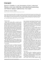
vaginal wall epithelium (Figs. 1, 2). Uroflowmetry was not<br />
obtained but post-voiding residuals of 200-250 ml were documented.<br />
Cystocolpodefecography (Fig. 3) and examination<br />
by a proctologist confirmed a stage 3 rectocoele together<br />
with good anal sphincter function and a tonic <strong>pelvic</strong> <strong>floor</strong>. In<br />
April 2003 the patient underwent colpoperineoplasty.<br />
Normal micturition was restored and at follow up 51<br />
months after surgery uroflowmetry was normal with no post<br />
void residual. The external urethral meatus was now visible<br />
and the rectocoele had disappeared.<br />
Case 2. In March 2006 an 83 years old woman presented<br />
with urgency, frequency, urge-incontinence and perineal discomfort.<br />
She had previously undergone a laparoscopic hysterectomy<br />
for fibroids. Urine analysis and urinary cytology<br />
were normal. She had a stage 4 rectocoele and a stage 1<br />
anterior colpocele (HWS). Micturition diaries showed 20<br />
voids per day with a mean voided amount of 70 ml. Bladderscan<br />
documented 280-300 ml post void residual. The rectocoele<br />
was reduced with a ring pessary and a urodynamic<br />
assessment was performed. All parameters were normal and<br />
the bladder capacity was 400 mL. The patient chose to<br />
follow a conservative course with a ring pessary. Twelve<br />
months later the ring pessary was still well-tolerated while<br />
urinary symptoms had improved significantly. At follow up<br />
the urinary diary revealed 8-10 micturitions per day with no<br />
more incontinence and no measurable post void residual.<br />
Case 3: In April 2006 a 63-year-old woman was admitted<br />
into our urology unit for investigation of hypogastric<br />
pain and obstructive urinary symptoms which had appeared<br />
10 days before. She reported long term constipation but<br />
the constipation had been worsening in the last few months<br />
and was associated with the appearance of genital prolapse.<br />
On examination we found bladder overdistension and a 3rd<br />
stage rectocoele (HWS) with significant faecal impaction<br />
causing direct pressure on the anterior wall of the vagina<br />
and urethra.<br />
A urethral catheter was placed, with slow drainage of<br />
1000mls of urine, and the faecal impaction was resolved<br />
using digitation and enemas.<br />
A <strong>pelvic</strong> ultrasound was normal. The rectocoele was<br />
reduced using a ring pessary while a programme of regular<br />
Fig. 1. – Case 1. - Posterior colpocele evident with the patient<br />
straining in the supine position: labia minora and clitoris are absent<br />
and the urethral meatus is not visible.<br />
Fig. 2. – Case 1. - Straddling and pulling up the labia majora the<br />
urethral meatus becomes visible and it is appreciable the obstructive<br />
effect on it by the rectocele.<br />
104<br />
<strong>Pelviperineology</strong> 2007; 26: 104-105<br />
http://www.pelviperineology.org
Urinary retention in women with isolated Stage 3 rectocoele<br />
We have reported 3 cases in which a significant (Stage 3<br />
or 4) rectocoele has been associated with urinary retention.<br />
Rectocoele exerted a direct pressure to cause obstruction<br />
of the urethral meatus in 2 patients, while in third woman<br />
urinary obstruction resulted from the pressure of a rectocoele<br />
that was distended with impacted faecal material. In<br />
all cases surgical repair of the rectocoele or reduction of the<br />
rectocoele using a ring pessary resulted in cure of the urinary<br />
retention.<br />
Fig. 3. – Case 1. - Cystocolpodefecography in sitting position<br />
during straining; the posterior colpocele is caused by a significant<br />
rectocoele.<br />
bowel evacuations was suggested. The patient resumed satisfactory<br />
urinary voiding without any post void residual. A<br />
year later she is still using a ring pessary without any urinary<br />
problems.<br />
DISCUSSION AND CONCLUSION<br />
Isolated rectocoele, often asymptomatic, can sometimes<br />
lead to rectal symptoms and occasionally can cause urinary<br />
symptoms. Only one case of urinary obstruction following<br />
rectocoele could be found in the literature. 5<br />
REFERENCES<br />
1. Marinkovic SP, Stanton SL. Incontinence and voiding difficulties<br />
associated with prolapse. J Urol 2004; 171: 1021-8.<br />
2. Fitzgerald MP, Kulkarni N, Fenner D. Postoperative resolution<br />
of urinary retention in patients with advanced <strong>pelvic</strong> organ prolapse.<br />
Am J Obstet Gynecol 2000; 183: 1361-3.<br />
3. Murthy VK, Orkin BA, Smith LE, Glassman LM. Excellent<br />
outcome using selective criteria for rectocele repair. Dis Colon<br />
Rectum 1996; 39: 374-8.<br />
4. Baden WF, Walker TA, Lindsay HJ. The vaginal profile. Tex<br />
Med J 1968; 64: 56-58.<br />
5. Nitti VW, Tu LM, Gitin J. Diagnosing bladder outlet obstruction<br />
in women. J Urol 1999; 161: 1535-40.<br />
6. JimAonez Cidre M. Obstruction of the lower urinary tract in<br />
women. Arch Esp Urol 2003; 55: 989-999.<br />
7. Nichols DH. Vaginal prolapse affecting bladder function. Urol<br />
Clin North America 1985; 12: 222-231.<br />
Correspondence to:<br />
Dr. ELENA ANDRETTA<br />
Urology Department, General Hospital<br />
Riviera XXIX Aprile, 2<br />
30031 Dolo (Venice) Italy<br />
Tel and Fax +39 041 5133481<br />
E-mail: elenaandretta@libero.it<br />
105
Pelvic Floor Digest<br />
This section presents a small sample of the Pelvic Floor Digest, an<br />
online publication (www.<strong>pelvic</strong><strong>floor</strong>digest.org) that reproduces titles and<br />
abstracts from over 200 <strong>journa</strong>ls. The goal is to increase interest in all the<br />
compartments of the <strong>pelvic</strong> <strong>floor</strong> and to develop an interdisciplinary culture<br />
in the reader.<br />
1 – THE PELVIC FLOOR<br />
Variations in stress incontinence and prolapse management by surgeon specialty. Anger JT, Litwin MS, Wang Q, et al. J Urol. 2007 Aug<br />
14; epub. Numerous studies have documented a relationship between provider specialty and outcomes for surgical procedures. In this study the<br />
effect of surgeon specialty was determined on outcomes of sling surgery for women with stress urinary incontinence. Early prolapse management<br />
by gynecologists corresponded to fewer prolapse repairs in the year following the sling, suggesting that gynecologists are more likely to<br />
identify and manage prolapse at the time of the evaluation of urinary incontinence, a strategy that appears to avoid the morbidity and cost of<br />
repeat surgery.<br />
Symptoms of anal incontinence and difficult defecation among women with prolapse and a matched control cohort. Morgan DM,<br />
Delancey JO, Guire KE, Fenner DE. Am J Obstet Gynecol. 2007 Aug 20; epub. One-third of women with <strong>pelvic</strong> prolapse have anal incontinence<br />
of flatus on “most” or “every” day or difficult defecation with “most” or “every” bowel movement. This study quantifies the risk for<br />
these symptoms comparing women with and without prolapse of similar age, body mass index, race, and hysterectomy status. Length of the<br />
perineal body, mean parity, and a positive standing cough stress test are associated with greater symptom severity.<br />
A prospective, randomized controlled trial of the use of an anal purse-string suture to decrease contamination during <strong>pelvic</strong> reconstructive<br />
surgery. Biller DH, Guerette NL, Bena JF, Davila GW. Int Urogynecol J Pelvic Floor Dysfunct. 2007 Apr 11; epub. An anal pursestring<br />
suture is an effective way of reducing fecal contamination of the sterile field when performing vaginal <strong>pelvic</strong> reconstructive surgery. No<br />
wound infections, graft erosions, or healing abnormalities however were noted without the suture.<br />
Medium-term efficacy of <strong>pelvic</strong> <strong>floor</strong> muscle training for female urinary incontinence in daily practice. Lamers BH, van der Vaart CH.<br />
Int Urogynecol J Pelvic Floor Dysfunct. 2007;18:301. Pelvic <strong>floor</strong> muscle training for urinary incontinence is effective for their quality of life<br />
in half of the women. If not successful, women seem to benefit significantly from incontinence surgery. This is the result of a study on 355<br />
women treated by specialized physiotherapists, with a mean follow-up of 32 months.<br />
Deformation of the <strong>pelvic</strong> <strong>floor</strong> muscles during a vaginal delivery. Parente MP, Jorge RM, Mascarenhas T et al. Int Urogynecol J Pelvic<br />
Floor Dysfunct. 2007 May 24; epub. Since <strong>pelvic</strong> <strong>floor</strong> injuries during a vaginal delivery can be considered a significant factor in the development<br />
of urinary and fecal incontinence and <strong>pelvic</strong> organ prolapse, to clarify the mechanisms behind <strong>pelvic</strong> <strong>floor</strong> disorders related to a vaginal<br />
delivery, a finite element method and a three-dimensional computer model of the <strong>pelvic</strong> <strong>floor</strong> and fetus were used. Results for the <strong>pelvic</strong> <strong>floor</strong><br />
stretch values obtained during the passage of the fetus head show that the maximum deformation obtained is 0.66 for a vertical displacement<br />
of the fetal head of approximately 60 mm.<br />
2 – FUNCTIONAL ANATOMY<br />
Morphology of the levator ani muscle. Li D, Guo M. Dis Colon Rectum. 2007 Aug 16; epub. Previous studies have suggested that both the<br />
levator ani and the puborectalis muscles lift the anus. CT defecography in a sitting position shows that the levator ani is funnel-shaped at rest in<br />
the sitting position; it ascends, becoming plate-shaped during squeeze, and it descends, becoming basin-shaped during defecation. There is no<br />
muscle to lift the anus during defecation, the puborectalis lifting it during squeeze. The main function of the levator ani is to open the genital<br />
hiatus and the anus during defecation, the puborectalis shuts the genital hiatus and anus during squeeze.<br />
Levator co-activation is a significant confounder of <strong>pelvic</strong> organ descent on Valsalva maneuver. Orno AK, Dietz HP. Ultrasound Obstet<br />
Gynecol. 2007;30:346. The Valsalva maneuver is used clinically and on imaging to determine <strong>pelvic</strong> organ prolapse, but is frequently accompanied<br />
by a <strong>pelvic</strong> <strong>floor</strong> muscle contraction. Levator co-activation may be a substantial confounder, reducing <strong>pelvic</strong> organ descent. Without<br />
repetition and digital, auditory or visual biofeedback, women may not perform a correct Valsalva maneuver. Biofeedback markedly reduces the<br />
likelihood of levator co-activation but does not abolish it completely.<br />
Anal canal anatomy showed by three-dimensional anorectal ultrasonography. Regadas FS, Murad-Regadas SM, Lima DM et al. Surg<br />
Endosc. 2007 May 4; epub. 3-D anal endosonography enables measurement of the different anatomical structures of the anal canal and demonstrates<br />
its asymmetrical configuration. The shorter anterior external anal sphincter and internal anal sphincter associated with a longer gap<br />
(distance from the anterior external sphincter to the anorectal junction) can justify the higher incidence of <strong>pelvic</strong> <strong>floor</strong> dysfunction in females,<br />
especially fecal incontinence and anorectocele with rectal intussusception.<br />
Effects and mechanisms of vaginal electrical stimulation on rectal tone and anal sphincter pressure. Song GQ, Zhu H, Chen JD. Dis<br />
Colon Rectum. 2007 Aug 14; epub. Vaginal electrical stimulation with long pulses or trains of long pulses but not trains of short pulses reduces<br />
rectal tone and increases anal sphincter pressure in conscious dogs. The inhibitory effect of vaginal electrical stimulation on rectal tone is mediated<br />
by the sympathetic pathway. These findings suggest that vaginal electrical stimulation may be a potential therapy for fecal incontinence.<br />
Rectoanal sensorimotor response in humans during rectal distension. De Ocampo S, Remes-Troche JM, Miller MJ, Rao SS. Dis Colon<br />
Rectum. 2007 Aug 16; epub. Rectal perception, that facilitates maintenance of continence and defecation, is associated with motor changes<br />
in anorectum. Sensory and motor responses of the anorectum during rectal distention were examined, the sensorimotor response first occurring<br />
synchronously with the sensation of fullness or more often with the desire to defecate which is associated with a unique, consistent, and<br />
reproducible anal contractile response.<br />
Role of phospholipase A2 in the genesis of basal tone in the internal anal sphincter smooth muscle. de Godoy MA, Rattan S. Am J Physiol<br />
Gastrointest Liver Physiol. 2007 Aug 23; epub. Phospholipase A2 plays a critical role in the genesis of tone in the internal anal sphincter and<br />
its inhibitors may provide potential therapeutic target for treating anorectal motility disorders.<br />
3 – DIAGNOSTICS<br />
Clinical and urodynamic parameters associated with history of urinary tract infections in women. Athanasiou S, Anstaklis A, Betsi GI et<br />
al. Acta Obstet Gynecol Scand. 2007;86:1130. Urodynamic testing does not help in identifying specific urogynecologic mechanisms that could<br />
improve medical and/or surgical management or prevent recurrent urinary tract infections.<br />
106<br />
<strong>Pelviperineology</strong> 2007; 26: 106-135 http://www.pelviperineology.org
Pelvic Floor Digest<br />
Effect of anatomic urethral length on the correlation between the Q-tip test and descent at point Aa of the POP-Q system. Larrieux<br />
JR, Balgobin S.Int Urogynecol J Pelvic Floor Dysfunct. 2007 Aug 9; epub. The objective of this study is to evaluate the effect of anatomic<br />
urethral length on the relationship between descent at point Aa of the <strong>pelvic</strong> organ prolapse quantification system and the Q-tip straining angle.<br />
Urethral length being measured with a urethral profilometer, a substantial correlation was found between descent at point Aa and the straining<br />
Q-tip angle, while there was no correlation between the anatomic urethral length and straining Q-tip angle. So urethral length does not affect<br />
the straining Q-tip angle, and point Aa is a strong predictor of an abnormal straining Q-tip angle in women with stage I anterior vaginal wall<br />
prolapse or greater.<br />
Segmental colonic transit studies: comparison of a radiological and a scintigraphic method. Lundin E, Graf W, Garske U et al. Colorectal<br />
Dis. 2007;9:344. Colonic transit studies are used to diagnose slow transit constipation (STC) and to evaluate segmental colonic transit before<br />
segmental or subtotal colectomy. The aim of the study was to compare a single X-ray radio-opaque marker method with a scintigraphic technique<br />
(111Indium-DTPA) to assess total and segmental colonic transit in patients with STC. Segmental colonic delay was a common finding.<br />
The two methods gave similar results for groups of patients, except in the descending colon. The variation of the results for individuals suggests<br />
that a repeated transit test may improve the assessment of total and segmental transit.<br />
MR colonography. Kinner S, Lauenstein TC. Radiol Clin North Am. 2007;45:377. Combining the advantages of unsurpassed soft tissue contrast<br />
and lack of ionizing radiation, MR imaging of the gastrointestinal tract has become increasingly used clinically. Both bowel inflammation<br />
and tumor disease of the large bowel can be well visualized by means of MR colonography.<br />
Nurse specialist led flexible sigmoidoscopy in an outpatient setting. Kelly SB, Murphy J, Smith A. Colorectal Dis. 2007 May 17; epub.<br />
Objective there has been an increasing demand for diagnostic flexible sigmoidoscopy. In order to improve our diagnostic services, we established<br />
a nurse specialist led flexible sigmoidoscopy clinic in 1999. A total of 3956 patients had a flexible sigmoidoscopy performed between<br />
1999 and 2004. Two patients only sustained an iatrogenic rectal perforation. The nurse specialist offers an efficient diagnostic service for<br />
patients presenting with colorectal symptoms.<br />
Digital rectal examination: national survey of undergraduate medical training in Ireland. Fitzgerald D, Connolly SS, Kerin MJ. Postgrad<br />
Med J. 2007;83:599. The experience gained in at least one digital rectal examination (DRE) by medical students by the completion of<br />
undergraduate training was assessed. No experience was reported in 24%, with mannequin-only experience in a further 20%, 56% performed<br />
DRE on at least one patient, but one third reported no confidence in their ability to interpret their findings properly.<br />
4 – PROLAPSES<br />
The natural history of posterior vaginal wall support after abdominal sacrocolpopexy with and without posterior colporrhaphy. Yau JL,<br />
Rahn DD, McIntire DD et al. Am J Obstet Gynecol. 2007;196:e45. For abdominal sacrocolpopexy with concomitant posterior colporrhaphy,<br />
POP-Q point Ap significantly improved and persisted at 34 months after surgery. Ten months after surgery, descent of POP-Q point Bp returned<br />
to preoperative levels and was the same regardless of whether a site-specific posterior colporrhaphy was performed at the time of an abdominal<br />
sacrocolpopexy.<br />
Symptomatic Pelvic Organ Prolapse: prevalence and risk factors in a population-based, racially diverse cohort. Rortveit G, Brown JS,<br />
Thom DH et al. Obstet Gynecol. 2007;109:1396. To estimate the prevalence of and identify risk factors associated with symptomatic <strong>pelvic</strong><br />
organ prolapse and level of distress in racially diverse women aged older than 40 years, the Reproductive Risks for Incontinence Study evaluated<br />
a population of 2,001 randomly selected women. Symptomatic prolapse was reported by 6% of women. Almost 50% of these reported<br />
moderate or great distress, and in 35% the symptoms affected at least one physical, social or sexual activity. In multivariable logistic regression<br />
analysis the risk of prolapse was significantly increased in women with vaginal deliveries compared with nulliparous women. Irritable<br />
bowel syndrome, constipation, and self-reported fair or poor health status were strongly associated with prolapse. African-American were<br />
significantly less likely to report symptomatic prolapse compared with white women.<br />
Functional outcome after transperineal rectocele repair with porcine dermal collagen implant. Smart NJ, Mercer-Jones MA. Dis Colon<br />
Rectum. 2007 Apr 9; epub. To assess the safety and efficacy of transperineal rectocele repair with porcine dermal collagen (Permacol (R)) 10<br />
females were followed for 5-16 months with objective assessment for constipation, excessive straining, incomplete evacuation, vaginal bulging,<br />
and vaginal digitations (always, usually, occasionally, never) and Medical Outcomes Study Short Form 36 questionnaires. All patients had<br />
an improvement in two or more symptoms and 70 percent of patients in three or more symptoms. Improvements in digitations and SF-36 scores<br />
were not significant.<br />
A novel technique for rectocele repair in elderly women. Nano M, Ferronato M, Solej M, D’Amico S. Tech Coloproctol. 2007 May 25; epub.<br />
The rectal wall was separated from the rectovaginal septum. The vaginal wall was divided in the middle. The first flap was sewn to the second<br />
and this onto the first. In 22 elderly women followed-up for 24-84 months the need to digitally assist evacuation disappeared.<br />
Heterogeneity in anatomic outcome of sacrospinous ligament fixation for prolapse: a systematic review. Morgan DM, Rogers MA, Huebner<br />
M, Wei JT, Delancey JO. Obstet Gynecol. 2007;109:1424. To explore why failure rates vary so much between published reports of sacrospinous<br />
ligament fixation to correct POP and what the potential sources of heterogeneity may be, Medline was queried for studies between<br />
1966 and 2005 and 187 studies were reviewed. The anterior compartment was the most common site of failure for any given grade. This was<br />
most striking when the criterion for failure was grade 1 (40.1% anterior, 11.0% apical, 18.2% posterior) or grade 2 prolapse (21.3% anterior,<br />
7.2% apical, 6.3% posterior). Areas of vaginal support were more equally affected when the criterion for failure was grade 3 prolapse (3.7%<br />
anterior, 2.7% apical, 2.3% posterior). Among cohorts using grade 2 prolapse as the criterion for objective failure, the pooled measure of<br />
failure to relieve symptoms was 10.3% and to provide patient satisfaction was 13.0%. In conclusion the variation in published failure rates after<br />
sacrospinous ligament fixation is, in part, accounted for by differences in how anatomical outcomes are evaluated and which compartment of<br />
vaginal support is being considered.<br />
Midline anterior repair alone vs anterior repair plus vaginal paravaginal repair: a comparison of anatomic and quality of life outcomes.<br />
Morse AN, O’dell KK, Howard AE. Int Urogynecol J Pelvic Floor Dysfunct. 2007;18:245. A retrospective study to compare the anatomic<br />
recurrence rates and quality of life outcomes of patients who had undergone either anterior colporrhaphy or anterior colporrhaphy and<br />
vaginal paravaginal repair as part of surgery for <strong>pelvic</strong> organ prolapse did not suggest that adding paravaginal repair is superior in terms of<br />
anatomic or quality of life outcomes.<br />
Mesh-related infections after <strong>pelvic</strong> organ prolapse repair surgery. Falagas ME, Velakoulis S, Iavazzo C, Athanasiou S. Eur J Obstet<br />
Gynecol Reprod Biol. 2007 Apr 23; epub. Although the use of vaginal meshes has become a new effective method of <strong>pelvic</strong> organ prolapse<br />
surgery clinicians should be aware of the various post-operative complications, including mesh-related infections. The incidence of meshrelated<br />
infections and erosion ranges from 0 to 8%, and 0 to 33%, respectively, in the published studies.<br />
The PFD continues on page 111<br />
107
Original article<br />
A pilot study: The anal sphincter support procedure<br />
for the treatment of anal incontinence<br />
MAXWELL E. HAVERFIELD<br />
The Northern Hospital, Epping, Victoria, Australia<br />
Abstract: Faecal incontinence is a significant and a very debilitating condition. The true prevalence of which is unknown but up to 3% of<br />
the general population have stool incontinence which increases with age. Faecal incontinence is much more common in women, increases<br />
with parity, and up to 30% have associated urinary incontinence and/or <strong>pelvic</strong> organ prolapse. Patients who have an identifiable external<br />
sphincter defect who fail conservative treatment options are best managed by a primary repair of the defect. This study presents a novel way<br />
of supporting the external anal sphincter with a circlage tape prosthesis. Based on the success of tension-free suburethral tapes used to treat<br />
stress urinary incontinence a pilot study was devised to assess Anal Sphincter Support System (ASSP) in patients presenting with mild faecal<br />
incontinence. Fourteen patients were followed up for a mean period of eighteen months. Outcomes were very encouraging with resolution<br />
of symptoms between 70 and 100%. There were no observed complications. This pilot study demonstrates that the ASSP provides a safe<br />
and effective method of restoring anal continence and can be used successfully with concomitant procedures which may be necessary for<br />
restoration of <strong>pelvic</strong> <strong>floor</strong> anatomy and function.<br />
Key words: Faecal incontinence; Anal Sphincter Support Procedure; Hammock hypothesis.<br />
INTRODUCTION<br />
Faecal incontinence is rarely due to one single factor. Vaginal<br />
childbirth and obstetric trauma are widely accepted as significant<br />
causes of faecal and flatal incontinence in women.<br />
Approximately 10-15% report symptoms following vaginal<br />
delivery culminating in 2-5 % of women having faecal incontinence.<br />
1 The most common contributing cause is anal sphincter<br />
injury occurring at the time of the second stage of labour.<br />
Other significant factors are instrumental delivery, midline<br />
episiotomy and prolonged second stage of labour. 2, 3 Also<br />
thought to be of significance is traction or entrapment of the<br />
pudendal nerve and its branches of the perineal nerve fibres,<br />
associated with injury to the levator ani plate (iliococcygeous,<br />
pubococcygeous and puborectalis muscles).<br />
Women with symptoms of faecal incontinence following<br />
their first pregnancy will deteriorate after their second pregnancy.<br />
4 In one study 26.9% of women had an identifiable<br />
occult anal sphincter injury on endoanal ultrasound. With<br />
each increase in parity the chance of a new defect increased<br />
by 8.5%, while 75-83% of women presenting with faecal<br />
incontinence will have an identifiable anal sphincter defect. 5<br />
Follow up of women who underwent primary repair of a<br />
third degree vaginal tear has shown that 85% will have persistent<br />
structural defects and over 50% of this group will<br />
develop faecal incontinence. 6-10<br />
A wide range of causes may contribute to anorectal dysfunction<br />
and anal incontinence. 7 Some iatrogenic causes<br />
that may result in injury to the anal sphincter complex<br />
include anorectal procedures such as fistulotomy, sphincterotomy<br />
and haemorrhoidectomy. Medical etiological factors<br />
include psychiatric conditions, chronic constipation, malabsorption<br />
syndromes, laxative overuse, diabetes, thyroid disease,<br />
gastrointestinal inflammatory conditions, neuropathies<br />
and spinal cord disease.<br />
Anatomical distortion associated with chronic straining<br />
is commonly known as <strong>pelvic</strong> <strong>floor</strong> prolapse syndrome and<br />
may also be associated with progressive denervation of the<br />
colon, rectum and anus at the <strong>pelvic</strong> <strong>floor</strong>, in conjunction<br />
with obstructive defaecation and intussusception. Lateral,<br />
central and posterior rectal defects including rectal diverticulum<br />
may also contribute to anorectal dysfunction with<br />
incontinence.<br />
It is difficult to know the true incidence of faecal incontinence<br />
due to variations in definitions, age distribution,<br />
108<br />
underreporting due to reluctance to broach the condition,<br />
coexistence with other <strong>pelvic</strong> symptoms such as voiding<br />
dysfunction and <strong>pelvic</strong> organ prolapse. 11 A complex, interactive<br />
psychological process is necessary for faecal/flatal<br />
continence including rectal motility and compliance with<br />
neurophysiology of sensation. Hyposensitivity associated<br />
with low resting pressure may indicate internal and external<br />
sphincter damage resulting in passive incontinence, whereas<br />
hypersensitivity with urge incontinence and the inability<br />
to postpone defaecation may be associated with low voluntary<br />
squeeze pressure thus indicating external anal sphincter<br />
malfunction.<br />
The aim of this study was to explore the possibility that<br />
a circumferential mesh support for the anal sphincter would<br />
reduce symptoms in patients with faecal or flatal incontinence.<br />
This hypothesis arose from consideration of the need<br />
for additional anchoring support of the Apogee TM (American<br />
Medical Systems, Inc, Minnetonka, MN, USA) mesh used<br />
for the repair of posterior vaginal wall prolapse (rectocele<br />
and associated perineal repair). It was found that a percentage<br />
of these patients reported a significant improvement in<br />
bowel function and in particular a reduction in severity of<br />
their previously underreported anal incontinence. 12<br />
The use of mid urethral slings to treat Stress Urinary<br />
Incontinence (SUI) is based on the Hammock Hypothesis of<br />
DeLancey. 13 Since the anatomy 14, 15 and pathophysiology of<br />
defects arising in the anterior <strong>pelvic</strong> compartment have been<br />
successfully corrected using a mid urethral sling to treat SUI<br />
it was postulated that a mesh sling prosthesis placed around<br />
the anal sphincter may improve the anal sphincter mechanism<br />
of action and address the problem of faecal and flatal<br />
incontinence. It was decided to embark on a pilot study of<br />
patients with mild faecal incontinence using such a mesh<br />
prosthesis and to monitor the outcomes of this group.<br />
ANORECTAL ANATOMY<br />
The anal canal extends from the anorectal junction which<br />
lies just above the level of the puborectalis sling and sphincters<br />
(internal and external) to the anus below. It is surrounded<br />
by an external sphincter of voluntary muscle fibres<br />
and an internal sphincter of involuntary muscle fibres. 16<br />
Between the two sphincters lies the longitudinal anal muscle<br />
of the rectum (LAM) 17 composed of muscle fibres which<br />
receive contributions from puborectalis, pubococcygeous<br />
<strong>Pelviperineology</strong> 2007; 26: 108-111 http://www.pelviperineology.org
A pilot study: The anal sphincter support procedure for the treatment of anal incontinence<br />
and iliococcygeous. 18 Recently Petros described that the<br />
LAM fibres contributed by the levator plate are incorporated<br />
into the sphincteric action as described by the integral<br />
theory. 19 This is probably important in closure and relaxation<br />
of the anal canal and that of the urethra. Individuals<br />
show significant variations in detail of the sphincter anatomy.<br />
16<br />
The external anal sphincter is a voluntary muscle, cylindrical<br />
in form being approximately 2 cm deep placed around<br />
the anal canal for approximately 5-7 cm. It has three parts.<br />
The subcutaneous part is slender and encircles the anal orifice.<br />
The superficial part supports the anus in an ovoid circular<br />
fashion and extends from the tip of the coccyx and<br />
anorectal raphe to the perineal body in the median plane.<br />
The deep part encircles the anal canal with some fibres joining<br />
the superficial transverse perineal muscle and blends<br />
with the puborectalis muscle of the levator ani. The internal<br />
sphincter surrounds the superior two thirds of the anal canal<br />
and is supported by the levator ani. The inferior two thirds<br />
are surrounded by the external sphincter as described above.<br />
The longitudinal muscle is comprised of three layers and<br />
lies between the internal and external sphincters. 17<br />
PATIENTS AND METHODS<br />
The ASSP was performed on 14 patients between June<br />
2005 and March 2007 with a total follow up time of 20.3<br />
months. Mean follow up time was 18 months. Inclusion<br />
criteria for the study were symptoms of mild to moderate<br />
faecal soiling and flatal incontinence. Ten patients had a<br />
Wexner grading symptom score of 5-8 and four patients<br />
had a Wexner grading symptom score of 8-10. Past gynaecological<br />
and colorectal surgeries were as follows: Four<br />
patients had a past history of vaginal hysterectomy and anterior<br />
<strong>pelvic</strong> organ prolapse repair, six had abdominal hysterectomy<br />
(two also Burch colposuspension), four anterior<br />
and posterior vaginal repair, two had a haemorrhoidectomy,<br />
one an partial sphincterotomy for anal fissure, one an anal<br />
stretch procedure. The ages of patients ranged from 45 to<br />
78 years, mean age 64.7 years. Obstetric history: all patients<br />
were parous. Five patients were parous 1-3, nine patients<br />
3-5. Two patients had caesarian sections only. Interestingly<br />
both these patients had a past history of anorectal surgery.<br />
Of the 12 patients who had vaginal births more than 33%<br />
had instrumental deliveries. Mean foetal size was 3,700 g,<br />
ranging between 4,300 to 2,800 g.<br />
All patients were studied using Magnetic Resonance<br />
Imaging (MRI) of the anal sphincter. At imaging 5 patients<br />
had evidence of external sphincter defects mainly right, lateral<br />
or central. Defects were considered small varying from<br />
2-3 mm. Two patients aged 75 and 78 years had evidence<br />
of mild atrophy of the external anal sphincter while 50%<br />
of patients had no obvious anal sphincter defects on MRI.<br />
All patients were assessed pre and post operatively with<br />
resting anorectal pressures and maximal squeeze pressure.<br />
Six patients had a concomitant posterior organ prolapse procedure<br />
using an Apogee TM (American Medical Systems, Inc,<br />
Minnetonka MN, USA) mesh. Patients were assessed using<br />
the POPQ system. 20<br />
Operative technique: All patients had a 24 hour preoperative<br />
bowel preparation. Systemic triple antibiotics<br />
were given intraoperatively and this combination was continued<br />
for 24 hours postoperatively. Clindamycin cream was<br />
inserted vaginally as a single dose approximately 1-2 hours<br />
preoperatively. The operation was performed in the lithotomy<br />
position. Aqueous Betadine antiseptic solution (10%<br />
w/v Povidone/Iodine) was used as skin preparation and liberally<br />
applied to inner thighs, intravaginally and to the perineal<br />
and suprapubic areas. The patient was draped in the<br />
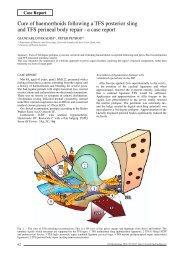
Fig. 1. – Anorectal Anatomy. 1. External anal sphincter. 2. Transverse<br />
rectal folds; rectal ampulla. 3. Anal sinuses. 4. Skin. 5. Subcutaneous<br />
part of external anal sphincter. 6. Superficial part of<br />
external anal sphincter. 7. Deep part of external anal sphincter and<br />
pubo rectalis. 8. Semi tendinosus. 9. Gluteous maximus. 10. Ischio<br />
rectal fossa. 11. The longitudinal muscle coat. 12. Pubo coccygeous.<br />
13. Obturator internus. 14. Iliococcygeous. 15. Pelvi-rectal<br />
space. 16. Para rectal fossa. 17. Internal circular muscle coat/ internal<br />
ano rectal sphincter.<br />
usual fashion facilitating good exposure of the operative site<br />
with the buttocks extending just below the end of the table.<br />
Infiltration of the perianal area and perineal body including<br />
the posterior vaginal mucosa was performed using a hydrodissection<br />
technique with dilute local anaesthetic. A stab<br />
incision of approximately 1 cm using an eleven blade was<br />
made 3 cm from the anal verge in the 5 o’clock and 7<br />
o’clock position or a single incision was made at 6 o’clock<br />
(Fig. 4, 5).<br />
At the vestibule of the vaginal orifice a transverse incision<br />
of approximately 4-5 cm was performed with dissection<br />
supramedially and laterally to expose the superior aspect of<br />
the perineal body. In the case of an adjuvant posterior prolapse<br />
procedure, blunt and minimal sharp tunneling dissection<br />
is continued proximally to create a pathway and identify the<br />
ischial spine and tendinous arch of the lateral <strong>pelvic</strong> wall. 21<br />
This dissection is performed bilaterally (Fig. 2, 3).<br />
Gentamycin solution 1 mg per 1cc of saline is used to liberally<br />
irrigate the operative site throughout the procedure.<br />
Fig. 2. – Placement of ASSP Monarc TM tape. 1. Incision over the<br />
perineal body at the hymenal ridge to expose the perineal body.<br />
2. Anal sphincter support prosthesis, Monarc in situ / surrounding<br />
the superficial part of the external anal sphincter. 3. Alternative<br />
incision site for placement of the Monarc tape.<br />
109
M. E. Haverfield<br />
track made by the helical needles emerging through the perianal<br />
incisions posterior to the anal verge. Gentle traction is<br />
used on the mesh contralaterally until it is seen and felt to sit<br />
undistorted and without tension over the perineal body. The<br />
needles are then detached. The plastic sheaths are removed<br />
and the mesh trimmed at the buttock incisions (Fig. 6). All<br />
incisions are then closed with 2-0 Vicryl interrupted mattress<br />
sutures.<br />
Fig. 3. – Position of Monarc TM and Apogee TM prosthesis when ASSP<br />
is performed together with a prolapse repair using the Apogee TM<br />
system. 1. External anal sphincter. 2. Perineal body. 3. Pubo rectalis<br />
and pubo vaginalis. 4. Superficial transverse perineal muscle. 5.<br />
Anal sphincter support tape (Monarc TM ) in situ. 6. Posterior <strong>pelvic</strong><br />
compartment support prosthesis (Apogee TM ) in situ.<br />
The recommended entry site for the Apogee (American<br />
Medical Systems, Inc, Minnetonka MN, USA) needles is<br />
5-6 cm posterolateral to the anal verge which is 3 cm posterolateral<br />
to the incisions used for the ASSP. The posterior<br />
compartment mesh repair Apogee should be performed<br />
before the perianal support procedure. The Apogee mesh<br />
must be in position and the excess inferior cape trimmed<br />
approximately 1cm above the transverse perineal incision<br />
before the positioning of the ASSP is performed. The stab<br />
incisions are performed as explained above and with the<br />
index finger of a double gloved left hand the helical needle<br />
of a Monarc (American Medical Systems, Inc, Minnetonka<br />
MN, USA) prosthesis is inserted into the incision and<br />
directed approximately 2-3 cm in depth before rotation of<br />
the needle through its natural arc creates a track around<br />
the external anal sphincter to emerge laterally through the<br />
perineal body enabling visualization of the needle tip at the<br />
perineal incision. The same technique is performed on the<br />
contra lateral side of the external anal sphincter. With the<br />
tips of both needles at the perineal body, the Monarc TM sling<br />
is attached to both needle tips firmly with an audible click.<br />
The mesh is then gently positioned posteriorly through the<br />
Fig. 4.<br />
Fig. 5.<br />
RESULTS<br />
All 14 patients were studied over a minimum period of 6<br />
months. Mean average time of follow up was 18 months.<br />
During that time there were no perioperative or postoperative<br />
complications. In particular there were no cases of mesh<br />
erosion or rejection and no cases of rectal perforation or<br />
trauma. Four cases had evidence of perianal bruising which<br />
resolved spontaneously with the judicious use of ice packs<br />
applied to the perianal area for approximately 24 hours.<br />
There were no cases of overt haematoma formation, infection<br />
or abscess formation. Ten patients whose initial Wexner<br />
score 22 was between 5 and 8 were reassessed to have<br />
between 90 and 100% improvement in symptoms. The<br />
other 4 patients with initial Wexner Score of 8-10 noted an<br />
improvement of 70% or better resulting in a Wexner score<br />
between 2 and 3.<br />
CONCLUSIONS<br />
The purpose of this study was primarily to evaluate the<br />
safety and efficacy of the ASSP and its impact on quality<br />
of life with respect to anorectal dysfunction and faecal<br />
incontinence. The ASSP was found to be a simple procedure<br />
with reproducible results and not associated with any<br />
significant peri or post operative complications within a<br />
mean follow up period of 18 months. In particular there<br />
was no evidence of mesh erosion, rejection or anorectal<br />
trauma. Subjective improvement of symptoms was noted<br />
using the Wexner grading profile and quality of life was<br />
maintained in all patients and improved in more than 90%.<br />
As a consequence of this pilot study it will be important<br />
to consider longer term prospective randomized controlled<br />
studies, comparing outcomes of other techniques for<br />
treating faecal incontinence with ASSP. The use of ASSP<br />
in conjunction with other concomitant procedures for posterior<br />
<strong>pelvic</strong> organ prolapse and dysfunction is efficacious<br />
and safe. This study suggests that there is a significant<br />
enhancement when used in conjunction with a posterior<br />
vaginal mesh procedure using the Apogee TM . Further studies<br />
in mesh design and in particular a wider dimension in<br />
the mesh prosthesis may be desirable. Further research into<br />
the role of <strong>pelvic</strong> <strong>floor</strong> imaging will increase our knowledge<br />
into the pathogenesis and management of functional<br />
<strong>pelvic</strong> <strong>floor</strong> disorders. 23 There is also a need for standardized<br />
terminology and diagnostic criteria for defaecatory<br />
dysfunction and for a comprehensive classification of<br />
site specific anatomical variations of the posterior <strong>pelvic</strong><br />
compartment. This will also need to be addressed from<br />
a <strong>multidisciplinary</strong> perspective as recently proposed by<br />
Farnsworth and Dodi. 24<br />
REFERENCES<br />
1. Arramorowitz L, Sobhani I, Ganansia R, et al. Are sphincter<br />
defects the cause of anal incontinence after vaginal delivery?<br />
Results of a prospective study. Diseases of the colon and<br />
rectum 2000; 43: 590-596.<br />
2. Donnelly V, Fynes M, Campbell D, et al. Obstetric events leading<br />
to anal sphincter damage. Obstetrics & Gynaecology 1998;<br />
92: 955-961.<br />
110
A pilot study: The anal sphincter support procedure for the treatment of anal incontinence<br />
3. Kamm MA. Obstetric damage and faecal incontinence. Lancet<br />
1994; 344: 730-733.<br />
4. Fynes M, Donnelly V, Beham M, et al. Effects of second vaginal<br />
delivery on ano-rectal physiology and faecal incontinence:<br />
Prospective study. Lancet. 1999; 354: 983-986.<br />
5. Varma A, Gunn J, Gardinera A, et al. Obstetric anal sphincter<br />
injury: prospective evaluation of incidence. Diseases of colon<br />
and rectum. 1999; 42: 1537-1542.<br />
6. Faltin DC, Sangalli MR, Curtin F, et al. Prevalence of anal<br />
incontinence and other ano rectal symptoms in women. International<br />
Urogynaecology Journal 2001; 12: 117-120.<br />
7. Kiff ES, Barnes PR, Swash M. Evidence of pudendal neuropathy<br />
in patients with perineal descent and chronic straining at<br />
stool. GUT 1984; 25: 1279-1282.<br />
8. Oberwalder M, Conner J, Wexner SD. Meta analysis to determine<br />
the incidence of obstetric anal sphincter damage. British<br />
Journal of Surgery 2003; 90: 1333-1337.<br />
9. Poen AL, Felt-Bersma RJ, Strijers RL, et al. Third degree<br />
obstetric perineal tear: long term clinical and functional results<br />
after primary repair. British Journal of Surgery. 1998; 85:<br />
1433-1438.<br />
10. Gjessing H, Racke B, Sahlin Y. Third degree obstetric tears:<br />
outcome after primary repair. Acta Obstetricia and Gynecologica<br />
Scandanavia 1998; 77: 736-740.<br />
11. Ench P, Bielefeldt K, Raschmann W, et al. Epidemiology of<br />
faecal incontinence in selected patient groups. Int Urogyn J<br />
1991; 6: 143-146.<br />
12. Reid R. Recto-enterocele repair - past problems and new horizons<br />
(review). <strong>Pelviperineology</strong> 2007; 26: 9-16.<br />
13. De Lancey JOL. Structural supports of the urethra as it related<br />
to stress urinary incontinence: The Hammock Hypothesis. Am<br />
J Obstet Gynec 1994; 170: 1713-1723.<br />
14. Versi E, Cardozo LD, Studd JWW, Brincatt M, O’Dowell DJ.<br />
Internal urinary sphincter in maintenance of urinary continence.<br />
BMJ 1986; 292: 166-167.<br />
15. Zaccharin RF. The anatomical supports of the female urethra.<br />
Obstets Gynaecology. 1969; 33: 754-759.<br />
16. Last’s Anatomy. Regional and applied. McMinn RMH. Churchill<br />
and Livingstone. Edinburgh, London, Melbourne, New<br />
York. 1990; 401-406.<br />
17. Macchi V, Porzionato A, Stecco C, Benettazzo F, Stecco A,<br />
Portenti A, Dodi G, De Caro R. Histo-topographic study of the<br />
longitudinal anal muscle. <strong>Pelviperineology</strong> 2007; 26: 30-32.<br />
18. Shafik A. A new concept of the anatomy of the anal sphincter<br />
mechanism and physiology of defaecation. XL11. Rectopuborectalis<br />
reflex. Coloproctology 1990; 12: 170-172.<br />
19. Petros P. The internal continence society and integral theory<br />
systems for the management of the incontinent female - a comparative<br />
analysis. <strong>Pelviperineology</strong> 2007; 26: 25-29.<br />
20. Bump RC, Matthiasson A, Bo K, et al. The standardization of<br />
terminology of female <strong>pelvic</strong> organ prolapse and <strong>pelvic</strong> <strong>floor</strong><br />
dysfunction. Am J Obstet Gynecol 1996; 175: 10-17.<br />
21. Davila GW, Beyer R, Moore R, DeRio S. Restoration of<br />
the vaginal apical and posterior support with Apogee system<br />
(Abstract). Int Urologynecology 2005; 16 (Supplement 2):<br />
5118.<br />
22. Jorge JM, Wexner SD. Etiology and management of fecal<br />
incontinence. Dis Colon Rectum 1993; 36: 77-97.<br />
23. Stoker J, Halligan S, Bartram C. State of the art colon <strong>pelvic</strong><br />
<strong>floor</strong> imaging. Radiology 2001; 218: 621-641.<br />
24. Farnsworth B, Dodi G. Short IPGH system for assessment of<br />
<strong>pelvic</strong> <strong>floor</strong> disease. <strong>Pelviperineology</strong> 2007; 26: 73-77.<br />
Competing Interests: None<br />
Acknowledgements:<br />
The author acknowledges Greg Gurr for the line drawings.<br />
Correspondence to:<br />
Dr. MAXWELL E. HAVERFIELD<br />
Senior Consultant,<br />
Department of Obstetrics and Gynaecology<br />
The Northern Hospital - 185 Cooper Street,<br />
Epping, Victoria. Australia 3076.<br />
Email: max.haverfield@nh.org.au<br />
Pelvic Floor Digest<br />
continued from page 107<br />
Prolapse repair by vaginal route using a new protected low-weight polypropylene mesh: 1-year functional and anatomical outcome in<br />
a prospective multicentre study. de Tayrac R, Devoldere G, Renaudie J et al. Int Urogynecol J Pelvic Floor Dysfunct. 2007;18:251. A lowweight<br />
polypropylene mesh coated with a hydrophilic absorbable film for vaginal repair of genital prolapse (anterior, posterior and anteriorposterior<br />
repair) seems to decrease local morbidity (vaginal erosions 6.3%, de novo dyspareunia 12.8%) while maintaining low recurrence<br />
rates (6.8% for cystocele and 2.6% for rectocele). The report is based on the analysis of the first 143 patients of a multicentre study evaluated<br />
after at least 10 months follow-up. The improvement of Pelvic Floor Distress Inventory and Pelvic Floor Impact Questionnaire were highly<br />
significant.<br />
Changes in the extracellular matrix in the anterior vagina of women with or without prolapse. Lin SY, Tee YT, Ng SC et al. Int Urogynecol<br />
J Pelvic Floor Dysfunct. 2007;18:43. To investigate the changes in the connective tissues (collagen type I, III, IV, V, VI, elastin, and glycoproteins)<br />
located in the upper portion of the anterior vaginal wall associated with prolapse, in 23 women with prolapse an immunohistochemical<br />
study demonstrated that collagen III is significantly less than in a control group with a positive correlations with ageing.<br />
Vaginal mesh erosion after transvaginal repair of cystocele using Gynemesh or Gynemesh-Soft in 138 women: a comparative study.<br />
Deffieux X, de Tayrac R, Huel C et al. Int Urogynecol J Pelvic Floor Dysfunct. 2007;18:73. In 138 women follow-up for 7-60 months cystocele<br />
repair was performed according to the technique of tension-free polypropylene mesh. Anatomically, the success rate was 95%. Vaginal erosion<br />
was reported in 20% of the patients with no statistically significant difference between Gynemesh and Gynemesh-Soft meshes. Cystocele stage<br />
>2 HWS is a protective factor against vaginal erosion. A partial excision of the mesh was necessary in 13/27 symptomatic patients (48%),<br />
associated with a vaginal mucosal closure, 2/27 underwent a complete excision. The incidence of de novo dyspareunia was 9% in patients with<br />
vaginal erosion and 11% in patient without erosion.<br />
A prospective, randomised, controlled trial comparing 3 hour and 24 hour postoperative removal of bladder catheter and vaginal pack<br />
following vaginal prolapse surgery. Glavind K, Morup L, Madsen H, Glavind J. Acta Obstet Gynecol Scand. 2007;86:1122. The aim of this<br />
prospective randomised study was to determine whether or not there was a higher incidence of bleeding, reoperation, urinary retention or<br />
bacterial count in the urine depending on whether urinary catheter and vaginal pack were removed 3 or 24 h after prolapse surgery. Pack and<br />
catheter removal after 3 h is recommended with careful monitoring of the patient’s voiding.<br />
Robot-assisted vs. conventional laparoscopic rectopexy for rectal prolapse: a comparative study on costs and time. Heemskerk J, de<br />
Hoog DE, van Gemert WG et al. Dis Colon Rectum. 2007 Aug 10; epub. Robot-assisted laparoscopic rectopexy is a safe and feasible procedure,<br />
but results in increased time and higher costs than conventional laparoscopy.<br />
Stapled hemorrhoidopexy and Milligan Morgan hemorrhoidectomy in the cure of fourth-degree hemorrhoids: long-term evaluation<br />
and clinical results. Mattana C, Coco C, Manno A et al. Dis Colon Rectum. 2007 Aug 16; epub. Long follow-up seems to indicate more<br />
favorable results in Milligan-Morgan procedure compared to stapled hemorrhoidopexy (mean follow-up 92 and 54 months respectively) in<br />
terms of resumption of symptoms and risk of recurrence.<br />
The PFD continues on page 112<br />
111
Three day workshops are regularly conducted at Sydney Adventist Hospital with both theoretical<br />
and practical course content. Temporary registration and hospital accreditation is necessary<br />
to enable delegates to scrub into live surgery. Numbers are limited to a maximum of 6<br />
delegates to each workshop.<br />
Dr BRUCE FARNSWORTH, Dr JEFF TARR, Dr VINCENT TSE, Dr MARK KRIEGER<br />
<br />
10 <br />
<br />
, CNS Pelvic Surgery - Centre for Pelvic Reconstructive Surgery<br />
Suite 505, San Clinic - 185 Fox Valley Rd - WAHROONGA NSW 2076 - AUSTRALIA<br />
Tel: +61 2 94738555 - Fax: +61 2 94738559<br />
E-mail: robynte@yahoo.com.au - http://www.aavis.org/workshops.htm<br />
Pelvic Floor Digest<br />
continued from page 111<br />
5 – RETENTIONS<br />
Postoperative urinary retention after primary colorectal cancer resection via laparotomy: a prospective study of 2,355 consecutive<br />
patients. Changchien CR, Yeh CY, Huang ST. Dis Colon Rectum. 2007 Aug 18; epub. Risk factors for postoperative urinary retention after<br />
colorectal cancer surgery are: male gender, age, lung disease, rectal cancer, longer operation duration, and additional <strong>pelvic</strong> procedure.<br />
Causes of failed urethral botulinum toxin A treatment for emptying failure. Liao YM, Kuo HC. Urology. 2007 Aug 17; epub. Urethral<br />
injection of BTX-A can reduce urethral resistance in patients with voiding dysfunction. However some patients do not benefit from this treatment.<br />
The causes of failed treatment in 23/200 patients were detrusor underactivity with very low abdominal straining pressure in 7, a tight<br />
urethral sphincter in 7, bladder neck obstruction in 7, and psychological inhibition of voiding in 2. Transurethral incision of the bladder neck<br />
was performed in 7 patients, and all had an improved result.<br />
Evaluation of constipation by abdominal radiographs correlated with treatment outcome in children with dysfunctional elimination.<br />
Allen HA, Austin JC, Boyt MA et al. Urology. 2007;69:966. No correlation between any uroflowmetry parameter and the presence of fecal<br />
distention of rectum could be demonstrated, nor a statistical significance between fecal distention of rectum on abdominal radiograph and<br />
outcome of wetting symptoms was established.<br />
A novel three-dimensional dynamic anorectal ultrasonography technique (echodefecography) to assess obstructed defecation, a comparison<br />
with defecography. Murad-Regadas SM, Regadas FS, Rodrigues LV et al. Surg Endosc. 2007 Aug 20; epub. Echodefecography may<br />
be used as an alternative method to assess patients with obstructed defecation as it has been shown to detect the same anorectal dysfunctions<br />
(anorectocele and rectal intussusception) observed in conventional defecography. It is minimally invasive, well tolerated, inexpensive, avoids<br />
exposure to radiation, and clearly demonstrates all the anatomic structures involved with defecation.<br />
6 – INCONTINENCES<br />
Anatomic restoration technique of continence mechanism and preservation of puboprostatic collar: a novel modification to achieve<br />
early urinary continence in men undergoing robotic prostatectomy. Tewari AK, Bigelow K, Rao S. Urology. 2007;69:726. The complex<br />
of puboprostatic ligaments, puboperinealis muscle, and arcus tendineus (acting in unison to provide continence in men and women) can be<br />
disrupted during robotic prostatectomy. The preservation of the puboprostatic collar helped to restore early continence in 50 men undergoing<br />
robotic prostatectomy for clinically localized prostate cancer. The ligaments were reconnected to the urethrovesical anastomosis, reapproximated<br />
the muscles and fixed the distal bladder to the arcus tendineus. The average additional time was only 2 to 5 minutes, and the continence<br />
rate was 29% in the first week and 95% in 16 weeks after catheter removal.<br />
Anal sphincter laceration at vaginal delivery: is this event coded accurately? Brubaker L, Bradley CS, Handa VL et al. Obstet Gynecol.<br />
2007;109:114.<br />
Effectiveness of a new self-positioning pessary for the management of urinary incontinence in women. Farrell SA, Baydock S, Amir<br />
B, Fanning C. Am J Obstet Gynecol. 2007;196:474. The Uresta, a new self-positioning women’s incontinence pessary, significantly reduces<br />
urinary incontinence and is easy for women to use.<br />
The PFD continues on page 120<br />
112
Review article<br />
Colonic manometry and sacral nerve stimulation<br />
in patients with severe constipation<br />
PHILIP G. DINNING<br />
Department of Medicine University of New South Wales, St George Hospital, Kogarah, NSW 2217, Australia<br />
Abstract: The current treatment options that are available for patients with severe chronic constipation are unsatisfactory. Long-term high dose<br />
laxative therapy produces significant morbidity in some, with ongoing bloating and abdominal pain. In refractory cases subtotal colectomy<br />
has become increasingly popular. However this is a major abdominal procedure with all the normal associated risks. Less invasive procedures<br />
for the treatment of constipation are being sought. However, improved therapies can only stem from a better understanding of the phenomena<br />
underlying severe constipation. Colonic propagating pressure wave sequences (PSs) are responsible for discrete movements of content and<br />
are vital for normal defaecation. Deficiencies in PS frequency, amplitude and extent of propagation are all implicated in severe defaecatory<br />
dysfunction. Mechanisms that can normalise these aberrant motor patterns may help rectify the problem. Recently the novel therapy of sacral<br />
nerve stimulation (SNS) has been utilized for the treatment of severe constipation. The results from a limited number of studies are encouraging,<br />
with improved stool frequency commonly reported. However, little is known of the effects of SNS upon colonic motor patterns. Colonic<br />
manometry provides the ideal test-bed to examine this phenomenon. Additionally colonic manometry can be used as a measurement tool to<br />
evaluate a range of stimulus parameters and determine those that give the optimal colonic response.<br />
Key words: Constipation; Colonic manometry; Sacral nerve stimulation.<br />
CONSTIPATION, EPIDEMIOLOGY<br />
AND HEALTH CARE BURDEN<br />
Constipation, a common cause of morbidity, is estimated<br />
to affect between 15 and 27% of the western world. 1 The<br />
prevalence increases to 30-40% of people aged over 65. 2<br />
Direct and indirect costs and resource utilisation are substantial.<br />
Chronic constipation in the US accounts for 13.7<br />
million days of restricted activity and 3.4 million days of<br />
bed disability. 3 The diagnosis and management of constipation<br />
leads to 5.7 million physician visits and 0.6 million hospitalisations<br />
per year, accounting for total costs of $US235M<br />
(2006 value). 4 Drug costs are high with $US368M per yr<br />
(1985 value) being spent on over the counter remedies 5<br />
and an additional $US22M per year spent on prescription<br />
drugs. 6<br />
For many constipated patients laxative use will sufficiently<br />
alleviate their symptoms. However, for patients<br />
in whom laxatives do not restore normal bowel habit<br />
increased abdominal pain and bloating can result. Some<br />
patients, particularly those with obstructed defaecation can<br />
undergo a trial of biofeedback therapy, which can demonstrate<br />
significant improvement in quality of life and<br />
stool frequency. 7, 8 However, the long-term efficacy (>1yr)<br />
9, 10<br />
in patients with severe slow transit constipation is poor.<br />
Overall at least 36% of those presenting to the clinic subsequently<br />
fail non-surgical therapies (diet, bulking agents,<br />
laxatives, biofeedback). 11 These patients can be extremely<br />
debilitated with physiological functioning, mental health,<br />
general health and bodily pain all scoring poorly on quality<br />
of life questionnaires in comparison to health. 12 For such<br />
cases subtotal colectomy becomes an option. However as<br />
this is a major abdominal procedure it comes with all of<br />
the normal associated risks. In addition patients can develop<br />
post-operative small and large bowel complications such as<br />
intractable diarrhoea, small bowel obstruction, faecal incontinence<br />
and recurrent<br />
13, 14<br />
constipation.<br />
COLONIC PROPULSIVE MOTOR PATTERNS<br />
IN HEALTH AND PATIENTS WITH CONSTIPATION<br />
The cause of severe constipation remains undetermined;<br />
however abnormal colonic motor patterns are implicated.<br />
In health studies utilising combined colonic manometry and<br />
scintigraphy have shown that colonic propagating sequences<br />
(PS) and high amplitude propagating sequences (HAPS) are<br />
temporally associated with discrete movement of colonic<br />
content. 15-17 Studies in health also demonstrate that defaecation<br />
is preceded by a series of PSs and HAPSs in which the<br />
site of origin of each PS approaching stool expulsion moves<br />
in an orad direction (Fig. 1). 18 These data indicate that defaecation<br />
is a complex process incorporating the entire colon.<br />
Indeed in health motor activity in the proximal colon is an<br />
essential component of defaecation. Our own studies have<br />
also demonstrated that this pre-defaecatory colonic response<br />
is absent in patients with obstructed defaecation. 19<br />
It is recognised that both HAPSs and long-extent PSs are<br />
deficient or absent in severe slow transit constipation 20-22<br />
although the neural apparatus necessary for the generation<br />
of these motor patterns appears to be intact because intraluminal<br />
irritant laxatives can trigger them. 16, 23 This observation<br />
suggests that extrinsic or intrinsic factors capable of<br />
modulating the propulsive characteristics of PSs are likely<br />
to contribute to the pathogenesis of constipation.<br />
The actual mechanisms involved in the induction of these<br />
propulsive pressure waves are only partially understood.<br />
Fig. 1. – The pre-defaecatory array of propagating sequences (PS).<br />
Note that with each PS approaching stool expulsion the site of<br />
origin of the PS shifts in an orad direction.<br />
<strong>Pelviperineology</strong> 2007; 26: 113-116<br />
http://www.pelviperineology.org<br />
113
P. G. Dinning<br />
TABLE 1. – Sacral nerve stimulation in patients with constipation.<br />
Reference No.<br />
No. of Constipation Intervention Study Outcome<br />
patients Type # Technique Design Measure<br />
Response<br />
Ganio et al. 38 12 OD PNE Uncontrolled<br />
Successfull Evacuation 66%*<br />
StoolFrequency 25%<br />
Malouf et al. 39 8 STC PNE Uncontrolled Stool Frequency 25%<br />
PNE Stool Frequency 100%<br />
Kenefick et al. 40 4 STC & Uncontrolled<br />
Permanent Stool Frequency 75%<br />
Kenefick et al. 41 2 STC Permanent RCT Stool Frequency 100%<br />
Dinning et al. 46 8 STC PNE Uncontrolled<br />
Coloc motor response 100%*<br />
Stool Frequency 75%*<br />
# OD = Obstructed defaecation; STC = Slow Transit Constipation * P < 0.05<br />
The enteric nervous system provides the direct neuronal<br />
control of colonic motility, modulated through the sympathetic,<br />
parasympathetic and extrinsic afferent pathways. The<br />
vagal nerves provide parasympathetic innervation to the<br />
caecum, ascending colon and most of the transverse colon,<br />
whilst parasympathetic fibres from the second to the fourth<br />
sacral sections of the spinal cord innervate the distal part<br />
of the transverse colon, the descending colon and the rectosigmoid<br />
colon. Therefore intuitively, stimulation of <strong>pelvic</strong><br />
nerves would be expected to have a motor response confined<br />
to the distal colon and ano-rectum. Yet evidence exists to<br />
suggest that stimulation of <strong>pelvic</strong> nerves is capable of inducing<br />
pan-colonic motor patterns. For example rectal chemical<br />
stimulation in the healthy human colon induces proximal<br />
colonic PSs presumably through long recto-colonic afferent<br />
pathways. 24 This pathway appears to be blocked in at least<br />
one form of constipation. 25<br />
It is possible that this attenuated pathway can be re-established<br />
through electrical stimulation of <strong>pelvic</strong> nerves. Indirect<br />
evidence supporting this hypothesis can be extrapolated<br />
from a case study in which a young female with severe<br />
constipation, received direct electrical stimulation to the<br />
anal canal and demonstrated significantly increased stool<br />
frequency. 26 As defaecation in health is preceded by proximal<br />
colonic pressure waves and that patient demonstrated<br />
improved stool frequency, we could make the reasonable<br />
assumption that the electrical stimulation of the anal canal<br />
helped to re-establish pre-defaecatory, pan-colonic pressure<br />
waves. Similar results were also obtained in patients with<br />
severe constipation and spinal cord injury. Here direct<br />
high voltage stimulation to the sacrum induced spontaneous<br />
stool evacuation. 27-30 While high voltage stimulation can<br />
not be applied to patients with an intact spinal cord, applying<br />
direct low-voltage stimulation to the sacral nerves can<br />
achieve comparable results. For example a high proportion<br />
of patients undergoing sacral nerve stimulation treatment<br />
for urinary or faecal incontinence reported an incidental<br />
increase in stool frequency. 31, 32 Finally data recorded in vivo<br />
from a canine colon suggested that electrical stimulation of<br />
sacral nerves can generated a similar colonic pre-defaecatory<br />
PS response to that observed during spontaneous defaecation.<br />
33 Taken collectively we can form the hypothesis that<br />
electrical stimulation of the <strong>pelvic</strong> <strong>floor</strong> nerves may be capable<br />
of inducing proximal colonic propulsive pressure waves<br />
in severe constipation, which in turn may improve constipation<br />
symptoms. This hypothesis prompted our lab to examine<br />
both the symptomatic and colonic response of the novel<br />
therapy sacral nerve stimulation (SNS) in a severely constipated<br />
cohort of patients.<br />
SACRAL NERVE STIMULATION IN CONSTIPATION<br />
The techniques of sacral nerve stimulation (SNS) and its<br />
use in patient’s with urinary and fecal incontinence has<br />
been documented in several recent reviews. 34-37 Briefly, SNS<br />
is a minimally invasive surgical technique that allows for<br />
direct electrical stimulation of the sacral nerves S2-S4 via<br />
an electrode placed through the sacral foramen. Of the three<br />
sacral roots used S3, which contains afferent sensory, efferent<br />
autonomic motor nerves and voluntary somatic nerves,<br />
provides the most satisfactory clinical response. 37 The SNS<br />
technique involves two stages. The first, commonly termed<br />
Fig. 2. – Iso-contour map of antegrade colonic motility in a patient with severe slow transit constipation, pre- and post sacral nerve stimulation.<br />
Each individual ridge represents an individual propagating sequence (PS) at the time the PS was initiated. The start of each ridge indicates the<br />
site of origin of the PS and the length of the ridge the extent of propagation of the propagating sequence. The shades of grey within each ridge<br />
represent the amplitude of each individual propagating pressure wave. The map highlights the dramatic increase in the frequency, amplitude<br />
and extent of propagation of PSs during sacral nerve stimulation.<br />
114
Colonic manometry and sacral nerve stimulation in patients with severe constipation<br />
the peripheral nerve evaluation (PNE), is conducted over<br />
two to three weeks and involves a temporary wire, with a<br />
single electrode, being introduced to the sacral root and connected<br />
to an external stimulator. Patients that respond favorably<br />
to the PNE move on to the second stage where a pulse<br />
generator (Interstim ® ) connected to a tined lead with 4 electrodes,<br />
is implanted permanently. 37<br />
In comparison to SNS use in urinary and faecal incontinence,<br />
investigation of the effects of SNS in patients with<br />
constipation is still in its infancy. Only 4 previous studies<br />
had been published each with a small sample size (< 12). 38-41<br />
(Table 1). The patients chosen to participate in these studies<br />
were carefully selected with all having long standing<br />
symptoms of constipation (unrelated to <strong>pelvic</strong> surgery) that<br />
had failed to respond to non-surgical therapy. 38-41 In such<br />
patients the data suggests that SNS can improve stool frequency<br />
and reduce the percentage of time patients suffer<br />
from bloating and pain. Importantly these studies also report<br />
very few adverse events. However, it should be stressed that<br />
the majority of these data are derived from the short-term<br />
PNE phase (Table 1).<br />
While SNS appears to influence stool frequency in constipated<br />
subjects the in vivo effects of SNS upon colonic<br />
motor function remained unknown. The only available data<br />
in humans had come from previous studies of patients with<br />
faecal incontinence, from which SNS had been shown to<br />
32, 42-45<br />
alter ano-rectal motor function. In our own study of<br />
SNS in severe constipation we used our validated technique<br />
of pan-colonic manometry to simultaneously record colonic<br />
motor patterns during periods of SNS. 46 The data obtained<br />
from this study indicates that SNS appears to induce both<br />
proximal and distal colonic motor patterns. Furthermore we<br />
observed an increase in the frequency of long extent PSs<br />
and the frequency of HAPSs (Fig. 2). As mentioned above<br />
these particular motor patterns are linked to both colonic<br />
transit and defaecation in health. During the 3-week PNE<br />
phase 75% of the patients reported improvement in stool<br />
frequency (Table 1).<br />
The mechanism of action of SNS in relation to initiating<br />
pan-colonic motor patterns remains unknown. It is likely<br />
that efferent neural pathways are activated but it is almost<br />
certain that afferent pathways are also activated. The rapidity<br />
of the colonic response to SNS 46 is certainly compatible<br />
with a neural pathway.<br />
THE FUTURE OF SACRAL NERVE STIMULATION<br />
IN PATIENTS WITH SEVERE CONSTIPATION.<br />
The reported positive outcome in carefully selected<br />
patients, coupled with minimal adverse side effects suggests<br />
that SNS is a reasonable option for patients who are faced<br />
with surgical procedures such as a colectomy in order to<br />
relieve their constipation symptoms. In addition SNS, at<br />
least in treating faecal incontinence, has been shown to be<br />
highly cost effective in comparison to other surgical intervention.<br />
47 However, further work is still required. As yet<br />
only one randomized control trial has assessed the effects<br />
of SNS in costipated patients and that study had a sample<br />
size of two. 40 Cleary data derived from adequately powered<br />
radomised control trials and long-term follow-up in patients<br />
with permanent implantation are still required. As is data<br />
determining which patients may benefit form SNS treatment.<br />
In addition while a colonic response to SNS in constipation<br />
has been shown, the stimulation parameters necessary<br />
to optimise this colonic response remain unclear. Assessing<br />
the merits of various combinations of parameters (i.e. alteration<br />
to pulse width, frequency and amplitude) can be time<br />
consuming if the yardstick is a clinical response which can<br />
take weeks or even months to develop. 41 Measurement of<br />
the immediate colonic contractile responses in the laboratory<br />
setting may prove to be a direct and powerful means of<br />
evaluating a wide range of stimulus parameters in order to<br />
help define the optimal ones.<br />
REFERENCES<br />
1. Thompson WG, Irvine EJ, Pare P, Ferrazzi S, Rance L. Functional<br />
gastrointestinal disorders in Canada: first populationbased<br />
survey using Rome II criteria with suggestions for<br />
improving the questionnaire. Dig Dis Sci 2002; 47: 225-35.<br />
2. Talley NJ, Fleming KC, Evans JM, O’Keefe EA, Weaver AL,<br />
Zinsmeister AR, Melton LJ. Constipation in an elderly community<br />
: a study of prevalence and potential risk factors. Am J<br />
Gastroenterol 1996; 91: 19-25.<br />
3. Sonnenberg A, Koch TR. Epidemiology of constipation in the<br />
United States. Dis Colon Rectum 1989; 32: 1-8.<br />
4. Martin BC, Barghout V, Cerulli A. Direct medical costs of constipation<br />
in the United States. Manag Care Interface 2006; 19:<br />
43-9.<br />
5. Glaser M, Chi J. Thirty-fifth annual report on consumer spending.<br />
Drug Topics 1983; 126: 18-20.<br />
6. Sonnenberg A, Everhart JE, Brown DM. The economic cost of<br />
constipation. In: Kamm MA, Lennard-Jones JE, eds. Constipation.<br />
Bristol (PA): Wrightson Biomedical Pub Ltd, 1994.<br />
7. Heymen S, Scarlett Y, Jones K, Ringel Y, Drossman D, Whitehead<br />
WE. Randomized, controlled trial shows biofeedback to<br />
be superior to alternative treatments for patients with <strong>pelvic</strong><br />
<strong>floor</strong> dyssynergia-type constipation. Dis Colon Rectum 2007;<br />
50: 428-41.<br />
8. Rao SSC, Seaton K, Miller M, Brown K, Nygaard I, Stumbo<br />
P, Zimmerman B, Schulze K. Randomized controlled trial of<br />
biofeedback, sham feedback, and standard therapy for dyssynergic<br />
defecation. Clin Gastroenterol Hepatol 2007; 5: 331-8.<br />
9. Battaglia E, Serra AM, Buonafede G, Dughera L, Chistolini F,<br />
Morelli A, Emanuelli G, Bassotti G. Long-term study on the<br />
effects of visual biofeedback and muscle training as a therapeutic<br />
modality in <strong>pelvic</strong> <strong>floor</strong> dyssynergia and slow-transit<br />
constipation. Dis Colon Rectum 2004; 47: 90-5.<br />
10. Chiarioni G, Salandini L, Whitehead WE. Biofeedback benefits<br />
only patients with outlet dysfunction, not patients with<br />
isolated slow transit constipation. Gastroenterology 2005; 129:<br />
86-97.<br />
11. Rantis PC, Jr., Vernava AM, 3 rd , Daniel GL, Longo WE.<br />
Chronic constipation – is the work-up worth the cost? Dis<br />
Colon Rectum 1997; 40: 280-6.<br />
12. Dennison C, Prasad M, Lloyd A, Bhattacharyya SK, Dhawan R,<br />
Coyne K. The health-related quality of life and economic burden<br />
of constipation. Pharmacoeconomics 2005; 25: 461-76.<br />
13. Lubowski DZ, Chen FC, Kennedy ML, King DW. Results<br />
of colectomy for severe slow transit constipation. Dis Colon<br />
Rectum 1996; 39: 23-9.<br />
14. Knowles CH, Scott M, Lunniss PJ. Outcome of colectomy for<br />
slow transit constipation. Ann Surg 1999; 230: 627-38.<br />
15. Cook IJ, Furukawa Y, Panagopoulos V, Collins PJ, Dent J.<br />
Relationships between spatial patterns of colonic pressure and<br />
individual movements of content. Am J Physiol Gastrointest<br />
Liver Physiol 2000; 278: G329-G341.<br />
16. Kamm MA, van der Sijp JRM, Lennard-Jones JE. Observations<br />
on the characteristics of stimulated defaecation in severe<br />
idiopathic constipation. Int J Colorect Dis 1992; 7: 197-201.<br />
17. Bazzocchi G, Ellis J, Villanueva-Meyer J, Narasimha Reddy S,<br />
Mena I, Snape W, Jr. Effect of eating on colonic motility and<br />
transit in patients with functional diarrhea: Simultaneous scintigraphic<br />
and manometric evaluation. Gastroenterology 1991;<br />
101: 1298-1306.<br />
18. Bampton PA, Dinning PG, Kennedy ML, Lubowski DZ, de<br />
Carle DJ, Cook IJ. Spatial and temporal organization of pressure<br />
patterns throughout the unprepared colon during spontaneous<br />
defecation. Am J Gastroenterol 2000; 95: 1027-1035.<br />
115
P. G. Dinning<br />
19. Dinning PG, Bampton PA, Andre J, Kennedy ML, Lubowski<br />
DZ, King DW, Cook IJ. Abnormal predefecatory colonic motor<br />
patterns define constipation in obstructed defaecation. Gastroenterology<br />
2004; 127: 49-56.<br />
20. Bassotti G, Gaburri M, Imbimbo BP, Rossi L, Farroni F, Pelli<br />
MA, Morelli A. Colonic mass movements in idiopathic chronic<br />
constipation. Gut 1988; 29: 1173-1179.<br />
21. Bassotti G, Imbimbo BP, Betti C, Dozzini G, Morelli A.<br />
Impaired colonic motor response to eating in patients with slow<br />
transit constipation. Am J Gastroenterol 1992; 87: 504-508.<br />
22. Rao SS, Sadeghi P, Beaty J, Kavlock R. Ambulatory 24-hour<br />
colonic manometry in slow-transit constipation. Am J Gastroenterol<br />
2004; 99: 2405-16.<br />
23. De Schryver AMP, Samson M, Smout AJPM. Effects of a meal<br />
and bisacodyl on colonic motility in healthy volunteers and<br />
patients with slow transit constipation. Dig Dis Sci 2003; 48:<br />
1206-1212.<br />
24. Bampton PA, Dinning PG, Kennedy ML, Lubowski DZ,<br />
Cook IJ. The proximal colonic motor response to rectal<br />
mechanical and chemical stimulation. Am J Physiol 2002; 282:<br />
B443-G449.<br />
25. Dinning PG, Bampton PA, Kennedy ML, Lubowski DZ, King<br />
DW, Cook IJ. Impaired proximal colonic motor response to<br />
rectal mechanical and chemical stimulation in Obstructed Defecation.<br />
Dis Colon Rectum 2005; 48: 1777-1784.<br />
26. Chang HS, Myung SJ, Yang SK, Yoon IJ, Kwon OR, Jung HY,<br />
Hong WS, Kim JH, Min YI, Yu CS. Functional constipation<br />
with impaired rectal sensation improved by electrical stimulation<br />
therapy: report of a case. Dis Colon Rectum 2004; 47:<br />
933-936.<br />
27. Varma JS, Binnie N, Smith AN, Creasey GH, Edmond P.<br />
Differential efects of sacral anterior root stimulation on anal<br />
sphincter and colorectal motility in spinally injured men. Br J<br />
Surg 1986; 73: 478-82.<br />
28. MacDonagh RP, Sun WM, Smallwood R, Forster D, Read NW.<br />
Control of defaecation in patients with spinal injuries by stimulation<br />
of sacral anterior nerve roots. Br Med J 1990; 300:<br />
1494-7.<br />
29. Chia YW, Lee TK, Kour NW, Tung KH, Tan ES. Microchip<br />
implants on the anterior sacral roots in patients with spinal<br />
trauma: does it improve bowel function? Dis Colon Rectum<br />
1996; 39: 690-4.<br />
30. Binnie NR, Smith AN, Creasey GH, Edmond P. Constipation<br />
associated with chronic spinal cord injury: the effect of <strong>pelvic</strong><br />
parasympathetic stimulation by the Brindley stimulator. Paraplegia<br />
1991; 29: 463-9.<br />
31. Pettit PD, Thompson JR, Chen AH. Sacral neuromodulation:<br />
new applications in the treatment of female <strong>pelvic</strong> <strong>floor</strong> dysfunction.<br />
Curr Opin Obstet Gynecol 2002; 14: 521-5.<br />
32. Kenefick NJ, Vaizey CJ, Cohen RC, Nicholls RJ, Kamm MA.<br />
Medium-term results of permanent sacral nerve stimulation for<br />
faecal incontinence. Br J Surg 2002; 89: 896-901.<br />
33. Hirabayashi T, Matsufuji H, Yokoyama J, Hagane K, Hoshino<br />
K, Morikawa Y, Kitajima M. Colorectal motility induction by<br />
sacral nerve electrostimulation in a canine model: implications<br />
for colonic pacing. Dis Colon Rectum 2003; 46: 809-17.<br />
34. Tjandra JJ, Lim JF, Matzel K. Sacral nerve stimulation: an<br />
emerging treatment for faecal incontinence. ANZ J Surg 2004;<br />
74: 1098-106.<br />
35. Matzel KE, Stadelmaier U, Besendorfer M. Sacral nerve stimulation.<br />
Acta Chirurgica Iugoslavica 2004; 51: 49-51.<br />
36. Kenefick NJ, Christiansen J. A review of sacral nerve stimulation<br />
for the treatment of faecal incontinence. Colorec Dis 2004;<br />
6: 75-80.<br />
37. Jarrett MED, Mowatt G, Glazener CMA, Fraser C, Nicholls<br />
RJ, Grant AM, Kamm MA. Systematic review of sacral nerve<br />
stimulation for faecal incontinence and constipation. Br J Surg<br />
2004; 91: 1559-1569.<br />
38. Ganio E, Masin A, Ratto C, Altomare DF, Ripetti V, Clerico<br />
G, Lise M, Doglietto GB, Memeo V, Landolfi V, Del Genio<br />
A, Arullani A, Giardiello G, de Seta F. Short-term sacral nerve<br />
stimulation for functional anorectal and urinary disturbances:<br />
results in 40 patients: evaluation of a new option for anorectal<br />
functional disorders. Dis Colon Rectum 2001; 44: 1261-7.<br />
39. Malouf AJ, Wiesel PH, Nicholls T, Nicholls RJ, Kamm Ma.<br />
Short term effects of sacral nerve stimulation for idiopathic<br />
slow transit constipation. World J Surg 2002; 26: 166-70.<br />
40. Kenefick NJ, Nicholls RJ, Cohen RG, Kamm Ma. Permanent<br />
sacral nerve stimulation for treatment of idiopathic constipation.<br />
Br J Surg 2002; 89: 882-888.<br />
41. Kenefick NJ, Vaizey CJ, Cohen CR, Nicholls RJ, Kamm<br />
MA. Double-blind placebo-controlled crossover study of sacral<br />
nerve stimulation for idiopathic constipation. Br J Surg 2002;<br />
89: 1570-1.<br />
42. Matzel KE, Stadelmaier U, Hohenfellner M, Hohenberger W.<br />
Chronic sacral spinal nerve stimulation for fecal incontinence:<br />
Long-term results with foramen and cuff electrodes. Dis Colon<br />
Rectum 2001; 44: 59-66.<br />
43. Leroi AM, Michot F, Grise P, Denis P. Effects of sacral nerve<br />
stimulation in patients with fecal and urinary incontinence. Dis<br />
Colon Rectum 2001; 44: 779-789.<br />
44. Kenefick NJ, Vaizey CJ, Nicholls RJ, Cohen R, Kamm MA.<br />
Sacral nerve stimulation for faecal incontinence due to systemic<br />
sclerosis. Gut. 2002; 51: 881-3.<br />
45. Ganio MD, Luc AR, Clerico G, Trompetto M. Sacral nerve<br />
stimulation for treatment of fecal incontinence: A Novel<br />
Approach for Intractable Fecal Incontinence. Dis Colon Rectum<br />
2001; 44: 619-631.<br />
46. Dinning PG, Fuentealba SE, Kennedy ML, Lubowski DZ,<br />
Cook IJ. Sacral nerve stimulation induces pan-colonic propagating<br />
pressure waves and increases defaecation in patients<br />
with slow transit constipation. Colorectal Disease. 2007; 9:<br />
123-32.<br />
47. Hetzer FH, Bieler A, Hahnloser D, Lohlein F, Clavien PA,<br />
Demartines N. Outcome and cost analysis of sacral nerve stimulation<br />
for faecal incontinence. Br J Surg 2006; 93: 1411-7.<br />
Supported by: NHMRC & Medtronic Australia.<br />
Correspondence to:<br />
Dr. PHIL DINNING PhD<br />
Department of Gastroenterology<br />
St George Hospital, Kogarah, NSW, 2217 - Australia<br />
Tel +61 2 91132208 - Fax +61 2 91133993<br />
Email: P.Dinning@unsw.edu.au<br />
116
Original article<br />
Reducing mesh exposure in Posterior Intra-Vaginal Slingplasty<br />
(PIVS) for vaginal apex suspension<br />
MENAHEM NEUMAN (*) - YUVAL LAVY (**)<br />
(*) Urogynecology, Dept. of Gynecology: “Shaare Zedek”, Ben-Gurion University of the Negev, “Hadassah-Har-Hazofim” and “Assuta” Medical Centers, “Leumit<br />
Advanced Personal Healthcare” and “Meuhedet Health Fund”<br />
(**) The Hebrew University of Jerusalem, Women Health Centers of “Clalit Medical Services”<br />
Abstract: OBJECTIVES. Urogynecologists are constantly looking for simple, safe and effective ways to cure vaginal apex supportive defects.<br />
A novel surgical technique, Posterior Intra-vaginal Slingplasty (PIVS), was described recently to concomitantly achieve a high therapeutic<br />
efficiency with a low complication rate. Mesh exposure was reported to complicate up to 16% of the operations. This study evaluates surgical<br />
steps aimed to mesh exposure reduction in PIVS-operated patients. STUDY DESIGN. A total of 140 patients with vaginal apex prolapse were<br />
subjected to the PIVS operation in a daycare set-up by one of two surgeons: In the first surgeon’s patients group (N=66) the surgical vaginal<br />
incisions were made as small as feasible, the para-rectal dissection was performed at the infra-fascial level, and the mucosal edges were not<br />
trimmed. These surgical procedures, assumed to have some anti-mesh exposure value, were not performed in the second group of patients<br />
(N=74), who were operated by a different surgeon. Preoperative demographics, operative details and postoperative follow-up data were prospectively<br />
collected for all patients. RESULTS. The demographics in both PIVS patient’s groups were similar. A statistically non-significant<br />
improvement regarding the mesh exposure rate was observed in the patient’s group where the three anti-mesh-exposure surgical steps had<br />
been applied. CONCLUSIONS. Reduction of vaginal mesh exposure rate following PIVS might be achieved by performing three simple antiexposure<br />
surgical steps. However, more and long-term data is required for being able to draw solid conclusions concerning the superiority of<br />
the discussed operative techniques.<br />
Key words: Mesh exposure; Vaginal apical defect; Posterior Intra-Vaginal Slingplasty.<br />
INTRODUCTION<br />
Pelvic organ prolapse is very common, and to some degree<br />
normal, especially among older women. Yet, up to 30% of<br />
all females suffer from <strong>pelvic</strong> <strong>floor</strong> relaxation advanced to<br />
a level, which has a negative impact upon their quality of<br />
life. 1 The affected women frequently require manual assistance<br />
to urinate and to defecate, and report urinary and fecal<br />
frequency, urgency and urge incontinence, as well as sex<br />
function-related symptoms. 2-4 The lifetime risk to undergo<br />
prolapse surgery is one in eleven, and up to 30% of those<br />
who underwent surgery will have repeat prolapse surgery. 5-8<br />
There are two primary routes of access in reconstructive<br />
<strong>pelvic</strong> surgery: the abdominal (either by laparotomy or laparoscopy<br />
9-10 ) and the vaginal approach. 11-12 Vaginal sacrospinal<br />
fixation and abdominal sacrocolpopexy have remained<br />
the “gold-standard” for repair of vaginal apical suspension<br />
defects. Yet, being less invasive, the vaginal approach offers<br />
a safer option for the anatomical correction of this suspension<br />
defect. 13-21<br />
Though the best approach for restoration of vaginal apical<br />
support among the commonly utilized abdominal and vaginal<br />
routes remains controversial, the uterosacral ligament<br />
vault suspension is the most anatomical among the repairs.<br />
Hence, it is most unlikely that the uterosacral ligament support<br />
for the vaginal apical prolapse will create a predisposition<br />
to future anterior or posterior vaginal vault defects<br />
or compromise vaginal function. 22 Given that vaginal vault<br />
herniation is the result of separation of the pubocervical<br />
fascia from the recto-vaginal and paracolpion fascia, resulting<br />
in an apical enterocele, it should be corrected by meticulous<br />
herniorraphy including reattachment of the vaginal<br />
vault to the uterosacral ligaments. 23<br />
These considerations encouraged Petros to design an innovative<br />
procedure for the correction of the apical vaginal<br />
support defect, through replacement of the uterovaginal ligament<br />
encoding with a synthetic sling, positioned at the<br />
levator plate level space via vaginal approach to the pararectal<br />
area, performed in a daycare setting. 23-25 Mesh exposure<br />
has been described to complicate the postoperative course<br />
of these and similar procedures in up to 16% of the patients,<br />
necessitating additional operations. 26-29 The current study<br />
<strong>Pelviperineology</strong> 2007; 26: 117-120 http://www.pelviperineology.org<br />
was conducted to evaluate the feasibility of reducing the<br />
vaginal mesh exposure rate by three simple surgical procedures:<br />
infra-fascial dissection, minimization of incisions<br />
and non-trimming of the vaginal incision edges. These procedural<br />
steps are intended at precluding mucosal mal-healing<br />
and hence to prevent vaginal mesh exposure.<br />
PATIENTS AND METHODS<br />
Patients suffering from an advanced vaginal apical supportive<br />
defect, diagnosed clinically according to the International<br />
Continence Society (ICS) Pelvic Organ Prolapse<br />
Quantification (POPQ) standard scoring system, were<br />
referred for a PIVS (Tyco healthcare) operation. Between<br />
1/2003 and 6/2005, 140 PIVS procedures were performed<br />
according to Petros by two surgeons in daycare set-ups, after<br />
an informed consent form had been signed. 24-26 All patients<br />
were given one gram Monocef (Cefonicid, Beecham Healthcare)<br />
intravenously, one hour prior to surgery. Prior to the<br />
commencement of surgery, all patients were subjected to<br />
prophylactic antiseptic iodine vaginal wash. The mode of<br />
anesthesia depended upon the patient’s request. Patients presenting<br />
with additional significant features of <strong>pelvic</strong> <strong>floor</strong><br />
relaxation had anterior and posterior colporrhaphy, concomitant<br />
with the apical supportive surgery. Patients with uterine<br />
prolapse were asked to elect either vaginal hysterectomy<br />
or preservation of the uterus, while the uterine cervix was<br />
amputated if it dilated over more than half of the vaginal<br />
length. 30 The first patient’s (study) group was subjected to<br />
three anti-mesh-exposure surgical procedures: 1) The initial<br />
incision at the posterior vaginal wall was minimized, ending<br />
more than 2 cm from the tape anchoring point at the vaginal<br />
apex. 2) The medial-to-lateral dissection developing<br />
the para-rectal space was made under the fascia rather then<br />
the traditional supra-fascial method of dissection. 3) The<br />
vaginal wall free edges were not trimmed prior to incision<br />
closure as usually is done with colporrhaphy. These 3 additional<br />
surgical procedures were not performed in the second<br />
(control) group of patients. Intra-operative and post-operative<br />
complications were prospectively recorded. The patients<br />
were interviewed in the first and sixth postoperative month<br />
117
M. Neuman - Y. Lavy<br />
TABLE 1. – Patients’ demographics and personal details.<br />
Feature Study group (N=66) Control group (N=74) Statistical significance<br />
Age (Yrs): Av., (SD 1 .) 62.5(8.9) 58.3(13.8) NS 2<br />
Parity: Av., (SD) 3.0(1.5) 4.1(2.3) NS<br />
Chronic illness 3 : N (%) 34(51.5%) 30 NS<br />
Previous hysterectomy: N (%) 37(56%) 19(25.7%) NS<br />
Previous anti-incontinence surgery: N (%) 1(1.5%) 0 NS<br />
Pre-operative BOA 4 : N (%) 20 (30.0%) 70 (94.0 %) P=0.022<br />
1<br />
Standard Deviation; 2 Not significant; 3 D.M., Bronchial Asthma, Hypertension, etc.; 4 Bladder overactivity.<br />
and yearly thereafter, with 6 to 24 months follow-up. Subjective<br />
data was prospectively recorded regarding urinal<br />
and fecal urgency, frequency, stress and urge incontinence,<br />
sexual function impairments, voiding habits and <strong>pelvic</strong> pain<br />
and bulging. Objective findings, including verification of<br />
urine and feces leakage, relaxation and prolapse of <strong>pelvic</strong><br />
<strong>floor</strong> and organs, were also prospectively collected through<br />
a physical <strong>pelvic</strong> examination according to the ICS standards<br />
terminology. All statistical analyses were performed<br />
with SPSS 10.1.4 (SPSS Inc, Chicago, IL). Student’s T test<br />
was used for the quantitative variants analysis, while Fisher’s<br />
exact test and the Chi-square test were applied for the<br />
categorical variants. All statistical tests were evaluated at<br />
the 0.05 significance level.<br />
RESULTS<br />
One hundred and forty patients diagnosed with vaginal<br />
vault prolapse stage 3 or 4 according to the POPQ standard<br />
scoring system (D point 1 cm or more beyond the hymeneal<br />
ring) were enrolled into this study. All demographic<br />
and personal details are tabulated in Table 1. Fifty-eight<br />
(41.4%) patients were 0.5 to 25 years post hysterectomy (26<br />
by abdominal and 32 by vaginal approach), 10 (7.1%) had<br />
undergone previous <strong>pelvic</strong> <strong>floor</strong> reconstructive surgery and<br />
7 (5.0%) had undergone previous anti-incontinence surgery.<br />
One hundred thirty-seven (97.9%) of the patients presenting<br />
with significant features of <strong>pelvic</strong> <strong>floor</strong> relaxation underwent<br />
successful anterior and posterior colporrhaphy. Forty-four<br />
(31.4%) among those underwent vaginal hysterectomy and<br />
nine (6.4%) underwent cervical amputation concomitantly<br />
with PIVS. Thirty-five (25.0%) underwent anti-incontinence<br />
surgery (either TVT or TVT-Obturator) in addition to PIVS<br />
(Table 2). The only statistically significant difference in<br />
pre-operative details between the study and control patients<br />
groups found related to bladder over-activity, 30% versus<br />
94%, respectively.<br />
The patients were operated on by one of two surgeons:<br />
one performed the three anti-mesh-exposure surgical steps<br />
in the study group (N=66); these were omitted by the second<br />
surgeon who operated on the control group (N=74). When<br />
comparing the study and control patient groups, no significant<br />
differences regarding operative details or intra-operative<br />
complications were recorded with the exception of<br />
the mode of anesthesia, general or regional: 78% and 22%<br />
respectively for the study group, compared to 28% and 72%<br />
respectively for the control group. Two study group patients<br />
and four control group patients suffered early post-operative<br />
hematoma within the para-rectal fossa. These patients were<br />
treated orally with prophylactic broad-spectrum antibiotics<br />
and recovered spontaneously without need for infusion of<br />
blood products or bleeding control procedures.<br />
Three (2.1%) patients, two of the study group and one of<br />
the control group, presented with operative failure, as the D<br />
point was found to be over 1 cm beyond the hymeneal ring.<br />
Four patients (2.9%) had a significant postoperative vaginal<br />
wall defect: three (of the control group) had cystocele and<br />
one (of the study group) had rectocele beyond the hymeneal<br />
ring, necessitating further corrective procedures. According<br />
to the POPQ measurements of the post-operative procedures,<br />
the cystocele, rectocele and vaginal vault prolapse<br />
corrections were satisfying in 93.9% of the study group<br />
patients and 95.9% of the control group patients (Table 3).<br />
One patient of the control group, who developed post-operative<br />
unilateral gluteal skin infection, was treated by surgical<br />
removal of the affected hemi-tape; the vaginal apex<br />
remained well suspended. Thirteen patients, four of the<br />
study group and nine of the control group, had vaginal mesh<br />
exposure. Ten of these patients underwent segmental tape<br />
resection at the outpatient clinic and three remained without<br />
treatment. Two patients (1.4%), one of each patient<br />
group, had complete spontaneous tape expulsion while the<br />
vaginal apex remained well suspended. One control group<br />
patient suffered post-operative fever of unknown origin,<br />
TABLE 2. – Operative details.<br />
Statistical significance Number(%) of patients per Feature<br />
Control group (N=74)<br />
Study group (N=66)<br />
Anesthesia<br />
P=0.000 21(28%) 52(78%) General<br />
P=0.000 53(72%) 14(22%) Regional<br />
Additive surgery<br />
NS* 73(98.6%) 64(97.0%) Colporrhaphy<br />
NS 19(25.7%) 25(37.9%) Vaginal hysterectomy<br />
NS 16(21.6%) 19(28.8%) Anti-incontinence surgery<br />
NS 7(9.5%) 2(3.0%) Cervical amputation<br />
*Not significant<br />
118
Reducing mesh exposure in Posterior Intra-Vaginal Slingplasty (PIVS) for vaginal apex suspension<br />
which recovered with oral antibiotics. All patients desiring<br />
sexual intercourse were able to do so, dyspareunia was not<br />
reported, and no de-novo post-operative urinary incontinence<br />
was recorded.<br />
DISCUSSION<br />
Pelvic organ prolapse (POP) may occur in up to 50% of<br />
parous women. It may cause a variety of urinary, bowel<br />
and sexual symptoms, 1-4 and is reported to necessitate surgical<br />
correction in 11% of the female population. 4 Previous<br />
hysterectomy, vaginal rather than abdominal, aggravates the<br />
risk for further vaginal prolapse. This probably is due to<br />
surgical damage as well as to pre-existing weakness of the<br />
<strong>pelvic</strong> <strong>floor</strong>. 7 Neither simple colporrhaphy, with or without<br />
plication of the uterosacral ligaments, nor sacrospinous and<br />
sacral colpopexies, seem to be the preferred procedures<br />
for repairing vaginal prolapse. Some authors observed that<br />
these procedures are associated with an up to 58% recurrence<br />
rate in terms of objective POPQ scoring and prolapse<br />
related subjective symptoms, 6 while others reported a recurrent<br />
surgery rate for <strong>pelvic</strong> <strong>floor</strong> reconstruction of 30%. 7-8<br />
The use of mesh for fixing the vaginal apex to achieve sacral<br />
anchorage frequently resulted in vaginal mesh erosion and<br />
protrusion, complicating colposacral fixation. 15-18<br />
Sacro-spinous colpopexy, thought to be less invasive and<br />
safer than the abdominal route, 11 was reported to be complicated<br />
by post-operative dyspareunia, buttock pain, urinary<br />
and fecal incontinence, cystocele and rectocele formation, 17<br />
altered defecation and constipation, bladder injuries, urinary<br />
retention and infections. 14 Laparoscopic sacral colpopexy<br />
necessitates meticulous and proper prior training 9-10 and,<br />
therefore, was unpopular.<br />
Against this background, Petros was encouraged to<br />
develop the novel PIVS, entailing minimal invasiveness<br />
via a vaginal approach together with anatomical restoration<br />
of the uterosacral ligament suspension of the vaginal<br />
apex, performed in a daycare set-up. 24-26 Magnetic resonance<br />
imaging showed that significant improvements in<br />
the restoration of the vaginal configuration were achieved<br />
in patients who underwent PIVS. 21 The operative results<br />
in the current series of patients are in agreement with previously<br />
reported data regarding the safety and efficacy of<br />
the PIVS method for vaginal apex support. 24-26 The PIVS<br />
operation facilitates uterine conservation, even in the event<br />
of advanced uterine prolapse. The restoration of the uterosacral<br />
ligaments support enables the surgeon to re-suspend<br />
the uterine isthmus, hereby avoiding the necessity<br />
to perform vaginal hysterectomy for the treatment of uterine<br />
prolapse. In the current series of patients, the restoration<br />
of the uterosacral ligaments support along with uterine<br />
preservation was performed in 38 (27.1%) of the women<br />
according to their personal preferences. One of the repeatedly<br />
reported PIVS complications is post-operative vaginal<br />
mesh exposure. 29-33 This study was aimed at evaluating<br />
the introduction of three preventative surgical measures,<br />
namely infra-fascial undermining, minimization of the<br />
incision and non-trimming. The study and control group<br />
showed statistically significant differences regarding preoperative<br />
bladder over-activity (30% versus 94% respectively)<br />
and regarding the mode of anesthesia, being general<br />
or regional: 78% and 22% respectively in the study group<br />
versus 28% and 72% respectively in the control group.<br />
According to the POPQ measurements on the post-operative<br />
patients, the cystocele, rectocele and vaginal vault<br />
prolapse corrections were satisfactory in 133 (95%) of the<br />
patients, 93.9% in the study group and 95.9% in the control<br />
group. Bladder over-activity symptoms, preoperatively<br />
troublesome for 30% of the study group patients and for<br />
94% of the control group patients were reduced post-operatively<br />
to 6.0% and 6.7% respectively. An explanation<br />
for this finding was offered earlier by Petros, suggesting<br />
that the well supported bladder tends to fire less neurological<br />
electrical activity than the poorly supported one. 26<br />
The two patients groups are similar, except for the bladder<br />
over-activity and the mode of anesthesia. Those differences<br />
should not bias the study conclusions regarding<br />
the value of the three preventive steps to avoid tape protrusion,<br />
performed with the study group patients, but not with<br />
those in the control group. This included minimization<br />
of the incision at the posterior vaginal wall, making the<br />
para-rectal space dissection below the fascial level rather<br />
than the traditional supra-fascial method and not trimming<br />
the vaginal wall free edges. These steps, which seemingly<br />
improve mesh covering and mucosal healing, and might<br />
bring about some reduction of the tape exposure rate, do<br />
not seem to interfere with the well-proven efficacy and<br />
safety of the PIVS operation. The fact that the difference<br />
between the study and control group exposure rates (6.0%<br />
and 12.2% respectively) was not statistically significant<br />
might be attributed to the rather relatively small patient<br />
groups. It was retrospectively calculated that, for the<br />
above-reported exposure percentages, a sample size of 280<br />
women per group would be required to prove a significant<br />
difference in mesh exposure rate between the two patient<br />
groups at a power of 80% (alpha=5%).<br />
Implementation of the above mentioned anti-exposure<br />
surgical procedures might ameliorate the patient’s satisfaction<br />
and reduce the need for reparations after PIVS. This<br />
should be proved by additional studies before being adopted<br />
as proven preventive measures for vaginally implanted mesh<br />
exposure.<br />
CONCLUSIONS<br />
Three simple surgical steps, which are safe, effective and<br />
easy-to-perform procedures, might reduce mesh exposure<br />
after Posterior Intra-vaginal Slingplasty. If supported by<br />
additional and long-term data, this might be adopted for<br />
other vaginal mesh implants to cure <strong>pelvic</strong> <strong>floor</strong> supportive<br />
defects, to avoid one of the rather frequent complications of<br />
these operations.<br />
REFERENCES<br />
1. Nygaard I, Bradlley C, Brandt D. Pelvic organ prolapse in<br />
older women: prevalence and risk factors. Obstet Gynecol<br />
2004; 104: 489-97.<br />
2. Burrows LJ, Meyn LA, Mark D et al. Pelvic symptoms in<br />
women with <strong>pelvic</strong> organ prolapse. Obstet Gynecol 2004; 104:<br />
982-8.<br />
3. Marikovic SP, Stanton SL. Incontinence and voiding difficulties<br />
associated with prolapse. J Urol 2004; 171: 1021-8.<br />
4. Ghetti C, Gregory T, Edwards R et al. Pelvic organ descent<br />
and symptoms of <strong>pelvic</strong> <strong>floor</strong> disorders. Am J Obstet Gynecol<br />
2005; 193: 53-7.<br />
5. Deval B, Haab F.What’s new in prolapse surgery? Curr Opin<br />
Urol 2003; 13: 315-23.<br />
6. Whitesides JL, Weber AM, Meyn LA, Walters MD. Risk factors<br />
for prolapse recurrence after vaginal repair. Am J Obstet<br />
Gynecol 2004; 191: 1533-8.<br />
7. Barrington JW, EdWards J. Post hysterectomy vaginal prolapse.<br />
Int Urogynecol J Pevic Floor Dysfunc 2000; 11: 241-5.<br />
8. DeLancey LOL. The hidden epidemic of <strong>pelvic</strong> <strong>floor</strong> dysfunction:<br />
achievable goals for improved prevention and treatment.<br />
Am J Obstet Gynecol 2005; 192: 1488-95.<br />
9. Cook JR, Seman EI, O’Shea RT. Laparoscopic treatment of<br />
enterocele: three years evaluation. Aust N Z J Obstet Gynaecol<br />
2004; 44: 107-10.<br />
119
M. Neuman - Y. Lavy<br />
10. Carter JE, Winter M, Mendehlsohn S et al. Vaginal vault suspension<br />
and enterocele repair by Richardson-Saye laparoscopic<br />
technique: description of training technique and results. JSLS<br />
2001; 5: 29-36 .<br />
11. Lovatsis D, Drutz H.Vaginal surgical approach to vaginal vault<br />
prolapse: considerations of anatomic correction and safety.<br />
Curr Opin Obstet Gynecol 2003; 15: 435-7 .<br />
12. Neuman M, Zuckerman B, Lavie O et al. Vaginal vault prolapse<br />
repair by sacrospinous fixation. Harefuah 2000; 138:<br />
7-9.<br />
13. Ginsberg DA. Vaginal vault prolapse: evaluation and repair.<br />
Curr Urol Rep 2003; 4: 404-8.<br />
14. David Montefiore E, Garbin O, Hummel M, Nisand I. Sacrospinous<br />
ligament fixation peri-operative complication in 195<br />
cases. Eur J Obstet Gynecol Reprod Biol 2004; 116: 71-8.<br />
15. Hopkins MP, Rooney C. Entero mesh vaginal fistula secondary<br />
to abdominal sacral colpopexy. Obstet Gynecol 2004; 103:<br />
1035-6.<br />
16. Hart SR, Weiser EB. Abdominal sacral colpopexy mesh erosion<br />
resulting in a sinus tract formation and sacral abscess.<br />
Obstet Gynecol 2004; 103: 1037-40.<br />
17. Lovatsis D, Drutz HP. Safety and efficacy of sacrospinous<br />
vault suspension. Int Urogynecol J Pelvic Floor Dysfunction<br />
2002; 13: 308-13.<br />
18. Visco AG, Weinder AC, Barber MD et al. Vaginal mesh erosion<br />
after abdominal sacral colpopexy. Am J Obstet Gynecol<br />
2001;184(3):297-302.<br />
19. Baessler K, Schuessler B. Abdominal sacrocolpopexy and<br />
anatomy and function of the posterior compartment. Obstet<br />
Gynecol 2001; 97: 678-84.<br />
20. Maher C, Baessler K, Glazener CM et al. Surgical management<br />
of <strong>pelvic</strong> organ prolapse in women. Cochrane Database<br />
Syst Rev 2004; 4: CD004014.<br />
21. Rane A, Lim YN, Withey G, Muller R. Magnetic resonance<br />
imaging findings following three different vaginal vault prolapse<br />
repair procedure: a randomized study. Aust New Zealand<br />
J Obstet Gynaecol 2004; 44: 135-9.<br />
22. Flynn BJ, Webster GD. Surgical management of the apical<br />
vaginal defect. Curr Opin Urol 2002; 12: 353-8.<br />
23. Carter JE. Enterocele repair and vaginal vault suspension. Curr<br />
Opin Obstet Gynecol 2000; 12: 321-30.<br />
24. Papa Petros PE. Vault prolapse 1: dynamic support of the<br />
vagina. Int Urogynecol J 2001; 12: 292-5.<br />
25. Papa Petros PE. Vault prolapse 2: Restoration of dynamic vaginal<br />
support by infracoccygeal sacropexy, an axial day-care<br />
vaginal procedure. Int Urogynecol J 2001; 12: 296-303.<br />
26. Farnsworth BN. Posterior intravaginal slingplasty (infracoccygeal<br />
sacropexy) for severe post hysterectomy vaginal vault<br />
prolapse – a preliminary report on efficacy and safety. Int Urogynecol<br />
J 2001; 13: 4-8.<br />
27. Siegel AL, Kim M, Goldstein M et al. High incidence of vaginal<br />
mesh extrusion using the intravaginal slingplasty. J Urol<br />
2005; 174: 1308-11.<br />
28. Collinet P, Belot F, Debodinance P. Transvaginal mesh technique<br />
for <strong>pelvic</strong> organ prolapse repair: mesh exposure management<br />
and risk factors. Int Urogynecol J 2006; 17: 315-20.<br />
29. Neuman M, Lavy Y. Conservation of the prolapsed uterus is a<br />
valid option: Medium term results of a prospective comparative<br />
study with the posterior intra-vaginal slingoplasty operation.<br />
Int Urogynecol J 2007; 18: 889-93.<br />
30. Neuman M, Lavy Y. Posterior Intra-Vaginal Sling (PIVS) for<br />
the treatment of vaginal apex prolapse: medium term results of<br />
the 140 operations with a novel procedure. Eur J Obstet Gynecol<br />
Reprod Biol Published online 2007.<br />
Correspondence to:<br />
MENAHEM NEUMAN, M.D.<br />
Urogynecology, Dept. of Gynecology<br />
Shaare-Zedek Medical Center POB 3235<br />
Jerusalem 91031, Israel.<br />
Phone: +972-546-444-033 - Fax: +972-8-9287-983<br />
E-mail: mneuman@netvision.net.il<br />
Pelvic Floor Digest<br />
continued from page 112<br />
Tension-free vaginal tape versus transobturator tape as surgery for stress urinary incontinence: results of a multicentre randomised<br />
trial. Porena M, Costantini E, Frea B et al. Eur Urol. 2007 Apr 27; epub. Seventy three women were randomised to TVT and 75 to TOT: both<br />
procedures appear safe and effective with minimal complications at mean follow-up of 31 months.<br />
Long-term results of perianal silicone injection for faecal incontinence. Maeda Y, Vaizey CJ, Kamm MA. Colorectal Dis. 2007; 9:357.<br />
This study is the first to determine the efficacy of silicone biomaterial (PTQ) injection in the long term for passive incontinence, in 6 patients<br />
followed up at 61 months: 1 patient had undergone a colostomy, in the score little was changed, but there was a substantial improvement in<br />
physical and social function on the SF-36 scores.<br />
Biofeedback for fecal incontinence: short-term outcomes of 513 consecutive patients and predictors of successful treatment. Byrne CM,<br />
Solomon MJ, Young JM et al. Dis Colon Rectum. 2007;50:417. Little is known about factors that may be associated with biofeedback effectiveness.<br />
This retrospective study on 513 patients assessed short-term outcomes, predictors of patients who completed treatment, and predictors of<br />
treatment success. More than 70 percent of patients demonstrated improved short-term outcomes. Treatment success was more likely in those<br />
who completed six training sessions, were female, older, or had more severe incontinence.<br />
7 – PAIN<br />
Urethral pain syndrome and its management. Kaur H, Arunkalaivanan AS. Obstet Gynecol Surv. 2007;62:348. Persistent or recurrent episodic<br />
urethral pain usually on voiding with daytime frequency and nocturia, in the absence of proven infection or other obvious pathology is a<br />
condition of uncertain etiology. Treatment includes antibiotics, alpha-blockers, acupuncture, laser therapy and psychological support.<br />
Chronic prostatitis/chronic <strong>pelvic</strong> pain syndrome and male interstitial cystitis: enigmas and opportunities. Forrest JB, Nickel JC, Moldwin<br />
RM. Urology. 2007;69(4 Suppl):S60.<br />
Successful management of interstitial cystitis in clinical practice. Forrest JB, Dell JR. Urology. 2007;69(4 Suppl):S82.<br />
The emerging presence of interstitial cystitis in gynecologic patients with chronic <strong>pelvic</strong> pain. Stanford EJ, Dell JR, Parsons CL. Urology.<br />
2007;69(4 Suppl):S53. It is important to consider the bladder as a generator of symptoms early in the evaluation of the gynecologic patient<br />
with chronic <strong>pelvic</strong> pain. New tools have been developed to aid the gynecologist in ruling out interstitial cystitis in these patients, including a<br />
new IC symptom questionnaire and the Potassium Sensitivity Test (PST).<br />
The PFD continues on page 126<br />
120
Original article<br />
TVT-SECUR:<br />
100 teaching operations with a novel anti-incontinence procedure<br />
MENAHEM NEUMAN<br />
Urogynecology, Dept. of Gynecology, Shaare Zedek Medical Center, Jerusalem,<br />
Ben-Gurion University of the Negev; and Assuta Medical Centers, Israel<br />
Abstract: AIM. To evaluate the technical aspects and training process of the TVT-SECUR – a novel minimally invasive anti-incontinence<br />
operative procedure. METHODS. With this prospective, observational and consecutive patient series, the TVT-SECUR operation was taught by<br />
one trainer to experienced <strong>pelvic</strong> <strong>floor</strong> surgeons on 100 patients with urodynamically proven stress urinary incontinence. Peri-operative data<br />
was prospectively collected. RESULTS. The surgical aspects of these 100 patient’s parameters were evaluated. No voiding difficulties, significant<br />
pain, or any other patient inconvenience was observed post-operatively. The early therapeutic failure rate for the TVT-SECUR procedure was<br />
9.0%. Four patients had vaginal wall penetration with the inserters, requiring withdrawal and re-insertion as well as vaginal wall repair. Three<br />
other patients needed trimming of a vaginaly extruded tape segment, done in the office with satisfactory results. Five patients had un-intended<br />
tape removal at the time of inserter removal, necessitating the usage of a second TVT-SECUR. No signs for bowel, bladder, or urethral injuries,<br />
intra-operative bleeding or post-operative infections were evident. CONCLUSIONS. Use of the TVT-SECUR, a novel mid-urethral sling, seems to<br />
be a safe anti-incontinence procedure. Operative complications associated with the TVT, such as bladder penetration and post-operative outlet<br />
obstruction as well as the TVT-Obturator complications such as post operative thigh pain and bladder outlet obstruction seem to be reduced<br />
with the TVT-SECUR.<br />
Key words: TVT-SECUR; Urinary Stress Incontinence; Training.<br />
INTRODUCTION<br />
The Tension-free Vaginal Tape (TVT) procedure is a wellestablished<br />
surgical procedure for the treatment of female<br />
stress urinary incontinence. The operation, described by<br />
Ulmsten in 1996, which is based on a mid-urethral Prolene<br />
tape support, is accepted worldwide as an easy-to-learn,<br />
effective and safe surgical technique. 1-5 Some typical TVT<br />
operative complications of concern to the operating surgeons<br />
include: bladder penetration, urinary outlet obstruction,<br />
potential bowel penetration, intra-operative bleeding<br />
and post-operative infections. 2, 3, 5-9 Against this background,<br />
Jean de Leval was encouraged to design a novel mid urethral<br />
sling in the form of an “inside-out” trans-obturator<br />
TVT-like procedure. In such, the TVT needle bypasses the<br />
retropubic area, which is in intimate proximity with the<br />
bladder, bowel and blood vessels, by making the needle<br />
route pass through the relatively safe medial compartment<br />
of the obturator fossa area, remote from the <strong>pelvic</strong> viscera<br />
and vessels. 10 The TVT-Obturator was shown to be a safe<br />
and easily performed minimally invasive anti-incontinence<br />
procedure. 11-12<br />
The novel TVT-SECUR was designed to overcome two of<br />
the peri-operative complications reported with use of TVT-<br />
Obturator: thigh pain and bladder outlet obstruction. 11-12<br />
This was addressed by tailoring the tape to be only 8 cm<br />
long and anchoring the tape edges into the internal obturator<br />
muscle, rather than passing it through the obturator<br />
foramen, muscles and membrane. The initial pull-out force<br />
of the tape and further tissue ingrowth were studied in the<br />
sheep model, revealing satisfactory figures. 13 The aim of the<br />
current analysis was to evaluate the operative data collected<br />
with early training in the first 100 novel, minimally invasive<br />
anti-incontinence procedures.<br />
METHODS<br />
Patients suffering from urinary stress incontinence with<br />
no intrinsic sphincteric deficiency, based on subjective complaints<br />
and objective clinical signs and confirmed with urodynamic<br />
diagnosis including cystometry, uroflowmetry and<br />
stress test, were prospectively and consecutively referred<br />
for corrective surgery from 25/9/2006 to 25/12/2006. One<br />
hundred TVT -SECUR training procedures were performed<br />
<strong>Pelviperineology</strong> 2007; 26: 121-123 http://www.pelviperineology.org<br />
after receiving profound consultation and explanation of the<br />
informed consent, highlighting the novelty of the procedure,<br />
the lack of experience and the training issues. This operative<br />
series of Hammock approach was done at 13 hospitals<br />
with one single trainer having previous experience with 35<br />
TVT-SECUR operations. All patients were given one gram<br />
of Monocef (Cefonicid, Beecham Healthcare) intravenously,<br />
one hour prior to surgery and were subjected to an iodine<br />
antiseptic prophylactic vaginal wash prior to commencement<br />
of the operation. The mode of anesthesia depended on patient<br />
request. No Foley catheter was placed and no diagnostic cystoscopy<br />
was performed. Pelvic <strong>floor</strong> relaxation was recorded<br />
in accordance with the ICS <strong>pelvic</strong> organ prolapse quantification<br />
system (POPQ). 14 Patients presenting with significant<br />
<strong>pelvic</strong> organ prolapse had colporrhaphies (anterior and posterior)<br />
with or without implantation of vaginal mesh (Pro-<br />
Lift TM , Gynecare, Summerville, NJ) implantation for <strong>pelvic</strong><br />
<strong>floor</strong> concomitant with the anti-incontinence surgery. Hysterectomies<br />
were not performed with this series. Operative<br />
bleeding was managed with hemostatic suture placement<br />
via vaginal approach. 15 Intra-operative and early post-operative<br />
complications within this patient series were recorded.<br />
Patients were interviewed and subjected to <strong>pelvic</strong> examination<br />
at the ends of the first and second post-operative months.<br />
The clinical findings regarding urine and feces leakage and<br />
prolapse were also collected according to the ICS standards<br />
terminology. 14 Therapeutic failure was defined as persistent<br />
urinary stress incontinence, that affected her quality of life,<br />
reported by patient and clinically confirmed. Minimal residual<br />
leakage, not deteriorating the patient’s quality of life, was<br />
mentioned but not regarded as therapeutic failure.<br />
RESULTS<br />
Patient’s pre-operative, operative and post-operative details<br />
have been tabulated in Tables 1 and 2, respectively. According<br />
to the POPQ system, 14 48 patients (48.0%) had an advanced<br />
cystocele (Aa/Ba>+1), 19 (19.0%) had an advanced rectocele<br />
(Ap/Bp>+1), 2 (2.0%) had uterine prolapse (C>+1)<br />
and 5 (5.0%) had vaginal vault prolapse (C>+1). All patients<br />
had the TVT-SECUR as primary anti-incontinence operation.<br />
Fifty one patients (51.0%) underwent concomitant operative<br />
procedures in addition to the TVT-SECUR: 48 patients<br />
121
M. Neuman<br />
TABLE 1. – Pre-operative details (No. = 100).<br />
Cystocele (Aa/Ba>+1)* 48 Pts (48.0%)<br />
Rectocele (Ap/Bp>+1)* 19 Pts (19.0%)<br />
Uterine prolapse (C>+1)* 2 Pts (2.0%)<br />
Vaginal vault prolapse (C>+1)* 5 Pts (5.0%)<br />
(*) In accordance with the POP-Q system.<br />
(48.0%) had anterior and 19 (19.0%) had posterior colporrhaphies,<br />
Eleven patients (11.0%) had anterior ProLift, 2<br />
(2.0%) had posterior ProLift and 5 (5.0%) had total ProLift<br />
TM operation (Gynecare) for the support of the vaginal walls<br />
and apex. No hysterectomies where performed with this<br />
patient’s series. The 82 trainees where experienced <strong>pelvic</strong><br />
<strong>floor</strong> staff surgeons, 28 (34.1%) of them where Urologists and<br />
54 (65.9%) Gynecologists, each performed one or two operations.<br />
The mode of anesthesia was subject to patient’s request,<br />
resulting in general in 52 (52.0%) operations, regional in 11<br />
operations and local in 37 (37%) operations. No anesthetic<br />
mode appeared to be superior in terms of facilitating the procedure<br />
or the recovery.<br />
The TVT-SECUR patients were followed up for period of<br />
2 to 5 months. Therapeutic failure, meaning sustained urinary<br />
stress incontinence, was diagnosed in 7 out of the first<br />
35 patients (20.0%). Seven (20.0%) other patients reported<br />
residuals non-significant post operative leakage, not influencing<br />
quality of life and hence not regarded as therapeutic<br />
failures. Acknowledging these figures, the mesh tension was<br />
subsequently minimally increased with the last 65 patients<br />
and a further two failure patients (3%) were observed. In<br />
total 9 patients (9%) failed. No clinical signs for operative<br />
bleeding, bladder or intestinal penetration, post-operative<br />
infection, bladder over activity or outlet obstruction were<br />
observed. Four patients (4.0) had vaginal wall penetration<br />
with the inserters, requiring withdrawal and re-insertion<br />
as well as vaginal wall repair. This was avoided later by<br />
making the preliminary sub-mucosal tunnel as wide as 12<br />
mm to permit the device to slip in smoothly. With such, no<br />
further vaginal penetrations were noted. Three other patients<br />
(3.0%) presented with vaginal tape extrusion, this was easily<br />
resected in office and no morbid sequela was recorded.<br />
After incorporating suburethral dissection to the procedural<br />
steps and allowing the tape to sink away from the vaginal<br />
mucosa – no more mesh extrusion occurred. Five patients<br />
had un-intended tape removal at the time of inserter removal,<br />
TABLE 2. – Operative and post-operative patient’s details (No. =<br />
100).<br />
Anesthesia: general /regional/local (Pts) 52(52.0%)/11(11.0%)/37(37.0)<br />
Concomitant corrective operations (Pts)<br />
Colporrhaphy: Anterior / Posterior 48 (48.0%) / 19 (19.0%)<br />
Vaginal Mesh:<br />
ProLift - anterior/posterior/total 11 (11.0%) / 2 (2.0%)/5(5.0)<br />
Un-intended tape removal (Pts) 5 (5.0%)<br />
Vaginal wall penetration (Pts) 4 (4.0%)<br />
Post-operative vaginal tape protrusion (Pts) 3 (3.0%)<br />
Post operative para-vesical hematoma (Pts) 1 (1.0%)<br />
Early therapeutic failure (Pts) 9 (9.0%)<br />
Residual non significant urinary leakage (Pts) 7 (7.0%)<br />
Clinical signs for post-operative bleeding, bladder<br />
penetration, bowel and/or urethral injury, postoperative<br />
outlet obstruction or infection (Pts) 0 (0.0%)<br />
necessitating the usage of a second TVT-SECUR. This was<br />
addressed by proper inserter separation from the tape prior<br />
to its withdrawal, and with such, no further unintended tape<br />
displacements were recorded. One patient had to be taken<br />
back to theater for evacuation of an early post operative<br />
para-vesical hematoma of 50 ml. Hematocrit level was not<br />
altered and neither blood transfusion nor bleeding control<br />
measures were required. All the above mentioned complications,<br />
other than the one case of hemorrhage and one case of<br />
vaginal tape extrusion, occurred with the first 42 patients.<br />
DISCUSSION<br />
The TVT procedure has become very popular ever since<br />
it was first described by Ulmsten in 1996. Common complications<br />
in previously performed surgeries for the treatment<br />
of stress urinary incontinence, such as intra-operative blood<br />
loss, <strong>pelvic</strong> and abdominal organ injury, post-operative de<br />
novo detrusor instability, dyspareunia and urethral erosion,<br />
are rare in the TVT era. 1-5 Prospective randomized multicenter<br />
studies, comparing TVT to the former gold standard<br />
Burch colposuspension, demonstrated similar therapeutic<br />
impact for both. However, TVT was associated with a higher<br />
intra-operative complication rate while colposuspension was<br />
associated with a higher post-operative complication rate and<br />
a longer recovery period. 16-17 The previously reported TVTrelated<br />
complications included bladder penetration, intraoperative<br />
bleeding, post-operative infection and vessel and<br />
bowel injuries. 1-3, 5-8 Since surgical procedures are more likely<br />
to cure stress urinary incontinence rather than non-surgical<br />
procedures, 18 de Leval adapted the TVT-Obturator procedure<br />
to avoid the aforementioned complications. His novel type<br />
of surgery enables mid urethral support for the treatment of<br />
female urinary stress incontinence, while not encroaching on<br />
the bladder, the femoral blood vessels, or the bowel. This is<br />
achieved by exploiting the obturator fossa as a route for the<br />
Prolene tape, replacing the retropubic space. The reported<br />
data regarding efficacy of the TVT-Obturator in terms of cure<br />
as well as intra-operative and early post-operative complication<br />
rates is encouraging. 11-12 Bladder penetration, previously<br />
reported in relation to “outside-in” trans-obturator designed<br />
mid urethral tape procedures, 19-20 has not been described in<br />
association with an “inside-out” trans-obturator procedure.<br />
Though bladder perforation could not be ruled out as diagnostic<br />
cystoscopy is not routinely performed, the absence of<br />
any indicative signs provides additional support to the idea<br />
that the TVT-Obturator does not cause bladder penetration.<br />
Therapeutic failure, intra-operative bleeding, post-operative<br />
infection and voiding difficulties also seem to occur less with<br />
2, 3, 5, 8,<br />
the TVT-Obturator than previously reported for TVT.<br />
11-12, 15-17<br />
However, the TVT-Obturator is not free of operative<br />
complications: thigh-pain is reported to interfere with patient<br />
satisfaction, operative infections and post-operative bladder<br />
outlet obstruction still occur as well as occasional operative<br />
hemorrhage.<br />
The TVT-SECUR was designed to minimize the operative<br />
procedure as much as possible in order to reduce those<br />
undesired complications. 21 This new device is composed of<br />
an 8 cm long laser cut polypropylene mesh and is introduced<br />
to the internal obturator muscle (Hammock position) by a<br />
metallic inserter, while no exit skin cuts are needed. This<br />
approach imitates the sub-mid-urethral support provided with<br />
the TVT-obturator, yet imitating the TVT is possible as well,<br />
by introducing the TVT-SECUR arms retropubically rather<br />
than to the obturator area. This “U” position approach necessitates<br />
urethral catheterization as well as diagnostic cystoscopy<br />
for recognition of possible bladder penetration. As the<br />
main possible advantage of the TVT-SECUR is minimalisation<br />
of the procedure and its side-effects, the simpler “ham-<br />
122
TVT-SECUR: 100 teaching operations with a novel anti-incontinence procedure<br />
mock” approach was elected for this patient’s series. The 100<br />
teaching operations reported herein served for training TVT-<br />
SECUR to experienced <strong>pelvic</strong> <strong>floor</strong> surgeons. It was obvious<br />
that the first trainee’s tended to use their previous knowledge<br />
and experience gained with the former mid-urethral slings<br />
to the performance of this newly developed surgical device.<br />
Given that the new laser cut tape and novel inserters are different<br />
than the former equipment, one could understand the<br />
trainer’s early learning curve difficulties. Laser cutting of the<br />
Secur tape is thought to greatly diminish the fraying previously<br />
seen with the mechanically cut tape. The elasticity of<br />
the laser cut mesh is, however, the same as the mechanically<br />
cut mesh within the physiologic range of forces applied<br />
to a mid-urethral tape. However, it does not “rope out” and<br />
remains flat under the urethra. The extra tension applied to<br />
the TVT and TVT-Obturator tapes during removal of the covering<br />
plastic sleeves, does not occur with the TVT-SECUR.<br />
Hence, some extra tension needs to be applied to the TVT-<br />
SECUR compared to the TVT in order to achieve the desired<br />
therapeutic result. Even doing so, no clinical signs for postoperative<br />
bladder outlet obstruction were observed. To<br />
accommodate the flatter, wider tape under the urethra that<br />
laser cutting produced, further mucosal undermining was<br />
done in order to permit the tape to sink deeper, away from<br />
the vaginal mucosa. The inserters, being more than twice as<br />
wide as TVT and TVT-Obturator needles, necessitate wider<br />
tunnels; 12 mm at least, in order to permit smooth passage<br />
of the tape and inserter and avoid gathering of vaginal skin<br />
which might lead to vaginal wall penetration. The tunnel<br />
depth should not go beyond the bone edge to avoid damaging<br />
the tissue meant to hold the coated tape edge; otherwise<br />
the initial pull out force might be impaired. The unique locking<br />
mechanism, attaching the tape to the inserter, should be<br />
unlocked properly and detached gently, to avoid unwanted<br />
tape removal with withdrawal of the inserter. Doing these<br />
simple surgical steps the author was able to lead the trainees<br />
toward successful completion of the operation.<br />
In summary, the TVT-SECUR procedure appears to be<br />
potentially easier to perform and relatively trouble-free<br />
for both surgeons and patients and might not require urethral<br />
catheterization or diagnostic cystoscopy during surgery.<br />
Paying respect to the above mentioned procedural<br />
specific surgical steps might shorten the TVT-SECUR learning<br />
curve. The novel TVT-SECUR’s actual place among<br />
TVT and TVT-related procedures can only be determined<br />
with randomized prospective longitudinal comparisons.<br />
CONCLUSIONS<br />
The data presented here supports the notion that the TVT-<br />
SECUR, a novel mid-urethral sling operation for the treatment<br />
of female stress urinary incontinence, seems to be safe<br />
and easy to perform. Intra-operative diagnostic cystoscopy<br />
and bladder catheterization might not be mandatory for an<br />
experienced surgeon when using the Hammock approach.<br />
The TVT-SECUR procedure might be associated with fewer<br />
complications, both intra-operatively and post-operatively,<br />
than previously reported for the TVT and TVT-related procedures.<br />
One should respect the above mentioned special<br />
features of this novel procedure to ensure simplicity, safety<br />
and security. Randomized comparative controlled trials and<br />
long-term follow-ups are still required to clarify the relative<br />
places of the different mid-urethral tape anti-incontinence<br />
techniques.<br />
REFERENCES<br />
1. Ulmsten U, Henriksson L, Johnson P, Varhos G. An ambulatory<br />
surgical procedure under local anesthesia for treatment of<br />
female urinary incontinence. Int Urogynecol J 1996; 7: 81-86.<br />
2. Olsson I, Kroon U. A three-year postoperative evaluation of<br />
tension-free vaginal tape. Gynecol Obstet Invest 1999; 48:<br />
267-269.<br />
3. Kuuva N, Nilsson CG. A nationwide analysis of complications<br />
associated with the tension-free vaginal tape (TVT) procedure.<br />
Acta Obstet Gynecol Scand 2002; 81: 72-77.<br />
4. Paraiso MFR, Muir TW, Sokol AI. Are mid-urethral slings<br />
the gold standard surgical treatment for primary genuine<br />
stress incontinence? J Am Assoc Gynecol Laparosc 2002; 9:<br />
405-407.<br />
5. Waetjen LE, Subak LL, Shen H et al. Stress urinary incontinence<br />
surgery in the United States. Obstet Gynecol 2003; 101:<br />
671-676.<br />
6. Neuman M. Tension-free vaginal tape bladder penetration and<br />
long-lasting transvesical Prolene material. J Pelvic Med Surg<br />
2004; 10: 307-309.<br />
7. Neuman M. Post tension-free vaginal tape voiding difficulties<br />
– prevention and management. J Pelvic Med & Surg 2004; 10:<br />
19-21.<br />
8. Neuman M. Trans vaginal tape readjustment after unsuccessful<br />
tension-fee vaginal tape operation. Neurourology and Urodynamics<br />
2004; 23: 282-3.<br />
9. Neuman M. Infected hematoma following tension-free vaginal<br />
tape implantation. J Urol 2002; 168: 2549.<br />
10. De Leval J. Novel surgical technique for the treatment of<br />
female stress urinary incontinence: Transobturator vaginal tape<br />
inside-out. European Urology 2003; 44: 724-730.<br />
11. Neuman M. TVT and TVT-Obturator: comparison of two operative<br />
procedures. Eur J Obstet Gynecol Reprod Biol, 2007;<br />
131: 89-92.<br />
12. Neuman M. TVT-Obturator: Short-term data on an operative<br />
procedure for the cure of female stress urinary incontinence<br />
performed on 300 patients. Eur Urol 2007; 51: 1083-1087.<br />
13. Rezapour M, Novara G, Meier PA et al. A three month preclinical<br />
trail to assess the performance of a new TVT-like mesh<br />
(TVTx) in a sheep model. Int Urogynecol J Pelvic Floor Dysfunc<br />
2007; 18: 183-187.<br />
14. Bump RC, Mattiasson AB, Brubaker LP et al. The standardization<br />
of terminology of female <strong>pelvic</strong> organ prolapse and<br />
<strong>pelvic</strong> <strong>floor</strong> dysfunction. Am J Obstet Gynecol 1996; 175:<br />
10-5.<br />
15. Neuman M. Transvaginal suture placement for bleeding control<br />
with the tension-free vaginal tape procedure. Int Urogynecology<br />
J and Pelvic Floor Dysfunction 2006; 17: 176-7.<br />
16. Liapis A, Bakas P, Creatsas G. Burch colposuspension and tension<br />
free vaginal tape for the management of stress incontinence<br />
in women. Eur Urol 2002; 41: 469-73.<br />
17. Ward KL, Hilton P. United Kingdom and Ireland tension-free<br />
vaginal tape Trial group. A prospective multicentric randomized<br />
trail of tension-free vaginal tape and colposuspension<br />
for primary urodynamic stress incontinence: two-year follow-up.<br />
Am J Obstet Gynecol 2004;190:324-31.<br />
18. Nygaard IE, Heit M. Stress urinary incontinence. Obstet Gynecol<br />
2004; 104: 607-20.<br />
19. Hermieu JF, Messas A, Delmas V et al. Bladder injury after<br />
TVT transobturator. Prog Urol 2003; 13: 115-7.<br />
20. Minaglia S,Ozel B,Klutke C et al. Bladder injury during transobturator<br />
sling. Urology 2004; 64: 376-7.<br />
21. Martan A, Masata J and Svabik K. TVT SECUR System – tension<br />
free support of the urethra in women suffering from stress<br />
urinary incontinence – technique and initial experience. Ceska<br />
Gynekol 2007; 72: 42-9.<br />
Disclosure: The author is a TVT SECUR trainer for Gynecare,<br />
Summerville, NJ.<br />
Correspondence to:<br />
MENAHEM NEUMAN, M.D.<br />
Urogynecology, Dept. of Gynecology<br />
Shaare-Zedek Medical Center<br />
Jerusalem 91031, Israel.<br />
Phone: +972-546-444-033 - Fax: +972-8-9287-983<br />
E-mail: mneuman@netvision.net.il<br />
123
Clinical audit<br />
Personal evolution of surgical technique using mesh<br />
for severe prolapse - a prospective audit of outcomes in 172 cases<br />
PHILIP PARIS-BROWNE<br />
Gynaecologist, Nowra Private and Shoalhaven District Memorial Hospitals, Nowra, Australia<br />
Abstract: A series of patients who underwent surgery for POPQ Stage 3 or 4 prolapse between 1999 and 2006 using multifilament polypropylene<br />
sling, autologous vaginal grafts or monofilament polypropylene mesh are presented. The change of mesh from Surgipro to Gynemesh<br />
resulted in a reduction of mesh erosion from 44 to 9%. Ongoing audit of results has brought about alterations in surgical technique and utilization<br />
of technologies.<br />
Key words: Prolapse; Surgery; Mesh; Audit.<br />
INTRODUCTION<br />
This paper is a clinical audit of the <strong>pelvic</strong> reconstructive<br />
surgery of a consultant gynaecologist since commencement<br />
of private specialist practice in 1995. Initial techniques for<br />
management of utero-vaginal prolapse were vaginal hysterectomy<br />
1 anterior and posterior repair 2 and sacrospinous<br />
colpopexy. 3 These techniques frequently failed to adequately<br />
deal with major degrees of prolapse (POPQ Ordinal Stages<br />
3 & 4). Either the apex of the prolapse failed to be adequately<br />
reattached (failure of Level 1 supports) or, over<br />
weeks to months, the vaginal epithelium sagged and collapsed<br />
to create a recurrent prolapse (failure of level 2 supports).<br />
A chance meeting with Professor Peter Petros in 1999<br />
lead to training in the use of the IVS Tunneller (Tyco, USA)<br />
to produce neo-ligaments in the anterior and the Posterior<br />
IVS operations, and the adoption of the Bridge Repair technique<br />
where the recycling of vascularised islands of vaginal<br />
epithelium provided new Level 2 and 3 supports. Subsequently,<br />
a standardised technique using mesh in conjunction<br />
with the Posterior IVS was developed and this clinical audit<br />
relates to the outcomes of this technique.<br />
METHODS<br />
A prospective audit of clinical outcomes enabled ongoing<br />
evaluation of surgical results and modification of surgical<br />
technique. All cases involved women with POPQ Ordinal<br />
Stage 3 or 4 prolapse who were treated with mesh. Cure of<br />
prolapse was defined as achievement of POPQ ordinal stage 0<br />
or 1. Improvement of prolapse was defined as achievement of<br />
POPQ ordinal stage 2. Failure of prolapse repair was defined<br />
as either unchanged physical findings post operatively. From<br />
1999 to 2001 a standardised operative dissection technique<br />
was used. This technique remained unaltered for the entire<br />
study period. The only changes which occurred during the<br />
study period were in the use of different mesh prostheses.<br />
OPERATIVE PROCEDURE<br />
Pararectal and paravesical spaces were opened trans<br />
vaginally using sharp dissection after development with<br />
hydrodissection using dilute 30-50 mls of Bupivicaine and<br />
adrenaline (Astra).<br />
1. A full thickness midline anterior vaginal wall incision<br />
was made down to the bladder. The vagina was dissected<br />
free from the bladder on each side, utilizing Gyrus bipolar<br />
forceps, laterally to the subpubic arch. The paravesical space<br />
on each side was entered at the level of the bladder neck<br />
by holding the tips of Metzenbaum scissors against the subpubic<br />
arch and advancing them through the urogenital diaphragm<br />
with a “push-open” technique. Entry to this space<br />
was confirmed by the appearance of perivesical fat. The<br />
124<br />
space was developed by inserting the index finger and<br />
sweeping away (in an anterior to posterior direction) any<br />
fibrous attachments of the bladder to the obturator fascia.<br />
A full thickness midline posterior vaginal incision was<br />
made down to the rectum. The vagina was dissected from<br />
the rectum along the full length from perineal body to uterus,<br />
utilizing Gyrus bipolar forceps. The avascular plane of perirectal<br />
fat was utilized to dissect down to the ischial spines.<br />
The peritoneum was elevated from the levator ani using<br />
Navratil retractors, thus exposing the sacrospinous ligament<br />
and freeing the rectum of any lateral attachments.<br />
2. Identify visually the sacrospinous ligaments and the<br />
Arcus Tendineus Fascia Pelvis (ATFP).<br />
3. Develop neo-uterosacral ligaments – using prostheses.<br />
Posterior Intravaginal Sling – using the technique previously<br />
described by Farnsworth. 4<br />
4. Develop neo vesico & rectovaginal fascia – securing<br />
repair tissue to the ATFP & levator ani.<br />
a. Bridge repairs – as described by Farnsworth. 5<br />
b. Polypropylene Mesh – Mesh was placed anteriorly in<br />
the paravesical and cervico-vaginal spaces and sutured to<br />
the ATFP and uterus using Maxon (Tyco Healthcare, USA)<br />
sutures. Mesh was placed posteriorly in the pararectal and<br />
rectovaginal spaces and sutured to the sacrospinous ligaments<br />
and the perineal body using Maxon sutures. The<br />
vagina was closed with continuous Vicryl (Johnson & Johnson,<br />
USA) sutures.<br />
c. Prolift Mesh – in the last year of the study, isolated<br />
pieces of mesh were replaced with Prolift Mesh (Johnson &<br />
Johnson, USA). This was inserted into the anterior and posterior<br />
spaces previously developed by the above mentioned<br />
dissection and was secured by utilizing transobturator wings<br />
perforating the obturator fascia and the posterior wings perforating<br />
the sacrospinous ligaments.<br />
RESULTS<br />
Posterior IVS and Bridge Repairs<br />
During 1999-2000 a Posterior IVS tape as a “level one”<br />
support and isolated islands of vaginal epithelium as level 2<br />
supports. These islands were sutured to the ATFP and levator<br />
ani. Table 1 shows the result obtained. In short, one third<br />
of patients were cured, one third improved and one third<br />
failed. The tape problems were those of infection and erosion<br />
through the overlying vagina. On analysing the failures,<br />
they appeared to occur because of poor longevity of<br />
the level 2 supports.<br />
Posterior IVS + Surgipro Mesh (Tyco Healthcare, USA)<br />
In order to improve the quality of Level 2 supports, Surgipro<br />
Mesh (Tyco Healthcare, USA) was substituted for<br />
<strong>Pelviperineology</strong> 2007; 26: 124-126 http://www.pelviperineology.org
Personal evolution of surgical technique using mesh for severe prolapse - a prospective audit of outcomes in 172 cases<br />
TABLE 1. – PIVS + Bridge.<br />
All 35<br />
Cure 13<br />
Improve 13<br />
Fail 9<br />
Tape Problems 5<br />
TABLE 2. – PIVS + Surgipro mesh.<br />
All 45<br />
Cure 39<br />
Improve 4<br />
Fail 2<br />
Mesh erosion 20<br />
Vaginal epithelial bridge repairs. Table 2 summarises the<br />
experience.<br />
This resulted in a dramatic improvement in cure rate. The<br />
two failures which occurred were due to Level 1 support<br />
failures in woman who had undergone a previous hysterectomy.<br />
Disturbingly, mesh erosions occurred in half the<br />
patients. These produced a foul smelling blood stained discharge.<br />
All patients so affected, requested treatment. This<br />
problem was resolved by surgically removing the exposed<br />
part of the mesh. Interestingly, there didn’t appear to be any<br />
worsening of prolapse repair results following partial mesh<br />
excision.<br />
Posterior IVS + Gynemesh (Gynecare, USA)<br />
In 2004, Johnson & Johnson began marketing in Australia<br />
a new macroporous mesh specifically designed for vaginal<br />
repairs – Gynemesh. The expectation was that because of its<br />
monofilament, macroporous design, that there should be a<br />
reduction in infection related extrusions of this mesh. Table<br />
3 summarises experience with this mesh.<br />
TABLE 3. – PIVS + Gynemesh.<br />
All 45<br />
Cure 40<br />
Improve 5<br />
Fail 0<br />
Erosion 4<br />
The same excellent anatomical results achieved with the<br />
Surgipro mesh (Tyco Healthcare, USA) were repeated with<br />
Gynemesh (Johnson & Johnson, USA) and there was a dramatic<br />
reduction in the number of mesh erosions. Moreover,<br />
these erosions were frequently asymptomatic.<br />
Prolift Mesh<br />
In June 2005 the Prolift kit (Ethicon Women’s Health and<br />
Urology, USA) for vaginal repair became available. This kit<br />
consisted of a unique preformed piece of Gynemesh with six<br />
extensions for placement through <strong>pelvic</strong> ligaments and purpose<br />
built instrumentation. This combined the insertion of<br />
Gynemesh, an equivalent to the posterior IVS procedure and<br />
a novel way of fixing the anterior wall mesh in position – fixation<br />
by perforation of the obturator fascia with lateral extensions<br />
of the mesh. During the time of this review the Johnson<br />
& Johnson approved technique was followed in which a<br />
TABLE 4. – Prolift.<br />
All 47<br />
Total Reconstruction 29<br />
Anterior 8<br />
Posterior 7<br />
Abdo/Vag 2<br />
Abandoned 1<br />
TVT-O 10<br />
Fascial Sling 2<br />
Hysterectomy 2<br />
Anal sphincter repair 1<br />
trocar is used to perforate the sacrospinous ligament with the<br />
mesh extension analogous to the Posterior IVS procedure.<br />
Table 4 summarises the number and types of procedures<br />
and additional procedures performed on patients whose<br />
operations occurred between August 2005 and April 2006.<br />
All patients had been followed for a minimum of 3 months.<br />
One procedure was abandoned following inadvertent<br />
cystotomy during the anterior dissection. In two combined<br />
abdomino-vaginal procedures the mesh was inserted transvaginally<br />
and then the abdomen was opened and the mesh<br />
was attached to the sacral periosteum. Both patients had previous<br />
POPQ Ordinal stage 4 prolapse and both had technical<br />
failure of an earlier transvaginal mesh procedure. Two<br />
hysterectomies were performed for patients who had both<br />
Grade 3 prolapse and dysfunctional uterine bleeding. An<br />
anal sphincter repair was performed on a patient with a<br />
grade 4 prolapse and faecal incontinence secondary to an<br />
incompletely repaired external anal sphincter tear.<br />
Table 5 summarises the results of Prolift repairs.<br />
One patient was lost to follow up. Two cases of improvement<br />
occurred in women with grade 4 prolapse and a previous<br />
hysterectomy – the lack of cure was due to less than<br />
ideal repair of the apical prolapse. In retrospect, this is possibly<br />
due to loosening of the apical portion of the mesh graft<br />
prior to complete healing. This might have been prevented<br />
by securing the apical portion of the graft to the sacrospinous<br />
ligament with permanent sutures.<br />
Table 6 describes the complications experienced in the<br />
Prolift repairs.<br />
Of the seven erosions, only 2 patients requested active<br />
treatment by excision of the exposed mesh.<br />
DISCUSSION<br />
TABLE 5.<br />
Cure 43<br />
Improve 2<br />
Fail 0<br />
TABLE 6.<br />
Mesh erosion 7<br />
Urinary Retention 1<br />
Deep Venous Thrombosis 1<br />
Over a six year period there has been a constant refinement<br />
of surgical technique in the management of POPQ<br />
Stage 3 and 4 prolapse. This constant refinement has been<br />
125
Original article<br />
Stapler-assisted trans-anal surgery for the treatment of outlet<br />
obstruction syndrome<br />
GIULIANO REBOA (*) - MARCO GIPPONI (*) - MATTEO LIGORIO (*)<br />
PAOLO MARINO (**) - MASSIMO COSTANTINI (***)<br />
(*) U.O. Chirurgia Generale con orientamento specialistico Colon-Retto-Ano<br />
(**) U.O. Radiologia, A.O.U. San Martino, Genova.<br />
(***) U.O. Cure Palliative, Istituto Nazionale per la Ricerca sul Cancro, Genova<br />
Abstract: Outlet obstruction syndrome (OOS) is related to anatomic alterations, such as rectocele, enterocele and distal intussusception which<br />
may be associated with functional disorders, such as paradoxical puborectalis contraction. Patients unresponsive to conservative treatment<br />
are eligible for surgical correction of the specific anatomic defect. Recently, new techniques of stapler-assisted trans-anal surgery have been<br />
proposed as an alternative to traditional trans-anal operations. This prospective study was undertaken with the aim of assessing the efficacy and<br />
postoperative morbidity of two trans-anal stapled techniques (stapled trans-anal prolapsectomy or stapled trans-anal rectal resection, STARR)<br />
which were selectively performed in patients with OOS, based on the specific clinical, manometric and defecographic findings. From January<br />
2004 to December 2006, 31 female patients (median age = 58.2 years: range = 27-77 years) underwent surgery at the Division of General<br />
Surgery, Colo-Rectal Disease Unit of San Martino Hospital in Genoa. Patients had preoperative colonoscopy, anorectal manometry, defecography,<br />
and a complete standardized questionnaire was completed preoperatively and at every six-month follow-up visit aimed at assessing<br />
the obstructed defecation score (ODS), the degree of symptoms (Gravity Disease Score = GDS), as well as the quality of life (PAC-QoL), and<br />
the satisfaction index by means of a visual analogue scale (VAS: 0-10). A complete re-assessment was performed after one year, including<br />
anorectal manometry and defecography. Six patients underwent stapled trans-anal prolapsectomy and 25 underwent STARR (double-STARR<br />
in seven, and single anterior STARR in 18 patients). All patients had a regular postoperative course. They had follow-up visits for a median<br />
period of 12 months (range: 4-27 months); 23 patients completed clinical and instrumental follow-up at one year, with a significant improvement<br />
of post-operative scores of outlet obstruction; moreover, 3 of them (13%) judged their final clinical outcome as excellent, and 18 (80%)<br />
as good or moderate. As regards anorectal manometry, pre- and postoperative resting and squeeze pressures were not different thus excluding<br />
any postoperative damage to the anal sphincter; conversely, an improvement of rectal sensation was observed in 15 patients (79%) as suggested<br />
by the decrease of rectal sensitivity threshold volumes (P = 0.01) and maximum tolerable volume (P < 0.01); moreover, in 7 out of 19 patients<br />
(36.8%) the balloon expulsion test became positive. With regard to postoperative defecography, normal findings were observed in 11 patients<br />
(61,2%) with a significant reduction of rectocele (P < 0.001); persistent abnormal findings were observed in 7 patients (38,8%). The accurate<br />
preoperative assessment and the selective trans-anal correction of rectocele and/or intussusception determined a significant improvement of<br />
outlet obstruction scores coupled with a normalization of defecographic and manometric findings, which was most relevant in patients undergoing<br />
STARR, without any serious postoperative complication.<br />
Key words: Outlet obstruction syndrome; Obstructed defecation; Rectocele.<br />
INTRODUCTION<br />
Outlet Obstruction Syndrome (OOS) mainly affects adult<br />
female patients (80-90%) presenting with difficulty in rectal<br />
emptying with painful efforts, prolonged time spent in the<br />
bathroom, insertion of fingers into vagina and/or the anal<br />
canal, laxative and/or enema abuse, and occasional bleeding.<br />
From the anatomic standpoint, various defects are detectable,<br />
such as rectocele, enterocele and internal mucosal<br />
prolapse with or without distal intussusception. Functional<br />
alterations may coexist, such as paradoxical puborectalis<br />
contraction or spastic external sphincter contraction which<br />
are likely to respond to biofeedback traning. 1-4 Patients<br />
unresponsive to conservative treatment (1.5 l/day of water,<br />
high-fiber diet, biofeedback training) are eligible to surgical<br />
correction of the specific anatomic defect (i.e., vaginal or<br />
perineal levatorplasty, laparotomic or laparoscopic rectopexies,<br />
resection-rectopexy, Delorme’s transrectal excision). 5-11<br />
Actually, surgical correction has achieved satisfactory results<br />
in 58% to 90% of patients in the short-middle term clinical<br />
follow-up, with favourable response in less than 50% in<br />
extended follow-up (6-7 years). 12-14 Recently, thanks to the<br />
development of stapler-assisted trans-anal surgery (such as<br />
stapled hemorrhoidopexy), new procedures have been proposed<br />
as an alternative to traditional trans-anal techniques<br />
for the treatment of OOS, which proved effective at least in<br />
short-term follow-up with a low postoperative complication<br />
rate. 15-16<br />
This prospective study was performed in consecutive<br />
patients with OOS with the aim of assessing the efficacy and<br />
postoperative morbidity of two trans-anal stapled techniques<br />
<strong>Pelviperineology</strong> 2007; 26: 127-131 http://www.pelviperineology.org<br />
(stapled trans-anal prolapsectomy and stapled trans-anal<br />
rectal resection, STARR) which were selectively performed<br />
based on the specific clinical, manometric and defecographic<br />
findings: stapled prolapsectomy was performed when the<br />
main anatomic defect was represented by internal mucosal<br />
prolapse and/or recto-anal intussusception while STARR<br />
was undertaken in patients whose rectocele was the most<br />
remarkable finding.<br />
PATIENTS AND METHODS<br />
From January 2004 to December 2006, 31 consecutive<br />
female patients with OOS (median age = 58.2 years: range<br />
= 27-77 years) were recruited with informed consent in<br />
this prospective clinical study which was performed at the<br />
Division of General Surgery, Colo-Rectal Disease Unit of<br />
San Martino Hospital in Genoa. Patients had preoperative<br />
colonoscopy, anorectal manometry and defecography. When<br />
colonic inertia was suspected, colonic transit time study<br />
using radiopaque markers was performed in order to detect<br />
slow colonic transit. A complete standardized questionnaire<br />
was completed preoperatively and at every six-month follow-up<br />
visit in order to assess obstructed defecation score<br />
(ODS), degree of symptoms (Gravity Disease Score = GDS),<br />
as well as the quality of life (PAC-QoL), and the satisfaction<br />
index by means of a visual analogue scale (VAS: 0-10). 17-18<br />
Intra- and postoperative complications (such as: bleeding,<br />
haematoma, intractable pain, local infection, recto-vaginal<br />
fistula, anastomotic stenosis, urinary tract infection or retention,<br />
outlet obstruction symptoms, urge to defecate, incontinence<br />
to flatus) were recorded. Clinical follow-up was<br />
127
G. Reboa - M. Gipponi - M. Ligorio - P. Marino - M. Costantini<br />
performed every six months, and a complete re-assessment<br />
was undertaken after one year, including anorectal manometry<br />
and defecography. Moreover, a qualitative self-assessment<br />
was determined considering the clinical outcome as<br />
excellent when patients were symptom-free; good when<br />
they had 1-2 episodes/month of use of laxatives without digital<br />
assistance, use of enema, or bleeding; moderate when<br />
they had more than 2 episodes/month of laxative use, and<br />
poor when their symptoms were unchanged as compared to<br />
the preoperative period. As regards statistical evaluation, all<br />
analyses were performed using SPSS for Windows (SPSS<br />
Inc., Chicago, IL) statistical software; t-test for paired data<br />
was used to compare the scores of questionnaires, and Fisher<br />
exact test to compare pre- and post-operative manometric<br />
and defecographic findings.<br />
Anorectal manometry: With the patient lying in the left<br />
lateral position and the hips flexed 90 degrees, a manometric<br />
probe, consisting of a silastic tube with an external diameter<br />
of 3 mm and bearing six open-ended-tip transducers<br />
with continuous water perfusion (Medtronic) was inserted<br />
into the anus. The transducers were in the anal canal, and<br />
an inflatable latex balloon was mounted on a polyvinyl<br />
tube at 5 cm from the anal verge. The transducers were<br />
connected via amplifiers to a multichannels computerized<br />
unit (PC Poligram VIII - Synectics Medical - USA) showing<br />
pressure/time curves in real-time. The main parameters<br />
which were assessed included: length of the anal canal, maximal<br />
anal resting pressure (MARP), maximal anal squeezing<br />
pressure (MASP), rectal sensitivity threshold volume<br />
(RSTV), objective recto-anal inhibitory reflex (RAIR), and<br />
maximum tolerable volume (MTV); moreover, the balloonexpulsion<br />
test was performed at the end of the manometric<br />
study. 19<br />
Defecography: Dynamic radiologic rectal evacuations<br />
were performed as described by Andromanakos et al. 20<br />
Three hours before the exam the patient was given hydrosoluble<br />
contrast by mouth in order to check the small bowel<br />
and exclude the co-existence of enterocele. The rectum and<br />
vagina were filled with different barium contrast medium.<br />
Radiographs were taken in the lateral projection at rest,<br />
during and after straining until the complete evacuation of<br />
the contrast; anterior-posterior projections were acquired in<br />
selected cases in order to exclude any lateralization of the<br />
anatomic defect. Defecography was used to document the<br />
extent of rectoanal intussusception according to Wexner’s<br />
classification (1 st degree: < 10 mm infolding on the rectal<br />
wall; 2 nd degree: > 10 mm infolding extending into the anal<br />
canal) and the size (in mm) and shape of rectocele according<br />
to Marti’s classification (Type I: digitiform rectocele; Type<br />
2: big sacculation with anterior rectal mucosal prolapse;<br />
Type 3: rectocele associated with intussusception and/or<br />
prolapse of the rectum). 21-22<br />
Surgical Techniques: Preoperatively a cleansing enema<br />
was given, and the patient received a routine antibiotic<br />
prophylaxis (single shot cefotaxime 2 g and metronidazole<br />
500 mg intravenously) immediately after the induction of<br />
anaesthesia. The operation was always performed under<br />
spinal anaesthesia with the patient in lithotomic position.<br />
Stapled Trans-Anal Prolapsectomy: According to the<br />
technique described by Longo for the treatment of hemorrhoids,<br />
15 the anal verge was gently dilated with one and then<br />
two fingers, and the lubricated dilator with obturator (CAD)<br />
of the PPH-03 TM kit (Ethicon Endo-Surgery, Inc., Pratica di<br />
Mare - Rome, Italy) was introduced and left in place for<br />
60 seconds. Four radial stitches were applied to the perineal<br />
skin and knotted in order to hold the CAD in place.<br />
After removing the obturator, the operative anoscope was<br />
introduced within the CAD, and one purse-string including<br />
mucosa and submucosa was prepared with Prolene 2-0<br />
(Ethicon, Somerville, NY, USA) 2.0 cm above the dentate<br />
line. The PPH-3 circular stapler was opened and its head<br />
was placed above the purse-string which was knotted and<br />
the ends of the sutures were brought through the specific<br />
holes of the stapler. Keeping the end of the sutures in traction,<br />
the stapler was closed, fired, and then gently withdrawn<br />
thus performing prolapse resection with rectopexy.<br />
The haemostasis was accurately controlled by supplementary<br />
haemostatic sutures of the anastomotic ring with Vicryl<br />
3-0 (Ethicon, Somerville, NY, USA). After 5-10 minutes of<br />
careful observation to check for bleeding, a piece of gauze<br />
was positioned into the anorectal canal for 4 to 6 hours, and<br />
the CAD was removed.<br />
Stapled Trans-Anal Rectal Resection (STARR): The preliminary<br />
operative phases were similar although a different<br />
kit was used (PPH-01 TM ; Ethicon Endo-Surgery, Inc., Pratica<br />
di Mare - Rome, Italy). The posterior rectal wall was<br />
protected by a retractor, inserted in the lower hole on the<br />
CAD 33 and pushed along the anal canal and lower rectal<br />
ampulla. The anoscope (PSA 33) was introduced into the<br />
CAD 33 and two half (180°; from 9 to 3 hours) purse-strings<br />
with Prolene 2-0 (Ethicon, Somerville, NY, USA), including<br />
prolapsed rectal wall with mucosa, submucosa and rectal<br />
muscle wall, were inserted 2 cm above the dentate line, 1-2<br />
cm apart, to include the top of rectocele. Should the anterior<br />
rectocele only be resected, the two purse-strings were a<br />
little wider, from 8 to 4 hours instead of 9 to 3 hours. The<br />
PPH-1 circular stapler was opened and its head was placed<br />
above the purse-strings which were knotted and the ends of<br />
the sutures were brought through the specific holes of the<br />
stapler. Before firing the stapler, the posterior vaginal wall<br />
was carefully checked with fingers and a vaginal valve was<br />
introduced to prevent mucosa entrapment. Keeping the end<br />
of the sutures in traction, the stapler was closed, fired, and<br />
then withdrawn. A minimal mucosal bridge with a staple<br />
connecting the two edges of the anterior anastomosis was<br />
sometimes found and cut by scissors. The haemostasis of<br />
the anterior stapled line was completed with haemostatic<br />
stitches using Vicryl 3-0 (Ethicon, Somerville, NY, USA).<br />
The procedure was repeated in the posterior rectal wall, with<br />
the retractor inserted into the upper hole of the dilator. Two<br />
half (180°) purse-strings with Prolene 2-0 were prepared in<br />
the posterior rectal wall above the dentate line including<br />
mucosa, submucosa and rectal wall, to reduce the posterior<br />
intussusception. Subsequent surgical manoeuvres were similar<br />
to the previous phase and, after careful inspection for<br />
bleeding, the operation was concluded by positioning a<br />
piece of gauze into the anorectal canal for 4 to 6 hours, and<br />
the CAD is removed. All removed tissue was always carefully<br />
checked and sent for definitive histologic assessment.<br />
RESULTS<br />
Six patients underwent stapled trans-anal prolapsectomy<br />
and 25 underwent STARR (double-STARR in seven, and<br />
single anterior STARR in 18 patients). All but one patient,<br />
who developed a peripheric neuropathy of the sciatic nerve,<br />
had a regular postoperative course. They had follow-up visits<br />
for a median period of 12 months (range: 4-27 months); 23<br />
patients completed clinical and instrumental follow-up at<br />
one year, and a significant improvement of the main scores<br />
of outlet obstruction was observed (Tab. 1). Moreover, three<br />
of them (13%) judged their final clinical outcome as excellent,<br />
16 patients (69.5%) as good, two patients (8.6%) as<br />
moderate, with only two patients (8.6%) having poor results<br />
(Tab. 2).<br />
128
Stapler-assisted trans-anal surgery for the treatment of outlet obstruction syndrome<br />
TABLE 1. – Preoperative and Postoperative scores in 31 patients operated for Outlet Obstruction.<br />
Items Preoperative 12 months P value<br />
Frequency 1.09 (0.259) 0.35 (0.135) 0.001<br />
Straining (intensity) 1.78 (0.108) 0.35 (0.119) 0.000<br />
Straining (time) 1.83 (0.081) 0.48 (0.124) 0.000<br />
Sense of rectal fullness 2.83 (0.120) 0.74 (0.237) 0.000<br />
Rectal pain 2.13 (0.269) 0.26 (0.180) 0.000<br />
Reduction of daily activity 1.83 (0.469) 0.26 (0.191) 0.001<br />
Use of laxatives 3.87 (0.678) 1.96 (0.567) 0.002<br />
Use of enema 2.74 (0.654) 0.48 (0.326) 0.001<br />
Manual assistance 1.65 (0.493) 0.30 (0.183) 0.006<br />
Failure 3.09 (0.301) 0.74 (0.229) 0.000<br />
Feeling of incomplete evacuation 3.83 (0.174) 1.00 (0.295) 0.000<br />
Painful evacuation effort 0.91 (0.288) 0.22 (0.177) 0.010<br />
Daily urge for defecation 0.22 (0.125) 0.09 (0.87) n.s.<br />
Rectal bleeding 1.30 (0.277) 0.22 (0.108) 0.002<br />
Continence Grading Scale 0.30 (0.183) 0.17 (0.136) n.s.<br />
PAC-QOL (unsatisfactory index) 50.13 (4.352) 13.22 (2.973) 0.000<br />
PAC-QOL (satisfactory index) 0.83 (0.306) 10.13 (1.118) 0.000<br />
VAS satisfactory index 3.52 (0.444) 7.39 (0.396) 0.000<br />
Values are expressed as means with standard errors.<br />
As regards anorectal manometry, pre- and postoperative<br />
resting and squeezing pressures were not different thus<br />
excluding any postoperative damage to the anal sphincter;<br />
conversely, an improvement of rectal sensation was observed<br />
in 15 out of 19 patients (79%) as suggested by the decrease<br />
of rectal sensitivity threshold volumes (P = 0.01) and maximum<br />
tolerable volume (P < 0.01); moreover, in 7 out of<br />
19 patients (36.8%) the balloon expulsion test became positive.<br />
In postoperative defecography normal findings were<br />
observed in 11 out of 18 patients (61.2%) while persistent<br />
abnormalities were observed in 7 patients (38.8%) (n = 3,<br />
second degree rectocele; n = 1, internal mucosal prolapse;<br />
n = 3, second degree rectocele with internal mucosal prolapse).<br />
As compared to preoperative defecographic findings,<br />
anterior rectocele was significantly reduced from 96 percent<br />
to 12 percent of patients (P < 0.001) while intussusception<br />
was reduced, although not significantly, from 48 percent to<br />
20 percent.<br />
The assessment of post-operative defecographic findings<br />
stratified by type of surgery showed that residual rectocele<br />
and intussusception were more frequent after stapled transanal<br />
prolapsectomy than after STARR (both anterior or<br />
double). Patients in the former group had residual rectocele<br />
and intussusception in two out of four cases (50%) while<br />
those undergoing single anterior STARR had residual intussusception<br />
in two out of ten patients (20%), and those<br />
TABLE 2. – Subjective evaluation of the outcome of surgery of 25<br />
patients at one-year follow-up.<br />
Subjective evaluation<br />
of the outcome of surgery<br />
n. pts %<br />
Excellent 3 13.0%<br />
Good 16 69.5%<br />
Moderate 2 8.69%<br />
Poor 2 8.69%<br />
undergoing double STARR had residual rectocele and intussusception<br />
in one out of four patients (25%). As regards<br />
post-operative manometric assessment, the improvement of<br />
rectal sensation was more frequently achieved by STARR<br />
than by stapled prolapsectomy, as suggested by persistently<br />
increased rectal sensitivity threshold volumes and maximum<br />
tolerable volumes in 33 percent of patients undergoing stapled<br />
prolapsectomy (1 out of 3) as compared to 9 percent<br />
of patients undergoing single anterior STARR (1 out of 11)<br />
and complete normalization of manometric parameters in all<br />
five patients undergoing double STARR.<br />
DISCUSSION<br />
Outlet Obstruction Syndrome (OOS) is a clinically relevant<br />
and emerging problem which mainly affects adult female<br />
patients (80-90%) referring with difficulty in rectal emptying<br />
with painful efforts, long time spent in bathroom, insertion<br />
of fingers into vagina and/or the anal canal, laxative and/or<br />
enema abuse, and occasional bleeding. This syndrome should<br />
not be confused with constipation related to colonic inertia or<br />
associated with irritable bowel syndrome, as they are characterized<br />
by a slow transit time of radio-opaque markers. 1 The<br />
pathophysiology of OOS is still far to be clearly understood<br />
because anatomic defects, such as rectocele, enterocele, internal<br />
mucosal prolapse with or without distal intussusception,<br />
may be associated with functional alterations, such as paradoxical<br />
puborectalis contraction or spastic external sphincter<br />
contraction. 1-4 Noteworthy, none of these alterations is<br />
regarded as pathognomonic because: 1) an anterior rectocele<br />
has been shown in 20 to 81 percent of asymtomatic females<br />
or with constipation; 2) only 23 to 70 percent of unselected<br />
patients with a rectocele have symptoms related to obstructed<br />
defecation; 3) clinical outcome after surgery is not strictly<br />
related to the complete repair of rectocele, and 4) notwithstanding<br />
rectocele repair, 30% al 72% of patients have still<br />
persistent difficulty with defecation. 12 The same considerations<br />
may be true for internal intussusception, a frequent find-<br />
129
G. Reboa - M. Gipponi - M. Ligorio - P. Marino - M. Costantini<br />
ing on defecography observed in 13 to 50 percent of control<br />
patients and in 28 to 50 percent of patients with OOS. 23-27<br />
Preoperative evaluation of patients with OOS should<br />
include a detailed clinical history by means of validated questionnaires,<br />
such as the Constipation Scoring System and the<br />
Continence Grading System, to allow a postoperative assessment<br />
of the effectiveness of treatment, and diagnostic investigations<br />
(defecography, anorectal manometry) to define the<br />
specific anatomic and functional abnormalities; colonoscopy<br />
should exclude concomitant colo-rectal disease. 16-18<br />
Patients should usually undergo preliminary conservative<br />
treatment (1.5 l/day of water, high-fiber diet, use of bulk<br />
laxatives); when paradoxical puborectalis contraction or<br />
descending perineum is confirmed, biofeedback training is<br />
valuable because in 30 to 38 percent of patients a prolonged<br />
clinical improvement has been reported. 28-31 Unresponsive<br />
patients are eligible to different surgical options for the<br />
repair of the specific anatomic defect, proven the absence<br />
of primarily colonic constipation. 32 Moreover, patients with<br />
impaired sphincter function should be excluded due to the<br />
high risk of inducing definitive postoperative incontinence;<br />
actually, postoperative anorectal manometry in patients<br />
treated by means of traditional trans-anal surgery demonstrated<br />
a significant reduction of maximum anal resting and<br />
squeezing pressures (P = 0.043) which may be related to<br />
anal stretching by the retractor, with damage of the anal<br />
sphincter complex, although a partial recovery was observed<br />
within two years after surgery. 33-35<br />
Different surgical options have been proposed, such as:<br />
vaginal or perineal levatorplasty, open or laparoscopic rectopexies,<br />
resection-rectopexy, and Delorme’s transrectal<br />
excision. 5-11 Some surgeons and urogynecologists are in<br />
favour of traditional transperineal anterior repair of rectal<br />
wall with levatorplasty, while rectocele with concomitant<br />
cystocele is best repaired by transvaginal anterior levatoplasty<br />
with posterior colporraphy; postoperative dyspareunia<br />
is frequently reported, with a negligible improvement<br />
of obstructed defecation symptoms. 36-39 The abdominal<br />
approach seems the best for the high type rectocele, as it<br />
makes possible combined treatment of rectal and urogynecologic<br />
alterations, and is also indicated in case of rectocele<br />
associated with enterocele, especially in young women. 40<br />
For low lying rectocele which is usually associated with<br />
OOS, the transanal repair is employed by colorectal surgeons<br />
and it seems to be more effective than traditional<br />
transperineal or vaginal operation in obstructed defecation<br />
symptoms improvement.<br />
Open and closed procedures have been proposed. The<br />
principles of open methods are: to resect superabundant<br />
layer of rectocele and anterior rectal wall prolapse: to<br />
restore solidity of the anterior rectal wall by means of<br />
submucosa and muscularis plication and by contemporary<br />
fibrosis induced from submucosa surgical trauma; a longitudinal<br />
plication by transverse suture of the rectal wall in<br />
the dissection area has been proposed by Khubchandani et<br />
al. 41 and Sullivan et al. 42 while transversal plication by longitudinal<br />
suture has been suggested by Sarles. 43 The most<br />
common closed technique for trans-anal rectocele repair<br />
(i.e., obliterative suture) has been proposed by Block et al. 44<br />
As regards the outcome of patients, the improvement of<br />
obstructed defecation ranges from 62 to 84 percent with<br />
closed Block technique, up to 78 to 92 percent with open<br />
techniques by Sarles, Sullivan e Khubchandani. 45 However,<br />
postoperative complications are rather frequent, as suggested<br />
by an Italian multicentric study: postoperative bleeding<br />
(7.8%), dehiscence of endorectal suture (5%), distal<br />
rectal stenosis (2%), recto-vaginal fistula (1.4%) and acute<br />
urinary retention (3.5%). 46<br />
Recently, thanks to the development of stapler-assisted<br />
trans-anal surgery (such as, the stapled hemorrhoidopexy<br />
proposed by Longo 15 ), new procedures have been proposed<br />
as an alternative to traditional trans-anal techniques for the<br />
treatment of OOS, which proved effective at least in shortterm<br />
follow-up with a low postoperative complication rate. 16<br />
The STARR procedure is performed using two circular staplers<br />
(PPH-01 TM ), the first to anteriorly reduce the intussusception<br />
and the anterior rectocele, thus correcting the<br />
anterior rectal wall muscle defect, and the second to posteriorly<br />
correct the intussusception.<br />
In the present experience, trans-anal prolapsectomy and<br />
STARR were adapted to the clinical and anatomic and functional<br />
findings in the specific patient, the former being performed<br />
when the most relevant defect was represented by<br />
internal mucosal prolapse and/or recto-anal intussusception<br />
while single anterior STARR (limited to anterior rectal wall,<br />
from 8 to 4 hours) was undertaken in patients whose anterior<br />
rectocele was the most remarkable finding, and double<br />
STARR in patients with anterior and posterior rectocele.<br />
Actually, trans-anal prolapsectomy allowed only a partial<br />
correction of the anatomic defect, with a high rate of<br />
recurrent rectocele and intussusception detected at defecography<br />
coupled with a less than optimal normalization of<br />
manometric findings. Conversely, both anterior and double<br />
STARR could achieve a persistent correction of rectocele<br />
and intussusception coupled with normalization of manometric<br />
parameters.<br />
As regards post-operative complications, no serious bleeding,<br />
recto-vaginal fistula or local infection did occur; this<br />
low morbidity rate seems to be related to the careful haemostasis<br />
of the anastomotic ring, and to the proper use of<br />
retractor and vaginal valve in order to avoid any entrapment<br />
of adjacent viscera, mostly associated enterocele or the posterior<br />
wall of vagina. Moreover, no significant reduction of<br />
maximal anal resting pressure and maximal anal squeezing<br />
pressure was observed as compared to traditional trans-anal<br />
surgery, which can be related both to the reduced operative<br />
time (20 to 25 minutes) and minimal anal divulsion produced<br />
by the use of CAD 33 (36 mm) as compared to Park’s<br />
rectal retractor which has been associated with transient<br />
sphincteric impairment. 33-35<br />
CONCLUSIONS<br />
The results of this prospective clinical study suggest that:<br />
1) more than 90% of patients had a satisfactory surgical<br />
result of OOS with stapler-assisted trans-anal surgery, coupled<br />
with a negligible postoperative morbidity;<br />
2) the clinical resolution of symptoms was associated<br />
with an improvement of functional parameters in over 80%<br />
of patients, with normal defecographic findings in more than<br />
60% of patients;<br />
3) although the symptoms of OOS were significantly<br />
improved with either operations, trans-anal prolapsectomy<br />
allowed only a partial correction of the anatomic defect,<br />
with a high rate of recurrent rectocele and intussusception<br />
detected at defecography as well as less than optimal normalization<br />
of manometric findings, while STARR achieved<br />
a persistent correction of rectocele and intussusception coupled<br />
with normalization of manometric parameters.<br />
REFERENCES<br />
1. Keighley MRB, Williams NS. Chirurgia di Ano-Retto e Colon.<br />
Piccin Ed. 2000; Vol. 1: 623-38.<br />
2. Gosselink MJ, Hop WCI, Schouten WR. Rectal compliance in<br />
females with obstructed defecation. Dis Colon Rectum 2001;<br />
44: 971.<br />
130
Stapler-assisted trans-anal surgery for the treatment of outlet obstruction syndrome<br />
3. Turbull GK, Lennard-Jones JE, Bartram CI. Failure of rectal<br />
expulsion as a cause of constipation: why fibre and laxative<br />
sometimes fail. Lancet 1986; 767-9.<br />
4. Gosselink MJ, Schouten WR. Rectal sensory perception in<br />
females with obstructed defecation. Dis Colon Rectum 2001;<br />
44: 1337.<br />
5. Singh K, Cortes E, Reid WM. Evaluation of the fascial technique<br />
for surgical repair of isolated posterior vaginal wall prolapse.<br />
Obstet Gynecol 2003; 101: 320-4.<br />
6. Watson SJ, Loder PB, Halligan S, Bartram CI, Kamm MA,<br />
Phillips RK. Transperineal repair of symptomatic rectocele<br />
with Marlex mesh: a clinical, physiological and radiological<br />
assessment of treatment. J Am Coll Surg 1996; 183: 257-61.<br />
7. Holmstrom B, Broden G, Dolk A. Results of the Ripstein operation<br />
in the treatment of rectal prolapse and internal rectal procidentia.<br />
Dis Colon Rectum 1986; 29: 845-8.<br />
8. Bruch HP, Herold A, Schiedeck T, Schwandner O. Laparoscopic<br />
surgery for rectal prolapse and outlet obstruction. Dis<br />
Colon Rectum 1999; 42: 1189-95.<br />
9. Orrom WJ, Bartolo DC, Miller R, Mortensen NJ, Roe AM.<br />
Rectopessy is ineffective treatment of obstructed defecation.<br />
Dis Colon Rectum 1991; 34: 41-6.<br />
10. Bossasanta P. Rosati R, Venturi M et al. Surgical treatment<br />
of complete rectal prolapse: results of abdominal and perineal<br />
procedures. J Laparoendosc Adv Surg Tech A 1999; 9: 235-8.<br />
11. Lieberman H, Hughs C, Dippolito A. Evaluation and outcome<br />
of the Delorme procedure in the treatment of rectal outlet<br />
obstruction. Dis Colon Rectum 2000; 43: 188-92.<br />
12. Van Laarhoven C, Kamm MA, Bartram CI, Halligan S, Hawley<br />
PR, Phillips RK. Relationship between anatomic and symptomatic<br />
long-term results after rectocele repair for impaired defecation.<br />
Dis Colon Rectum 1999; 42: 204-11.<br />
13. Ayav A, Bresler L, Brunaud L, Boiseel P. Long-term results<br />
of transanal repair of rectocele using linear stapler. Dis Colon<br />
Rectum 2004; 47: 889-94.<br />
14. Tjandra JJ, Ooi BS, Tang CL, Dwyer P, Carey M. Transanal<br />
repair of rectocele corrects obstructed defecation if it not associated<br />
with anismus. Dis Colon Rectum 1999; 42: 1544-50.<br />
15. Longo A: Treatment of hemorrhoids disease by reduction of<br />
mucosa and hemorrhoidal prolapse with a circular suturing<br />
device: a new procedure. Proc. 6 th World Congress of Endscopic<br />
Surgery, 1998: 777-84.<br />
16. Boccasanta P, Venturi M, Stuto A et al. Stapled transanal rectal<br />
resection for outlet obstruction: a prospective, multicenter trial.<br />
Dis Colon Rectum 2004; 47: 1285-97.<br />
17. Agachan F, Chen T, Pfeiffer J, Reisman P, Wexner SD. A constipation<br />
scoring system to simplify evaluation and management<br />
of constipated patients. Dis Colun Rectum 1996; 39:<br />
681-5.<br />
18. Oliveira J, Pfeiffer J, Wexner SD. Physiological and clinical<br />
outcome of anterior sphincteroplasty. Br J Surg 1996; 83:<br />
502-5.<br />
19. Rotholtz NA, Efron JE, Weiss EG, Nougueras JJ, Wexner SD.<br />
Anal manometric predictors of significant rectocele in constipated<br />
patients. Tech Coloproctol 2002; 6: 73-7.<br />
20. Karasick S. Defecography for the diagnosis of abnormalities in<br />
patients with fecal incontinence. Am J Roentgenol 2006; 186:<br />
1166-72.<br />
21. Andromanakos N, Skandalakis P, Troupis T, Filippou D. Constipation<br />
of anorectal outlet obstruction: pathophysiology, evaluation<br />
and management. J Gastroenterol Hepatol 2006; 21: 638-46.<br />
22. Rao SS, Ozturk R, Laine L. Clinical utility of diagnostic tests<br />
for constipation in adults: a systematic review. Am J Gastroenterol<br />
2005; 100: 1605-15.<br />
23. Selvaggi F, Pesce G, Scotto Di Carlo E, Maffettone V, Canonico<br />
S. Evaluation of normal subjects by defecographic techniques.<br />
Dis Colon Rectum 1990; 33: 698-702.<br />
24. Goei R, van Engelsoven L, Schouten H, Beaten C, Stassen C.<br />
Anorectal function: defecographic measurement in asymptomatic<br />
subjects. Radiology 1989; 173: 137-41.<br />
25. Mellgren A, Bremmer S, Johansson C, Dolk A, Uden R, Ahlback<br />
SO. Defecography: results of investigations in 2,816<br />
patients. Dis Colon Rectum 1994; 37: 1133-41.<br />
26. Bartolo DC, Roe AM, Virjee J, Mortensen NJ, Locke-<br />
Eddmunds JC. An analysis of rectal morphology in obstructed<br />
defecation. Int J Colorectal Dis 1988; 3: 17-22.<br />
27. Van Dam JH, Ginai AZ, Gosselink MJ, Drogendijk AC, Hop WC,<br />
Schouten WR. Role of defecography in predicting clinical outcome<br />
of rectocele repair. Dis Colon Rectum 1997; 40: 201-7.<br />
28. Johansson C, Nilson BY, Holmstrom B, Dolk A, Mellgren<br />
A. Association between rectocele and paradoxical sphincter<br />
response. Dis Colon Rectum 1992; 35: 503-9.<br />
29. Lau CW, Heymen S, Alabaz O, Iroatulam AJ, Wexner SD.<br />
Prognostic significance of rectocle, intussusception, and abnormal<br />
perineal descent in biofeedback treatment for constipated<br />
patients with paradoxical puborectalis contraction. Dis Colon<br />
Rectum 2000; 43: 478-82.<br />
30. Mimura T, Roy AJ, Storrie JB, Kamm M. Treatment of impaired<br />
defecation associated with rectocele by behavioural retraining<br />
(biofeedback). Dis Colon Rectum 2000; 43: 1267-72.<br />
31. Choi JS, Hwang YH, Salum MR, Weis EG, Pikarsky AJ,<br />
Nogueras JJ. Outcome and management of patients with large<br />
rectoanal intussusception. Am J Gastroeneterol 2001; 96:<br />
740-4.<br />
32. Karlbom U, Graf W, Nilsson S, Pahlman L. Does surgical<br />
repair of a rectocele improve rectal emptying. Dis Colon<br />
Rectum 1996; 39: 1296-302.<br />
33. Ho YH, Ang M, Nyam D, Tan M, Seow-Coen F. Transanal<br />
approach to rectocele repair may compromise anal sphincter<br />
pressures. Dis Colon Rectum 1998; 41: 354-8.<br />
34. Van Dam JH, Huisman WM, Hop WCJ, Schouten WR. Fecal<br />
incontinence after rectocele repair: a prospective study. Int J<br />
Colorectal Dis 2000; 15: 54-7.<br />
35. Van Tets WF, Kuijpers JH, Tran K, Mollen R, van Goor H.<br />
Influence of Park’s anal retractor on anal sphincter pressures.<br />
Dis Colon Rectum 1997; 40: 1042-5.<br />
36. Zbar AP, Lienemann A, Fritsch H, Beer-gabel M, Pescatori M.<br />
Rectocele: pathogenesis and surgical management. Int J Colorectal<br />
Dis 2003; 18: 369-84.<br />
37. Maeda K, Maruta M, Hanait T, Sato H, Masumori K, Koide Y.<br />
Transvaginal anterior levatorplasty with posterior colporraphy<br />
for symptomatic rectocele. Tech Coloproctol 2003; 7: 181-5.<br />
38. Deval B, Rafii A. For anterior levatorplasty in the treatment of<br />
rectocele in women. Gynecol Obstet Fertil 2003; 31: 971-3.<br />
39. Lehur PA, Mirallie E. Against anterior levatorplasty in the<br />
treatment of rectocele in women. Gynecol Obstet Fertil 2003;<br />
31: 969-71.<br />
40. Villet R, Morice P, Bech A, Salet-Lizee D, Zafirolupo M.<br />
Approche abdominale des rectoceles et des elitroceles. Ann<br />
Chir 1993; 47: 626-30.<br />
41. Khubchandani IT, Clancy JP, Rosen L, Riether RD, Stasik JJ<br />
Jr. Endorectal repair of rectocele revisited. Br J Surg 1997; 84:<br />
1034.<br />
42. Sullivan ES, Leaverton GH, Hardwick CE. Transrectal perineal<br />
repair, an adjunct to improved function after anorectal surgery.<br />
Dis Colon Rectum 1968; 11: 106-14.<br />
43. Sarles C, Sielezneff I. Il rettocele sottoelevatore ed il suo trattamento<br />
chirurgico per via endorettale. Riv Ital Colonproct 1993;<br />
12: 172-5.<br />
44. Block IR. Transrectal repair of rectocele using obliterative<br />
suture. Dis Colon Rectum 1986; 29: 707-11.<br />
45. D’Avolio M, Ferrara A, Chimenti C. Transanal rectocele repair<br />
using EndoGia: short-term results of a prospective study. Tech<br />
Coloproctol 2005; 9: 108-14.<br />
46. Boccasanta P, Venturi, Calabrò G et al. Which surgical approach<br />
for rectocele? A multicentric report from Italian coloproctologists.<br />
Tech Coloproctol 2001; 5: 149-56.<br />
Correspondence to:<br />
MARCO GIPPONI, M.D.<br />
U.O. Chirurgia Generale con orientamento specialistico<br />
Malattie Colon-Retto-Ano<br />
Azienda Ospedaliera Universitaria “San Martino” - Genoa, Italy<br />
Ph: +39 10-5553113; Fax: +39 10-5556664<br />
E-mail: marco.gipponi@unige.it<br />
131
Case reports<br />
Persistent dyschezia after double stapled transanal rectal resection<br />
for outlet obstruction: four case reports<br />
FILIPPO PUCCIANI - MARIA NOVELLA RINGRESSI - JACOPO GIANI<br />
Dipartimento di Area Critica Medico-Chirurgica - Università di Firenze<br />
Abstract: Sometimes dyschezia may be due to rectocele and/or recto-anal intussusception. Stapled transanal rectal resection (STARR) is a<br />
surgical option for dyschezia caused by rectocele and/or recto-anal intussusception. We reported four cases of patients evaluated for persistent<br />
symptoms of impaired defecation after STARR. Goals of the study were to identify the causes of failure, to outline an effective rehabilitative<br />
treatment program and to evaluate the post-rehabilitation results. Four females (mean age: 48.5 ± 3.3 years old), who had a previous STARR<br />
and who were symptomatic for dyschezia, underwent clinical evaluation, defecography, and anorectal manometry. A rehabilitative treatment<br />
was successively planned on the basis of the diagnostic instrumental data. A post-rehabilitative clinical evaluation was performed and the<br />
instrumental data of patients were compared with those of ten healthy women (mean age 54 years, range 43-67) with normal bowel habits.<br />
Clinical evaluation, defecographic X-rays, and anorectal manometry made diagnosis of outlet obstruction, supported by <strong>pelvic</strong> <strong>floor</strong> dyssynergia.<br />
The <strong>pelvic</strong> <strong>floor</strong> dyssynergia was also preoperatively present in all patients. An appropriate cycle of rehabilitation was outlined for each<br />
patient. After the rehabilitation the patients were clinically evaluated and Agachan constipation score lowered in all 4 cases. The persistent<br />
dyschezia after STARR was caused by <strong>pelvic</strong> <strong>floor</strong> dyssynergia, which was present pre-operatively. Rehabilitation was a useful therapeutic<br />
option in these patients.<br />
Key words: Obstructed defecation; Chronic constipation; Rehabilitation; Biofeedback.<br />
INTRODUCTION<br />
Dyschezia, which presents with symptoms of outlet<br />
obstruction and difficult defecation including straining, feeling<br />
of incomplete evacuation after defecation, and manual<br />
manoeuvres to facilitate defecation, 1 is sometimes due to an<br />
intussusception extending into the anal canal (i.e. recto-anal<br />
intussusception) and/or to anterior rectocele. After failed<br />
first line medical and/or rehabilitative therapy, both diseases<br />
may be treated with surgical repair, but there is little agreement<br />
on the best approach. A recent technique, the double<br />
stapled trans-anal rectal resection (STARR), has been proposed<br />
for obstructed defecation due to recto-anal intussusception<br />
and rectocele. 2 The first stapler is used anteriorly, to<br />
correct rectocele and reduce intussusception, the second stapler<br />
is fired posteriorly, to complete the resection of intussusception.<br />
In this study we present four cases of failed treatment, i.e.<br />
post-operative persistent difficulty in defecation following<br />
STARR. The aims of the study were to identify the causes of<br />
failure, outline an effective rehabilitative treatment program<br />
and evaluate post-rehabilitation results.<br />
METHODS<br />
Between June 2003 and January 2007, 4 females (mean<br />
age: 48.5 ± 3.3 years old), suffering from obstructed defecation<br />
that was not cured by conservative therapy, underwent<br />
a STARR in different surgical units. Some months<br />
after surgery, these patients came to the Surgical Clinic of<br />
the University of Florence (Italy) for persistent difficulty<br />
in defecation. All four patients were routinely studied<br />
for obstructed defecation by means of clinical evaluation,<br />
anorectal manometry, and defecography. The diagnostic<br />
tools suggested a rehabilitative program, which was performed<br />
in accordance to the pathophysiological profile<br />
of each patient. In order to attain the goals of the study,<br />
defecographic and manometric data were compared to the<br />
normal values of 10 healthy women (age range 43-67<br />
years; mean age 54 years) with normal bowel habits and<br />
who were never seen by a doctor for intestinal problems.<br />
The patients were clinically re-evaluated after the rehabilitation.<br />
132<br />
In accordance with the ethical guidelines of our University,<br />
all four of the participants gave a written consent to use<br />
their clinical data with full knowledge of the procedures to<br />
be undertaken.<br />
Clinical evaluation<br />
A clinical questionnaire, concerning births, previous <strong>pelvic</strong><br />
or anorectal surgery, concomitant diseases and bowel habits<br />
was administered to all the patients. A four weeks diary was<br />
used to evaluate the obstructed defecation 3 and the Agachan<br />
score gave objective evaluation of chronic constipation. 4<br />
A clinical examination was performed focusing on perineum,<br />
anus, rectum, and vagina. In particular, perineal<br />
descent, size and extension of rectocele, presence of genital<br />
prolapse by means of Pelvic Organ Prolapse Quantification<br />
(POPQ) examination, 5 were noted.<br />
Defecography<br />
The radiological assessment was carried out at rest, during<br />
contraction, and during expulsion of the barium in according<br />
to the methods suggested by the Italian Working Team. 6<br />
All the X-rays showed latero-lateral views. Radiological<br />
measurements included the anorectal angle (measured and<br />
expressed in degrees between the longitudinal axis of the<br />
anal canal and the tangential line to the posterior rectal wall)<br />
and <strong>pelvic</strong> <strong>floor</strong> descent (defined as the vertical distance<br />
between the pubococcygeal line and the anorectal junction).<br />
The latter was expressed in mm. A qualitative evaluation<br />
was made by noting the barium trapping, recto anal intussusception<br />
and the persistence of the puborectal indentation<br />
during evacuation. A rectocele was also identified as a herniation<br />
of the anterior wall of the rectum into the vagina; the<br />
size was measured in mm and was defined as the distance<br />
between the tip of the rectocele and the longitudinal axis of<br />
the anal canal.<br />
Anorectal manometry<br />
Anorectal manometry was carried out according to the previously<br />
described technique. 7 Anal resting pressure (ARP)<br />
was carried out four times with the stationary pull-through<br />
technique and recorded in mmHg. Computerized analysis<br />
identified the maximal anal pressure (Pmax) and the mean<br />
pressure (Pm) of the anal canal. The maximal voluntary con-<br />
<strong>Pelviperineology</strong> 2007; 26: 132-135 http://www.pelviperineology.org
Persistent dyschezia after double stapled transanal rectal resection for outlet obstruction: four case reports<br />
traction (MVC) was evaluated by asking the subject to voluntarily<br />
contract the anal sphincter as long as he could. The<br />
computer quantified the amplitude in mmHg. Straining test<br />
was performed and anal relaxation (AR) during attempted<br />
defecation was evaluated by asking the subject to voluntarily<br />
strain and relax the anal sphincters. A relaxation value<br />
>75% from the basal line was considered normal. The rectoanal<br />
inhibitory reflex (RAIR) was elicited four times in the<br />
proximal sphincter by inflating a balloon (40 ml of air) in<br />
the rectum. Forty ml of air is considered the normal value<br />
necessary to induce complete relaxation. RAIR was noted<br />
for its presence.<br />
The conscious distension volume threshold was identified<br />
as the first perceived transient sensation [conscious rectal<br />
sensitivity threshold (CRST)]. Values were expressed in ml.<br />
The maximal tolerated volume (MTV), also expressed in<br />
ml, was considered an expression of the rectal reservoir<br />
capacity. Compliance of the rectum (expression of the ratio<br />
mmHg/ml of inflated air) was measured by means of the<br />
pressure/volume curve.<br />
Rehabilitation<br />
The data of computerized anorectal manometry, carried<br />
out in all of the patients prior to the rehabilitation cycle,<br />
were used to guide rehabilitation. Signs of <strong>pelvic</strong> <strong>floor</strong> dyssynergia<br />
suggested the use of pelviperineal kinesitherapy<br />
and biofeedback, according to bimodal rehabilitation programme.<br />
8 Impaired (< 20 ml) or delayed (> 80 ml) CRST<br />
(normal values: 40.0 ± 10.0 ml) were treated using volumetric<br />
rehabilitation. 9 Impaired MTV (< 130 ml or > 240<br />
ml; normal values: 201.3 ± 19.4 ml) and impaired compliance<br />
of the rectum (ratio mmHg/ml > 0.5) were considered<br />
manometric signs for rehabilitative treatment using volumetric<br />
rehabilitation. 9 Electrostimulation was used as the<br />
preliminary step for biofeedback and kinesitherapy when<br />
the patients needed to better feel the anoperineal plane. 9 The<br />
sequence of rehabilitative program was adjusted according<br />
to the manometric reports of each patient. Therefore, it<br />
was aimed at the mechanisms of impaired defecation. This<br />
model resulted in individualized cycles of rehabilitation that<br />
were specifically designed for each subject.<br />
CASES REPORTS<br />
The clinical evaluation and diagnostic data of all the four<br />
women are reported in tables 1-4.<br />
Case 1. – A.R. - Female - 48 years old. One childbirth with episiotomy.<br />
I grade uterine prolapse. In 1998, the patient underwent<br />
a Sarles operation for a rectocele, but difficult evacuation continued.<br />
A later defecography (2001) showed a descending perineum<br />
with an anterior rectocele (Ø 4 cm), recto-anal intussusception (13<br />
mm) and the persistence of puborectal indentation at evacuation.<br />
The patient received no further anorectal manometry or other functional<br />
evaluations of her bowel habits. Nevertheless, in June 2003,<br />
the woman underwent STARR for dyschezia and outlet obstruction.<br />
Unfortunately, symptoms of difficult evacuation did not disappear<br />
even if defecatory frequency was higher than before surgery<br />
(8 times/week versus 3 times/week) and fecal soiling began. In<br />
November 2003, the patient came to our outpatient unit. She had<br />
a high residual fiber diet and good intake of water (> 1.5 l/die).<br />
The details of the clinical evaluation are reported in table 1. The<br />
patient underwent defecography and anorectal manometry: the<br />
instrumental data are reported in tables 2 and 3. Defecography<br />
showed a descending perineum with persistent puborectalis indentation<br />
during evacuation: no rectocele nor rectal invagination were<br />
noted, but barium trapping was 50%. Anorectal manometry showed<br />
a reduced ARP and a low amplitude of MVC. Impaired AR was<br />
noted at attempted defecation. Rectal capacity (MTV: 110 ml) and<br />
compliance (p/v > 0.6) were impaired.<br />
Due to the pathophysiological profile of her bowel dysfunction<br />
conservative rehabilitation therapy was recommended and commenced.<br />
The sequence was electrostimulation, to better perceive<br />
the anoperineal plane, volumetric rehabilitation, to restore the rectal<br />
capacity and impaired compliance and pelviperineal kinesitherapy<br />
and biofeedback, to coordinate <strong>pelvic</strong> <strong>floor</strong> activity during defecation<br />
and restore sphincteric endurance. Rehabilitative therapy was<br />
very useful. Difficult defecation disappeared and the patient had no<br />
further episodes of fecal soiling. The clinical goals were obtained<br />
(table 4).<br />
Case 2. – M.L. - Female - 49 years old. Two childbirths (1980<br />
and 1983) with II grade perineal tears. In 1985, sacral trauma and<br />
fracture of the coccyx. Previous surgery in 1996 (Milligan-Morgan<br />
hemorrhoidectomy) and 2000 (right ovariectomy). Dyschezia<br />
began at a young age, worsened after each childbirth and fracture<br />
of the coccyx. In June 2003, a defecography was performed. Rectocele<br />
(Ø 3.3 cm) and recto-anal intussusception (rectoanal infold-<br />
TABLE 1. – Clinical evaluation in outpatient unit.<br />
Age<br />
Births<br />
Previous<br />
Perineal<br />
Genital prolapse<br />
Agachan<br />
Rectocele<br />
surgery descent POPQ score<br />
Case 1 48 1 Sarles operation Present – Stage I 16<br />
Case 2 49 2<br />
Haemorrhoidectomy<br />
Right ovariectomy<br />
Case 3 51 – Haemorrhoidectomy<br />
– – – 18<br />
– – – 20<br />
Case 4 46 1 – Present Present Stage II 18<br />
TABLE 2. – Defecography.<br />
Anorectal angle (degrees)<br />
Pelvic <strong>floor</strong> descent (mm)<br />
Case 1 Case 2 Case 3 Case 4 Controls<br />
resting 96 91 96 100 94 ± 3<br />
evacuation 100 95 92 102 110 ± 3<br />
resting 59 22 16 52 17 ± 7<br />
evacuation 96 20 19 89 25 ± 2<br />
Rectocele size (mm) 0 0 0 15 0<br />
Barium trapping (% retained volume) 50% 40% 50% 40% 10%<br />
Recto-anal intussusception absent absent present absent 0<br />
Puborectalis indentation present present present present 0<br />
133
F. Pucciani - M. N. Ringressi - J. Giani<br />
TABLE 3. – Anorectal manometry.<br />
Case 1 Case 2 Case 3 Case 4 Controls<br />
Pmax (mmHg) 54.6 74.0 84.9 63.4 78.01 ± 10.2<br />
Pm (mmHg) 25.9 42.7 47.8 31.3 41.8 ± 6.6<br />
MVC (mmHg) 99.6 114.4 136.8 95.9 124.1 ± 13.7<br />
Anal relaxation (%) 37% absent absent absent > 75%<br />
RAIR present present present present present<br />
CRST (ml) 40 50 < 20 40 40.0 ± 10.0<br />
MTV (ml) 110 190 80 200 201.3 ± 19.4<br />
ing: 12 mm) combined with paradoxical puborectalis contraction<br />
at fecal evacuation were diagnosed. In November 2003 the patient,<br />
after a failed rehabilitative cycle with biofeedback, underwent a<br />
STARR. The dyschezia however persisted and in March 2004<br />
the patient came to our outpatient unit. The clinical evaluation is<br />
reported in table 1. The patient had low intakes of vegetal fiber<br />
and water (< 1 l/die). Stool frequency was 1-2 times/week and the<br />
stools were dry and hard. A diagnostic work up was made. A colonoscopy<br />
showed a “melanosis coli” due to laxative abuse. Colonic<br />
transit time was impaired in the recto-anal region with persistence<br />
of 75% of the radiopaque markers after 5 days. Defecographic<br />
and manometric data are reported in tables 2 and 3. Defecography<br />
confirmed the presence of puborectalis indentation at evacuation;<br />
neither recto-anal intussusception nor a rectocele were found.<br />
Anal endosonography showed a small hypoechoic area (width 27°,<br />
length 1.4 cm) in the anterior wall of the external anal sphincter.<br />
Anorectal manometry reported anal contraction during attempted<br />
defecation. The conclusive diagnosis, based on defecographic and<br />
manometric reports, was <strong>pelvic</strong> <strong>floor</strong> dyssynergia. 10 Preliminarily, a<br />
high residual fiber diet and hydration (> 1.5 l/die) were prescribed.<br />
A successive cycle of <strong>pelvic</strong> <strong>floor</strong> rehabilitation was performed:<br />
bimodal rehabilitation, including pelviperineal kinesitherapy and<br />
biofeedback, was used. 8 At the end of the therapeutic program,<br />
bowel function was normal (Tab. 4). Stool frequency was daily, and<br />
there were no longer any symptoms of difficult defecation.<br />
Case 3. – C.V. - Female - 51 years old. Nulliparous. Previous<br />
Milligan-Morgan hemorrhoidectomy (1994). Dyschezia began 5<br />
years ago, and worsened until digital evacuation was found to provide<br />
relief. In December 2003, a defecographic evaluation showed<br />
impaired emptying of the rectum with increased puborectalis indentation<br />
at evacuation, rectocele (Ø 3.8 cm) and recto-anal intussusception<br />
(recto-anal infolding: 16 mm). The patient had no further<br />
functional evaluation and in June 2004 underwent STARR after<br />
unsuccessful rehabilitative therapy with biofeedback. A few days<br />
after surgery, the 5-6 bowel movements/day were already characterized<br />
by urgency and symptoms of incomplete rectal emptying<br />
requiring digitation. During the next months, this defecation behavior<br />
continued and for this reason, in October 2004, the patient came<br />
to our outpatient unit. The frequency of bowel movements was<br />
high (5-8/day) but excreta were few for each stool, sometimes with<br />
digital evacuation. Preliminary endoscopic evaluation was negative<br />
for organic lesions of the colon and rectum. A new defecography<br />
was performed and radiological pictures showed an increased<br />
puborectalis indentation at evacuation combined with a recto-anal<br />
intussusception (Tab. 2). Anorectal manometry showed anal contractions<br />
during attempted defecation and impaired values for rectal<br />
sensation (CRST < 20 ml), rectal capacity (MTV= 80 ml) and rectal<br />
compliance (p/v > 0.5) (table 3). On the basis of the pathophysiological<br />
data (impairment of functional rectal properties and the<br />
presence of <strong>pelvic</strong> <strong>floor</strong> dyssynergia), a rehabilitative program was<br />
TABLE 4. – Agachan score.<br />
Pre-rehabilitation cycle<br />
Post-rehabilitation cycle<br />
Case 1 16 3<br />
Case 2 20 3<br />
Case 3 18 6<br />
Case 4 18 5<br />
outlined. Volumetric rehabilitation, used to restore normal rectal<br />
volumes of sensation, capacity and compliance, was the first step<br />
of the rehabilitative program. Bimodal rehabilitation (i.e. pelviperineal<br />
kinesitherapy and biofeedback) was the second rehabilitative<br />
step. At the end of the rehabilitation cycle the patient was better:<br />
the number of bowel movements was about twice a day, but some<br />
degree of urgency persisted (Tab. 4). Rectal emptying was complete<br />
and digital evacuation was no longer necessary.<br />
Case 4. – P.G. - Female - 46 years old. One childbirth. Uterine<br />
prolapse (II grade). From the time the patient was in her twenties,<br />
chronic constipation was treated with laxatives. In 2002, a gastroenterological<br />
evaluation diagnosed “descending perineum syndrome:<br />
rectocele”, suggesting rehabilitative therapy. The patient,<br />
however, received no treatment. Symptoms of difficult defecation<br />
worsened over time until digital evacuation was practiced and in<br />
July 2004 a new defecography was performed. The radiological<br />
report confirmed the abnormal and mobile descending <strong>pelvic</strong> <strong>floor</strong><br />
with the persistence of puborectalis indentation at evacuation and<br />
the appearance of a rectocele (Ø 3.4 cm). Anorectal manometry<br />
pointed out low ARP and absent sphincter relaxation at attempted<br />
defecation.<br />
A rehabilitation program with biofeedback proved unsuccessful<br />
and in February 2006 the patient underwent STARR. After surgery,<br />
bowel evacuation occurred daily, but the symptoms of dyschezia<br />
persisted, sometimes with post-defecatory fecal soiling. In January<br />
2007, the patient was evaluated in our outpatient unit (Tab. 1).<br />
The diagnostic work up included defecography, anorectal manometry<br />
and neurophysiological tests (external anal sphincter electromyography,<br />
latency of pudendo-anal reflex, evoked sensory/motor<br />
potentials). The instrumental data are reported in tables 2 and 3. Pre<br />
and post operative defecography studies were similar but the rectocele<br />
was smaller (Ø 1.5 cm). Anorectal manometry confirmed<br />
the low ARP (Pm: 31.3 mmHg) and the absence of anal relaxation<br />
at straining. Neurophysiological tests excluded pudendal neuropathy.<br />
The patient was treated using bimodal rehabilitation which led<br />
to improved bowel function, requiring the use of a micro-enema<br />
once/week (Tab. 3).<br />
DISCUSSION<br />
Outlet obstruction is a complex disorder. A wide variety<br />
of non-specific symptoms are present and there is an overlap<br />
among the clinical profiles of functional (<strong>pelvic</strong> <strong>floor</strong><br />
dyssynergia) and organic (rectocele and rectal intussusception)<br />
diseases. 11 Moreover, anatomic abnormalities, when<br />
present, are involved with functional disorders, such as<br />
<strong>pelvic</strong> <strong>floor</strong> dyssynergia, in the pathophysiology of this syndrome.<br />
Therefore, it follows that the surgical treatment of<br />
rectocele and/or recto-anal intussusception, after failed conservative<br />
treatment, is debatable. When to operate and which<br />
type of surgical technique to use are the two important questions,<br />
and both are without a decisive answer. 12 Three years<br />
ago, an Italian group published the results of a prospective<br />
multicenter trial on the new procedure, STARR, for outlet<br />
obstruction. 13 The level of evidence was II-2: 14 This was not<br />
a randomized clinical trial and there was no control group.<br />
The results were encouraging, but some symptoms of dyschezia<br />
persisted in a group of patients. For example, the<br />
134
Persistent dyschezia after double stapled transanal rectal resection for outlet obstruction: four case reports<br />
feeling of incomplete evacuation was present in 18.9% of<br />
the patients at a 12 month follow-up. In spite of the low<br />
evidence-based evaluation, 15 implementation of the technique<br />
has become widespread in Italy. Over a relatively<br />
short period of time (November 2003-January 2007), we<br />
evaluated four women affected by persistent dyschezia after<br />
STARR. Our case reports help to better understand the surgical<br />
failures. All four patients had pre-operative <strong>pelvic</strong> <strong>floor</strong><br />
dyssynergia, combined in different ways with rectocele and<br />
recto-anal intussusception. These instrumental data could<br />
have been used as exclusion criteria for STARR 13 but surgeons<br />
intervened following “failed biofeedback”. Surgery<br />
solved some pre-operative signs of obstructed defecation.<br />
In fact, post-operative evaluation showed a resolution of<br />
morphologic lesions (rectocele and/or rectoanal intussusception),<br />
but clinically there were unchanged symptoms of<br />
impaired defecation. A recent paper 16 underlined the symptomatic<br />
failure in 58.3% patients treated by STARR for<br />
obstructed defecation where the failure was due to <strong>pelvic</strong><br />
<strong>floor</strong> dyssynergia. In our patients there was a similar pathophysiological<br />
pathway and it can be reasonably supposed<br />
that their pre-operative “failed biofeedback” could be due to<br />
a poorly performed pre-operative rehabilitation programme,<br />
since dyssynergic patients who do not respond to bimodal<br />
rehabilitation are very few. 8 We don’t know if STARR was<br />
useful or not but, surely, a more complex rehabilitative<br />
programme would have been more helpful. Two patients<br />
required multimodal rehabilitation for the physiopathological<br />
complexity of the obstructed defecation.<br />
In conclusion, this report advocates caution when considering<br />
surgery for patients affected by obstructed defecation<br />
with <strong>pelvic</strong> <strong>floor</strong> dyssynergia and emphasizes the importance<br />
of an effective rehabilitation program in the treatment<br />
of this condition.<br />
REFERENCES<br />
1. Whitehead WE, Devroede G, Habib FI, Meunier P, Wald. A<br />
Functional disorders of the anorectum. Gastroenterol Int 1992;<br />
5: 92-108.<br />
2. Landolfi V, Boccasanta P, Venturi M et al. Risultati preliminari<br />
della resezione transanale del retto con doppia stapler<br />
(PPH) nel trattamento della defecazione ostruita. Trial prospettico<br />
multicentrico. Atti 104° Congresso SIC. Roma 2002; 2:<br />
Pelvic 253-262. Floor Digest<br />
3. Bassotti G, Bellini M, Pucciani F et al. Italian Constipation<br />
Study Group: An extended assessment of bowel habits in a<br />
general population. World J Gastroenterol 2004; 10: 713-716.<br />
4. Agachan F, Chen T, Pfeifer J et al. A constipation scoring<br />
system to simplify evaluation and management of constipated<br />
patients. Dis Colon Rectum 1996; 39: 681-685.<br />
5. Bump RC, Mattiasson A, Bo K et al. The standardization of<br />
terminology of female <strong>pelvic</strong> <strong>floor</strong> dysfunction. Am J Obstet<br />
Gynecol 1996; 175: 10-17.<br />
6. Piloni V, Genovesi N, Grassi R et al. Italian Working Team<br />
report on Defecography. Radiol Med 1993; 85: 784-793.<br />
7. Pucciani F, Rottoli ML, Bologna A et al. Anterior rectocele and<br />
anorectal dysfunction. Int J Colorect Dis 1996; 11: 1-9.<br />
8. Pucciani F, Rottoli ML, Bologna A et al. Pelvic <strong>floor</strong> dyssynergia<br />
and bimodal rehabilitation: results of combined pelviperineal<br />
kinesitherapy and biofeedback training. Int J Colorect Dis<br />
1998; 13: 124-130.<br />
9. Pucciani F, Iozzi L, Masi A et al. Multimodal rehabilitation of<br />
faecal incontinence: experience of an Italian centre devoted to<br />
faecal disorder rehabilitation. Tech Coloproctol 2003; 7: 139-147.<br />
10. Whitehead WE, Wald A, Diamant N et al. Functional disorders<br />
of the anus and rectum. International Working Party Consensus.<br />
Rome Criteria II. Gut 1999; 45 (Suppl II): 55-59.<br />
11. Dvorkin L, Knowles C, Scott SM et al. Rectal intussusception:<br />
characterization of symptomatology. Dis Colon Rectum 2005;<br />
48: 824-831.<br />
12. Boccasanta P, Venturi M, Calabrò G et al. Which surgical<br />
approach for rectocele? A multicentric report from Italian coloproctologists.<br />
Tech Coloproctol 2001; 5: 149-156.<br />
13. Boccasanta P, Venturi M, Stuto A et al. Stapler transanal rectal<br />
resection for outlet obstruction: a prospective multicenter trial.<br />
Dis Colon Rectum 2004; 47: 1285-1297.<br />
14. McLeod RS, Wille-Jørgensen. What is evidence-based surgery?<br />
Seminars in Colon &Rectal Surgery 2002; 13: 3-9.<br />
15. Jayne DG, Finan PJ. Stapled transanal rectal resection for<br />
obstructed defaecation and evidence-based practice. Brit J Surg<br />
2005; 92: 793-794.<br />
16. Pechlivanides G, Tsiaoussis J, Athanasakis E et al. Stapled<br />
transanal rectal resection (STARR) to reverse the anatomic<br />
disorders of <strong>pelvic</strong> <strong>floor</strong> dyssynergia. World J Surg 2007; 31:<br />
1329-1335.<br />
Correspondence to:<br />
Prof. FILIPPO PUCCIANI<br />
Dipartimento di Area Critica Medico-Chirurgica<br />
Viale Morgagni 85 - 50134 Firenze<br />
Phone 0039-055-7947083 - Fax 0039-055-42201333<br />
E-mail: pucciani@unifi.it<br />
Pelvic Floor Digest<br />
continued from page 126<br />
Repeat transanal advancement flap repair: impact on the overall healing rate of high transsphincteric fistulas and on fecal continence.<br />
Mitalas LE, Gosselink MP, Zimmerman DD, Schouten WR. Dis Colon Rectum. 2007 Aug 14; epub. Repeat transanal advancement flap repair<br />
increases the overall healing rate of high transsphincteric fistulas from 67 percent after one attempt to 90 percent after two attempts without a<br />
deteriorating effect on fecal continence (87 patients, median follow-up 15 months range 2-50).<br />
9 – BEHAVIOUR, PSYCHOLOGY, SEXOLOGY<br />
Male sexual dysfunction after <strong>pelvic</strong> fracture. Metze M, Tiemann AH, Josten C. J Trauma. 2007;63:394. Major <strong>pelvic</strong> trauma may impair<br />
sexual function in men. The results demonstrate an objective measurement of erectile dysfunction by the International Index of Erectile Function<br />
as well as an extended spectrum of complaints (ejaculatory dysfunction, sensory loss in genital region, and pain during sexual activity).<br />
Patients that need further medical evaluation must be identified.<br />
Sexual function in adult patients with spina bifida and its impact on quality of life. Lassmann J, Garibay Gonzalez F et al. J Urol. 2007<br />
Aug 15; epub.<br />
Should we avoid stapled hemorrhoidopexy in males and females who practice receptive anal sex? Mlakar B. Dis Colon Rectum. 2007<br />
Aug 9; epub.<br />
10 – MISCELLANEOUS<br />
Modified posterior sagittal transanorectal approach in repair of urogenital sinus anomalies. Pratap A, Agrawal CS, Kumar A et al. Urology.<br />
2007 Aug 2; epub. The modified posterior sagittal transanorectal approach is a safe and effective technique in the treatment of urogenital<br />
sinus anomalies and can be performed without the need for a protective colostomy.<br />
135
Letter to the Editor<br />
Professor PETER PETROS MB BS (Syd) D Med Sc (Uppsala) DS (UWA) MD (Syd) FRCOG<br />
(Lond) FRANZCOG CU<br />
Reconstructive Pelvic Floor Surgeon and Certified Urogynaecologist<br />
14A Osborne Pde Claremont WA 6010 AUSTRALIA<br />
Email kvinno@highway1.com.au<br />
Website www.integraltheory.org<br />
Re<br />
Posterior IVS for vault suspension: A re-evaluation<br />
by BRUCE FARNSWORTH<br />
Dr Farnsworth is to be complimented on an excellent<br />
historical summation. He has played a central role in simplifying<br />
the teaching of this technique. I endorse his comments<br />
on method and sterile technique, and would add, it<br />
is absolutely necessary to create a fascial layer below the<br />
tape. Approximation of suburethral or rectovaginal fascia<br />
will give a better symptomatic result, and vastly reduce tape<br />
rejections. Any direct contact with the tape on the incision<br />
will initiate the action of lytic enzymes, and may explain the<br />
high rejection rate reported by some surgeons.<br />
I present some comments below which I hope will stimulate<br />
further debate.<br />
1. THE CONTENTION THAT THE OPERATION<br />
IS A LEVEL 2 REPAIR.<br />
The tape is inserted through the uterosacral ligaments. As<br />
the uterosacral ligaments attach to the posterior part of the<br />
cervix, the operation is by definition, a level 1 repair. The<br />
penetration point for the muscles is 1cm medial and posterior<br />
to the ischial spine.<br />
The accompanying xray studies demonstrate that the ischial<br />
spine is above the cervix in the resting position, figure 1, and<br />
well above the cervix during straining, figure 2.<br />
2. SUGGESTIONS BY OTHERS<br />
THAT THE TAPE SHOULD PENETRATE<br />
THE SACROSPINOUS LIGAMENT<br />
I agree entirely with Dr Farnsworth, that the tape should<br />
not penetrate the sacrospinous ligament. This new trend has,<br />
I believe, 3 major flaws. Firstly, there would be increased<br />
risk of causing major haemorrhage by damage to the venous<br />
plexus below the sacrospinous ligament (SSL). 2 Secondly,<br />
a rigid attachment is created between vagina and SSL. This<br />
would inhibit the backward/downward stretching of vagina<br />
and bladder base by the levator muscles, evident on comparing<br />
figures 1 & 2. This movement restores support to<br />
the bladder base stretch receptors, a pre-requisite for the<br />
reported clinical cure of urgency, frequency and nocturia. 3<br />
There is anecdotal evidence from surgeons in the field that<br />
the 80% cure rate reported for such symptoms 3 has halved<br />
with the use of large mesh systems which attach to the<br />
sacrospinous ligament.<br />
However, his advice that “A separate apical attachment<br />
using independent permanent nonabsorbable sutures can be<br />
placed on the posterior and medial end of the sacrospinous<br />
ligament on each side”, is, I believe, a sacrospinous fixation<br />
(SSF) in all but name, something the “PIVS” or “infracoccygeal<br />
sacropexy” 4 was specifically designed to avoid.<br />
Any excess stretching of the vagina during surgical<br />
attachment to the SSL would bring back the post-operative<br />
pain associated with sacrospinous fixation (SSF).<br />
Any sacrospinous fixation risks the vascular complications<br />
described by Nichols. A penetration point of the tunneller<br />
1cm medial and behind the ischial spine is safer, allows<br />
greater muscle stretching of the organs, figure 2, and is<br />
potentially a less painful procedure than PIVS with a SSF<br />
component.<br />
3. OTHER ISSUES<br />
Speaking from an experience of >4000 IVS cases, I make<br />
the following observations. It is important to distinguish<br />
between “infection” and foreign body reaction. Both are<br />
inflammatory reactions, and both may present as purulent<br />
reactions. Patients with tape “rejections” are invariably afebrile.<br />
The presenting symptom is usually a painless purulent<br />
Fig. 1. – Xray of a nulliparous patient, resting sitting position. Note<br />
that the cervix I situated just below the level of the ischial spine. X<br />
marks the penetration point of the tunneller just medial and behind<br />
the ischial spine (IS). SSL=sacrospinous ligament; LP=levator<br />
plate; V=vagina; R=rectum; PB=perineal body; B=bladder. The<br />
tape and line of SSL have been superimposed on the original xray.<br />
136<br />
<strong>Pelviperineology</strong> 2007; 26: 136-138<br />
http://www.pelviperineology.org
Letter to the Editor<br />
Fig. 2. – Xray of a nulliparous patient, straining. Same patient as<br />
figure 1. The tape and line of SSL have been superimposed on the<br />
original xray. Note the natural movement of the cervix and vagina<br />
activated by the posterior muscle forces. Whereas a tape allows<br />
such movements, fixation on SSL is far more rigid, and would inhibit<br />
them.<br />
discharge. With my patients, I took bacterial swabs from<br />
every tape rejection observed, and rarely was a significant<br />
bacterial growth returned. Histology showed macrophages<br />
and giant cells, the cardinal diagnostic sign of a foreign<br />
body reaction. Partial rejection was dealt with simply by<br />
excision of a loop, total rejection simply by pulling on the<br />
tape, as an office procedure. Such tape rejection, in a small<br />
percentage of patients, is a small penalty to pay for vastly<br />
reducing morbidity, mortality, urinary retention, and hospital<br />
stay.<br />
A foreign body reaction depends on the quantum of foreign<br />
material inserted. A large mesh used in conjunction,<br />
will greatly increase the foreign body reaction rate. The<br />
IVS multifilament tape was used in 1999 because it was<br />
the only non-stretch tape available at the time. Even when<br />
rejected, it is much easier to remove than a monofilament<br />
tape. Unlike the monofilament TVT tape, there is no known<br />
report of a urethral fistula.<br />
Used alone, my rejection rate was 1% for the midurethral<br />
sling, and 2% for the PIVS. The explanation for the high<br />
rates of rejection experienced by Dr Farnsworth may well<br />
lie in the combination of PIVS tape with large mesh or biological<br />
mesh. Mixed use does not seem so well tolerated.<br />
The PIVS operation was never designed to repair the<br />
middle zone of the vagina, which has an entirely different<br />
fascial/ligamentous support system. Repair of the posterior<br />
zone will inevitably divert the intraabdominal forces to<br />
a subclinically damaged middle zone, causing cystocoele.<br />
This occurred in 16% of patients. 4<br />
REFERENCES<br />
1. Farnsworth B. Posterior IVS for vault suspension: a re-evaluation.<br />
<strong>Pelviperineology</strong> 2007; 26: 70-73.<br />
2). Barksdale PA, Elkins TE, Sanders CK, Jaramillo FE, Gasser<br />
RF. An anatomic approach to <strong>pelvic</strong> hemorrhage during sacrospinous<br />
ligament fixation of the vaginal vault. Obstetrics and<br />
Gynecology 1998; 91: 715-8.<br />
3. Farnsworth BN. Posterior Intravaginal Slingplasty (Infracoccygeal<br />
Sacropexy) for Severe Posthysterectomy Vaginal Vault<br />
Prolapse - A preliminary Report on Efficacy and Safety. Int Urogynecol<br />
J 2002; 13: 4-8.<br />
4. Petros PE. Vault Prolapse II: Restoration of Dynamic Vaginal<br />
Supports by Infracoccygeal Sacropexy, an Axial Day-case Vaginal<br />
Procedure. Int Urogynecol J 2001; 12: 296-303.<br />
Response to the letter from Professor Petros<br />
I thank Professor Petros for his letter to the editor. Whilst<br />
the anatomic points made by Professor Petros are correct the<br />
reality is that the Posterior IVS does not achieve a true Level<br />
1 apical attachment. In order to provide a true apical attachment<br />
the IVS tape would need to pull the apex of the vagina<br />
up and back towards the insertion point of the uterosacral<br />
ligament over the medial end of the sacrospinous ligament,<br />
whereas the Posterior IVS tape passes down from where it<br />
attaches to the vaginal apex through the levator plate and<br />
into the ischiorectal fossa. It secures the vault to the levator<br />
muscle below the apex of the vagina. This is acknowledged<br />
by Jelovsek et al. when they performed cadaver studies on<br />
the anatomy of the Posterior IVS and commented that the<br />
“posterior IVS procedure appears to give support to the<br />
mid posterior vaginal wall rather than the vaginal apex”. 1<br />
They classified the procedure as a Level 2 support procedure<br />
rather than a Level 1 operation.<br />
In order for the IVS tape to truly replicate the uterosacral<br />
ligament it would have to pass from one insertion point of<br />
the ligament to the other. Umek et al. 2 have shown that in<br />
82% of patients the proximal attachment of the uterosacral<br />
ligament overlies the sacrospinous ligament and coccygeus<br />
muscle complex. My own clinical experience with posterior<br />
compartment reconstruction supports this finding and I have<br />
found an advantage in placing an independent apical support<br />
at this point. 3 The more posterior and medial the attachment<br />
the better the anatomical restoration. When combined with<br />
an independent apical attachment the Posterior IVS is an<br />
excellent Level 2 support and is critical in ensuring adequate<br />
vaginal length and a normal vaginal orientation as it pulls<br />
the posterior fornix of the vagina downwards and backwards<br />
into the pit of the sacrum.<br />
Professor Petros describes how creating a rigid attachment<br />
between the vagina and sacrospinous ligament reduces<br />
functionality. I agree that use of a high density mesh in conjunction<br />
with a posterior IVS leads to excessive fibrosis and<br />
reduces the cure rate for urinary urgency and other functional<br />
symptoms. This finding was reported in an abstract<br />
presented to the ICS. 4<br />
Professor Petros has come to the conclusion that an<br />
anatomically lower placed tension free attachment using<br />
the posterior IVS is preferable to an immovable tensioned<br />
attachment of the vagina to the sacrospinous ligament.<br />
However, the technique that I advocate does not rely on<br />
such a fixation, rather it is also a tension free Level 1 non<br />
absorbable suture attachment using quite a long suture<br />
bridge which extends from the proximal, medial end of<br />
the sacrospinous ligament to the cervix on each side (Fig.<br />
1). The higher posterior apical attachment is more effective<br />
in recreating Level 1 and complements the important<br />
role of the Posterior IVS at Level 2 described above<br />
(Figs. 2, 3).<br />
137
Letter to the Editor<br />
Fig. 1. – The uterosacral ligaments (USL) extend from the coccyx<br />
to the cervix. The USL shares a common origin adjacent to the<br />
cervix with the sacrospinous ligament (SSL) which extends from<br />
the lower sacrum and coccyx to the ischial spine (IS). A suture<br />
bridge from position A to position B accurately recreates theUSL<br />
while a tape from position B to X or X1 does not. The traditional<br />
point of attachment of the Posterior IVS to the <strong>pelvic</strong> side wall is at<br />
position X below and medial to IS. Some surgeons advocate attachment<br />
of the Posterior IVS to the SSL itself at position X1. Position<br />
X1 is also the usual point of a attachment when performing a<br />
sacrospinous hitch procedure. A more physiological attachment of<br />
the vaginal apex can be achieved when the sacrospinous hitch is<br />
made to position A.<br />
Fig. 3. – Superior (“birds eye”) view of <strong>pelvic</strong> support ligaments.<br />
The Cervix (C) is the key support of the vaginal apex and uterus<br />
with ligamentous attachments to the pelvis (a: uterosacral ligaments,<br />
b: cardinal ligaments and c: arcus tendineous fascia pelvis.<br />
The uterosacral ligaments (USL) help support the rectum (R)<br />
which passes between them. The USL and SSL share a common<br />
insertion adjacent to the coccyx and lower sacrum at position A<br />
which is the best position for apical support. The Posterior IVS<br />
is attached to the levator plate at position B.<br />
There is no doubt that the Posterior IVS has proven to be<br />
a landmark innovative procedure and Professor Petros has<br />
been responsible for a paradigm shift in our understanding<br />
of <strong>pelvic</strong> dysfunction. As the years go by we will all modify<br />
our clinical practice in response to our own experiences and<br />
the experiences we share with colleagues and each surgeon<br />
will individually determine the role of this procedure in his<br />
or her future surgical practice.<br />
BRUCE FARNSWORTH<br />
Centre for Pelvic Reconstructive Surgery<br />
Sydney Adventist Hospital<br />
Fig. 2. – Lateral view of the position of a posterior IVS sling which<br />
extends from the cervix (position 1) to the point of insertion through<br />
the levator muscle (position 2) and then down into the ischiorectal<br />
fossa (3) to exit the skin of the buttocks. This does not replicate the<br />
true attachment of the uterosacral ligaments at position 2a.<br />
Finally, the argument that Professor Petros uses to defend<br />
the multifilament tape is unlikely to sway the large number<br />
of <strong>pelvic</strong> surgeons who now only use monofilament tapes<br />
and no longer see tape related problems. We need to move<br />
forward as the battle to defend the multifilament tape is<br />
lost. Surgeons have continued to report mesh erosion, rejection<br />
and extrusion with the multifilament IVS tape up to ten<br />
years after the original implantation. These problems are not<br />
seen with the new monofilament IVS tape.<br />
REFERENCES<br />
1. Jelovsek JE, Sokol AI, Barber MD, Paraiso MF, and Walters<br />
MD. Anatomical Relationships of Infracoccygeal Sacropexy<br />
(Posterior Intravaginal Slingplasty) Trocar Insertion. Am J<br />
Obstet Gynecol 2005; 193: 2099-2104.<br />
2. Umek WH, Morgan DM, Ashton-Miller JA. Quantitative Analysis<br />
of Uterosacral Ligament Origin and Insertion Points by<br />
Magnetic Resonance Imaging. Am J Obstet Gynecol 2004; 103:<br />
447-451.<br />
3. Farnsworth B. Posterior IVS – a Re-evaluation. <strong>Pelviperineology</strong><br />
2007; 26: 70-72.<br />
4. Farnsworth B, Parodi M. Total Vaginal Reconstruction with<br />
Polypropylene Mesh. Objective and Functional Outcome Assessment.<br />
Int Urogynecol J 2005; 16 (Supplement 2 No. 55).<br />
138
LATEST NEWS FROM THE <br />
<br />
<br />
The name was used for the first time in 1990 by Giuseppe Dodi. This colo-proctological surgeon<br />
used the name in an editorial of the <strong>journa</strong>l Rivista Italiana di Colon-proctologia to speak about a new speciality<br />
dedicated to the management of all the functional <strong>pelvic</strong> <strong>floor</strong> disorders. 1<br />
Independently, a group of French and Belgian physicians and physiotherapists created the Groupement<br />
Européen de Périnéologie (GEP) and developed the concept of perineology, wrote a book about the subject 2 and<br />
published the definition of perineology 2-6 developing the subject well beyond Dodi’s original vision.<br />
<br />
– an interdisciplinary (only one specialist dealing with the patient), three axis (gynaecological, urological<br />
and coloproctological) and holistic approach to the patient<br />
– dealing only with the functional problems of the perineum<br />
– using “defect specific” treatments (restoration ad integrum)<br />
– avoiding side effects on each of the three axis (primum non nocere)<br />
Because of the strong limits of the concept (avoiding certain surgical procedures, only one specialist dealing<br />
with the patient...) and of a more “from the top” (endo<strong>pelvic</strong>) vision of the <strong>pelvic</strong> <strong>floor</strong>, some physicians in France<br />
and Italy are more comfortable with the term pelviperineology. <strong>Pelviperineology</strong> includes all the aspects of the<br />
management of the <strong>pelvic</strong> <strong>floor</strong> without any restriction and refers to a more <strong>multidisciplinary</strong> approach.<br />
Whilst perineology and pelviperineology are both using a global approach to obtain better patient outcomes, there<br />
may be differences in how this goal is achieved but confrontation of ideas is the only way to progress. It will be<br />
our pleasure to present a “GEP’s corner” in the <strong>journa</strong>l to review regularly some aspects of perineology and to<br />
introduce the concept to those of you who may be interested in learning more about this exciting and evolving<br />
area of medicine.<br />
Jacques Beco <br />
Jack Mouchel <br />
<br />
1. Dodi G. Perineologia: un neologismo? Rivista Italiana di Colon-Proctologia 1990; 9: 113.<br />
2. Beco J, Mouchel J, Nelissen G. La Périnéologie... comprendre un équilibre et le préserver. Verviers (Belgium): Odyssée<br />
1998; 1372.<br />
3. Beco J, De Bisschop G, Dijkstra R, Nelissen G, Mouchel J. [Perineology...reaching equilibrium and preserving it?]. J Gynecol<br />
Obstet Biol Reprod (Paris) 1999; 28: 855-6.<br />
4. Beco J, Mouchel J. La périnéologie: un nouveau nom, un nouveau concept, une nouvelle spécialité... Gunaïkeia 2000; 5:<br />
212-214.<br />
5. Beco J, Mouchel J. Understanding the concept of perineology. Int Urogynecol J Pelvic Floor Dysfunct 2002; 13: 275-7.<br />
6. Beco J, Mouchel J. Perineology: a new area. Urogynaecologia International Journal 2003; 17: 79-86.<br />
<br />
Contact the GEP via the website at <br />
Dr Jacques Beco E-mail: <br />
<br />
<br />
<br />
<br />
<br />
139
142
VENICE, ITALY 2008<br />
10 th AAVIS Annual Scientific Meeting<br />
International <strong>Pelviperineology</strong> Congress<br />
October 2nd–4th, 2008<br />
LATE PRESS: AAVIS 2008 Venue and Dates are confirmed<br />
Westin Excelsior Hotel, Venice Lido<br />
October 2nd–4th, 2008<br />
FOR FURTHER INFORMATION:<br />
Bruce Farnsworth Tel: +61 2 94738555 Email: drbruce505@yahoo.com.au<br />
Giuseppe Dodi Tel: +39 3355329304 Email: Giuseppe.dodi@unipd.it<br />
www.aavis.org
INSTRUCTIONS FOR AUTHORS<br />
<strong>Pelviperineology</strong> publishes original papers on clinical and experimental<br />
topics concerning the diseases of the <strong>pelvic</strong> <strong>floor</strong> in the<br />
fields of Urology, Gynaecology and Colo-Rectal Surgery from a<br />
<strong>multidisciplinary</strong> perspective. All submitted manuscripts must adhere<br />
strictly to the following Instructions for Authors.<br />
The manuscript and illustrations must be mailed in three separate<br />
copies printed on A4 size paper with double spacing. In addition an<br />
electronic copy in Word for Windows format (example.doc) or rich<br />
text format (example.rtf) must be sent with the manuscript or emailed<br />
separately to one of the joint editors. Images must be in JPEG<br />
format (.jpg) with a definition not less than 300 dpi.<br />
Submission address: Manuscripts and letters can be sent to one<br />
of the joint editors:<br />
Prof G. Dodi, Dept Surgery, Policlinico University of Padova,<br />
Italy; Padova, e-mail: giuseppe.dodi@unipd.it<br />
Dr B Farnsworth, PO Box 1094, Wahroonga 2076 Australia<br />
e-mail: drbruce505@yahoo.com.au<br />
Prof Ghislain Devroede, Dept de Chirurgie, CHUS, site<br />
Fleurimont, Sherbrooke (Quebec) Canada jlh5n4; e-mail:<br />
Ghislain.Devroede@USherbrooke.ca<br />
Address for correspondence with the Authors: Full details of<br />
postal and e-mail addresses of the author(s) should accompany each<br />
submitted manuscript.<br />
Responsibility of the Authors: <strong>Pelviperineology</strong> takes no responsibility<br />
for the Authors’ statements. The manuscripts, once accepted,<br />
become property of the <strong>journa</strong>l and cannot be published elsewhere<br />
without the written permission of the <strong>journa</strong>l <strong>Pelviperineology</strong>. All<br />
manuscripts must carry the following statement that must be signed<br />
by all the Authors: “the Authors transfer the property of the copyright<br />
to the <strong>journa</strong>l <strong>Pelviperineology</strong>, in case their contribution ‘xyz’<br />
will be published”. They must also make a written statement that the<br />
submitted article is original and has never been submitted for publication<br />
to any other <strong>journa</strong>l, nor has it ever been published elsewhere,<br />
except as an Abstract or as a part of a lecture, review, or thesis.<br />
Evaluation and review of the manuscripts: All manuscripts are<br />
evaluated by a scientific committee and/or by two or more experts<br />
anonymously. Only manuscripts that strictly adhere to these Instructions<br />
for Authors will be evaluated. Contributions are accepted on<br />
the basis of their importance, originality, validity and methodology.<br />
Comments of Peer Reviewers may be forwarded to the Author(s) in<br />
cases where this is considered useful. The Author(s) will be informed<br />
whether their contribution has been accepted, refused, or if it has been<br />
returned for revision and further review. The Editors review all manuscripts<br />
prior to publication to ensure that the best readability and brevity<br />
have been achieved without distortion of the original meaning.<br />
Reprints: Request forms for reprints are mailed to the Author(s)<br />
with the proofs.<br />
Preparation of the manuscript: The manuscript should be typed<br />
with double spacing and generous margins. Each page must be numbered<br />
including the title page.<br />
Abbreviations should only be used when a lengthy term is repeated<br />
frequently. Words must appear in full initially with the abbreviation<br />
in brackets. All measurements must be expressed in SI units.<br />
Drugs must be described by their generic names. If research papers<br />
include a survey a copy of the questions must be supplied.<br />
Each of the following sections must start on a new page: 1)<br />
Title, Summary and Key Words, 2) Introduction, 3) Materials and<br />
Methods, 4) Results, 5) Discussion, 6) References, 7) Tables, 8)<br />
Legends.<br />
Title page: The title page must contain: 1) the title of the article;<br />
2) full name and family name, institution for each of the Authors;<br />
3) full name and full address and e-mail of the Author responsible<br />
for the correspondence; 4) any grants, pecuniary interests or financial<br />
support of the Authors.<br />
Summary/Abstract: The summary must not exceed 250 words<br />
and should possibly follow the format below: 1. a sentence indicating<br />
the problem and the objective of the study; 2. one or two sentences<br />
reporting the methods; 3. a short summary on the results, detailed<br />
enough to justify the conclusions. Avoid writing “the results are presented”<br />
or “… discussed”; 4. a sentence with the conclusions.<br />
Key words: Below the summary, 2 to 5 key words must be listed.<br />
Introduction: Clearly state the objective of the study. Give only<br />
strictly relevant references and don’t review extensively their topics.<br />
Methods: Clearly explain the methods and the materials in detail<br />
to allow the reader to reproduce the results.<br />
Results: Results must be presented in a logic sequence with text,<br />
tables and illustrations. All data in the tables and figures must not<br />
be repeated in text. Underline or summarize only the most important<br />
observation.<br />
Discussion: Emphasize only the new and most important aspects<br />
of the study and their conclusions.<br />
Acknowledgments: Mention only those that give a substantial<br />
contribution.<br />
References: References in the text must be numbered in the order<br />
of citation. References in text, tables and legends must be identified<br />
with Arabic numerals in superscript. The style of references and<br />
abbreviated titles of <strong>journa</strong>ls must follow that of Index Medicus or<br />
one of the examples illustrated below:<br />
1) Article from a Journal (Index Medicus):<br />
a) Standard:<br />
MacRae HM, McLeod RS. Comparison of haemorrhoid treatment<br />
modalities: a metanalysis. Dis Colon Rectum 1995; 38: 687-94.<br />
Court FG, Whiston RJ, Wemyss-Holden SA, Dennison AR, Maddern<br />
GJ. Bioartificial liver support devices: historical perspectives.<br />
ANZ J Surg 2003; 73: 793-501.<br />
or:<br />
Court FG, Whiston RJ, Wemyss-Holden SA, et al. Bioartificial<br />
liver support devices: historical perspectives. ANZ J Surg 2003; 73:<br />
793-501.<br />
b) Committees and Groups of Authors<br />
The Standard Task Force, American Society of Colon and Rectal<br />
Surgeons: Practice parameters for the treatment of haemorrhoids.<br />
Dis Colon Rectum 1993; 36: 1118-20.<br />
c) Cited paper:<br />
Treitz W. Ueber einem neuen Muskel am Duodenum des Menschen,<br />
uber elastiche Sehnen, und einige andere anatomische Verhaltnisse.<br />
Viertel Jarhrsxhrift Prar. Heilkunde (Prager) 1853; 1: 113-114<br />
(cited by Thomson WH. The nature of haemorrhoids. Br J Surg<br />
1975; 62: 542-52. and by: Loder PB, Kamm MA, Nicholls RJ, et al.<br />
Haemorrhoids: pathology, pathophysiology and aetiology. Br J Surg<br />
1994; 81: 946-54).<br />
2) Chapter from a book:<br />
Milson JW. Haemorrhoidal disease. In: Beck DE, Wexner S, eds.<br />
Fundamentals of Anorectal Surgery. 1 st ed. New York: McGraw-Hill<br />
1992; 192-214.<br />
Tables: Each table must be typed on a separate page, numbered,<br />
and with a short title. Each table must be captioned and self explanatory.<br />
The layout should be as simple as possible with no shading or<br />
tinting.<br />
Illustrations: Only images relating to the text may be used. Illustrations<br />
should be professionally produced and of a standard suitable<br />
for reproduction in print. The name of the first author, the number<br />
of the figure and an arrow to indicate the top should be written on<br />
the back of each illustration, using a soft pencil. The identity of<br />
any individual in a photograph or illustration should be concealed<br />
unless written permission from the patient to publish is supplied.<br />
Each table and illustration must be cited in the text in consecutive<br />
order. Electronic submission of images must include identification of<br />
each image by number (e.g., 1.jpg, 2.jpg) in order of citation. The<br />
appropriate position in the text should be indicated in the margin of<br />
the manuscript.<br />
Legends must be typed on a separate page.<br />
Proof reading and correction of manuscripts: Final proofs will<br />
be sent to the first Author and should be reviewed, corrected and<br />
returned within 7 days of receipt.
InterStim TM Therapy<br />
for Bowel Control<br />
For patients with faecal incontinence<br />
Patients with severe<br />
Faecal Incontinence who have<br />
failed conservative therapies<br />
often face social isolation or<br />
undergo major surgical<br />
intervention such as stoma<br />
formation.<br />
InterStim offers a<br />
minimally invasive therapy<br />
which can restore balance to<br />
the <strong>pelvic</strong> <strong>floor</strong> improving<br />
quality of life.<br />
Why wait any longer?<br />
Step up to InterStim<br />
Therapy.<br />
For Australian customers<br />
contact Paul O’Sullivan<br />
Ph: 02 9857 9000<br />
InterStim Bowel for Faecal Incontinence is<br />
currently not available in the US<br />
Product manuals must be reviewed prior to use for detailed disclosure contact Medtronic Australasia Pty Ltd, ABN 47 001 162 661,<br />
97 Waterloo Road North Ryde NSW 2113 Australia. Phone: (02) 9857 9000 Fax: (02) 9878 5100 Toll Free: 1800 668 670. TM = Trademark of Medtronic Inc.