PowerPoint Presentation (PDF) - Perfusion.com
PowerPoint Presentation (PDF) - Perfusion.com
PowerPoint Presentation (PDF) - Perfusion.com

You also want an ePaper? Increase the reach of your titles
YUMPU automatically turns print PDFs into web optimized ePapers that Google loves.
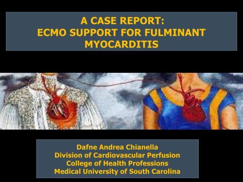
A CASE REPORT:<br />
ECMO SUPPORT FOR FULMINANT<br />
MYOCARDITIS<br />
Dafne Andrea Chianella<br />
Division of Cardiovascular <strong>Perfusion</strong><br />
College of Health Professions<br />
Medical University of South Carolina
HISTORY OF MYOCARDITIS<br />
• Diagnostic Dilemma:<br />
• Shifting diagnostic criteria<br />
• Complex classification<br />
• Changing pattern of infectious disease<br />
• Consensus definition in 1984: “Dallas Criteria”<br />
• Defined by the American College of Cardiology<br />
• Used for the multicenter NIH myocarditis treatment<br />
trials<br />
• Remain standards histological classification
MYOCARDITIS: DEFINITION<br />
• Myocardial process characterized by the presence of an<br />
inflammatory infiltrate and myocyte damage or<br />
necrosis not typical of the myocardial damage of<br />
ischemic heart disease.<br />
Ventricular<br />
Dysfunction<br />
Cardiogenic Shock<br />
MYOCARDITIS<br />
Acute-Onset Heart<br />
Failure<br />
MI/Stroke<br />
Myocarditis is implicated in sudden cardiac death of<br />
young adults (8.6% to 12%)
• Chest pain<br />
• Arrhythmia<br />
SYMPTOMS<br />
• Shortness of breath, at rest or during physical<br />
activity<br />
• Fluid retention with swelling of legs, ankles and<br />
feet<br />
• Fatigue
CLASSIFICATION<br />
• Etiology<br />
• Ex: viral, bacterial, fungal, protozoal,<br />
parasitic, toxic, hypersensitive,<br />
immunologic syndrome<br />
• Histologically<br />
• Clinical <strong>Presentation</strong><br />
• Acute vs. Fulminant
PATHOLOGY OF MYOCARDITIS<br />
• Phase I: Viral Infection<br />
• Phase II: Autoimmunity<br />
• Phase III/IV: Dilated<br />
Cardiomyopathy<br />
Enterovirus<br />
(gut or respiratory tract)<br />
Coxsackieviruse<br />
Adenoviruses<br />
(respiratory tract)<br />
Encephalomyocarditis<br />
virus
Phase I: Viral Infection<br />
• Cardiotropic RNA virus enters host<br />
• Transported to target organ sites (ex. heart)<br />
• Co-receptors aid membrane binding (DAF and CD5 <strong>com</strong>plex)<br />
• Viral genome enters host cell via CAR (coxsackie-adenoviral<br />
receptor)<br />
• Protein<br />
translation
PHASE II: INFLAMMATION<br />
• Inflammatory cells infiltrate the myocardium<br />
• Cytokine release<br />
• (Ex. interferons, interleukins, tumor necrosis factor)<br />
• Macrophages, lymphocytes, and endothelial cell activation<br />
• Inflammatory mediators = myocarditis<br />
• Vascular endothelium disrupted:<br />
• Endothelial dysfunction<br />
• Vasoconstriction<br />
• Necrosis of myocardial tissues
PHASE III: CELL-MEDIATED IMMUNITY<br />
• Recruitment of immune cells to infected myocardium<br />
• T helper cells + cytotoxic T clear the virally infected cells<br />
• Activated T lymphocytes lysis virus-infected cardiomyocytes<br />
• Viral clearing<br />
• Detrimental effects:<br />
• Myocyte signal transduction<br />
• Calcium homeostasis<br />
• Cardiac bioenergetics
RESOLUTION:<br />
PHASE 4: RESOLUTION OR DILATED<br />
CARDIOMYOPATHY<br />
• Virus effectively cleared<br />
• Myocardial healing and<br />
recovery<br />
CARDIOMYOPATHY:<br />
• Persistent activation of T cells<br />
• Continued antibody-mediated<br />
myocyte destruction<br />
• Ventricular dysfunction<br />
• Fulminant myocarditis or<br />
Chronic persistent myocarditis<br />
• Fibrosis/ necrosis<br />
• Ventricular dilatation<br />
• End-stage heart failure.
FIGURE 1 PATHOGENESIS OF VIRAL MYOCARDITIS CAN BE DIVIDED INTO FOUR PHASE S
Case Report
HISTORY & PHYSICAL<br />
• 24 year old African American Female<br />
• Ht: 167.59 cm<br />
BSA: 1.75m²<br />
• Wt: 68.14 kg<br />
• Presented to ER<br />
• Chest pain X3 days<br />
• Fever, diaphoresis, nausea, vomiting<br />
• ST elevation<br />
• Troponin of 92.5 ng/mL on admission
LEFT HEART CATHETERIZATION<br />
• Normal coronary arteries<br />
• Diffuse left ventricular hypokinesis<br />
• EF = 25%<br />
• LVEDP 30mmHg
ICU AND RIGHT HEART CATH<br />
• VTach with loss of consciousness<br />
• Cardioversion with 200 joules and return to sinus rhythm<br />
• Amiodarone and Norepinephrine administered<br />
• Intermittent <strong>com</strong>plete heart block<br />
Right Heart Catheterization:<br />
• Placement precordial pacemaker and IABP<br />
• PCWP= 22 mmHg<br />
• PAP mean= 23 mmHg<br />
Echocardiogram:<br />
•Hypokinesis of the LV<br />
•EF= 25% to 40% depending on the heart rate
VA-ECMO REPORT:<br />
• Preoperative diagnosis:<br />
• Cardiogenic shock and myocardial stunning<br />
• Myocarditis with biventricular failure<br />
• Coagulopathy<br />
• Acute renal failure<br />
• 8,000 IU Heparin to pt<br />
• Pump primed with 2 units of PRBC, 1 unit FFP<br />
• Cannulation:<br />
• 23-French femoral venous cannula (right femoral vein)<br />
• 17-French arterial cannula (left femoral artery)
MUSC-ECMO<br />
•Equipment:<br />
•Stockert roller pump<br />
•Jostra Quadrox-D oxygenator w/ Safeline® coating<br />
•Gish biomedical circuit w/ GBS coating<br />
•Safety Devices:<br />
•Super Tygon tubing<br />
•Bubble detector<br />
•Transonic flow probe<br />
•Anti Factor Xa levels measured<br />
•Servoregulation:<br />
•Venous line pressure -40 mmHg<br />
•Arterial line pressure +350 mmHg
MUSC ECMO Circuit :<br />
Quadrox D<br />
Membrane Oxygenator
• Cardiac Stun:<br />
ECHO: DAY 1 OF ECMO<br />
• Third-degree heart block with ventricular<br />
paced rhythm.<br />
• Marked global LV hypokinesis<br />
• Paradoxical septal motion and some minor<br />
regional variability.<br />
• EF < 10%.<br />
• RV hypokinesis, mild hypertrophy.
ECHO: DAY 3 OF ECMO<br />
• Concentric left ventricular hypertrophy<br />
• Increased wall thickness<br />
• Increased myocardial mass index<br />
• Severe global hypokinesis of the LV<br />
• EF= 26%.
ECHO: DAY 6 OF ECMO<br />
• Trial Off- evaluate LV function after clamping<br />
ECMO.<br />
• Hypokinesis of the anterior wall, anterior<br />
septum, and inferior septum<br />
• EF increased to 52%.<br />
• RV function appeared recovered<br />
• After 60 minutes of clamping, there was no<br />
significant change in ventricular performance
ECHO: DAY 7 OF ECMO<br />
• EF= 55%<br />
• RV systolic pressure =14mmHg<br />
• Intraoperative removal of the left femoral<br />
ECMO cannula<br />
• Postoperative Diagnosis:<br />
• Fulminant Myocarditis
6<br />
14<br />
20<br />
34<br />
38<br />
46<br />
54<br />
62<br />
70<br />
82<br />
90<br />
102<br />
110<br />
118<br />
126<br />
134<br />
142<br />
150<br />
Anti-Xa Level (IU/mL)<br />
1.2<br />
Anti-Factor Xa Level on ECMO<br />
1<br />
0.8<br />
0.6<br />
0.4<br />
0.2<br />
0<br />
Hours on ECMO
Cardiac Index (l/min/m²)<br />
1<br />
9<br />
17<br />
25<br />
33<br />
41<br />
49<br />
57<br />
65<br />
73<br />
81<br />
89<br />
97<br />
105<br />
113<br />
121<br />
129<br />
137<br />
145<br />
153<br />
2.5<br />
CARDIAC INDEX ON ECMO<br />
2<br />
1.5<br />
1<br />
0.5<br />
0<br />
Hours on ECMO
fib (mg/dL) 215 191 208 229 233 306 377<br />
Plasma Free HGB<br />
(mg/dL) 40 40 90 100 40 30<br />
Lab Test Initiation Day 1 Day 2 Day 3 Day 4 Day 5 Day 6 Day 7<br />
PRBC units<br />
administered 2 1 2 2 2 0 4 2<br />
PLT units administered 1 0 0 0 0 1 2 1<br />
BUN (mg/dL) 41 81 92 67 51 53 47<br />
Creatinite (mg/dL) 2.4 4.8 5.3 3.5 3.3 3.5 3.5<br />
Average ACT (sec ) 200 210 195 175 160 175 180 185<br />
D-Dimer (mg/L) >20 >20 9.1 9.86 5.26<br />
AT3 % 69<br />
PT (sec) 30.3 26 18.3 18.4 17.3<br />
PTT (sec) 150 118 90 66 108 89 88<br />
INR 2.9 2.4 1.43 1.49 1.38
DISCHARGE<br />
• 8 days S/P ECMO ECHO:<br />
• EF= 69% with improved LV systolic<br />
function<br />
• Peak right ventricular systolic pressure is<br />
34 mmHg
EF (%)<br />
80<br />
Patient EF Through Hospital Course<br />
70<br />
69<br />
60<br />
50<br />
40<br />
30<br />
20<br />
25<br />
mean 33<br />
(25-40)<br />
26<br />
52<br />
55<br />
10<br />
0<br />
8
Day 1 on ECMO<br />
Discharge
CLINICAL PRESENTATION OF<br />
MYOCARDITIS<br />
Acute vs. Fulminant
FULMINANT: DEFINITION<br />
• Fulminant :<br />
• Any event or process that occurs suddenly and<br />
quickly<br />
• With intensity and severity to the point of<br />
lethality.<br />
The word <strong>com</strong>es from Latin fulmināre,<br />
to strike with lightning.
METHODS OF FM DIAGNOSIS<br />
• Echocardiography and EKG<br />
• Cardiac MRI<br />
• 67 Ga and 111 In antimyosin antibody<br />
scintigraphy,<br />
• Angiography<br />
• Biopsy
• FM:<br />
FM VS ACUTE<br />
• Fever and symptoms of heart failure<br />
• Distinct Prodrome (usually viral)<br />
• Severe cardiovascular <strong>com</strong>promise<br />
• Ventricular dysfunction that either resolves<br />
spontaneously or results in death.<br />
• Acute:<br />
• Patients present with established ventricular<br />
dysfunction<br />
• May respond to immunosuppressive therapy<br />
• May progress to dilated cardiomyopathy.
LABORATORY EVIDENCE FOR FM VS.<br />
ACUTE<br />
Lab Test<br />
Fulminant<br />
Non-Fulminant<br />
(Acute)<br />
Normal<br />
Creatinine 10.0±5.7 mmmol/L 6.4±3.6 mmol/L<br />
BUN 97.2±44.3 mol/L 79.6±26.5 mol/L<br />
Creatinine Kinase 1708±1217 U/L 617±447 U/L<br />
0.5 to 1.0 mg/dL<br />
7–21 mg/dL<br />
60 and 400<br />
IU/L<br />
Transaminases 1158±595 U/L 189±59 U/L 0.0-45 IU/L<br />
C-reative Protein 590±350 mg/L 340±260 mg/L
FM VS ACUTE<br />
Cardiac Test<br />
Fulminant<br />
Non-Fulminant<br />
(Acute)<br />
Inraventricular Conduction<br />
Delays 73% 25%<br />
Ventricular Arrhythmias 27% 4%<br />
Depressed Left Ventricular<br />
EF% 57% 41%<br />
Further FM lab evidence:<br />
• leukocytosis, eosinophilia, and erythrocyte<br />
sedimentation rate<br />
• troponins and creatinine kinase MB isoenzyme<br />
• Prolonged QRS <strong>com</strong>plex and depressed left ventricular EF
Fulminant<br />
Chronic<br />
HISTOLOGICAL CLASSIFICATION OF FM<br />
Normal<br />
Borderline
Fulminant<br />
Normal Cardiac Muscle<br />
Acute
LONG-TERM OUTCOME OF FULMINANT MYOCARDITIS AS<br />
COMPARED WITH ACUTE (NONFULMINANT) MYOCARDITIS<br />
ROBERTE, MCCARTHY (N ENGL J MED 2000;342: 690-5)<br />
Conclusions<br />
Fulminant myocarditis is a distinct clinical entity with an<br />
excellent long term prognosis. Aggressive hemodynamic<br />
support is warranted for patients with this condition.
MECHANICAL CIRCULATORY SUPPORT FOR<br />
PATIENTS WITH ACUTE-FULMINANT<br />
MYOCARDITIS<br />
(ANN THORAC SURG 2001;71:S73–6) 2001<br />
• An aggressive use of mechanical support is strongly justified.<br />
•Survival (full, BTR, or BTT) approaches 70%.<br />
•Transplantation can often be avoided.
EXPERIENCE AND RESULT OF ECMO IN<br />
TREATING FM WITH SHOCK:<br />
WHAT MECHANICAL SUPPORT SHOULD BE<br />
Conclusion:<br />
CONSIDERED FIRST?<br />
YIH-SHARNG CHEN, MD<br />
J HEART LUNG TRANSPLANT 2005;24: 81–7.<br />
ECMO can be considered the first-line treatment of<br />
mechanical support for FM when the IABP is inadequate or<br />
infeasible.<br />
• Fewer <strong>com</strong>plications<br />
• Easier application<br />
• Biventricular support
ADVANTAGES OF ECMO VS. VAD<br />
AS FIRST-LINE TREATMENT FOR FM<br />
• Easy and rapid setup without sternotomy<br />
• Suitable with CPR<br />
• Weaning to be attempted over several hours to days.<br />
• Bridge to long-term mechanical devices<br />
• Flexibility in ECMO application.<br />
• Cost
CONCLUSION<br />
Teamwork and aggressive treatment lead to the<br />
patients extraordinary recovery
QUESTIONS??
Initial Tn levels were elevated (21.8 11.9 ng/dl) in the patient group. The absolute level of Tn could<br />
not differentiate patients who would survive or not. Nevertheless, daily Tn level over the first 72<br />
hours clearly showed that patients with a steep decline in Tn (6.53 3.73, ranged from 2.494 to<br />
14.8, n 13) were able to be weaned off mechanical support (Figure 1). This finding may suggest<br />
that initially high Tn values do not necessarily indicate irreversible damage to the myocardium;<br />
rather, rapid decline in Tn slope may reflect early recovery of the myocardium. Conversely, initial<br />
Tn levels, even if not particularly elevated, might reflect irreversible damage of the myocardium<br />
without good prospects for recovery if they remain persistently elevated.<br />
Figure 1. Trend of daily troponin<br />
(Tn) levels.<br />
Patients 1 to 5 used Tn T, and the<br />
others used Tn I. There are similarities<br />
in the slope in reversible fulminant<br />
myocarditis, 6.53 3.73 ng/ml/day (n<br />
13;<br />
patient 12 with failed weaning and<br />
patient 10 with only one Tn datum<br />
were excluded from analysis).
Two-dimensional echocardiograms from patients with<br />
fulminant and acute myocarditis at presentation. The top<br />
panels show the parasternal long axis (A) and short axis<br />
(B) views of a 20-year-old man with fulminant myocarditis<br />
who presented after five days of a viral syndrome<br />
followed by acute hemodynamic collapse. Note the severe<br />
ventricular thickening (septal thickness 2.1 cm) but small<br />
ventricular cavity size (LVEDD 2.5 cm). After hemodynamic<br />
support with intravenous inotropic agents and a left ventricular<br />
assist device, this patient recovered near normal left<br />
ventricular function.<br />
Echo of normal heart –<br />
Parasternal View
#3 JOURNAL OF THE AMERICAN<br />
COLLEGE OF CARDIOLOGY VOL. 36,<br />
NO. 1, 2000<br />
ECHOCARDIOGRAPHIC FINDINGS IN<br />
FULMINANT AND ACUTE MYOCARDITIS<br />
G. MICHAEL FELKER, MD,*<br />
CONCLUSIONS:<br />
Fulminant myocarditis is distinguishable<br />
from acute myocarditis by echocardiography.<br />
Patients with fulminant myocarditis exhibit a<br />
substantial improvement in ventricular function at six<br />
months <strong>com</strong>pared with those with acute myocarditis.<br />
Echo has value in classifying patients with<br />
myocarditis and may provide prognostic information.<br />
Figure 1. Echocardiographic findings in patients with fulminant and acute<br />
myocarditis at baseline and six months. (A) Fractional shortening, (B) left<br />
ventricular end-diastolic dimension, (C) septal thickness. *p , 0.01 vs.<br />
acute; †p , 0.01 for interaction between time and type of myocarditis.
This study demonstrated that 70% of patients with fulminant myocarditis supported<br />
by percutaneous ECMO could be saved. Cardiac function was severely<br />
depressed in the acute phase but improved markedly in the chronic phase. The<br />
clinical course in the chronic phase in the patientswith fulminant myocarditis<br />
who were weaned from ECMO was similar to that in patients with non-fulminant<br />
myocarditis.
FAVOURABLE CLINICAL OUTCOME IN PATIENTS WITH CARDIOGENIC SHOCK<br />
DUE TO FULMINANT MYOCARDITIS SUPPORTED BY PERCUTANEOUS<br />
EXTRACORPOREAL MEMBRANE OXYGENATION<br />
YASUHIDE ASAUMI<br />
EUROPEAN HEART JOURNAL (2005) 26, 2185–2192<br />
Figure 2:<br />
Acute changes in left ventricular function<br />
before and immediately<br />
after the support and at weaning from<br />
ECMO in patients with fulminant<br />
myocarditis (F group). (A) Fractional<br />
shortening (B) end-systolic dimentions.<br />
Open circles indicate patients who<br />
were weaned from ECMO and closed<br />
diamonds indicate F patients who were<br />
not weaned from ECMO and died.
• Myocardial Stunning and Hibernation<br />
• Acute, coronary occlusion causes a rapid decline (within seconds) in ventricular<br />
performance. If coronary flow is reestablished, ventricular function will slowly return to<br />
normal. The duration of reduced performance (myocardial stunning), depends on the<br />
duration of the preceding ischemia. Stunning is <strong>com</strong>pletely reversible, and therefore there<br />
is no permanent damage to the myocardium. The stunned myocardium, however, is less<br />
responsive to inotropic drugs and cardiogenic shock can result. Stunning is found<br />
following coronary angioplasty, thrombolysis, coronary vasospasm, and cardiopulmonary<br />
bypass.<br />
• Myocardial hibernation is a term used to describe the condition in which regional cardiac<br />
function is depressed due to chronic ischemia. Restoration of normal coronary flow (e.g.,<br />
by coronary bypass) will restore normal function in the affected region.<br />
• The mechanisms of stunning and hibernation are not <strong>com</strong>pletely understood, but there is<br />
evidence that it is related to impaired Ca ++ handling by the sarcoplasmic reticulum as well<br />
as due to damage caused by oxygen free radicals.
SPINALE IDEAS<br />
• Myocardial stanning<br />
• Dysfunction not death yet hybernating myocard
MAJOR ETIOLOGICAL CLASSIFICATIONS OF<br />
MYOCARDITIS<br />
Viral<br />
Bacterial<br />
Fungal<br />
Protozoal<br />
Adenovirus<br />
Coxsackievirus<br />
HCV<br />
HIV<br />
Mycobacterial<br />
Streptococcal species<br />
Mycoplasma pneumoniae<br />
Treponema pallidum<br />
Aspergillus<br />
Candida<br />
Coccidiodes<br />
Cryptococcus<br />
Histoplasma<br />
Trypanosoma cruzi<br />
Parasitic<br />
Toxins<br />
Hypersensitivity<br />
Schistosomiasis<br />
Larva migrans<br />
Anthracyclines<br />
Cocaine<br />
Interleukin-2<br />
Cocaine<br />
Sulfonamides<br />
Cephalosporins<br />
Diuretics<br />
Digoxin<br />
Tricyclic antidepressants<br />
Dobutamine<br />
Immunologic syndromes<br />
Churg-Strauss<br />
Inflammatory bowel disease<br />
Giant cell myocarditis<br />
Diabetes mellitus<br />
Sarcoidosis<br />
Systemic lupus erythematosus<br />
Thyrotoxicosis<br />
Takayasu’s arteritis<br />
Wegener’s granulomatosis
FIGURE 2.<br />
A CASE OF FULMINANT<br />
MYOCARDITIS, MANAGED<br />
USING INTRA-AORTIC<br />
BALLOON PUMPING AND<br />
MECHANICAL VENTILATOR,<br />
SHOWED THAT ON DAY 7<br />
WHEN THE CPK LEVEL<br />
PEAKED, TH1 DOMINANT<br />
POLARITY WAS<br />
DETECTED. IN THE<br />
HEALING PHASE, AT DAY<br />
14, T-CELLS WITH TH2<br />
FUNCTION DRAMATICALLY<br />
INCREASED AND<br />
OVERCAME TH1 T-CELLS.<br />
AT DAY 20, BOTH TH1 AND<br />
TH2 T-CELLS DECREASED.
SUGGESTED GUIDELINES FOR CASE REPORT PRESENTATIONS<br />
Speakers for each case report will be allotted a 30-minute period on the program. You should plan to spend 20 minutes for the<br />
case report followed by a 10-minute question and answer period guided by the session moderators. This 10 -minute period<br />
is an important part of the case report under discussion and will add to the overall quality of the information conveyed.<br />
Content (at presenters discretion) may be posted on International <strong>Perfusion</strong> Association (IPA) web site. IPA is a non -profit<br />
perfusion education organization. Please sign at bottom of abstract form to exercise this option. The following guidelines<br />
are re<strong>com</strong>mended from past experience to help you <strong>com</strong>municate most effectively.<br />
FORMAT<br />
• 1. Title and Introduction Each presenter should give a brief description of the case report and how it pertains to perfusion<br />
practice.<br />
• 2. Case Report The specific case should be described including: patient age, gender, and other pertinent demographics<br />
such as height, weight and body surface area, brief medical history, extracorporeal equipment and circuitry, operative and<br />
perfusion procedure, out<strong>com</strong>e and possible follow-up.<br />
• 3. Discussion This part could include evolution of the technique, history and bibliographic references. Additional experience<br />
or expansion of alternative techniques or results by other practitioners can be described. The discussion of problems,<br />
speculations or future suggested approaches should be included here.<br />
• 4. Conclusion A brief concluding statement summarizing the case with the major points should be described.<br />
• GENERAL GUIDELINES<br />
• Whenever possible, generic drug names should be used; doses of each drug should be stated when appropriate.<br />
Laboratory values should be expressed in proper units of measure. Extracorporeal equipment should be described (model<br />
number and manufacturer) to give the audience a frame of reference. Medical history of the patient should be brief and<br />
contain only information pertinent to the noteworthy aspects of the case report. The year the case was performed should be
CALL FOR ABSTRACTS<br />
ABSTRACTS FOR CASE REPORTS PRESENTATIONS SHOULD<br />
CONTAIN THE FOLLOWING INFORMATION:<br />
• TITLE: State the title of your presentation.<br />
• INTRODUCTION: Briefly describe the case and how it pertains to perfusion practice.<br />
• CASE REPORT: Include patient age, gender and other pertinent demographics such as<br />
height, weight and body surface area, brief medical history, extracorporeal equipment and<br />
circuitry, operative and perfusion procedure, out<strong>com</strong>e and follow-up (if available).<br />
• DISCUSSION: This may include evolution of the technique, history and bibliographic<br />
references, expansion of alternative techniques or results, problems, speculations or<br />
future suggested approaches.<br />
• CONCLUSION: The conclusion should summarize the main points.<br />
• QUESTIONS: Submit five questions that can be asked about your presentation at the<br />
Case Reports Meeting.
ABSTRACT<br />
Title: A case report: ECMO support for Fulminant Myocarditis<br />
Introduction:<br />
This case report describes the use of ECMO for biventricular support for a patient diagnosed with fulminant myocarditis (FM).<br />
Case Report:<br />
In this case study, we discuss a patient diagnosed with FM. A 25 year old woman present to the ER at MUSC hospital <strong>com</strong>plaining of three days of chest pain, nausea, and<br />
vomiting. She had a height of 167 cm, weighed 68 kg, and had a BSA of 1.75m². Upon admission and a diagnostic cardiac catheterization, her EF was 69%. After 21 days in the hospital, she was released to go home.<br />
Discussion:<br />
Fulminant myocarditis (FM) is caused by the inflammation of the myocardium that results in ventricular systolic dysfunction and acute-onset heart failure. Symptoms progress<br />
rapidly and are life threatening. Patients who are diagnosed with FM often die of sudden cardiac arrest, arrhythmias, or sever heart failure unless mechanical circulatory support is<br />
introduced. When patients are aggressively treated with biventricular support devices, there is a 70-75% of full recovery from FM. Heart transplantation can often be avoided.<br />
Conclusion:<br />
This paper presents the diagnostic and clinical aspects of an FM patient. A detailed account follows the clinical presentation, methods of diagnosis, patient management,<br />
treatment options, and the inevitable prognosis of FM. Lastly, the pathophysiological, histological, and electrophysical distinguishing features of FM are identified.<br />
Overall, the aggressive treatment, rapid mechanical support, and extraordinary collaboration of the heart team lead to the patient’s excellent recovery.<br />
Questions:<br />
1. Do other ventricular support devices provide the same results as ECMO?<br />
2. How many case of FM occur every year?<br />
3. How did ECMO effect the patient’s other organs?<br />
4. What are the<br />
5. How is FM classified from other forms of myocarditis?