novel species of Mycosphaerellaceae and ... - Persoonia
novel species of Mycosphaerellaceae and ... - Persoonia
novel species of Mycosphaerellaceae and ... - Persoonia

You also want an ePaper? Increase the reach of your titles
YUMPU automatically turns print PDFs into web optimized ePapers that Google loves.
<strong>Persoonia</strong> 23, 2009: 119–146<br />
www.persoonia.org<br />
RESEARCH ARTICLE<br />
doi:10.3767/003158509X479531<br />
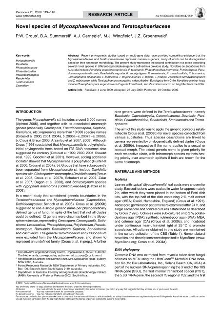
Novel <strong>species</strong> <strong>of</strong> <strong>Mycosphaerellaceae</strong> <strong>and</strong> Teratosphaeriaceae<br />
P.W. Crous 1 , B.A. Summerell 2 , A.J. Carnegie 3 , M.J. Wingfield 4 , J.Z. Groenewald 1<br />
Key words<br />
Mycosphaerella<br />
Penidiella<br />
Phaeophleospora<br />
Phaeothecoidea<br />
Pseudocercospora<br />
Readeriella<br />
Teratosphaeria<br />
Zasmidium<br />
Abstract Recent phylogenetic studies based on multi-gene data have provided compelling evidence that the<br />
<strong>Mycosphaerellaceae</strong> <strong>and</strong> Teratosphaeriaceae represent numerous genera, many <strong>of</strong> which can be distinguished<br />
based on their anamorph morphology. The present study represents the second contribution in a series describing<br />
several <strong>novel</strong> <strong>species</strong> in different capnodealean genera defined in a previous study. Novelties on Eucalyptus from<br />
Australia include: Penidiella pseudotasmaniensis, P. tenuiramis, Phaeothecoidea intermedia, P. minutispora, Pseudocercospora<br />
tereticornis, Readeriella angustia, R. eucalyptigena, R. menaiensis, R. pseudocallista, R. tasmanica,<br />
Teratosphaeria alboconidia, T. complicata, T. majorizuluensis, T. miniata, T. pr<strong>of</strong>usa, Zasmidium aerohyalinosporum<br />
<strong>and</strong> Z. nabiacense, while Teratosphaeria xenocryptica is described on Eucalyptus from Chile. Novelties on other hosts<br />
include Phaeophleospora eugeniicola on Eugenia from Brazil, <strong>and</strong> Zasmidium nocoxi on twig litter from the USA.<br />
Article info Received: 4 June 2009; Accepted: 26 July 2009; Published: 29 October 2009.<br />
Introduction<br />
The genus Mycosphaerella s.l. includes around 3 000 names<br />
(Aptroot 2006), <strong>and</strong> together with its associated anamorph<br />
genera (especially Cercospora, Pseudocercospora, Septoria,<br />
Ramularia, etc.) represents more than 10 000 <strong>species</strong> names<br />
(Crous et al. 2000, 2001, 2004a, b, 2006a–c, 2007a–c, 2008a,<br />
b, Crous & Braun 2003, Arzanlou et al. 2007, 2008). Although<br />
Crous (1998) postulated that Mycosphaerella is polyphyletic,<br />
initial phylogenetic trees based on ITS DNA sequence data<br />
suggested the contrary (Crous et al. 1999, 2000, 2001, Stewart<br />
et al. 1999, Goodwin et al. 2001). However, adding additional<br />
loci later showed that Mycosphaerella is polyphyletic (Hunter et<br />
al. 2006, Crous et al. 2007a, b). Groups that have subsequently<br />
been separated from Mycosphaerella s.l. include Davidiella<br />
<strong>species</strong> with Cladosporium anamorphs (Davidiellaceae) (Braun<br />
et al. 2003, Crous et al. 2007b, Schubert et al. 2007, Zalar<br />
et al. 2007, Dugan et al. 2008), <strong>and</strong> Schizothyrium <strong>species</strong><br />
with Zygophiala anamorphs (Schizothyriaceae) (Batzer et al.<br />
2008).<br />
In a recent study that considered generic boundaries in the<br />
Teratosphaeriaceae <strong>and</strong> <strong>Mycosphaerellaceae</strong> (Capnodiales,<br />
Dothideomycetes; Schoch et al. 2006), Crous et al. (2009b)<br />
suggested to use a single generic name per phylogenetically<br />
defined genus <strong>of</strong> fungi. In spite <strong>of</strong> the fact that not all clades<br />
could be defined, 12 genera were circumscribed in the <strong>Mycosphaerellaceae</strong>,<br />
representing Cercospora, Cercosporella, Dothistroma,<br />
Lecanosticta, Phaeophleospora, Polythrincium, Pseudocercospora,<br />
Ramularia, Ramulispora, Septoria, Sonderhenia<br />
<strong>and</strong> Zasmidium. The genera Ramichloridium <strong>and</strong> Dissoconium<br />
were excluded from the <strong>Mycosphaerellaceae</strong>, <strong>and</strong> shown to<br />
represent an undefined family (Crous et al. in prep.). A further<br />
1<br />
CBS-KNAW Fungal Biodiversity Centre, Uppsalalaan 8, 3584 CT Utrecht,<br />
The Netherl<strong>and</strong>s; corresponding author e-mail: p.crous@cbs.knaw.nl.<br />
2<br />
Royal Botanic Gardens <strong>and</strong> Domain Trust, Mrs. Macquaries Road, Sydney,<br />
NSW 2000, Australia.<br />
3<br />
Forest Resources Research, NSW Department <strong>of</strong> Primary Industries, P.O.<br />
Box 100, Beecr<strong>of</strong>t, New South Wales 2119, Australia.<br />
4<br />
Department <strong>of</strong> Genetics, Forestry <strong>and</strong> Agricultural Biotechnology Institute<br />
(FABI), University <strong>of</strong> Pretoria, Pretoria 0002, South Africa.<br />
nine genera were defined in the Teratosphaeriaceae, namely<br />
Baudoinia, Capnobotryella, Catenulostroma, Devriesia, Penidiella,<br />
Phaeothecoidea, Readeriella, Staninwardia <strong>and</strong> Teratosphaeria.<br />
The aim <strong>of</strong> this study was to apply the generic concepts established<br />
in Crous et al. (2009b) for <strong>novel</strong> <strong>species</strong> collected from<br />
various substrates. Thus <strong>species</strong> descriptions are linked to<br />
genera represented by phylogenetically defined clades (Crous<br />
et al. 2009b), irrespective if the name applies to a sexual or<br />
asexual morph. The oldest generic name is given priority for<br />
each respective clade, with teleomorph <strong>species</strong> epithets having<br />
priority over anamorph epithets if both are known for the<br />
same holomorph.<br />
Materials <strong>and</strong> Methods<br />
Isolates<br />
Leaves with typical ‘Mycosphaerella’ leaf spots were chosen for<br />
study. Excised lesions were soaked in water for approximately<br />
2 h, after which they were placed in the bottom <strong>of</strong> Petri dish<br />
lids, with the top half <strong>of</strong> the dish containing 2 % malt extract<br />
agar (MEA; Oxoid, Hampshire, Engl<strong>and</strong>) (Crous et al. 1991).<br />
Ascospore germination patterns were examined after 24 h, <strong>and</strong><br />
single ascospore <strong>and</strong> conidial cultures established as described<br />
by Crous (1998). Colonies were sub-cultured onto 2 % potatodextrose<br />
agar (PDA), synthetic nutrient-poor agar (SNA), MEA,<br />
<strong>and</strong> oatmeal agar (OA) (Crous et al. 2009c), <strong>and</strong> incubated<br />
under continuous near-ultraviolet light at 25 °C to promote<br />
sporulation. All cultures obtained in this study are maintained<br />
in the culture collection <strong>of</strong> the CBS (Table 1). Nomenclatural<br />
<strong>novel</strong>ties <strong>and</strong> descriptions were deposited in MycoBank (www.<br />
MycoBank.org; Crous et al. 2004a).<br />
DNA phylogeny<br />
Genomic DNA was extracted from mycelia taken from fungal<br />
colonies on MEA using the UltraClean TM Microbial DNA Isolation<br />
Kit (Mo Bio Laboratories, Inc., Solana Beach, CA, USA). A<br />
part <strong>of</strong> the nuclear rDNA operon spanning the 3’ end <strong>of</strong> the 18S<br />
rRNA gene (SSU), the first internal transcribed spacer (ITS1),<br />
the 5.8S rRNA gene, the second ITS region (ITS2) <strong>and</strong> the first<br />
© 2009 Nationaal Herbarium Nederl<strong>and</strong> & Centraalbureau voor Schimmelcultures<br />
You are free to share - to copy, distribute <strong>and</strong> transmit the work, under the following conditions:<br />
Attribution:<br />
You must attribute the work in the manner specified by the author or licensor (but not in any way that suggests that they endorse you or your use <strong>of</strong> the work).<br />
Non-commercial: You may not use this work for commercial purposes.<br />
No derivative works: You may not alter, transform, or build upon this work.<br />
For any reuse or distribution, you must make clear to others the license terms <strong>of</strong> this work, which can be found at http://creativecommons.org/licenses/by-nc-nd/3.0/legalcode. Any <strong>of</strong> the above conditions can be<br />
waived if you get permission from the copyright holder. Nothing in this license impairs or restricts the author’s moral rights.
120 <strong>Persoonia</strong> – Volume 23, 2009<br />
Table 1 Details <strong>of</strong> the isolates for which <strong>novel</strong> sequences were generated <strong>and</strong> <strong>of</strong> other isolates <strong>of</strong> importance to this study.<br />
Species Accession number 1 Host Country Collector GenBank number (ITS) 2<br />
Dissoconium commune CPC 12397 Eucalyptus globulus Australia I.W. Smith GQ852738<br />
Dissoconium dekkeri CPC 13097 Corymbia variegata Australia A.J. Carnegie GQ852739<br />
CPC 13098 Eucalyptus "argophloia" Australia A.J. Carnegie GQ852740<br />
CPC 13264 Eucalyptus molucana Australia B.A. Summerell GQ852741<br />
CPC 13279 Eucalyptus sp. Australia P.W. Crous GQ852742<br />
CPC 13479 Eucalyptus camaldulensis Thail<strong>and</strong> W. Himaman GQ852743<br />
'Mycosphaerella' heimii CPC 11000 Eucalyptus sp. Colombia M.J. Wingfield GQ852744<br />
CPC 13099 Eucalyptus dunnii Australia A.J. Carnegie GQ852745<br />
'Mycosphaerella' konae CPC 10992 Eucalyptus sp. Colombia M.J. Wingfield GQ852746<br />
'Mycosphaerella' marksii CPC 13273 Eucalyptus sp. Australia P.W. Crous GQ852747<br />
CPC 13724 Eucalyptus globulus Australia I.W. Smith GQ852748<br />
'Penidiella' pseudotasmaniensis CBS 124991; CPC 12400 Eucalyptus globulus Australia I.W. Smith GQ852749<br />
Penidiella tenuiramis CBS 124993; CPC 13692 Eucalyptus tenuiramis Tasmania B.A. Summerell GQ852750<br />
'Phaeocryptopus' qaeumannii CPC 14373 Pseudotsuga menziesii – – GQ852751<br />
CPC 14374 Pseudotsuga menziesii – – GQ852752<br />
CPC 14375 Pseudotsuga menziesii – – GQ852753<br />
Phaeothecoidea intermedia CBS 124994; CPC 13711 Eucalyptus globulus Australia B.A. Summerell GQ852754<br />
Phaeothecoidea minutispora CBS 124995; CPC 13710 Eucalyptus globulus Australia B.A. Summerell GQ852755<br />
Pseudocercospora crousii CBS 119487; Lynfield 1260 Eucalyptus sp. New Zeal<strong>and</strong> C.F. Hill GQ852756<br />
Pseudocercospora pseudoeucalyptorum CPC 12406 Eucalyptus globulus Australia I.W. Smith GQ852757<br />
CPC 12568 Eucalyptus nitens Australia C. Mohammed GQ852758<br />
CPC 12802 Eucalyptus globulus Portugal A.J.L. Phillips GQ852759<br />
CPC 12957 Eucalyptus deonei Australia B.A. Summerell GQ852760<br />
CPC 13455 Eucalyptus sp. Portugal P.W. Crous GQ852761<br />
CPC 13769 Eucalyptus punctata South Africa P.W. Crous GQ852762<br />
CPC 13816 Eucalyptus glanscens United Kingdom S. Denman GQ852763<br />
CPC 13926 Eucalyptus sp. USA:California S. Denman GQ852764<br />
Pseudocercospora schizolobii CBS 124990; CPC 13492 Eucalyptus camaldulensis Thail<strong>and</strong> W. Himaman GQ852765 3<br />
Pseudocercospora sp. CPC 12497 Fraxinus rhynchophylla South Korea H.D. Shin GQ852766<br />
CPC 14621 Eucalyptus sp. Madagascar – GQ852767<br />
Pseudocercospora tereticornis CBS 124996; CPC 12960 Eucalyptus nitens Australia A.J. Carnegie GQ852768<br />
CPC 13008 Eucalyptus tereticornis Australia A.J. Carnegie GQ852769<br />
CPC 13299 Eucalyptus tereticornis Australia P.W. Crous & B.A. Summerell GQ852770<br />
CPC 13315 Eucalyptus tereticornis Australia P.W. Crous & B.A. Summerell GQ852771<br />
Readeriella angustia CBS 124997; CPC 13608 Eucalyptus delegatensis Tasmania B.A. Summerell GQ852772<br />
CBS 124998; CPC 13618 Eucalyptus delegatensis Tasmania B.A. Summerell GQ852773<br />
CPC 13621 Eucalyptus regnans Tasmania B.A. Summerell, P. Summerell & A. Summerell GQ852774<br />
CPC 13630 Eucalyptus delegatensis Tasmania B.A. Summerell GQ852775<br />
Readeriella callista CBS 124986; CPC 13615 Eucalyptus sclerophylla Australia B.A. Summerell GQ852776<br />
CPC 12715 Eucalyptus deanei Australia B.A. Summerell GQ852777<br />
CPC 12727 Eucalyptus sp. Australia B.A. Summerell GQ852778<br />
CPC 12841 Eucalyptus cannonii Australia B.A. Summerell GQ852779<br />
CPC 13605 Eucalyptus multicaulis Australia B.A. Summerell GQ852780<br />
Readeriella eucalypti CPC 13401 Eucalyptus sp. Portugal P.W. Crous GQ852781<br />
Readeriella eucalyptigena CBS 124999; CPC 13026 Eucalyptus dives Australia B.A. Summerell GQ852782<br />
Readeriella menaiensis CBS 125003; CPC 14447 Eucalyptus oblonga Australia B.A. Summerell GQ852783<br />
Readeriella mirabilis CBS 125000; CPC 12370 Eucalyptus sp. Australia R. Park FJ493192<br />
CPC 12379 Eucalyptus sp. Australia R. Park GQ852784<br />
CPC 13611 Eucalyptus delegatensis Tasmania B.A. Summerell GQ852785<br />
Readeriella nontingens CPC 14444 Eucalyptus oblonga Australia B.A. Summerell GQ852786<br />
Readeriella patrickii CBS 124987; CPC 13602 Eucalyptus amygdalina Tasmania P. Summerell & B.A. Summerell GQ852787<br />
Readeriella pseudocallista CBS 125001; CPC 13599 Eucalyptus prominula Australia B.A. Summerell GQ852788<br />
Readeriella tasmanica CBS 125002; CPC 13631 Eucalyptus delegatensis Tasmania B.A. Summerell GQ852789<br />
Teratosphaeria alboconidia CPC 14597 Eucalyptus miniata Australia B.A. Summerell FJ493197<br />
Teratosphaeria complicata CPC 14535 Leaf litter <strong>of</strong> Eucalyptus miniata Australia B.A. Summerell GQ852790<br />
Teratosphaeria considenianae CPC 13032 Eucalyptus sp. Australia B.A. Summerell GQ852791<br />
CPC 14057 Eucalyptus stellatata Australia B.A. Summerell GQ852792
P.W. Crous et al.: Mycosphaerella <strong>and</strong> Teratosphaeria<br />
121<br />
Teratosphaeria corymbiae CBS 124988; CPC 13125 Eucalyptus henryii Australia A.J. Carnegie FJ493185<br />
Teratosphaeria cryptica CPC 12415 Eucalyptus globulus Australia I.W. Smith GQ852793<br />
CPC 12424 Eucalyptus globulus Australia I.W. Smith GQ852794<br />
CPC 12559 Eucalyptus nitens Australia C. Mohammed GQ852795<br />
CPC 12562 Eucalyptus nitida Australia C. Mohammed GQ852796<br />
CPC 12565 Eucalyptus amygdalina – C. Mohammed GQ852797<br />
CPC 13839 Eucalyptus globulus Australia I.W. Smith GQ852798<br />
CPC 13842 Eucalyptus globulus Australia I.W. Smith GQ852799<br />
Teratosphaeria destructans CBS 111370; CPC 1368 Eucalyptus sp. Indonesia P.W. Crous GQ852800<br />
Teratosphaeria eucalypti CPC 12552 Eucalyptus nitens Australia C. Mohammed GQ852801<br />
Teratosphaeria majorizuluensis CBS 120040; CPC 12712 Eucalyptus botryoides Australia B.A. Summerell GQ852802<br />
Teratosphaeria miniata CBS 125006; CPC 14514 Leaf litter <strong>of</strong> Eucalyptus miniata Australia B.A. Summerell GQ852803<br />
Teratosphaeria molleriana CPC 12232 Eucalyptus globulus Portugal A.J.L. Phillips GQ852804<br />
CPC 12246 Eucalyptus globulus Portugal A.J.L. Phillips GQ852805<br />
Teratosphaeria nubilosa CPC 11926 Acacia auriculiformis Thail<strong>and</strong> W. Himaman GQ852806<br />
CPC 12235 Eucalyptus globulus Portugal A.J.L. Phillips GQ852807<br />
CPC 12243 Eucalyptus globulus Portugal A.J.L. Phillips GQ852808<br />
CPC 12830 Eucalyptus globulus Portugal A.J.L. Phillips GQ852809<br />
CPC 13452 Eucalyptus sp. Portugal P.W. Crous GQ852810<br />
CPC 13825 Eucalyptus globulus Australia I.W. Smith GQ852811<br />
CPC 13828 Eucalyptus globulus Australia I.W. Smith GQ852812<br />
CPC 13831 Eucalyptus globulus Australia I.W. Smith GQ852813<br />
CPC 13833 Eucalyptus globulus Australia I.W. Smith GQ852814<br />
CPC 13835 Eucalyptus globulus Australia I.W. Smith GQ852815<br />
CPC 13837 Eucalyptus globulus Australia I.W. Smith GQ852816<br />
CPC 13844 Eucalyptus globulus Australia I.W. Smith GQ852817<br />
CPC 13847 Eucalyptus coniocalyx South Africa P.W. Crous GQ852818<br />
CPC 13849 Eucalyptus obliqua South Africa P.W. Crous GQ852819<br />
'Teratosphaeria' parva CPC 12249 Eucalyptus globulus Portugal A.J.L. Phillips GQ852820<br />
CPC 12419 Eucalyptus globulus Australia I.W. Smith GQ852821<br />
Teratosphaeria pr<strong>of</strong>usa CBS 125007; CPC 12821 Eucalyptus nitens Australia A.J. Carnegie FJ493196<br />
Teratosphaeria sp. CPC 13680 Eucalyptus placita Australia B.A. Summerell GQ852822<br />
Teratosphaeria stellenboschiana CBS 124989; CPC 13767 Eucalyptus punctata South Africa P.W. Crous GQ852823<br />
CPC 12283 Eucalyptus sp. Corsica J. Dijksterhuis GQ852824<br />
CPC 13764 Eucalyptus punctata South Africa P.W. Crous GQ852825<br />
'Teratosphaeria' suberosa CPC 13090 Eucalyptus agglomerata Australia A.J. Carnegie GQ852826<br />
CPC 13091 Eucalyptus dunnii Australia A.J. Carnegie GQ852827<br />
CPC 13093 Eucalyptus mollucana Australia A.J. Carnegie GQ852828<br />
CPC 13094 Eucalyptus dunnii Australia A.J. Carnegie GQ852829<br />
CPC 13095 Eucalyptus cloeziana Australia A.J. Carnegie GQ852830<br />
CPC 13096 Eucalyptus dunnii Australia A.J. Carnegie GQ852831<br />
CPC 13104 Eucalyptus dunnii Australia A.J. Carnegie GQ852832<br />
CPC 13106 Eucalyptus argophloia Australia A.J. Carnegie GQ852833<br />
CPC 13111 Eucalyptus dunnii Australia A.J. Carnegie GQ852834<br />
CPC 13115 Eucalyptus dunnii Australia A.J. Carnegie GQ852835<br />
CPC 13736 Eucalyptus aquatica Australia B.A. Summerell GQ852836<br />
Teratosphaeria suttonii CBS 119973; CMW 23440 Eucalyptus pellita Vietnam T.I. Burgess GQ852837<br />
CPC 12218 Eucalyptus sp. Indonesia M.J. Wingfield GQ852838<br />
Teratosphaeria viscidus CBS 124992; CPC 13306 Eucalyptus sp. Australia P.W. Crous FJ493186<br />
Teratosphaeria xenocryptica CBS 122905; CPC 355 Leaves <strong>of</strong> Eucalyptus sp. Chile M.J. Wingfield AF309622<br />
'Zasmidium' aerohyalinosporum CBS 125011; CPC 14636 Eucalyptus tectifica Australia B.A. Summerell GQ852839<br />
Zasmidium citri CPC 13467 Eucalyptus sp. Thail<strong>and</strong> W. Himaman GQ852840<br />
Zasmidium lonicericola CBS 125008; CPC 11671 Lonicera japonica South Korea H.D. Shin DQ303097<br />
'Zasmidium' nabiacense CBS 125010; CPC 12748 Eucalyptus sp. (red gum) Australia A.J. Carnegie GQ852841<br />
Zasmidium nocoxi CBS 125009; CPC 14044 Litter USA P.W. Crous GQ852842<br />
1<br />
CBS: Centraalbureau voor Schimmelcultures, Utrecht, The Netherl<strong>and</strong>s; CMW: Culture Collection <strong>of</strong> the Forestry <strong>and</strong> Agricultural Biotechnology Institute (FABI) <strong>of</strong> the University <strong>of</strong> Pretoria, Pretoria, South Africa; CPC: Culture collection <strong>of</strong> Pedro Crous, housed at CBS; Lynfield:<br />
Private culture collection <strong>of</strong> Frank Hill.<br />
2<br />
ITS: Internal transcribed spacers 1 <strong>and</strong> 2 together with 5.8S nrDNA.<br />
3<br />
This isolate was not included in the analyses; see Crous et al. 2009b for position in 28S nrDNA phylogeny.
122 <strong>Persoonia</strong> – Volume 23, 2009<br />
Cladosporium colocasiae AF393692<br />
Cladosporium sphaerospermum AF455481<br />
97 Dissoconium commune AY725541<br />
Dissoconium commune CPC 12397<br />
100<br />
Dissoconium australiensis EF394854<br />
Dissoconium dekkeri DQ302975<br />
Dissoconium dekkeri CPC 13264<br />
Dissoconium dekkeri CPC 13479<br />
Dissoconium dekkeri CPC 13097<br />
Dissoconium dekkeri CPC 13098<br />
Dissoconium dekkeri CPC 13279<br />
Dissoconium dekkeri AF309625<br />
Dissoconium dekkeri AF309624<br />
74 Phaeophleospora eugeniae AF309613<br />
100<br />
100 ‘Mycosphaerella’ stramenti DQ303042<br />
Phaeophleospora eugeniicola CPC 2557<br />
100 Phaeophleospora eugeniicola CPC 2558<br />
Ramularia endophylla AY490763<br />
100 Ramularia pratensis var. pratensis EU019284<br />
Ramularia coleosporii EF535674<br />
85 Ramularia eucalypti EF394861<br />
10 changes<br />
Fig. 1 First <strong>of</strong> 1 000 equally most parsimonious trees obtained from a heuristic search with 100<br />
r<strong>and</strong>om taxon additions <strong>of</strong> the <strong>Mycosphaerellaceae</strong> ITS sequence alignment using PAUP v4.0b10<br />
(Sw<strong>of</strong>ford 2003). The scale bar shows 10 changes, <strong>and</strong> bootstrap support values 1 000 replicates are<br />
shown at the nodes. Thickened lines indicate the strict consensus branches <strong>and</strong> ex-type sequences<br />
are printed in bold face. The tree was rooted to Cladosporium colocasiae (GenBank accession<br />
AF393692) <strong>and</strong> Cladosporium sphaerospermum (GenBank accession AF455481).<br />
95<br />
86<br />
100<br />
96<br />
Ramularia proteae EU707899<br />
100 ‘Mycosphaerella’ marksii CPC 13273<br />
‘Mycosphaerella’ marksii CPC 13724<br />
‘Pseudocercospora’ epispermogonia DQ267596<br />
‘Mycosphaerella’ marksii DQ267587<br />
‘Stenella’ eucalypti EF394865<br />
‘Zasmidium’ aerohyalinosporum CPC 14636<br />
‘Periconiella’ levispora EU041780<br />
‘Zasmidium’ nabiacense CPC 12748<br />
‘Mycosphaerella’ vietnamensis DQ632675<br />
‘Mycosphaerella’ vietnamensis DQ632678<br />
‘Mycosphaerella’ parkii AF309590<br />
‘Mycosphaerella’ parkii AY626979<br />
‘Phaeophleospora’ stonei EF394856<br />
72<br />
100<br />
96<br />
100<br />
Zasmidium citri AY752145<br />
Zasmidium citri CPC 13467<br />
‘Mycosphaerella’ eucalyptorum DQ302954<br />
‘Ramichloridium’ strelitziae EU041803<br />
Periconiella arcuata EU041779<br />
‘Mycosphaerella’ pseudovespa DQ530216<br />
Periconiella velutina EU041781<br />
98<br />
100<br />
99<br />
84<br />
Rasutoria pseudotsugae EF114687<br />
Rasutoria tsugae EF114688<br />
Zasmidium cellare DQ681316<br />
Zasmidium cellare DQ681317<br />
Ramichloridium cerophilum AF050286<br />
Ramichloridium cerophilum EU041798<br />
‘Mycosphaerella aleuritidis’ EU167594<br />
Zasmidium nocoxi CPC 14044<br />
Zasmidium lonicericola DQ303097<br />
‘Stenella’ xenoparkii DQ303026<br />
‘Stenella’ xenoparkii DQ303028<br />
100<br />
‘Mycosphaerella’ yunnanensis DQ632687<br />
‘Mycosphaerella’ yunnanensis DQ632686<br />
‘Mycosphaerella’ yunnanensis DQ632688<br />
100<br />
95<br />
99<br />
75<br />
100<br />
‘Phaeocryptopus’ gaeumannii EF114685<br />
‘Phaeocryptopus’ gaeumannii CPC 14375<br />
‘Phaeocryptopus’ gaeumannii CPC 14374<br />
‘Phaeocryptopus’ gaeumannii CPC 14373<br />
‘Mycosphaerella’ thail<strong>and</strong>ica AY752156<br />
‘Mycosphaerella’ thail<strong>and</strong>ica EF394848<br />
‘Mycosphaerella’ heimii AF309606<br />
‘Mycosphaerella’ heimii CPC 13099<br />
‘Mycosphaerella’ heimii CPC 11000<br />
‘Mycosphaerella’ konae EF394842<br />
‘Mycosphaerella’ konae CPC 10992<br />
Pseudocercospora vitis DQ073923<br />
Pseudocercospora gracilis DQ302960<br />
71<br />
81<br />
Pseudocercospora crousii CBS 119487<br />
Pseudocercospora crousii DQ303090<br />
Pseudocercospora pseudoeucalyptorum AY725526<br />
Pseudocercospora pseudoeucalyptorum CPC 13769<br />
Pseudocercospora pseudoeucalyptorum CPC 12957<br />
Pseudocercospora pseudoeucalyptorum CPC 13455<br />
Pseudocercospora pseudoeucalyptorum CPC 12802<br />
Pseudocercospora pseudoeucalyptorum CPC 12568<br />
Pseudocercospora pseudoeucalyptorum CPC 12406<br />
Pseudocercospora pseudoeucalyptorum CPC 13816<br />
Pseudocercospora pseudoeucalyptorum CPC 13926<br />
Pseudocercospora cydoniae EF535716<br />
Pseudocercospora abelmoschi EF535719<br />
Pseudocercospora lythracearum EF535720<br />
Pseudocercospora basiramifera AF309595<br />
Pseudocercospora paraguayensis DQ267602<br />
Pseudocercospora sp. CPC 12497<br />
Pseudocercospora sp. CPC 14621<br />
91<br />
‘Mycosphaerella’ tumulosa DQ530217<br />
‘Mycosphaerella’ tumulosa DQ530218<br />
Pseudocercospora tereticornis CPC 12960<br />
Pseudocercospora tereticornis CPC 13008<br />
Pseudocercospora tereticornis CPC 13315<br />
Pseudocercospora tereticornis CPC 13299<br />
100<br />
900 bp at the 5’ end <strong>of</strong> the 28S rRNA gene (LSU) was amplified<br />
<strong>and</strong> sequenced as described by Cheewangkoon et al. (2008).<br />
The generated ITS sequences were compared with other fungal<br />
DNA sequences from NCBI’s GenBank sequence database using<br />
a megablast search <strong>of</strong> the nr database; sequences with high<br />
similarity were added to the alignments. The ITS sequences<br />
were subsequently divided into a <strong>Mycosphaerellaceae</strong> <strong>and</strong> a<br />
Teratosphaeriaceae alignment to improve the alignment quality.<br />
Both alignments were subjected to phylogenetic analyses<br />
as described by Cheewangkoon et al. (2008) <strong>and</strong> only the first<br />
1 000 equally parsimonious trees were saved. Novel sequences<br />
were lodged in GenBank (Table 1) <strong>and</strong> the alignments <strong>and</strong><br />
phylogenetic trees in TreeBASE (http://www.treebase.org).
P.W. Crous et al.: Mycosphaerella <strong>and</strong> Teratosphaeria<br />
123<br />
Cladosporium colocasiae AF393692<br />
Cladosporium sphaerospermum AF455481<br />
10 changes<br />
Fig. 2 (two parts) First <strong>of</strong> 1 000 equally most parsimonious trees<br />
obtained from a heuristic search with 100 r<strong>and</strong>om taxon additions <strong>of</strong><br />
the Teratosphaeriaceae ITS sequence alignment using PAUP v4.0b10<br />
(Sw<strong>of</strong>ford 2003). The scale bar shows 10 changes, <strong>and</strong> bootstrap support<br />
values 1 000 replicates are shown at the nodes. Thickened lines<br />
indicate the strict consensus branches <strong>and</strong> ex-type sequences are<br />
printed in bold face. The tree was rooted to Cladosporium colocasiae<br />
(GenBank accession AF393692) <strong>and</strong> Cladosporium sphaerospermum<br />
(GenBank accession AF455481).<br />
100<br />
73<br />
100<br />
95<br />
100<br />
Penidiella tenuiramis CPC 13692<br />
Penidiella eucalypti EU882131<br />
100<br />
100<br />
100<br />
88<br />
95<br />
‘Teratosphaeria’ flexuosa FJ493194<br />
‘Teratosphaeria’ flexuosa AF309603<br />
‘Teratosphaeria’ parva CPC 12419<br />
‘Teratosphaeria’ parva DQ303004<br />
‘Teratosphaeria’ parva CPC 12249<br />
‘Teratosphaeria’ parva EU707875<br />
100<br />
84<br />
83<br />
100<br />
‘Penidiella’ tasmaniensis AF310107<br />
‘Penidiella’ pseudotasmaniensis CPC 12400<br />
‘Penidiella’ pseudotasmaniensis AY045515<br />
‘Teratosphaeria’ excentrica EF394834<br />
Phaeothecoidea intermedia CPC 13711<br />
Phaeothecoidea minutispora CPC 13710<br />
Phaeothecoidea eucalypti EF394857<br />
Phaeothecoidea eucalypti EU019280<br />
78 98<br />
‘Teratosphaeria’ suberosa CPC 13106<br />
‘Teratosphaeria’ suberosa CPC 13090<br />
‘Teratosphaeria’ suberosa CPC 13736<br />
‘Teratosphaeria’ suberosa CPC 13111<br />
‘Teratosphaeria’ suberosa CPC 13096<br />
‘Teratosphaeria’ suberosa CPC 13095<br />
‘Teratosphaeria’ suberosa CPC 13091<br />
‘Teratosphaeria’ suberosa CPC 13093<br />
‘Teratosphaeria’ suberosa AY725579<br />
‘Teratosphaeria’ suberosa CPC 13094<br />
‘Teratosphaeria’ suberosa CPC 13115<br />
‘Teratosphaeria’ suberosa CPC 13104<br />
Readeriella dendritica EF394829<br />
Readeriella dendritica EF394830<br />
Readeriella novaezel<strong>and</strong>iae AY725578<br />
Readeriella novaezel<strong>and</strong>iae DQ267603<br />
Readeriella tasmanica CPC 13631<br />
Readeriella patrickii CPC 13602<br />
83<br />
100<br />
Readeriella dimorphospora FJ493182<br />
Readeriella dimorphospora EF394850<br />
Readeriella minutispora EF394851<br />
Readeriella minutispora EF394852<br />
Readeriella nontingens CPC 14444<br />
Readeriella nontingens EF394847<br />
Readeriella nontingens FJ493183<br />
Readeriella angustia CPC 13608<br />
Readeriella angustia CPC 13630<br />
Readeriella angustia CPC 13621<br />
Readeriella angustia CPC 13618<br />
Readeriella eucalyptigena CPC 13026<br />
97<br />
82<br />
Readeriella menaiensis CPC 14447<br />
Readeriella mirabilis FJ493192<br />
Readeriella mirabilis DQ303094<br />
Readeriella mirabilis AY725529<br />
Readeriella mirabilis FJ493193<br />
Readeriella mirabilis CPC 13611<br />
Readeriella mirabilis CPC 12379<br />
Readeriella readeriellophora FJ493195<br />
Readeriella readeriellophora DQ303013<br />
Readeriella readeriellophora AY725577<br />
Readeriella callista CPC 12715<br />
Readeriella callista CPC 13615<br />
Readeriella callista CPC 13605<br />
Readeriella callista CPC 12841<br />
Readeriella callista CPC 12727<br />
Readeriella eucalypti CPC 13401<br />
Readeriella eucalypti DQ303092<br />
Readeriella pseudocallista CPC 13599<br />
Catenulostroma elginense AY260093<br />
Catenulostroma abietis AY128699<br />
Catenulostroma germanicum EU019253<br />
Catenulostroma microsporum AY260097<br />
83 90 Catenulostroma microsporum EU707873<br />
100 Batcheloromyces sedgefieldii EU707893<br />
73 Batcheloromyces proteae AY260099<br />
Batcheloromyces leucadendri EU552103<br />
Batcheloromyces leucadendri EU707892<br />
Teratosphaeria xenocryptica AF309622<br />
71<br />
Teratosphaeria cryptica AF309623<br />
Teratosphaeria cryptica CPC 12415<br />
Teratosphaeria cryptica DQ302951<br />
Teratosphaeria cryptica CPC 12559<br />
Teratosphaeria cryptica CPC 12424<br />
Teratosphaeria cryptica CPC 13839<br />
Teratosphaeria cryptica CPC 13842<br />
Teratosphaeria cryptica CPC 12562<br />
Teratosphaeria cryptica CPC 12565<br />
100<br />
86<br />
94<br />
73<br />
92<br />
Teratosphaeria alboconidia FJ493197<br />
100 Teratosphaeria multiseptata DQ530225<br />
Teratosphaeria multiseptata DQ530224<br />
Teratosphaeria multiseptata DQ530223<br />
Teratosphaeria eucalypti DQ632711<br />
Teratosphaeria eucalypti CPC 12552<br />
Teratosphaeria eucalypti AY626988<br />
Teratosphaeria eucalypti DQ267599<br />
Teratosphaeria viscidus FJ493186<br />
Teratosphaeria destructans CBS 111370<br />
Teratosphaeria destructans DQ267595<br />
Teratosphaeria destructans AF309614
124 <strong>Persoonia</strong> – Volume 23, 2009<br />
10 changes<br />
73<br />
75<br />
94<br />
Teratosphaeria toledana AY725581<br />
100 Teratosphaeria corymbiae FJ493185<br />
Teratosphaeria corymbiae EF011656<br />
73 Teratosphaeria corymbiae EF011657<br />
100<br />
Teratosphaeria complicata CPC 14535<br />
Teratosphaeria brunneotingens EF394853<br />
100 Teratosphaeria verrucosa FJ023540<br />
Teratosphaeria verrucosa AY725515<br />
Teratosphaeria juvenalis AY725513<br />
Teratosphaeria juvenalis AY725514<br />
Teratosphaeria ovata FJ023538<br />
Teratosphaeria miniata CPC 14514<br />
Teratosphaeria sp. CPC 13680<br />
Teratosphaeria considenianae CPC 13032<br />
Teratosphaeria considenianae DQ923527<br />
Teratosphaeria considenianae CPC 14057<br />
Teratosphaeria blakelyi DQ923526<br />
Teratosphaeria sp. FJ493184<br />
Teratosphaeria molleriana AY150675<br />
90<br />
76<br />
83<br />
Teratosphaeria molleriana AY725530<br />
Teratosphaeria molleriana CPC 12246<br />
Teratosphaeria molleriana CPC 12232<br />
Teratosphaeria molleriana EF394844<br />
87<br />
Teratosphaeria sp. EU301006<br />
Teratosphaeria majorizuluensis CPC 12712<br />
Teratosphaeria sp. EU301007<br />
Teratosphaeria sp. DQ240187<br />
Teratosphaeria zuluensis DQ240203<br />
Teratosphaeria zuluensis DQ303067<br />
Teratosphaeria zuluensis DQ239976<br />
Teratosphaeria zuluensis DQ240207<br />
Teratosphaeria sp. EU301008<br />
Teratosphaeria gauchensis DQ303068<br />
Teratosphaeria gauchensis EU019290<br />
Teratosphaeria stellenboschiana CPC 12283<br />
Teratosphaeria stellenboschiana DQ303069<br />
Teratosphaeria stellenboschiana AY725518<br />
Teratosphaeria sp. DQ240201<br />
Teratosphaeria sp. DQ240202<br />
Teratosphaeria stellenboschiana CPC 13767<br />
84<br />
100<br />
Teratosphaeria stellenboschiana CPC 13764<br />
Teratosphaeria pseudocryptica DQ303009<br />
Teratosphaeria pseudocryptica DQ303010<br />
94<br />
94<br />
100<br />
99<br />
Teratosphaeria suttonii AF309621<br />
Teratosphaeria suttonii CPC 12218<br />
Teratosphaeria suttonii DQ632705<br />
Teratosphaeria suttonii DQ632702<br />
Teratosphaeria suttonii CBS 119973<br />
Teratosphaeria suttonii DQ303053<br />
Teratosphaeria suttonii DQ303052<br />
Teratosphaeria alcornii EF394866<br />
Teratosphaeria fimbriata EF394836<br />
Teratosphaeria angophorae EF011652<br />
Teratosphaeria angophorae EF011653<br />
Teratosphaeria syncarpiae DQ530219<br />
Teratosphaeria syncarpiae DQ530220<br />
Teratosphaeria fibrillosa AY260094<br />
Teratosphaeria wingfieldii EU707896<br />
Teratosphaeria macowanii AY260095<br />
Teratosphaeria macowanii AY260096<br />
Teratosphaeria pr<strong>of</strong>usa FJ493196<br />
Teratosphaeria pluritubularis DQ303007<br />
Teratosphaeria dimorpha FJ023537<br />
Teratosphaeria dimorpha DQ923529<br />
Teratosphaeria dimorpha DQ923528<br />
100<br />
75<br />
70<br />
96<br />
83<br />
92<br />
96<br />
Teratosphaeria veloci FJ023539<br />
Teratosphaeria nubilosa CPC 13831<br />
Teratosphaeria nubilosa AY725574<br />
Teratosphaeria nubilosa CPC 13833<br />
Teratosphaeria nubilosa CPC 11926<br />
Teratosphaeria nubilosa CPC 13849<br />
Teratosphaeria nubilosa CPC 13847<br />
Teratosphaeria nubilosa CPC 13452<br />
Teratosphaeria nubilosa CPC 12830<br />
Teratosphaeria nubilosa CPC 12243<br />
Teratosphaeria nubilosa CPC 12235<br />
Teratosphaeria nubilosa CPC 13825<br />
Teratosphaeria nubilosa AY725572<br />
Teratosphaeria nubilosa CPC 13844<br />
Teratosphaeria nubilosa CPC 13837<br />
Teratosphaeria nubilosa CPC 13835<br />
Teratosphaeria nubilosa CPC 13828<br />
Fig. 2, part 2<br />
Taxonomy<br />
Wherever possible, 30 measurements (× 1 000 magnification)<br />
were made <strong>of</strong> structures mounted in lactic acid, with the extremes<br />
<strong>of</strong> spore measurements given in parentheses. Colony<br />
colours (surface <strong>and</strong> reverse) were assessed after 1 mo on<br />
MEA, OA <strong>and</strong> PDA at 25 °C in the dark, using the colour charts<br />
<strong>of</strong> Rayner (1970).<br />
Results<br />
Phylogenetic analyses<br />
The manually adjusted ITS alignment for the <strong>Mycosphaerellaceae</strong><br />
contained 93 taxa (including the two outgroup sequences)<br />
<strong>and</strong>, <strong>of</strong> the 547 characters used in the phylogenetic analysis,<br />
268 were parsimony-informative, 30 were variable <strong>and</strong> parsimony-uninformative,<br />
<strong>and</strong> 249 were constant. Neighbour-joining
P.W. Crous et al.: Mycosphaerella <strong>and</strong> Teratosphaeria<br />
125<br />
a<br />
b<br />
c<br />
d<br />
e<br />
f<br />
g<br />
h i j<br />
k<br />
l<br />
Fig. 3 Penidiella pseudotasmaniensis (CPC 12400). a. Leaf spot; b. somewhat superficial ascomata; c. broken ascoma with asci; d–f. asci; g. germinating<br />
ascospore; h. colony on PDA; i. sporulation on pine needle; j–m. conidiophores giving rise to catenulate conidia. — Scale bars = 10 µm.<br />
m
126 <strong>Persoonia</strong> – Volume 23, 2009<br />
analysis using the three substitution models on the sequence<br />
data yielded trees with identical topology <strong>and</strong> bootstrap values.<br />
For the parsimony analysis, only the first 1 000 equally most<br />
parsimonious trees were retained, the first <strong>of</strong> which is shown in<br />
Fig. 1 (TL = 1234, CI = 0.484, RI = 0.876, RC = 0.424).<br />
The manually adjusted ITS alignment for the Teratosphaeriaceae<br />
contained 93 taxa (including the two outgroup sequences)<br />
<strong>and</strong>, <strong>of</strong> the 521 characters used in the phylogenetic analysis,<br />
265 were parsimony-informative, 35 were variable <strong>and</strong> parsimony-uninformative,<br />
<strong>and</strong> 221 were constant. Neighbour-joining<br />
analysis using the three substitution models on the sequence<br />
data yielded trees with identical topology <strong>and</strong> bootstrap values.<br />
For the parsimony analysis only the first 1 000 equally most<br />
parsimonious trees were retained, the first <strong>of</strong> which is shown in<br />
Fig. 2 (TL = 1473, CI = 0.411, RI = 0.892, RC = 0.367).<br />
Taxonomy<br />
This study has lead to the discovery <strong>of</strong> several <strong>novel</strong> taxa, which<br />
are treated in alphabetical order below.<br />
Penidiella complex (Teratosphaeriaceae;<br />
Clades 21, 23 in Crous et al. 2009b)<br />
Penidiella pseudotasmaniensis Crous, sp. nov. — MycoBank<br />
MB509742; Fig. 3<br />
Statui anamorpho Teratosphaeriae tasmaniensis similis, sed germinatione<br />
ascosporarum typi G, ascosporis minoribus, (8–)9(–10) × 3(–3.5) µm.<br />
Etymology. Name refers to the similarity <strong>of</strong> the Penidiella anamorph to<br />
that <strong>of</strong> ‘Teratosphaeria’ tasmaniensis.<br />
a<br />
b<br />
c<br />
d<br />
e<br />
f<br />
g h i j<br />
Fig. 4 Penidiella tenuiramis (CPC 13692). a. Colony on PDA; b. colony sporulating on pine needle; c–f. macroconidiophores <strong>and</strong> catenulate conidia; g, h. microconidiophores;<br />
i, j. conidia. — Scale bars = 10 µm.
P.W. Crous et al.: Mycosphaerella <strong>and</strong> Teratosphaeria<br />
127<br />
Leaf spots amphigenous, irregular to subcircular or circular,<br />
2–5 mm diam, medium brown, with a raised border <strong>and</strong> thin,<br />
red-purple margin. Ascomata pseudothecial, amphigenous,<br />
black, erumpent to almost superficial <strong>and</strong> loose, attached to the<br />
surface by means <strong>of</strong> a hyphal stroma; hyphae brown, septate,<br />
branched, 2–6 µm diam, arising from the base <strong>and</strong> sides <strong>of</strong><br />
ascomatal body, but predominantly basal; globose, up to 100<br />
µm wide; apical ostiole 5–10 µm wide; wall consisting <strong>of</strong> 2–3<br />
layers <strong>of</strong> medium brown textura angularis. Asci aparaphysate,<br />
fasciculate, bitunicate, subsessile, obovoid to broadly ellipsoidal,<br />
straight to slightly curved, 8-spored, 25–35 × 8–10 µm.<br />
Ascospores bi- to tri-seriate, overlapping, hyaline, guttulate,<br />
thin-walled, straight, fusoid-ellipsoidal with obtuse ends, widest<br />
in middle <strong>of</strong> apical cell, medianly 1-septate, constricted<br />
at the septum, tapering towards both ends, but more prominently<br />
towards the lower end, prominently guttulate, covered<br />
in mucilaginous sheath, which largely disappears at maturity,<br />
(8–)9(–10) × 3(–3.5) µm; ascospores germinate from one or<br />
both polar ends, with germ tubes parallel to the long axis <strong>of</strong><br />
the spore; spore swelling, distorting <strong>and</strong> becoming prominently<br />
constricted at the septum, verruculose <strong>and</strong> brown, up to 6 µm<br />
wide (also observed on host surface) (germination Type G<br />
sensu Crous 1998). Conidiophores brown, erect, variable in<br />
length when on ends <strong>of</strong> creeping hyphae, 1–2 µm wide. Conidiogenous<br />
cells predominantly terminal, but at times also lateral,<br />
apex swollen with 1–4 subdenticulate loci that are thickened<br />
<strong>and</strong> somewhat darkened. Secondary ramoconidia (Schubert<br />
et al. 2007) subcylindrical, aseptate, pale brown, smooth,<br />
subcylindrical to narrowly fusoid, 5–9 × 1.5–2 µm. Intercalary<br />
<strong>and</strong> terminal conidia aseptate, pale brown, smooth, subcylindrical<br />
to narrowly fusoid, 5–9 × 1.5–2 µm; scars thickened <strong>and</strong><br />
somewhat darkened.<br />
Culture characteristics — Colonies on MEA erumpent, somewhat<br />
spreading, powdery due to pr<strong>of</strong>use sporulation, sparse to<br />
moderate aerial mycelium, margin even but catenulate; surface<br />
honey to buff; reverse isabelline; colonies reaching 20 mm after<br />
1 mo; on OA smoke-grey with moderate aerial mycelium <strong>and</strong><br />
smooth, catenulate margins; reaching 30 mm diam after 1 mo;<br />
fertile.<br />
Specimen examined. Australia, Victoria, on leaves <strong>of</strong> Eucalyptus globulus,<br />
Sept. 2005, coll. I.W. Smith, CBS H-20252 holotype, isol. P.W. Crous,<br />
cultures ex-type CPC 12400 = CBS 124991, CPC 12401, 12402.<br />
Notes — Penidiella pseudotasmaniensis is phylogenetically<br />
related to T. tasmaniensis. Morphologically, however,<br />
the two <strong>species</strong> are quite distinct, <strong>and</strong> they also have different<br />
ascospore germination patterns (Crous 1998). Although the<br />
present collection was made in Victoria, a sequence lodged in<br />
GenBank by A. Milgate (AY045515), suggests that this <strong>species</strong><br />
is also present in Tasmania.<br />
Penidiella tenuiramis Crous & Summerell, sp. nov. — Myco-<br />
Bank MB509743; Fig. 4<br />
Penidiellae columbianae similis, sed conidiophoris apice non ramoso, sine<br />
apparato conidiogeno, et conidiis (6–)8–10(–12) × 3–4 µm.<br />
Etymology. Name refers to the host <strong>species</strong>, Eucalyptus tenuiramis.<br />
Isolated from leaves incubated in damp chambers. Mycelium<br />
spreading, superficial, but also internal in host tissue, pale<br />
brown, smooth, septate, branched, 1.5–2.5 µm, becoming<br />
dark brown, warty, up to 5 µm wide. Conidiophores dimorphic.<br />
Microconidiophores reduced to conidiogenous cells, lateral on<br />
hyphae, with or without a basal septum, up to 15 µm long, 2–3<br />
µm wide. Macroconidiophores erect, arising as lateral branches<br />
from superficial hyphae, or as terminal ends <strong>of</strong> creeping hyphae,<br />
variable in length, pale to dark brown, smooth, 2–4 µm wide.<br />
Conidiogenous cells pale brown, smooth, subcylindrical, at<br />
times with clavate apex, 5–15 × 2.5–3.5 µm; scars somewhat<br />
thickened <strong>and</strong> darkened. Ramoconidia subcylindrical, pale<br />
to medium brown, smooth, 10–25 × 3–3.5 µm. Secondary<br />
ramoconidia narrowly to broadly ellipsoidal or subcylindrical,<br />
with 1–3 apical or lateral loci. Intercalary <strong>and</strong> terminal conidia<br />
ellipsoid to narrowly fusoid with obtuse ends, brown, smooth,<br />
(6–)8–10(–12) × 3–4 µm.<br />
Culture characteristics — Colonies on MEA cottony, spreading<br />
with moderate aerial mycelium, <strong>and</strong> uneven margins, greyolivaceous,<br />
reverse olivaceous-black; colonies covering plate<br />
after 1 mo; on OA cottony, smoke-grey to grey-olivaceous,<br />
covering plate after 1 mo.<br />
Specimen examined. Australia, Tasmania, Tasman Peninsula, Brown<br />
Mountain walk, 43°11'13.9"S, 147°51'00.8"E, on leaves <strong>of</strong> Eucalyptus tenuiramis,<br />
14 Oct. 2006, coll. B.A. Summerell & P. Summerell, CBS H-20253<br />
holotype, isol. P.W. Crous, cultures ex-type CPC 13692 = CBS 124993, CPC<br />
12693, 13694.<br />
Notes — Penidiella tenuiramis clusters with the type <strong>species</strong>,<br />
P. columbiana (culture ex-type CBS 486.80, fig. 1 in Crous et<br />
al. 2009b). Morphologically it is distinct from the latter <strong>species</strong>,<br />
having microconidiophores, <strong>and</strong> pale brown conidia.<br />
Phaeophleospora (<strong>Mycosphaerellaceae</strong>;<br />
Clade 5 in Crous et al. 2009b)<br />
Phaeophleospora eugeniicola Crous & Alfenas, sp. nov.<br />
— MycoBank MB509744; Fig. 5<br />
Conidia solitaria, exsudata in cirris, subcylindrica ad obclavata, apice obtuso,<br />
basis obconice truncata, latissima in medio vel in triente basale conidii,<br />
crassitunicata, (9–)11–13(–15)-euseptata, recta vel irregulatiter curvata,<br />
hyalina vel subhyalina, levia, granulata, (40–)60–80(–90) × (4–)5–6 µm;<br />
hila cum segmento marginali minuto.<br />
Etymology. Named after Eugenia, the host on which it occurs.<br />
Leaf spots amphigenous, circular, 1–5 mm diam, brown, surrounded<br />
by a wide red-purple margin. Conidiomata epiphyllous,<br />
pycnidial, aggregated in the center <strong>of</strong> lesions, becoming<br />
erumpent, lifting the epidermis, which turns grey, until the<br />
spore mass breaks through in a black cirrus; pycnidia black,<br />
subglobose or somewhat flattened, unilocular, up to 150 µm<br />
high <strong>and</strong> 250 µm wide; wall <strong>of</strong> brown textura epidermoidea in<br />
surface view, <strong>and</strong> <strong>of</strong> textura angularis to textura intricata in<br />
vertical section, base <strong>of</strong> 2–3 layers, but lateral walls wider,<br />
consisting <strong>of</strong> up to 7 layers, with hyphal elements that frequently<br />
extend into the cavity (possibly as precursors to conidiophores);<br />
ostiole irregular, central. Conidiophores mostly reduced to<br />
conidiogenous cells, or multi-septate, subcylindrical, branched<br />
or not, brown, verruculose, 15–25 × 4–5 µm. Conidiogenous<br />
cells terminal, discrete, brown, verruculose, subcylindrical or<br />
doliiform, with 1–5 inconspicuous percurrent proliferations, or<br />
at times with sympodial proliferation, 8–15 × (3–)4–6(–8) µm.<br />
Conidia solitary, exuded in cirri, subcylindrical to obclavate,<br />
apex obtuse, base obconically truncate, widest in middle or basal<br />
third <strong>of</strong> conidium, thick-walled, (9–)11–13(–15)-euseptate,<br />
straight to irregularly curved, hyaline to subhyaline, smooth,<br />
granular, (40–)60–80(–90) × (4–)5–6 µm; hila with a minute<br />
marginal frill. Spermatogenous cells developing in vitro on CLA<br />
in conidiomata before the development <strong>of</strong> conidia, hyaline,<br />
ampulliform, 5–7 × 4–6 µm. Spermatia hyaline, smooth, rodshaped,<br />
3–4 × 1 µm.<br />
Culture characteristics — Colonies pale white (surface),<br />
<strong>and</strong> pale mouse-grey (reverse), with smooth, slightly irregular<br />
margins, obtaining 13 mm diam after 14 d at 25 °C in the dark<br />
on MEA.<br />
Specimen examined. Brazil, Pará, Belém, living leaves <strong>of</strong> Eugenia<br />
klotzschiana, 28 Apr. 1999, coll. A.C. Alfenas, PREM 56551 holotype, isol.<br />
P.W. Crous, cultures ex-type CPC 2555–2558 (cultures no longer viable,<br />
<strong>and</strong> therefore not deposited in CBS).
128 <strong>Persoonia</strong> – Volume 23, 2009<br />
b<br />
d<br />
f<br />
a<br />
c<br />
e<br />
g<br />
h i j<br />
k l<br />
m n<br />
o<br />
p<br />
q<br />
r<br />
Fig. 5 Phaeophleospora spp. a–g. Phaeophleospora eugenia. a. Colony on OA; b–d. conidiogenous cells giving rise to conidia; e, f. macroconidia; g. microconidia.<br />
— h–o. P. eugeniicola (CPC 2555). h–j. Vertical sections through conidiomata; k. hyphal elements in conidioma; l. conidiogenous cells; m–o. conidia.<br />
— p–s. P. stonei. p. Leaf spot with conidia cirrus (arrow); q. colony on OA; r. conidiogenous cells giving rise to conidia; s. conidia. — Scale bars = 10 µm.<br />
s
P.W. Crous et al.: Mycosphaerella <strong>and</strong> Teratosphaeria<br />
129<br />
Notes — Phaeophleospora is based on P. eugeniae, which<br />
commonly occurs on leaf spots <strong>of</strong> Eugenia uniflora in Brazil<br />
(Crous et al. 1997), <strong>and</strong> is an anamorph <strong>of</strong> the <strong>Mycosphaerellaceae</strong>.<br />
Phaeophleospora eugeniicola is unusual in the sense<br />
that it has brown, verruculose hyphal elements that have also<br />
been observed to extend from the pycnidial wall into the cavity,<br />
giving rise to conidiogenous cells in some instances, but mostly<br />
appearing as sterile paraphyses between fertile, aseptate conidiophores.<br />
Phaeophleospora eugeniicola is also distinguished<br />
by its pycnidia with thick, almost stromatic lateral walls. This is in<br />
contrast to the type <strong>species</strong>, which tends to have more uniform<br />
walls, 3–4 cell layers thick. Phaeophleospora eugeniae <strong>and</strong><br />
Ph. eugeniicola cluster apart (Fig. 1) from the recently described<br />
Ph. stonei (Crous et al. 2007c), which does not appear to be<br />
genetically related to the type <strong>species</strong> <strong>of</strong> Phaeophleospora.<br />
Phaeothecoidea (Teratosphaeriaceae;<br />
Clade 29 in Crous et al. 2009b)<br />
Phaeothecoidea intermedia Crous & Summerell, sp. nov.<br />
— MycoBank MB509745; Fig. 6<br />
Phaeothecoideae eucalypi similis, sed conidiis minoribus, 10–13 × 5–8 µm.<br />
Etymology. Name refers to the conidia that are intermediate in size<br />
between those <strong>of</strong> P. eucalypti <strong>and</strong> P. minutispora.<br />
Hyphae in vitro creeping, subhyaline, verruculose, branched,<br />
septate, 3–6 µm wide, becoming swollen, up to 15 µm wide,<br />
verruculose, medium brown; end cells dividing into several<br />
endoconidia, which are released upon rupture <strong>of</strong> the cell wall.<br />
Endoconidia pale to medium brown, smooth to verruculose,<br />
thick-walled, ellipsoid to obovoid or obclavate, 4–7 µm diam;<br />
after liberation swelling, becoming 1-septate, medium to dark<br />
brown, verruculose to verrucose, 10–13 × 5–8 µm.<br />
Culture characteristics — Colonies on OA irregular, erumpent,<br />
with sparse aerial mycelium <strong>and</strong> feathery margins; center<br />
hazel, outer zone olivaceous-black, reaching 12 mm diam after<br />
1 mo; fertile.<br />
Specimen examined. Australia, Bruny Isl<strong>and</strong>, Adventure Bay Beach,<br />
43°20'55.3"S, 147°19'21.8"E, on leaves <strong>of</strong> Eucalyptus globulus, 11 Oct. 2006,<br />
coll. B.A Summerell, P. Summerell & A. Summerell, CBS H-20254 holotype,<br />
isol. P.W. Crous, cultures ex-type CPC 13711 = CBS 124994.<br />
Notes — Conidia <strong>of</strong> P. intermedia are slightly smaller than<br />
those <strong>of</strong> P. eucalypti, 10–15 × 5–7 µm (Crous et al. 2007c), but<br />
larger than those <strong>of</strong> P. minutispora, 5–8 × 4–6 µm. The two<br />
Phaeothecoidea <strong>species</strong> described here were isolated from<br />
the same leaves, <strong>and</strong> were originally separated based on their<br />
distinct cultural morphologies. Colonies <strong>of</strong> P. intermedia are very<br />
irregular <strong>and</strong> erumpent in growth, while those <strong>of</strong> P. minutispora<br />
are much less so, <strong>and</strong> more uniform.<br />
Phaeothecoidea minutispora Crous & Summerell, sp. nov.<br />
— MycoBank MB509746; Fig. 7<br />
Phaeothecoideae eucalypi similis, sed conidiis 5–8 × 4–6 µm.<br />
Etymology. Name refers to the small conidia observed in this fungus.<br />
Hyphae in vitro creeping, subhyaline, verruculose, branched,<br />
septate, 3–5 µm wide, becoming swollen, up to 10 µm wide,<br />
verruculose, medium brown; end cells dividing into several<br />
endoconidia, which are released upon rupture <strong>of</strong> the cell wall.<br />
Endoconidia pale to medium brown, smooth to verruculose,<br />
thick-walled, ellipsoid to obovoid or obclavate, 3–5 µm diam;<br />
after liberation swelling, becoming 1-septate, medium to dark<br />
brown, verruculose to verrucose, 5–8 × 4–6 µm.<br />
Culture characteristics — Colonies on MEA slimy, erumpent,<br />
without aerial mycelium, <strong>and</strong> irregular, crenate margin, <strong>and</strong> folded<br />
surface, iron-grey, reverse greenish black; colonies reaching<br />
15 mm diam after 1 mo; on OA fuscous-black, erumpent, with<br />
sparse to no aerial mycelium, <strong>and</strong> even margin; reaching 8 mm<br />
diam after 1 mo; fertile.<br />
Specimen examined. Australia, Bruny Isl<strong>and</strong>, Adventure Bay Beach,<br />
43°20'55.3"S, 147°19'21.8"E, on leaves <strong>of</strong> Eucalyptus globulus, 11 Oct. 2006,<br />
coll. B.A Summerell, P. Summerell & A. Summerell, CBS H-20255 holotype,<br />
isol. P.W. Crous, cultures ex-type CPC 13710 = CBS 124995.<br />
Notes — Phaeothecoidea minutispora can easily be distinguished<br />
from P. eucalypti (conidia 10–15 × 5–7 µm; Crous et<br />
al. 2007c), in that it has smaller conidia, 5–8 × 4–6 µm. Phylogenetically<br />
these <strong>species</strong> also cluster apart (Fig. 2, part 1).<br />
Pseudocercospora (<strong>Mycosphaerellaceae</strong>;<br />
Clade 16 in Crous et al. 2009b)<br />
Pseudocercospora tereticornis Crous & Carnegie, sp. nov.<br />
— MycoBank MB509747; Fig. 8<br />
Statui anamorpho Mycosphaerellae tumulosae similis, sed conidiis subcylindricis,<br />
(30–)40–65(–85) × (4–)5(–6) µm, hilis truncatis.<br />
Etymology. Name refers to Eucalyptus tereticornis, on which it causes a<br />
prominent leaf spot disease.<br />
c<br />
a<br />
b<br />
d<br />
e<br />
Fig. 6 Phaeothecoidea intermedia (CPC 13711). a. Colony on OA; b–d. hyphae giving rise to endoconidia; e. conidia. — Scale bar = 10 µm.
130 <strong>Persoonia</strong> – Volume 23, 2009<br />
a<br />
b<br />
c<br />
d<br />
e<br />
f<br />
Fig. 7 Phaeothecoidea minutispora (CPC 13710). a. Colony on SNA; b. colony on OA; c–e. hyphae giving rise to endoconidia; f. conidia. — Scale bar = 10 µm.<br />
Leaf spots amphigenous, irregular to subcircular, 1–5 mm diam,<br />
medium brown with a raised border <strong>and</strong> thin, red-purple margin.<br />
Mycelium predominantly internal, medium brown, consisting <strong>of</strong><br />
septate, branched, roughened hyphae, 2–4 µm wide. Caespituli<br />
fasciculate, amphigenous, medium brown on leaves, up to 80<br />
µm wide <strong>and</strong> 170 µm high. Conidiophores aggregated in loose<br />
fascicles arising from the upper cells <strong>of</strong> a brown stroma, up to<br />
40 µm wide <strong>and</strong> 30 µm high; conidiophores medium brown,<br />
roughened, 2–10-septate, subcylindrical, straight to variously<br />
curved or geniculate-sinuous, unbranched, 40–140 × 4–6 µm.<br />
Conidiogenous cells terminal, unbranched, medium brown,<br />
roughened, tapering to a flat-tipped apex, proliferating percurrently<br />
at apex by means <strong>of</strong> several irregular proliferations,<br />
10–30 × 4–5 µm. Conidia solitary, medium brown, smooth,<br />
guttulate, subcylindrical, apex obtuse, at times conidia somewhat<br />
elongated, then apex subobtuse, widest at truncate base,<br />
straight to curved, 1–6-septate, (30–)40–65(–85) × (4–)5(–6)<br />
µm; hila inconspicuous, with minute marginal frill.<br />
Culture characteristics — Colonies on MEA erumpent, with<br />
moderate aerial mycelium <strong>and</strong> smooth, lobate margins; olivaceous-grey<br />
with patches <strong>of</strong> white; reverse iron-grey; reaching<br />
35 mm diam after 1 mo; on OA flat, spreading with moderate<br />
aerial mycelium, <strong>and</strong> even catenulate margins, olivaceous-grey;<br />
reaching 35 mm after 1 mo.<br />
Specimens examined. Australia, Queensl<strong>and</strong>, Cairns, 48 km from<br />
Mareeba, Eureka Creek, 17°17'13.2"S, 145°02'27.4"E, 468 m, on leaves<br />
<strong>of</strong> E. tereticornis, 27 Sept. 2006, leg. P.W. Crous & B. Summerell, holotype<br />
CBS H-20256, isol. P.W. Crous, cultures ex-type CPC 13299–13301,<br />
13315–13317; Bago State Forest, on leaves <strong>of</strong> Eucalyptus nitens, 22 Dec.<br />
2005, A.J. Carnegie, CBS H-20257, culture CPC 12960 = CBS 124996; New<br />
South Wales, Bonalbo, Morpeth Park, on leaves <strong>of</strong> E. tereticornis, 8 Feb.<br />
2006, coll. A.J. Carnegie, CBS H-20258, isol. P.W. Crous, cultures CPC<br />
13008, 13009.<br />
Notes — Phylogenetically Ps. tereticornis is closely related<br />
to Mycosphaerella tumulosa (Fig. 1), which also occurs on<br />
E. tereticornis (Carnegie et al. 2007). Morphologically, however,<br />
conidia <strong>of</strong> M. tumulosa have more obconically subtruncate basal<br />
cells than the truncate bases observed in Ps. tereticornis.<br />
Pseudocercospora tereticornis is most similar to Ps. pseudobasitruncata<br />
(Braun & Dick 2002), but distinct in that it has<br />
longer conidiophores, several irregular, percurrent proliferations<br />
on its conidiogenous cells, <strong>and</strong> shorter, more subcylindrical<br />
conidia with less septa.
P.W. Crous et al.: Mycosphaerella <strong>and</strong> Teratosphaeria<br />
131<br />
a<br />
b<br />
c<br />
d<br />
e<br />
Fig. 8 Pseudocercospora tereticornis (CPC 13299). a. Leaf spots; b–d. conidiophores with conidiogenous cells (arrow indicates percurrent proliferation);<br />
e. conidia with guttules. — Scale bar = 10 µm.<br />
c<br />
a<br />
b<br />
d<br />
e<br />
f<br />
g h i<br />
Fig. 9 Readeriella angustia (CPC 13608). a. Leaf spots; b. colony on PDA; c. Readeriella state among Cibiessia conidiophores; d. Readeriella conidia; e–i.<br />
conidia <strong>of</strong> Cibiessia state. — Scale bars = 10 µm.
132 <strong>Persoonia</strong> – Volume 23, 2009<br />
Readeriella (incl. Nothostrasseria, with Teratosphaerialike<br />
teleomorphs, <strong>and</strong> Cibiessia synanamorphs)<br />
(Teratosphaeriaceae; Clade 24 in Crous et al. 2009b)<br />
Readeriella angustia Crous & Summerell, sp. nov. — Myco-<br />
Bank MB509748; Fig. 9<br />
Readeriellae minutisporae similis, sed conidiis angustioribus, 3–5 × 1.5–2<br />
µm.<br />
Etymology. Name refers to the narrow conidia in this fungus.<br />
Leaf spots amphigenous, raised, dark brown with a raised border<br />
<strong>and</strong> thin red-brown margin, subcircular to angular, 1–3 mm<br />
diam. Hyphae pale brown, smooth, 2–3 µm wide, disarticulating<br />
at septa to form short, pale brown, cylindrical conidia with<br />
obtusely rounded to subtruncate ends; aseptate conidia 2–4<br />
× 2–3 µm, 1(–3)-septate conidia 5–10 × 2–4.5 µm; conidia<br />
developing further, becoming medium brown, predominantly<br />
aseptate, verruculose, ellipsoidal to subglobose or globose, with<br />
dehiscence scars clearly visible on conidial body; inner layer <strong>of</strong><br />
the dehiscence scar extends past the outer layer. Readeriella<br />
synanamorph: only observed in culture, with Cibiessia state<br />
dominant. Conidiomata oozing a dark brown conidial masses;<br />
conidiomata pycnidial, subglobose, unilocular; wall consisting<br />
<strong>of</strong> 3–4 layers <strong>of</strong> brown textura angularis. Conidiophores 0–1-<br />
septate, subcylindrical to ampulliform, hyaline to pale brown,<br />
smooth, 5–10 × 3–4 µm, mono- or polyphialide with visible<br />
periclinal thickening, or phialides proliferating percurrently<br />
near apex. Conidia narrowly ellipsoid to subcylindrical with<br />
rounded ends, pale brown, smooth to finely verruculose, 3–5<br />
× 1.5–2 µm.<br />
Culture characteristics — Colonies on MEA spreading, flat,<br />
with sparse to moderate aerial mycelium, <strong>and</strong> smooth, catenulate<br />
margins; surface olivaceous-grey with outer region irongrey;<br />
reverse iron-grey; colonies reaching 60 mm diam after<br />
1 mo; on OA spreading, flat, with sparse to moderate aerial<br />
mycelium, olivaceous-grey; colonies covering the plate after<br />
1 mo.<br />
Specimens examined. Australia, Tasmania, Mount Wellington, 42°55'0"S,<br />
147°15'0"E, on leaves <strong>of</strong> Eucalyptus delegatensis, 14 Oct. 2006, coll. B.A.<br />
Summerell, CBS H-20259 holotype, isol. P.W. Crous, cultures ex-type CPC<br />
13608 = CBS 124997, CPC 13609, 13610, CPC 13618 = CBS 124998, CPC<br />
13619, 13620; Mt Wellington, 42°55'0"S, 147°15'0"E, on leaves <strong>of</strong> Eucalyptus<br />
delegatensis, 14 Oct. 2006, B.A. Summerell, CPC 13630; Mt Wellington,<br />
42°41'0"S, 146°44'0"E, on leaves <strong>of</strong> Eucalyptus regnans, 14 Oct. 2006, coll.<br />
B.A. Summerell, P. Summerell & A. Summerell, CBS H-20260, isol. P.W.<br />
Crous, cultures CPC 13621–13623.<br />
Notes — The anamorph genus Cibiessia was erected for<br />
Teratosphaeria-like <strong>species</strong> that form anamorphs that have<br />
disarticulating chains <strong>of</strong> brown conidia born in aerial mycelium,<br />
<strong>and</strong> Readeriella synanamorphs (Crous et al. 2007a). Presently<br />
c<br />
a<br />
b<br />
d<br />
e<br />
g<br />
i<br />
j<br />
f<br />
h<br />
Fig. 10 Readeriella eucalyptigena (CPC 13026). a. Leaf spot; b. colony on PDA; c, d. microconidiophores; e. microconidia; f, g, i, j. conidiogenous cells giving<br />
rise to conidia; h, k. conidia. — Scale bars = 10 µm.<br />
k
P.W. Crous et al.: Mycosphaerella <strong>and</strong> Teratosphaeria<br />
133<br />
three <strong>species</strong> <strong>of</strong> Cibiessia are known, from which R. angustia<br />
can be distinguished based on its shorter, narrower conidia. In<br />
an attempt to deal with pleomorphism in this clade, we proposed<br />
that the older genus name, Readeriella is applied (Crous et al.<br />
2009b).<br />
Readeriella eucalyptigena Crous & Summerell, sp. nov.<br />
— MycoBank MB509749; Fig. 10<br />
Readeriellae mirabilis similis, sed conidiis minoribus, (7–)8–9(–10) µm longis,<br />
ad apicem (7–)8(–9) µm latis.<br />
Etymology. Name refers to an association with Eucalyptus.<br />
Leaf spots amphigenous, irregular to angular, pale to medium<br />
brown, with a raised, dark brown border, 2–5 mm diam. Description<br />
on OA. Conidiomata pycnidial, brown, up to 500 µm<br />
diam; wall consisting <strong>of</strong> 2–3 layers <strong>of</strong> brown textura angularis.<br />
Conidiophores reduced to conidiogenous cells, pale brown,<br />
finely verruculose, ampulliform to dolliform, proliferating several<br />
times percurrently near apex, mono- or polyphialidic, 6–15 ×<br />
3–6 µm. Conidia solitary, medium brown, aseptate, smooth,<br />
granular, base truncate, with three apical, lateral, obtuse projections,<br />
deltoid, thick-walled, with darker pigmentation in<br />
the lateral projections, but with more prominent constriction<br />
between the projections <strong>and</strong> the base, (7–)8–9(–10) µm long,<br />
(7–)8(–9) µm wide at apex. Microconidiophores occurring in<br />
same conidioma, subcylindrical, irregularly branched, 1–3-septate,<br />
up to 35 µm long, 3–5 µm wide. Microconidia ellipsoidal<br />
with subtruncate bases <strong>and</strong> obtuse apices, hyaline, smooth,<br />
3–5 × 2–3.5 µm.<br />
Culture characteristics — Colonies on MEA spreading with<br />
moderate aerial mycelium <strong>and</strong> uneven, feathery margins, pale<br />
olivaceous-grey, reverse iron-grey; covering the dish after 1 mo;<br />
on OA olivaceous-grey, with patches <strong>of</strong> pale olivaceous-grey to<br />
white, <strong>and</strong> moderate aerial mycelium, covering the dish after<br />
1 mo; colonies fertile.<br />
Specimen examined. Australia, New South Wales, Southern Tablel<strong>and</strong>s,<br />
on leaves <strong>of</strong> Eucalyptus dives, Apr. 2006, coll. B.A. Summerell, CBS H-20261<br />
holotype, isol. P.W. Crous, cultures ex-type CPC 13026 = CBS 124999, CPC<br />
13027, 13028.<br />
Notes — Although conidia <strong>of</strong> R. eucalyptigena (7–10 × 7–9 µm)<br />
are somewhat smaller than those <strong>of</strong> R. mirabilis <strong>and</strong> R. menaiensis,<br />
the most obvious difference lies in the more prominent<br />
constrictions between the lateral projections <strong>and</strong> base <strong>of</strong> R. eucalyptigena<br />
conidia.<br />
Readeriella mirabilis Syd. & P. Syd., Ann. Mycol. 6: 484.<br />
1908.<br />
Illustration — Crous et al. (2007a).<br />
Leaf spots amphigenous, irregular to subcircular, medium<br />
brown, border raised, dark brown, 2–8 mm diam. Description<br />
on OA. Conidiomata pycnidial, brown, up to 300 µm diam; wall<br />
consisting <strong>of</strong> 2–3 layers <strong>of</strong> brown textura angularis. Conidiophores<br />
ampulliform to dolliform, 0–1-septate, pale brown, finely<br />
verruculose, predominantly unbranched, 10–20 × 3–5 µm.<br />
Conidiogenous cells terminal, unbranched, hyaline, becoming<br />
pale brown, smooth to finely verruculose, proliferating several<br />
times percurrently near apex, mono- or polyphialidic, ampulliform<br />
to dolliform, 6–15 × 3–5 µm. Conidia solitary, medium<br />
brown, aseptate, smooth, granular, base truncate, with three<br />
apical, lateral, obtuse projections, deltoid, thick-walled, with<br />
darker pigmentation in the lateral projections, (7–)9–10(–11)<br />
µm long, (7–)8–9(–10) µm wide at apex.<br />
Culture characteristics — Colonies on MEA spreading, erumpent,<br />
with moderate aerial mycelium <strong>and</strong> droplets <strong>of</strong> slime; surface<br />
uneven, pale olivaceous-grey to olivaceous-grey, with<br />
feathery margins; reverse olivaceous-grey; reaching 20 mm<br />
after 2 wk; on OA spreading, woolly with moderate aerial<br />
mycelium, <strong>and</strong> even, catenulate margins; surface pale olivaceous-grey<br />
to olivaceous-grey; reaching 30 mm diam after<br />
2 wk; colonies fertile.<br />
Specimens examined. Australia, Victoria, on leaves <strong>of</strong> Eucalyptus capitellata,<br />
June 1907, F.M. Reader, S, holotype; Victoria, on leaves <strong>of</strong> Eucalyptus<br />
globulus, Sept. 2005, coll. I.W. Smith, CBS H-20262 epitype designated here,<br />
isol. P.W. Crous, cultures ex-type CPC 12370 = CBS 125000, CPC 12371,<br />
12372.<br />
Notes — Readeriella mirabilis is compared here with two<br />
similar <strong>species</strong> that have the same conidial shape <strong>and</strong> dimensions.<br />
Although R. menaiensis has conidia that are 9–10(–11)<br />
µm long, (7–)8–9 µm wide <strong>and</strong> thus similar to those in R. mirabilis,<br />
it has somewhat longer conidiophores (up to 30 µm<br />
long), <strong>and</strong> has faster growing, darker colonies. Readeriella<br />
eucalyptigena has somewhat smaller conidia to these two<br />
<strong>species</strong>, (7–)8–9(–10) µm long, (7–)8(–9) µm wide, with a<br />
more prominent lateral constriction between the projections<br />
<strong>and</strong> the conidial base.<br />
Readeriella pseudocallista Crous & Summerell, sp. nov. —<br />
MycoBank MB509750; Fig. 11<br />
Readeriellae callistae similis, sed conidiis uniformiter modice brunneis et latitudine<br />
maximo in medio.<br />
Etymology. Name refers to the fact that this fungus is very similar to<br />
Readeriella callista.<br />
Leaf spots amphigenous, circular to irregular, medium brown,<br />
with a raised, brown border, <strong>and</strong> thin red-purple margin, up to<br />
6 mm diam. Description on OA. Conidiomata pycnidial, brown,<br />
up to 300 µm diam; wall consisting <strong>of</strong> 3–4 layers <strong>of</strong> brown<br />
textura angularis. Conidiophores reduced to conidiogenous<br />
cells, hyaline to pale brown, smooth, aseptate, ampulliform<br />
to dolliform, with visible periclinal thickening near apex, 4–7 ×<br />
2–4 µm; polyphialides also observed in culture. Conidia solitary,<br />
medium brown, aseptate, finely verruculose, ellipsoidal,<br />
tapering towards a subobtuse apex <strong>and</strong> subtruncate or truncate<br />
base (1 µm wide), (5–)7–8 × (4.5–)5–5.5 µm.<br />
Culture characteristics — Colonies on MEA spreading,<br />
woolly, pale olivaceous-grey to olivaceous-grey; reverse irongrey;<br />
covering the plate in 1 mo; on OA pale olivaceous-grey,<br />
with patches <strong>of</strong> olivaceous-grey; covering the plate in 1 mo;<br />
fertile.<br />
Specimen examined. Australia, New South Wales, Central Coast,<br />
33°05'01"S, 151°07'39"E, on leaves <strong>of</strong> Eucalyptus prominula, Oct. 2006, coll.<br />
B.A. Summerell, CBS H-20263 holotype, isol. P.W. Crous, cultures ex-type<br />
CPC 13599 = CBS 125001, CPC 13600, 13601.<br />
Notes — This fungus is very similar to R. callista in conidial<br />
dimensions, 7–8.5 × 4–5 µm, <strong>and</strong> conidiogenesis. It has a very<br />
distinct conidial shape, <strong>and</strong> pigmentation, with conidia being<br />
more uniformly medium brown, being widest in the middle, <strong>and</strong><br />
lacking the prominent taper towards an obtuse apex observed<br />
in R. callista.<br />
Readeriella tasmanica Crous & Summerell, sp. nov. —<br />
MycoBank MB509751; Fig. 12<br />
Readeriellae dendriticae similis, sed conidiis minoribus, (7–)8–10(–11) ×<br />
(2.5–)3–3.5(–4) µm.<br />
Etymology. Name refers to Tasmania, where the type was collected.<br />
Leaf spots predominantly hypophyllous, irregular to subcircular,<br />
medium brown, 1–4 mm diam, raised above the leaf<br />
lamina, with pycnidia oozing black spore masses onto the leaf<br />
surface. Description on OA. Conidiomata black, globose, pycnidial,<br />
almost superficial on agar surface, oozing black conidial
134 <strong>Persoonia</strong> – Volume 23, 2009<br />
a<br />
b<br />
c<br />
d<br />
Fig. 11 Readeriella pseudocallista (CPC 13599). a. Leaf spot; b–d. conidiogenous cells giving rise to conidia (arrows denote loci); e. conidia. — Scale bars<br />
= 10 µm.<br />
e<br />
a<br />
b<br />
c<br />
f<br />
d<br />
e<br />
g<br />
Fig. 12 Readeriella tasmanica (CPC 13631). a. Leaf spot; b. colony on OA; c–e. conidiogenous cells giving rise to conidia; f, g. conidia. — Scale bars = 10 µm.
P.W. Crous et al.: Mycosphaerella <strong>and</strong> Teratosphaeria<br />
135<br />
masses, at times appearing multilocular, up to 400 µm diam.<br />
Conidiophores ampulliform to lageniform, hyaline, smooth, 0–3-<br />
septate, mono- to polyphialidic, rarely proliferating percurrently,<br />
rarely branched, with loci terminal <strong>and</strong> lateral, 10–20 × 3–5<br />
µm. Conidia consisting <strong>of</strong> an ellipsoid body with obtuse apex,<br />
tapering to a tubular basal appendage; body medium brown,<br />
verruculose, (7–)8–10(–11) × (2.5–)3–3.5(–4) µm; tubular<br />
appendage separated from the conidium body by a septum,<br />
unbranched, hyaline, smooth, (5–)6–7(–8) × 1–1.5 µm.<br />
Culture characteristics — Colonies on MEA erumpent, spreading<br />
with moderate to abundant aerial mycelium, <strong>and</strong> feathery<br />
margins; surface pale olivaceous-grey, margins olivaceousgrey;<br />
reverse olivaceous-grey; colonies reaching 35 mm diam<br />
after 1 mo; on OA erumpent, cottony, with moderate aerial<br />
mycelium, uneven but with smooth margins, pale olivaceousgrey;<br />
colonies reaching 40 mm after 1 mo.<br />
Specimen examined. Australia, Tasmania, Mount Wellington Park,<br />
42°55'0"S, 147°15'0"E, on leaves <strong>of</strong> Eucalyptus delegatensis, 14 Oct. 2006,<br />
coll. B.A. Summerell, CBS H-20264 holotype, isol. P.W. Crous, cultures extype<br />
CPC 13631 = CBS 125002, CPC 13632.<br />
Notes — Conidia <strong>of</strong> Readeriella tasmanica (7–11 × 2.5–4<br />
µm, appendages 5–8 × 1–1.5 µm) are smaller than those <strong>of</strong><br />
R. dendritica (6–12 × 3–4 µm, appendages 4–15 × 1–1.5 µm;<br />
Summerell et al. 2006).<br />
Readeriella menaiensis Crous & Summerell, sp. nov. — Myco-<br />
Bank MB509752; Fig. 13<br />
Readeriellae mirabilis similis, sed conidiophoris longioribus et coloniis in<br />
vitro (MEA) cito crescentibus.<br />
Etymology. Name refers to Menai, Australia where the fungus was first<br />
collected.<br />
Leaf spots amphigenous, irregular to subcircular, medium<br />
brown, frequently grey-brown in center, border raised, dark<br />
brown, 2–5 mm diam. Description on OA. Conidiomata pycnidial,<br />
brown, up to 300 µm diam; wall consisting <strong>of</strong> 2–3 layers <strong>of</strong><br />
brown textura angularis. Conidiophores ampulliform to dolliform,<br />
0–2-septate, pale brown, finely verruculose, predominantly<br />
unbranched, 8–30 × 3–6 µm. Conidiogenous cells terminal,<br />
unbranched, medium brown, finely verruculose, proliferating<br />
several times percurrently near apex, mono- or polyphialidic,<br />
ampulliform to dolliform, 8–15 × 3–6 µm. Conidia solitary, medium<br />
brown, aseptate, smooth, granular, base truncate, with<br />
three apical, lateral, obtuse projections, deltoid, thick-walled,<br />
with darker pigmentation in the lateral projections, 9–10(–11)<br />
µm long, (7–)8–9 µm wide at apex.<br />
Culture characteristics — Colonies on MEA spreading with<br />
moderate aerial mycelium <strong>and</strong> uneven, feathery margins, fuscous-black,<br />
reverse black; reaching 70 mm after 1 mo; on OA<br />
black, covering the dish after 1 mo, with sparse aerial mycelium;<br />
colonies fertile.<br />
a<br />
b<br />
c<br />
d<br />
f<br />
e<br />
g h i<br />
Fig. 13 Readeriella menaiensis (CPC 14447). a. Leaf spot; b. colony on OA; c–f. conidiogenous cells giving rise to conidia; g–i. conidia. — Scale bars =<br />
10 µm.
136 <strong>Persoonia</strong> – Volume 23, 2009<br />
Specimen examined. Australia, New South Wales, Menai, Central Coast,<br />
34°00'38"S, 151°00'57"E, on leaves <strong>of</strong> Eucalyptus oblonga, 23 Sept. 2007,<br />
coll. B.A. Summerell, CBS H-20265 holotype, isol. P.W. Crous, cultures extype<br />
CPC 14447 = CBS 125003, CPC 14448, 14449.<br />
Notes — Readeriella menaiensis is distinguished from R. mirabilis<br />
based on its longer conidiophores <strong>and</strong> darker, faster<br />
growing colonies.<br />
Teratosphaeria (with Kirramyces <strong>and</strong> Colletogloeopsis<br />
anamorphs, <strong>and</strong> Batcheloromyces-like synanamorphs)<br />
(Teratosphaeriaceae; Clade 31 in Crous et al. 2009b)<br />
Teratosphaeria alboconidia Crous & Summerell, sp. nov. —<br />
MycoBank MB509753; Fig. 14<br />
Teratosphaeriae zuluensis similis, sed conidiis (4–)5–6(–7) × (2–)2.5–3 µm,<br />
pallide brunneis.<br />
Etymology. Name refers to the pale brown conidia in this fungus.<br />
Leaf spots amphigenous, irregular, 3–6 mm diam, grey, with<br />
a thin, raised, brown border. Conidiomata amphigenous, pycnidial,<br />
substomatal, brown, globose, up to 120 µm diam, exuding<br />
black conidial masses onto the leaf surface; wall consisting<br />
<strong>of</strong> 3–4 layers <strong>of</strong> brown textura angularis; becoming acervular<br />
in culture. Description on OA. Conidiophores reduced to<br />
conidiogenous cells, brown, verruculose, aseptate, dolliform,<br />
proliferating percurrently near apex, 5–8 × 2.5–3.5 µm. Conidia<br />
solitary, pale brown to brown, aseptate, guttulate, verruculose,<br />
ellipsoidal to subcylindrical, apex obtuse, base truncate or<br />
bluntly rounded, (4–)5–6(–7) × (2–)2.5–3 µm.<br />
Culture characteristics — Colonies on MEA white, erumpent,<br />
spreading with moderate aerial mycelium <strong>and</strong> smooth, even<br />
margins; reverse scarlet; colonies reaching 25 mm diam after<br />
1 mo; on OA flat, spreading, with even, catenulate margins,<br />
lacking aerial mycelium; surface slimy, flesh coloured with<br />
patches <strong>of</strong> scarlet, reaching 30 mm diam after 1 mo; fertile.<br />
Specimen examined. Australia, Northern Territory, ENE Pine Creek,<br />
13°40'49"S, 131°59'04.9"E, on leaf litter <strong>of</strong> Eucalyptus miniata, 23 Sept.<br />
2007, coll. B.A. Summerell, CBS H-20266 holotype, isol. P.W. Crous, cultures<br />
ex-type CPC 14597, 14598 = CBS 125004, CPC 14599.<br />
Notes — Morphologically T. alboconidia is quite distinct from<br />
other taxa in the genus, based on its rather small, (4–)5–6(–7)<br />
× (2–)2.5–3 µm, ellipsoid, pale brown conidia.<br />
Teratosphaeria complicata Crous & Summerell, sp. nov. —<br />
MycoBank MB509754; Fig. 15<br />
Conidiomata sporodochiales, amphigena. Cellulae conidiogenae terminals,<br />
subcylindricae, brunneae, verruculosae, ad apicem cum proliferationibus<br />
irregularibus, percurrentibus, 5–12 × 3–5 µm. Conidia subcylindrica vel ellipsoidea,<br />
recta vel curvata, brunnea, verruculosa, 0–3-septata; in vitro conidia<br />
3-septata usque ad 30 µm longa, conidia 2-septata usque ad 20 µm, conidia<br />
1-septata usque ad 13 µm, conidia non septata (6–)7–8 × (4.5–)5–6 µm.<br />
Etymology. Name reflects the complicated situation surrounding the<br />
generic affinity <strong>of</strong> this fungus.<br />
Leaf spots amphigenous, circular, 10–20 mm diam, brown, with<br />
a raised, thin, red-purple border. Conidiomata sporodochial, amphigenous,<br />
black, up to 60 µm diam on leaves, but up to 1 mm<br />
diam in culture on sterile pine needles on WA, <strong>and</strong> even larger<br />
on MEA. Conidiophores reduced to conidiogenous cells, or<br />
1–3-septate, brown, verruculose, subcylindrical, unbranched or<br />
branched at apex, 10–20 × 3–5 µm. Conidiogenous cells terminal,<br />
subcylindrical, brown, verruculose, with several irregular,<br />
rough, flaring percurrent proliferations near apex, 5–12 × 3–5<br />
µm; in culture conidiogenous cells frequently without percurrent<br />
proliferation visible. Conidia subcylindrical to ellipsoid, straight<br />
to curved, brown, verruculose, almost warty, with longitudinal<br />
striations, apex obtuse, base bluntly rounded or truncate, with<br />
a flaring marginal frill; wall thick, 0–3-septate, eventually disarticulating<br />
with age into aseptate conidia; 3-septate conidia up to<br />
30 µm long, 2-septate up to 20 µm long, 1-septate up to 13 µm<br />
long, aseptate conidia, (6–)7–8 × (4.5–)5–6 µm in vivo.<br />
Culture characteristics — Colonies on MEA spreading, with<br />
sparse aerial mycelium; surface catenulate, uneven; margin<br />
feathery; olivaceous-grey with patches <strong>of</strong> iron-grey due to sporulation;<br />
reverse greenish black; reaching 25 mm after 1 mo; on OA<br />
spreading, woolly, with moderate aerial mycelium, olivaceousgrey;<br />
margin irregular, forming a diffuse red-brown pigment in<br />
agar; colonies reaching 18 mm diam after 1 mo; fertile.<br />
Specimen examined. Australia, Northern Territory, ENE Pine Creek,<br />
13°40'49"S, 131°59'04.9"E, on leaf litter <strong>of</strong> Eucalyptus miniata, 23 Sept.<br />
2007, coll. B.A. Summerell, CBS H-20267 holotype, isol. P.W. Crous, cultures<br />
ex-type CPC 14535–14537.<br />
Notes — Following Ellis (1971), the present collection would<br />
be defined as a <strong>species</strong> <strong>of</strong> Stigmina. Morphologically it is similar<br />
to T. eucalyptorum, described from E. maidenii leaves in Tanzania<br />
(Crous 1998). Conidia <strong>of</strong> the latter <strong>species</strong> are smaller, however,<br />
being 0–2-septate, 8–15 × 3.5–6 µm. However, based<br />
d<br />
b<br />
a<br />
c<br />
Fig. 14 Teratosphaeria alboconidia (CPC 14597). a. Leaf spot; b. colony on OA; c, d. conidiogenous cells giving rise to conidia (arrows denote loci); e. conidia.<br />
— Scale bar = 10 µm.<br />
e
P.W. Crous et al.: Mycosphaerella <strong>and</strong> Teratosphaeria<br />
137<br />
a<br />
b<br />
c<br />
f<br />
d<br />
e<br />
Fig. 15 Teratosphaeria complicata (CPC 14535). a. Leaf spot; b. colony on OA; c. colony on inoculated pine needle; d, e. fascicles with conidiogenous cells<br />
giving rise to conidia; f, g. conidia. — Scale bars = 10 µm.<br />
g<br />
on the findings <strong>of</strong> Crous et al. (2006b), Stigmina was shown to<br />
be a synonym <strong>of</strong> Pseudocercospora, <strong>and</strong> hence Crous et al.<br />
(2007a) decided to place these taxa occurring on Eucalyptus<br />
in Batcheloromyces (see Crous et al. 2008a). This morphology<br />
type has evolved in several clades, <strong>and</strong> the Eucalyptus<br />
<strong>species</strong>, which lack the superficial mycelial plaques typical <strong>of</strong><br />
Batcheloromyces (Taylor et al. 1999), belong elsewhere. Because<br />
they cluster in Teratosphaeria s.s., we choose to name<br />
the fungus in this genus, rather than erecting a new anamorph<br />
genus to accommodate it. Furthermore, Crous (1998) illustrated<br />
T. suttonii, which has a Kirramyces anamorph, to form such a<br />
cercosporoid synanamorph in culture.<br />
Teratosphaeria majorizuluensis Crous & Summerell, sp. nov.<br />
— MycoBank MB509755; Fig. 16<br />
Teratosphaeriae zuluensis similis, sed conidiis majoribus, (4–)5–6(–7) ×<br />
(1.5–)2(–2.5) µm.<br />
Etymology. Name refers to the fact that the fungus is morphologically<br />
similar to Teratosphaeria zuluensis, but with larger conidia.<br />
Leaf spots amphigenous, irregular blotches up to 3 cm diam,<br />
medium brown with a thin, raised, dark brown border, <strong>and</strong><br />
red-purple margin. Conidiomata hypophyllous, substomatal,<br />
exuding conidia in black masses; conidiomata pycnidial on host,<br />
globose, brown to black, up to 50 µm diam, but in agar becoming<br />
acervular, convulated <strong>and</strong> huge, up to 1 mm diam; wall<br />
consisting <strong>of</strong> 3–4 cell layers <strong>of</strong> brown cells <strong>of</strong> textura angularis.<br />
Conidiogenous cells brown, verruculose, aseptate, doliiform to<br />
ampulliform, proliferating percurrently near the apex, 5–7 × 2–3<br />
µm; sympodial proliferation also observed in culture. Conidia<br />
brown, verruculose, ellipsoidal to subcylindrical, apex obtuse to<br />
subobtuse, tapering to a subtruncate or truncate base (1–2 µm<br />
wide) with inconspicuous, minute marginal frill, (4–)5–6(–7) ×<br />
(1.5–)2(–2.5) µm in vivo; 6–9 × 2.5–4 µm in vitro.<br />
Culture characteristics — Colonies on PDA reaching 35 mm<br />
diam after 5 wk at 25 °C; colonies spreading but erumpent,<br />
with moderate aerial mycelium <strong>and</strong> irregular margins; surface<br />
grey-olivaceous to dull green or green-olivaceous; reverse<br />
iron-grey.<br />
Specimen examined. Australia, New South Wales, Middle Head, Sydney<br />
Harbour National Park, 33°49'51"S, 151°15'31"E, on leaves <strong>of</strong> Eucalyptus<br />
botryoides, Feb. 2006, coll. B.A. Summerell, CBS H-19773 holotype, isol. P.W.<br />
Crous, cultures ex-type CPC 12712 = CBS 120040, CPC 12713, 12714.<br />
Notes — Teratosphaeria majorizuluensis resembles several<br />
<strong>species</strong> known from Eucalyptus in conidial shape, <strong>and</strong> is<br />
phylogenetically closely related to T. zuluensis (Crous et al.<br />
2009a). Conidia <strong>of</strong> T. majorizuluensis are longer <strong>and</strong> narrower<br />
than those <strong>of</strong> T. zuluensis (4–)4.5–5(–6) × 2–2.5(–3.5) µm,<br />
<strong>and</strong> narrower than those <strong>of</strong> T. gauchensis (4–)5–6(–7.5) ×<br />
(2–)2.5(–3) µm (Cortinas et al. 2006). Teratosphaeria majorizuluensis<br />
is associated with prominent leaf spots, whereas T. zuluensis<br />
<strong>and</strong> T. gauchensis primarily cause stem cankers. Based<br />
on their nucleotide phylogeny, it is clear that the latter two<br />
taxa as defined by Cortinas et al. (2006) incorporate several<br />
phylogenetic <strong>species</strong>. Furthermore, it is possible that certain<br />
clades or groups <strong>of</strong> isolates have the ability to cause both leaf<br />
spots <strong>and</strong> stem cankers, <strong>and</strong> that T. majorizuluensis represents<br />
such an isolate within this complex (see Fig. 2, part 2). Further<br />
pathogenicity tests will have to be conducted, however, to test<br />
this hypothesis.
138 <strong>Persoonia</strong> – Volume 23, 2009<br />
a<br />
b<br />
c<br />
d<br />
Fig. 16 Teratosphaeria majorizuluensis (CPC 12712). a. Leaf spot; b. colony on PDA; c. conidia; d. globose pycnidium; e. conidiogenous cells giving rise to<br />
conidia. — Scale bars = 10 µm.<br />
e<br />
a<br />
b<br />
c<br />
d<br />
e<br />
Fig. 17 Teratosphaeria miniata (CPC 14514). a. Leaf spot; b. colony on PDA; c, d. conidiogenous cells giving rise to conidia; e, f. conidia. — Scale bars =<br />
10 µm.<br />
f
P.W. Crous et al.: Mycosphaerella <strong>and</strong> Teratosphaeria<br />
139<br />
Teratosphaeria miniata Crous & Summerell, sp. nov. — Myco-<br />
Bank MB509756; Fig. 17<br />
Teratosphaeriae pr<strong>of</strong>usae similis, sed conidiis saepe aseptatis, (5–)6–7(–8)<br />
× (2.5–)3(–3.5) µm.<br />
Etymology. Name refers to the Eucalyptus miniata host on which the<br />
fungus is found.<br />
Leaf spots amphigenous, irregular, 3–12 mm diam, medium<br />
brown, with a raised, brown border, <strong>and</strong> thin, red-purple margin.<br />
Conidiomata hypophyllous (Pseudocercospora sp. on<br />
some spots, epiphyllous), pycnidial, substomatal, brown, globose,<br />
up to 90 µm diam, exuding black conidial masses onto<br />
the leaf surface; wall consisting <strong>of</strong> 3–4 layers <strong>of</strong> brown textura<br />
angularis; becoming acervular in culture, up to 1 mm diam.<br />
Description on OA. Conidiophores brown, verruculose, reduced<br />
to conidiogenous cells, or up to 2-septate with terminal <strong>and</strong><br />
lateral conidiogenous cells, subcylindrical, branched or not,<br />
up to 15 µm long, 3–4 µm wide. Conidiogenous cells, brown,<br />
verruculose, aseptate, dolliform to ampulliform, proliferating<br />
percurrently near apex, 4–8 × 2.5–3.5 µm; sympodial proliferation<br />
also observed in culture. Conidia solitary, brown, aseptate,<br />
verruculose, ellipsoidal to subcylindrical, apex obtuse to subobtuse,<br />
tapering to a subtruncate or truncate base (1–2 µm wide),<br />
with inconspicuous marginal frill, (5–)6–7(–8) × (2.5–)3(–3.5)<br />
µm; older cultures have conidia that become swollen, broadly<br />
ellipsoid, 1-septate, up to 10 µm long <strong>and</strong> 5 µm wide (more<br />
pronounced on MEA than on OA).<br />
Culture characteristics — Colonies on MEA spreading,<br />
erumpent, surface sectored, with deep creases, fuscous-black,<br />
with umber aerial mycelium in middle; margins catenulate,<br />
smooth, reverse fuscous-black; reaching 40 mm after 1 mo;<br />
on OA spreading with sparse to moderate aerial mycelium,<br />
pale olivaceous-grey with olivaceous-grey margins; reaching<br />
50 mm diam after 1 mo.<br />
Specimen examined. Australia, Northern Territory, ENE Pine Creek,<br />
13°40'49"S, 131°59'04.9"E, on leaf litter <strong>of</strong> Eucalyptus miniata, 23 Sept.<br />
2007, coll. B.A. Summerell, CBS H-20269 holotype, isol. P.W. Crous, cultures<br />
ex-type CPC 14514 = CBS 125006, CPC 14515, 14516.<br />
Notes — Teratosphaeria miniata is somewhat reminiscent<br />
<strong>of</strong> T. angophorae, in that conidia appear to develop further<br />
in culture, obtaining additional septa, <strong>and</strong> also undergoing<br />
microcyclic conidiation. Morphologically, however, these two<br />
<strong>species</strong> differ markedly in their conidial dimensions (Andjic et<br />
al. 2007).<br />
Teratosphaeria pr<strong>of</strong>usa Crous & Carnegie, sp. nov. — Myco-<br />
Bank MB509757; Fig. 18<br />
Teratosphaeriae miniatae similis, sed conidiis majoribus, (7–)8–10(–13) ×<br />
(2.5–)3(–3.5) µm.<br />
Etymology. Name refers to the pr<strong>of</strong>use sporulation on agar.<br />
Isolated from leaf spots together with several other fungi. Conidiomata<br />
amphigenous, pycnidial, substomatal, brown, globose, up<br />
to 90 µm diam; wall consisting <strong>of</strong> 3–4 layers <strong>of</strong> brown textura<br />
angularis; becoming very large in agar, <strong>and</strong> acervular. Description<br />
on OA. Conidiophores brown, verruculose, subcylindrical,<br />
frequently branched, up to 4-septate, variable in length, but also<br />
reduced to conidiogenous cells when arranged in a dense stroma<br />
(less so on OA, more prominent on MEA). Conidiogenous<br />
cells brown, verruculose, aseptate, dolliform to ampulliform,<br />
proliferating percurrently near apex, 5–12 × 3.5–6 µm; sympodial<br />
proliferation also observed in culture. Conidia solitary,<br />
brown, verruculose, with two prominent guttules, ellipsoidal to<br />
subcylindrical, 0(–1)-septate, apex subobtuse, tapering to a<br />
subtruncate or truncate base (1–2 µm wide), with inconspicuous<br />
marginal frill, (7–)8–10(–13) × (2.5–)3(–3.5) µm.<br />
Culture characteristics — Colonies on MEA erumpent, with<br />
moderate aerial mycelium, sectored, with uneven, catenulate<br />
margin, smoke-grey with patches <strong>of</strong> olivaceous-black due to<br />
sporulation; reverse olivaceous-black; reaching 25 mm after<br />
1 mo; on OA spreading with sparse to moderate smoke-grey<br />
aerial mycelium; surface iron-grey, with smooth, catenulate<br />
margin, reaching 50 mm diam after 1 mo.<br />
Specimen examined. Australia, Bago State Forest, on leaves <strong>of</strong> Eucalyptus<br />
nitens, 22 Dec. 2005, coll. A.J. Carnegie, CBS H-20270 holotype, isol.<br />
P.W. Crous, culture ex-type CPC 12821 = CBS 125007.<br />
Notes — Teratosphaeria pr<strong>of</strong>usa is phylogenetically (Fig. 2,<br />
part 2) closely related to T. pluritubularis, a sexual <strong>species</strong><br />
originally described from E. globulus leaves collected in Spain<br />
(Crous et al. 2006f). Morphologically it resembles several <strong>species</strong><br />
recently described in Colletogloeopsis or Kirramyces, but<br />
is distinct based on its conidial dimensions, <strong>and</strong> conidia that<br />
also develop an additional septum with age (Crous et al. 2006f,<br />
2007a, c, Andjic et al. 2007).<br />
Teratosphaeria xenocryptica Crous & M.J. Wingf., sp. nov.<br />
— MycoBank MB509758<br />
Anamorph. Kirramyces sp.<br />
Teratosphaeriae crypticae similis, sed ascosporis minoribus, (10–)11–12(–13)<br />
× 3–3.5(–4) µm.<br />
Etymology. Name refers to Teratosphaeria cryptica, to which it is morphologically<br />
similar.<br />
Leaf spots occurring on juvenile <strong>and</strong> older leaves, circular to<br />
irregular, forming larger blotches with age, red-brown, with<br />
a prominent purple, raised margin. Ascomata amphigenous,<br />
black, globose, subepidermal to erumpent, ostiolate, up to 110<br />
µm diam; wall consisting <strong>of</strong> 2–3 layers <strong>of</strong> medium brown textura<br />
angularis. Asci obovoid to broadly ellipsoid, aparaphysate, subsessile,<br />
bitunicate, 8-spored, 30–40 × 7–10 µm. Ascospores trito<br />
multi-seriate, hyaline, ellipsoidal with rounded ends, medianly<br />
1-septate, constricted at the septum, guttulate, thick-walled,<br />
straight, (10–)11–12(–13) × 3–3.5(–4) µm; mucoid sheath visible<br />
on some ascospores. Acervuli amphigenous, dark brown to<br />
black, subcuticular, erumpent, up to 150 µm diam, intermingled<br />
among ascomata. Conidiogenous cells arising from upper<br />
cells <strong>of</strong> the stroma, doliiform to subcylindrical, 5–15 × 3–5<br />
µm, thin-walled, pale to medium brown, proliferating 1–4 times<br />
percurrently. Conidia single, pale to medium brown, aseptate,<br />
smooth to verruculose, subcylindrical, straight or curved, apex<br />
obtuse, base truncate with a marginal frill, 7–15 × 2.5–3.5 µm.<br />
Ascospores germinating after 24 h on MEA by several germ<br />
tubes (from polar ends <strong>and</strong> lateral), that grow irregular in direction;<br />
spore darkening, <strong>and</strong> distorting upon germination.<br />
Cultural characteristics <strong>and</strong> illustrations — Wingfield et al.<br />
(1995).<br />
Specimen examined. Chile, on leaves <strong>of</strong> Eucalyptus sp., 1994, coll. M.J.<br />
Wingfield, holotype deposited at PREM, isol. P.W. Crous, culture ex-type<br />
CPC 355 = CBS 122905.<br />
Notes — Teratosphaeria xenocryptica was originally reported<br />
from Chile, as ‘M. cryptica’ based on its ascospore shape<br />
<strong>and</strong> unique anamorph. It was collected on hosts such as E.<br />
bicostata, E. globulus, E. maidenii <strong>and</strong> E. nitens (Wingfield et al.<br />
1995, Crous 1998). In the present study, however, this <strong>species</strong><br />
was shown to cluster close to, but apart from authentic strains <strong>of</strong><br />
T. cryptica originating from Australia (Fig. 2, part 1). A re-examination<br />
<strong>of</strong> the material revealed ascospores to be smaller than<br />
normally seen in T. cryptica, <strong>and</strong> the ascospore germination<br />
patterns differ from those <strong>of</strong> the typical Type A reported from<br />
T. cryptica (Crous 1998, Crous et al. 2006f).
140 <strong>Persoonia</strong> – Volume 23, 2009<br />
a<br />
b<br />
c<br />
d<br />
e<br />
f<br />
Fig. 18 Teratosphaeria pr<strong>of</strong>usa (CPC 12821). a. Colony on PDA; b. colony on OA; c. colony on SNA; d, e. conidiogenous cells giving rise to conidia; f, g. conidia.<br />
— Scale bars = 10 µm.<br />
g<br />
Zasmidium-complex (Mycosphaerella-like <strong>and</strong><br />
Rasutoria teleomorphs; <strong>Mycosphaerellaceae</strong>;<br />
Clade 8 in Crous et al. 2009b)<br />
Zasmidium lonicericola (Y.H. He & Z.Y. Zhang) Crous &<br />
U. Braun, comb. nov. — MycoBank MB509759<br />
Basionym. Cladosporium lonicericola Y.H. He & Z.Y. Zhang, Mycosystema<br />
20: 469. 2001; <strong>and</strong> in Zhang et al., Flora Fungorum Sinicorum 14: 116. 2003.<br />
≡ Stenella lonicericola (Y.H. He & Z.Y. Zhang) K. Schub., H.D. Shin &<br />
U. Braun, Fung. Diversity 20: 204. 2005.<br />
= Cladosporium lonicerae Sawada, Rep. Gov. Res. Inst. Formosa 86:<br />
163. 1943, nom. inval.<br />
Leaf spots inconspicuous, visible only as pale to dark olivaceous-brown<br />
discolouration on the abaxial leaf surface. Description<br />
on host (adapted from Schubert & Braun 2005): Colonies<br />
hypophyllous, inconspicuous, between or on leaf hairs. Primary<br />
mycelium internal, subcuticular; external mycelium creeping or<br />
climbing leaf hairs, sparingly branched, 2–4 μm wide, septate,<br />
pale to medium olivaceous or brown, walls slightly thickened,<br />
verruculose, but at the base <strong>of</strong> conidiophores smooth or almost<br />
so, <strong>of</strong>ten somewhat swollen <strong>and</strong> darker, medium to medium<br />
dark brown, concolourous with conidiophores. Conidiophores<br />
solitary, arising from superficial hyphae, erect, straight to flexuous,<br />
cylindrical-oblong or <strong>of</strong>ten filiform, unbranched, 22–350<br />
× 3–5.5 μm, pluriseptate, occasionally slightly constricted at<br />
the septa, medium to medium dark brown, not or somewhat<br />
paler towards the apex, smooth, thick-walled; occasionally<br />
proliferating enteroblastically. Conidiogenous cells integrated,<br />
terminal, rarely intercalary, 6–34 μm long, occasionally terminally,<br />
somewhat swollen, with several, <strong>of</strong>ten somewhat crowded<br />
conidiogenous loci, slightly convex, 0.5–1.5 μm wide, only<br />
slightly thickened <strong>and</strong> darkened-refractive. Conidia catenate,<br />
in unbranched or branched chains, ovoid, ellipsoid, fusiform,<br />
3–19 × 1.5–5 μm, usually aseptate, rarely with a single or up<br />
to 3 septa, septa not very conspicuous, pale brown, smooth,<br />
walls unthickened or only slightly thickened, apex rounded or<br />
attenuated towards the apex <strong>and</strong> base, with up to 4 apical hila,<br />
hila slightly convex, 0.5–1.5 μm wide, only slightly thickened,<br />
somewhat refractive or darkened-refractive. Description on<br />
WA with sterile pine needles: Mycelium pale brown, consisting<br />
<strong>of</strong> septate, branched, smooth to finely verruculose hyphae,<br />
1.5–2.5 µm wide. Conidiophores arising singly from superficial<br />
mycelium, medium brown, finely verruculose, 1–6-septate, subcylindrical,<br />
straight to slightly curved, unbranched, or branched<br />
above, up to 120 µm long, 3–4 µm wide. Conidiogenous cells<br />
terminal, but also lateral, unbranched, subcylindrical, but apex<br />
frequently swollen, straight or once geniculate, medium brown,<br />
finely verruculose, tapering to flat-tipped apical loci, proliferating<br />
sympodially, at times with a weakly developed rachis, 15–20 ×<br />
3–5 µm. Ramoconidia rarely observed, subcylindrical, medium<br />
brown, finely verruculose, 15–25 × 2 µm. Secondary ramoconidia,<br />
10–17 × 2–3 µm. Terminal <strong>and</strong> intercalary conidia<br />
medium brown, finely verruculose, subcylindrical to ellipsoid, in<br />
branched chains, aseptate, (4–)7–9(–11) × 2–2.5(–3) µm; hila<br />
<strong>and</strong> scars somewhat thickened <strong>and</strong> refractive, 1 µm wide.<br />
Culture characteristics — Colonies on MEA spreading with<br />
smooth, lobate margins, <strong>and</strong> moderate aerial mycelium, olivaceous-grey;<br />
reverse greenish black, reaching 40 mm diam after<br />
1 mo; on OA olivaceous-grey with moderate aerial mycelium<br />
<strong>and</strong> smooth margins, forming a diffuse red pigment in the agar;<br />
fertile.<br />
Specimens examined. Korea, Yangpyong, on leaves <strong>of</strong> Lonicera japonica,<br />
23 July 2004, H.-D. Shin (HAL); Hongchon, on leaves <strong>of</strong> Lonicera japonica,<br />
30 Oct. 2004, coll. H.-D. Shin, epitype designated here CBS H-20271 (dried<br />
culture), isol. P.W. Crous, cultures ex-epitype CPC 11671 = CBS 125008,<br />
CPC 11672, 11673. – Taiwan, Taipei, on leaves <strong>of</strong> Lonicera japonica var. sempervillosa,<br />
20 Dec. 1914, K. Sawada, BPI 427243 syntype <strong>of</strong> Cladosporium<br />
lonicerae.
P.W. Crous et al.: Mycosphaerella <strong>and</strong> Teratosphaeria<br />
141<br />
Notes — The taxonomic history <strong>of</strong> Cladosporium lonicerae<br />
<strong>and</strong> C. lonicericola was discussed by Schubert & Braun (2005).<br />
On account <strong>of</strong> the scar type <strong>and</strong> the presence <strong>of</strong> verruculose,<br />
superficial hyphae, this <strong>species</strong> was reallocated to Stenella.<br />
Zhang et al. (2003) cited additional collections <strong>of</strong> this <strong>species</strong><br />
on Abelia biflora, Leycesteria formosa, Lonicera japonica <strong>and</strong><br />
Lonicera japonica var. sempervillosa from China.<br />
The availability <strong>of</strong> Stenella lonicericola cultures enabled us to<br />
study the fungus in more detail. Although the overall morphology<br />
agrees with the description given by Schubert & Braun (2005),<br />
conidia could be clearly distinguished into primary <strong>and</strong> secondary<br />
ramoconidia, as well as intercalary <strong>and</strong> terminal conidia.<br />
The latter were aseptate, <strong>and</strong> much smaller, (4–)7–9(–11) ×<br />
2–2.5(–3) µm, than the range provided from host material,<br />
3–19 × 1.5–5 μm, which is underst<strong>and</strong>able, as it incorporated<br />
different conidial types. Based on its ITS DNA phylogeny (see<br />
Crous et al. 2006f bottom <strong>of</strong> fig. 3, as ‘Stenella sp. CPC 11671’)<br />
<strong>and</strong> scar type (planate instead <strong>of</strong> pileate), this <strong>species</strong> is better<br />
placed in Zasmidium.<br />
Zasmidium nocoxi Crous, sp. nov. — MycoBank MB509760;<br />
Fig. 19<br />
Conidiophora modice brunnea, verruculosa, 1–6-septata, subcylindrica,<br />
usque ad 100 µm longa, 2–3 µm lata. Cellulae conidiogenae terminales,<br />
non ramosae, sympodialiter proliferantes, 15–30 × 2–2.5 µm. Conidia ramicatenulata,<br />
modice brunnea, subtiliter verruculosa, anguste obclavata vel<br />
subcylindrica, (0–)3–6-septata, (20–)40–60(–80) × 2–3(–3.5) µm.<br />
a<br />
b<br />
c<br />
d<br />
e<br />
f<br />
g<br />
h i j k<br />
Fig. 19 Zasmidium nocoxi (CPC 14044). a. Collection site, Front Royal, Virginia; b. colony on PDA; c–f. synanamorph forming in aerial hyphae (arrow denotes<br />
point <strong>of</strong> attachment); g. conidia <strong>of</strong> synanamorph; h–k. conidia <strong>and</strong> conidiogenous cells <strong>of</strong> Zasmidium state. — Scale bars = 10 µm.
142 <strong>Persoonia</strong> – Volume 23, 2009<br />
b<br />
d<br />
a<br />
c<br />
e<br />
f<br />
g h i<br />
Fig. 20 Zasmidium nabiacense (CPC 12748). a. Leaf spot; b–e. conidiogenous cells; f–i. conidia. — Scale bar = 10 µm.<br />
Etymology. Name refers to the DNA Barcoding meeting organised by the<br />
Consortium for the Barcode <strong>of</strong> Life (CBOL) at Front Royal, Virginia, where<br />
it was decided that Cox1 would not be the st<strong>and</strong>ard DNA barcode locus for<br />
fungi, <strong>and</strong> that the ITS region would rather be used.<br />
Occurring on twig litter <strong>of</strong> unidentified host. Mycelium internal<br />
<strong>and</strong> external, medium brown, consisting <strong>of</strong> septate, branched,<br />
finely verruculose hyphae, 1.5–2.5 µm wide. Sporulating on<br />
WA with sterile pine needles. Conidiophores arising singly from<br />
superficial mycelium, medium brown, finely verruculose or<br />
verruculose, 1–6-septate, subcylindrical, straight to flexuous,<br />
predominantly unbranched, up to 100 µm long, 2–3 µm wide.<br />
Conidiogenous cells terminal, unbranched, medium brown,<br />
finely verruculose, tapering to flat-tipped apical loci, proliferating<br />
sympodially, 15–30 × 2–2.5 µm. Conidia catenulate, occurring<br />
in branched chains, medium brown, finely verruculose, narrowly<br />
obclavate to subcylindrical, apex subobtuse, base long obconically<br />
subtruncate, straight to slightly curved, (0–)3–6-septate,<br />
(20–)40–60(–80) × 2–3(–3.5) µm; hila <strong>and</strong> scars thickened,<br />
darkened <strong>and</strong> refractive, 1–1.5 µm wide. Synanamorph state<br />
formed in aerial mycelium. Conidiophores subcylindrical, straight<br />
to curved with terminal conidiogenous cells that give rise to<br />
curved, irregularly branched, hyaline propagules with numerous<br />
lateral projections that are irregularly curved, thick-walled,<br />
transversely multi-septate; terminal <strong>and</strong> lateral projections with<br />
obtusely rounded ends; short projections 5–10 µm long, 3–7<br />
µm wide.<br />
Culture characteristics — Colonies on MEA flat, spreading<br />
with moderate aerial mycelium; surface folded, margin entire,<br />
catenulate, iron-grey; reverse greenish black; reaching 40 mm<br />
diam after 1 mo; on OA olivaceous-grey with sparse aerial mycelium<br />
<strong>and</strong> entire, catenulate margin, reaching 45 mm diam after<br />
1 mo.<br />
Specimen examined. USA, Virginia, Front Royal, on twig debris, 14 May<br />
2007, P.W. Crous, CBS H-20272 holotype, cultures ex-type CPC 14044 =<br />
CBS 125009.<br />
Notes — Zasmidium nocoxi is particularly interesting as it<br />
has a synanamorph resembling that observed in Zasmidium<br />
aerohyalinosporum, illustrating the fact that this synanamorph<br />
occurs in more than one clade. Presently no <strong>species</strong> in Gen-<br />
Bank has a similar sequence to that <strong>of</strong> Z. nocoxi (Fig. 1), <strong>and</strong><br />
it is consequently described as new.<br />
Zasmidium-like (<strong>Mycosphaerellaceae</strong>;<br />
Clade 2 in Crous et al. 2009b)<br />
Zasmidium nabiacense Crous & Carnegie, sp. nov. — Myco-<br />
Bank MB509761; Fig. 20<br />
Stenellae pseudoparkii similis, sed locis conidiogenis saepe solitariis, conidiis<br />
longioribus, saepe 3–5-septatis, (20–)35–45(–55) × (2–)3–3.5 µm.<br />
Etymology. Named for Nabiac, the locality where this fungus was collected<br />
in Australia.<br />
Leaf spots amphigenous, irregular to angular, 3–15 mm diam,<br />
medium brown with a concolorous, raised border. Mycelium<br />
internal <strong>and</strong> external, pale to medium brown, consisting <strong>of</strong><br />
septate, branched, finely verruculose to verruculose hyphae,<br />
2–3 µm wide. Sporulating on WA with sterile pine needles.<br />
Conidiophores arising singly from superficial mycelium, medium<br />
brown, finely verruculose or verruculose, 1–2-septate,
P.W. Crous et al.: Mycosphaerella <strong>and</strong> Teratosphaeria<br />
143<br />
a<br />
b<br />
c<br />
d<br />
e<br />
f<br />
g<br />
h<br />
i j k l<br />
Fig. 21 Zasmidium aerohyalinosporum (CPC 14636). a. Leaf spot with black spermatogonia (small black dots); b. germinating ascospores on MEA; c–l.<br />
conidiogenous cells giving rise to conidia (arrow denotes point <strong>of</strong> attachment). — Scale bars = 10 µm.<br />
or reduced to conidiogenous cells, subcylindrical, straight to<br />
slightly curved, unbranched, up to 70 µm long, 2.5–3.5 µm<br />
wide. Conidiogenous cells terminal, unbranched, medium<br />
brown, finely verruculose, tapering to one or two flat-tipped apical<br />
loci, proliferating sympodially, 15–30 × 2.5–3 µm. Conidia<br />
solitary, medium brown, finely verruculose, guttulate, narrowly<br />
obclavate, apex obtuse, base long obconically subtruncate,<br />
straight to slightly curved, 3–5-septate, (20–)35–45(–55) ×<br />
(2–)3–3.5 µm; hila <strong>and</strong> scars thickened, darkened <strong>and</strong> refractive,<br />
1–1.5 µm wide.
144 <strong>Persoonia</strong> – Volume 23, 2009<br />
Culture characteristics — Colonies on MEA spreading with<br />
sparse aerial mycelium <strong>and</strong> smooth, lobate margins, striate,<br />
greenish black with patches <strong>of</strong> pale olivaceous-grey; reverse<br />
greenish black, reaching 30 mm diam after 1 mo; on OA flat,<br />
spreading with sparse to moderate aerial mycelium, margins<br />
entire, smooth, olivaceous-grey with sectors <strong>of</strong> pale olivaceousgrey,<br />
reaching 30 mm after 1 mo.<br />
Specimen examined. Australia, New South Wales, Nabiac, on leaves <strong>of</strong><br />
Eucalyptus sp. (red gum), 30 Nov. 2005, coll. A.J. Carnegie, CBS H-20273<br />
holotype, isol. P.W. Crous, cultures ex-type CPC 12748 = CBS 125010, CPC<br />
12749, 12750.<br />
Notes — Morphologically <strong>and</strong> phylogenetically, Z. nabiacense<br />
is distinct from other <strong>species</strong> <strong>of</strong> ‘Stenella’ presently known<br />
from Eucalyptus (Crous 1998, Crous et. al. 2006f, 2007c). Although<br />
it resembles other taxa in the Zasmidium clade, the<br />
fungus clusters in a sister clade with taxa in the Mycosphaerella<br />
parkii complex, which also have Zasmidium-like anamorphs<br />
(Fig. 1).<br />
Zasmidium aerohyalinosporum Crous & Summerell, sp. nov.<br />
— MycoBank MB509762; Fig. 21<br />
Conidiophora brunnea, variabiliter longa, ad 40 µm, 3–4 µm lata, non ramosa,<br />
loco singulo, conidia hyaline, crassitunicata, subcylindrica, 3–30 transverse<br />
septata, irregulariter inflata, interdum ramosa, 4–7 µm lata, 40–150 µm longa,<br />
apice conidiorum et ramulorum obtuso.<br />
Etymology. Name refers to the dominant asexual state in this fungus,<br />
which forms hyaline, twisted aerial conidia.<br />
Leaf spots amphigenous, irregular grey-brown, surrounded by<br />
a dark brown to red-purple margin; spots varying from specks<br />
to spots, up to 7 mm diam; hypophyllous surface well colonised<br />
by black, submerged spermatogonia. Cultures derived from<br />
single ascospores, sporulating on WA with sterile pine needles,<br />
as well as OA (not on MEA), forming mycelium consisting <strong>of</strong><br />
subhyaline to pale brown, smooth, branched, septate, 1–1.5 µm<br />
wide hyphae; hyphae giving rise to huge swollen propagules<br />
that occur terminal or lateral on hyphal str<strong>and</strong>s. Conidiophores<br />
medium to dark brown, variable in length, up to 40 µm long <strong>and</strong><br />
3–4 µm wide, unbranched, smooth to verruculose, becoming<br />
constricted at septa, eventually disarticulating, with each<br />
conidiophore giving rise to a single conidium. Conidia hyaline,<br />
thick-walled, subcylindrical, with 3–30 transverse septa, cells<br />
5–15 µm long, developing irregular swellings which can form<br />
branches, 4–7 µm wide, 40–150 µm long, apex <strong>and</strong> lateral<br />
branches with obtuse ends; body granular, basal cell tapering<br />
prominently towards the conidiophore. Typical brown, verruculose,<br />
obclavate conidia with thickened scars rarely observed.<br />
Culture characteristics — Colonies on MEA erumpent, irregular<br />
with lobed edge, center mouse-grey, edge pale mousegrey,<br />
reverse iron-grey, reaching 17 mm diam after 1 mo; on<br />
OA spreading with radial striations <strong>and</strong> lobed margin; surface<br />
pale olivaceous-grey with sparse, flattened aerial mycelium;<br />
reaching 27 mm diam after 1 mo.<br />
Specimen examined. Australia, New South Wales, Road to Robin Falls,<br />
13°31'01.3"S, 131°16'22.5"E, 126 m, on leaves <strong>of</strong> Eucalyptus tectifica, 23<br />
Sept. 2007, coll. B.A. Summerell, CBS H-20274 holotype, isol. P.W. Crous,<br />
cultures ex-type CPC 14636 = CBS 125011, CPC 14637.<br />
Notes — The fungus was grown in culture from discharged<br />
single ascospores that were hyaline, 10–14 × 3–4 µm. Attempts<br />
to relocate fertile ascomata were unsuccessful. Conidia <strong>of</strong> this<br />
anamorph state have been observed previously in <strong>species</strong> <strong>of</strong><br />
<strong>Mycosphaerellaceae</strong> grown in culture, <strong>and</strong> they have hitherto<br />
generally been overlooked. However, in Z. aerohyalinosporum,<br />
these propagules were observed to clearly detach, forming<br />
loose conidia. In older cultures (especially MEA), the lateral<br />
conidial branches were observed to become constricted at<br />
the septa, <strong>and</strong> to disarticulate into single-celled, subcylindrical,<br />
hyaline conidial propagules. No such propagules have been<br />
observed in vivo. It is possible that if they form on leaves, the<br />
conidial propagules would occur as the smaller, single-celled<br />
propagules observed on MEA. Similar propagules have been<br />
observed in cultures <strong>of</strong> Zasmidium <strong>species</strong> <strong>and</strong> treated as a<br />
synanamorph. The only published record <strong>of</strong> an anamorph such<br />
as this is found in ‘Mycosphaerella’ scytalidii, which forms a<br />
Zasmidium <strong>and</strong> hyaline, aerial synanamorph in culture <strong>and</strong> is<br />
phylogenetically associated with the ‘Mycosphaerella’ endophytica<br />
complex <strong>and</strong> ‘Mycosphaerella’ stramenti (Crous et al.<br />
2006f).<br />
Discussion<br />
In this study several new <strong>species</strong> are introduced that would<br />
previously have been described in Mycosphaerella or one <strong>of</strong> its<br />
formerly accepted anamorph genera. We have rather applied<br />
the taxonomic foundation proposed by Crous et al. (2009b), in<br />
which more natural genera are defined, employing the concept<br />
<strong>of</strong> a single generic name per clade. Thus, this study employs<br />
these concepts to describe 20 <strong>novel</strong> <strong>species</strong> representing the<br />
genera Phaeophleospora, Pseudocercospora, Zasmidium<br />
(<strong>Mycosphaerellaceae</strong>), Penidiella, Phaeothecoidea, Readeriella<br />
<strong>and</strong> Teratosphaeria (Teratosphaeriaceae) as they are defined<br />
by Crous et al. (2009b).<br />
The application <strong>of</strong> single generic names to new <strong>species</strong> in this<br />
study relates strongly to the interpretation <strong>of</strong> Article 59 <strong>of</strong> the<br />
International Code <strong>of</strong> Botanical Nomenclature (ICBN). This<br />
article sought to accommodate a dual nomenclature for the<br />
fungi <strong>and</strong> thus to present an acceptable mechanism to accommodate<br />
names <strong>of</strong> sexual <strong>and</strong> asexual states. In recent years,<br />
phylogenetic inference based on DNA sequence analyses has<br />
made it clear that a single nomenclature for fungal groups where<br />
the phylogenetic relationships are known would be preferable,<br />
although this issue has been the subject <strong>of</strong> considerable debate<br />
(Gams 1995, Seifert et al. 2000, Rossman 2006). In Seifert et<br />
al. (2000), the underlying idea was that fungal taxonomy should<br />
strive to obtain a 1 : 1 correlation <strong>of</strong> teleomorph <strong>and</strong> anamorph<br />
generic concepts. Although this goal seemed attainable for<br />
some groups, such as Calonectria : Cylindrocladium (Crous<br />
& Wingfield 1994, Crous et al. 2004c, 2006b), others like<br />
the Botryosphaeria-complex represented numerous different<br />
genera, <strong>and</strong> required a different solution (Crous et al. 2006e,<br />
Marincowitz et al. 2008, Phillips et al. 2008).<br />
A recent emendment to the ICBN has provided a means to<br />
name asexual states <strong>of</strong> fungi in sexual genera where these<br />
relationships are known from phylogenetic inference, even<br />
though these states were not physically observed. Crous et al.<br />
(2009b) applied this approach to the <strong>Mycosphaerellaceae</strong> <strong>and</strong><br />
Teratosphaeriaceae, <strong>and</strong> we have utilised the genera provided<br />
in this taxonomic study for the naming <strong>of</strong> several <strong>species</strong> from<br />
leaf specimens. Furthermore, we support the view <strong>of</strong> Rossman<br />
& Samuels (2005) that the oldest generic name should have<br />
priority. Although the code has made careful provision for naming<br />
anamorphs by installing Article 59, the latter need not be<br />
followed, <strong>and</strong> if relationships are known, a single (teleomorph)<br />
name can be chosen to name a <strong>novel</strong>, apparently asexual<br />
<strong>species</strong>. In the case at h<strong>and</strong>, this option appears to be the<br />
only way forward for the taxonomy <strong>of</strong> several genera within the<br />
Teratosphaeriaceae <strong>and</strong> <strong>Mycosphaerellaceae</strong>.<br />
The fungal collections that made this study possible originated<br />
from various parts <strong>of</strong> the world, but the majority were from Australia.<br />
The fact that so many new taxa continue to arise from<br />
relatively limited collections reaffirms the view <strong>of</strong> Crous et al.<br />
(2008b, 2009a) that many more <strong>species</strong> <strong>of</strong> capnodialean fungi
P.W. Crous et al.: Mycosphaerella <strong>and</strong> Teratosphaeria<br />
145<br />
remain to be discovered. Most <strong>of</strong> these <strong>species</strong> could never<br />
have been recognised prior to the advent <strong>of</strong> DNA sequence<br />
comparisons for <strong>species</strong> recognition. It is commonly accepted<br />
that only a small percentage <strong>of</strong> the fungi have been collected<br />
<strong>and</strong> named (Hawksworth 1991, 2004, Crous et al. 2006d) <strong>and</strong><br />
it seems likely that many <strong>of</strong> these will be <strong>species</strong> residing in the<br />
<strong>Mycosphaerellaceae</strong> <strong>and</strong> the Teratosphaeriaceae.<br />
‘Mycosphaerella’ was recognised as a megadiverse group long<br />
before DNA-based phylogenetic inference was used to define<br />
<strong>species</strong> in this group (Barr 1972). If a single genus were retained<br />
for the growing numbers <strong>of</strong> <strong>species</strong> in the <strong>Mycosphaerellaceae</strong><br />
<strong>and</strong> Teratosphaeriaceae, these fungi would be impossible to<br />
h<strong>and</strong>le effectively. This would also raise serious practical problems,<br />
especially because many <strong>of</strong> these morphologically similar<br />
taxa represent important plant pathogens on a diverse range <strong>of</strong><br />
hosts. Thus segregating this complex into different families, <strong>and</strong><br />
families into genera that correlate with monophyletic lineages<br />
attributed to single generic names, is much more practical. Although<br />
the majority <strong>of</strong> the taxa described in this study can still<br />
be identified based on morphology, this will soon no longer be<br />
the case, as the thous<strong>and</strong>s <strong>of</strong> <strong>species</strong> all have an ascospore<br />
range from approximately 8–30 × 2–6 µm. Furthermore, the<br />
majority <strong>of</strong> these <strong>species</strong> are sterile in culture, suggesting that<br />
morphology is <strong>of</strong> limited value in this group, <strong>and</strong> new technologies<br />
such as DNA barcoding (Summerbell et al. 2005, Seifert<br />
2009), may be the only solution for ready identification in future.<br />
This conclusion also supports initiatives to recollect <strong>novel</strong> <strong>and</strong><br />
already described <strong>species</strong>, to enable mycologists to build the<br />
DNA barcode library <strong>of</strong> ‘Mycosphaerella’, to enable users <strong>of</strong><br />
these data to identify these pathogens when they are encountered<br />
on agricultural <strong>and</strong> forestry produce.<br />
Acknowledgements Pr<strong>of</strong>. dr U. Braun (Martin-Luther-Universität, Halle,<br />
Germany) is thanked for providing the Latin diagnoses. The authors thank<br />
the technical staff, A. van Iperen (cultures), M. Vermaas (photo plates), <strong>and</strong><br />
M. Starink-Willemse (DNA isolation, amplification <strong>and</strong> sequencing) for their<br />
invaluable assistance. Various colleagues collected material used in this<br />
study, for which we are grateful, namely Pr<strong>of</strong>. dr A.C. Alfenas (University <strong>of</strong><br />
Viçosa, MG, Brazil) <strong>and</strong> Dr I.W. Smith (University <strong>of</strong> Melbourne, Australia).<br />
We are also grateful to Alex Buchanan, Tasmanian Herbarium <strong>and</strong> Ian<br />
Cowie, Northern Territory Herbarium for assistance with the collection <strong>and</strong><br />
identification <strong>of</strong> Eucalyptus spp. <strong>and</strong> to Richard Johnston <strong>and</strong> Andrew Orme,<br />
Royal Botanic Gardens <strong>and</strong> Domain Trust for the collection <strong>of</strong> Eucalyptus<br />
in New South Wales.<br />
REFERENCES<br />
Andjic V, Barber PA, Carnegie AJ, Hardy GEStJ, Wingfield MJ, Burgess TI.<br />
2007. Phylogenetic reassessment supports accommodation <strong>of</strong> Phaeophleospora<br />
<strong>and</strong> Colletogloeopsis from eucalypts in Kirramyces. Mycological<br />
Research 111: 1184–1198.<br />
Aptroot A. 2006. Mycosphaerella <strong>and</strong> its anamorphs: 2. Conspectus <strong>of</strong> Mycosphaerella.<br />
CBS Biodiversity Series 5: 1–231.<br />
Arzanlou M, Groenewald JZ, Fullerton RA, Abeln ECA, Carlier J, Zapater M-F,<br />
Buddenhagen IW, Viljoen A, Crous PW. 2008. Multiple gene genealogies<br />
<strong>and</strong> phenotypic characters differentiate several <strong>novel</strong> <strong>species</strong> <strong>of</strong> Mycosphaerella<br />
<strong>and</strong> related anamorphs on banana. <strong>Persoonia</strong> 20: 19–37.<br />
Arzanlou M, Groenewald JZ, Gams W, Braun U, Shin H-D, Crous PW. 2007.<br />
Phylogenetic <strong>and</strong> morphotaxonomic revision <strong>of</strong> Ramichloridium <strong>and</strong> allied<br />
genera. Studies in Mycology 58: 57–93.<br />
Barr ME. 1972. Preliminary studies on the Dothideales in temperate North<br />
America. Contributions <strong>of</strong> the University <strong>of</strong> Michigan Herbarium 9: 523–<br />
638.<br />
Batzer JC, Mercedes Diaz Arias M, Harrington TC, Gleason ML, Groenewald<br />
JZ, Crous PW. 2008. Four <strong>species</strong> <strong>of</strong> Zygophiala (Schizothyriaceae, Capnodiales)<br />
are associated with the sooty blotch <strong>and</strong> flyspeck complex on<br />
apple. Mycologia 100: 232–244.<br />
Braun U, Crous PW, Dugan F, Groenewald JG, Hoog SG de. 2003. Phylogeny<br />
<strong>and</strong> taxonomy <strong>of</strong> Cladosporium-like hyphomycetes, including Davidiella<br />
gen. nov., the teleomorph <strong>of</strong> Cladosporium s.str. Mycological Progress 2:<br />
3–18.<br />
Braun U, Dick MA. 2002. Leaf spot diseases <strong>of</strong> eucalypts in New Zeal<strong>and</strong><br />
caused by Pseudocercospora <strong>species</strong>. New Zeal<strong>and</strong> Journal <strong>of</strong> Forestry<br />
Science 32: 221–234.<br />
Carnegie AJ, Burgess TI, Beilharz V, Wingfield MJ. 2007. New <strong>species</strong> <strong>of</strong><br />
Mycosphaerella from Myrtaceae in plantations <strong>and</strong> native forests in eastern<br />
Australia. Mycologia 99: 461–474.<br />
Cheewangkoon R, Crous PW, Hyde KD, Groenewald JZ, To-anan C. 2008.<br />
Species <strong>of</strong> Mycosphaerella <strong>and</strong> related anamorphs on Eucalyptus leaves<br />
from Thail<strong>and</strong>. <strong>Persoonia</strong> 21: 77–91.<br />
Cortinas MN, Crous PW, Wingfield BD, Wingfield MJ. 2006. Multi-gene<br />
phylogenies <strong>and</strong> phenotypic characters distinguish two <strong>species</strong> within<br />
the Colletogloeopsis zuluensis complex associated with Eucalyptus stem<br />
cankers. Studies in Mycology 55: 133–146.<br />
Crous PW. 1998. Mycosphaerella spp. <strong>and</strong> their anamorphs associated with<br />
leaf spot diseases <strong>of</strong> Eucalyptus. Mycologia Memoir 21: 1–170.<br />
Crous PW, Aptroot A, Kang J-C, Braun U, Wingfield MJ. 2000. The genus<br />
Mycosphaerella <strong>and</strong> its anamorphs. Studies in Mycology 45: 107–121.<br />
Crous PW, Braun U. 2003. Mycosphaerella <strong>and</strong> its anamorphs. 1. Names<br />
published in Cercospora <strong>and</strong> Passalora. CBS Biodiversity Series 1:<br />
1–571.<br />
Crous PW, Braun U, Groenewald JZ. 2007a. Mycosphaerella is polyphyletic.<br />
Studies in Mycology 58: 1–32.<br />
Crous PW, Braun U, Schubert K, Groenewald JZ. 2007b. Delimiting Cladosporium<br />
from morphologically similar genera. Studies in Mycology 58:<br />
33–56.<br />
Crous PW, Ferreira FA, Sutton BC. 1997. A comparison <strong>of</strong> the fungal genera<br />
Phaeophleospora <strong>and</strong> Kirramyces (Coelomycetes). South African Journal<br />
<strong>of</strong> Botany: 63: 111–115.<br />
Crous PW, Gams W, Stalpers JA, Robert V, Stegehuis G. 2004a. MycoBank:<br />
an online initiative to launch mycology into the 21st century. Studies in<br />
Mycology 50: 19–22.<br />
Crous PW, Groenewald JZ, Groenewald M, Caldwell P, Braun U, Harrington<br />
TC. 2006a. Species <strong>of</strong> Cercospora associated with grey leaf spot <strong>of</strong> maize.<br />
Studies in Mycology 55: 189–197.<br />
Crous PW, Groenewald JZ, Mansilla JP, Hunter GC, Wingfield MJ. 2004b.<br />
Phylogenetic reassessment <strong>of</strong> Mycosphaerella spp. <strong>and</strong> their anamorphs<br />
occurring on Eucalyptus. Studies in Mycology 50: 195–214.<br />
Crous PW, Groenewald JZ, Risède J-M, Simoneau P, Hyde KD. 2006b.<br />
Calonectria <strong>species</strong> <strong>and</strong> their Cylindrocladium anamorphs: <strong>species</strong> with<br />
clavate vesicles. Studies in Mycology 55: 213–226.<br />
Crous PW, Groenewald JZ, Risède J-M, Simoneau P, Hywel-Jones NL.<br />
2004c. Calonectria <strong>species</strong> <strong>and</strong> their Cylindrocladium anamorphs: <strong>species</strong><br />
with sphaeropedunculate vesicles. Studies in Mycology 50: 415–430.<br />
Crous PW, Groenewald JZ, Summerell BA, Wingfield BD, Wingfield MJ.<br />
2009a. Co-occurring <strong>species</strong> <strong>of</strong> Teratosphaeria on Eucalyptus. <strong>Persoonia</strong><br />
22: 38–48.<br />
Crous PW, Hong L, Wingfield MJ, Wingfield BD, Kang J. 1999. Uwebraunia<br />
<strong>and</strong> Dissoconium, two morphologically similar anamorph genera with<br />
distinct teleomorph affinity. Sydowia 52: 155–166.<br />
Crous PW, Kang J-C, Braun U. 2001. A phylogenetic redefinition <strong>of</strong> anamorph<br />
genera in Mycosphaerella based on ITS rDNA sequence <strong>and</strong> morphology.<br />
Mycologia 93: 1081–1101.<br />
Crous PW, Liebenberg MM, Braun U, Groenewald JZ. 2006c. Re-evaluating<br />
the taxonomic status <strong>of</strong> Phaeoisariopsis griseola, the causal agent <strong>of</strong><br />
angular leaf spot <strong>of</strong> bean. Studies in Mycology 55: 163–173.<br />
Crous PW, Rong IH, Wood A, Lee S, Glen H, Botha W, Slippers B, Beer WZ<br />
de, Wingfield MJ, Hawksworth DL. 2006d. How many <strong>species</strong> <strong>of</strong> fungi are<br />
there at the tip <strong>of</strong> Africa? Studies in Mycology 55: 13–33.<br />
Crous PW, Slippers B, Wingfield MJ, Rheeder J, Marasas WFO, Philips AJL,<br />
Alves A, Burgess T, Barber P, Groenewald JZ. 2006e. Phylogenetic lineages<br />
in the Botryosphaeriaceae. Studies in Mycology 55: 235–253.<br />
Crous PW, Summerell BA, Carnegie AJ, Mohammed C, Himaman W, Groenewald<br />
JZ. 2007c. Foliicolous Mycosphaerella spp. <strong>and</strong> their anamorphs<br />
on Corymbia <strong>and</strong> Eucalyptus. Fungal Diversity 26: 143–185.<br />
Crous PW, Summerell BA, Carnegie AJ, Wingfield MJ, Hunter GC, Burgess<br />
TI, Andijc V, Barber PA, Groenewald JZ. 2009b. Unravelling Mycosphaerella:<br />
do you believe in genera? <strong>Persoonia</strong> 23: 99–118.<br />
Crous PW, Summerell BA, Mostert L, Groenewald JZ. 2008a. Host specificity<br />
<strong>and</strong> speciation <strong>of</strong> Mycosphaerella <strong>and</strong> Teratosphaeria <strong>species</strong> associated<br />
with leaf spots <strong>of</strong> Proteaceae. <strong>Persoonia</strong> 20: 59–86.<br />
Crous PW, Verkley GJM, Groenewald JZ, Samson RA (eds). 2009c. Fungal<br />
Biodiversity. CBS Laboratory Manual Series 1. Centraalbureau voor Schimmelcultures,<br />
Utrecht, Netherl<strong>and</strong>s.<br />
Crous PW, Wingfield MJ. 1994. A monograph <strong>of</strong> Cylindrocladium, including<br />
anamorphs <strong>of</strong> Calonectria. Mycotaxon 51: 341–435.<br />
Crous PW, Wingfield MJ, Mansilla JP, Alfenas AC, Groenewald JZ. 2006f.<br />
Phylogenetic reassessment <strong>of</strong> Mycosphaerella spp. <strong>and</strong> their anamorphs<br />
occurring on Eucalyptus. II. Studies in Mycology 55: 99–131.
146 <strong>Persoonia</strong> – Volume 23, 2009<br />
Crous PW, Wingfield MJ, Park RF. 1991. Mycosphaerella nubilosa a synonym<br />
<strong>of</strong> M. molleriana. Mycological Research 95: 628–632.<br />
Crous PW, Wood AR, Okada G, Groenewald JZ. 2008b. Foliicolous micr<strong>of</strong>ungi<br />
occurring on Encephalartos. <strong>Persoonia</strong> 21: 135–146.<br />
Dugan FM, Braun U, Groenewald JZ, Crous PW. 2008. Morphological plasticity<br />
in Cladosporium sphaerospermum. <strong>Persoonia</strong> 21: 9–16.<br />
Ellis MB. 1971. Dematiaceous Hyphomycetes. Commonwealth Mycological<br />
Institute, Kew.<br />
Gams W. 1995. How natural should anamorph genera be? Canadian Journal<br />
<strong>of</strong> Botany 73: S747–S753.<br />
Goodwin SB, Dunkle DL, Zismann VL. 2001. Phylogenetic analysis <strong>of</strong> Cercospora<br />
<strong>and</strong> Mycosphaerella based on the internal transcribed spacer region<br />
<strong>of</strong> ribosomal DNA. Phytopathology 91: 648–658.<br />
Hawksworth DL. 1991. The fungal dimension <strong>of</strong> biodiversity: magnitude, significance,<br />
<strong>and</strong> conservation. Mycological Research 95: 641–655.<br />
Hawksworth DL. 2004. Fungal diversity <strong>and</strong> its implications for genetic resource<br />
collections. Studies in Mycology 50: 9–17.<br />
Hunter GC, Wingfield BD, Crous PW, Wingfield MJ. 2006. A multi-gene<br />
phylogeny for <strong>species</strong> <strong>of</strong> Mycosphaerella occurring on Eucalyptus leaves.<br />
Studies in Mycology 55: 147–161.<br />
Marincowitz S, Groenewald JZ, Wingfield MJ, Crous PW. 2008. Species <strong>of</strong><br />
Botryosphaeriaceae occurring on Proteaceae. <strong>Persoonia</strong> 21: 111–118.<br />
Phillips AJL, Alves A, Pennycook SR, Johnston PR, Ramaley A, Akulov A,<br />
Crous PW. 2008. Resolving the phylogenetic <strong>and</strong> taxonomic status <strong>of</strong><br />
dark-spored teleomorph genera in the Botryosphaeriaceae. <strong>Persoonia</strong><br />
21: 29–55.<br />
Rayner RW. 1970. A mycological colour chart. Commonwealth Mycological<br />
Institute <strong>and</strong> British Mycological Society. Kew, Surrey, UK.<br />
Rossman AY. 2006. Roundtable at IMC8 debates a separate Code for<br />
naming fungi <strong>and</strong> revisits dual nomenclature. Mycological Research 110:<br />
1255–1256.<br />
Rossman AY, Samuels GJ. 2005. Towards a single scientific name for <strong>species</strong><br />
<strong>of</strong> fungi. Inoculum 56, 3: 3–6.<br />
Schoch CL, Shoemaker RA, Seifert, KA, Hambleton S, Spatafora JW, Crous<br />
PW. 2006. A multigene phylogeny <strong>of</strong> the Dothideomycetes using four<br />
nuclear loci. Mycologia 98: 1043–1054.<br />
Schubert K, Braun U. 2005. Taxonomic revision <strong>of</strong> the genus Cladosporium<br />
s.l. 4. Species reallocated to Asperisporium, Dischloridium, Fusicladium,<br />
Passalora, Pseudoasperisporium <strong>and</strong> Stenella. Fungal Diversity 20:<br />
187–208.<br />
Schubert K, Groenewald JZ, Braun U, Dijksterhuis J, Starink M, Hill CF,<br />
Zalar P, Hoog GS de, Crous PW. 2007. Biodiversity in the Cladosporium<br />
herbarum complex (Davidiellaceae, Capnodiales), with st<strong>and</strong>ardisation <strong>of</strong><br />
methods for Cladosporium taxonomy <strong>and</strong> diagnostics. Studies in Mycology<br />
58: 105–156.<br />
Seifert KA. 2009. Progress towards DNA barcoding <strong>of</strong> fungi. Molecular Ecology<br />
Resources 9 (Suppl. 1): 83–89.<br />
Seifert KA, Gams W, Crous PW, Samuels GJ (eds). 2000. Molecules,<br />
morphology <strong>and</strong> classification: towards monophyletic genera in the Ascomycetes.<br />
Studies in Mycology 45: 1–230.<br />
Stewart EL, Liu Z, Crous PW, Szabo L. 1999. Phylogenetic relationships<br />
among some cercosporoid anamorphs <strong>of</strong> Mycosphaerella based on rDNA<br />
sequence analysis. Mycological Research 103: 1491–1499.<br />
Summerbell RC, Lévesque CA, Seifert KA, Bovers M, Fell JW, Diaz MR,<br />
Boekhout T, Hoog GS de, Stalpers J, Crous PW. 2005. Microcoding: the<br />
second step in DNA barcoding. Philosophical Transactions <strong>of</strong> the Royal<br />
Society B 360 (1462): 1897–1903. doi:10.1098/rstb.2005.1721.<br />
Summerell BA, Groenewald JZ, Carnegie AJ, Summerbell RC, Crous PW.<br />
2006. Eucalyptus micr<strong>of</strong>ungi known from culture. 2. Alysidiella, Fusculina<br />
<strong>and</strong> Phlogicylindrium genera nova, with notes on some other poorly known<br />
taxa. Fungal Diversity 23: 323–350.<br />
Sw<strong>of</strong>ford DL. 2003. PAUP*. Phylogenetic Analysis Using Parsimony (* <strong>and</strong><br />
other methods). Version 4. Sinauer Associates, Sunderl<strong>and</strong>, Massachusetts.<br />
Taylor JE, Crous PW, Wingfield MJ. 1999. The genus Batcheloromyces on<br />
Proteaceae. Mycological Research 103: 1478–1484.<br />
Wingfield MJ, Crous PW, Peredo HL. 1995. A preliminary, annotated list <strong>of</strong><br />
foliar pathogens <strong>of</strong> Eucalyptus spp. in Chile. South African Forestry Journal<br />
173: 53–57.<br />
Zalar P, Hoog GS de, Schroers H-J, Crous PW, Groenewald JZ, Gunde-<br />
Cimerman N. 2007. Phylogeny <strong>and</strong> ecology <strong>of</strong> the ubiquitous saprobe<br />
Cladosporium sphaerospermum, with descriptions <strong>of</strong> seven new <strong>species</strong><br />
from hypersaline environments. Studies in Mycology 58: 157–183.<br />
Zhang ZY, Liu YL, Zhang T, Li TF, Wang G, Zhang H, He YH, Peng HH.<br />
2003. Cladosporium, Fusicladium, Pyricularia. Flora Fungorum Sinicorum<br />
14: 1–297.