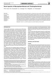
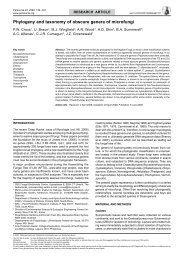
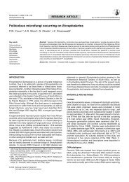
R.A. Samson et al.: <strong>Polyphasic</strong> <strong>taxonomy</strong> <strong>of</strong> Byssochlamys 23 a b c d e f g h i Fig. 7 Paecilomyces saturatus. a–e. Conidiophores; f. conidia. — Paecilomyces formosus. g, h. Conidiophores; i. conidia. — Scale bars = 10 µm.
24 <strong>Persoonia</strong> – Volume 22, 2009 Ram (1968) described two Paecilomyces species, P. lecythidis and P. maximus, based on <strong>the</strong>ir cultural characters and <strong>the</strong>ir large-sized conidia. The results <strong>of</strong> <strong>the</strong> sequencing <strong>of</strong> <strong>the</strong> ITS region, and parts <strong>of</strong> <strong>the</strong> protein coding genes β-tubulin and calmodulin, showed that P. formosus may consist <strong>of</strong> three taxa, P. formosus, P. lecythidis and P. maximus. However, <strong>the</strong> three species could not be distinguished by microscopical examination. One difference between strains belonging to <strong>the</strong> ‘P. maximus-clade’ and <strong>the</strong> o<strong>the</strong>r members <strong>of</strong> this diverse group, is <strong>the</strong> faster growth rate <strong>of</strong> this species at 37 °C than at 30 °C. The sequence and morphological diversity was not detected by <strong>the</strong> extrolite analyses: <strong>the</strong> ex-type cultures <strong>of</strong> P. lecythidis and P. maximus produced similar extrolite pr<strong>of</strong>iles, while <strong>the</strong> ex-type culture <strong>of</strong> P. formosus is degenerated and is a weak producer <strong>of</strong> extrolites. For a more detailed conclusion and delimitation <strong>of</strong> <strong>the</strong>se three groups more strains should be studied, particularly emphasising conidial shape and extrolites production. For <strong>the</strong> time being we propose to place P. lecythidis and P. maximus in synonymy with P. formosus. This species is morphologically similar to P. variotii and <strong>the</strong> main difference is <strong>the</strong> consistent acid production on CREA. Paecilomyces formosus has been isolated from tropical and subtropical soils, wood, sponge, man (bone marrow, blood), air in a bedroom (Denmark) and pot plant soil <strong>of</strong> Senseviera trifasciata (Denmark). Paecilomyces saturatus (Nakaz., Y. Takeda & Suematsu) Samson & Houbraken, comb. nov. — MycoBank MB512560; Fig. 7a–f Basionym. Paecilomyces mandshuricus (Saito) Thom var. saturatus Nakaz., Y. Takeda & Suematsu, J. Agric. Chem. Soc. Japan 10: 102. 1934. = Penicillium viniferum Sakag., May. Inoue & Tada, Zentralbl. Bakteriol., 2. Abt. 100: 303. 1939. = Paecilomyces dactylethromorphus Bat. & H. Maia, Anais Soc. Biol. Pernambuco 15: 152. 1957. The oldest basionym available for this taxon is P. mandshuricus var. saturatus and <strong>the</strong>refore <strong>the</strong> name <strong>of</strong> this taxon is derived from this varietal name. Isolates growing on MEA at 30 °C cover <strong>the</strong> Petri dish within 7 d, with strong olive-brown sporulation. Good growth at 37 °C, though slower at 30 °C. Good growth on MEA with 0.5 % acetic acid and CYA with 1 000 ppm propionic acid (pH 3.8). No growth observed on CYA with 5 % NaCl. Poor growth and no acid production on CREA. Morphological examination <strong>of</strong> various strains showed that this species forms fairly regularly branched, penicillium-like conidiophores, with ellipsoidal and/or cylindrical conidia without a distinct truncation. Chlamydospores present, hyaline and smooth walled. No teleomorph observed. The production <strong>of</strong> extrolites depends very much on <strong>the</strong> growth medium; patulin or brefeldin A can be produced. Paecilomyces saturatus is an easily recognisable species with its ellipsoidal and/or cylindrical conidia and its fairy regularly branched, penicillium-like conidiophores. Sakaguchi et al. (1939) described Penicillium viniferum and this species was subsequently placed in Paecilomyces by Raper & Thom (1949). In retrospect, <strong>the</strong> placement in Paecilomyces was correct, although this species could be interpreted, with its penicillium-like conidiophores and <strong>the</strong> presence <strong>of</strong> chlamydospores, as an intermediate form between Penicillium and Paecilomyces. Molecular studies now show that this species belongs to <strong>the</strong> Byssochlamys clade and is different from o<strong>the</strong>r olive-brown coloured species such as Hamigera avellanea (Luangsa-ard et al. 2004), Penicillium digitatum and P. cylindrosporum (unpubl. data). This species has been isolated from a variety <strong>of</strong> substrates, e.g. acetic acid, lea<strong>the</strong>r, medicine containing quinine, a dispersion <strong>of</strong> fenylacetate and dibutylmaleinate and Lepidium sativum. Notes on <strong>the</strong> ecology and extrolites production Byssochlamys and Paecilomyces species are <strong>of</strong>ten found in acidic habitats such as silage (Scurti et al. 1973, Escoula 1975a, b, Anderson et al. 1979), and in common with Penicillium series Roqueforti species (Frisvad & Samson 2004) can also tolerate microaerophilic conditions (Escoula 1975a, b, Taniwaki 1995). Byssochlamys nivea was initially reported to produce patulin under <strong>the</strong> name Gymnoascus sp. (Karow & Forster 1944, Kuehn 1958), later confirmed by Kis et al. (1969), Scurti et al. (1973), Rice et al. (1977) and Draughon & Ayres (1980). Byssochlamys fulva was also reported to produce patulin, albeit by few strains (Escoula 1975a, b, Percebois et al. 1975, Rice et al. 1977). Besides <strong>the</strong>ir presence in pasteurised fruit, B. fulva and B. nivea also form toxic extrolites, such as byssotoxin A and byssochlamic acid (Kramer et al. 1976, Rice et al. 1977). Besides mycotoxins, also an antitumor metabolite, byssochlamysol, a steroid against IGF-1 dependent cancer cells, is produced by B. nivea (Mori et al. 2003). Paecilomyces variotii s.l. also produces mycotoxins (Scott 1965), such as patulin (Escoula 1975a, b), sphing<strong>of</strong>ungin E and F (Frommer et al. 1992) and viriditoxin, reported originally from an isolate named Spicaria divaricata (Jiu & Mizuba 1974). Apart from being reported as being a potential mycotoxin, viriditoxin has also been reported to be a candidate for treatment <strong>of</strong> antibiotic <strong>resistant</strong> bacteria (Wang et al. 2003). Among <strong>the</strong> known extrolites are <strong>the</strong> antifungal drug variotin (Takeuchi et al. 1959, 1964, Suzuki et al. 1990, Omolo et al. 2000), and o<strong>the</strong>r drug candidates such as cornexistins (Nakajima et al. 1991, Fields et al. 1996), SCH 643432 (Hegde et al. 2003) and a penicillin-like compound (Burton 1949). The sideramins ferrirubrin and fusigen (Diekmann 1967, Domsch et al. 1980) and <strong>the</strong> organic acids 3-indole-acetic acid (Bakalinerov 1968, Voinova-Raikova et al. 1969), citric acid (Loesecke 1945), ethyleneoxide-α, β-dicarboxylic acid (Sakaguchi et al. 1939), (3Z,5E)-octa-3,5-diene -1,3,4-tricarboxylic acid 3,4-anhydride (Aldridge et al. 1980) have also been reported. The possible mycotoxins and/or potential drugs byssotoxin A, sphing<strong>of</strong>ungin E and F, SCH 643432 and byssochlamysol were not available to us as standards. Given <strong>the</strong> taxonomic revision presented here, it remains to be seen which species produce <strong>the</strong>se extrolites. Some connections between species and bioactive extrolites were confirmed or established here and several species had a high consistent extrolite pr<strong>of</strong>ile. However, <strong>the</strong> chemo<strong>taxonomy</strong> <strong>of</strong> P. saturatus is unresolved, because <strong>the</strong>re appear to be two chemotypes, which may or may not indicate that <strong>the</strong>re are two species ra<strong>the</strong>r than one. Likewise, B. fulva appears to have two chemotypes, with only byssochlamic acid as a common extrolite in all isolates examined. Key to Byssochlamys and related PaecilomYces anamorphs* 1. Conidia with conspicuously truncate ends or a flattened base . . . . . . . . . . . . . . . . . . . . . . . . . . . . . . . . . . . . . . . . . 2 1. Conidia ellipsoidal or cylindrical with inconspicuously truncate ends . . . . . . . . . . . . . . . . . . . . . . . . . . . . P. saturatus 2. Conidia predominantly ellipsoidal and/or cylindrical chlamydospores absent or present; Byssochlamys teleomorph absent or present . . . . . . . . . . . . . . . . . . . . . . . . . . . . . . . . . . . . 3 2. Conidia predominantly globose to subglobose, chlamydospores present; Byssochlamys teleomorph present . . . . 8