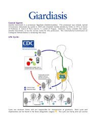
Causal Agent: Life Cycle:
Causal Agent: Life Cycle:
Causal Agent: Life Cycle:

Create successful ePaper yourself
Turn your PDF publications into a flip-book with our unique Google optimized e-Paper software.
<strong>Causal</strong> <strong>Agent</strong>:<br />
Pneumocystis jiroveci (previously classified as Pneumocystis carinii) was previously classified as a<br />
protozoa. Currently, it is considered a fungus based on nucleic acid and biochemical analysis.<br />
<strong>Life</strong> <strong>Cycle</strong>:<br />
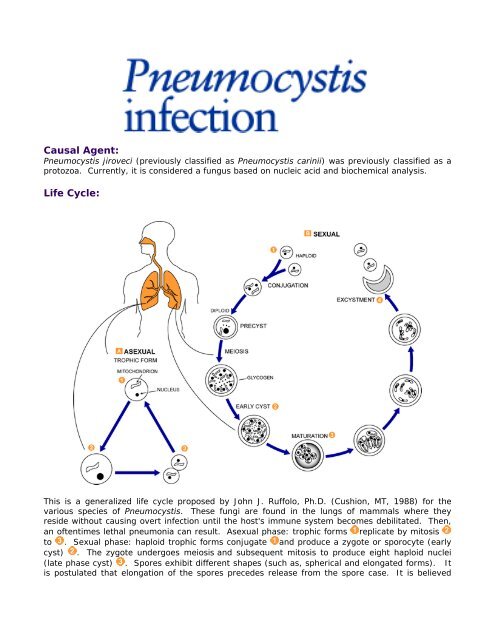
This is a generalized life cycle proposed by John J. Ruffolo, Ph.D. (Cushion, MT, 1988) for the<br />
various species of Pneumocystis. These fungi are found in the lungs of mammals where they<br />
reside without causing overt infection until the host's immune system becomes debilitated. Then,<br />
an oftentimes lethal pneumonia can result. Asexual phase: trophic forms replicate by mitosis<br />
to . Sexual phase: haploid trophic forms conjugate and produce a zygote or sporocyte (early<br />
cyst) . The zygote undergoes meiosis and subsequent mitosis to produce eight haploid nuclei<br />
(late phase cyst) . Spores exhibit different shapes (such as, spherical and elongated forms). It<br />
is postulated that elongation of the spores precedes release from the spore case. It is believed
that the release occurs through a rent in the cell wall. After release, the empty spore case usually<br />
collapses, but retains some residual cytoplasm . A trophic stage, where the organisms probably<br />
multiply by binary fission is also recognized to exist. The organism causes disease in<br />
immunosuppressed individuals.<br />
Geographic Distribution:<br />
Worldwide, in humans and animals. Serologic evidence indicates that most healthy children have<br />
been exposed by age 3 to 4. Pneumocystis pneumonia (PCP) occurs in immunosuppressed<br />
individuals and in premature, malnourished infants.<br />
Clinical Features:<br />
The symptoms of Pneumocystis pneumonia (PCP) include dyspnea, nonproductive cough, and<br />
fever. Chest radiography demonstrates bilateral infiltrates. Extrapulmonary lesions occur in a<br />
minority (