Lysosome Enrichment Kit for Tissue and Cultured Cells - Pierce
Lysosome Enrichment Kit for Tissue and Cultured Cells - Pierce
Lysosome Enrichment Kit for Tissue and Cultured Cells - Pierce

You also want an ePaper? Increase the reach of your titles
YUMPU automatically turns print PDFs into web optimized ePapers that Google loves.
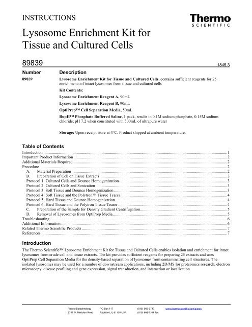
INSTRUCTIONS<br />
<strong>Lysosome</strong> <strong>Enrichment</strong> <strong>Kit</strong> <strong>for</strong><br />
<strong>Tissue</strong> <strong>and</strong> <strong>Cultured</strong> <strong>Cells</strong><br />
89839<br />
Number<br />
Description<br />
89839 <strong>Lysosome</strong> <strong>Enrichment</strong> <strong>Kit</strong> <strong>for</strong> <strong>Tissue</strong> <strong>and</strong> <strong>Cultured</strong> <strong>Cells</strong>, contains sufficient reagents <strong>for</strong> 25<br />
enrichments of intact lysosomes from tissue <strong>and</strong> cultured cells<br />
<strong>Kit</strong> Contents:<br />
<strong>Lysosome</strong> <strong>Enrichment</strong> Reagent A, 90mL<br />
<strong>Lysosome</strong> <strong>Enrichment</strong> Reagent B, 90mL<br />
OptiPrep Cell Separation Media, 50mL<br />
BupH Phosphate Buffered Saline, 1 pack, results in 0.1M sodium phosphate, 0.15M sodium<br />
chloride; pH 7.2 when constituted with 500mL of ultrapure water<br />
1845.3<br />
Storage: Upon receipt store at 4°C. Product shipped at ambient temperature.<br />
Table of Contents<br />
Introduction ................................................................................................................................................................................. 1<br />
Important Product In<strong>for</strong>mation .................................................................................................................................................... 2<br />
Additional Materials Required ..................................................................................................................................................... 2<br />
Procedure ..................................................................................................................................................................................... 2<br />
A. Material Preparation ...................................................................................................................................................... 2<br />
B. Preparation of Cell or <strong>Tissue</strong> Extracts ........................................................................................................................... 3<br />
Protocol 1: <strong>Cultured</strong> <strong>Cells</strong> <strong>and</strong> Dounce Homogenization ........................................................................................................ 3<br />
Protocol 2: <strong>Cultured</strong> <strong>Cells</strong> <strong>and</strong> Sonication ............................................................................................................................... 3<br />
Protocol 3: Soft <strong>Tissue</strong> <strong>and</strong> Dounce Homogenization ............................................................................................................. 3<br />
Protocol 4: Soft <strong>Tissue</strong> <strong>and</strong> the Polytron <strong>Tissue</strong> Tearer ....................................................................................................... 4<br />
Protocol 5: Hard <strong>Tissue</strong> <strong>and</strong> Dounce Homogenization ............................................................................................................ 4<br />
Protocol 6: Hard <strong>Tissue</strong> <strong>and</strong> the Polytron <strong>Tissue</strong> Tearer ......................................................................................................... 4<br />
C. Preparation of the Sample <strong>for</strong> Density Gradient Centrifugation.................................................................................... 5<br />
D. Removal of <strong>Lysosome</strong>s from OptiPrep Media .............................................................................................................. 5<br />
Troubleshooting ........................................................................................................................................................................... 6<br />
Additional In<strong>for</strong>mation ................................................................................................................................................................ 6<br />
Related Thermo Scientific Products ............................................................................................................................................ 7<br />
References ................................................................................................................................................................................... 7<br />
Introduction<br />
The Thermo Scientific <strong>Lysosome</strong> <strong>Enrichment</strong> <strong>Kit</strong> <strong>for</strong> <strong>Tissue</strong> <strong>and</strong> <strong>Cultured</strong> <strong>Cells</strong> enables isolation <strong>and</strong> enrichment <strong>for</strong> intact<br />
lysosomes from crude cell <strong>and</strong> tissue extracts. The kit provides sufficient reagents <strong>for</strong> preparing 25 extracts <strong>and</strong> uses<br />
OptiPrep Cell Separation Media <strong>for</strong> the density-based separation of lysosomes from contaminating cell structures. The<br />
isolated lysosomes may be used <strong>for</strong> a number of downstream applications, including 2D/MS <strong>for</strong> proteomics research, electron<br />
microscopy, disease profiling <strong>and</strong> gene expression, signal transduction, <strong>and</strong> interaction or localization.<br />
<strong>Pierce</strong> Biotechnology PO Box 117 (815) 968-0747 www.thermoscientific.com/pierce<br />
3747 N. Meridian Road Rock<strong>for</strong>d, lL 61105 USA (815) 968-7316 fax
Important Product In<strong>for</strong>mation<br />
• The OptiPrep Cell Separation Media is a 60% solution of iodixanol - 5,5´-[(2-hydroxy-1,3-propanediyl)-<br />
bis(acetylamino)] bis [N,N´-bis(2,3 dihydroxypropyl-2,4,6-triiodo-1,3-benzenecarboxamide).<br />
• The density of the lysosomes varies depending on the cell <strong>and</strong> tissue source. There<strong>for</strong>e, optimization of the gradient<br />
concentrations is necessary <strong>for</strong> best results. Refer to the Axis-Shield website <strong>for</strong> helpful density gradient concentrations<br />
<strong>for</strong> various cell <strong>and</strong> tissue types: www.axis-shield-density-gradient-media.com<br />
• The procedure was developed using a tabletop ultracentrifuge. A floor centrifuge may also be used with appropriate<br />
scaling of reagents.<br />
Additional Materials Required<br />
• Ultracentrifuge: tabletop or floor-model<br />
• Ultracentrifuge tubes: either open- or closed-top tubes, 8mL<br />
• Bench-top microcentrifuge with refrigeration<br />
• 2mL microcentrifuge tubes<br />
• 15mL conical tubes<br />
• Vortex mixer<br />
• Protease inhibitors, such as Thermo Scientific <strong>Pierce</strong> Protease Inhibitor Mini Tablets, EDTA-Free (Product No.<br />
88666)<br />
• Dounce tissue grinder, such as 2mL Kontes or Wheaton Dounce <strong>Tissue</strong><br />
• <strong>Tissue</strong> Tearer, such as the Kinematica AG Polytron PT1200<br />
• Sonicator, such as the Misonix Sonicator 3000<br />
• Surgical scissors <strong>for</strong> mincing tissue<br />
Procedure<br />
A. Material Preparation<br />
Phosphate-buffered Saline<br />
(PBS)<br />
Gradient Dilution Buffer<br />
OptiPrep Gradients<br />
Dissolve the dry-blend buffer with 500mL of ultrapure water. For long-term storage of excess<br />
buffer, sterile-filter the solution <strong>and</strong> store at 4°C.<br />
Mix equal volumes of the <strong>Lysosome</strong> <strong>Enrichment</strong> Reagents A <strong>and</strong> B. Each sample requires<br />
2.5mL of Gradient Dilution Buffer.<br />
The OptiPrep Cell Separation Media is supplied as a 60% solution. Prepare five gradient<br />
solutions using the Gradient Dilution Buffer as indicated in the table below. Each volume is <strong>for</strong><br />
one sample preparation.<br />
Gradient<br />
OptiPrep Cell<br />
Separation Media<br />
Volume (µL)<br />
Table 1. OptiPrep Gradient Preparation<br />
Gradient Dilution<br />
Buffer<br />
Volume (µL)<br />
Final<br />
Volume<br />
(µL)<br />
Final Gradient<br />
Percent<br />
(%)<br />
1 283.3 716.7 1000 17<br />
2 333.3 666.7 1000 20<br />
3 191.7 308.3 500 23<br />
4 450 550 1000 27<br />
5 250.0 250.0 500 30<br />
<strong>Pierce</strong> Biotechnology PO Box 117 (815) 968-0747 www.thermoscientific.com/pierce<br />
3747 N. Meridian Road Rock<strong>for</strong>d, lL 61105 USA (815) 968-7316 fax<br />
2
B. Preparation of Cell or <strong>Tissue</strong> Extracts<br />
Protocol 1: <strong>Cultured</strong> <strong>Cells</strong> <strong>and</strong> Dounce Homogenization<br />
• Immediately be<strong>for</strong>e use, add protease inhibitors to <strong>Lysosome</strong> <strong>Enrichment</strong> Reagents A <strong>and</strong> B; only add inhibitors to the<br />
volume being used <strong>for</strong> the procedure <strong>and</strong> not to the stock solutions.<br />
• Process one sample at a time.<br />
• Pre-chill Dounce tissue grinder on ice be<strong>for</strong>e use.<br />
• Empirically determine the number of Dounce strokes required <strong>for</strong> optimal cell lysis with minimal damage to the<br />
lysosomes <strong>for</strong> each tissue type. See the Additional In<strong>for</strong>mation Section <strong>for</strong> cell lysis in<strong>for</strong>mation.<br />
1. Pellet 50-200mg of cells by centrifuging harvested cell suspension in a 2mL microcentrifuge tube at ~850 × g <strong>for</strong><br />
2 minutes. Carefully remove <strong>and</strong> discard the supernatant.<br />
2. Add 800µL of <strong>Lysosome</strong> <strong>Enrichment</strong> Reagent A. Vortex at medium speed <strong>for</strong> 5 seconds <strong>and</strong> incubate on ice <strong>for</strong> exactly<br />
2 minutes. Do not exceed the 2 minute incubation.<br />
3. Transfer cell suspension to a Dounce tissue grinder <strong>and</strong> homogenize cells on ice. Per<strong>for</strong>m enough strokes to effectively<br />
lyse the cells.<br />
Note: To check lysis efficiency, place 5µL of lysate onto a glass slide, add coverslip <strong>and</strong> view with a microscope.<br />
Compare results with 5µL of the non-lysed cells.<br />
4. Transfer lysed cells into a 2mL microcentrifuge tube <strong>and</strong> add 800µL of <strong>Lysosome</strong> <strong>Enrichment</strong> Reagent B. Invert tube<br />
several times to mix – do not vortex.<br />
5. Centrifuge tube at 500 × g <strong>for</strong> 10 minutes at 4°C. Collect supernatant in a new tube <strong>and</strong> keep on ice until needed.<br />
6. Proceed to Sections C <strong>and</strong> D <strong>for</strong> gradient centrifugation <strong>and</strong> isolation of lysosomes.<br />
Protocol 2: <strong>Cultured</strong> <strong>Cells</strong> <strong>and</strong> Sonication<br />
• Immediately be<strong>for</strong>e use, add protease inhibitors to <strong>Lysosome</strong> <strong>Enrichment</strong> Reagents A <strong>and</strong> B; only add inhibitors to the<br />
reagent amount being used <strong>for</strong> the procedure <strong>and</strong> not to the stock solutions.<br />
1. Pellet 50-200mg of cells by centrifuging harvested cell suspension in a 2mL microcentrifuge tube at ~850 × g <strong>for</strong><br />
2 minutes. Carefully remove <strong>and</strong> discard the supernatant.<br />
2. Add 800µL of <strong>Lysosome</strong> <strong>Enrichment</strong> Reagent A. Vortex at medium speed <strong>for</strong> 5 seconds <strong>and</strong> incubate on ice <strong>for</strong> exactly<br />
2 minutes. Do not exceed the 2-minute incubation.<br />
3. Sonicate cell suspension on ice, per<strong>for</strong>ming a sufficient number of bursts <strong>for</strong> effective cell lysis (e.g., 10-15 bursts, at<br />
6-9W of power).<br />
Note: To check the lysis efficiency, place 5µL of cell lysate onto a glass slide, add coverslip <strong>and</strong> view under a<br />
microscope. Compare with 5µL of non-lysed cells.<br />
4. Add 800µL of <strong>Lysosome</strong> <strong>Enrichment</strong> Reagent B. Invert tube several times to mix – do not vortex.<br />
5. Centrifuge tube at 500 × g <strong>for</strong> 10 minutes at 4°C. Collect supernatant in a new tube <strong>and</strong> keep on ice until needed.<br />
6. Proceed to Sections C <strong>and</strong> D <strong>for</strong> gradient centrifugation <strong>and</strong> isolation of lysosomes.<br />
Protocol 3: Soft <strong>Tissue</strong> <strong>and</strong> Dounce Homogenization<br />
• Pre-chill the Dounce tissue grinder on ice be<strong>for</strong>e use.<br />
• Immediately be<strong>for</strong>e use, add protease inhibitors to the <strong>Lysosome</strong> <strong>Enrichment</strong> Reagents A <strong>and</strong> B; add inhibitors only to<br />
the volume being used <strong>for</strong> the procedure <strong>and</strong> not to the stock solutions.<br />
• Empirically determine the number of Dounce strokes required <strong>for</strong> optimal cell lysis with minimal damage to the<br />
lysosomes <strong>for</strong> each tissue type. See the Additional In<strong>for</strong>mation Section <strong>for</strong> cell lysis in<strong>for</strong>mation.<br />
1. Wash 50-200mg of tissue with 2-4mL of PBS. Carefully remove <strong>and</strong> discard the PBS wash.<br />
2. Mince tissue into small pieces (< 3mm 3 ) <strong>and</strong> add 800µL of <strong>Lysosome</strong> <strong>Enrichment</strong> Reagent A.<br />
<strong>Pierce</strong> Biotechnology PO Box 117 (815) 968-0747 www.thermoscientific.com/pierce<br />
3747 N. Meridian Road Rock<strong>for</strong>d, lL 61105 USA (815) 968-7316 fax<br />
3
3. Per<strong>for</strong>m Dounce homogenization on ice.<br />
Note: To check lysis efficiency, place 5µL of lysate onto a glass slide, add coverslip <strong>and</strong> view with a microscope.<br />
Compare results with 5µL of the non-lysed cells.<br />
4. Add 800µL of <strong>Lysosome</strong> <strong>Enrichment</strong> Reagent B. Invert the tube several times to mix - do not vortex.<br />
5. Centrifuge tube at 500 × g <strong>for</strong> 10 minutes at 4°C. Collect supernatant in a new tube <strong>and</strong> keep on ice until needed.<br />
6. Proceed to Sections C <strong>and</strong> D <strong>for</strong> gradient centrifugation <strong>and</strong> isolation of lysosomes.<br />
Protocol 4: Soft <strong>Tissue</strong> <strong>and</strong> the Polytron <strong>Tissue</strong> Tearer<br />
• Immediately be<strong>for</strong>e use, add protease inhibitors to the <strong>Lysosome</strong> <strong>Enrichment</strong> Reagents A <strong>and</strong> B; add inhibitors only to<br />
the reagent amount being used <strong>for</strong> the procedure <strong>and</strong> not to the stock solutions.<br />
• The speed <strong>and</strong> time duration <strong>for</strong> homogenization varies depending on tissue type.<br />
1. Wash 50-200mg of tissue with 2-4mL of PBS. Carefully remove <strong>and</strong> discard the PBS wash.<br />
2. Mince tissue into small pieces (less than 3mm 3 ) <strong>and</strong> add 800µL of <strong>Lysosome</strong> <strong>Enrichment</strong> Reagent A.<br />
3. Homogenize on ice at approximately 8000-9000 rpm <strong>for</strong> 45 seconds; however, depending on tissue type, the time<br />
required <strong>for</strong> lysis may be longer or shorter.<br />
Note: To check lysis efficiency, place 5µL of lysate onto a glass slide, add coverslip <strong>and</strong> view with a microscope.<br />
Compare results with 5µL of the non-lysed cells.<br />
4. Add 800µL of <strong>Lysosome</strong> <strong>Enrichment</strong> Reagent B. Invert the tube several times to mix – do not vortex.<br />
5. Centrifuge at 500 × g <strong>for</strong> 10 minutes at 4°C. Collect supernatant in a new tube <strong>and</strong> keep on ice until needed.<br />
6. Proceed to Sections C <strong>and</strong> D <strong>for</strong> gradient centrifugation <strong>and</strong> isolation of lysosomes.<br />
Protocol 5: Hard <strong>Tissue</strong> <strong>and</strong> Dounce Homogenization<br />
• Pre-chill the Dounce tissue grinder on ice be<strong>for</strong>e use.<br />
• Immediately be<strong>for</strong>e use, add protease inhibitors to the <strong>Lysosome</strong> <strong>Enrichment</strong> Reagents A <strong>and</strong> B; add inhibitors only to<br />
the reagent amount being used <strong>for</strong> the procedure <strong>and</strong> not to the stock solutions.<br />
• Empirically determine the number of Dounce strokes required <strong>for</strong> optimal cell lysis with minimal damage to the<br />
lysosomes <strong>for</strong> each tissue type. See the Additional In<strong>for</strong>mation Section <strong>for</strong> cell lysis in<strong>for</strong>mation.<br />
1. Wash 50-200mg of tissue with 2-4mL of PBS. Carefully remove <strong>and</strong> discard the PBS wash.<br />
2. Mince tissue into small pieces (less than 3mm 3 ) <strong>and</strong> add 800µL of <strong>Lysosome</strong> <strong>Enrichment</strong> Reagent A.<br />
3. Per<strong>for</strong>m Dounce homogenization on ice.<br />
Note: To check lysis efficiency, place 5µL of lysate onto a glass slide, add coverslip <strong>and</strong> view with a microscope.<br />
Compare results with 5µL of the non-lysed cells.<br />
4. Add 800µL of <strong>Lysosome</strong> <strong>Enrichment</strong> Reagent B. Invert the tube several times to mix – do not vortex.<br />
5. Centrifuge at 500 × g <strong>for</strong> 10 minutes at 4°C. Collect supernatant in a new tube <strong>and</strong> keep on ice until needed.<br />
6. Proceed to Sections C <strong>and</strong> D <strong>for</strong> gradient centrifugation <strong>and</strong> isolation of lysosomes.<br />
Protocol 6: Hard <strong>Tissue</strong> <strong>and</strong> the Polytron <strong>Tissue</strong> Tearer<br />
• Immediately be<strong>for</strong>e use, add protease inhibitors to the <strong>Lysosome</strong> <strong>Enrichment</strong> Reagents A <strong>and</strong> B; add inhibitors only to<br />
the reagent amount being used <strong>for</strong> the procedure <strong>and</strong> not to the stock solutions.<br />
• The speed <strong>and</strong> time duration <strong>for</strong> homogenization varies depending on cell/tissue type.<br />
1. Wash 50-200mg of tissue with 2-4mL of PBS. Carefully remove <strong>and</strong> discard the PBS wash.<br />
2. Mince tissue into small pieces (< 3mm 3 ) <strong>and</strong> add 800µL of <strong>Lysosome</strong> <strong>Enrichment</strong> Reagent A.<br />
<strong>Pierce</strong> Biotechnology PO Box 117 (815) 968-0747 www.thermoscientific.com/pierce<br />
3747 N. Meridian Road Rock<strong>for</strong>d, lL 61105 USA (815) 968-7316 fax<br />
4
3. Homogenize on ice at approximately 8000-9000 rpm <strong>for</strong> 45 seconds; however, depending on tissue type, the time<br />
required <strong>for</strong> lysis may be longer or shorter.<br />
Note: To check lysis efficiency, place 5µL of lysate onto a glass slide, add coverslip <strong>and</strong> view with a microscope.<br />
Compare results with 5µL of the non-lysed cells.<br />
4. Add 800µL of <strong>Lysosome</strong> <strong>Enrichment</strong> Reagent B. Invert the tube several times to mix - do not vortex.<br />
5. Centrifuge at 500 × g <strong>for</strong> 10 minutes at 4°C. Collect supernatant in a new tube <strong>and</strong> keep on ice until needed.<br />
6. Proceed to Sections C <strong>and</strong> D <strong>for</strong> gradient centrifugation <strong>and</strong> isolation of lysosomes.<br />
C. Preparation of the Sample <strong>for</strong> Density Gradient Centrifugation<br />
• The concentrations of the density media <strong>for</strong> lysosome isolation may require optimization, depending on the cell or tissue<br />
type. Refer to references <strong>for</strong> examples of enrichment gradient concentrations <strong>and</strong> cell/tissue sources.<br />
• Use either an open- or closed-top ultracentrifuge tube <strong>for</strong> enrichment.<br />
1. In an ultracentrifuge tube, prepare a discontinuous density gradient by carefully overlaying the prepared OptiPrep<br />
Gradients (see Section A) in descending concentrations. For example, first add the 30% gradient <strong>and</strong> then the 27%, 23%,<br />
20% <strong>and</strong> 17% gradients.<br />
2. Mix the prepared cell or tissue extract (Section B) with the OptiPrep Cell Separation Media to make a final concentration<br />
of 15% OptiPrep Media. For example, add 1500µL of extract to 500µL of OptiPrep Cell Separation Media.<br />
3. Overlay the sample containing the 15% OptiPrep Media on top of the density gradients prepared in Step 1 (Figure 1).<br />
4. Ultracentrifuge the samples at 145,000 × g <strong>for</strong> 2 hours at 4°C.<br />
5. After centrifugation, several b<strong>and</strong>s will <strong>for</strong>m in the gradient (Figure 2). The lysosome b<strong>and</strong> is located in the top 2mL of<br />
the gradient. Carefully remove the b<strong>and</strong> <strong>and</strong> save on ice.<br />
Figure 1. Be<strong>for</strong>e ultracentrifugation, the sample is<br />
visible on top of the gradient. The sample is A431<br />
cell extract prepared from ~200mg of cell pellet.<br />
Figure 2. After ultracentrifugation, b<strong>and</strong>s <strong>for</strong>m<br />
in the gradient. The top b<strong>and</strong> contains the<br />
isolated lysosomes.<br />
D. Removal of <strong>Lysosome</strong>s from OptiPrep Media<br />
1. Mix the isolated lysosome fraction with 2-3 volumes of PBS to decrease the concentration of the OptiPrep Media. Gently<br />
vortex to mix the sample.<br />
2. Transfer the sample into a microcentrifuge tube <strong>and</strong> centrifuge at 18,000 × g <strong>for</strong> 30 minutes at 4°C.<br />
3. Remove the supernatant <strong>and</strong> keep the lysosome pellet on ice.<br />
4. To surface-wash the pellet, add 1mL of the Gradient Dilution Buffer <strong>and</strong> centrifuge at 18,000 × g <strong>for</strong> 30 minutes at 4°C.<br />
5. Remove supernatant <strong>and</strong> maintain the lysosome pellet on ice until downstream processing. Freezing <strong>and</strong> thawing may<br />
compromise lysosomal integrity.<br />
<strong>Pierce</strong> Biotechnology PO Box 117 (815) 968-0747 www.thermoscientific.com/pierce<br />
3747 N. Meridian Road Rock<strong>for</strong>d, lL 61105 USA (815) 968-7316 fax<br />
5
Troubleshooting<br />
Problem Possible Cause Solution<br />
Small lysosome pellet Number of Dounce strokes were<br />
insufficient to achieve optimal lysis<br />
Optimize the number of Dounce strokes <strong>for</strong> each tissue<br />
type<br />
<strong>Cells</strong> are difficult to lyse<br />
Increase the number of Dounce strokes <strong>and</strong> consider<br />
using the Sonicator <strong>and</strong> Polytron <strong>Tissue</strong> Tearer<br />
Hard-tissue sample is not completely<br />
homogenized<br />
Mince hard tissue into small pieces be<strong>for</strong>e<br />
homogenization<br />
Fats/lipids are present in<br />
the sample<br />
Dietary intake contributes to<br />
fats/lipids in the system<br />
Starve animal 24 hours be<strong>for</strong>e sacrifice to minimize the<br />
amount of fats/lipids present in the system<br />
Rats are mature or overweight Young lean rats have less fats/lipids in their systems<br />
<strong>Lysosome</strong>s are in several<br />
fractions or dispersed in the<br />
gradient<br />
<strong>Lysosome</strong>s are not<br />
completely pelleted<br />
Additional In<strong>for</strong>mation<br />
<strong>Lysosome</strong> density differs depending<br />
on the cell or tissue type<br />
Inadvertent mixing of gradients during<br />
sample removal from the gradient<br />
The density gradient concentration<br />
was not low enough<br />
Optimize the gradient concentrations <strong>for</strong> each cell or<br />
tissue type<br />
Siphon lysosome b<strong>and</strong> or other fraction carefully <strong>and</strong><br />
slowly – quickly removing the sample causes mixing<br />
Dilute the gradient concentration with PBS to pellet the<br />
lysosomes<br />
A. Cell Lysis In<strong>for</strong>mation<br />
Note: Regardless of the cell lysis method, monitor the lysis efficiency during optimization. Do not jeopardize organelle<br />
integrity by significantly increasing cell lysis efficiency.<br />
Dounce Homogenization: The number of Dounce homogenization strokes necessary <strong>for</strong> optimal lysis varies depending upon<br />
cell or tissue type. For each sample type, determine the number of strokes required <strong>for</strong> optimal lysis with minimal damage to<br />
the lysosomes. Use the table below as a general guide.<br />
<strong>Tissue</strong>*<br />
<strong>Cells</strong>**<br />
<strong>Tissue</strong> Type Amount<br />
(mg)<br />
Number of<br />
Strokes<br />
Cell Type Amount<br />
(mg)<br />
Number of<br />
Strokes<br />
Soft (Liver) 50 20-30 A431 50 30-50<br />
200 50-75 200 80-100<br />
Hard (Heart) 50 10 HeLa 50 20-30<br />
200 30-50 200 70-80<br />
*Mince the tissue into small pieces be<strong>for</strong>e homogenization.<br />
** Determine lysis efficiency by visual examination with a microscope.<br />
Sonication: The robustness of the cell type affects the power output <strong>and</strong> the number of bursts needed <strong>for</strong> effective cell lysis.<br />
The procedure detailed here was tested on HeLa, HepG2 <strong>and</strong> A431 cells <strong>and</strong> used 9W of power <strong>and</strong> 15 sonication bursts.<br />
During optimization, monitor lysis efficiency by spotting sample onto a glass slide <strong>and</strong> comparing with a whole cell control.<br />
Polytron <strong>Tissue</strong> Tearer: Depending on the tissue type, adjust speed <strong>and</strong> duration as needed. For soft tissue, such as liver,<br />
<strong>and</strong> hard tissue, such as heart <strong>and</strong> kidney, the protocol suggests using 8000-9000 rpm <strong>for</strong> 45 seconds. The speed <strong>and</strong> duration<br />
required also varies depending on the amount of starting tissue material. During optimization, monitor the lysis efficiency by<br />
placing sample onto a glass slide <strong>and</strong> comparing with a non-lysed control.<br />
<strong>Pierce</strong> Biotechnology PO Box 117 (815) 968-0747 www.thermoscientific.com/pierce<br />
3747 N. Meridian Road Rock<strong>for</strong>d, lL 61105 USA (815) 968-7316 fax<br />
6
B. Optimization of OptiPrep Cell Separation Media Concentration <strong>and</strong> Centrifugation Speed/time<br />
Depending on the cell or tissue type, lysosomes differ in morphology <strong>and</strong> relative density. <strong>Lysosome</strong>s vary from 0.2 to 2µm<br />
<strong>and</strong> can fuse with other lysosomes or endosomes. Organellar fusion affects relative densities of the organelle <strong>and</strong> directly<br />
affects the concentration of OptiPrep Media required <strong>for</strong> an effective enrichment. There<strong>for</strong>e, the optimum gradient<br />
concentration must be determined empirically. See references <strong>for</strong> examples of various OptiPrep Media concentrations with<br />
different cell <strong>and</strong> tissue types. In the initial experiment, collect small volumes of the sample/gradient, <strong>for</strong> example 500-<br />
1000µL (post-centrifugation), from the top of the gradient going down. Analyze the fractions by Western blot to determine<br />
the location of the lysosomes <strong>and</strong> the contaminating organelles to help determine a good starting concentration to use.<br />
Centrifugation speed <strong>and</strong> time also may require optimization. Depending on the sample, it may be necessary to centrifuge<br />
longer or faster. Per<strong>for</strong>m the developed protocol initially <strong>and</strong> make the necessary optimizations afterwards.<br />
C. <strong>Lysosome</strong> Lysis<br />
For protein analysis using the Thermo Scientific Coomassie Plus (Brad<strong>for</strong>d) Assay <strong>Kit</strong> (Product No. 23236), the<br />
lysosomes may be lysed with 2% CHAPS in Tris-buffered saline (TBS; 25mM Tris, 0.15M NaCl; pH 7.2; Product No.<br />
28379) as follows:<br />
• For direct analysis by SDS-PAGE or Western blotting, boil lysosomes pellet with SDS-PAGE sample buffer <strong>and</strong> apply<br />
to a protein electrophoresis gel.<br />
• Depending on the size of the lysosome pellet or protein concentration needed, add 100-400µL of 2% CHAPS in TBS to<br />
the lysosome pellet <strong>and</strong> vortex <strong>for</strong> 1 minute.<br />
1. Incubate at room temperature <strong>for</strong> 10 minutes.<br />
2. Vortex sample <strong>for</strong> 1 minute.<br />
3. Centrifuge sample at 18,000 × g at 4°C <strong>for</strong> 5 minutes. The clarified supernatant will contain soluble lysosomal proteins<br />
that can be analyzed by the Coommassie Plus (Brad<strong>for</strong>d) Assay <strong>Kit</strong> (Product No. 23236).<br />
Related Thermo Scientific Products<br />
89840 Peroxisome <strong>Enrichment</strong> <strong>Kit</strong> <strong>for</strong> <strong>Tissue</strong><br />
89841 Nuclei <strong>Enrichment</strong> <strong>Kit</strong> <strong>for</strong> <strong>Tissue</strong><br />
78833 NE-PER Nuclear <strong>and</strong> Cytoplasmic Extraction <strong>Kit</strong><br />
89826 Mem-PER Eukaryotic Membrane Protein Extraction <strong>Kit</strong><br />
78415 <strong>Pierce</strong> Protease Inhibitor Mini Tablets, EDTA-Free, 30 tablets/vial<br />
23236 Coomassie Plus (Brad<strong>for</strong>d) Assay <strong>Kit</strong><br />
78501 M-PER Mammalian Protein Extraction Reagent, 250mL<br />
34095 SuperSignal West Femto Maximum Sensitivity Chemiluminescent Substrate, 100mL<br />
References<br />
Gille, L. <strong>and</strong> Nohl, H. (2000). The existence of a lysosomal redox chain <strong>and</strong> the role of ubiquinone. Arch Biochem Biophys 375:347-54.<br />
Gjoen, T., et al. (1997). <strong>Lysosome</strong>s <strong>and</strong> endocytosis. Subcellular fractionation: A practical approach. Eds. Graham, J.M. <strong>and</strong> Rickwood, D., IRL Press<br />
Limited, Ox<strong>for</strong>d, Engl<strong>and</strong>, pp. 169-200.<br />
Goswami, P. <strong>and</strong> Cooney, J.J. (1999). Subcellular location of enzymes involved in oxidation of n-alkane by Cladosporium resinae. Appl Microbiol<br />
Biotechnol 51:860-4.<br />
Graham, J.M. (1997). Homogenization of tissues <strong>and</strong> cells. Subcellular fractionation: A practical approach. Eds. Graham, J.M. <strong>and</strong> Rickwood, D., IRL Press<br />
Limited, Ox<strong>for</strong>d, Engl<strong>and</strong>, pp. 1-28.<br />
Hinton, R.H. <strong>and</strong> Mullock, B.M. (1997). Isolation of subcellular fractions. Subcellular fractionation: A practical approach. Eds. Graham, J.M. <strong>and</strong><br />
Rickwood, D., IRL Press Limited, Ox<strong>for</strong>d, Engl<strong>and</strong>, pp. 31-69.<br />
Lukong, K.E., et al. (2001). Intracellular distribution of lysosomal sialidase is controlled by the internalization signal in its cytoplasmic tail. J Biol Chem<br />
276:46172-81.<br />
Miyazaki., T., et al. (2003). Regulation of cytochrome C oxidase activity by c-Src in osteoclasts. J Cell Biol 160:709-18.<br />
Murata, Y., et al. (2002). Differential localization of the vacuolar H + pump with G subunit iso<strong>for</strong>ms (G1 <strong>and</strong> G2) in mouse neurons. J Biol Chem 277:36296-<br />
303.<br />
Nathanson, C-M., et al. (2002). Regulated expression <strong>and</strong> intracellular localization of cystatin F in human U937 cells. Eur J Biochem 269:5502-51.<br />
<strong>Pierce</strong> Biotechnology PO Box 117 (815) 968-0747 www.thermoscientific.com/pierce<br />
3747 N. Meridian Road Rock<strong>for</strong>d, lL 61105 USA (815) 968-7316 fax<br />
7
Prigozy, T.I., et al. (2001). glycolipid antigen processing <strong>for</strong> presentation by CD1d molecules. Science 291:664-7.<br />
Seyrantepe, V., et al. (2004). Neu4, a novel human lysosomal lumen sialidase, confers normal phenotype to sialidosis <strong>and</strong> galactosialidosis cells. J Biol<br />
Chem 279:37021-37029.<br />
Simonova, M., et al. (2000). Tyrosinase mutants are capable of prodrug activation in transfected nonmelanotic cells. Cancer Res 60:6656-62.<br />
Weissleder, R., et al. (1999). In vivo imaging of tumors with protease-activated near-infrared fluorescent probes. Nature Biotech 17:375-8.<br />
Products are warranted to operate or per<strong>for</strong>m substantially in con<strong>for</strong>mance with published Product specifications in effect at the time of sale, as set <strong>for</strong>th in<br />
the Product documentation, specifications <strong>and</strong>/or accompanying package inserts (“Documentation”). No claim of suitability <strong>for</strong> use in applications regulated<br />
by FDA is made. The warranty provided herein is valid only when used by properly trained individuals. Unless otherwise stated in the Documentation, this<br />
warranty is limited to one year from date of shipment when the Product is subjected to normal, proper <strong>and</strong> intended usage. This warranty does not extend to<br />
anyone other than Buyer. Any model or sample furnished to Buyer is merely illustrative of the general type <strong>and</strong> quality of goods <strong>and</strong> does not represent that<br />
any Product will con<strong>for</strong>m to such model or sample.<br />
NO OTHER WARRANTIES, EXPRESS OR IMPLIED, ARE GRANTED, INCLUDING WITHOUT LIMITATION, IMPLIED WARRANTIES OF<br />
MERCHANTABILITY, FITNESS FOR ANY PARTICULAR PURPOSE, OR NON INFRINGEMENT. BUYER’S EXCLUSIVE REMEDY FOR NON-<br />
CONFORMING PRODUCTS DURING THE WARRANTY PERIOD IS LIMITED TO REPAIR, REPLACEMENT OF OR REFUND FOR THE NON-<br />
CONFORMING PRODUCT(S) AT SELLER’S SOLE OPTION. THERE IS NO OBLIGATION TO REPAIR, REPLACE OR REFUND FOR PRODUCTS<br />
AS THE RESULT OF (I) ACCIDENT, DISASTER OR EVENT OF FORCE MAJEURE, (II) MISUSE, FAULT OR NEGLIGENCE OF OR BY BUYER,<br />
(III) USE OF THE PRODUCTS IN A MANNER FOR WHICH THEY WERE NOT DESIGNED, OR (IV) IMPROPER STORAGE AND HANDLING OF<br />
THE PRODUCTS.<br />
Unless otherwise expressly stated on the Product or in the documentation accompanying the Product, the Product is intended <strong>for</strong> research only <strong>and</strong> is not to<br />
be used <strong>for</strong> any other purpose, including without limitation, unauthorized commercial uses, in vitro diagnostic uses, ex vivo or in vivo therapeutic uses, or<br />
any type of consumption by or application to humans or animals.<br />
Current product instructions are available at www.thermoscientific.com/pierce. For a faxed copy, call 800-874-3723 or contact your local distributor.<br />
© 2013 Thermo Fisher Scientific Inc. All rights reserved. OptiPrep is a trademark of Axis-Shield. Polytron is a trademark of Kinematica AG. Sonicator is a<br />
trademark of Mettler Electronics Corp. All (other) trademarks are the property of Thermo Fisher Scientific Inc. <strong>and</strong> its subsidiaries. Printed in the USA.<br />
<strong>Pierce</strong> Biotechnology PO Box 117 (815) 968-0747 www.thermoscientific.com/pierce<br />
3747 N. Meridian Road Rock<strong>for</strong>d, lL 61105 USA (815) 968-7316 fax<br />
8