Microspheres & Particles Handling Guide - Polysciences, Inc.
Microspheres & Particles Handling Guide - Polysciences, Inc.
Microspheres & Particles Handling Guide - Polysciences, Inc.

Create successful ePaper yourself
Turn your PDF publications into a flip-book with our unique Google optimized e-Paper software.
<strong>Microspheres</strong> & <strong>Particles</strong><br />
<strong>Handling</strong> <strong>Guide</strong>
<strong>Microspheres</strong> & <strong>Particles</strong> <strong>Handling</strong> <strong>Guide</strong><br />
<strong>Microspheres</strong> and <strong>Particles</strong> <strong>Handling</strong> <strong>Guide</strong><br />
<strong>Polysciences</strong>, <strong>Inc</strong>. offers a wide selection of microspheres and particles. The information contained in this section covers many<br />
of the frequently asked questions about these products. Please contact us with any further questions you might have.<br />
General Information<br />
<strong>Microspheres</strong> offer a highly convenient and flexible system for developing reagents for assays and bioseparations and for use as instrument<br />
standards. As there are many varieties of microspheres available, it is important to think about the demands the application will<br />
place on them when making a base bead selection. Physical and optical properties should be considered in the context of handling and<br />
detection and thought should also be given to requirements for diameter and size distribution, composition, surface chemistry and any<br />
other needed properties.<br />
Property<br />
Size<br />
Composition<br />
Surface chemistry<br />
Special properties<br />
Considerations<br />
Diameter<br />
Uniformity / distribution<br />
Density<br />
Refractive index<br />
Hydrophobicity / -philicity<br />
Nonspecific binding<br />
Autofluorescence<br />
Reactive groups<br />
Level of functionalization<br />
Charge<br />
Visible dye / fluorophore<br />
Superparamagnetic<br />
Particle Size<br />
Microsphere size may be critical to the proper function of an assay, or it may be secondary to other characteristics. Considering<br />
traditional diagnostic methods, the test or assay format commonly dictates particle size, such as the use of very small spheres (~0.1-<br />
0.4µm) to ensure satisfactory wicking in lateral flow tests, or the use of larger, cell-sized spheres (~4-10µm) for bead-based flow<br />
cytometric assays.<br />
In magnetic separations, particularly those involving capture and elution of target, the exact size of the magnetic particle may be<br />
unimportant provided that the particles are in some general size range and offer desired separation characteristics.<br />
Diameter also determines surface area. Small-diameter spheres present more surface area per unit weight, while larger spheres present<br />
more surface area per bead. Size also effects ease of handling, process considerations (such as the method used for separations<br />
[centrifugation, dialysis, filtration]) and the amount of reagent needed for coating.<br />
All sizes listed in this catalog are nominal sizes. For most products, the mean diameter of your particles will be printed on the label with<br />
the standard deviation.<br />
2<br />
For technical questions, info@polysciences.com or (800) 523-2575
<strong>Microspheres</strong> & <strong>Particles</strong> <strong>Handling</strong> <strong>Guide</strong><br />
Particle Composition<br />
Common microsphere compositions include polystyrene (PS), poly(methyl methacrylate) (PMMA) and silica. These materials possess<br />
different physical and optical properties, which may present advantages or limitations for different applications.<br />
Polymer beads are generally hydrophobic and, as such, have high protein binding abilities. However, they often require the use of<br />
some surfactant (e.g. 0.01-0.1% Tween ® 20 or SDS) in the storage buffer to ensure ease of handling. During synthesis, functional<br />
monomers may be co-polymerized with styrene or methyl methacrylate to develop beads with reactive surface groups. Functional<br />
groups may be used in covalent binding reactions and also aid in stabilizing the suspension.<br />
Silica microspheres are inherently hydrophilic and negatively charged. Consequently, aqueous silica suspensions rarely require the<br />
use of surfactants or other stabilizers. Carboxyl- and amine-functionalized silica spheres are available for use in common covalent<br />
coating protocols and plain silica microspheres may be modified using a variety of silanes to generate functional groups or alter<br />
surface properties.<br />
Composition Refractive Index Density Glass Transition<br />
(589nm) (g/cm 3 ) Temperature (˚C)<br />
PS<br />
1.59<br />
1.05<br />
95<br />
PMMA<br />
1.49<br />
1.19<br />
105<br />
Silica<br />
1.43-1.46*<br />
2.0*<br />
>>1000<br />
*Determined using representative samples. Other values are as reported in the literature for bulk polymer or silica.<br />
Particle Suspension<br />
The number of particles per ml will vary with the specified weight to volume (w/v) concentration, diameter of the particle and density<br />
of particle composition. The number of particles per milliliter can be calculated using the following equation:<br />
6x • 10 12<br />
y • π • z 3<br />
x = particle weight (g) per ml<br />
y = density (g/ml)<br />
z = diameter (µm)<br />
For more information please call (800) 523-2575 or visit: www.polysciences.com<br />
3
<strong>Microspheres</strong> & <strong>Particles</strong> <strong>Handling</strong> <strong>Guide</strong><br />
The following grid gives the estimated particles per ml for polystyrene (y = 1.05g/ml) suspended at 2.5% solids (w/v) and silica<br />
(y = 2.0g/ml) suspended at 10% solids, at common diameters:<br />
Polystyrene Silica<br />
Polystyrene Silica<br />
3.00 1.68 x 10 9 3.61 x 10 9<br />
Diameter 2.5% Solids 10% Solids Diameter 2.5% Solids 10% Solids<br />
(µm) (particles/ml) (particles/ml) (µm) (particles/ml) (particles/ml)<br />
0.05 3.64 x 10 14 N/A<br />
4.50 4.99 x 10 8 1.07 x 10 9<br />
0.10 4.55 x 10 13 9.74 x 10 13<br />
6.00 2.10 x 10 8 4.5 x 10 8<br />
0.20 5.68 x 10 12 1.22 x 10 13<br />
10.0 4.55 x 10 7 N/A<br />
0.35 1.06 x 10 12 2.27 x 10 12<br />
15.0 1.35 x 10 7 N/A<br />
0.50 3.64 x 10 11 7.8 x 10 11<br />
20.0 5.68 x 10 6 N/A<br />
0.75 1.08 x 10 11 2.31 x 10 11<br />
25.0 2.91 x 10 6 N/A<br />
1.00 4.55 x 10 10 9.74 x 10 10<br />
45.0 4.99 x 10 5 N/A<br />
1.50 1.35 x 10 10 2.89 x 10 10<br />
75.0 1.08 x 10 5 N/A<br />
2.00 5.68 x 10 9 1.22 x 10 10<br />
90.0 6.24 x 10 4 N/A<br />
Surface to Volume Ratios<br />
Use the following formulas as a rough guide to estimate the surface area or the volume of a sphere.<br />
Surface Area = 4 π r 2<br />
r<br />
Volume = 4 π r 3 /3<br />
Determination of the surface area of polystyrene spheres is complicated by the unique form of the polymer. These beads are made by the<br />
formation of many single chain polymers which may be likened to a ball of wool. Thus, the surface area will be much greater than that<br />
predicted by the simple formula. This is particularly important for protein binding applications and charge calculations.<br />
Centrifugation<br />
Washing particles may be done via centrifugation. This procedure must be performed carefully as excess centrifugation may result in<br />
resuspension difficulties. For the purposes of pelleting, it is important to understand the settling velocities of particles. Centrifuging magnetic<br />
particles is not recommended. For spherical particles, settling velocity can be calculated using Stokes’ Law:<br />
V=<br />
2Ga 2 (ρ 1 - ρ 2 )<br />
9n<br />
V = Velocity (cm/sec)<br />
G = G force (cm/sec 2 )<br />
ρ 1<br />
= density of particle (g/cm 3 )<br />
ρ 2<br />
= density of suspending media (g/cm 3 )<br />
n = coefficient of viscosity (poise; g/cm-sec)<br />
a = radius of spherical particle in cm<br />
4<br />
For technical questions, info@polysciences.com or (800) 523-2575
<strong>Microspheres</strong> & <strong>Particles</strong> <strong>Handling</strong> <strong>Guide</strong><br />
For calculating the settling velocity of polystyrene spheres at 1 G in 20°C water, Stokes Law can be expressed in the following<br />
formula where d = diameter (µm) (ρ 1<br />
= 1.05 g/cm 3 , ρ 2 = 1.00g/cm 3 and n = 1.002 cp); V= 2.77 x 10 -6 d 2 . To estimate appropriate<br />
times for centrifugation, settling velocity is multiplied by the G forces generated by the centrifuge. The resultant velocity is then<br />
compared to the height of the centrifuge tube.<br />
For example:<br />
A 1.0µm polystyrene particle placed in a microcentrifuge generating 10,000 G will settle at a velocity of 2.77 x 10 -2 cm/sec.<br />
Pelleting the particle in a 4cm high tube would require a 144 second (minimum) centrifuge run. The actual time required to form<br />
an acceptable pellet could possibly be 50% longer. These calculations are intended to be used as guidelines to assist in determining<br />
centrifugation time. Different size particles yield dramatically different settling velocities. A 10.0µm particle could settle in 2 seconds<br />
under the aforementioned conditions, whereas a 0.01µm particle could take at least 4 hours to settle. Brownian motion and<br />
particle concentration also affect settling rate.<br />
<strong>Handling</strong> and Storage<br />
Our microspheres are synthesized in water and should be stored in water. Deionized water is the best suspending medium since<br />
high concentrations of ions may result in irreversible aggregation. Storage of particles over long periods of time should be at 4-8°C<br />
to deter the growth of microbes and the particle suspensions must not be allowed to freeze. Dyed and fluorescent particles should<br />
be protected from light. Biocides may be added for extended storage.<br />
Washing<br />
<strong>Microspheres</strong> sold as instrument standards can often be used as-is, or simply diluted in an appropriate buffer or aqueous solution.<br />
Conversely, microspheres that will be coated or otherwise modified should be washed to remove additives and residuals that could<br />
interfere with the binding reactions or other processes.<br />
Common washing and separation methods for non-magnetic beads include centrifugation, filtration and dialysis. Selection of the<br />
“best” method will depend on scale, required throughput and microsphere characteristics. Centrifugation is often used for smallscale<br />
separations of ≥0.5µm, dialysis for spheres
<strong>Microspheres</strong> & <strong>Particles</strong> <strong>Handling</strong> <strong>Guide</strong><br />
Polystyrene <strong>Microspheres</strong><br />
Polystyrene Microsphere General Characteristics<br />
Parameter<br />
Size<br />
Monodispersity<br />
Concentration<br />
Suspending Medium<br />
Color<br />
Functionality<br />
Stability<br />
Protein Affinity<br />
Glass Transition<br />
Bead Density<br />
Refractive Index (589nm)<br />
Biocides<br />
Description<br />
0.05 - 150µm for a wide range of applications.<br />
Coefficient of variance
<strong>Microspheres</strong> & <strong>Particles</strong> <strong>Handling</strong> <strong>Guide</strong><br />
Embedding Tissues Containing Polystyrene <strong>Microspheres</strong><br />
Polystyrene microspheres have been visualized by light microscopy in unembedded coverslip monolayers, in fixed or unfixed frozen<br />
sections, in paraffin sections and in glycol methacrylate kits. For paraffin sections, n-butyl alcohol must be used for clearing and<br />
deparaffination since the typical organic solvents, such as toluene, THF, or ethyl acetate, will destroy the beads. The beads cannot<br />
be embedded in methyl or butyl methacrylate media. TEM embedments in Epon and Spurrs have been successful.<br />
Polybead ® and Fluoresbrite ® Dyed <strong>Particles</strong><br />
Types of Dyes Used<br />
For our Fluoresbrite ® , Polybead ® and some of our Flow Check ® products, water insoluble dyes* are used. This minimizes the incidence<br />
of dye leaching from the particles into aqueous buffers. Our visibly dyed microspheres are available as black, blue, red, violet, orange,<br />
green and yellow. Other colors and intensities are available at the customer’s request. Our Flow Check ® Alignment and Compensation<br />
particles are “surface dyed” using the same fluorochromes used with conjugated antibodies. <strong>Polysciences</strong> can custom manufacture<br />
Fluoresbrite ® particles with a customer’s dye of choice. <strong>Polysciences</strong>’ most popular fluorescent dyes match the following filter settings:<br />
<strong>Polysciences</strong>’<br />
Fluorescent <strong>Particles</strong><br />
BB (Bright Blue)<br />
YG (Yellow Green)<br />
YO (Yellow Orange)*<br />
PC (Polychromatic Red)<br />
Filter Setting<br />
Coumarin<br />
Fluorescein<br />
Rhodamine<br />
Phycoerythrin<br />
Dyed Excitation Emission<br />
Particle Max. (nm) Max. (nm)<br />
BB 360 407<br />
YG 441 486<br />
YO 529 546<br />
PC 491; 512 554<br />
Ruby Red 475; 600 663<br />
FITC 492 512<br />
PE 492 512<br />
Rhodamine 556 573<br />
Sulforhodamine 540 645<br />
APC 583 654<br />
Dyed <strong>Microspheres</strong> and Microscopy<br />
A 6µm visibly dyed (non-fluorescent) particle is the smallest colored particle that can reasonably be observed under light microscopy<br />
conditions (400x). Infinite magnification of a dyed particle will result in an undyed appearance. Fluorescently-labeled Fluoresbrite ®<br />
microspheres are recommended for microscopic viewing of particles smaller than 6µm. Fluoresbrite ® 0.05µm particles have been<br />
identified using a fluorescent microscope set at 100x objective and 10x ocular magnification.<br />
* YO has limited water solubility, some leaching may occur with aggressive washing.<br />
For more information please call (800) 523-2575 or visit: www.polysciences.com<br />
7
<strong>Microspheres</strong> & <strong>Particles</strong> <strong>Handling</strong> <strong>Guide</strong><br />
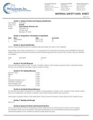
Fluoresbrite ® for Phagocytosis or Retrograde Transport<br />
These consistently sized polystyrene particles are ideal for cell interaction. Identification is made<br />
easy by the intense fluorescence and polystyrene has long been recognized as a biologically<br />
active surface for cell attachment. Using two different fluorescent particles allows researchers<br />
to track two populations in one study.<br />
Data for this chart was obtained on a EPICS V instrument with<br />
fluorescent polystyrene beads, 2µm in diameter. Data courtesy<br />
of Kristi Harkins, University of Nebraska / Lincoln.<br />
Fluoresbrite ® to Calibrate Flow Cytometers<br />
Our Fluoresbrite ® particles have had a long history of use as flow<br />
cytometry standards. As a manufacturer of the base particle, as well as<br />
an innovator in the incorporation of fluorescent dyes, we have advised<br />
even instrument manufacturers on the proper types of beads to use for<br />
aligning flow cytometers.<br />
BioMag ® , BioMag ® Plus & BioMag ® Maxi<br />
BioMag ® Physical Characteristics<br />
Our conventional BioMag ® are irregularly shaped iron oxide particles that vary in size between<br />
1-2µm, with most particles measuring ~1.5µm. BioMag ® Plus are similar, but have undergone<br />
additional processing for the reduction of outliers. BioMag ® Maxi are ~6µm. The particles are<br />
covered with an aminosilane coating, which provides functional groups for the attachment of<br />
proteins or antibodies. The irregular shape of the BioMag ® , BioMag ® Plus and BioMag ® Maxi<br />
particles provide increased surface area and therefore increased binding capacity.<br />
BioMag ® particle surface area: >100m 2 /g<br />
BioMag ® particle density:<br />
BioMag ® settling rate:<br />
2.5g/cc<br />
4% in 30 minutes<br />
BioMag ® Stability<br />
Since the BioMag ® base particle is composed of coated iron oxide, the particle itself is very stable. However, any proteins or<br />
antibodies attached to the BioMag ® particle are susceptible to degradation over time. BioMag ® should not be frozen or exposed<br />
to extreme heat.<br />
8<br />
For technical questions, info@polysciences.com or (800) 523-2575
<strong>Microspheres</strong> & <strong>Particles</strong> <strong>Handling</strong> <strong>Guide</strong><br />
BioMag ® Stability in Solvents<br />
BioMag ® particles have been used in various coupling buffers at pH ranging from 5.5 to 8.0. It is best to test the suitability of<br />
BioMag ® usage in organic solvents or extreme pH conditions.<br />
BioMag ® Magnetic Responsiveness<br />
BioMag ® particles are superparamagnetic meaning they have no magnetic memory and will readily resuspend if the magnetic force<br />
is removed. The particles are greater than 90% magnetite content and have a magnetization of 25-35 emu/g (Electromagnetic Units).<br />
Positive and Negative Selection with BioMag ®<br />
BioMag ® particles can be used for both positive and negative selections. In negative selection, the unwanted components are pulled<br />
out of solution by the BioMag ® particles. After magnetic separation, the resulting supernatant is enriched for the target cells or molecules.<br />
In positive selection, the BioMag ® particles are used to pull out of solution only the target cells or molecules of interest. All<br />
unwanted cell populations, cells or molecules will be left in the supernatant and can be discarded, resulting in a purified suspension<br />
of the target components.<br />
Magnetic Separator for BioMag ®<br />
For maximum performance, BioMag ® particles should be used with one of the <strong>Polysciences</strong><br />
magnets. Since BioMag ® particles are so small, they require a strong magnetic field in order to<br />
separate them in a timely fashion. <strong>Polysciences</strong>’ magnets are rare earth (Neodymium-Iron-Boron)<br />
magnets embedded in plastic housings, with magnetic strengths ranging from 27 megagauss<br />
Oersteds to 35 megagauss Oersteds, depending on the magnet.<br />
Coupling Efficiency Determination<br />
Coupling efficiency can be determined by measuring the change in absorbance<br />
of the supernatant before and after coupling.<br />
1. Set spectrophotometer wavelength to 280nm. Blank with the Coupling Buffer.<br />
2. Measure the absorbance of the Pre-Coupling Solution. A further dilution may be necessary to read an absorbance depending<br />
upon the amount of protein added (D = dilution factor).<br />
3. Measure the absorbance of the Post-Coupling Solution. A dilution may be necessary to read the absorbance (D = dilution factor).<br />
4. Calculate the coupling efficiency, expressed as the % Protein Uptake, as follows. Typical values of Protein Uptake are >60%.<br />
[(A 280 Pre-Coupling Solution 1 x D) – (A 280 Pre-Coupling Solution 2 x D)] x 100<br />
(A 280 Pre-Coupling Solution 1 x D)<br />
For complete technical information for each BioMag ® product, refer to the Technical Information page of our website.<br />
For more information please call (800) 523-2575 or visit: www.polysciences.com<br />
9
<strong>Microspheres</strong> & <strong>Particles</strong> <strong>Handling</strong> <strong>Guide</strong><br />
Coating <strong>Microspheres</strong><br />
<strong>Microspheres</strong> may be coated with capture molecules such as antibodies, oligonucleotides, peptides, etc. for use in diagnostic or separation<br />
applications. Microsphere coatings are typically optimized to achieve desired specific activity, while minimizing nonspecific interactions.<br />
Consideration should also be given to the required stability, development timeframe and budget and the specific biomolecule to<br />
be coated. These factors will aid in determining the most fitting coating strategy for both short- and long-term objectives.<br />
Standard microsphere products support three basic coating strategies: passive adsorption, covalent coupling and affinity binding. It is<br />
important to note that each binding strategy has benefits and limitations, which should be weighed in the context of study objectives<br />
and the demands that will be placed upon the finished reagent.<br />
Passive Adsorption<br />
Passive adsorption relies primarily on hydrophobic interactions between the biomolecule and the polymer particle. Such coatings are<br />
fairly simple to conduct, involving incubation of the microspheres with the purified biomolecule. They typically require little optimization<br />
and reagents may be developed relatively quickly. However, as adsorption relies on the formation of multiple attachment points between<br />
the molecule and the particle, this strategy is typically reserved for use with proteins and non-functionalized polymer spheres. Adsorption<br />
is generally not suitable for hormones, peptides, or nucleic acids in hybridization-based applications and protein adsorption to silica<br />
is expected to be less efficient than to polymer. Most techniques using passive adsorption technology report four to six months of bead<br />
stability.<br />
Covalent Coupling<br />
Covalent coupling results in the permanent attachment of the molecule to the functionalized (e.g. carboxyl or amine) microsphere. It can<br />
provide needed stability when developing a commercial reagent and for multiplexed assays, where analyte-specific bead populations are<br />
mixed. Additionally, specialized chemical linkers may be employed to address steric effects or to optimally orient the molecule. If surfactant<br />
is required as an additive in the assay, covalent coupling procedures are recommended as surfactants can displace adsorbed proteins from<br />
the surface. Covalent binding is also important for the immobilization of oligonucleotides or peptides, where end-point attachment is required.<br />
Although covalent binding protocols often involve a higher level of optimization than other approaches, coupling kits are available<br />
to simplify the process.<br />
When Coupling to <strong>Particles</strong> Less Than 0.5µm<br />
The chemical aspects of the protocols are universally applied, but the mechanical separations of these particles must be adapted for<br />
specific sizes. Most protocols suggest centrifugation to separate the particles from the reagents. This is not practical for particle sizes less<br />
than 0.5µm, since most microcentrifuges cannot spin these particles down within 30 minutes. Even extremely high G forces are not recommended,<br />
as resuspension becomes arduous. Other separation techniques can be utilized, such as dialysis, forced membrane filtration<br />
or centrifugal filter devices. <strong>Polysciences</strong> offers coupling kits that use hollow fiber filtration techniques to effect separation of 0.1-0.5µm<br />
particles.<br />
Coupling to Dyed <strong>Particles</strong><br />
The surfaces of dyed beads are available for protein adsorption or covalent attachment via functional groups. <strong>Polysciences</strong>’ capability to<br />
manufacture custom lots with exact sizing and relative control of dye content per bead has enhanced its prominence as a world leader in<br />
supplying the diagnostic assay market.<br />
10<br />
For technical questions, info@polysciences.com or (800) 523-2575
<strong>Microspheres</strong> & <strong>Particles</strong> <strong>Handling</strong> <strong>Guide</strong><br />
Affinity Binding<br />
Affinity binding is a straightforward method for immobilizing primary antibodies or biotinylated molecules. Protein A and G and<br />
Fc-specific antibody coatings permit the directed immobilization of primary antibodies and streptavidin is used extensively for the<br />
binding of biotinylated molecules, such as antibodies, peptides and oligonucleotides.<br />
Biomolecule Coating Strategy Notes<br />
Peptides<br />
Nucleic acids<br />
Proteins (e.g.<br />
antibodies)<br />
Covalent<br />
Streptavidin / biotin<br />
Covalent<br />
Streptavidin / biotin<br />
Covalent<br />
Adsorption<br />
End-point attachment to preserve the activity of the peptides.<br />
End-point attachment to permit hybridization of probe sequence with target<br />
sequence.<br />
Common proteins are generally large enough that multi-point attachment and<br />
nonspecific orientation do not compromise their activity. However, linkers or<br />
spacers (covalent or SA/B) may be employed to address steric effects or<br />
sub-optimal orientation.<br />
Alternative for BSA as a Blocking Agent<br />
Any innocuous protein may be used to block the effects of non-specific adsorption. In selecting an alternative to BSA, it is suggested that<br />
the size of the active protein and the size of the blocking protein be compared. BSA is highly recommended for IgG coupling. However,<br />
the large size of BSA will obscure the activity of smaller, active proteins. Glycine or small polypeptides may be used as alternatives.<br />
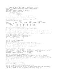
Well dispersed 10µm Polybead ® <strong>Microspheres</strong><br />
For more information please call (800) 523-2575 or visit: www.polysciences.com<br />
11
<strong>Microspheres</strong> & <strong>Particles</strong> <strong>Handling</strong> <strong>Guide</strong><br />
Protein Binding Protocols<br />
The following sequences serve as guidelines for protein binding. Technical Data Sheets (TDS) with detailed step-by-step protocols can<br />
be downloaded from our website.<br />
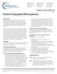
Adsorbing Protein on <strong>Particles</strong><br />
TDS #238E<br />
Plain <strong>Particles</strong><br />
• Initial Buffer: 0.1M Borate Buffer,<br />
pH 8.5<br />
• Suspend in buffer, spin down and<br />
resuspend 2 or 3 times.<br />
• Suspend in borate buffer.<br />
• Add protein and mix end-to-end<br />
overnight.<br />
• Spin and save supernatant for<br />
protein determination.<br />
• Resuspend in BSA in appropriate<br />
buffer and spin down twice.<br />
• Resuspend in PBS, pH 7.4,<br />
containing BSA and glycerol<br />
(storage buffer).<br />
• Protein bound directly on surface.<br />
PolyPointer<br />
Easily find all of our Technical<br />
Data Sheets by visiting www.<br />
polysciences.com<br />
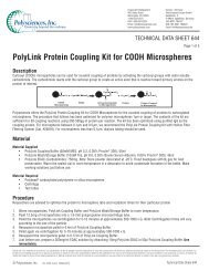
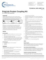
Coupling by Carbodiimide<br />
TDS #238C & #644<br />
Carboxylate Functional <strong>Particles</strong><br />
• Initial Buffer: 0.1M Carbonate Buffer<br />
• Suspend in buffer, spin down and<br />
resuspend 2 or 3 times.<br />
• Suspend in phosphate buffer.<br />
• Add fresh carbodiimide solution<br />
dropwise and incubate for 15-30<br />
minutes.<br />
• Wash to remove excess<br />
carbodiimide, then resuspend<br />
in borate buffer.<br />
• Add protein and mix end-to-end<br />
for 2-4 hours.<br />
• Add ethanolamine, mix for<br />
30 minutes.<br />
• Spin and save supernatant for<br />
protein determination.<br />
• Resuspend in BSA-containing<br />
buffer and spin down twice.<br />
• Resuspend in PBS, pH 7.4,<br />
containing BSA and glycerol<br />
(storage buffer).<br />
• Protein bound 2-3 carbon atoms<br />
from surface.<br />
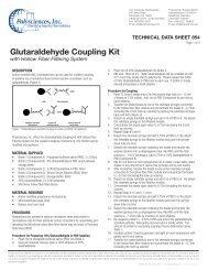
Coupling by Glutaraldehyde<br />
TDS #238D & #238G<br />
Amino or Blue Dyed <strong>Particles</strong><br />
• Initial Buffer: 0.02M PBS, pH 7.4<br />
• Suspend in buffer, spin down and<br />
resuspend 2 or 3 times.<br />
• Suspend in PBS.<br />
• Suspend in 8% glutaraldehyde in<br />
PBS, pH 7.4 and mix end-to-end<br />
for 4-6 hours.<br />
• Wash to remove excess<br />
glutaraldehyde and resuspend in<br />
PBS buffer.<br />
• Add protein and mix end-to-end<br />
overnight.<br />
• Add ethanolamine, mix for 30<br />
minutes.<br />
• Spin and save supernatant for<br />
protein determination.<br />
• Resuspend in BSA-containing<br />
buffer and spin down twice.<br />
• Resuspend in PBS, pH 7.4,<br />
containing BSA and glycerol<br />
(storage buffer).<br />
• Protein bound 5 carbon atoms<br />
from surface of blue dyed beads<br />
and 11-12 carbon atoms from<br />
surface of amino beads.<br />
12<br />
For technical questions, info@polysciences.com or (800) 523-2575
<strong>Microspheres</strong> & <strong>Particles</strong> <strong>Handling</strong> <strong>Guide</strong><br />
Protein Coupling Troubleshooting<br />
Below are some solutions for protein coupling troubleshooting. If you do not see a solution to the problem you are<br />
experiencing, please email us at: info@polysciences.com<br />
Problem<br />
Clumping prior to use<br />
Clumping after procedure<br />
• Carbodiimide addition causes clumping<br />
• Glutaraldehyde addition causes clumping<br />
Solution<br />
Careful sonication<br />
Isolate which step causes clumping<br />
Add slowly, agitate beads, decrease bead concentration<br />
Add slowly, agitate beads, decrease bead concentration; add an excess of glutaraldehyde<br />
to avoid chemical crosslinking of particles; clumping will typically<br />
resolve by the conclusion of the protein coupling<br />
• Protein addition causes clumping<br />
• Washing causes clumping<br />
Low binding<br />
Variable coating<br />
Coating, but no reaction<br />
Centrifuge not practical<br />
Nonspecific adsorption<br />
Small proteins bound, but not reactive<br />
<strong>Inc</strong>rease protein concentration<br />
Add surfactant or reduce washing steps<br />
Move pH of binding closer to protein isoelectric point<br />
Use pure water - no contaminants; use fresh reagents<br />
Optimize pH away from isoelectric point<br />
Use membrane filtration, dialysis, or spin fibers for small particles<br />
Use an alternative for BSA (glycine, casein)<br />
Use a crosslinking agent to extend coupling away from<br />
the surface of the bead<br />
Long-term storage leaches protein<br />
Try covalent attachment or lyophilize final product<br />
Special Properties<br />
Many applications in the life sciences demand added properties, such as fluorescence or a visible color, or iron oxide inclusions for<br />
magnetic separations. Polymer spheres (and some polymer-based magnetic spheres) are often internally dyed via organic solvent swelling<br />
and many standard products are available. Dye concentrations can be adjusted to produce beads with different intensities to meet<br />
special needs, such as QuantumPlex for multiplexed flow cytometric assays or our Dragon Green or Flash Red Intensity standards, which<br />
support imaging applications and associated instrument QC. Many surface- or internally-labeled fluorescent beads are also available as<br />
specialized flow cytometry standards.<br />
Various types of superparamagnetic microparticles are available as well – with different matrices, magnetite content, surface groups,<br />
etc. For new assays or applications, magnetic beads should be evaluated with application demands in mind.<br />
The following tables provide product suggestions for common microsphere applications. These are offered as general guidelines only.<br />
Further literature research and screening experiments may be appropriate.<br />
For more information please call (800) 523-2575 or visit: www.polysciences.com<br />
13
<strong>Microspheres</strong> & <strong>Particles</strong> <strong>Handling</strong> <strong>Guide</strong><br />
Microsphere Selection for Common Test and Assay Formats:<br />
Test / Assay Format Bead Size Bead Type Coating Strategy Detection Strategy<br />
Flow cytometric<br />
2-15µm<br />
QuantumPlex <br />
Covalent<br />
Flow cytometer<br />
(suspension array)<br />
QuantumPlexM (encoded<br />
Streptavidin / biotin<br />
populations for multiplexing)<br />
Non-fluorescent<br />
Lateral flow<br />
0.1-0.4µm<br />
Dyed (visible or<br />
Covalent<br />
Visual or automated reader<br />
fluorescent)<br />
Adsorption<br />
(absorbance, fluorescence)<br />
Lateral flow -<br />
0.1-0.4µm<br />
Dyed (visible mobile phase)<br />
Covalent<br />
Visual<br />
Boulders in the Stream<br />
~2-3µm<br />
Undyed (capture phase)<br />
Adsorption<br />
Dipstick<br />
0.1-0.4µm<br />
Dyed (visible)<br />
Covalent<br />
Visual<br />
Adsorption<br />
Latex Agglutination Tests<br />
0.2-1.0µm<br />
Undyed<br />
Covalent<br />
Visual (may be microscope-<br />
(LATs)<br />
Visibly dyed<br />
Adsorption<br />
assisted)<br />
Light Scattering<br />
0.36-0.76µm<br />
Undyed<br />
Covalent<br />
Nephelometer<br />
(automated LAT -<br />
(diameter equal<br />
Turbidimeter<br />
Nephelometric or<br />
to wavelength<br />
Turbidimetric)<br />
of light being<br />
scattered)<br />
Suggested Products for Magnetic Assays:<br />
Assays<br />
Immunoassays<br />
Hybridization-based assays<br />
Suggested Products<br />
ProMag or BioMag ®<br />
ProMag <br />
Suggested Products for Magnetic Separations:<br />
Magnetic Separations<br />
Cells<br />
Subcellular organelles<br />
Immunoprecipitates<br />
mRNA<br />
Biotinylated oligonucleotide capture or binding<br />
Biopanning<br />
Suggested Products<br />
BioMag ® anti-CD marker or secondary antibody<br />
BioMag ®<br />
BioMag ®<br />
BioMag ® Oligo dT (20) or mRNA Purification System<br />
ProMag or BioMag ®<br />
ProMag or BioMag ®<br />
14<br />
For technical questions, info@polysciences.com or (800) 523-2575
<strong>Microspheres</strong> & <strong>Particles</strong> <strong>Handling</strong> <strong>Guide</strong><br />
Instrument Quality Control<br />
For confidence in test or assay results, a facility must employ a comprehensive quality assurance program that encompasses employee<br />
training and routine instrument maintenance and quality control. Facilities should also ensure that studies, particularly those involving<br />
quantitative fluorescence measurements, are conducted with appropriate standardization.<br />
Microsphere standards aid in defining the instrument’s capabilities and limitations in terms of sensitivity, precision and accuracy and<br />
provide a means for ensuring that the instrument is stable and suitable for use. They are also helpful in understanding the effects of<br />
extraneous factors, such as temperature, humidity and electronic noise. The comparison of daily and historical QC data aids in the<br />
identification of random errors (due to electronic noise, air bubbles, etc.) and systematic errors (due to bias, shifts and trends caused by<br />
temperature variation, laser deterioration, misalignment, etc.) so that suitable corrective action may be taken.<br />
Daily Controls<br />
Flow cytometers are highly configurable and results can vary dramatically with different instrument settings. Establishing a common “Window<br />
of Analysis” for each detector, with the upper and lower fluorescence limits defined, allows reference populations to be positioned in<br />
approximately the same place on the same scale. This type of standardized instrument set-up ensures consistency of results from specific<br />
instruments and enables meaningful comparison between instruments. Therefore, the general status and stability of the cytometer must<br />
be checked daily. In addition, by plotting values over time, random and systemic errors may be identified and corrected. Manually aligned<br />
instruments must be aligned daily. If the data from multiple instruments and/or sites are to be compared, all instruments must be standardized<br />
daily, using the same standard bead. If multi-color analysis and/or quantitation is being performed, then the appropriate standards<br />
must be run on that same day.<br />
Weekly Controls<br />
If only qualitative analyses are being run, weekly checks of the detection threshold, resolution and linearity are sufficient to establish sensitivity.<br />
Some cytometers are fixed alignment instruments. For these, alignment may be verified weekly rather than established daily.<br />
In addition to a regular QA/QC program, specific analyses may require other controls, such as count standards, size standards or<br />
reference standards. Experiments and analyses must be critically evaluated for inclusion of the appropriate controls prior to<br />
instrumental analysis.<br />
For more information please call (800) 523-2575 or visit: www.polysciences.com<br />
15
The following chart outlines a basic program of quality control and standardization that includes suggestions regarding specific<br />
products and their frequency and method of use.<br />
Category Purpose Frequency Products<br />
Daily QC<br />
General check of instrument<br />
Daily<br />
Full Spectrum (multi), Ultra Rainbow<br />
stability / status<br />
Fluorescent <strong>Particles</strong> (multi), Fluorescence<br />
Reference Standards (single), Flow Check ®<br />
Ruby Red Fluorescent <strong>Microspheres</strong> (single)<br />
Daily QC<br />
General check of instrument<br />
Daily<br />
Full Spectrum (multi), Ultra Rainbow<br />
optical system<br />
Fluorescent <strong>Particles</strong> (multi), Fluorescence<br />
Reference Standards (single), Flow Check ®<br />
Ruby Red Fluorescent <strong>Microspheres</strong> (single)<br />
Daily QC<br />
Optical alignment<br />
Daily<br />
Right Reference Standard , Flow Check ®<br />
Alignment and Compensation Particle Sets<br />
Daily QC<br />
Fluidics check<br />
Daily<br />
Surface-labeled fluorescent microspheres,<br />
e.g. QC Windows ® , QC3 , Fluorescence<br />
Reference Standards, Quantum MESF<br />
Weekly QC<br />
Optical system sensitivity,<br />
Weekly<br />
Quantum MESF<br />
resolution for linearity (for<br />
specific lasers / PMTs)<br />
Daily Set-Up<br />
Standardized instrument<br />
Daily or between runs if settings are changed<br />
QC Windows ® , QC3 <br />
set-up<br />
Daily Set-Up<br />
Standardized compensation<br />
Daily or between runs if settings are changed<br />
FITC/PE Compensation Standard, Simply<br />
settings for multi-color<br />
Cellular ® Compensation Standard,<br />
analyses<br />
Quantum Simply Cellular ®<br />
Application<br />
Fluorescence quantitation in<br />
Daily when quantitative analyses are<br />
Quantum MESF, Quantum Simply Cellular ®<br />
cellular expression studies or<br />
performed or between different applications<br />
bead-based assays<br />
if fluorescence PMT settings are changed<br />
Application<br />
F:P ratio determination for<br />
As needed, i.e. with each new lot of<br />
Simply Cellular ® (used in conjunction with<br />
quantitative fluorescence<br />
fluorochrome-conjugated antibody<br />
Quantum MESF)<br />
analyses<br />
Application<br />
Compensation for multi-color<br />
Daily or between different<br />
FITC/PE Compensation Standard, Simply<br />
flow cytometry<br />
applications if fluorescence PMT<br />
Cellular ® Compensation Standard,<br />
settings are changed<br />
Quantum Simply Cellular ®<br />
Application<br />
Cell counting<br />
As needed<br />
Flow Cytometry Absolute Count Standard <br />
Application<br />
Cell size estimation<br />
As needed<br />
Size Calibration Standards Kit<br />
Application<br />
Suspension array<br />
Platform for development of bead-based flow<br />
QuantumPlex , QuantumPlex M<br />
cytometric assays<br />
U.S. Corporate Headquarters | 400 Valley Road, Warrington, PA 18976 | 1(800) 523-2575 / (215) 523-2575 | Fax 1(800) 343-3291 | info@polysciences.com<br />
<strong>Polysciences</strong> Europe GmbH | Handelsstrasse 3 D-69214 Eppelheim, Germany | +(49) 6221-765767 | Fax +(49) 6221-764620 | info@polysciences.de