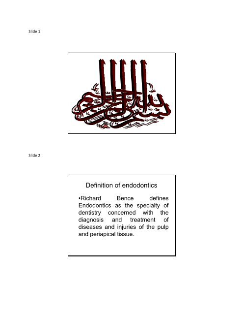
Definition of endodontics
Definition of endodontics
Definition of endodontics

You also want an ePaper? Increase the reach of your titles
YUMPU automatically turns print PDFs into web optimized ePapers that Google loves.
Slide 1<br />
Slide 2<br />
<strong>Definition</strong> <strong>of</strong> <strong>endodontics</strong><br />
•Richard Bence defines<br />
Endodontics as the specialty <strong>of</strong><br />
dentistry concerned with the<br />
diagnosis and treatment <strong>of</strong><br />
diseases and injuries <strong>of</strong> the pulp<br />
and periapical tissue.
Slide 3<br />
•The American Association <strong>of</strong><br />
Endodontists defines this field as<br />
that branch <strong>of</strong> dentistry concerned<br />
with the morphology, physiology,<br />
and pathology <strong>of</strong> the human<br />
dental pulp and periradicular<br />
tissues.<br />
Slide 4<br />
BASIC PHASES OF<br />
ENDODONTIC THERAPY<br />
a) the diagnostic phase, in which<br />
the cause <strong>of</strong> the disease is<br />
identified and the treatment plan<br />
is prepared;
Slide 5<br />
Slide 6
Slide 7<br />
Slide 8<br />
b) the preparatory phase, in<br />
which, by cleaning and shaping<br />
the root canal, the root canal<br />
contents are removed and the<br />
root canal space itself is shaped<br />
to receive a three-dimensional<br />
filling; and
Slide 9<br />
Slide 10<br />
c) the obturation phase, in which<br />
the root canal system is filled with<br />
an inert material to ensure a tight<br />
seal.
Slide 11<br />
Slide 12
Slide 13<br />
Anatomy & access<br />
cavities<br />
Slide 14<br />
Pulpal floor<br />
Pulpal ro<strong>of</strong><br />
Dentin<br />
Enamel<br />
Pulp horn<br />
Pulp chamber<br />
Bifurcation canal<br />
Lateral canal<br />
Canal orifice<br />
Pulp canal<br />
Apical delta<br />
Apical foramen<br />
Major anatomic components <strong>of</strong> the root canal system.
Slide 15<br />
Endodontic terminology:<br />
Apical delta:<br />
When the main canal branches<br />
apically into two small canals i.e<br />
there will be 2 apical foramina for<br />
the same canal<br />
Slide 16<br />
Apical ramifications:<br />
When the main canal branches<br />
apically into more than two<br />
branches i.e there will be more<br />
than 2 apical foramina for the<br />
same canal.
Slide 17<br />
Dentino-cemental junction is<br />
also called the ( apical<br />
constriction, minor apical<br />
diameter ). It is the area where<br />
cememntum meets dentin near<br />
the apex <strong>of</strong> the root.<br />
Slide 18<br />
Dentino-cemental junction<br />
( apical constriction )
Slide 19<br />
Lateral canal:<br />
It is a lateral branching <strong>of</strong> the<br />
main canal . It connects the main<br />
canal with the periodontal<br />
membrane.<br />
Slide 20<br />
Vertucci Classification<br />
Vertucci Classification<br />
1 canal 1 orifice<br />
2 canals join then 1 orifice<br />
1 two 1
Slide 21<br />
Vertucci Classification-<br />
Two canals at the apex<br />
2 canals 2 orifices<br />
1 two<br />
2 one 2<br />
1 two 1 two<br />
Slide 22<br />
Vertucci Classification<br />
3 canals 3 orifices
Slide 23<br />
A successful outcome in<br />
endodontic treatment essentially<br />
depends on three factors:<br />
1) cleaning and shaping<br />
2) disinfection<br />
3) three-dimensional obturation <strong>of</strong><br />
the root canal system.<br />
Slide 24<br />
Access cavity permits:<br />
•localization,<br />
•cleaning,<br />
•shaping,<br />
•disinfection, and<br />
•three-dimensional obturation <strong>of</strong><br />
the root canal system.
Slide 25<br />
REQUIREMENTS OF THE ACCESS<br />
CAVITY<br />
1)Permit the removal <strong>of</strong> all the<br />
chamber contents<br />
All the ro<strong>of</strong> <strong>of</strong> the chamber should<br />
be removed. It should include<br />
both the mesio-distal and buccolingual<br />
width <strong>of</strong> the pulp chamber.<br />
Slide 26
Slide 27<br />
2) Permit complete, direct vision<br />
<strong>of</strong> the floor <strong>of</strong> the pulp chamber<br />
and canal openings.<br />
Slide 28<br />
•To meet this second requirement,<br />
the access cavity must<br />
sometimes be slightly modified to<br />
give it the so-called “convenient<br />
shape”.<br />
•This gives the walls a slight<br />
anterior inclination that facilitates<br />
inspection <strong>of</strong> the floor and thus<br />
localization <strong>of</strong> the canal openings.
Slide 29<br />
Slide 30<br />
3) Facilitate the introduction <strong>of</strong><br />
canal instruments into the root<br />
canal openings<br />
•Access should allow instruments<br />
to slide in the canal.
Slide 31<br />
4) Provide access as direct as<br />
possible to the apical one third <strong>of</strong><br />
the canal for both preparation<br />
instruments and canal filling<br />
instruments.<br />
Slide 32<br />
5) Provide a positive support for<br />
temporary fillings<br />
6) Always have four walls
Slide 33<br />
GENERAL PRINCIPLES FOR THE<br />
PREPARATION OF THE ACCESS<br />
CAVITY<br />
Regardless <strong>of</strong> the tooth, there are<br />
three phases in the preparation <strong>of</strong><br />
the access cavity:<br />
•penetration,<br />
•enlarging, and<br />
•finishing.<br />
Slide 34<br />
1-Penetration phase<br />
•This phase is performed using a<br />
round diamond bur mounted on a<br />
high-speed handpiece.<br />
•The objective <strong>of</strong> this phase is to<br />
“penetrate” the pulp chamber by<br />
breaking through the ro<strong>of</strong> with the<br />
bur.
Slide 35<br />
If the pulp chamber is wide<br />
enough, there is a sensation <strong>of</strong><br />
“falling into a vacuum” when the<br />
ro<strong>of</strong> is penetrated.<br />
Slide 36<br />
2-Enlargement phase<br />
•This phase is performed with a<br />
round bur mounted on a low-speed<br />
handpiece.<br />
•Its diameter should be slightly<br />
smaller than that <strong>of</strong> the preceding<br />
bur, and it should have a long shaft<br />
for improved penetration and<br />
visibility.
Slide 37<br />
Slide 38<br />
3-Finishing and flaring phase<br />
•This phase requires a non-endcutting<br />
diamond bur, also called<br />
self-guiding bur, or Batt’s bur or<br />
endo Z bur mounted on a highspeed<br />
handpiece.
Slide 39