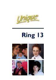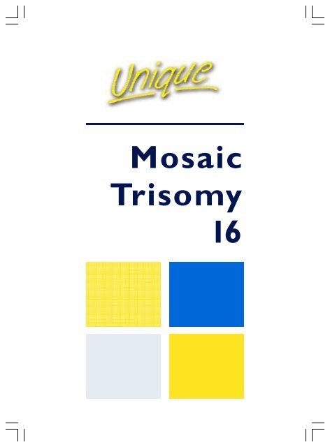
Mosaic Trisomy 16
Mosaic Trisomy 16
Mosaic Trisomy 16

Create successful ePaper yourself
Turn your PDF publications into a flip-book with our unique Google optimized e-Paper software.
<strong>Mosaic</strong><br />
<strong>Trisomy</strong><br />
<strong>16</strong>
What is<br />
<strong>Mosaic</strong><br />
<strong>Trisomy</strong> <strong>16</strong>?<br />
There are usually 46<br />
chromosomes in the<br />
cells of the body.<br />
Chromosomes come<br />
in different sizes and<br />
are numbered from<br />
largest to smallest<br />
according to their size,<br />
from number 1 to<br />
number 22, in addition<br />
to the sex<br />
chromosomes, X and<br />
Y. We have two<br />
copies of each of the<br />
chromosomes (23<br />
pairs), one inherited<br />
from the father and<br />
one inherited from<br />
the mother but<br />
someone with mosaic<br />
trisomy <strong>16</strong> (MT<strong>16</strong>)<br />
has some cells with a<br />
third chromosome <strong>16</strong>,<br />
making 47<br />
chromosomes in<br />
those cells.<br />
Cells with 46 and 47<br />
chromosomes exist<br />
alongside each other<br />
in different body<br />
tissues and organs,<br />
exerting different<br />
effects. The picture<br />
changes, with the<br />
number of cells with<br />
the extra chromosome<br />
usually falling in<br />
time, especially<br />
in blood.<br />
How common is MT<strong>16</strong>?<br />
Full trisomy <strong>16</strong> with an extra chromosome <strong>16</strong> in every<br />
cell is believed to be the most common of all<br />
chromosome abnormalities at conception (when a<br />
baby is made), and is found in 1 to 2 per cent of all<br />
confirmed pregnancies. However, babies with full<br />
trisomy <strong>16</strong> are almost always lost in very early<br />
pregnancy and virtually none have been born. Even<br />
babies with MT<strong>16</strong> are very rare. Researchers who<br />
recently surveyed every MT<strong>16</strong> pregnancy published in<br />
the medical literature gathered <strong>16</strong>2 pregnancies<br />
worldwide. Among these pregnancies they confirmed<br />
that mosaic trisomy <strong>16</strong> appears to be slightly more<br />
common in girls than in boys (Yong 2003; Benn 1998).<br />
Chromosome<br />
<strong>16</strong><br />
A note<br />
This leaflet describes what is<br />
known from published medical<br />
research and from the experience<br />
of Unique and of the Disorders of<br />
Chromosome <strong>16</strong> Foundation<br />
support group. Although recent<br />
research into MT<strong>16</strong> is of a very<br />
high quality, children who come<br />
to doctors’ attention and feature<br />
in research reports are likely to<br />
be the ones who have<br />
experienced effects on their<br />
health or development from their<br />
unusual chromosomes. Those<br />
who experience few or no health<br />
problems will not be described.<br />
This means that the picture we<br />
paint may appear more negative<br />
than it really is. Unique has<br />
published a companion booklet of<br />
<strong>Mosaic</strong> <strong>Trisomy</strong><strong>16</strong> Stories,<br />
describing families’ experiences.<br />
References to medical research<br />
are shown in brackets and a full<br />
reference list is available<br />
from Unique.<br />
2
How is mosaic trisomy <strong>16</strong> diagnosed in pregnancy?<br />
<strong>Mosaic</strong> trisomy <strong>16</strong> is usually first suspected and diagnosed during pregnancy. The<br />
first tests are non-specific. Usually either ultrasound scans or blood tests for certain<br />
chemicals (maternal serum screening) suggest there may be a problem and a general<br />
test for the most common chromosome abnormalities is performed either on the<br />
developing placenta (with chorionic villus sampling, CVS) or on amniotic fluid (from<br />
an amniocentesis). If this shows trisomy <strong>16</strong> in some cells, the pregnancy has mosaic<br />
trisomy <strong>16</strong>.<br />
There are three possible outcomes for the baby:<br />
• Either the only effects are on the baby’s growth rate<br />
• Or the baby will be apparently unaffected<br />
• Or the baby will be affected.<br />
You will be offered tests to try to clarify which is the case for you.<br />
The tests that show that mosaic trisomy <strong>16</strong> is present cannot unfortunately show<br />
directly how slightly or severely affected your baby will be. However, when the tests<br />
are put together with information from ultrasound scans, a clearer picture will<br />
emerge.<br />
CVS (chorionic villus sampling)<br />
Two types of early pregnancy CVS test can be<br />
done on the cells from the developing placenta.<br />
A rapid test (called direct sampling) gives a<br />
result within a day or two.<br />
The placenta cells are cultured (grown), with<br />
the results in a week or two.<br />
You should wait for both results. Most often,<br />
they show that all the cells in the developing<br />
placenta have the extra chromosome <strong>16</strong>. Less<br />
often, they show a mixture of trisomy <strong>16</strong> cells<br />
and ordinary cells. This means that the placenta<br />
does contain trisomy <strong>16</strong> cells but usually none<br />
are found in amniotic fluid and your baby does<br />
not have any. This is called confined placental<br />
mosaicism. Even when the cultured CVS shows<br />
trisomy <strong>16</strong>, the fetus does not usually turn out<br />
to have mosaic trisomy <strong>16</strong>.<br />
The chromosomes in the placenta and in the<br />
baby can differ because the placenta and the<br />
baby develop from separate clusters of cells.<br />
But early in the pregnancy it is only safe to take<br />
cells from the developing placenta.<br />
Confined<br />
placental<br />
mosaicism<br />
In confined placental<br />
mosaicism, the<br />
developing placenta and<br />
the tissues outside the<br />
baby have the extra<br />
chromosome, but the<br />
baby has entirely or<br />
mostly normal cells.<br />
Your baby is likely to<br />
grow slowly in the<br />
womb and be born tiny<br />
or very tiny. Babies with<br />
mosaic trisomy <strong>16</strong> are<br />
almost always born after<br />
a pregnancy with<br />
confined placental<br />
mosaicism. A blood or<br />
skin sample from the<br />
baby usually reveals no<br />
trisomy <strong>16</strong> cells.<br />
3
Amniocentesis<br />
Cells from the baby’s skin, urine, intestinal tract and lungs are shed into the<br />
amniotic fluid and give a more direct picture of the presence of trisomy <strong>16</strong><br />
cells in the baby than CVS. Generally speaking, the outlook is less good if<br />
trisomy <strong>16</strong> cells are found in amniotic fluid. However, even finding trisomic<br />
cells in amniotic fluid does not mean that your baby will necessarily have<br />
trisomy <strong>16</strong> mosaicism. If they are found in the baby at all, they are usually<br />
confined to just one body tissue such as the skin or the lungs. The trisomy <strong>16</strong><br />
cells may diminish over time and they may vanish altogether. In some<br />
pregnancies, no trisomy <strong>16</strong> cells are found in amniotic fluid but they are<br />
found later in the baby.<br />
If mosaic trisomy <strong>16</strong> has been diagnosed during pregnancy, a blood sample<br />
will be taken at birth from the umbilical cord. This often shows no trisomy<br />
<strong>16</strong> cells. There is a specialist research centre at the University of British<br />
Columbia that uses a particularly sensitive molecular test to identify trisomy<br />
<strong>16</strong> cells but even if this is negative, it is not possible to entirely rule out low<br />
levels of trisomy <strong>16</strong> cells in other parts of the body. You can find them at this<br />
website: www.medgen.ubc.ca/wrobinson/mosaic/index.htm.<br />
Ultrasound<br />
Ultrasound scans, ideally undertaken at a fetal wellbeing centre, will give<br />
important information about the baby’s health. In skilled hands at least 50 per<br />
cent of birth defects can be identified and the baby’s growth rate can be<br />
carefully monitored, especially in the third trimester when it is most likely to<br />
slow down. However, even an apparently clear scan is no guarantee.<br />
In the end a couple has to brace themselves for the<br />
possible worst case scenario where the baby would have<br />
undetected birth defects, developmental delay<br />
or learning difficulties - Clinical geneticist<br />
Hope for the best, prepare for the worst - Parent<br />
4
Does the percentage of trisomic cells matter?<br />
The proportion of T<strong>16</strong> cells and their distribution in the tissues of the body<br />
depends partly on the time in the pregnancy when mosaicism was<br />
established and partly on a tendency for the ordinary cells without the extra<br />
chromosome to grow more effectively.<br />
Your geneticist or genetic counsellor will very likely tell you the percentage<br />
of cells with trisomy <strong>16</strong> but what this means is not always clear. It is helpful<br />
to know the percentage for a CVS result because when the CVS (either<br />
rapid-result or cultured) shows 100% trisomy <strong>16</strong> cells rather than mosaic<br />
trisomy <strong>16</strong>, the baby’s growth delay is likely to be more severe.<br />
It is much less helpful to know this for an amniocentesis result.<br />
There does not appear to be any obvious link between either<br />
the degree or the distribution of trisomy cells at amniocentesis<br />
and the severity of the effects on your baby, either in terms<br />
of growth delay or birth defects.<br />
The authors of the most recent and authoritative survey of mosaic trisomy<br />
<strong>16</strong> say that an amniocentesis should not be considered to be anything more<br />
than a snapshot (Yong 2003; Hsu 1998; Benn 1998).<br />
This seems to be true after birth as well. Common sense suggests that<br />
tissues and organs with more T<strong>16</strong> cells will be more obviously affected<br />
than tissues with lower levels or none. That is not necessarily the case.<br />
Different levels of T<strong>16</strong> cells are found in different parts of the body<br />
and it is especially common for no T<strong>16</strong> cells to be found in the baby’s<br />
blood. In one case, doctors who performed surgery on a baby girl to<br />
correct narrowing of the aorta, the main artery from the heart, took<br />
samples from her aorta, her lungs, skin, lymph and blood.<br />
They found no link between her defects (in the heart)<br />
and the levels of trisomy <strong>16</strong> cells (Greally 1996).<br />
5
6<br />
Making a decision<br />
It is very hard to make a decision when there is little certainty.<br />
Many couples have praised the support available from the Disorders of<br />
Chromosome <strong>16</strong> Foundation (DOC<strong>16</strong> at www.trisomy<strong>16</strong>.org), founded by<br />
the mother of a girl with mosaic trisomy <strong>16</strong>. At Unique we also aim to<br />
support parents with our information, helpline and contact with similarly<br />
affected families.<br />
The UK support group Antenatal Results and Choices (www.arc-uk.org;<br />
+44 (0) 207 631 0285) has a series of handbooks for family members.<br />
On the net, you can also turn to:<br />
http://bbs.babycenter.com/board/pregnancy/prenatalhealth/<br />
where there are a number of helpful message boards, including Continuing<br />
a Pregnancy with Poor Prenatal Diagnosis and Termination for Medical<br />
Reasons.<br />
What is likely<br />
to happen<br />
during pregnancy<br />
If you make the decision to continue<br />
with your pregnancy, you will have<br />
regular scans and tests of your baby’s<br />
heart beat and movements. This is to<br />
monitor your baby’s growth and to<br />
identify any signs that it is time to<br />
intervene. Your own blood pressure<br />
will also be monitored carefully as<br />
hypertension (high blood pressure) is<br />
more common in mosaic trisomy <strong>16</strong><br />
pregnancies.<br />
A large review of mosaic trisomy <strong>16</strong><br />
pregnancies suggested strongly that<br />
many would end in a premature birth.<br />
It showed that babies’ average age at<br />
birth was just under 36 weeks and out<br />
of 70 babies, 30 were born between<br />
35 and 37 weeks and 22 were born<br />
between 27 and 34 weeks. It isn’t<br />
known whether the premature birth<br />
was spontaneous or induced due to<br />
concern over the baby’s growth or<br />
any complications (Yong 2003).<br />
Will my baby<br />
survive?<br />
Until very recently the outlook for<br />
mosaic trisomy <strong>16</strong> pregnancies was<br />
thought to be very bleak. A large recent<br />
survey of <strong>16</strong>2 pregnancies has shown a<br />
much more optimistic picture. Two out<br />
of three pregnancies ended in the birth of<br />
a baby and the great majority of these<br />
babies survived the newborn period.<br />
Seventeen per cent of babies died before<br />
birth or in the newborn period (Yong<br />
2003). No long term studies of the<br />
outcomes have been published yet.<br />
However, the short term outcomes for<br />
babies look very reassuring. Of the babies<br />
born with MT<strong>16</strong>, 93 per cent survived<br />
the newborn period and so long as babies<br />
are in good health at birth they are<br />
believed to usually progress quite well.<br />
You may find it helpful to read the stories<br />
in Unique’s <strong>Mosaic</strong> <strong>Trisomy</strong> <strong>16</strong> Stories<br />
leaflet and those on the website of the<br />
DOC<strong>16</strong> Foundation.
Common effects on babies and children<br />
Two recent surveys have shown that most babies with MT<strong>16</strong> are born small for<br />
their gestational age but are otherwise healthy. Isolated medical conditions have<br />
been found in a minority of babies, but most of these were no more common than<br />
among babies without MT<strong>16</strong>. Just two conditions did occur more commonly –<br />
although they were still unusual: a hole in the heart (between the left and right<br />
chambers) and in boys, hypospadias, a condition where the hole normally at the end<br />
of the penis is on the underside instead (Yong 2003; Benn 1998).<br />
When your baby is examined, the clinicians will be particularly alert to the following<br />
body tissues and systems. This is because they have been most often affected in the<br />
minority of babies with MT<strong>16</strong> who have been found to have a clinical condition.<br />
• Heart • Lungs • Hands • Kidneys, urinary and genital systems<br />
• Digestion and intestines • Head and face • Joints and bones • Skin<br />
Growth<br />
The typical growth delay before birth is generally attributed to inadequate placental<br />
functioning. This means that growth after birth may be normal but we don’t know<br />
yet whether children completely catch up in height.<br />
Two copies of<br />
the mother’s<br />
chromosome <strong>16</strong><br />
Children without mosaic trisomy<br />
have two chromosome <strong>16</strong>s, one<br />
from their father and one from<br />
their mother. In at least one third<br />
of mosaic trisomy <strong>16</strong> pregnancies,<br />
the cell line with two<br />
chromosome <strong>16</strong>s instead of three<br />
contains two of the mother’s<br />
chromosome <strong>16</strong>s and none of the<br />
father’s. This is called maternal<br />
uniparental disomy <strong>16</strong> (UPD <strong>16</strong><br />
mat) and may intensify the growth<br />
delay before birth caused by the<br />
trisomy <strong>16</strong> cells. It is possible that<br />
maternal UPD <strong>16</strong> may have<br />
additional effects on the baby, but<br />
if so these are likely to be subtle<br />
(Yong 2003/2).<br />
<strong>Trisomy</strong> rescue<br />
After conception, there are almost always<br />
three chromosome <strong>16</strong>s, usually two from the<br />
mother and one from the father. A natural<br />
attempt to correct the mistake is then partly<br />
successful, leaving two cell lines, one with the<br />
extra chromosome <strong>16</strong>, the other without.<br />
Skewed X-inactivation<br />
The mechanism that results in trisomy rescue<br />
also leads to a situation known as 100%<br />
skewed X-chromosome inactivation. This<br />
means that a baby transcribes genes from<br />
either a maternal or the paternal X<br />
chromosome, but not both. Research<br />
suggests that this seems to be linked with<br />
poorer outcomes at birth but it should have<br />
no important effects afterwards (Penaherrera<br />
2000). The only likely effect is that in a girl,<br />
the risk of rare X-linked recessive disorders<br />
(such as muscular dystrophy or haemophilia)<br />
might rise to that of a typical boy.<br />
7
Why did it happen?<br />
Almost all MT<strong>16</strong> pregnancies start as a pregnancy with a complete extra<br />
chromosome <strong>16</strong>. During the formation of the egg cells in the mother, an error<br />
occurs in the natural process by which chromosomes separate. This leaves an extra<br />
chromosome <strong>16</strong> in the egg cell. Much more rarely the same mistake occurs when<br />
the father’s sperm cells are forming. After conception, there are three chromosome<br />
<strong>16</strong>s, usually two from the mother and one from the father. A natural attempt to<br />
correct the mistake after conception is then partly successful, leaving two cell lines,<br />
one with the extra chromosome <strong>16</strong>, the other without. This is the process called<br />
correction or trisomy rescue.<br />
Can it happen again?<br />
There is no evidence that couples are likely to have another baby affected by MT<strong>16</strong>.<br />
However, if the original trisomy (third chromosome) was caused by a failure of the<br />
mother’s chromosomes to separate when her egg cells were forming and she is an<br />
older mother (over 35), she may have a raised chance of having another pregnancy<br />
affected by trisomy.<br />
Rare Chromosome Disorder Support Group,<br />
PO Box 2189,<br />
Caterham,<br />
Surrey CR3 5GN, UK.<br />
Tel/Fax: +44(0)1883 330766<br />
info@rarechromo.org<br />
www.rarechromo.org<br />
Disorders of Chromosome <strong>16</strong> Foundation<br />
www.trisomy<strong>16</strong>.org<br />
Unique mentions other organisations’ message boards and websites to help families looking for<br />
information. This does not imply that we endorse their content or have any responsibility for it.<br />
This leaflet is not a substitute for personal medical or genetic advice. Families should<br />
consult a medically qualified clinician in all matters relating to genetic diagnosis,<br />
management and health. The information is believed to be the best available at the<br />
time of publication and the medical content has been verified by<br />
Dr Wendy Robinson, Associate Professor of Medical Genetics, University of British<br />
Columbia and Monica Pearson BSc 2004.<br />
Copyright © Unique 2005<br />
8<br />
Rare Chromosome Disorder Support Group Charity Number 1110661<br />
Registered in England and Wales Company Number 5460413